Article

Minimally Invasive Spine Stabilization for Pyogenic Spondylodiscitis: A 23-Case Series and Review of Literature

1
Department of Orthopaedic Surgery, Ota Memorial Hospital, Gunma 373-8585, Japan
2
Department of Orthopaedic Surgery, School of Medicine, International University of Health and Welfare (IUHW), Narita 286-0048, Japan
3
Department of Orthopaedic Surgery, International University of Health and Welfare Mita Hospital, Tokyo 108-8329, Japan
4
Department of Orthopaedic Surgery, International University of Health and Welfare Narita Hospital, Narita 286-8520, Japan
5
Department of Orthopaedic Surgery, Kansai Medical University, Osaka 573-1191, Japan
*
Authors to whom correspondence should be addressed.
These authors contributed equally to this work.
Medicina 2022, 58(6), 754; https://doi.org/10.3390/medicina58060754
Submission received: 3 April 2022 / Revised: 29 May 2022 / Accepted: 30 May 2022 / Published: 1 June 2022

Abstract

:
Background and Objectives: The incidence of pyogenic spondylodiscitis has been increasing due to the aging of the population. Although surgical treatment is performed for refractory pyogenic spondylodiscitis, surgical invasiveness should be considered. Recent minimally invasive spine stabilization (MISt) using percutaneous pedicle screw (PPS) can be a less invasive approach. The purpose of this study was to evaluate surgical results and clinical outcomes after MISt with PPS for pyogenic spondylodiscitis. Materials and Methods: Clinical data of patients who underwent MISt with PPS for pyogenic spondylitis were analyzed. Results: Twenty-three patients (18 male, 5 female, mean age 67.0 years) were retrospectively enrolled. The mean follow-up period was 15.9 months after surgery. The causative organism was identified in 16 cases (69.6%). A mean number of fixed vertebrae was 4.1, and the estimated blood loss was 145.0 mL. MISt with PPS was successfully performed in 19 of 23 patients (82.6%). Four cases (17.4%) required additional anterior debridement and autologous iliac bone graft placement. CRP levels had become negative at an average of 28.4 days after surgery. There was no major perioperative complication and no screw or rod breakages during follow-up. Conclusions: MISt with PPS would be a less invasive approach for pyogenic spondylodiscitis in elderly or immunocompromised patients.

1. Introduction

The incidence of pyogenic spondylodiscitis ranges from 0.2 to 2.4 per 100,000 per year [1,2,3,4] and is more common in males [5]. In recent years, the incidence has been on the rise due to the increase in elderly and immunocompromised patients [5,6,7], in addition to advances in imaging techniques such as magnetic resonance imaging (MRI) for diagnosis [8]. Pyogenic spondylodiscitis is often difficult to treat, and mortality rates have been reported to be between 2–20% [5,9,10,11]. One of the reasons for the difficulty in treatment is the low positive rate of the causative organism [12,13,14]. Although some reports recommend early surgery including debridement [15,16], the first-line is conservative treatment [12,17,18]. The most important aspect of conservative treatment is the administration of antibiotics for a sufficient period of time as well as rest and external orthosis [2,5]. However, the failure rate of conservative treatment has been reported to be between 12%and 18% [12,19,20,21,22], and surgical treatment should be considered when there is a refractory infection, progressive bone destruction, excruciating pain, or neurological deterioration.
Traditionally, the ideal treatment is debridement and anterior column reconstruction; however, it is sometimes inadvisable in patients with poor medical conditions. Recent reports have described posterior fixation techniques with an open approach for pyogenic spondylodiscitis [23,24]. Moreover, minimally invasive spine stabilization (MISt) using a percutaneous pedicle screw (PPS) can be a less invasive approach for patients with multiple comorbidities. Although there are a few reports describing the efficacy of PPS [25,26], its superiority is still controversial. The purpose of this study was to evaluate surgical results and clinical outcomes after MISt with PPS for pyogenic spondylodiscitis.

2. Materials and Methods

A multicenter retrospective analysis was conducted. This study was approved by the institutional review board at our hospital and all subjects were given an option to opt-out. Patients who underwent MISt with PPS for refractory pyogenic spondylodiscitis in the thoracic and lumbar spine between 2008 and 2014 were enrolled. Medical charts and radiographs were retrospectively reviewed. All patients underwent blood culture, urine culture, sputum culture, or needle biopsy to identify the causative organism. After the culture tests were performed, broad-spectrum antibiotics were administered for a sufficient period of time, and de-escalation was applied when the culture results were available. All patients had adequate conservative treatment before surgery; however, patients presented with residual symptoms, such as intractable pain, neurological deterioration, elevated serum C reactive protein (CRP), and destructive radiographic changes. Basically, posterior instrumentation with 2-level above and 2-level below fixation is considered in MISt with PPS procedure. The fixation range of instrumentation is finally decided after considering the degree of intervertebral disc or vertebral body destruction, age, physical activity, bone strength, and spinal motion segment or junctional lesion. Posterior fixation with PPS was performed, and patients were allowed to move immediately postoperatively, with rehabilitation performed with a corset within 1 week after surgery. If there was a large bony defect or unhealed infection, additional anterior debridement and autologous iliac bone graft placement was performed.
The following clinical data were collected: age; gender; comorbidities and predisposing condition; level of infection; causative organism; operative time; estimated blood loss; the number of fixed vertebrae; with or without anterior debridement and autologous iliac bone graft placement; time until CRP became negative after surgery; and perioperative complications. Perioperative complications included surgical site infection, postoperative neurologic deficit, postoperative hematoma, rod/screw breakage, implant replacement, and medical complications (symptomatic anemia, pneumonia, urinary tract infection, sepsis, and other) [27]. Major perioperative complications were determined as follows; mortality, massive bleeding, mechanical failure requiring revision, pulmonary embolism, sepsis, and other fatal events. There was no patient who had a history of surgery using artificial implants such as aortic vascular surgery or joint surgery in this study.

3. Results

A total of 23 subjects (mean age, 67.0 years old) were enrolled, comprising of 18 men and 5 women. Table 1 summarizes the characteristics of subjects enrolled in the present study. Twenty (87.0%) patients had comorbidities with a history of solid cancer being the most common comorbidity (eight patients, 34.8%). Other comorbidities included diabetes mellitus in five patients (21.7%), renal failure in three patients (13.0%), cerebrovascular disease in three patients (13.0%), liver cirrhosis in two patients (8.7%), angina pectoris in one patient (4.3%), pancreatitis in one patient (4.3%), and depression in one patient (4.3%) (including one duplicate case). The infected intervertebral discs and vertebral bodies were located at the thoracic spine in four patients (17.4%), thoracolumbar in four patients (17.4%), lumbar in eleven patients (47.8%), and lumbosacral in five patients (21.7%) (including one duplicate case) (Table 1).
Causative organisms for all patients are shown in Table 2. The most common causative organism identified was Staphylococcus aureus in six patients (26.1%), followed by methicillin resistant Staphylococcus aureus (MRSA) in three patients (13.0%), Streptococcus dysgalactiae in two patients (8.7%), Streptococcus intermedius in one patient (4.3%), Streptococcus mutans in one patient (4.3%), Escherichia coli in one patient (4.3%), Enterobacter aerogens in one patient (4.3%), and Corynebacterium in one patient (4.3%) The causative organism was unknown in seven patients (30.4%) (Table 2).
A detailed presentation of patient data is shown in Table 3. The mean number of fixed vertebrae was 4.1 (2–6 vertebrae). The mean operative time was 205.1 min (55–399 min), and the mean estimated blood loss was 145.0 mL (5–550 mL).
The mean time until CRP became negative after operation was 28.4 days (10–56 days). Additional anterior debridement and autologous iliac bone graft placement were required in four cases (17.4%). No major perioperative complications were observed, and there were no cases of rod/screw breakage, or implant replacement during follow-up (Table 4).

Case

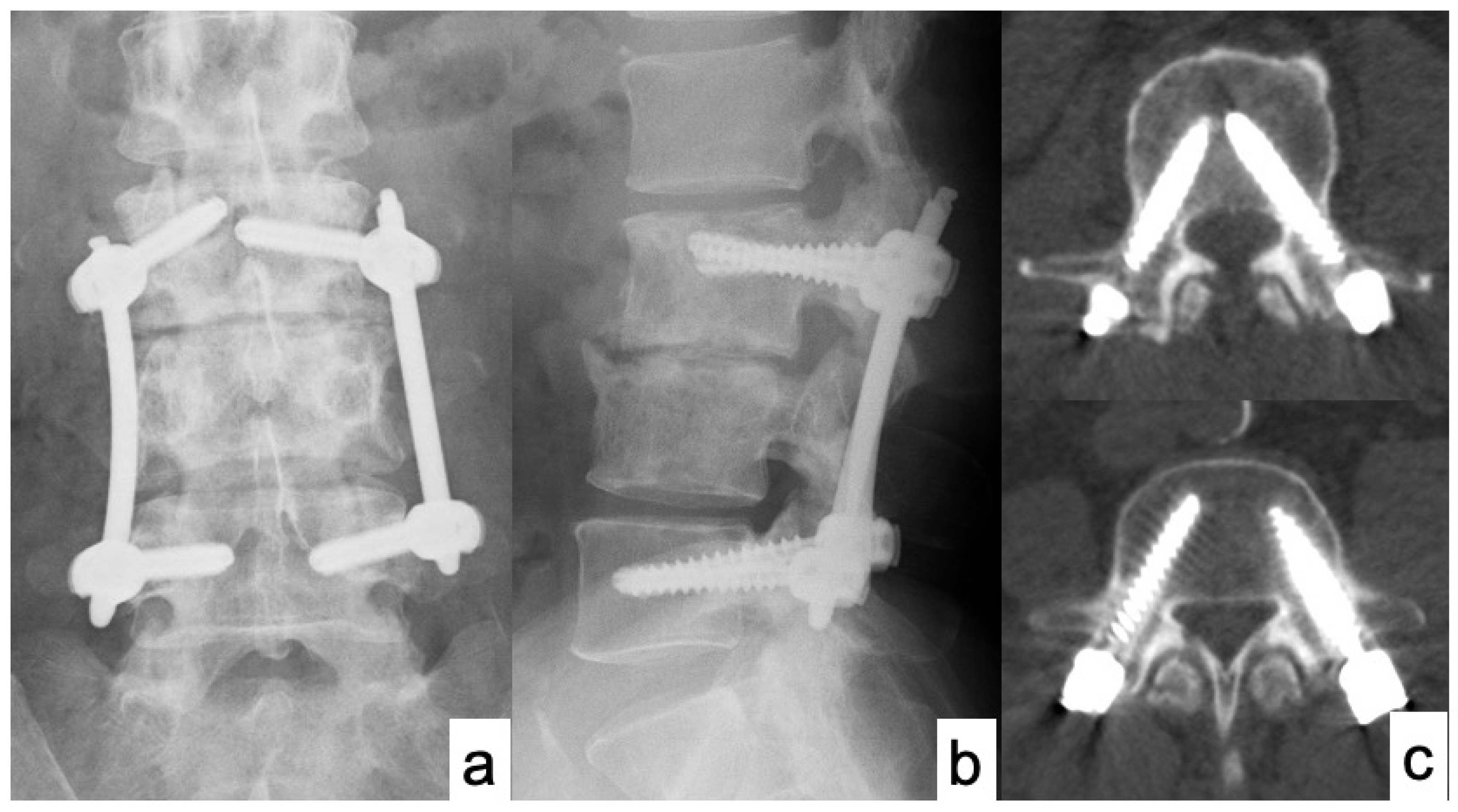
50 years old male presented with severe low back pain and fever. Despite the administration of antibiotics for a sufficient period of time, bone destruction progressed and the patient was unable to leave the bed due to pain, thus MISt with PPS was performed (Figure 1 and Figure 2). CRP became negative 22 days postoperatively, and bony union was achieved on CT images at 12 months after surgery (Figure 3).

4. Discussion

In recent years, the incidence of pyogenic spondylodiscitis has been rising due to an increase in the elderly population [5,6,7]. Pyogenic spondylodiscitis is often difficult to treat in elderly patients with multiple comorbidities or immunocompromised patients. It is indisputable that first-line treatment for pyogenic spondylodiscitis is adequate conservative treatment. Multiple randomized controlled trials suggest that antibiotics are required for 6 weeks with or without surgery and 8 weeks or more for Staphylococcus aureus and MRSA [28,29,30,31]. Surgical treatment is considered when conservative treatment fails, and the indications for surgical treatment have been reported in the literature: the presence of neurological symptoms, spinal instability, spinal deformity, and failure of conservative treatment [1,2,6,9,10].
In this study, MISt with PPS was successfully performed in 19 of 23 patients (82.6%). Although mortality rates of pyogenic spondylodiscitis have been reported to be between 2% and 20%, all 23 patients were cured of the infection within a mean of 28.4 days without any serious complications. The first reason for this high healing rate was the minimally invasive approach with PPS fixation. The conventional open method leads to massive bleeding, especially in long spinal fixation; however, PPS allows for long spinal fixation without significant bleeding and soft tissue damage. Posterior spinal fixation using PPS is clearly less invasive than the conventional open method, leading to earlier healing and mobilization without fatal complications. The second factor may be that we could administer adequate antibiotics for a sufficient period of time after conducting various culture tests and selecting sensitive antibiotics. The positivity rate of culture results is said to be 42 to 100%, and the high positivity rate of 69.6% in this study was a major advantage in the treatment of pyogenic spondylodiscitis [5,12,13,14]. These factors are thought to have contributed to the high healing rate of refractory pyogenic spondylodiscitis in this series. It is important to detect the causative organisms not only for the treatment of spondylodiscitis but also for the prevention and treatment of surgical site infection with spinal instrumentation that can result in serious consequences.
However, if the infection is not eradicated or bone union is not achieved after MISt with PPS, additional anterior debridement and reconstruction should be considered. In our case series, 4 of 23 (17.4%) patients required additional anterior debridement and autologous iliac bone graft placement; of which, 3 of 4 (75%) patients were presented with spondylodiscitis in the mid to lower lumbar spine. Because there might be hypermobility in the mid to lower lumbar spine, anterior reconstruction was required due to potential instability. Secure minimally invasive sacropelvic fixation is also required to provide a rigid distal foundation for spondylodiscitis in the lumbosacral spine [32]. Though Lin et al. reported that anterior debridement followed by PPS fixation was effective, it might be better to perform anterior debridement when a significant epidural abscess is present [11]. It is also reasonable to perform laminectomy posteriorly with a separate incision for an epidural abscess associated with neurological deficit. Recently introduced techniques such as full endoscopic surgery via a transforaminal approach can also be performed for the debridement of an epidural abscess [25]. The administration of teriparatide may also be effective as an additional treatment to obtain bony union around the infected vertebrae [33]. However, since Morita et al. reported that teriparatide did not suppress the progression of bone destruction in a murine osteomyelitis model, the administration of teriparatide for pyogenic spondylodiscitis remains controversial [34].
The present study has several limitations. The first is that this is a retrospective observational study, and the cohort was small due to the rarity of refractory pyogenic spondylodiscitis. In order to obtain a higher evidence level, it is desirable to conduct a prospective cohort study comparing the conventional open method and MISt with PPS. In addition, a long-term follow-up is required to observe the recurrence of infection, nonunion, mechanical failure, and potential risk of spinal malalignment. Second, PPS using fluoroscopy is not applicable to the cervical and upper thoracic spine, and it is also difficult to perform in osteoporotic and obese patients because the pedicle cannot be clearly visualized under fluoroscopy. Because we excluded the patients with pyogenic spondylodiscitis in the cervical and upper thoracic spine from this study, it can be a limitation to prove the effectiveness of MISt with PPS procedure in those patients.

5. Conclusions

In summary, MISt with PPS was successfully performed in 19 of 23 patients (82.6%) with refractory pyogenic spondylodiscitis without serious perioperative complications. MISt with PPS could be a feasible surgical option for refractory pyogenic spondylodiscitis, especially in elderly and immunocompromised patients.

Author Contributions

Conceptualization, S.I., H.F. and K.I.; data collection and analysis S.I., H.F., N.I., M.I., T.S. and K.I.; writing—original draft preparation, S.I. and H.F.; writing—review and editing, S.I., H.F. and K.I.; supervision, K.I. All authors have read and agreed to the published version of the manuscript.

Funding

This research received no external funding.

Institutional Review Board Statement

This study was conducted according to the guidelines of the Declaration of Helsinki and approved by the International University of Health and Welfare (IUHW) institutional ethics committee (approval number, 5-26-55).

Informed Consent Statement

We applied the opt-out method to obtain informed consent in the study.

Data Availability Statement

Not applicable.

Conflicts of Interest

The authors declare no conflict of interest.

References

  1. Gouliouris, T.; Aliyu, S.H.; Brown, N.M. Spondylodiscitis: Update on diagnosis and management. J. Antimicrob. Chemother. 2010, 65 (Suppl. 3), iii11–iii24. [Google Scholar] [CrossRef] [PubMed] [Green Version]
  2. Cheung, W.Y.; Luk, K.D.K. Pyogenic spondylitis. Int. Orthop. 2012, 36, 397–404. [Google Scholar] [CrossRef] [PubMed] [Green Version]
  3. Tsantes, A.G.; Papadopoulos, D.V.; Vrioni, G.; Sioutis, S.; Sapkas, G.; Benzakour, A.; Benzakour, T.; Angelini, A.; Ruggieri, P.; Mavrogenis, A.F.; et al. Spinal Infections: An Update. Microorganisms 2020, 8, 476. [Google Scholar] [CrossRef] [PubMed] [Green Version]
  4. Kourbeti, I.S.; Tsiodras, S.; Boumpas, D.T. Spinal infections: Evolving concepts. Curr. Opin. Rheumatol. 2008, 20, 471–479. [Google Scholar] [CrossRef] [PubMed]
  5. Rutges, J.P.H.J.; Kempen, D.H.; van Dijk, M.; Oner, F.C. Outcome of conservative and surgical treatment of pyogenic spondylodiscitis: A systematic literature review. Eur. Spine J. 2016, 25, 983–999. [Google Scholar] [CrossRef] [PubMed] [Green Version]
  6. Guerado, E.; Cerván, A.M. Surgical treatment of spondylodiscitis. An update. Int. Orthop. 2012, 36, 413–420. [Google Scholar] [CrossRef] [Green Version]
  7. Cervan, A.M.; Colmenero, J.D.; Del Arco, A.; Villanueva, F.; Guerado, E. Spondylodiscitis in patients under haemodyalisis. Int. Orthop. 2012, 36, 421–426. [Google Scholar] [CrossRef] [Green Version]
  8. Karchevsky, M.; Schweitzer, M.E.; Morrison, W.B.; Parellada, J.A. MRI findings of septic arthritis and associated osteomyelitis in adults. Am. J. Roentgenol. 2004, 182, 119–122. [Google Scholar] [CrossRef]
  9. Skaf, G.S.; Domloj, N.T.; Fehlings, M.G.; Bouclaous, C.H.; Sabbagh, A.S.; Kanafani, Z.A.; Kanj, S.S. Pyogenic spondylodiscitis: An overview. J. Infect. Public Health 2010, 3, 5–16. [Google Scholar] [CrossRef] [Green Version]
  10. Zarghooni, K.; Rollinghoff, M.; Sobottke, R.; Eysel, P. Treatment of spondylodiscitis. Int. Orthop. 2012, 36, 405–411. [Google Scholar] [CrossRef] [Green Version]
  11. Lin, T.Y.; Tsai, T.T.; Lu, M.L.; Niu, C.C.; Hsieh, M.K.; Fu, T.S.; Lai, P.L.; Chen, L.H.; Chen, W.J. Comparison of two-stage open versus percutaneous pedicle screw fixation in treating pyogenic spondylodiscitis. BMC Musculoskelet Disord. 2014, 15, 443. [Google Scholar] [CrossRef] [PubMed] [Green Version]
  12. Valancius, K.; Hansen, E.S.; Høy, K.; Helmig, P.; Niedermann, B.; Bünger, C. Failure modes in conservative and surgical management of infectious spondylodiscitis. Eur. Spine J. 2013, 22, 1837–1844. [Google Scholar] [CrossRef] [PubMed] [Green Version]
  13. Herren, C.; Jung, N.; Pishnamaz, M.; Breuninger, M.; Siewe, J.; Sobottke, R. Spondylodiscitis: Diagnosis and Treatment Options. Dtsch. Arztebl. Int. 2017, 114, 875–882. [Google Scholar] [CrossRef] [PubMed] [Green Version]
  14. Özmen, D.; Özkan, N.; Guberina, N.; Fliessbach, K.; Suntharalingam, S.; Theysohn, J.; Büchter, M.; Forsting, M.; Buer, J.; Dudda, M.; et al. Computed-tomography-guided biopsy in suspected spondylodiscitis: Single-center experience including 201 biopsy procedures. Orthop. Rev. 2019, 11, 7793. [Google Scholar] [CrossRef] [Green Version]
  15. Tsai, T.T.; Yang, S.C.; Niu, C.C.; Lai, P.L.; Lee, M.H.; Chen, L.H.; Chen, W.J. Early surgery with antibiotics treatment had better clinical outcomes than antibiotics treatment alone in patients with pyogenic spondylodiscitis: A retrospective cohort study. BMC Musculoskelet Disord. 2017, 18, 175. [Google Scholar] [CrossRef]
  16. Guo, W.; Wang, M.; Chen, G.; Chen, K.H.; Wan, Y.; Chen, B.; Zou, X.; Peng, X. Early surgery with antibiotic medication was effective and efficient in treating pyogenic spondylodiscitis. BMC Musculoskelet Disord. 2021, 22, 288. [Google Scholar] [CrossRef]
  17. Butler, J.S.; Shelly, M.J.; Timlin, M.; Powderly, W.G.; O’Byrne, J.M.O. Nontuberculous pyogenic spinal infection in adults: A 12-year experience from a tertiary referral center. Spine 2006, 31, 2695–2700. [Google Scholar] [CrossRef]
  18. Flury, B.B.; Elzi, L.; Kolbe, M.; Frei, R.; Weisser, M.; Scharen, S.; Widmer, A.F.; Battegay, M. Is switching to an oral antibiotic regimen safe after 2 weeks of intravenous treatment for primary bacterial vertebral osteomyelitis? BMC Infect. Dis. 2014, 14, 226. [Google Scholar]
  19. Bettini, N.; Girardo, M.; Dema, E.; Cervellati, S. Evaluation of conservative treatment of non spcific spondylodiscitis. Eur. Spine J. 2009, 18 (Suppl. 1), 143–150. [Google Scholar] [CrossRef] [Green Version]
  20. O’Daly, B.J.; Morris, S.F.; O’Rourke, S.K. Long-term functional outcome in pyogenic spinal infection. Spine 2008, 33, E246–E253. [Google Scholar] [CrossRef]
  21. Cottle, L.; Riordan, T. Infectious spondylodiscitis. J. Infect. 2008, 56, 401–412. [Google Scholar] [CrossRef]
  22. Karadimas, E.J.; Bunger, C.; Lindblad, B.E.; Høy, K.; Helmig, P.; Kannerup, A.S.; Niedermann, B. Spondylodiscitis. A retrospective study of 163 patients. Acta Orthop. 2008, 79, 650–659. [Google Scholar] [CrossRef] [Green Version]
  23. Pee, Y.H.; Park, J.D.; Choi, Y.G.; Lee, S.H. Anterior debridement and fusion followed by posterior pedicle screw fixation in pyogenic spondylodiscitis: Autologous iliac bone strut versus cage. J. Neurosurg. Spine 2008, 8, 405–412. [Google Scholar] [CrossRef] [Green Version]
  24. Gonzalvo, A.; Abdulla, I.; Riazi, A.; De La Harpe, D. Single- level/single-stage debridement and posterior instrumented fusion in the treatment of spontaneous pyogenic osteomyelitis/discitis: Long-term functional outcome and health-related quality of life. J. Spinal Disord. Tech. 2011, 24, 110–115. [Google Scholar] [CrossRef]
  25. Duan, K.; Qin, Y.; Ye, J.; Zhang, W.; Hu, X.; Zhou, J.; Gao, L.; Tang, Y. Percutaneous endoscopic debridement with percutaneous pedicle screw fixation for lumbar pyogenic spondylodiscitis: A preliminary study. Int. Orthop. 2020, 44, 495–502. [Google Scholar] [CrossRef] [Green Version]
  26. Janssen, I.K.; Jörger, A.K.; Barz, M.; Sarkar, C.; Wostrack, M.; Meyer, B. Minimally invasive posterior pedicle screw fixation versus open instrumentation in patients with thoracolumbar spondylodiscitis. Acta Neurochir. 2021, 163, 1553–1560. [Google Scholar] [CrossRef]
  27. Farshad, M.; Aichmair, A.; Gerber, C.; Bauer, D.E. Classification of perioperative complications in spine surgery. Spine J. 2020, 20, 730–736. [Google Scholar] [CrossRef]
  28. Bernard, L.; Dinh, A.; Ghout, I.; Simo, D.; Zeller, V.; Issartel, B.; Le Moing, V.; Belmatoug, N.; Lesprit, P.; Bru, J.P.; et al. Antibiotic treatment for 6 weeks versus 12 weeks in patients with pyogenic vertebral osteomyelitis: An open-label, non-inferiority, randomised, controlled trial. Lancet 2015, 385, 875–882. [Google Scholar] [CrossRef]
  29. Loibl, M.; Stoyanov, L.; Doenitz, C.; Brawanski, A.; Wiggermann, P.; Krutsch, W.; Nerlich, M.; Oszwald, M.; Neumann, C.; Salzberger, B.; et al. Outcome-related co-factors in 105 cases of vertebral osteomyelitis in a tertiary care hospital. Infection 2014, 42, 503–510. [Google Scholar] [CrossRef]
  30. Roblot, F.; Besnier, J.M.; Juhel, L.; Vidal, C.; Ragot, S.; Bastides, F.; Le Moal, G.; Godet, C.; Mulleman, D.; Azais, I.; et al. Optimal duration of antibiotic therapy in vertebral osteomyelitis. Semin. Arthritis Rheum. 2007, 36, 269–277. [Google Scholar] [CrossRef]
  31. Park, K.H.; Chong, Y.P.; Kim, S.H.; Lee, S.O.; Choi, S.H.; Lee, M.S.; Jeong, J.Y.; Woo, J.H.; Kim, Y.S. Clinical characteristics and therapeutic outcomes of hematogenous vertebral osteomyelitis caused by methicillin-resistant Staphylococcus aureus. J. Infect. 2013, 67, 556–564. [Google Scholar] [CrossRef]
  32. Funao, H.; Kebaish, K.M.; Isogai, N.; Koyanagi, T.; Matsumoto, M.; Ishii, K. Utilization of a technique of percutaneous S2 alariliacfixation in immunocompromised patients with spondylodiscitis. World Neurosurg. 2017, 97, e11–e18. [Google Scholar] [CrossRef]
  33. Shinohara, A.; Ueno, Y.; Marumo, K. Weekly teriparatide therapy rapidly accelerates bone healing in pyogenic spondylitis with severe osteoporosis. Asian Spine J. 2014, 8, 498–501. [Google Scholar] [CrossRef]
  34. Morita, M.; Iwasaki, R.; Sato, Y.; Kobayashi, T.; Watanabe, R.; Oike, T.; Nakamura, S.; Keneko, Y.; Miyamoto, K.; Ishihara, K.; et al. Elevation of pro-inflammatory cytokine levels following anti-resorptive drug treatment is required for osteonecrosis development in infectious osteomyelitis. Sci. Rep. 2017, 7, 46322. [Google Scholar] [CrossRef]
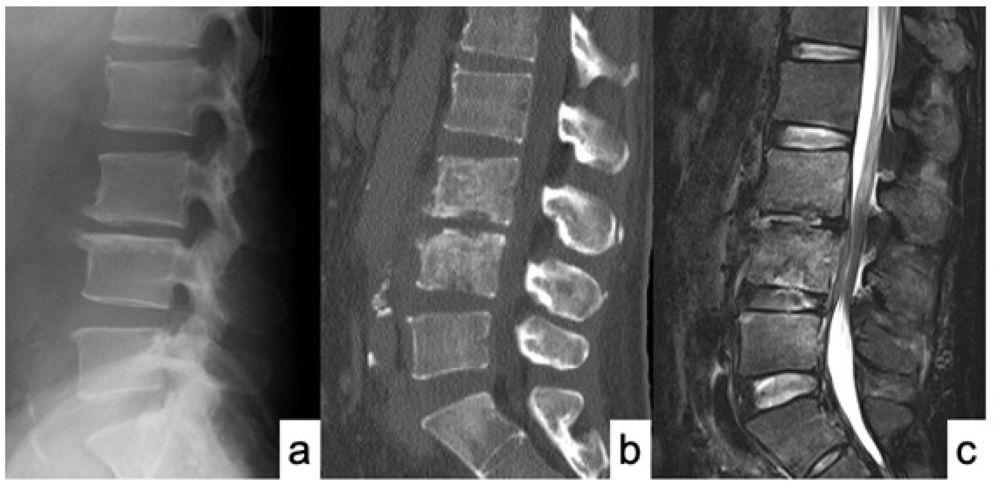
Figure 1. MRI and CT scan of case No. 14 at first admission. (a) X-ray of sagittal view; (b) Sagittal view of CT scan showed a destructive change of L3 and L4 vertebral bodies; and (c) T2-weighted sagittal view of MRI showed spondylodiscitis at L3-4 and L4-5 with epidural abscess.
Medicina 58 00754 g001
Figure 2. X-ray and CT scan of case No. 14 immediately after surgery. (a) X-ray of AP view; (b) X-ray of lateral view; and (c) Axial views of CT scan showed accurate PPS placements.
Medicina 58 00754 g002
Figure 3. Comparison of CT images before surgery and 12 months after surgery. (a,c) CT images before surgery showed destructive change of L3 and L4 vertebral bodies; (b,d) CT image at 12 months after surgery showed good bony union with callus bridging between L3 and L4.
Medicina 58 00754 g003
Table 1. Demographic data.
CharacteristicN = 23 (%)
Gender
Male18 (78.3%)
Female5 (21.7%)
Comorbidities (including duplications)
Solid cancer8 (34.8%)
Diabetes mellitus5 (21.7%)
Renal failure3 (13.0%)
Cerebrovascular disease3 (13.0%)
Liver cirrhosis2 (8.7%)
Angina pectoris1 (4.3%)
Pancreatitis1 (4.3%)
Depression1 (4.3%)
Location (including 1 duplication)
Thoracic4 (17.4%)
Thoracolumbar4 (17.4%)
Lumbar11 (47.8%)
Lumbosacral5 (21.7%)
Table 2. Causative organisms.
Bacterial StrainN = 23 (%)
Staphylococcus aureus6 (26.1%)
MRSA 13 (13.0%)
Streptococcus dysgalactiae2 (8.7%)
Streptococcus intermedius1 (4.3%)
Streptococcus mutans1 (4.3%)
Escherichia coli1 (4.3%)
Enterobacter aerogens1 (4.3%)
Corynebacterium1 (4.3%)
Unknown7 (30.4%)
1 MRSA: methicillin resistant staphylococcus aureus.
Table 3. Detailed presentation of patient data.
Patient No.AgeSexInvolved
Level
Fixed
Vertebrae
OrganismComorbiditiesCRP
Become Negative (Days)
Operative Time (min)EBL (mL)ADFF-U (Month)
164MT7-8T5-10Unknown 2714050No6
266MT8-9T6-11MRSAInfectious
endocarditis
161225No6
385FT9-10T6-L1S. aureusProstate cancer2825870No24
478MT9-10T7-12MRSAPancreatitis14130300No7
575FT9-11T6-L2Unknown 12278130No6
684MT10-11T7-L2S. aureusDM28265384No26
769MT10-11T7-L2UnknownRF and CD2528267No18
864ML1-2L1-3S. aureusLiver Cancer396520Yes6
960ML2-3T12-L5S. aureus 1121252No6
1062ML2-3
L5-S1
T12-S1
(Iliac)
S. dysgalactiaeRF56364260No6
1155ML3-4L1-S1E. aerogenesColon cancer47183158Yes43
1257ML3-4L1-5S. mutansLiver cancer10245100No16
1375ML3-4L1-5S. dysgalactiaeDM51154280No15
1450ML3-5L3-5S. intermediusDM and
Liver cancer
568590No12
1539FL4-5L4-5S. aureusDepression195520No18
1677ML4-5L2-S1
(S2AI *)
S. aureusLung cancer2019326No6
1772ML4-5L2-S1
(S2AI *)
UnknownGastric cancer3018110Yes24
1857FL4-5L2-S1
(S2AI *)
Unknown 3021543No18
1977ML4-5L2-S1
(S2AI *)
UnknownLung cancer and
angina
3018826No6
2077ML5-S1L3-S1
(Illiac)
UnknownDM14265340No13
2159ML5-S1L3-S1
(Illiac)
MRSACD30399550No24
2269FL5-S1L3-S1
(Illiac)
Coryne
bacterium
Uterine cancer30261172No36
2371ML5-S1L3-S1
(S2AI *)
E. coliDM, RF, and CD30196171Yes24
* S2AI: S2 alar iliac; MRSA: methicillin resistant staphylococcus aureus; DM: diabetes mellitus; RF: renal failure; CD: cerebrovascular disease; ADF: anterior debridement and fusion.
Table 4. Perioperative data.
Numbers of fixed vertebrae4.1 vertebrae (2–6)
Operative time205.1 min (55–399)
Estimated blood loss145.0 mL (5–550)
Anterior debridement and bone graft placement4 cases (17.4%)
CRP becomes negative after surgery28.4 days (10–56 days)
Major perioperative complicationnone
Publisher’s Note: MDPI stays neutral with regard to jurisdictional claims in published maps and institutional affiliations.