Case Report

A Case of Stevens–Johnson Syndrome Complicated with Multimatrix System Mesalamine in Ulcerative Colitis

Department of Gastroenterology, Dokkyo Medical University, 880 Kitakobayashi, Mibu 321-0293, Japan
*
Author to whom correspondence should be addressed.
Medicina 2022, 58(2), 276; https://doi.org/10.3390/medicina58020276
Submission received: 7 January 2022 / Revised: 2 February 2022 / Accepted: 10 February 2022 / Published: 11 February 2022
(This article belongs to the Section Gastroenterology & Hepatology)

Abstract

:
A 41-year-old man was treated with prednisolone (PSL) and multimatrix (MMX) mesalamine for remission induction therapy of ulcerative colitis. PSL was tapered due to successful remission induction treatment. During the treatment course, ocular foreign body sensation, eyelid swelling, ocular conjunctiva hyperemia, facial redness and swelling, watery nasal discharge, stomatitis, anal pain, and reddish puffiness on the bilateral dorsum of the hands appeared, and he was diagnosed with Stevens–Johnson syndrome (SJS). SJS was improved by PSL treatment and intravenous immunoglobulin. MMX mesalamine was the causative agent by drug-induced lymphocyte stimulation test. This is the first reported case of SJS with MMX mesalamine.

1. Introduction

The use of 5-aminosalicylic acid (5-ASA) as a basic treatment for ulcerative colitis (UC) has few side effects, and is a drug frequently used for induction and maintenance of remission. In recent years, a sustained-release tablet containing mesalamine (MMX mesalamine) using the multimatrix (MMX) delivery system has been widely used for UC. Currently, salazosulfapyridine (SASP) and three different forms of mesalamine are used in Japan.
Stevens–Johnson syndrome (SJS) and toxic epidermal necrolysis (TEN) are rare, but potentially life-threatening and severe disorders, diseases characterized by widespread epidermal necrosis, and are predominantly medication induced. Although there have been several reports of SJS and TEN caused by mesalamine and SASP, there have been no such reports with MMX mesalamine. Although 5-ASA is widely used as a basic therapeutic agent for UC without immune modulation, it is important to recognize that, like other drugs, it can cause serious side effects, such as SJS and TEN.

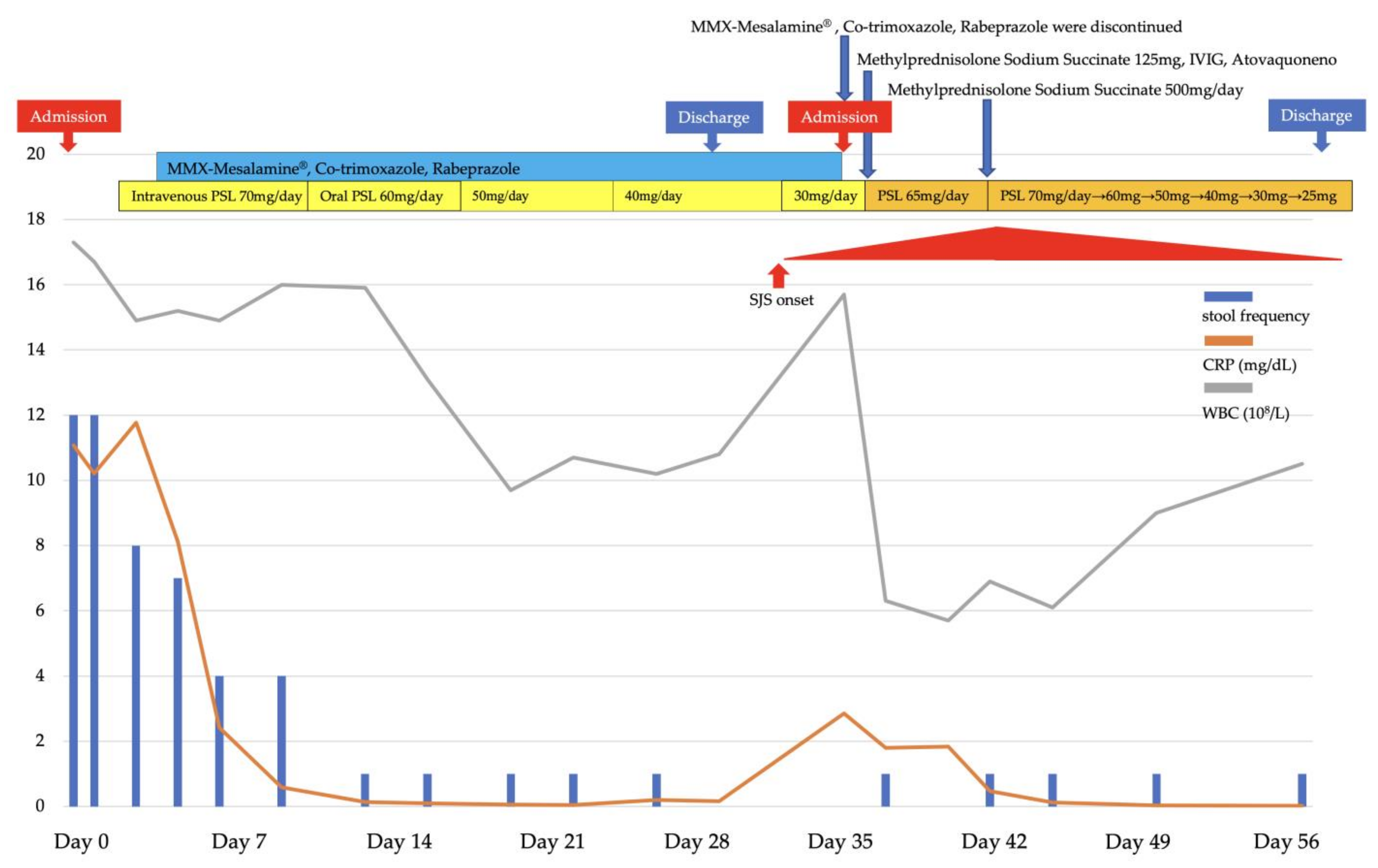
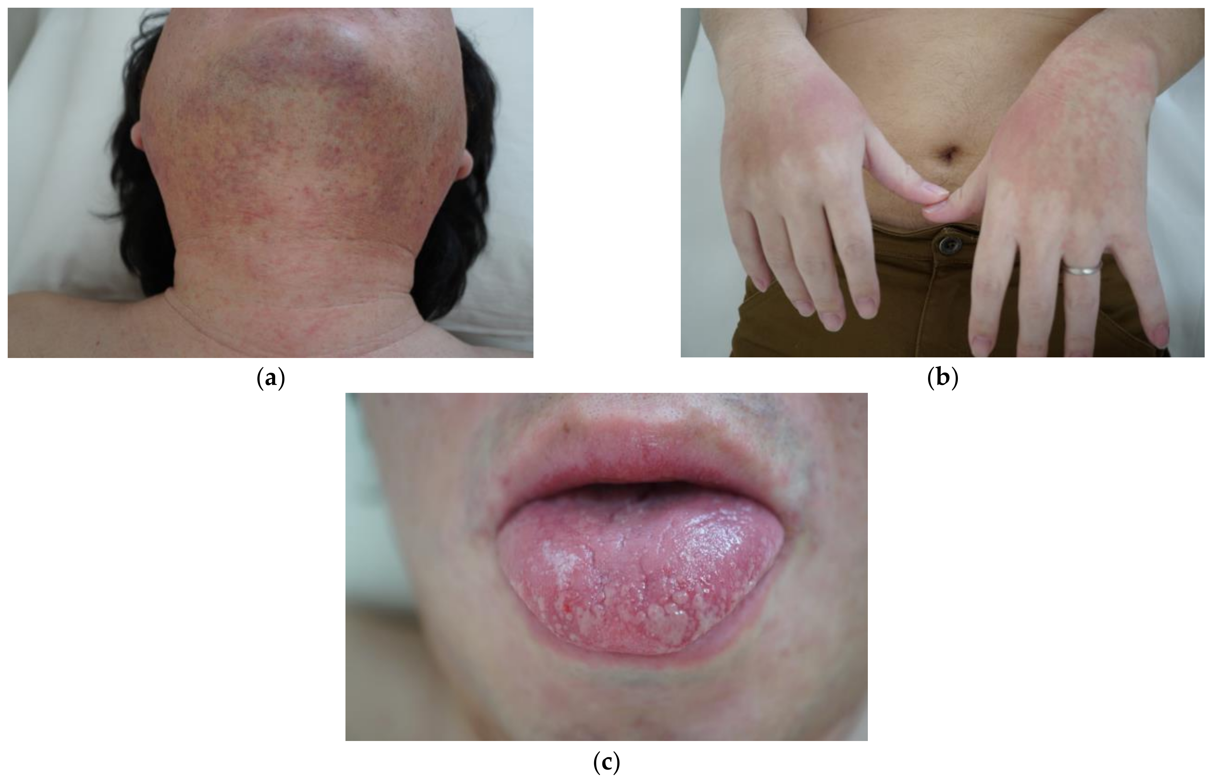
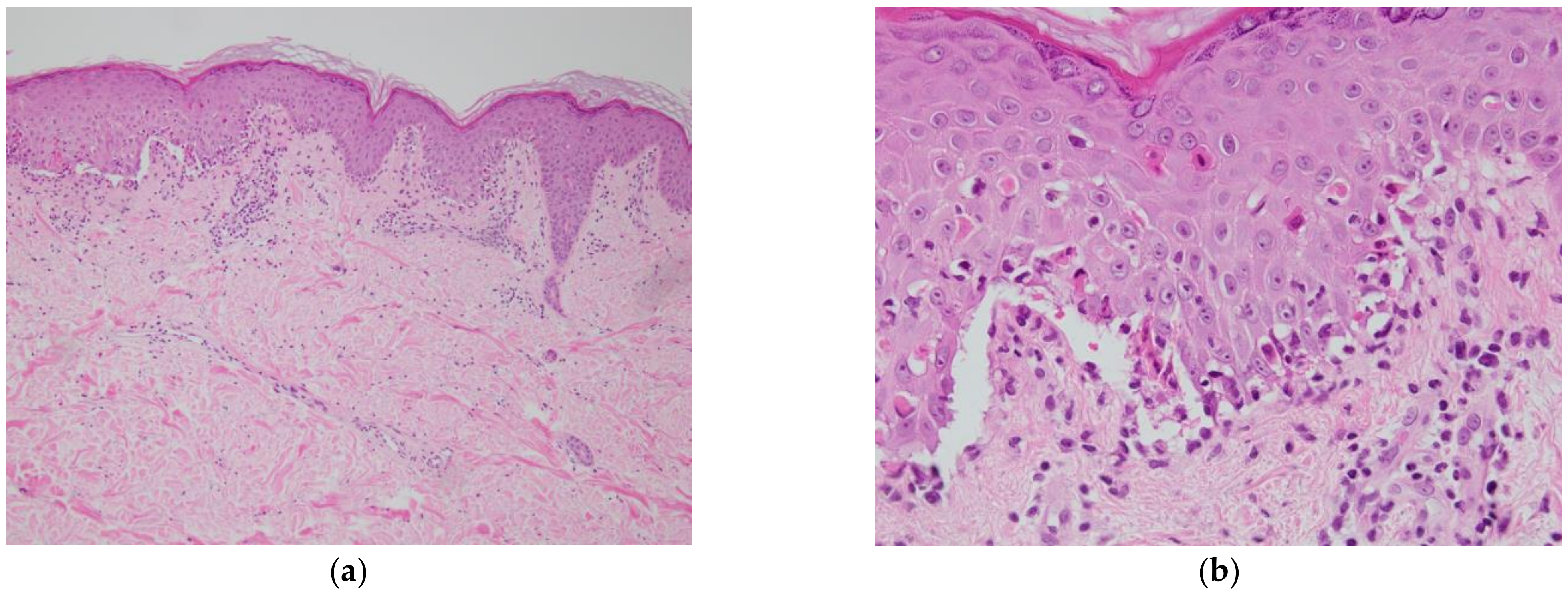
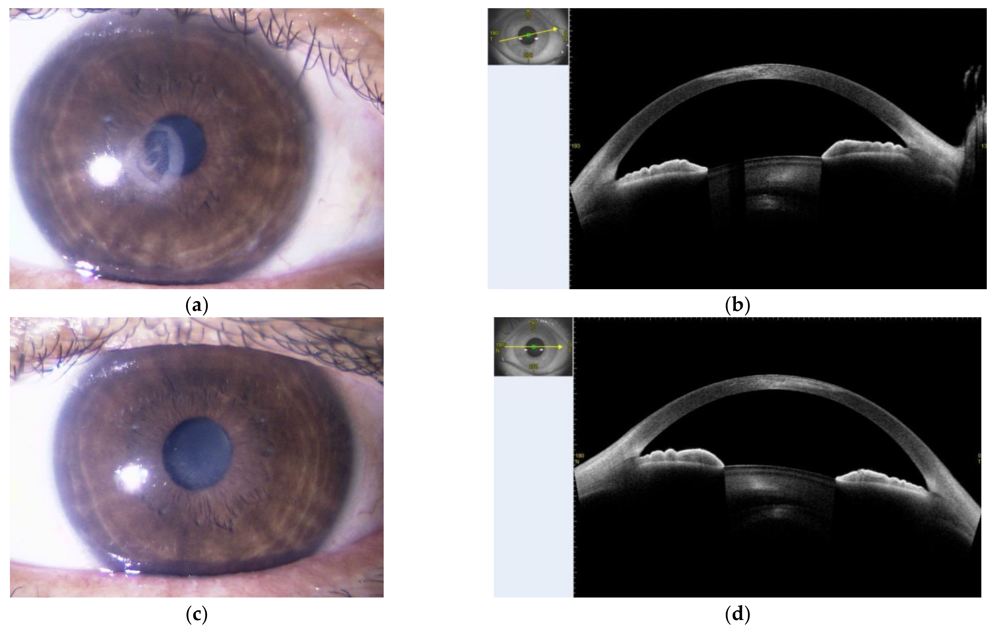
2. Presentation of Case Report

A 41-year-old man visited his local doctor in March 2020 due to abdominal pain, diarrhea, and bloody stools that started in February 2020. He was diagnosed with UC (total colitis) by colonoscopy and was soon referred to our hospital for treatment. He had no previous medical history and was not taking any medication. He did not consume alcohol and had been smoking 10 cigarettes/day for 21 years since the age of 20.
The severity of UC at the time of his visit was Lichtiger index of 13 (≥12: severe) [1]. The endoscopy showed a Mayo endoscopic subscore 3 in the entire colon. He was admitted for remission induction of UC and started remission induction therapy with intravenous prednisolone (PSL) 70 mg/day (1 mg/kg/day) and oral MMX mesalamine 4800 mg/day. Oral co-trimoxazole to prevent the development of pneumocystis pneumonia due to immunosuppression associated with high-dose PSL administration, and oral rabeprazole 10 mg/day to prevent gastrointestinal mucosal damage were also started. The UC symptoms improved in about 12 days, and PSL was tapered by 10 mg every week. He achieved remission, had a Lichtiger index of 2, and discharged. MMX mesalamine 4800 mg/day, co-trimoxazole, and rabeprazole 10 mg/day were continued. During the treatment course (Figure 1), PSL was tapered to 30 mg/day, and about 30 days after starting MMX mesalamine, he exhibited eyelid swelling, ocular conjunctiva hyperemia, facial redness and swelling, watery nasal discharge, stomatitis, anal pain, and erythematous swelling of the bilateral dorsum of the hands (Figure 2a–c). We suspected drug-related SJS, or viral infections associated with immunosuppression due to high-dose PSL administration.
Nikolsky’s sign (a phenomenon in which the epidermis of apparently normal skin becomes detached and erosive when mild pressure is applied) was positive. The ocular lesions included extensive corneal erosion and severe conjunctival hyperemia. Laboratory tests showed mild liver dysfunction with AST 31 U/L and ALT 65 U/L. WBC was 15.7 × 10³/μL and CRP was 2.85 mg/dL, and anemia was present with Hb 11.2 g/dL. Eosinophil was 0.8% and IgE was 12.6 IU/L. Although SJS may occur with mycoplasma and some viral infections, bacterial and viral infections were ruled out as the patient was HSV-IgG positive, mycoplasma negative, ASO negative, and CMVAg negative (Table 1). The skin biopsy showed interface dermatitis, and liquid degeneration at the epidermal–dermal border and cleft formation was prominent. There were necrotic keratinocytes in the epidermis and lymphocytic infiltration into the epidermis and around subepidermal microvessels (Figure 3a,b). We considered the patient’s symptoms to be drug-induced SJS and discontinued all three suspected drugs, which were MMX mesalamine, co-trimoxazole and rabeprazole sodium, and started intravenous methylprednisolone 125 mg/day and intravenous immunoglobulin. Before the results of the drug-induced lymphocyte stimulation test (DLST) became clear, we also started atovaquone instead of co-trimoxazole to prevent the onset of pneumocystis pneumonia due to immunosuppression associated with high doses of PSL, and famotidine instead of rabeprazole to prevent gastrointestinal mucosal damage. In the DLST that was submitted prior to the start of treatment to identify the causative drug, MMX mesalamine was positive, co-trimoxazole and rabeprazole negative, and MMX mesalamine was determined to be the causative agent.
The symptoms continued to worsen even after the start of treatment, with widely distributed erythema, blistering, and epidermal peeling over the entire body, including the palms and soles, and mucosal lesions in the transition area between the lips and the oral and anal mucosa. In addition, ocular pain increased and corneal opacity and pseudomembrane formation were observed, and consequently methylprednisolone 500 mg/day was administered intravenously for 3 days, and symptoms began to improve. Thereafter, we gradually tapered PSL from 70 mg/day. The skin and mucosal lesions showed a tendency to heal, and some nails were deformed and dropped off, but later recovered. As for the ocular lesions, visual impairment due to opacity of the corneal parenchyma remained as a sequela (Figure 4a–d). Fortunately, no exacerbation of UC was observed during this course. Since MMX causes SJS, it was decided that a 5-ASA formulation could not be used and that an immunomodulator would be used to maintain UC remission.

3. Discussion

This is a case of UC with SJS caused by MMX mesalamine. SJS and TEN develop 1–3 weeks after the initiation of the causative agent [2,3]. Based on international standards, SJS and TEN are distinguished by their percentage of body surface area, with SJS being less than 10%, overlap syndrome being 10–30%, and TEN being more than 30% [4]. The fatality rate can be high, ranging from 3–5% in SJS and 20–30% in TEN [5]. In addition, about 50–89% of SJS/TEN patients develop ocular complications, which may lead to serious adverse events such as corneal damage and vision loss [6,7,8]. In 1987, Roujeau et al. highlighted the significance of polymorphisms in human leukocyte antigen (HLA) in the pathogenesis of SJS/TEN [9]. The HLA-B*44:03 found in the present case has been reported to be associated with SJS/TEN with severe ocular complications related to common cold medications [10,11].
In general, DLST has low sensitivity and high specificity in drugs such as antituberculosis drugs and rheumatic drugs. Saito et al. reported that the sensitivity and specificity of DLST for mesalamine were 0.240 and 0.805, respectively [12]. As with other drugs, DLST for mesalamine has low sensitivity and high specificity, so a positive DLST is strongly suspected to be caused by the drug. Therefore, DLST may be useful in the definitive diagnosis of the causative agent of SJS and TEN. In this case, DLST of MMX mesalamine was positive, while co-trimoxazole and rabeprazole were negative. Based on the sensitivity and specificity, we thought that co-trimoxazole and rabeprazole were unlikely to be involved in the development of SJS in this case, and came to the diagnosis of SJS by MMX mesalamine.
The basic drug 5-ASA that can treat UC without immunosuppression, has few side effects, and is used from remission induction to long term remission maintenance. Several mesalamines with different drug delivery systems and SASP have been used to improve delivery to the colon, the main site of disease in UC. Among these, there have been several reports of SJS and TEN occurring when using mesalamine and SASP [13,14,15,16,17,18,19,20,21], but there have been no similar reports when using MMX mesalamine (Table 2).
The mechanism of drug release obtained with the MMX system avoids the release of the embedded mesalamine until the tablet is exposed to pH 7 or higher, which is normally reached in the terminal ileum. After reaching the terminal ileum, the activity of the tablet core, which is composed of hydrophilic excipients (thought to drive the tablet to swell into a viscous gel mass, and slowing the release of the drug) and lipophilic excipients (thought to slow the penetration of aqueous fluids into the tablet core), results in a homogenous and prolonged exposure of the entire colonic mucosa to the embedded mesalamine [22]. MMX mesalamine is capable of sustained release of mesalamine throughout the colon, which differentiates it from other 5-ASA formulations. In addition to mesalamine, the MMX system has been implemented in budesonide, parnaparin, rifampicin, and other drugs, and has been used or is being considered for use in clinical practice. All of the aforementioned drugs have been reported to have a high safety profile and have not been associated with serious adverse events [23,24,25,26,27,28]. We speculated that the MMX system, with its slow release of drugs, may reduce the incidence of SJS and TEN.
In particular, this patient was receiving a high dose of PSL for remission induction of UC at about the same time as the causative agent. Although the pathogenesis of SJS is unknown, it is thought that cytotoxic T cells activated by drugs taken into the body may directly induce apoptosis of epidermal cells, or cytokines (e.g., TNF-α) produced by activated cytotoxic T cells may indirectly cause cytotoxicity [29,30]. It has been shown that the dose of PSL is related to the function of cellular immunity [31,32] and it is possible that in the present case, the onset of SJS was suppressed by the use of high-dose PSL, and the symptoms only became apparent as PSL was gradually tapered. Furthermore, since this patient was receiving a relatively high dose of PSL at the time of onset, it is possible that SJS-induced fever could have been masked.
Lastly, based on previous reports, cases in which PSL was administered at the same time as the causative drug tended to have a longer time to onset than cases in which PSL was not administered Lastly, based on previous reports, cases in which PSL was administered at the same time as the causative drug tended to have a longer time to onset than cases in which PSL was not administered (Table 2).

4. Conclusions

A relatively safe drug with a low incidence of side effects, 5-ASA is widely used as a basic treatment for UC. However, it is important to recognize that it can cause serious side effects such as SJS and TEN in susceptible individuals. Although the incidence of SJS and TEN is extremely low, they have a relatively high fatality rate and can leave serious sequelae, therefore, prompt therapeutic intervention is critical when diagnosed.

Author Contributions

Conceptualization, M.K. and K.T.; wrote the manuscript and provided the images. M.K., K.T., A.K., T.T., S.M., S.W., K.A. and A.Y.; reviewed the manuscript K.G. and A.I.; article guarantor. K.T. All authors have read and agreed to the published version of the manuscript.

Funding

This case report received no external funding.

Institutional Review Board Statement

Not applicable.

Informed Consent Statement

Written informed consent has been obtained from the patient to publish this paper.

Data Availability Statement

Not applicable.

Acknowledgments

The authors would like to thank Mizuki Terada, Takashi Gonmori (Department of Dermatology, Dokkyo Medical University), and Haruki Mori (Department of Ophthalmology, Dokkyo Medical University) for patient’s treatment. The authors would also like to thank Ishida Miyako, Nishimoto Naoko, and Iizuka Sanae (Department of Gastroenterology, Dokkyo Medical University) for technical assistance.

Conflicts of Interest

The authors declare no conflict of interest.

References

  1. Lichtiger, S.; Present, D.H. Preliminary report: Cyclosporin in treatment of severe active ulcerative colitis. Lancet 1990, 336, 16–19. [Google Scholar] [CrossRef]
  2. Roujeau, J.C.; Stern, R.S. Severe adverse cutaneous reactions to drugs. N. Engl. J. Med. 1994, 331, 1272–1285. [Google Scholar] [CrossRef]
  3. Assier, H.; Bastuji-Garin, S.; Revuz, J.; Roujeau, J.C. Erythema multiforme with mucous membrane involvement and Stevens-Johnson syndrome are clinically different disorders with distinct causes. Arch. Dermatol. 1995, 131, 539–543. [Google Scholar] [CrossRef] [PubMed]
  4. Creamer, D.; Walsh, S.A.; Dziewulski, P.; Exton, L.S.; Lee, H.Y.; Dart, J.K.; Setterfield, J.; Bunker, C.B.; Ardern-Jones, M.R.; Watson, K.M.; et al. U.K. guidelines for the management of Stevens-Johnson syndrome/toxic epidermal necrolysis in adults 2016. Br. J. Dermatol. 2016, 174, 1194–1227. [Google Scholar] [CrossRef] [PubMed] [Green Version]
  5. Ferrándiz-Pulido, C.; García-Fernández, D.; Gómez-Morell, P.; Palao, R.; García-Patos, V. Síndrome de Stevens-Johnson y necrólisis epidérmica tóxica: Revisión de la experiencia clínica en un Hospital Universitario (1989–2008) [Stevens-Johnson syndrome and toxic epidermal necrolysis: A review of the clinical experience of a University Hospital (1989–2008)]. Med. Clin. 2011, 136, 583–587. [Google Scholar] [CrossRef]
  6. Revuz, J.; Penso, D.; Roujeau, J.C.; Guillaume, J.C.; Payne, C.R.; Wechsler, J.; Touraine, R. Toxic epidermal necrolysis. Clinical findings and prognosis factors in 87 patients. Arch. Dermatol. 1987, 123, 1160–1165. [Google Scholar] [CrossRef]
  7. Power, W.J.; Ghoraishi, M.; Merayo-Lloves, J.; Neves, R.A.; Foster, C.S. Analysis of the acute ophthalmic manifestations of the erythema multiforme/Stevens-Johnson syndrome/toxic epidermal necrolysis disease spectrum. Ophthalmology 1995, 102, 1669–1676. [Google Scholar] [CrossRef]
  8. Sotozono, C.; Ang, L.P.; Koizumi, N.; Higashihara, H.; Ueta, M.; Inatomi, T.; Yokoi, N.; Kaido, M.; Dogru, M.; Shimazaki, J.; et al. New grading system for the evaluation of chronic ocular manifestations in patients with Stevens-Johnson syndrome. Ophthalmology 2007, 114, 1294–1302. [Google Scholar] [CrossRef]
  9. Roujeau, J.C.; Huynh, T.N.; Bracq, C.; Guillaume, J.C.; Revuz, J.; Touraine, R. Genetic susceptibility to toxic epidermal necrolysis. Arch. Dermatol. 1987, 123, 1171–1173. [Google Scholar] [CrossRef]
  10. Ueta, M.; Kaniwa, N.; Sotozono, C.; Tokunaga, K.; Saito, Y.; Sawai, H.; Miyadera, H.; Sugiyama, E.; Maekawa, K.; Nakamura, R.; et al. Independent strong association of HLA-A*02:06 and HLA-B*44:03 with cold medicine-related Stevens-Johnson syndrome with severe mucosal involvement. Sci. Rep. 2014, 4, 4862. [Google Scholar] [CrossRef] [Green Version]
  11. Ueta, M.; Kannabiran, C.; Wakamatsu, T.H.; Kim, M.K.; Yoon, K.C.; Seo, K.Y.; Joo, C.K.; Sangwan, V.; Rathi, V.; Basu, S.; et al. Trans-ethnic study confirmed independent associations of HLA-A*02:06 and HLA-B*44:03 with cold medicine-related Stevens-Johnson syndrome with severe ocular surface complications. Sci. Rep. 2014, 4, 5981. [Google Scholar] [CrossRef] [PubMed] [Green Version]
  12. Saito, D.; Hayashida, M.; Sato, T.; Minowa, S.; Ikezaki, O.; Mitsui, T.; Miura, M.; Sakuraba, A.; Hisamatsu, T. Evaluation of the drug-induced lymphocyte stimulation test for diagnosing mesalazine allergy. Intest. Res. 2018, 16, 273–281. [Google Scholar] [CrossRef] [Green Version]
  13. Maddocks, J.L.; Slater, D.N. Toxic epidermal necrolysis, agranulocytosis and erythroid hypoplasia associated with sulphasalazine. J. R. Soc. Med. 1980, 73, 587–588. [Google Scholar] [CrossRef] [PubMed] [Green Version]
  14. Tolia, V. Sulfasalazine desensitization in children and adolescents with chronic inflammatory bowel disease. Am. J. Gastroenterol. 1992, 87, 1029–1032. [Google Scholar]
  15. Iemoli, E.; Piconi, S.; Ardizzone, S.; Bianchi, P.G.; Raimond, F. Erythroderma and toxic epidermal necrolysis caused by to 5-aminosalacylic acid. Inflamm. Bowel Dis. 2006, 12, 1007–1008. [Google Scholar] [CrossRef]
  16. Fukunaga, K.; Ohda, Y.; Inoue, T.; Kono, T.; Miwa, H.; Matsumoto, T. Toxic epidermal necrosis associated with mesalamine in a patient with ulcerative colitis. Inflamm. Bowel Dis. 2007, 13, 1055–1056. [Google Scholar] [CrossRef] [PubMed]
  17. Tremblay, L.; de Chambrun, G.P.; De Vroey, B.; Lavogiez, C.; Delaporte, E.; Colombel, J.F.; Cortot, A. Stevens-Johnson syndrome with sulfasalazine treatment: Report of two cases. J. Crohns Colitis 2011, 5, 457–460. [Google Scholar] [CrossRef] [Green Version]
  18. Zizi, N.; Elmrahi, A.; Dikhaye, S.; Fihmi, N.; Alami, Z. Stevens Johnson syndrome-Toxic Epidermal Necrolysis Overlap induced by sulfasalazine treatment: A case report. Tunis. Med. 2015, 93, 413–415. [Google Scholar]
  19. Núñez, O.A.; Trigo, S.C.; de la Cruz, R.M.D.; Herrera, J.J.M.; Leo, C.E. Topical mesalazine as a cause of Stevens-Johnson syndrome. Rev. Esp. Enferm. Dig. 2018, 110, 736–738. [Google Scholar] [CrossRef]
  20. Xiong, H.; Chen, S.; Luo, X. Salazosulphapyridine-related Stevens-Johnson Syndrome Caused by Sulphapyridine and Confirmed by Enzyme-linked Immunospot Assay. J. Crohns Colitis 2018, 12, 381–382. [Google Scholar] [CrossRef]
  21. Viola, A.; Caltagirone, A.M.; Campisi, G.; Guarneri, G.; Cappello, M. Stevens-Johnson syndrome on treatment with sulfasalazine for Crohn’s disease: Need for a multidisciplinary approach. Turk. J. Gastroenterol. 2019, 30, 211–212. [Google Scholar] [CrossRef] [PubMed]
  22. Nardelli, S.; Pisani, L.F.; Tontini, G.E.; Vecchi, M.; Pastorelli, L. MMX® technology and its applications in gastrointestinal diseases. Therap. Adv. Gastroenterol. 2017, 10, 545–552. [Google Scholar] [CrossRef] [PubMed] [Green Version]
  23. Lichtenstein, G.R. Budesonide Multi-matrix for the Treatment of Patients with Ulcerative Colitis. Dig. Dis. Sci. 2016, 61, 358–370. [Google Scholar] [CrossRef] [PubMed] [Green Version]
  24. Sherlock, M.E.; MacDonald, J.K.; Griffiths, A.M.; Steinhart, A.H.; Seow, C.H. Oral budesonide for induction of remission in ulcerative colitis. Cochrane Database Syst. Rev. 2015, 26, CD007698. [Google Scholar] [CrossRef]
  25. Sandborn, W.J.; Travis, S.; Moro, L.; Jones, R.; Gautille, T.; Bagin, R.; Huang, M.; Yeung, P.; Ballard, E.D., 2nd. Once-daily budesonide MMX® extended-release tablets induce remission in patients with mild to moderate ulcerative colitis: Results from the CORE I study. Gastroenterology 2012, 143, 1218–1226. [Google Scholar] [CrossRef] [PubMed] [Green Version]
  26. Travis, S.P.; Danese, S.; Kupcinskas, L.; Alexeeva, O.; D’Haens, G.; Gibson, P.R.; Moro, L.; Jones, R.; Ballard, E.D.; Masure, J.; et al. Once-daily budesonide MMX in active, mild-to-moderate ulcerative colitis: Results from the randomised CORE II study. Gut 2014, 63, 433–441. [Google Scholar] [CrossRef] [PubMed]
  27. Pastorelli, L.; Saibeni, S.; Spina, L.; Signorelli, C.; Celasco, G.; de Franchis, R.; Vecchi, M. Oral, colonic-release low-molecular-weight heparin: An initial open study of Parnaparin-MMX for the treatment of mild-to-moderate left-sided ulcerative colitis. Aliment. Pharmacol. Ther. 2008, 28, 581–588. [Google Scholar] [CrossRef]
  28. DuPont, H.L.; Petersen, A.; Zhao, J.; Mundt, A.; Jiang, Z.D.; Miller, S.; Flores, J.; Shringarpure, R.; Moro, L.; Bagin, R.G.; et al. Targeting of rifamycin SV to the colon for treatment of travelers’ diarrhea: A randomized, double-blind, placebo-controlled phase 3 study. J. Travel. Med. 2014, 21, 369–376. [Google Scholar] [CrossRef] [Green Version]
  29. Chung, W.H.; Hung, S.I. Recent advances in the genetics and immunology of Stevens-Johnson syndrome and toxic epidermal necrosis. J. Dermatol. Sci. 2012, 66, 190–196. [Google Scholar] [CrossRef]
  30. Nickoloff, B.J. Saving the skin from drug-induced detachment. Nat. Med. 2008, 14, 1311–1313. [Google Scholar] [CrossRef]
  31. Grijalva, C.G.; Chen, L.; Delzell, E.; Baddley, J.W.; Beukelman, T.; Winthrop, K.L.; Griffin, M.R.; Herrinton, L.J.; Liu, L.; Ouellet-Hellstrom, R.; et al. Initiation of tumor necrosis factor-α antagonists and the risk of hospitalization for infection in patients with autoimmune diseases. JAMA 2011, 306, 2331–2339. [Google Scholar] [CrossRef] [PubMed]
  32. Fan, P.T.; Yu, D.T.; Targoff, C.; Bluestone, R. Effect of corticosteroids on the human immune response. Suppression of mitogen-induced lymphocyte proliferation by “pulse” methylprednisolone. Transplantation 1978, 26, 266–267. [Google Scholar] [PubMed]
Figure 1. Clinical course of the patient’s condition.
Medicina 58 00276 g001
Figure 2. Photos were taken on 15 April 2020. (a) A reddish-purple rash is seen on the neck; (b) erythematous rash on the dorsum of both hands; (c) redness and blistering of the tongue.
Medicina 58 00276 g002
Figure 3. A skin biopsy showing interface dermatitis, and liquid degeneration at the epidermal-dermal border and cleft formation was prominent. There were necrotic keratinocytes in the epidermis and lymphocytic infiltration into the epidermis and around subepidermal microvessels. (a) Hematoxylin and eosin staining (100×); (b) hematoxylin and eosin staining (400×).
Medicina 58 00276 g003
Figure 4. Photos were taken on 29 July 2020. In the bilateral eye, there is opacity extending to the corneal parenchyma. (a) Right eye ball; (b) right eye ball’s optical coherence tomography; (c) left eye ball; (d) left eye ball’s optical coherence tomography.
Medicina 58 00276 g004
Table 1. Laboratory data on admission.
Normal Range Normal Range
AST31 U/L13–30WBC1.57 × 103/μL3.30–8.60
ALT65 U/L10–42RBC3.91 × 106/μL4.35–5.55
ALP254 U/L106–322Hb11.2 g/dL13.7–16.8
LD155 U/L124–222Plt385 × 109/L158–348
γGTP130 U/L13–64ESR (1 hr)40 mm2–10
T-Bil0.4 mg/dL0.4–1.5ferritin38.4 ng/mL21.8–274.6
UN16 mg/dL8–20IgG752 mg/dL870–1700
Cre0.59 mg/dL0.65–1.07IgE12.6 IU/mL<170
Alb3.0 mg/dL4.1–5.1HSV-IgG35.6 (+)<2.0
Na139 mmol/L138–145CMVAgnegative
K4.4 mmol/L3.6–4.8Mycoplasmanegative
Cl104 mmol/L101–108ASOnegative
CRP2.85 mg/dL<0.14TARC215 pg/mL<450
Abbreviations: AST: aspartate aminotransferase; ALT: alanine aminotransferase; ALP: alkaline phosphatase, LDH: lactate dehydrogenase; γGTP: γ-glutamyl transpeptidase; T-Bil: total bilirubin; UN: urea nitrogen; Cre: creatinine; Alb: albumin; Na: sodium; K: potassium; Cl: chloride; CRP: C-reactive protein; WBC: white blood cell; RBC: red blood cell; Hb: hemoglobin; Plt: platelet; ESR: erythrocyte sedimentation rate; IgG: immunoglobulin G; IgE: immunoglobulin E; HSV: Herpes simplex virus; CMVAg: cytomegalovirus antigenemia; ASO: anti-streptolysin O; TARC: thymus and activation-regulated chemokine.
Table 2. Reported cases of Mesalamine and Salazosulfapyridine related Stevens–Johnson syndrome/toxic epidermal necrolysis.
No.AuthorPublishedAgeSexDiseaseDiagnosisCausative DrugTime to Onset of SJSNikolsky’s SignTreatmentOutcome
1Maddocks
JL
198039MUCTENSASP60 days-intravenous methylprednisolonedeath
2Tolia V199217MUCSJSSASP---improved
3Tolia V199213FUCSJSSASP---improved
4Tolia V199216MUCSJSSASP---improved
5Lemoli E2006-MUCTENmesalamine--steroids, antimycotics, antibioticsimproved
6Fukunaga K200717MUCTENmesalamine19–34 dayspositiveintravenous methylprednisoloneimproved
7Tremblay L201136FUCSJSSASP11 dayspositivesymptomatic treatmentimproved
8Tremblay L201119FUCSJSSASP21 days-symptomatic treatmentimproved
9Zizi N201533F-SJS/
TEN
SASP15 dayspositivesymptomatic treatmentimproved
10Núñez Ortiz A201846FUCSJSmesalamine14 daysnegativeintravenous corticosteroidsimproved
11Xiong H201861FUCSJSSASP12 days---
12Viola A. 201932FCDSJSSASPwithin 30 days-intravenous steroid, antihistamineimproved
Present case 41MUCSJSMMX mesalamine28 dayspositiveintravenous steroid, IVIGimproved
Abbreviations; M: male, F: female, UC: ulcerative colitis, CD: Crohn’s disease, TEN: toxic epidermal necrolysis, SJS: Stevens–Johnson syndrome, SASP: salazosulfapyridine, MMX: multimatrix, IVIG: intravenous immunoglobulin.
Publisher’s Note: MDPI stays neutral with regard to jurisdictional claims in published maps and institutional affiliations.

