Bisphosphonate’s Effect on Tongue Mucosa: An Experimental Electron Microscopy Study

 ,
,  , , and
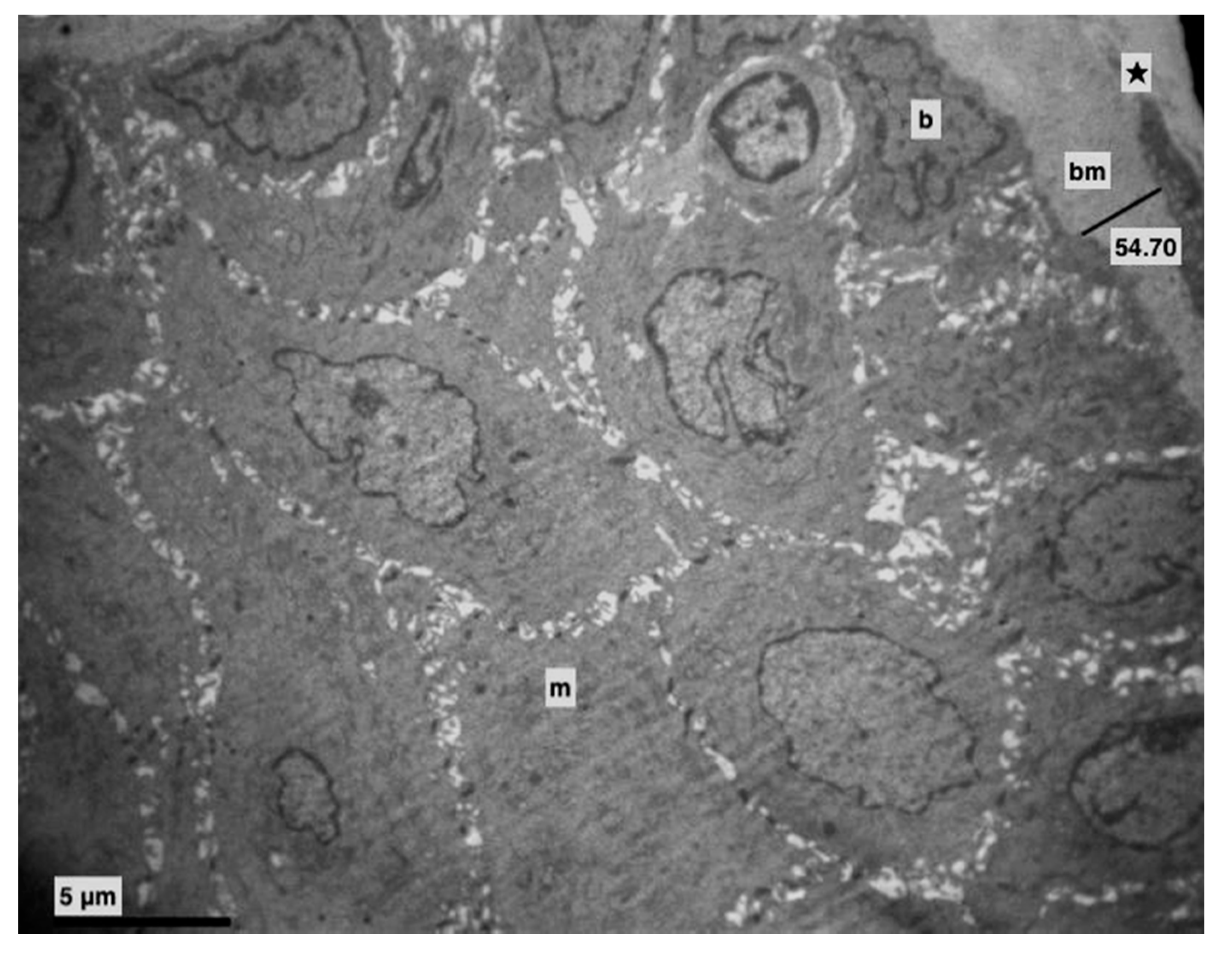
Abstract
1. Introduction
2. Materials and Methods
Transmission Electron Microscopy (TEM)
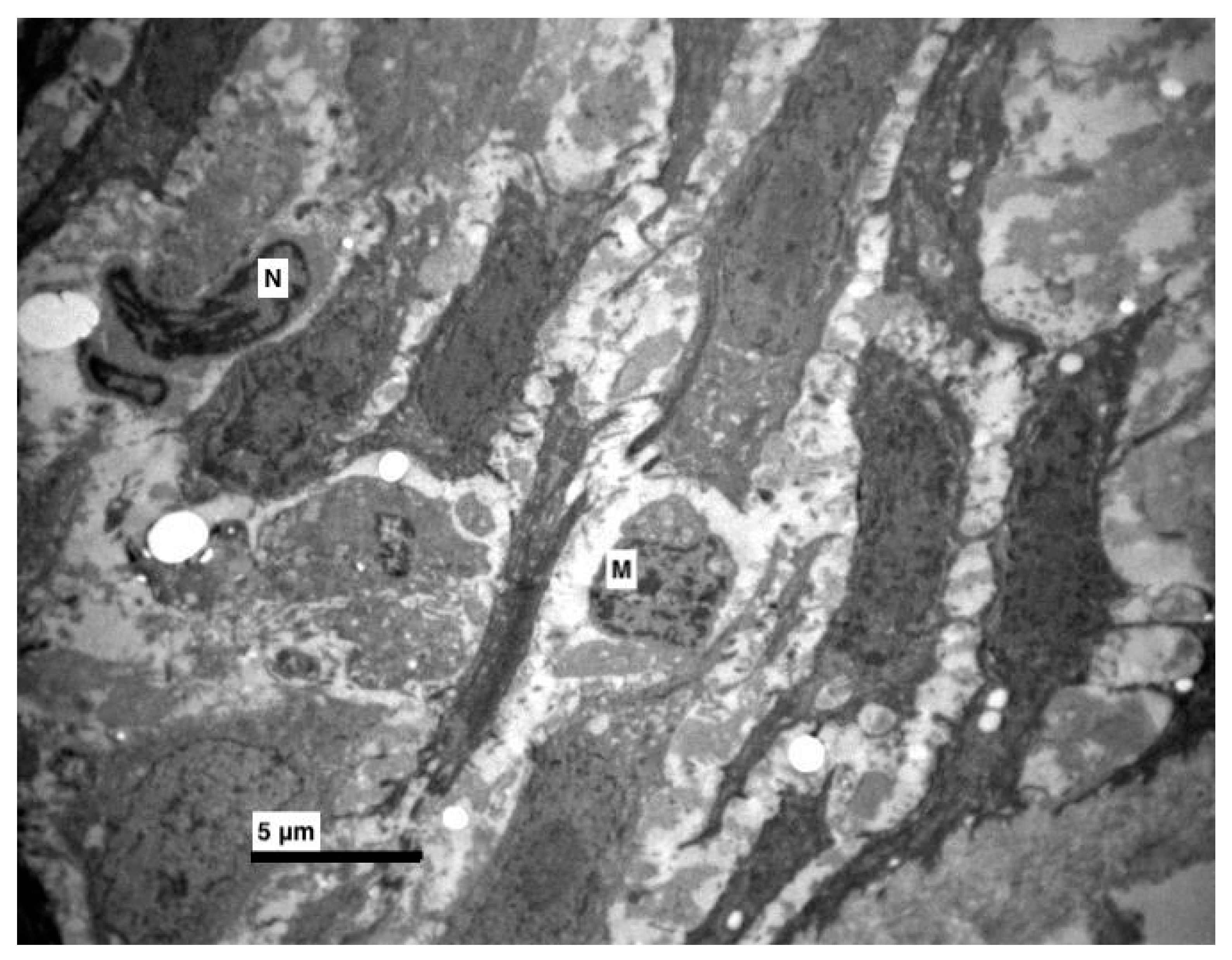
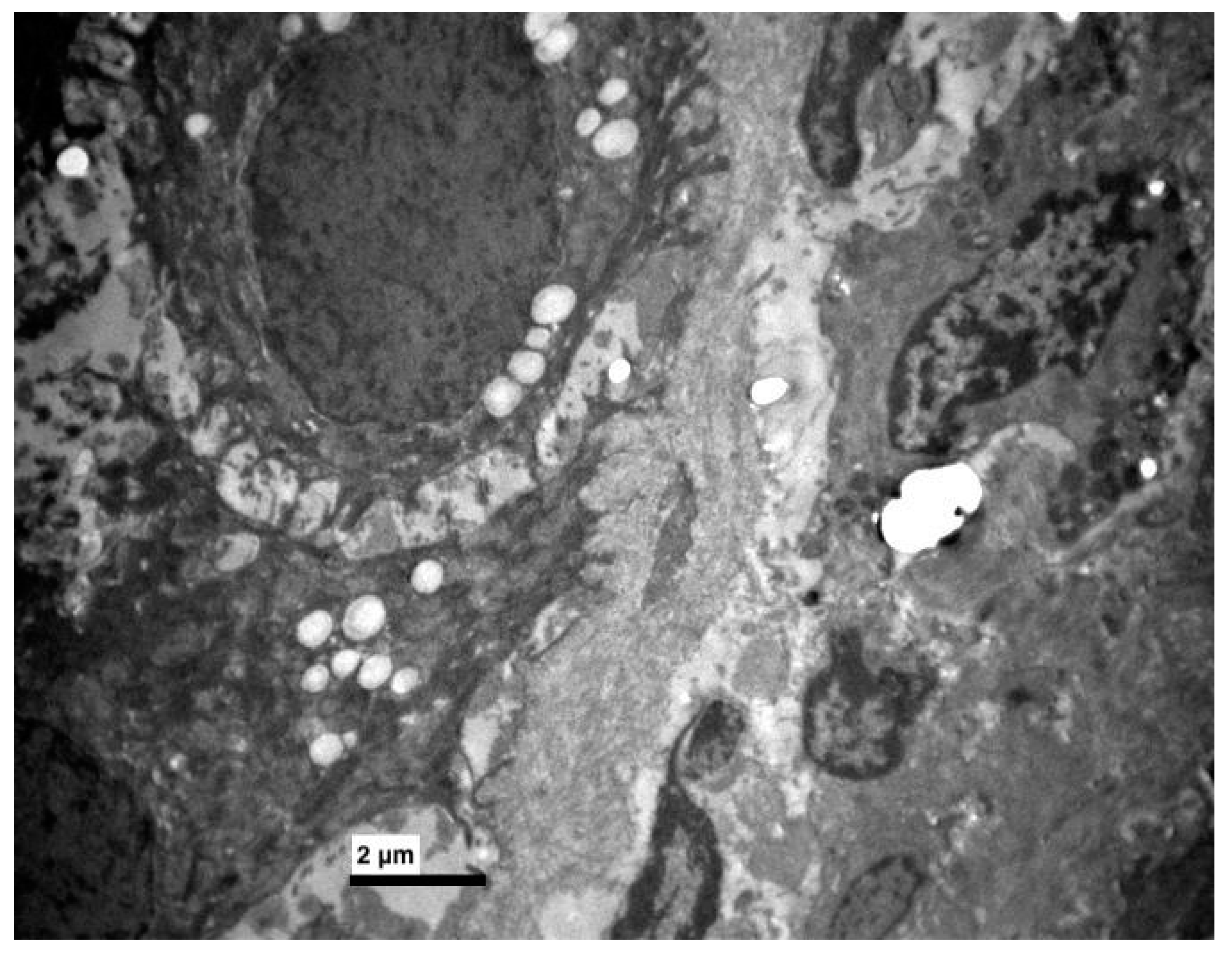
3. Results
4. Discussion
5. Conclusions
Author Contributions
Funding
Conflicts of Interest
References
- Serrano, A.J.; Begona, L.; Anitua, E.; Cobos, R.; Orive, G. Systematic review and meta-analysis of the efficacy and safety of alendronate and zoledronate for the treatment of postmenopausal osteoporosis. Gynecol. Endocrinol. 2013, 29, 1005–1014. [Google Scholar] [CrossRef] [PubMed]
- Lin, J.H.; Russell, G.; Gertz, B. Pharmacokinetics of alendronate: An overview. Int. J. Clin. Pract. 1999, 101, 18–26. [Google Scholar]
- Fernández, N.P.; Fresco, R.E.; Urizar, J.M.A. Bisphosphonates and oral pathology I. General and preventive aspects. Med. Oral Patol. Oral Cir. Bucal 2006, 11, 396–400. [Google Scholar]
- García-Font, M.; Curcó, N.; Prat, C.; Vives, P. Mouth Sores Caused by Alendronate Primary Cutaneous Cryptococcosis Presenting with a Sporotrichoid Pattern in a Cancer Patient. Blood 2009, 100, 77–83. [Google Scholar]
- Jeal, W.; Barradell, L.B.; McTavish, D. Alendronate. A review of its pharmacological properties and therapeutic efficacy in postmenopausal osteoporosis. Drugs 1997, 53, 415–434. [Google Scholar] [CrossRef]
- Sharpe, M.; Noble, S.; Spencer, C.M. Alendronate: An update of its use in osteoporosis. Drugs 2001, 61, 999–1039. [Google Scholar] [CrossRef]
- Papapetrou, P.D. Bisphosphonate-associated adverse events. Hormones 2009, 8, 96–110. [Google Scholar] [CrossRef]
- Cardwell, C.R.; Abnet, C.C.; Cantwell, M.M.; Murray, L.J. Exposure to oral bisphosphonates and risk of esophageal cancer. JAMA 2010, 304, 657–663. [Google Scholar] [CrossRef]
- Body, J.-J.; Bergmann, P.; Boonen, S.; Devogelaer, J.-P.; Gielen, E.; Goemaere, S.; Kaufman, J.-M.; Rozenberg, S.; Reginster, J.-Y. Extraskeletal benefits and risks of calcium, vitamin D and anti-osteoporosis medications. Osteoporos. Int. 2012, 23 (Suppl. 1), S1–S23. [Google Scholar] [CrossRef]
- Oh, Y.H.; Yoon, C.; Park, S.M. Bisphosphonate use and gastrointestinal tract cancer risk: Meta-analysis of observational studies. World J. Gastroenterol. 2012, 18, 5779–5788. [Google Scholar] [CrossRef]
- Wright, E.; Schofield, P.T.; Molokhia, M. Bisphosphonates and evidence for association with esophageal and gastric cancer: A systematic review and meta-analysis. BMJ Open 2015, 5, e007133. [Google Scholar] [CrossRef] [PubMed]
- Papamitsou, T.; Karachrysafi, S.; Toskas, A.; Dietrich, E.; Kostelidou, A.; Sioga, A. Bisphosphonate’s effect in hepatic rat cells: An electron microscopy study. Aristotle Univ. Med. J. 2016, 43, 7–11. [Google Scholar]
- Theodora, P.; Stella, F.; Angeliki, P.; Eva-Maria, D.; Dimitris, K.; Alexandros, T.; Sofia, K.; Antonia, S. Effect of Alendronic Acid on Buccal Mucosa. J. Dent. Oral Health 2018, 4, 0116. [Google Scholar]
- Pozzi, S.; Marcheselli, R.; Falorio, S.; Masini, L.; Stelitano, C.; Falcone, A.; Quarta, G.; Ponchio, L.; Pitini, V.V.; Luminari, S.; et al. Bisphosphonates-associated osteonecrosis of the jaw: A long-term follow-up of a series of 35 cases observed by GISL and evaluation of its frequency over time. Am. J. Hematol. 2009, 84, 850–852. [Google Scholar] [CrossRef]
- Isaacs, J.D.; Shidiak, L.; Harris, I.A.; Szomor, Z.L. Femoral Insufficiency Fractures Associated with Prolonged Bisphosphonate Therapy. Clin. Orthop. Relat. Res. 2010, 468, 3384–3392. [Google Scholar] [CrossRef]
- Dietrich, E.-M.; Theodora, P.; Antonia, S.; Georgios, K.; Esthelle, N. Ultrastructural alterations of the inferior alveolar nerve in wistar rats after alendronate administration per os: Hypothesis for the generation of the “numb chin syndrome. J. Histol. Histopathol. 2015, 2, 24. [Google Scholar] [CrossRef]
- Allgrove, J. Biphosphonates. Arch. Dis. Child. 1997, 76, 73–75. [Google Scholar] [CrossRef]
- Porras, A.G.; Holland, S.D.; Gertz, B.J. Pharmacokinetics of alendronate. Clin. Pharm. 1999, 36, 315–328. [Google Scholar] [CrossRef]
- Treister, N.S.; Richardson, P.; Schlossman, R.; Miller, K.; Woo, S.B. Painful tongue ulcerations in patients with bisphosphonate-associated osteonecrosis of the jaws. Oral Surg. Oral Med. Oral Pathol. Oral Radiol. Endodontol. 2008, 105, 8–11. [Google Scholar] [CrossRef]
- Ng, S.; Chow, L.; Zed, C. Oral erosive mucositis associated with improper administration of a drug. J. Can. Dent. Assoc. 2010, 76, a156. [Google Scholar]
- Kharazmi, M.; Sjöqvist, K.; Warfvinge, G. Oral ulcers, a little known adverse effect of alendronate: Review of the literature. J. Oral Maxillofac. Surg. 2012, 70, 830–836. [Google Scholar] [CrossRef] [PubMed]
- Donetti, E.; Gualerzi, A.; Sardella, A.; Lodi, G.; Carrassi, A.; Sforza, C. Alendronate impairs epithelial adhesion, differentiation and proliferation in human oral mucosa. Oral Dis. 2014, 20, 466–472. [Google Scholar] [CrossRef] [PubMed]
- Dhillon, S.; Lyseng-Williamson, K.A. Zoledronic acid: A review of its use in the management of bone metastases of malignancy. Drugs 2008, 68, 507–534. [Google Scholar] [CrossRef] [PubMed]
- Oliveira, T.C.; Bradaschia-Correa, V.; Castro, J.R.; Simoes, A.; Arana-Chavez, V.E. Ultrastructural and biochemical analysis of the effects of alendronate on salivary glands of young rats. Arch. Oral Biol. 2014, 59, 1307–1311. [Google Scholar] [CrossRef]
- Cruz, L.; Assumpcao, E.; Andrade, S.F.; Conrado, D.J.; Guterres, S.S.; Pohlmann, A.R. Microencapsulation of sodium alendronate reduces drug mucosal damage in rats. Drug Deliv. 2010, 17, 231–237. [Google Scholar] [CrossRef]
- Dik, E.A.; van Es, R.J.J.; Bergsma, J.E. A toxic reaction of the oral mucosa to alendronate (Fosamax). Ned. Tijdschr. Tandheelkd. 2010, 117, 387–390. (In Dutch) [Google Scholar] [CrossRef]
- Kharazmi, M.; Sjqvist, K.; Rizk, M.; Warfvinge, G. Oral ulcer associated with alendronate: A case report. Oral Surg. Oral Med. Oral Pathol. Oral Radiol. Endodontol. 2010, 110, e11–e13. [Google Scholar] [CrossRef]


© 2020 by the authors. Licensee MDPI, Basel, Switzerland. This article is an open access article distributed under the terms and conditions of the Creative Commons Attribution (CC BY) license (http://creativecommons.org/licenses/by/4.0/).
Share and Cite
Papamitsou, T.; Morsi-Yeroyannis, A.; Papanastasiou, A.; Bakalopoulos, N.; Dietrich, E.-M.; Karachrysafi, S.; Toskas, A.; Mareti, E.; Morsi-Yeroyanni, A.; Sioga, A. Bisphosphonate’s Effect on Tongue Mucosa: An Experimental Electron Microscopy Study. Medicina 2020, 56, 51. https://doi.org/10.3390/medicina56020051
Papamitsou T, Morsi-Yeroyannis A, Papanastasiou A, Bakalopoulos N, Dietrich E-M, Karachrysafi S, Toskas A, Mareti E, Morsi-Yeroyanni A, Sioga A. Bisphosphonate’s Effect on Tongue Mucosa: An Experimental Electron Microscopy Study. Medicina. 2020; 56(2):51. https://doi.org/10.3390/medicina56020051
Chicago/Turabian StylePapamitsou, Theodora, Antonios Morsi-Yeroyannis, Anastasios Papanastasiou, Nikolaos Bakalopoulos, Eva-Maria Dietrich, Sofia Karachrysafi, Alexandros Toskas, Evangelia Mareti, Anastasia Morsi-Yeroyanni, and Antonia Sioga. 2020. "Bisphosphonate’s Effect on Tongue Mucosa: An Experimental Electron Microscopy Study" Medicina 56, no. 2: 51. https://doi.org/10.3390/medicina56020051
APA StylePapamitsou, T., Morsi-Yeroyannis, A., Papanastasiou, A., Bakalopoulos, N., Dietrich, E.-M., Karachrysafi, S., Toskas, A., Mareti, E., Morsi-Yeroyanni, A., & Sioga, A. (2020). Bisphosphonate’s Effect on Tongue Mucosa: An Experimental Electron Microscopy Study. Medicina, 56(2), 51. https://doi.org/10.3390/medicina56020051