Ultrasound Assessment of Extracranial Carotids and Vertebral Arteries in Acute Cerebral Ischemia

 ,
,  ,
, 
Abstract
1. Introduction
- Assessment of ultrasound characteristics and risk of atherosclerotic plaques;
- Detection and evaluation of approximate degree of underlying stenoses;
- Diagnosis of occlusion of extracranial and (indirectly) of intracranial arteries;
- Referral of suitable patients for carotid endarterectomy or carotid artery stenting;
- Detection of other non-atherosclerotic lesions related to vessel wall pathology in extracranial and intracranial vessels (dissection, fibromuscular dysplasia, Takayasu disease, temporal arteritis, carotid-jugular fistula);
- Diagnosis of subclavian steal syndrome.
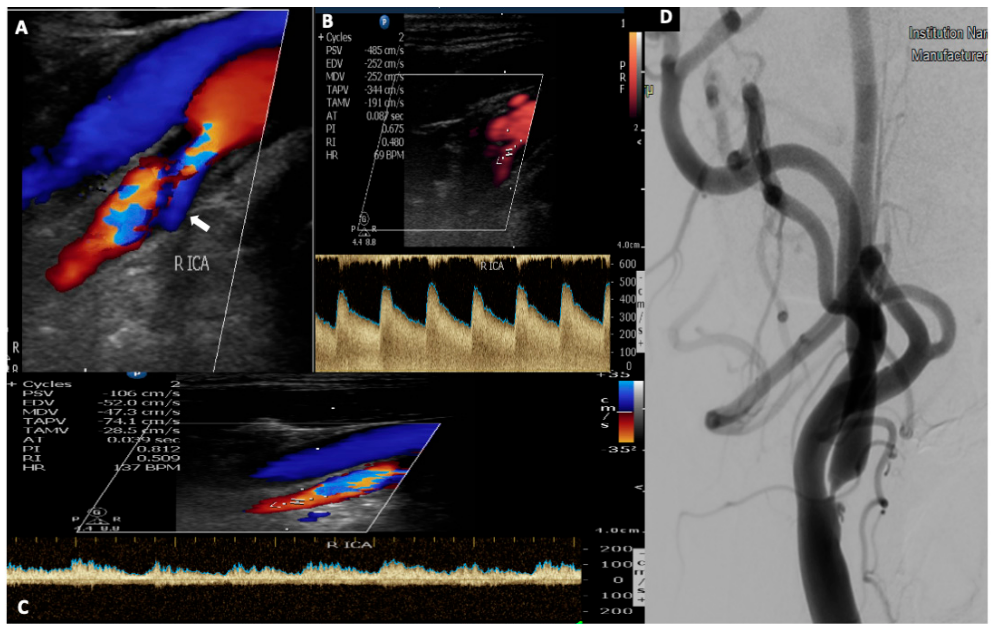
2. Carotid Stenosis
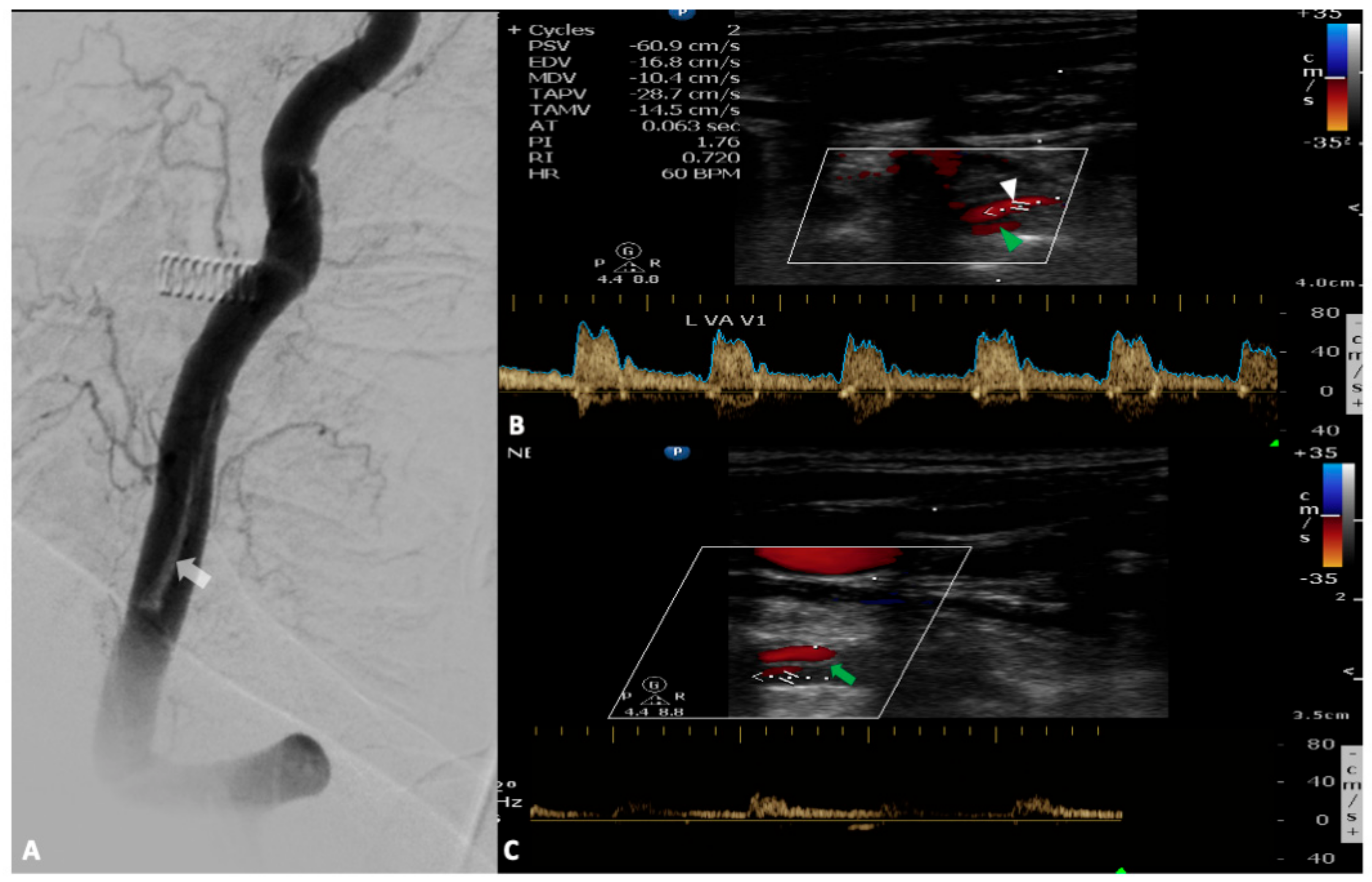
3. Vertebral Artery
4. Other Non-Atherosclerotic Vessel Pathologies
4.1. Dissection
4.2. Inflammation
4.3. Fibromuscular Dysplasia
5. Subclavian Steal Syndrome
6. Conclusions
Author Contributions
Funding
Conflicts of Interest
References
- Feigin, V.L.; Forouzanfar, M.H.; Krishnamurthi, R. For the Global Burden of Diseases, Injuries, and Risk Factors Study 2010 (GBD 2010) and the GBD Stroke Experts Group. Global and regional burden of stroke during 1990–2010: Findings from the Global Burden of Disease Study 2010. Lancet 2014, 383, 245–254. [Google Scholar] [CrossRef]
- Adams, H.P., Jr.; Bendixen, B.H.; Kappelle, L.J.; Biller, J.; Love, B.B.; Gordon, D.L.; Marsh, E.E., 3rd. Classification of subtype of acute ischemic stroke. Definitions for use in a multicenter clinical trial. TOAST. Trial of Org 10172 in Acute Stroke Treatment. Stroke 1993, 24, 35–41. [Google Scholar] [CrossRef]
- Hart, R.G.; Diener, H.C.; Coutts, S.B.; Easton, J.D.; Granger, C.B.; O’Donnell, M.J.; Sacco, R.L.; Connolly, S.J. Cryptogenic Stroke/ESUS International Working Group. Embolic strokes of undetermined source: The case for a new clinical construct. Lancet Neurol. 2014, 13, 429–438. [Google Scholar] [CrossRef]
- Ornello, R.; Degan, D.; Tiseo, C.; Di Carmine, C.; Perciballi, L.; Pistoia, F.; Carolei, A.; Sacco, S. Distribution and Temporal Trends From 1993 to 2015 of Ischemic Stroke Subtypes: A Systematic Review and Meta-Analysis. Stroke 2018, 49, 814–819. [Google Scholar] [CrossRef]
- Tsivgoulis, G.; Alexandrov, A.V. Ultrasound in Neurology. Continuum 2016, 22, 1655–1677. [Google Scholar] [CrossRef]
- Chernyshev, O.Y.; Garami, Z.; Calleja, S.; Song, J.; Campbell, M.S.; Noser, E.A.; Shaltoni, H.; Chen, C.I.; Iguchi, Y.; Grotta, J.C.; et al. Yield and accuracy of urgent combined carotid/transcranial ultrasound testing in acute cerebral ischemia. Stroke 2005, 36, 32–37. [Google Scholar] [CrossRef] [PubMed]
- Kargiotis, O.; Safouris, A.; Magoufis, G.; Georgala, M.; Roussopoulou, A.; Stamboulis, E.; Moulakakis, K.G.; Lazaris, A.; Geroulakos, G.; Vasdekis, S.; et al. The Role of Neurosonology in the Diagnosis and Management of Patients with Carotid Artery Disease: A Review. J. Neuroimaging 2018, 28, 239–251. [Google Scholar] [CrossRef] [PubMed]
- Touboul, P.J.; Hennerici, M.G.; Meairs, S.; Adams, H.; Amarenco, P.; Bornstein, N.; Csiba, L.; Desvarieux, M.; Ebrahim, S.; Hernandez, R.; et al. Mannheim carotid intima-media thickness and plaque consensus (2004-2006-2011). An update on behalf of the advisory board of the 3rd, 4th and 5th watching the risk symposia, at the 13th, 15th and 20th European Stroke Conferences, Mannheim, Germany, 2004, Brussels, Belgium, 2006, and Hamburg, Germany, 2011. Cerebrovasc. Dis. 2012, 34, 290–296. [Google Scholar] [PubMed]
- Touboul, P.J.; Elbaz, A.; Koller, C.; Lucas, C.; Adrai, V.; Chedru, F.; Amarenco, P. Common carotid artery intima-media thickness and brain infarction: The ‘Etude du Profil Génétique de l‘Infarctus Cérébral’ (GENIC) case-control study. The GENIC Investigators. Circulation 2000, 102, 313–318. [Google Scholar] [CrossRef] [PubMed]
- Lorenz, M.W.; von Kegler, S.; Steinmetz, H.; Markus, H.S.; Sitzer, M. Carotid intima-media thickening indicates a higher vascular risk across a wide age range: Prospective data from the Carotid Atherosclerosis Progression Study (CAPS). Stroke 2006, 37, 87–92. [Google Scholar] [CrossRef] [PubMed]
- Lorenz, M.W.; Markus, H.S.; Bots, M.L.; Rosvall, M.; Sitzer, M. Prediction of clinical cardiovascular events with carotid intima-media thickness: A systematic review and meta-analysis. Circulation 2007, 115, 459–467. [Google Scholar] [CrossRef] [PubMed]
- Tsivgoulis, G.; Vemmos, K.; Papamichael, C.; Spengos, K.; Manios, E.; Stamatelopoulos, K.; Vassilopoulos, D.; Zakopoulos, N. Common carotid artery intima-media thickness and the risk of stroke recurrence. Stroke 2006, 37, 1913–1916. [Google Scholar] [CrossRef] [PubMed]
- Roquer, J.; Segura, T.; Serena, J.; Cuadrado-Godia, E.; Blanco, M.; García-García, J.; Castillo, J. ARTICO Study.Value of carotid intima-media thickness and significant carotid stenosis as markers of stroke recurrence. Stroke 2011, 42, 3099–3104. [Google Scholar] [CrossRef] [PubMed][Green Version]
- Stary, H.C.; Chandler, A.B.; Dinsmore, R.E.; Fuster, V.; Glagov, S.; Insull, W., Jr.; Rosenfeld, M.E.; Schwartz, C.J.; Wagner, W.D.; Wissler, R.W. A definition of advanced types of atherosclerotic lesions and a histological classification of atherosclerosis. Circulation 1995, 92, 1355–1374. [Google Scholar] [CrossRef] [PubMed]
- Stary, H.C.; Chandler, A.B.; Glagov, S.; Guyton, J.R.; Insull, W., Jr.; Rosenfeld, M.E.; Schaffer, S.A.; Schwartz, C.J.; Wagner, W.D.; Wissler, R.W. A definition of initial, fatty streak, and intermediate lesions of atherosclerosis. A report from the Committee on Vascular Lesions of the Council on Arteriosclerosis, American Heart Association. Circulation 1994, 89, 2462–2478. [Google Scholar] [CrossRef] [PubMed]
- Marnane, M.; Prendeville, S.; McDonnell, C.; Noone, I.; Barry, M.; Crowe, M.; Mulligan, N.; Kelly, P.J. Plaque inflammation and unstable morphology are associated with early stroke recurrence in symptomatic carotid stenosis. Stroke 2014, 45, 801–806. [Google Scholar] [CrossRef]
- Geroulakos, G.; Ramaswami, G.; Nicolaides, A.; James, K.; Labropoulos, N.; Belcaro, G.; Holloway, M. Characterisation of symptomatic and asymptomatic carotid plaques using high-resolutionreal time ultrasonography. Br. J. Surg. 1993, 80, 1274–1277. [Google Scholar] [CrossRef]
- Gray-Weale, A.C.; Graham, J.C.; Burnett, J.R.; Byrne, K.; Lusby, R.J. Carotid artery atheroma: Comparison of preoperative B-mode ultrasound appearance with carotid endarterectomy specimen pathology. J. Cardiovasc. Surg. 1988, 29, 676–681. [Google Scholar]
- Jashari, F.; Ibrahimi, P.; Bajraktari, G.; Grönlund, C.; Wester, P.; Henein, M.Y. Carotid plaque echogenicity predicts cerebrovascular symptoms: A systematic review and meta-analysis. Eur. J. Neurol. 2016, 23, 1241–1247. [Google Scholar] [CrossRef]
- Tegos, T.J.; Sabetai, M.M.; Nicolaides, A.N.; Elatrozy, T.S.; Dhanjil, S.; Stevens, J.M. Patterns of brain computed tomography infarction and carotid plaque echogenicity. J. Vasc. Surg. 2001, 33, 334–339. [Google Scholar] [CrossRef]
- Salem, M.K.; Sayers, R.D.; Bown, M.J.; Eveson, D.J.; Robinson, T.G.; Naylor, A.R. Rapid access carotid endarterectomy can be performed in the hyperacute period without a significant increase in procedural risks. Eur. J. Vasc. Endovasc. Surg. 2011, 41, 222.e8. [Google Scholar] [CrossRef] [PubMed][Green Version]
- Kakkos, S.K.; Griffin, M.B.; Nicolaides, A.N.; Kyriacou, E.; Sabetai, M.M.; Tegos, T.; Makris, G.C.; Thomas, D.J.; Geroulakos, G. Asymptomatic Carotid Stenosis and Risk of Stroke (ACSRS) Study Group. The size of juxtaluminal hypoechoic area in ultrasound images of asymptomatic carotid plaques predicts the occurrence of stroke. J. Vasc. Surg. 2013, 57, 609–618.e1. [Google Scholar] [CrossRef] [PubMed]
- Nicolaides, A.N.; Kakkos, S.K.; Kyriacou, E.; Griffin, M.; Sabetai, M.; Thomas, D.J.; Tegos, T.; Geroulakos, G.; Labropoulos, N.; Doré, C.J.; et al. Asymptomatic internal carotid artery stenosis and cerebrovascular risk stratification. J. Vasc. Surg. 2010, 52, 1486–1496. [Google Scholar] [PubMed]
- Salem, M.K.; Bown, M.J.; Sayers, R.D.; West, K.; Moore, D.; Nicolaides, A.; Robinson, T.G.; Naylor, A.R. Identification of patients with a histologically unstable carotid plaque using ultrasonic plaque image analysis. J. Vasc. Endovasc. Surg. 2014, 48, 118–125. [Google Scholar] [CrossRef]
- Saba, L.; Saam, T.; Jäger, H.R.; Yuan, C.; Hatsukami, T.S.; Saloner, D.; Wasserman, B.A.; Bonati, L.H.; Wintermark, M. Imaging biomarkers of vulnerable carotid plaques for stroke risk prediction and their potential clinical implications. Lancet Neurol. 2019, 18, 559–572. [Google Scholar] [CrossRef]
- Prabhakaran, S.; Rundek, T.; Ramas, R.; Elkind, M.S.; Paik, M.C.; Boden-Albala, B.; Sacco, R.L. Carotid plaque calcification predicts incident stroke, myocardial infarction and vascular death: The Northern Manhattan Study. Stroke 2006, 37, 2696–2701. [Google Scholar] [CrossRef]
- De Bray, J.M.; Baud, J.M.; Dauzat, M. Consensus concerning the morphology and the risk of carotid plaques. Cerebrovasc. Dis. 1997, 7, 289–296. [Google Scholar] [CrossRef]
- Muraki, M.; Mikami, T.; Yoshimoto, T.; Fujimoto, S.; Tokuda, K.; Kaneko, S.; Kashiwaba, T. New criteria for the sonographic diagnosis of a plaque ulcer in the extracranial carotid artery. AJR Am. J. Roentgenol. 2012, 198, 1161–1166. [Google Scholar] [CrossRef]
- Comerota, A.J.; Katz, M.L.; White, J.V.; Grosh, J.D. The preoperative diagnosis of the ulcerated carotid atheroma. J. Vasc. Surg. 1990, 11, 505–510. [Google Scholar] [CrossRef]
- Fürst, H.; Hartl, W.H.; Jansen, I.; Liepsch, D.; Lauterjung, L.; Schildberg, F.W. Color-flow Doppler sonography in the identification of ulcerative plaques in patients with high-grade carotid artery steno-sis. AJNR Am. J. Neuroradiol. 1992, 13, 1581–1587. [Google Scholar]
- Rafailidis, V.; Chryssogonidis, I.; Xerras, C.; Nikolaou, I.; Tegos, T.; Kouskouras, K.; Rafailidis, D.; Charitanti-Kouridou, A. A comparative study of color Doppler imaging and contrast-enhanced ultrasound for the detection of ulceration in patients with carotid atherosclerotic disease. Eur. Radiol. 2019, 29, 2137–2145. [Google Scholar] [CrossRef] [PubMed]
- Akkus, Z.; Hoogi, A.; Renaud, G.; van den Oord, S.C.; Kate, G.L.T.; Schinkel, A.F.; Adam, D.; de Jong, N.; van der Steen, A.F.; Bosch, J.G. New quantification methods for carotid intra-plaque neovascularization using contrast-enhanced ultrasound. Ultrasound Med. Biol. 2014, 40, 25–36. [Google Scholar] [CrossRef] [PubMed]
- Van den Oord, S.C.; Akkus, Z.; van Lennep, J.E.R.; Bosch, J.G.; van der Steen, A.F.; Sijbrands, E.J.; Schinkel, A.F. Assessment of subclinical atherosclerosis and intraplaque neovascularization using quantitative contrast-enhanced ultrasound in patients with familial hypercholesterolemia. Atherosclerosis 2013, 231, 107–113. [Google Scholar] [CrossRef] [PubMed]
- Huang, R.; Abdelmoneim, S.S.; Ball, C.A.; Nhola, L.F.; Farrell, A.M.; Feinstein, S.; Mulvagh, S.L. Detection of carotid atherosclerotic plaque neovascularization using contrast enhanced ultrasound: A systematic review and meta-analysis of diagnostic accuracy studies. J. Am. Soc. Echocardiogr. 2016, 29, 491–502. [Google Scholar] [CrossRef]
- Saba, L.; Yuan, C.; Hatsukami, T.S.; Balu, N.; Qiao, Y.; DeMarco, J.K.; Saam, T.; Moody, A.R.; Li, D.; Matouk, C.C.; et al. Vessel Wall Imaging Study Group of the American Society of Neuroradiology.Carotid Artery Wall Imaging: Perspective and Guidelines from the ASNR Vessel Wall Imaging Study Group and Expert Consensus Recommendations of the American Society of Neuroradiology. AJNR 2018, 39, E9–E31. [Google Scholar] [CrossRef]
- Brinjikji, W.; Rabinstein, A.A.; Lanzino, G.; Murad, M.H.; Williamson, E.E.; DeMarco, J.K.; Huston, J., 3rd. Ultrasound characteristics of symptomatic carotid plaques: A systematic review and meta-analysis. Cerebrovasc. Dis. 2015, 40, 165–174. [Google Scholar] [CrossRef]
- Bhatti, A.F.; Leon, L.R., Jr.; Labropoulos, N.; Rubinas, T.L.; Rodriguez, H.; Kalman, P.G.; Schneck, M.; Psalms, S.B.; Biller, J. Free-floating thrombus of the carotid artery: Literature review and case reports. J. Vasc. Surg. 2007, 45, 199–205. [Google Scholar] [CrossRef]
- Purroy, F.; Montaner, J.; Molina, C.A.; Delgado, P.; Ribo, M.; Álvarez-Sabín, J. Patterns and predictors of early risk of recurrence after transient ischemic attack with respect to etiologic subtypes. Stroke 2007, 38, 3225–3229. [Google Scholar] [CrossRef]
- Tsivgoulis, G.; Krogias, C.; Georgiadis, G.S.; Mikulik, R.; Safouris, A.; Meves, S.H.; Voumvourakis, K.; Haršány, M.; Staffa, R.; Papageorgiou, S.G.; et al. Safety of early endarterectomy in patients with symptomatic carotid artery stenosis: An international multicenter study. Eur. J. Neurol. 2014, 21, 1251–1257. [Google Scholar] [CrossRef]
- Naylor, A.R.; Ricco, J.B.; de Borst, G.J.; Debus, S.; de Haro, J.; Halliday, A.; Hamilton, G.; Kakisis, J.; Kakkos, S.; Lepidi, S.; et al. European Society of Vascular Surgery guidelines on the management of atherosclerotic carotid and vertebral artery disease. Eur. J. Vasc. Endovasc. Surg. 2018, 55, 3e81. [Google Scholar]
- Kargiotis, O.; Tsivgoulis, G. The 2020 breakthroughs in early secondary prevention: Dual antiplatelet therapy versus single antiplatelet therapy. Curr. Opin. Neurol. 2020. [Google Scholar] [CrossRef]
- Wolff, T.; Guirguis-Blake, J.; Miller, T.; Gillespie, M.; Harris, R. Screening for carotid artery stenosis: An update of the evidence for the U.S. Preventive Services Task Force. Ann. Intern. Med. 2007, 147, 860–870. [Google Scholar] [CrossRef]
- Tahmasebpour, H.R.; Buckley, A.R.; Cooperberg, P.L.; Fix, C.H. Sonographic examination of the carotid arteries. Radiographics 2005, 25, 1561–1575. [Google Scholar] [CrossRef]
- Carpenter, J.P.; Lexa, F.J.; Davis, J.T. Determination of duplex Doppler ultrasound criteria appropriate to the North American Symptomatic Carotid Endarterectomy Trial. Stroke 1996, 27, 695–699. [Google Scholar] [CrossRef] [PubMed]
- AbuRahma, A.F.; Robinson, P.A.; Strickler, D.L.; Alberts, S.; Young, L. Proposed new duplex classification for threshold stenoses used in various symptomatic and asymptomatic carotid endarterectomy trials. Ann. Vasc. Surg. 1998, 12, 349–358. [Google Scholar] [CrossRef] [PubMed]
- Grant, E.G.; Benson, C.B.; Moneta, G.L. Carotid artery stenosis: Gray-scale and Doppler US diagnosis–Society of Radiologists in Ultrasound Consensus Conference. Radiology 2003, 229, 340–346. [Google Scholar] [CrossRef]
- Oates, C.P.; Naylor, A.R.; Hartshorne, T.; Charles, S.M.; Fail, T.; Humphries, K.; Aslam, M.; Khodabakhsh, P. Joint recommendations for reporting carotid ultrasound investigations in the United Kingdom. Eur. J. Vasc. Endovasc. Surg. 2009, 37, 251–261. [Google Scholar] [CrossRef] [PubMed]
- Dhanjil, S.; Jameel, M.; Nicolaides, A.; Belcaro, G.; Williams, M.; Griffin, M.; Ramaswami, G. Ratio of peak systolic velocity of internal carotid to end diastolic velocity of common carotid: New duplex criteria for grading internal carotid stenosis. J. Vasc. Technol. 1997, 21, 237e40. [Google Scholar] [CrossRef]
- Lee, V.S.; Hertzberg, B.S.; Workman, M.J.; Smith, T.P.; Kliewer, M.A.; DeLong, D.M.; Caroll, B.A. Variability of Doppler US measurements along the common carotid artery: Effects on estimates of internal carotid arterial stenosis in patients with angiographically proved disease. Radiology 2000, 214, 387e92. [Google Scholar] [CrossRef]
- Tsivgoulis, G.; Katsanos, A.H.; Köhrmann, M.; Caso, V.; Lemmens, R.; Tsioufis, K.; Paraskevas, G.P.; Bornstein, N.M.; Schellinger, P.D.; Alexandrov, A.V.; et al. Embolic strokes of undetermined source: Theoretical construct or useful clinical tool? Ther. Adv. Neurol. Disord. 2019, 12, 1756286419851381. [Google Scholar] [CrossRef]
- Hart, R.G.; Sharma, M.; Mundl, H.; Kasner, S.E.; Bangdiwala, S.I.; Berkowitz, S.D.; Swaminathan, B.; Lavados, P.; Wang, Y.; Wang, Y.; et al. Rivaroxaban for stroke prevention after embolic stroke of undetermined source. N. Engl. J. Med. 2018, 378, 2191–2201. [Google Scholar] [CrossRef] [PubMed]
- Diener, H.C.; Sacco, R.L.; Easton, J.D.; Granger, C.B.; Bernstein, R.A.; Uchiyama, S.; Kreuzer, J.; Cronin, L.; Cotton, D.; Grauer, C.; et al. Dabigatran for prevention of stroke after embolic stroke of undetermined source. N. Engl. J. Med. 2019, 380, 1906–1917. [Google Scholar] [CrossRef] [PubMed]
- Ntaios, G.; Swaminathan, B.; Berkowitz, S.D.; Gagliardi, R.J.; Lang, W.; Siegler, J.E.; Lavados, P.; Mundl, H.; Bornstein, N.; Meseguer, E.; et al. Efficacy and safety of rivaroxaban versus aspirin in embolic stroke of undetermined source and carotid atherosclerosis. Stroke 2019, 50, 2477–2485. [Google Scholar] [CrossRef] [PubMed]
- Kamtchum-Tatuene, J.; Wilman, A.; Saqqur, M.; Shuaib, A.; Jickling, G.C. Carotid Plaque with High-Risk Features in Embolic Stroke of Undetermined Source: Systematic Review and Meta-Analysis. Stroke 2020, 51, 311–314. [Google Scholar] [CrossRef] [PubMed]
- Komatsu, T.; Iguchi, Y.; Arai, A.; Sakuta, K.; Sakai, K.; Terasawa, Y.; Mitsumura, H.; Matsushima, M. Large but Nonstenotic Carotid Artery Plaque in Patients with a History of Embolic Stroke of Undetermined Source. Stroke 2018, 49, 3054–3056. [Google Scholar] [CrossRef] [PubMed]
- Buon, R.; Guidolin, B.; Jaffre, A.; Lafuma, M.; Barbieux, M.; Nasr, N.; Larrue, V. Carotid Ultrasound for Assessment of Nonobstructive Carotid Atherosclerosis in Young Adults with Cryptogenic Stroke. J. Stroke Cerebrovasc. Dis. 2018, 27, 1212–1216. [Google Scholar]
- Goyal, M.; Singh, N.; Marko, M.; Hill, M.D.; Menon, B.K.; Demchuk, A.; Coutts, S.B.; Almekhlafi, M.A.; Ospel, J.M. Embolic stroke of undetermined source and symptomatic nonstenotic carotid disease. Stroke 2020, 51, 1321–1325. [Google Scholar] [CrossRef]
- Cloud, G.C.; Markus, H.S. Diagnosis and management of vertebral artery stenosis. Q. J. Med. 2003, 96, 27–34. [Google Scholar] [CrossRef]
- Trattnig, S.; Hubsch, P.; Schuster, H.; Polzleitner, D. Color-coded Doppler imaging of normal vertebral arteries. Stroke 1990, 21, 1222–1225. [Google Scholar] [CrossRef]
- Bartels, E.; Fuchs, H.H.; Flugel, K.A. Duplex ultrasonography of vertebral arteries: Examination, technique, normal values, and clinical applications. Angiology 1992, 43, 169Y180. [Google Scholar] [CrossRef]
- Rozeman, A.D.; Hund, H.; Westein, M.; Wermer, M.J.H.; Lycklama, À.; Nijeholt, G.J.; Boiten, J.; Schimsheimer, R.J.; Algra, A. Duplex ultrasonography for the detection of vertebral artery stenosis: A comparison with CT angiography. Brain Behav. 2017, 7, e00750. [Google Scholar] [CrossRef] [PubMed]
- Khan, S.; Cloud, G.C.; Kerry, S.; Markus, H.S. Imaging of vertebral artery stenosis: A systematic review. J. Neurol. Neurosurg. Psychiatry 2007, 78, 1218–1225. [Google Scholar] [CrossRef] [PubMed]
- Hua, Y.; Meng, X.F.; Jia, L.Y.; Ling, C.; Miao, Z.R.; Ling, F.; Liu, J.B. Color Doppler imaging evaluation of proximal vertebral artery stenosis. AJR Am. J. Roentgenol. 2009, 193, 1434–1438. [Google Scholar] [CrossRef] [PubMed]
- De Bray, J.M.; Pasco, A.; Tranquart, F.; Papon, X.; Alecu, C.; Giraudeau, B.; Dubas, F.; Emile, J. Accuracy of color-Doppler in the quantification of proximal vertebral artery stenoses. Cereb. Dis. 2001, 11, 335–340. [Google Scholar] [CrossRef] [PubMed]
- Ackerstaff, R.G.; Hoeneveld, H.; Slowikowski, J.M.; Moll, F.L.; Eikelboom, B.C.; Ludwig, J.W. Ultrasonic duplex scanning in atherosclerotic disease of the innominate, subclavian and vertebral arteries. A comparative study with angiography. Ultrasound Med. Biol. 1984, 10, 409–418. [Google Scholar] [CrossRef]
- Buckenham, T.M.; Wright, I.A. Ultrasound of the extracranial vertebral artery. Br. J. Radiol. 2004, 77, 15–20. [Google Scholar] [CrossRef]
- Benninger, D.H.; Baumgartner, R.W. Ultrasound diagnosis of cervical artery dissection. Front Neurol. Neurosci. 2006, 21, 70–84. [Google Scholar]
- De Bray, J.M.; Lhoste, P.; Dubas, F.; Emile, J.; Saumet, J.L. Ultrasonic features of extracranial carotid dissections: 47 cases studied by angiography. J. Ultrasound Med. 1994, 13, 659–664. [Google Scholar] [CrossRef]
- Benninger, D.H.; Georgiadis, D.; Gandjour, J.; Baumgartner, R.W. Accuracy of color duplex ultrasound diagnosis of spontaneous carotid dissection causing ischemia. Stroke 2006, 37, 377–381. [Google Scholar] [CrossRef]
- Nebelsieck, J.; Sengelhoff, C.; Nassenstein, I.; Maintz, D.; Kuhlenbäumer, G.; Nabavi, D.G.; Ringelstein, E.B.; Dittrich, R. Sensitivity of neurovascular ultrasound for the detection of spontaneous cervical artery dissection. J. Clin. Neurosci. 2009, 16, 79–82. [Google Scholar]
- Arnold, M.; Baumgartner, R.W.; Stapf, C.; Nedeltchev, K.; Buffon, F.; Benninger, D.; Georgiadis, D.; Sturzenegger, M.; Mattle, H.P.; Bousser, M.G. Ultrasound diagnosis of spontaneous carotid dissection with isolated Horner syndrome. Stroke 2008, 39, 82–86. [Google Scholar] [CrossRef] [PubMed]
- Farina, F.; Brunner, C.; Schreiber, S.J.; Palmieri, A.; Struhal, W.; Baracchini, C.; Vosko, M.R. Ultrasound examination of the pupil suggestive for carotid dissection. Neurology 2017, 89, 973–974. [Google Scholar] [CrossRef] [PubMed]
- Sturzenegger, M.; Mattle, H.P.; Rivoir, A.; Rihs, F.; Schmid, C. Ultrasound findings in spontaneous extracranial vertebral artery dissection. Stroke 1993, 24, 1910–1921. [Google Scholar] [CrossRef] [PubMed]
- Engelter, S.T.; Brandt, T.; Debette, S.; Caso, V.; Lichy, C.; Pezzini, A.; Abboud, S.; Bersano, A.; Dittrich, R.; Grond-Ginsbach, C.; et al. Antiplatelets versus anticoagulation in cervical artery dissection. Stroke 2007, 38, 2605–2611. [Google Scholar] [CrossRef]
- Steinke, W.; Rautenberg, W.; Schwartz, A.; Hennerici, M. Noninvasive monitoring of internal carotid artery dissection. Stroke 1994, 25, 998–1005. [Google Scholar] [CrossRef]
- Caso, V.; Paciaroni, M.; Corea, F.; Hamam, M.; Milia, P.; Pelliccioli, G.P.; Parnetti, L.; Gallai, V. Recanalization of cervical artery dissection: Influencing factors and role in neurological outcome. Cerebrovasc. Dis. 2004, 17, 93–97. [Google Scholar] [CrossRef]
- Sengelhoff, C.; Nebelsieck, J.; Nassenstein, I.; Maintz, D.; Nabavi, D.G.; Kuhlenbaeumer, G.; Ringelstein, E.B.; Dittrich, R. Neurosonographical follow-up in patients with spontaneous cervical artery dissection. Neurol. Res. 2008, 30, 687–689. [Google Scholar] [CrossRef]
- Tsivgoulis, G.; Vadikolias, K.; Heliopoulos, I.; Patousi, A.; Iordanidis, A.; Souftas, V.; Piperidou, C. Aortic arch dissection causing acute cerebral ischemia: An uncommon contraindication for intravenous thrombolysis. Circulation 2011, 124, 657–658. [Google Scholar] [CrossRef][Green Version]
- Cinar, I.; Wang, H.; Stone, J.R. Clinically isolated aortitis: Pitfalls, progress, and possibilities. Cardiovasc. Pathol. 2017, 29, 23–32. [Google Scholar] [CrossRef]
- Kargiotis, O.; Safouris, A.; Petrou, V.N.; Magoufis, G.; Stamboulis, E.; Tsivgoulis, G. Teaching NeuroImages: Giant cell arteritis presenting with acute ischemic strokes due to diffuse intracranial stenoses. Neurology 2017, 89, e190–e191. [Google Scholar] [CrossRef]
- Dejaco, C.; Ramiro, S.; Duftner, C.; Besson, F.L.; Bley, T.A.; Blockmans, D.; Brouwer, E.; Cimmino, M.A.; Clark, E.; Dasgupta, B.; et al. EULAR recommendations for the use of imaging in large vessel vasculitis in clinical practice. Ann. Rheum. Dis. 2018, 77, 636–643. [Google Scholar] [CrossRef] [PubMed]
- Tsivgoulis, G.; Heliopoulos, I.; Vadikolias, K.; Patousi, A.; Giatromanolaki, A.; Sivridis, E.; Piperidou, C. Teaching NeuroImages: Ultrasound findings in giant-cell arteritis. Neurology 2010, 75, e67–e68. [Google Scholar] [CrossRef] [PubMed]
- Monti, S.; Floris, A.; Ponte, C.; Schmidt, W.A.; Diamantopoulos, A.P.; Pereira, C.; Piper, J.; Luqmani, R. The use of ultrasound to assess giant cell arteritis: Review of the current evidence and practical guide for the rheumatologist. Rheumatology 2018, 57, 227–235. [Google Scholar] [CrossRef] [PubMed]
- Karassa, F.B.; Matsagas, M.I.; Schmidt, W.A.; Ioannidis, J.P. Meta-analysis: Test performance of ultrasonography for giant-cell arteritis. Ann. Intern. Med. 2005, 142, 359–369. [Google Scholar] [CrossRef] [PubMed]
- Arida, A.; Kyprianou, M.; Kanakis, M.; Sfikakis, P.P. The diagnostic value of ultrasonography-derived edema of the temporal artery wall in giant cell arteritis: A second meta-analysis. BMC Musculoskelet. Disord. 2010, 11, 44. [Google Scholar] [CrossRef]
- Ball, E.L.; Walsh, S.R.; Tang, T.Y.; Gohil, R.; Clarke, J.M. Role of ultrasonography in the diagnosis of temporal arteritis. Br. J. Surg. 2010, 97, 1765–1771. [Google Scholar] [CrossRef]
- De Miguel, E.; Beltran, L.M.; Monjo, I.; Deodati, F.; Schmidt, W.A.; Garcia-Puig, J. Atherosclerosis as a potential pitfall in the diagnosis of giant cell arteritis. Rheumatology 2018, 57, 318–321. [Google Scholar] [CrossRef]
- Czihal, M.; Schröttle, A.; Baustel, K.; Lottspeich, C.; Dechant, C.; Treitl, K.M.; Treitl, M.; Schulze-Koops, H.; Hoffmann, U. B-mode sonography wall thickness assessment of the temporal and axillary arteries for the diagnosis of giant cell arteritis: A cohort study. Clin. Exp. Rheumatol. 2017, 35, 128–133. [Google Scholar]
- Ponte, C.; Serafim, A.S.; Monti, S.; Fernandes, E.; Lee, E.; Singh, S.; Piper, J.; Hutchings, A.; McNally, E.; Diamantopoulos, A.P.; et al. Early variation of ultrasound halo sign with treatment and relation with clinical features in patients with giant cell arteritis. Rheumatology 2020, 59, 3717–3726. [Google Scholar] [CrossRef]
- Maeda, H.; Handa, N.; Matsumoto, M.; Hougaku, H.; Ogawa, S.; Oku, N.; Itoh, T.; Moriwaki, H.; Yoneda, S.; Kimura, K.; et al. Carotid lesion detected by B-mode ultrasonography in Takayasu’s arteritis: ‘Macaroni sign’ as an indicator of the disease. Ultrasound Med. Biol. 1991, 17, 695–701. [Google Scholar] [CrossRef]
- Tsivgoulis, G.; Heliopoulos, I.; Vadikolias, K.; Birbilis, T.; Piperidou, C. Subclavian steal syndrome secondary to Takayasu arteritis in a young female Caucasian patient. J. Neurol. Sci. 2010, 296, 110–111. [Google Scholar] [PubMed]
- Olin, J.W.; Sealove, B.A. Diagnosis, management, and future developments of fibromuscular dysplasia. J. Vasc. Surg. 2011, 53, 826–836.e1. [Google Scholar] [CrossRef] [PubMed]
- Persu, A.; Touzé, E.; Mousseaux, E.; Barral, X.; Joffre, F.; Plouin, P.F. Diagnosis and management of fibromuscular dysplasia: An expert consensus. Eur. J. Clin. Invest. 2012, 42, 338–347. [Google Scholar] [CrossRef] [PubMed]
- Schievink, W.I.; Bjornsson, J. Fibromuscular dysplasia of the internal carotid artery: A clinicopathological study. Clin. Neuropathol. 1996, 15, 2–6. [Google Scholar] [PubMed]
- Sethi, S.; Lau, J.; Erwin, P.; Gustavson, S.; Olin, J.W. The S curve: A novel morphological finding in the internal carotid artery in patients with fibromuscular dysplasia [abstract]. J. Am. Coll. Cardiol. 2012, 59, E2051. [Google Scholar] [CrossRef]
- Olin, J.W.; Gornik, H.L.; Bacharach, J.M.; Biller, J.; Fine, L.J.; Gray, B.H.; Gray, W.A.; Gupta, R.; Hamburg, N.M.; Katzen, B.T.; et al. Fibromuscular dysplasia: State of the science and critical unanswered questions: A scientific statement from the American Heart Association. Circulation 2014, 129, 1048–1078. [Google Scholar] [CrossRef]
- Bolen, M.A.; Brinza, E.; Renapurkar, R.D.; Kim, E.S.H.; Gornik, H.L. Screening CT Angiography of the Aorta, Visceral Branch Vessels, and Pelvic Arteries in Fibromuscular Dysplasia. JACC Cardiovasc. Imaging 2017, 10, 554–561. [Google Scholar] [CrossRef]
- Chehab, B.M.; Gupta, K. Contemporary diagnosis of carotid fibromuscular dysplasia: Role of power Doppler and a review of other diagnostic modalities. Rev. Cardiovasc. Med. 2013, 14, e136–e143. [Google Scholar]
- Kargiotis, O.; Siahos, S.; Safouris, A.; Feleskouras, A.; Magoufis, G.; Tsivgoulis, G. Subclavian Steal Syndrome with or without Arterial Stenosis: A Review. J. Neuroimaging 2016, 26, 473–480. [Google Scholar]
- Labropoulos, N.; Nandivada, P.; Bekelis, K. Prevalence and impact of the subclavian steal syndrome. Ann. Surg. 2010, 252, 166–170. [Google Scholar] [CrossRef]
- Jung, K.H.; Kim, J.M.; Lee, S.T.; Chu, K.; Roh, J.K. Brain response characteristics associated with subclavian steal phenomenon. J. Stroke Cerebrovasc. Dis. 2014, 23, e157–e161. [Google Scholar] [CrossRef] [PubMed]
- Potter, B.J.; Pinto, D.S. Subclavian steal syndrome. Circulation 2014, 129, 2320–2323. [Google Scholar] [CrossRef] [PubMed]
- Clark, C.E.; Taylor, R.S.; Shore, A.C.; Ukoumunne, O.C.; Campbell, J.L. Association of a difference in systolic blood pressure between arms with vascular disease and mortality: A systematic review and meta-analysis. Lancet 2012, 379, 905–914. [Google Scholar] [CrossRef]
- Alexandrov, A.V.; Sloan, M.A.; Tegeler, C.H.; Newell, D.N.; Lumsden, A.; Garami, Z.; Levy, C.R.; Wong, L.K.; Douville, C.; Kaps, M.; et al. American Society of Neuroimaging Practice Guidelines Committee.Practice standards for transcranial Doppler (TCD) ultrasound. Part II. Clinical indications and expected outcomes. J. Neuroimaging 2012, 22, 215–224. [Google Scholar] [CrossRef]
- Valdueza, J.M.; Schreiber, S.J.; Roehl, J.-E.; Connolly, F.; Klingebiel, R. Neurosonology & Neuroimaging of Stroke, 2nd ed.; Georg Thieme Verlag: Stuttgart, Germany, 2017; p. 137. [Google Scholar]
- Sharon, M.; Asinger, R.W.; Hodges, M. Reactive hyperemia for the clinical diagnosis of subclavian steal syndrome: Report of a case. Stroke 1981, 12, 369–371. [Google Scholar] [CrossRef]
- Kliewer, M.A.; Hertzberg, B.S.; Kim, D.H.; Bowie, J.D.; Courneya, D.L.; Carroll, B.A. Vertebral artery Doppler waveform changes indicating subclavian steal physiology. AJR Am. J. Roentgenol. 2000, 174, 815–819. [Google Scholar] [CrossRef]
- Johnsen, S.; Schreiber, S.J.; Connolly, F.; Schepelmann, K.; Valdueza, K.M. Pitfall of vertebral artery insonation: Bidirectional flow without subclavian artery pathology. Perspect. Med. 2012, 1, 449–451. [Google Scholar] [CrossRef][Green Version]


| A. Clinical Diagnosis of Anterior Circulation Ischemia |
| STEP 1: Transcranial Doppler |
|
| STEP 2: Carotid/Vertebral Duplex |
|
| B. Clinical Diagnosis of Cerebral Ischemia in the Posterior Circulation |
| STEP 1: Transcranial Doppler |
|
| STEP 2: Vertebral/Carotid Duplex Ultrasound |
|
| Percentage Stenosis (NASCET) | ICA Peak Systolic Velocity (cm/s) | Peak Systolic Velocity Ratio ICAPSV/CCAPSV | St Mary’s Ratio ICAPSV/CCAEDV |
|---|---|---|---|
| <50% | <125 | <2 | <8 |
| 50–59% | >125 | 2–4 | 8–10 |
| 60–69% | 11–13 | ||
| 70–79% | >230 | >4 | 14–21 |
| 80–89% | 22–29 | ||
| >90 but less than near occlusion | >400 | >5 | >30 |
| Near Occlusion | High, low-string flow | Variable | Variable |
| Occlusion | No flow | Not Applicable | Not Applicable |
| Percentage Stenosis | PSVorigin (cm/s) | Ratio PSVorigin/PSVIV | EDVorigin (cm/s) |
|---|---|---|---|
| <50% | ≥85 | ≥1.3 | ≥27 |
| 50–69% | ≥140 | ≥2.1 | ≥35 |
| 70–99% | ≥210 | ≥4.0 | ≥50 |
Publisher’s Note: MDPI stays neutral with regard to jurisdictional claims in published maps and institutional affiliations. |
© 2020 by the authors. Licensee MDPI, Basel, Switzerland. This article is an open access article distributed under the terms and conditions of the Creative Commons Attribution (CC BY) license (http://creativecommons.org/licenses/by/4.0/).
Share and Cite
Psychogios, K.; Magoufis, G.; Kargiotis, O.; Safouris, A.; Bakola, E.; Chondrogianni, M.; Zis, P.; Stamboulis, E.; Tsivgoulis, G. Ultrasound Assessment of Extracranial Carotids and Vertebral Arteries in Acute Cerebral Ischemia. Medicina 2020, 56, 711. https://doi.org/10.3390/medicina56120711
Psychogios K, Magoufis G, Kargiotis O, Safouris A, Bakola E, Chondrogianni M, Zis P, Stamboulis E, Tsivgoulis G. Ultrasound Assessment of Extracranial Carotids and Vertebral Arteries in Acute Cerebral Ischemia. Medicina. 2020; 56(12):711. https://doi.org/10.3390/medicina56120711
Chicago/Turabian StylePsychogios, Klearchos, Georgios Magoufis, Odysseas Kargiotis, Apostolos Safouris, Eleni Bakola, Maria Chondrogianni, Panagiotis Zis, Elefterios Stamboulis, and Georgios Tsivgoulis. 2020. "Ultrasound Assessment of Extracranial Carotids and Vertebral Arteries in Acute Cerebral Ischemia" Medicina 56, no. 12: 711. https://doi.org/10.3390/medicina56120711
APA StylePsychogios, K., Magoufis, G., Kargiotis, O., Safouris, A., Bakola, E., Chondrogianni, M., Zis, P., Stamboulis, E., & Tsivgoulis, G. (2020). Ultrasound Assessment of Extracranial Carotids and Vertebral Arteries in Acute Cerebral Ischemia. Medicina, 56(12), 711. https://doi.org/10.3390/medicina56120711