Article

Kaempferol Regresses Carcinogenesis through a Molecular Cross Talk Involved in Proliferation, Apoptosis and Inflammation on Human Cervical Cancer Cells, HeLa

1
School of Life Sciences, Manipal Academy of Higher Education-Dubai Campus, Dubai 345050, United Arab Emirates
2
Department of Life and Environmental Sciences, College of Natural and Health Science, Zayed University, Academic City, Dubai 19282, United Arab Emirates
3
Department of Medical Biotechnology, College of Applied Medical Sciences, Qassim University, Buraydah 52266, Saudi Arabia
4
Department of Biochemistry, Faculty of Medicine and Health Science, UAE University, Abu Dhabi 51133, United Arab Emirates
5
School of Pharmacy, Al-Karim University, Katihar 854106, India
6
Nano Drug Delivery, School of Pharmacy, Raleigh-Durham, NC 27705, USA
*
Author to whom correspondence should be addressed.
Appl. Sci. 2022, 12(6), 3155; https://doi.org/10.3390/app12063155
Submission received: 17 January 2022 / Revised: 8 February 2022 / Accepted: 10 March 2022 / Published: 19 March 2022

Abstract

:
Kaempferol, a flavonoid, contains a plethora of therapeutic properties and has demonstrated its efficacy against cancer. This study aims to unravel the molecular targets that are being modulated by kaempferol on HeLa cells. Various assays were performed, namely: MTT assay, flow cytometry to analyze DNA content and quantitate apoptosis. Quantitative PCR and protein profiling were performed to evaluate the modulated manifestation of different genes involved in apoptosis, cell growth and inflammation. Kaempferol exhibited reduction in cell viability of HeLa cells (IC50 = 50 µM 48 h), whereas it did not show any significant effect on viability of the AC-16 cell line. Kaempferol-impacted apoptosis was definitive, as it induced DNA fragmentation, caused disruption of membrane potential, accumulation of cells in the G2-M phase and augmented early apoptosis. Consistently, kaempferol induced apoptosis in HeLa cells by modulating the expression of various genes at both transcript and protein levels. It upregulated the expression of pro-apoptotic genes, including APAF1, BAX, BAD, Caspases 3, and 9, etc., at the transcript level and Bad, Bax, p27, p53, p21, Caspases 3 and 8 etc. at the protein level, while it downregulated the expression of pro-survival gene BCL-2, BIRC8, MCL-1, XIAP, and NAIP at the transcript level and Bcl-2, XIAP, Livin, clap-2 at the protein level. Kaempferol attenuated oxidative stress by upregulating GSH activity and anti-inflammatory response by suppressing NF-kB pathways. Moreover, kaempferol averted rampant cell division and induced apoptosis by modulating AKT/MTOR and MAP kinase pathways. Hence, kaempferol can be considered as a natural therapeutic agent with a differential profile.

1. Introduction

Multistage and multifactorial carcinogenesis is an intricate route involving compromised apoptosis and unconstrained cell growth that causes tumor formation and may consequently lead to invasion and metastasis [1]. To overcome the toxicity associated with conventional treatment, novel therapeutics approaches, i.e., “chemoprevention” used by researchers, are non-cytotoxic and exhibit differential response and can inhibit cancer development at all stages [2,3]. Amidst all the chemopreventive agents, plant-derived polyphenols possess myriad anticancer properties by targeting and modulating the expression of different genes that consequently lead to inhibition of cell proliferation, resulting in cell cycle arrest and apoptosis, alleviated inflammation, etc. [3,4]. Polyphenols such as luteolin, chrysin, EGCG (Epigallatocatechin gallate) and quercetin have been reported to modulate PI3K/AKT/mTOR, MAPK/ P38, and WNT pathways and eventually deter cancer growth by halting cell cycle and induce cytotoxicity and apoptosis of the transformed cells [5,6,7].
Apoptosis is an intentional cell death mechanism which maintains homeostasis and is regulated by multiple signalling pathways. Cancer cells can escape cell death by progressing through cell cycle checkpoints and unregulated expression of pro-apoptotic and anti-apoptotic protein [8,9,10]. Apoptosis is mediated by two core pathways, the death receptor and the mitochondrial-mediated pathways. They congregate at the execution pathway that involves cleavage of caspase-3, which subsequently leads to nuclear fragmentation, and apoptotic body formation that eventually gets phagocytosed [11]. Polyphenols act as a novel therapeutic approach that induces apoptosis of transformed cells through activating pro-apoptotic proteins (BAD, BAX, BID, etc.) or repressing anti-apoptotic proteins (Bcl-2, Bcl-w)/ apoptotic inhibitors (IAFs) [11,12,13]. Polyphenols like EGCG, resveratrol and curcumin inhibit cell proliferation by downregulating IGF and Bcl-2 (anti-apoptotic) expression and upregulating the expression of Bax and bad (pro-apoptotic) proteins, etc. [9,14,15,16]. EGCG, resveratrol, curcumin, genistein, quercetin can also induce apoptosis by enhancing ROS and p53 expression in colon cancer [9,17].
Kaempferol (3,5,7-trihydroxy-2-[4-hydroxyphenyl]-4H-1-benzopyran-4-one) is a flavonol ubiquitously present in tea, cabbage, kale, strawberry, grapes, and tomatoes. Kaempferol exerts its anticancer effect by averting cell growth, arresting cell cycle, inducing apoptosis and attenuating oxidative stress and inflammation [18,19,20,21]. Studies have established that kaempferol shows cytotoxicity against HeLa cells and reduces their viability in a dose-dependent manner by downregulating PI3K/AKT and hTERT pathways [22,23], while it reduces viability of SiHa cells in both dose and time point mode and induces apoptosis by disrupting mitochondrial membrane potential [13]. Likewise, in breast cancer (MCF-7) cells, 25 μM of Kaempferol treatment suppresses metastasis, i.e., epithelial to mesenchymal transition by downregulating the expression E- and N- Cadherine, Slug, Snails, and MMPs (MMP2-9) [24]. Zhu et al. have reported that kaempferol can effectively reduce proliferation of TNBC (triple-negative BC), MDA-MB-231 cells by inhibiting G2/M transition and DNA damage by upregulating cleaved caspase-3, -9 and ϒH2AX compared to the control group [12]. Furthermore, the remarkable anti-inflammatory property of kaempferol was established both in vitro and in vivo conditions. Kaempferol inhibits the enzymes of iNOS and COX2, which consequently reduces the ROS level. In addition, it exhibits its protective effect by upregulating anti-oxidative enzymes that can scavenge the free radicals to reduce inflammation [18,19,25,26].
In this study, we intended to explore the anti-proliferative effect of kaempferol against HeLa (Human cervical cancer) and AC 16 cells, along with its other anticancer properties, such as apoptosis and inflammation, with an attempt to elucidate the molecular targets modulated by kaempferol to exert its anti-tumorigenic effect.

2. Materials and Methods

2.1. Cell Maintenance and Preparation of Kaempferol Solutions

HeLa cells (Human Cervical cancer) and AC 16 (Human cardiomyocyte) cells were cultured in DMEM (PANBIOTECH; Aidenbach, Germany) with 1% pen-strep (Sigma, St. Louis, MO, USA) complemented with 10% FBS (PANBIOTECH; Aidenbach, Germany) in 5% CO2 at a constant temperature of 37 °C. DMSO (dimethyl sulfoxide), PI (Propidium iodide), MTT (3-(4,5-dimethylthiazol-2-yl)-2, 5-diphenyl tetrazolium) and trypsin-EDTA were acquired from Sigma Chemical Co. (Sigma, St. Louis, MO, USA).
Kaempferol was procured from TCI (Tokyo Chemical Industries; Tokyo, Japan) (CAS:520–18–3 biosciences). A 1 mM standard solution of kaempferol was prepared in DMSO and stored at −20 °C. Dilutions were prepared in media with FBS ranging between 10 µM for treatment of cells.

2.2. Cell Viability Assay

The cytotoxicity of kaempferol on HeLa (Cervical cancer cell) and AC 16 (normal cell, Human cardiomyocyte) cells was assessed by MTT (thiazolyl blue tetrazolium bromide) assay. Both cell types were plated with ~1 × 10⁴ cells/well in 96 well plates and kept for attachment at 37 °C. The following day, the treatment started for 24 and 48 h with various concentrations of kaempferol from 1–100 µM in complete media; whereas AC-16 cells were treated using different concentrations of kaempferol between 1–200 µM over 24 h. Treatments for each concentration were done in triplicates. After adding MTT solution (5 mg/mL, Sigma) in each well, the plate was kept in dark for 3 h, consequently dissolving the formazan in DMSO. After 30 min, the O.D. value was measured at 570 nm by a microplate reader (BioTek, Winooski, VT, USA). Images were captured for both DMSO control and treated HeLa cells by an inverted microscope (Labomed, Los Angeles, CA, USA).

2.3. Analysis of Nuclear Morphology of Apoptotic Cells Using Propidium Iodide

The impact of kaempferol on treated HeLa cells’ nuclear morphology was evaluated through PI staining. ~2 × 10⁵ cells/well in a 24 well plate was treated for 24 and 48 h, followed by PBS wash. Then the cells were fixed with 70% ice-cold ethanol for 20 min, followed by staining with PI final (10 mg/mL in PBS) for 1–2 min. Excess stain was washed away by another PBS wash. Images were then taken using fluorescent microscope using Cell-F software (Olympus).

2.4. DNA Ladder Assay

Cultured cells in complete media were exposed to 0 μM (control), 30, 40 and 50 μM of kaempferol for 48 h. DNA isolation was done in suspension buffer as per the kit’s (Quick Apoptotic DNA Ladder Detection Kit by Ray biotech) protocol, followed by resolving the DNA in 1.2% agarose gel electrophoresis.

2.5. Cell Cycle Analysis

The DNA content of the treated HeLa cells was analysed by flow cytometry by a propidium iodide Flow Cytometry Kit (Abcam: ab139418, USA). About ~1 × 10⁶ cells/well were plated in a culture flask and were treated with 30 µM, 40 µM and 50 µM of kaempferol for 24 and 48 h. After the treatment, the cells were pooled and washed with PBS. To fix the cells, while vortexing the pellet, 70% alcohol was added dropwise then stored overnight at −20 °C. After staining, the cells were analysed by flow cytometer (FACS Calibur; BectonDickinson, Franklin Lakes, NJ, USA). FlowJo software was used to quantitate the cells in each phase.

2.6. Quantitation of Cell Apoptosis

Kaempferol-facilitated cell death was assessed through Flow cytometry using an Annexin V-FITC Apoptosis Detection kit from Abcam (ab 14085) following manufacturer’s protocol. About ~2 × 10⁵ cells/well were plated, followed by treatment with 30 μM, 40 μM, and 50 μM for 24 and 48 h. Then the cells were collected and centrifuged, followed by PBS wash to remove the media completely. Each sample was stained with 5 μL each of Annexin V-FITC conjugate and PI solution with binding buffer and kept in the dark at RT for 20 min. The sample was then analyzed to quantitate apoptosis by Flow cytometry (FACS Calibur; BectonDickinson, Franklin Lakes, NJ, USA). Despite the images representing a single experiment, it was repeated three times.

2.7. Detection of Mitochondrial Membrane Potential (∆ψm)

Alteration in ∆ψm (mitochondrial membrane potential) was detected by TMRE-Mitochondrial Potential assay kit from Abcam (ab113852, USA). About ~5 × 10³ cells/well in 96 well plates were treated with 20–50 µM of kaempferol for 48 h. After the treatment, TMRE dye was added in control and treated cells and kept at 37 °C for 20 min. The fluorescence reading was measured using a microplates spectrophotometer (Ex/Em is 549/575 nm). Fluorescence intensity was compared between treated and control cells by capturing images using a fluorescence microscope (Progress Fluorescent Microscope (Olympus, Tokyo, Japan) at 20×.

2.8. Gene Expression by TaqMan Apoptosis Array

RNA isolation was done from both control (untreated cells) and treated cells (30 µM and 50 µM) according to Gen Elute Mammalian Genomic Total RNA Kit’s procedure (Sigma, St. Louis, MO, USA). After a qualitative check, quantitation of RNA was done by Nanodrop (Nanodrop 2000c; Thermo Scientific™, MO, USA). Then, cDNA was synthesized with the help of a reverse transcription kit from Applied Biosystems™, USA. A pre-designed, ready-to-use TaqMan® quantitative gene Expression Array (Apoptosis Array 4414072 & oncogene array 4369514) was employed to evaluate cell cycle regulatory and apoptotic pathways’ gene expression in both treated and control cells. A mix of 10 μL of complementary DNA (100 ng) with 10 μL of master mix was added/well and the qPCR plate was run on a thermocycler (QuantiStudio3, Applied Biosystems, USA), and data was analyzed by DataAssistTM software through 2 −ΔΔCT method. The data was normalized using housekeeping gene 18S rRNA. Differential gene expression of the treated sample compared to control is shown through the RQ value displaying the fold change.

2.9. Quantitation of Apoptosis Related Protein

The protein expression of various genes associated with apoptosis was ascertained through RayBio® Human Apoptosis Arrays C1 (Cat. No. AAH-APO-1). Cell lysate was prepared from untreated control and cells treated with 30 µM and 50 µM kaempferol for 48 h. The experimental steps were conducted according to the manufacturer’s protocol. The lysate’s protein quantitation was performed by using Pierce™ BCA Protein quantitation kit (Thermo Fisher Scientific, Inc.; cat. no. 23225). Each membrane was incubated overnight in a cold room on a shaking surface with 500 μg of diluted cell lysate, followed by addition of biotinylated Antibody and HRP-Streptavidin for signal development. Consequently, a detection buffer was added and the developed signal’s measurement was done through a chemiluminescent detector gel doc system (Bio-Rad Laboratories, USA). Image Lab software was used for the quantitative analysis of the protein expression.

2.10. Detection of Caspase Multiplex Activity

The protease activity of caspases-3, -8, and -9 in HeLa cells after treatment was assessed using a caspase multiplex assay kit (fluorometric) from Abcam (ab219915), and the assay was performed as per the kit’s protocol. Intrinsic apoptosis pathway triggers the activation of Caspase-9, whereas extrinsic pathway activates Caspase-8. Both extrinsic and intrinsic pathways lead to activation of Caspase-3 (executioner). To determine the effect of kaempferol treatment on caspase activation, 1 × 10⁴ cells/well were seeded, followed by kaempferol treatment at 20, 30, 40 and 50 μM. Following treatment, respective caspase substrates were added (100 μL/well prepared in assay buffer) in each well and kept for 1 h in dark at RT. Fluorescence reading was measured at Ex/Em = 535/620 nm (Caspase 3), Ex/Em = 490/525 nm (Caspase 8) and Ex/Em = 370/450 nm (Caspase 9) to evaluate the alteration in caspase activity.

2.11. Quantitation of GSH Activity

To analyze the impact of kaempferol on oxidative stress in HeLa cells, GSH (colorimetric, Biovision Catalog #K261) assays were conducted as per the protocol. Both the treated and untreated cells supernatant was prepared in compliance with the protocol and was added to each well with reaction mix and substrate and kept at room temperature. Then O.D. value was measured at 405 nm and was followed by calculation of the GSH activity in fold change [19,27,28,29].

2.12. Quantitation of Inflammatory Cytokines Expression through Antibody Array

To establish the anti-inflammatory property of kaempferol, protein expression of DMSO control and treated HeLa cells (50 µM) was analyzed through inflammation antibody array (Abcam Cat no. ab134003). The experiment was done as per the protocol of the kit, which is similar to the protocol mentioned earlier under apoptosis proteome profiler [18,19,29,30,31,32,33,34].

2.13. Analysis of Phosphorylated Proteins Expression Pertaining to Various Signalling Pathways

To ascertain whether kaempferol alters phosphorylation level of different proteins that play a significant role in different cancer signalling pathways, Human Phosphorylation Pathway Profiling Array C55 (AAH-PPP-1-2, RAY BIOTEK) was employed. The modified expression of different phosphorylated proteins after treatment with kaempferol (50 µM) was compared to untreated control cells and the result was quantitated by image lab software version 6.0.1 by BIORAD.

2.14. Statistical Analysis

Two-way ANOVA was computed using GraphPad prism (version 9.3.1) software for comparisons, followed by Tukey’s post hoc test. The data are presented as the mean ± SD of three experiments, and the values with * p < 0.05 indicated significant differences.

3. Results

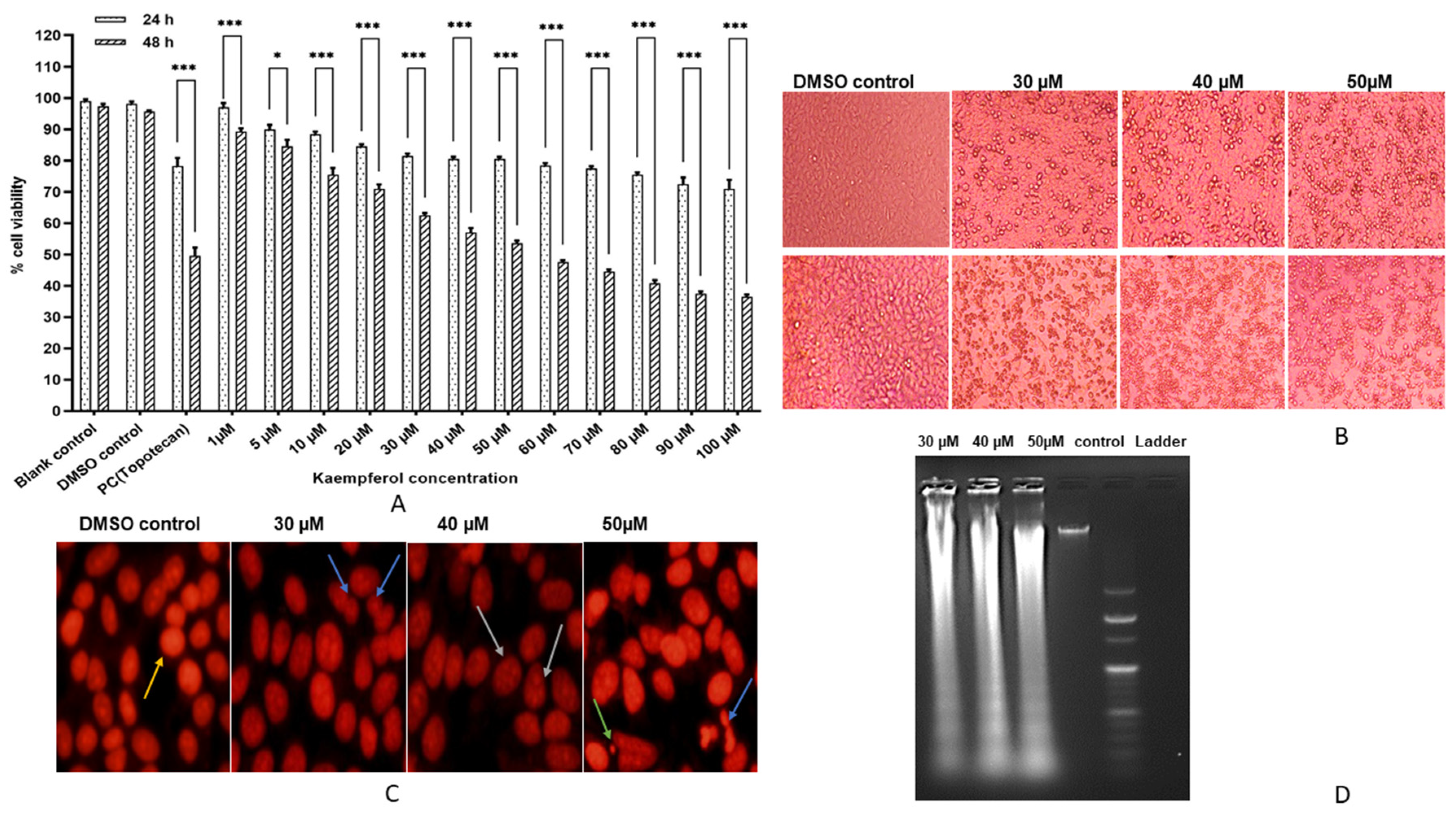
3.1. Kaempferol Inhibits HeLa Cell Proliferation

The cytotoxic effect of Kaempferol was assessed by MTT assay by exposing HeLa cells with different concentrations of Kaempferol (1–100 μM) for 24 and 48 h. It is evident from Figure 1A that kaempferol reduces cell viability in both dose- and time-dependent manner, which ranged from 96–73% at 24 h and 90–37% at 48 h. The IC50 value was estimated to be 50 μM at 48 h. The experiments were repeated at least thrice, and the average is shown in the given result (Figure 1A). On the contrary, kaempferol treatment (1–200 μM) exhibited an insignificant difference in the cell viability of cardiomyocyte (AC-16; normal cell line). Moreover, kaempferol (30, 40 and 50 µM) altered the appearance of treated HeLa cells compared to the control, which was assessed by an inverted microscope. The dead cells were rounded off, detached and floating, and their proportion increased with increasing dose of kaempferol (Figure 1B).

3.2. Kaempferol Mediates Nuclear Aberrations and DNA Fragmentation

Treated HeLa cells with kaempferol 30, 40, and 50 μM demonstrated fragmentation, nuclear blebbing, and apoptotic body formation, and the effect was found to be more pronounced with increasing concentration. In contrast, the control cell’s nucleus was intact and conspicuous. Moreover, DNA analysis of the kaempferol-treated cells (30, 40 and 50 μM) for 48 h demonstrated that kaempferol efficiently reduces the DNA integrity and consequently induced DNA ladder formation in treated cells in a dose-dependent manner, as evident via agarose gel electrophoresis, whereas the DNA of the control cells remained intact (Figure 1C,D).

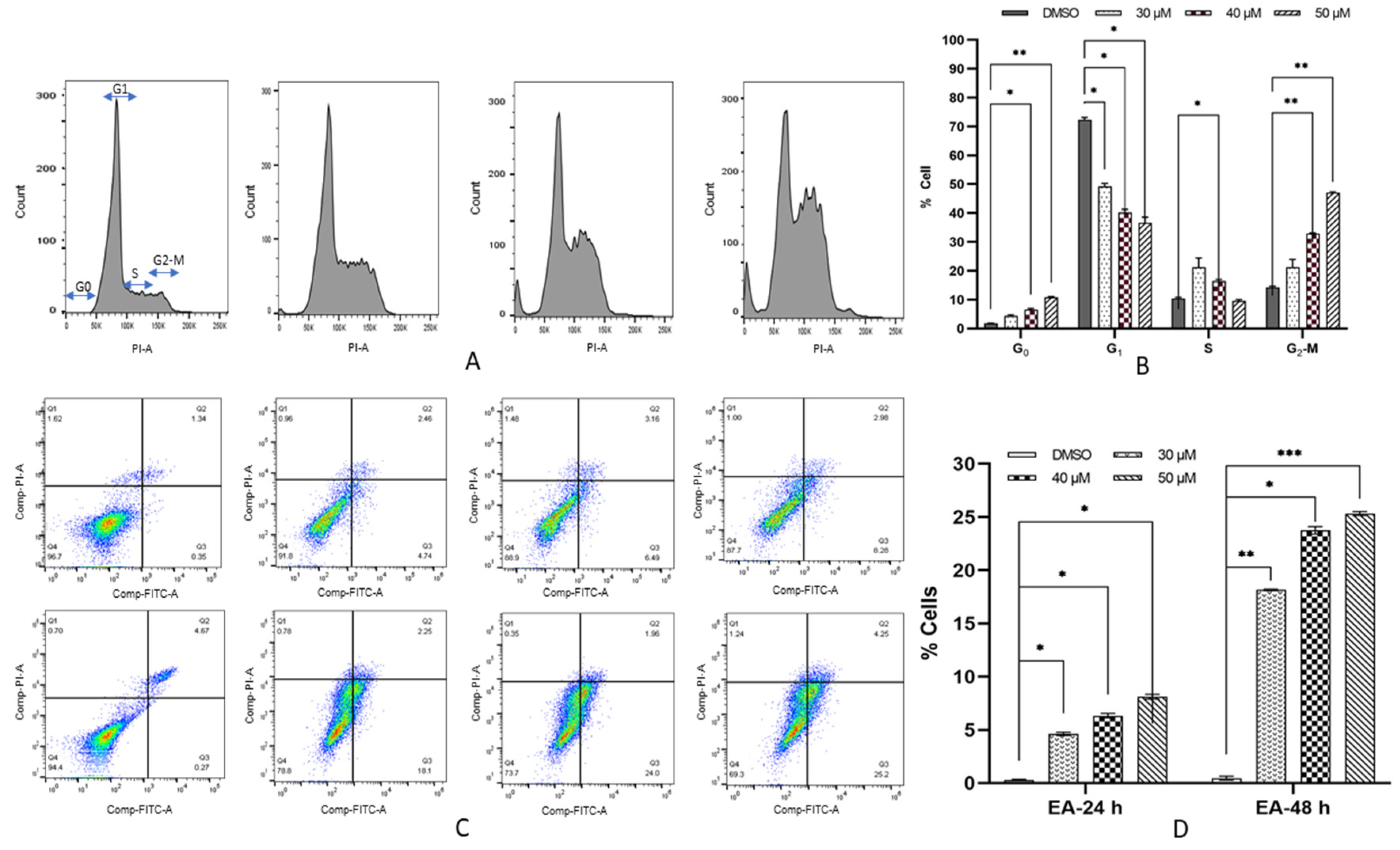
3.3. Kaempferol Induces G2/M Arrest in HeLa Cells

To determine whether Kaempferol affects HeLa cells’ proliferation by arresting the cell cycle, DNA content of different phases of the cell cycle was evaluated and compared with the untreated control. Kaempferol treatment halts the cell cycle at the G2/M phase; the cells’ population was raised in a dose-dependent manner from 19.4%, 32.8% to 46.7% at 30, 40 and 50 μM, respectively at 48 h, compared to 10.7% in the DMSO control. However, at 50 μM there was a concomitant increase in Sub-G1 population from 1.23% to 7.05% (Figure 2A,B).

3.4. Kaempferol Increases Early Apoptosis in HeLa Cells

To ascertain whether G2/M cell cycle arrest is accompanied by apoptosis induction by kaempferol, Annexin V/PI double staining was employed. Treated HeLa cells at 20, 30 and 50 μM for 48 h exhibited an increase in the proportion of early apoptotic cells with an insignificant change in the proportion of post apoptotic cells. In contrast, the percentage of live cells showed a decreasing trend. Early apoptotic cell population was found to be increased from 0.35% to 4.74%, 6.49% and 8.26% at 30, 40 and 50 μM in 24 h, whereas in 48 h treatment, it raised to 18.1%, 24% and 25.2%, respectively. Therefore, Annexin/PI staining clearly established the apoptosis-inducing property of kaempferol in HeLa cells (Figure 2C,D).

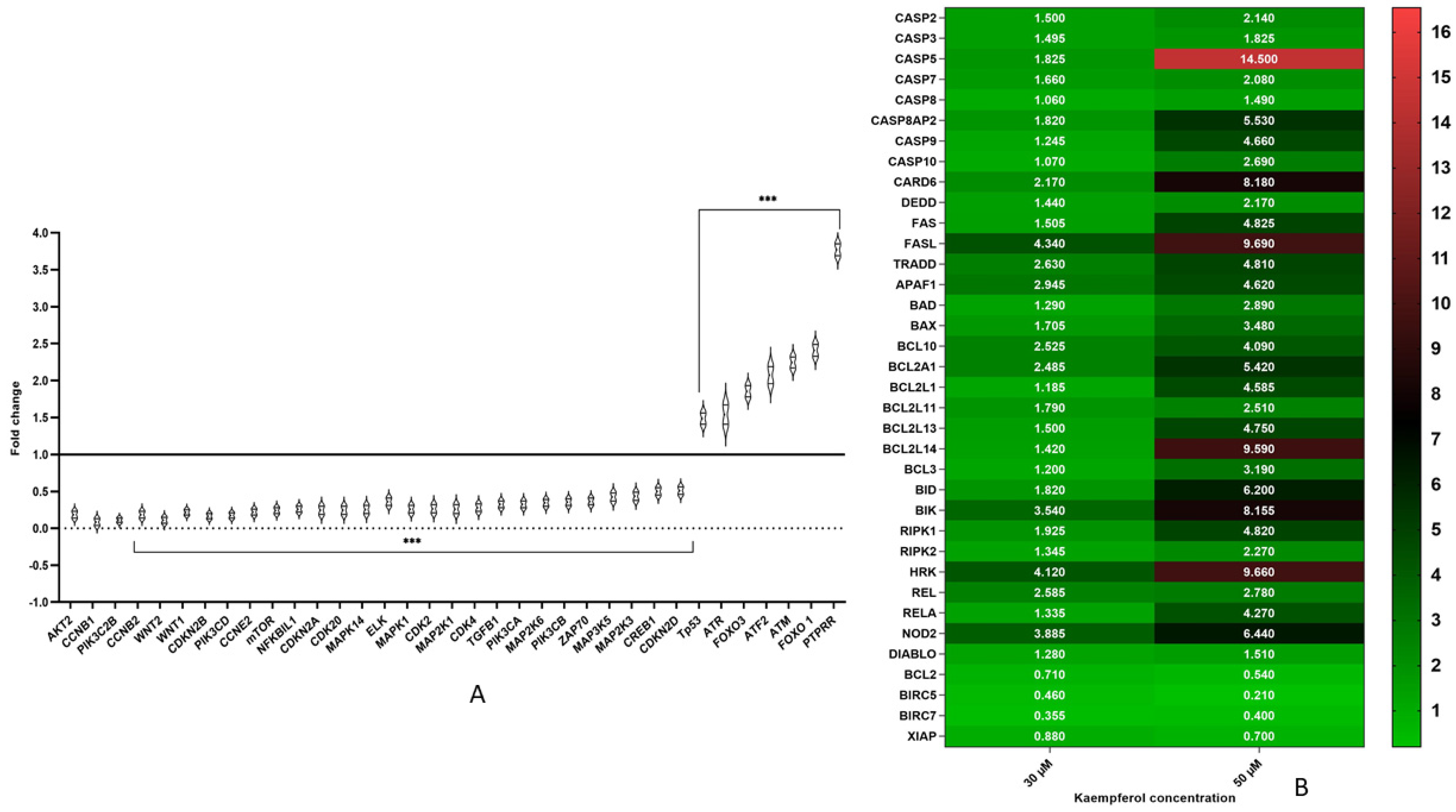
3.5. Kaempferol Modulates Expression of Various Genes Involved in Cell Cycle Regulation and Signalling Pathways

Overexpression of MAPK and AKT/mTOR leads to uncontrolled cell proliferation and survival, consequently causing transformation of cells. Kaempferol treatment (30 and 50 μM for 48 h) limits cancer cell proliferation by downregulating MAPK and AKT/mTOR pathways at the transcript level. Expression of various MAPK pathway genes was considerably downregulated, such as MAPK1, MAK14, MAP2K1, MAP2K3, MAP2K5, MAP2K6, MYC and ELK 1. Further, the transcript-level expression of PI3K/AKT/mTOR genes, such as AKT2, MTOR, PIK3C2B, PIK3CA, PIK3CB, PIK3CD, was also found to be significantly downregulated. However, expression of various tumor suppressor genes, such as PTPRR, ATM, ATR, FOXO 1, and FOXO 3, was upregulated.
Inhibition of G2-M progression through cell cycle checkpoints was further validated at the molecular level by analysing the expression of cell cycle regulator genes. Kaempferol treatment (50 μM) demonstrated a decreased expression of CCNB1, CCNB2, CCNE2, CDK2, CDKN2A, CDKN2B, and CDK4, which explains the G2-M arrest induced by kaempferol in comparison to control cells, whereas the expression of TP53, a TSG was significantly upregulated (Figure 3A).

3.6. Kaempferol Mediates Apoptosis Via Both Extrinsic and Intrinsic Pathways

Kaempferol-impacted cell death was validated by assessing the manifestation of various genes involved in apoptosis. HeLa cell treated with Kaempferol (30 and 50 µM) for 48 h demonstrated a surge in the pro-apoptotic transcripts’ expression, while anti-apoptotic transcripts exhibited a decline in its expression. The receptors and ligands involved in extrinsic pathways such as FAS, CARD6, DEDD and TRADD were upregulated, along with Caspases8Ap2, and a slight increase of Caspase 8 is indicative of extrinsic apoptosis. Pro-apoptotic genes of the BCL2 family, such as BCL10, BCL2A1, BCL2L1, BCL2L11, BCL2L13, BCL2L14, BCL3, BAD, BAX, BID, BIK, HRK, RIPK1, RIPK2 REL, RELA, DIABLO, and NOD2, were found to be significantly upregulated, whereas the anti-apoptotic genes such as of BCL2, BIRC5, BIRC7, and XIAP were found to be downregulated. Caspases such as caspase 2, caspase 3, caspase 5, caspase 7, caspase 9, and caspase 10 also exhibited upregulation along with APAF1 at the transcript level. Elevated caspase-8, caspase-7, caspase-3 and caspase-10 probably signifies an extrinsic pathway, whereas caspase 9 and caspase 3 with APAF1 suggests intrinsic apoptosis (Figure 3B).

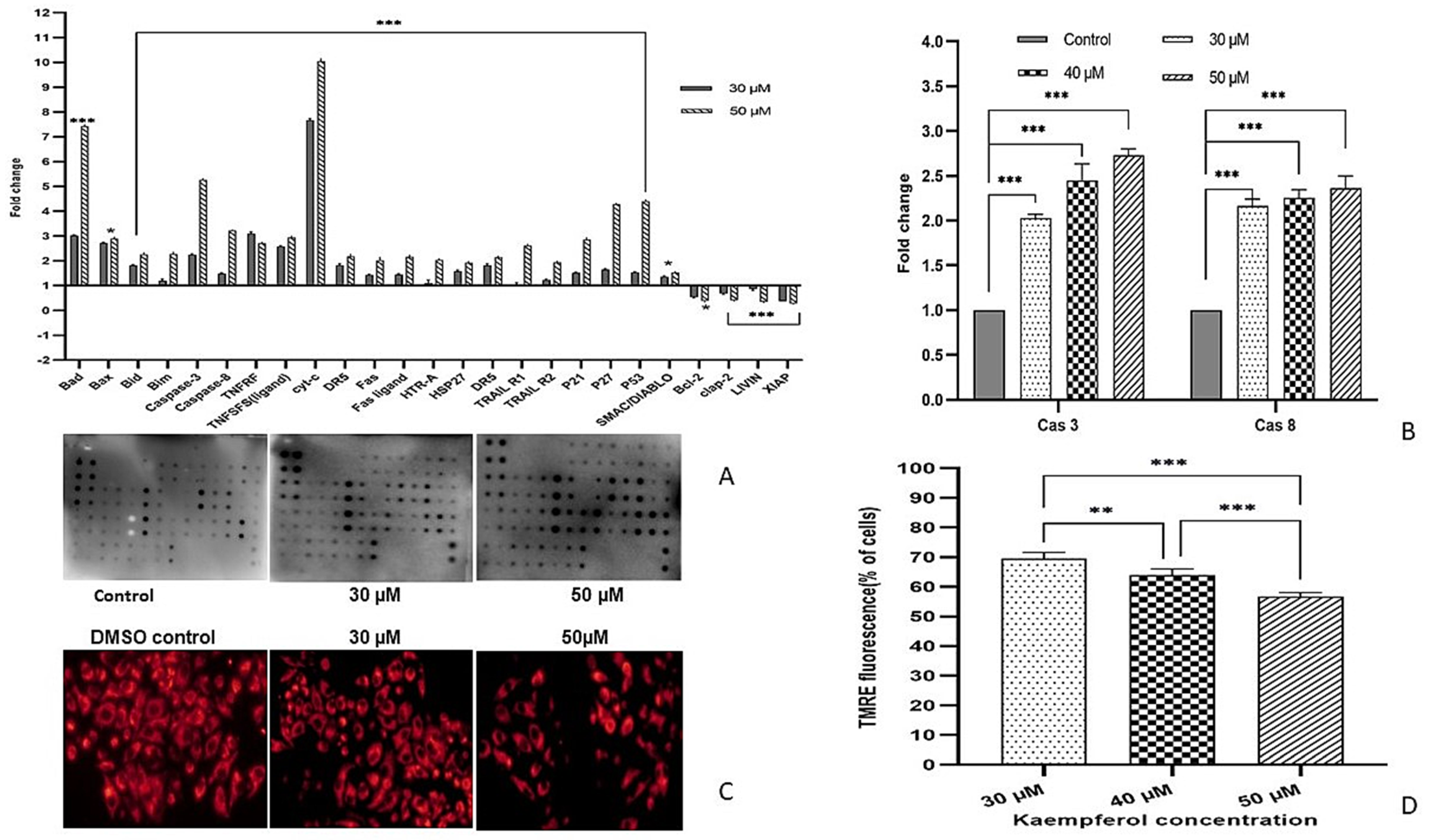
3.7. Modulation of Pro-Survival and Anti-Survival Proteins by Kaempferol

Kaempferol modulates the expression of both pro- and anti-apoptotic proteins in a dose-dependent manner, which corresponds with the result found at transcript level. The expression of pro-apoptotic proteins, such as Bad, Bid, Bim, p21, p53, p27 cyt-c, DR5 (TRAILR2), Fas, Fas ligand, HSP27, caspase-3, and caspase-8, were upregulated, whereas the anti-apoptotic proteins such as Bcl-2, clap-2, LIVIN, and XIAP expression were significantly decreased (Figure 4A).

3.8. Kaempferol Induces Apoptosis by Activating Caspase-3 and Caspase-8

Kaempferol-treated cells were analyzed for caspase-3, -8, and -9 expression by a fluorometric assay. It was determined that kaempferol-treated cells at 30, 40, and 50 µM for 48 h showed a relative dose-dependent elevation in caspase-3 activity by ≤2.5 folds and Caspase-8 activity increased to ≥3.5 folds; however, no significant change was observed in the activity of Caspase-9 (Figure 4B).

3.9. Kaempferol Reduces ∆ψm and Fluorescence Intensity

Alteration in mitochondrial membrane potential is one of the triggers for apoptotic induction. To analyze whether kaempferol induces mitochondrial dysfunction to release Cyt-c, the mitochondrial membrane potential of the DMSO control was compared with the kaempferol-treated cells. After staining the cells with TMRE for half an hour, observed red fluorescence intensity was found to decreased from 69%, 64% and 55% in a dose- dependent manner compared to untreated control at 30, 40 and 50 μM. (Figure 4C,D) [13].

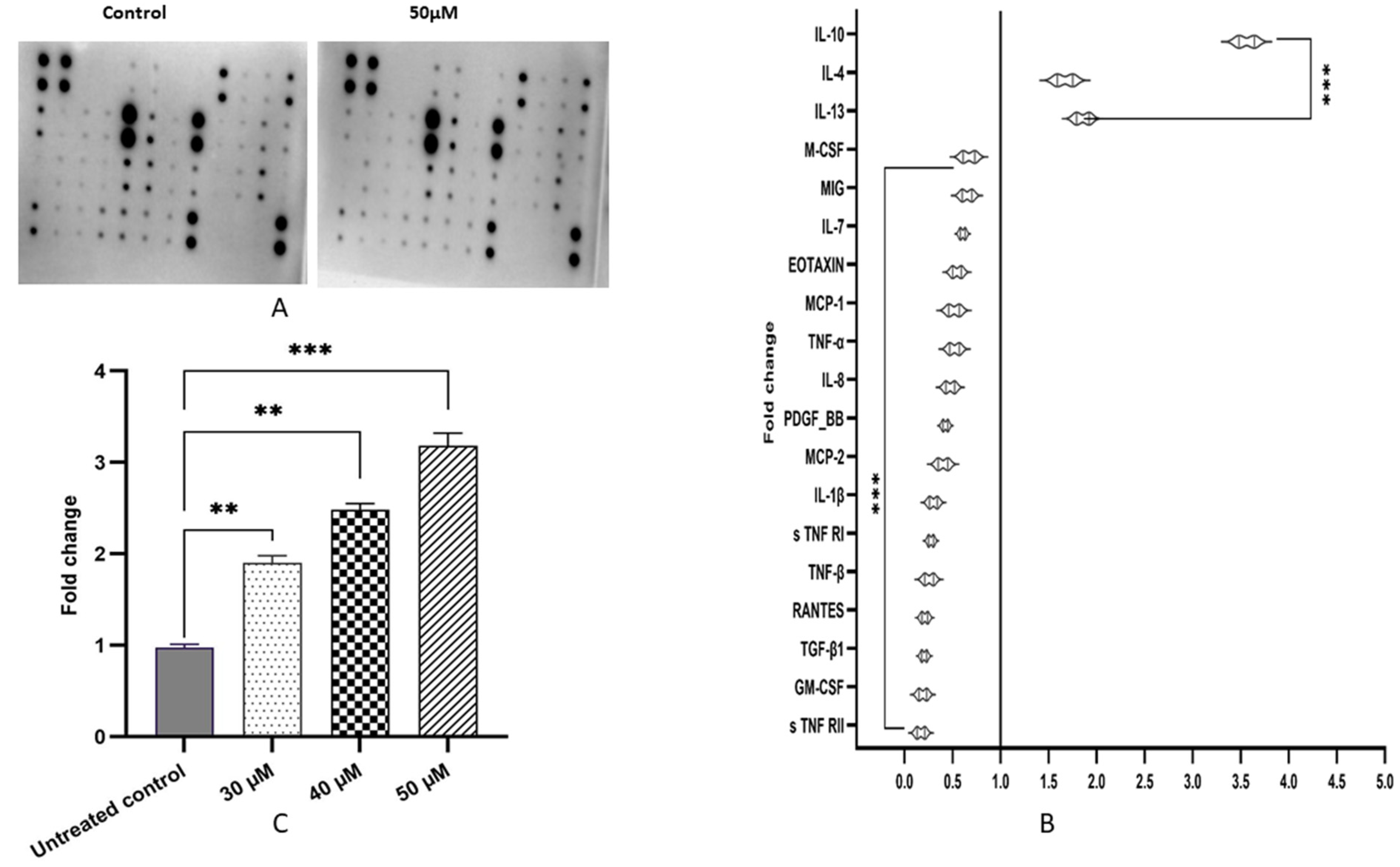
3.10. Kaempferol Ameliorates Inflammatory Response by Altering Pro-Inflammatory and Anti-Inflammatory Cytokines/Proteins

An indeterminate immune reaction owing to infection may lead to inflammation. Even though an inflammatory reaction is often self-limiting; however, unrestrained inflammation may drive the cells towards carcinogenesis. Kaempferol limited inflammation by suppressing the production of pro-inflammatory cytokines and chemokines, such as IL-7, IL-8, IL-16 MIG, M-CSF, that exhibited marginal alteration, whereas IL-1β, GM-CSF, TNF-α, TNF-β, s TNF RI, s TNF RII, RANTES, MCP-1, MCP-2 and EOTAXIN showed significant downregulation. On the contrary, anti-inflammatory cytokines such as IL-4, IL-10 and IL-13 were significantly upregulated by kaempferol treatment at 50 μM for 48 h (Figure 5A,B).

3.11. Kaempferol Upregulates GSH Activity

Research has confirmed that cervical cancer patients have significantly lower levels of GSH. An increase in GSH level will be able to restore the antioxidant defense system in cervical cancer patients. Kaempferol-exposed HeLa cells demonstrated an increase in GSH activity to 1.9, 2.4 to 3.1 fold at 30, 40 and 50 μM of drug at 48 h (Figure 5C).

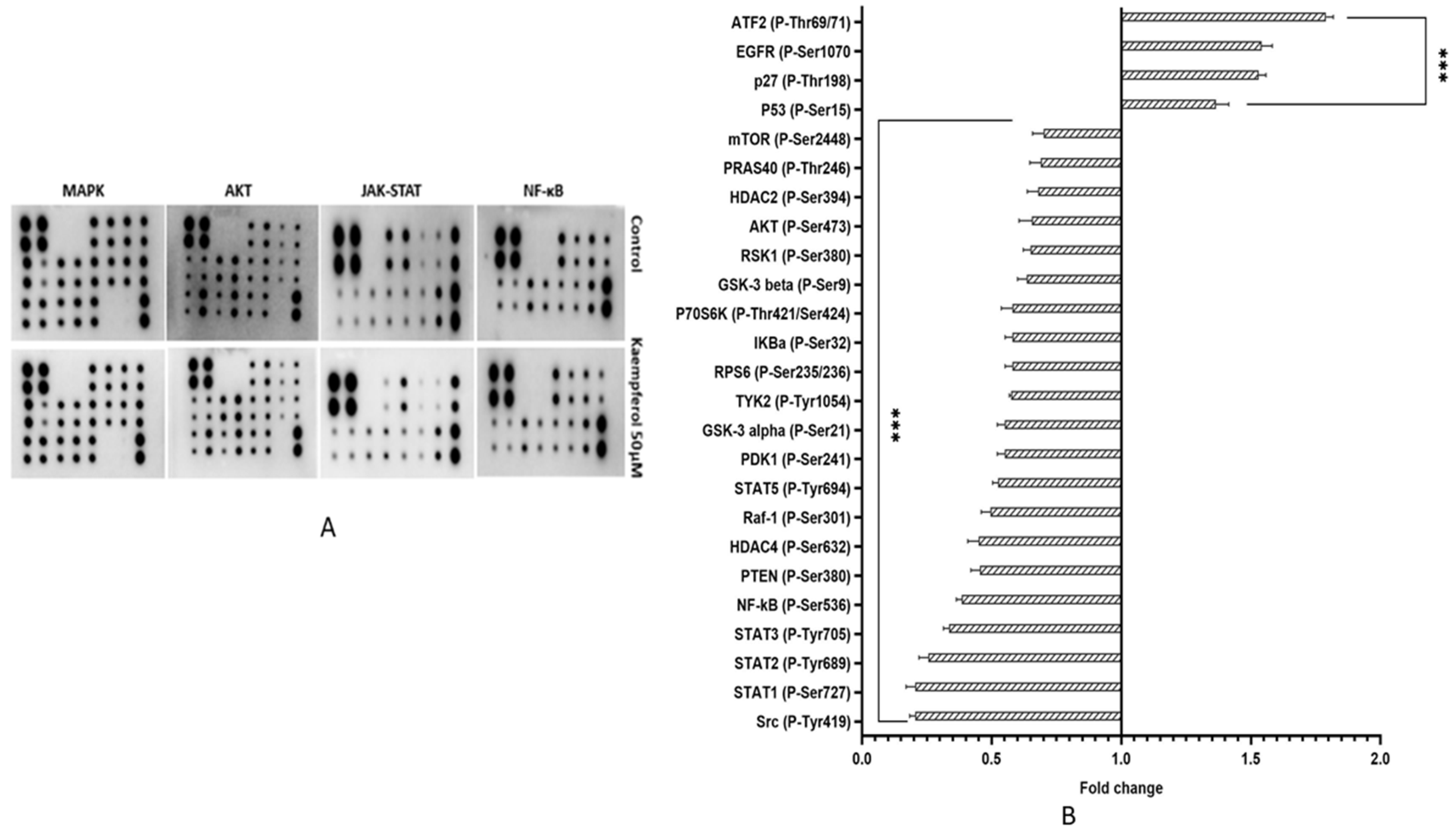
3.12. Kaempferol Suppresses Cell Growth, Survival and Inflammation by Regulating Aberrant MAPK, PI3K/AKT/mTOR, NF-kB and JAK-STAT Pathways

Kaempferol suppresses NF-kB and JAK-STAT pathways to reduce inflammation in HeLa cells. The phosphorylation level of IKBa (P-Ser32), NF-kB (P-Ser536), TBK1 (P-Ser172) and HDAC4 (P-Ser632) were reduced significantly, whereas HDAC2 (P-Ser394) TAK1 (P-Ser412) exhibited marginal reduction [18]. Expression of phosphorylated proteins of JAK-STAT pathways such as Src (P-Tyr419), STAT1 (P-Ser727), STAT2 (P-Tyr689), STAT3 (P-Tyr705), STAT5 (P-Tyr694), TYK2 (P-Tyr1054) was also reduced after treatment (50 µM) compared to control.
Consistently, at the protein level MAPK and AKT pathways were also suppressed. Expression of phosphorylated proteins pertaining to MAPK pathways, such as MEK (P-Ser217/221), HSP27 (P-Ser82), RSK1 (P-Ser380) and Raf-1 (P-Ser301), was reduced, whereas P53 (P-Ser15) was found to be upregulate. Likewise, the phosphorylated proteins of AKT pathway, such as GSK3a (p-ser21), GSK3b (p-ser9), MTOR (p-ser2448), PRAS 40 (p-Ther246), BAD (p-ser112), PTEN (p-ser380), AKT (p-ser473), RPS6 (P-Ser235/236), also exhibited reduced expression, whereas p27 (P-Thr198) expression was increased compared to the untreated control HeLa cells (Figure 6A,B).

4. Discussion

Despite tremendous progress in cancer treatment regimes, cancer mortality rate of is on the rise worldwide. This increasing rate of cancer mortality and morbidity has created the need to develop a treatment strategy that has fewer side effects and delivers specific responses. Chemoprevention through polyphenols from fruits and vegetables presents itself as an attractive candidate that can interfere with multiple cell signalling pathways to achieve therapeutic potential with a minimal cytotoxic profile and enhance the therapeutic index of the available treatment. Therefore, these agents can be used alone or combined with other therapeutics for better cancer management [2,3,4]. The current study extensively evaluated the anti-proliferative, apoptosis-inducing, anti-oxidative stress, and anti-inflammatory properties of kaempferol on HeLa cells through modulating various signalling pathways.
In the present study, cytotoxicity of kaempferol was confirmed through MTT assay and the IC50 was found to be of 50 µM at 48 h in HeLa cells; however, kaempferol did not demonstrate any significant difference in the viability of AC 16 cells (normal cell line) between treated and untreated control, which confirms the specific and safe profile of kaempferol (Figure 1A). Similarly, the IC50 value of kaempferol in different cell lines such as in SiHa the IC50 was reported to be 61.37 µg/mL, 48.6 µg/mL, and 27.06 µg/mL at 24, 48, and 72 h, respectively, in HeLa cells 45.63 and 22.87 µM at 24 and 48 h, and in SK-HEP-1 (hepatic cancer cells) the half inhibitory concentration was reported to be 100 µM in 24 h [4,22,23,35,36]. Light microscopic analysis confirmed the distinctive morphology of the treated cells as they were rounded off and moving free compared to the DMSO control cells. The extent of death was increased on both time and concentration-dependent measures (Figure 1B). PI staining confirmed the apoptotic morphology of the treated HeLa cells, such as membrane blebbing, chromatin condensation, apoptotic body, etc., that increased in a dose-dependent manner from 30–50 µM at 48 h. In contrast, the nucleus of the control cells was conspicuous with no significant alteration (Figure 1C). Kaempferol-mediated inter-nucleosomal degradation was further established through agarose gel electrophoresis, which exhibited DNA ladder formation with sharp bands in treated cells compared to untreated control (Figure 1D). Flavonoids such as quercetin, luteolin, fisetin, chrysin have shown similar results in HeLa cells [5,6,36,37,38,39,40].
The anti-proliferative property of kaempferol was illustrated by analyzing the cell cycle regulatory points through flow cytometry. This study demonstrated that kaempferol induces its anti-proliferative effect by G2/M cell cycle arrest, as there was a dose-dependent increase in the proportion of the cells at G2/M, which was accompanied by a slight increase in the sub-G1 population. At 48 h the population of cells being arrested was increased from 10.7 % in the DMSO control to 19.4 %, 32.8 % and 46.7 % at 30, 40, and 50 µM of kaempferol, respectively. Additionally, the proportion of cells in Sub-G1 was also elevated from 1.23 % to 6.86 % (Figure 2A,B). Several pieces of research have confirmed kaempferol-mediated G2/M arrest in different cell lines, such as MDA-MB-231 (Triple-negative breast cancer cell cline), HL-60 leukemia cells and SK-HEP-1 human hepatic cancer cells. This result is in agreement with the previously reported studies [4,12,35]. Apoptosis is accountable for maintaining homeostasis by regulating multiple biological processes, such as the disruption of mitochondrial membrane to release cytochrome-c, DNA laddering, chromatin condensation, and caspase cleavage, events that lead to deletion of unwanted cells. Kaempferol treatment showed an increasing trend in the early apoptotic cell population in a concentration-dependent manner that was found to be increased from 0.35% to 4.74%, 6.49% and 8.26% at 20, 30 and 50 μM at 24 h, whereas at 48 h treatment, it rose to 18.1%, 24% and 25.2%, respectively (Figure 2C,D). Therefore, Annexin/PI staining established the apoptosis-inducing property of kaempferol in HeLa cells. Consistent with this study, similar results have been reported earlier [12,13,41]. At the molecular level, kaempferol also modified cell cycle regulatory genes manifestation to avert the proliferation of transformed cells and induce cell cycle arrest. Kaempferol at 50 µM downregulates the expression of CCNB1, CCNB2, CCNE2, CDK2, CDKN2A, CDKN2B, and CDK4, which corresponds to G2-M arrest. The result is in concordance with the flow cytometry result, whereas the expression of various TSGs, such as TP53, ATM, ATR, PTPRR, FOXO1, FOXO3, was significantly upregulated. Activation of TP53, ATM, and ATR in response to DNA damage facilitates cell cycle arrest and apoptosis [42,43,44], along with FOXO1/3, which upregulates FasL and TRAIL (anti-survival factors) expression and induces apoptosis. In addition, PTPRR deters cell proliferation by inhibiting the MAP kinase pathway (Figure 3A) [4,6,12,22,37,41,45,46,47]. Zhu Wang et al. have reported that kaempferol ameliorates nephrotoxicity induced by cisplatin by altering the expression of various apoptotic, inflammatory, and anti-oxidant genes associated with cancer development and progression [22]. Respective modulation of these genes’ products via chemopreventive agents can be crucial to mitigate cancer growth and provide a safer treatment opportunity [16,17,19]. In this study, kaempferol treatment elicited increased expression of pro-apoptotic genes, such as BCL10, BCL2A1, BCL2L1, BCL2L11, BCL2L13, BCL2L14, BCL3, BAD, BAX, BID, BIK, HRK, RIPK1, RIPK2 REL, RELA, DIABLO, APAF1 and NOD2, whereas it decreased expression of the anti-apoptotic genes, such as BCL2, BIRC5, BIRC7, and XIAP [42,48,49]. The expression of various receptors associated with extrinsic pathways, such as FAS, CARD6, DEDD, TRADD, was upregulated along with Caspases8Ap2 and a slight increase of Caspase 8, which indicates the extrinsic pathway of apoptosis. Caspases such as caspase 2, caspase 3, caspase 5, caspase 7 and caspase 9 demonstrated an upregulation along with APAF1 at the transcript level. Elevated expression of caspase-8, caspase-7, caspase-3, and caspase-10 signified extrinsic pathway, whereas caspase 9, caspase 3 with APAF1 suggested intrinsic apoptosis (Figure 3B).
Concurrently with the transcript level at the protein level, kaempferol also reduced expression of pro-apoptotic proteins, whereas it increased anti-apoptotic proteins to ensure apoptosis. The expression of pro-apoptotic proteins, such as Bad, Bid, Bim, p21, p53, p27 TNFRF, TNFSFS (ligand), cyt-c, DR5 (TRAILR2), Fas, Fas ligand, HSP27, caspase-3, and caspase-8, were increased in kaempferol-treated cells compared to the control, whereas the expression of different pro-survival proteins like Bcl-2, BCL-w, clap-2, HSP70, LIVIN, Survivin, and XIAP was found to be decreased (Figure 4A). Earlier studies have reported modulation of pro- and anti-apoptotic genes and pathways; however, such an extensive list has not been studied before [41,49]. In the current study, transcript and protein level accumulation or buildup of caspase 3, 8, and 9 was reflected at the biochemical level. With this assumption, the biochemical activity of these caspases was also evaluated, and it was found to be consistent, as the expression of caspase 3 and 8 was found to be increased by ≤2.5 and ≥3.5 fold, whereas caspase 9 showed a marginal increase after kaempferol treatment (30, 40 and 50 μM) in comparison to the control (Figure 4B). Lee et al. and Yoshida et al. have confirmed kaempferol increased expression of various caspases in HT-29 and SW480 cell lines [41,45,50]. M. Isumera et al. have reported inhibition of DNA fragmentation upon using caspase inhibitor Z-Asp-CH2-DCB (200 µM) in MKN-45 cells [51]. EGCG-induced cell death was reported to be salvaged by caspase inhibitor in an intestinal cell line [52].
Presumably, apoptosis shall disrupt the mitochondrial membrane potential to release cyt-c and facilitate intrinsic apoptosis; the mitochondrial membrane potential of the kaempferol treated cells was measured. This study indeed showed that kaempferol-treated cells decreased mitochondrial membrane potential to 69%, 64% and 56% at 30, 40 and 50 µM against untreated control (Figure 4C,D). Kaempferol demonstrated a similar effect in MCF and SiHa cell lines [12,23,45]. Therefore, we can conclude that kaempferol certainly exhibits cytotoxicity against HeLa cells and induces apoptosis in a dose-dependent manner.
Persistent inflammation generates multiple types of ROS (free radicals), which causes oxidative stress. Anti-oxidative systems consist of various protective enzymes, such as Catalase, Glutathione, etc., that scavenge free radicals and subsequently reduce inflammation. Therefore, the anti-oxidative system protects the cells from inflammation. Inflammation can also be reduced by suppressing the expression of pro-inflammatory cytokines associated with NF-kB and JAK-STAT pathways [53,54,55,56,57]. It has been well established that increased ROS levels owing to inflammation activate the synthesis of pro-inflammatory cytokines, such as IL-1b and IL-6, by activating NF-kB and JAK-STAT pathways (57). Therefore, a potential anti-oxidative and anti-inflammatory therapeutic agent should increase glutathione levels and suppress inflammation by targeting the NF-kB pathway. In the present study, kaempferol treatment upregulated GSH levels from 1.8 2.4 to 3 fold at 30, 40, and 50 µM after 48 h. Earlier reports have confirmed that flavonoids such as myricetin, kaempferol, quercetin upregulated GSH activity in various in vitro and in vivo studies [58,59,60,61,62,63,64]. Kaempferol treatment (50 µM for 48 h) illustrated a downregulated expression of pro-inflammatory cytokines such as IL-1β, IL-7, IL-8, MIG, MCP-1, MCP-2, MIP-1β, MIP-1γ, M-CSF, GM-CSF, EOTAXIN, PDGF_BB, RANTES, TNF-α, TNF-β, s TNF RI and s TNF RII compared to control, whereas the expression of anti-inflammatory cytokines such as IL-4, IL-10 and IL-13 were significantly upregulated (Figure 5A,B). A similar result of Kaempferol-mediated reduction in TNF-α, IL1-β, IL-6, and increase in IL-10 expression, was reported earlier as well [65,66].
This study confirms that Kaempferol suppresses NF-kB pathways to reduce inflammation, growth and survival of HeLa cells. The phosphorylation level of IKBa (P-Ser32), NF-kB (P-Ser536), and other proteins of the pathway were reduced significantly, signifying suppression of NF-kB signalling and, hence, inflammation. Earlier studies have shown results in concordance with this study, i.e., inhibition of NF-kB and a few phosphorylated proteins [6,18,19,66]; however, this study is among the first that has evaluated expression of enormous genes at both transcript and protein levels. Ghose et al. have reported that activating cells by various agents can stimulate protein kinase C (PKC), which causes phosphorylation and degradation of IκB and, therefore, separation of the NF-κB/IκB complex. NF-κB then can then migrate to the nucleus and initiate the transcription of genes involved in inflammation [67,68]. This study demonstrated that kaempferol treatment inhibited phosphorylation of IKBa (P-Ser32), therefore preventing NF-κB activation and translocation to the nucleus.
JAK-STAT upon activation increases proliferation, inflammation and cell survival. Once activated, JAK phosphorylates STAT. Phosphorylated STAT can then translocate to the nucleus and upregulate the transcription of genes associated with growth and proliferation. Therefore, reduced phosphorylation shall suppress the pathway [69,70]. It was observed in this study that kaempferol treatment (50 µM) dephosphorylated Src (P-Tyr419), STAT1 (P-Ser727), STAT2 (P-Tyr689), STAT3 (P-Tyr705), STAT5 (P-Tyr694) and TYK2 (P-Tyr1054) proteins compared to control (Figure 6A,B).
Overexpression of MAPK and AKT/mTOR leads to uncontrolled cell proliferation and survival, consequently causing transformation of cells. Kaempferol treatment (30 and 50 μM for 48 h) limits cancer cell proliferation by downregulating MAPK and AKT/mTOR pathways at the transcript level. The expression of MAPK pathway genes, such as MAPK1, MAK14, MAP2K1, MAP2K3, MAP2K5, MAP2K6, MYC and ELK 1, was considerably downregulated. Further, the transcript-level expression of PI3K/AKT/mTOR genes, such as AKT2, MTOR, PIK3C2B, PIK3CA, PIK3CB, PIK3CD, was also found to be significantly downregulated. However, the expression of various tumor suppressor genes such as PTPRR, ATM, ATR, FOXO 1, and FOXO 3 was upregulated (Figure 3A). Consistently, at the protein level MAPK and AKT pathways were also suppressed. Expression of phosphorylated proteins pertaining to MAPK pathways, such as MEK (P-Ser217/221), HSP27 (P-Ser82), RSK1 (P-Ser380), and Raf-1 (P-Ser301), was reduced, whereas P53 (P-Ser15) was found to be upregulated. It was reported that p53 accumulation in the protein level induces phosphorylation of P53 at Ser15 residue, which increases the apoptosis in cancer cells [71]. Consistent with previous reports, this study also established that kaempferol upregulated both p53 and P53 (P-Ser15) expression.
Likewise, the phosphorylation levels of AKT pathway proteins, such as GSK3a (p-ser21), GSK3b (p-ser9), MTOR (p-ser2448), PRAS 40 (p-Ther246), BAD (p-ser112), PTEN (p-ser380), AKT (p-ser473), RPS6 (P-Ser235/236), were also modulated by kaempferol and exhibited reduced expression, whereas p27 (P-Thr198) expression was increased compared to the untreated control HeLa cells (Figure 6A,B). This result is consistent with the available literature on kaempferol [22,35,41]. The activated AKT and MAPK pathway molecules meet and phosphorylate BAD at two different serine residues, i.e., Ser-136 and Ser-112, respectively, ultimately reducing apoptosis. The P-BAD (Ser-136 and Ser-112) have been shown to be overexpressed in different cancers [4,70,71]. This study has confirmed that kaempferol treatment led to dephosphorylation of BAD (p–ser112), suppressing the AKT and MAPK pathway to enhance apoptosis and inhibit rampant cell growth.
Overall, kaempferol is one of the most ubiquitously present polyphenols. It has illustrated a strong potential as an anticancer agent with a safe profile, as it exhibited a differential effect on cell cytotoxicity. Even though kaempferol has a plethora of pharmacological properties, aptly designed clinical trials are still required to evaluate its efficiency and safety profiles, which may help provide a conclusive finding about the efficacy of this compound in humans and give a clear direction about the clinical use of this compound in the future.

5. Conclusions

Conclusively, it can be said that kaempferol brings about its antioxidant, anti-inflammatory, apoptosis-inducing and growth-averting properties by modulating different genes associated with AKT/PI3K, MAPK and NF-kB pathways, which was further substantiated by quantitating expression of various phosphorylated proteins pertaining to the aforementioned pathways. Therefore, these findings offer strong evidence to support the use of kaempferol as a multifaceted therapeutic agent.

Author Contributions

N.A. and A.H.; designed the study and wrote the manuscript. S.P., A.G.A., A.M.A., R.R. and N.R.; searched the literature, collected the articles and extracted the relevant data. T.A.R., M.F.; technical support and data analysis. All authors have read and agreed to the published version of the manuscript.

Funding

The present study was supported by the Zayed University (RIF grant no. R19056) and a MAHE internal research grant (grant no. R&DP/MUD/RL-06/2019). The authors are grateful to Manipal Research support grant provided to Naushad Rais.

Institutional Review Board Statement

The study was approved by the Institutional Review Board.

Informed Consent Statement

Not applicable.

Data Availability Statement

Data is contained within the article. The data presented in this study are available in manuscript itself.

Acknowledgments

The researchers would like to thank the Deanship of Scientific Research, Qassim University for funding the publication of this project.

Conflicts of Interest

The authors confirm that this article has no conflict of interest. The authors declare that there are no competing interests associated with the manuscript.

Abbreviations

Bak, Bcl-2 homologous antagonist/killer; Bax, Bcl-2-like protein 4; Bcl-2, B-cell lymphoma 2; TNF, Tumor necrotic factor; FASL, Fas ligand; TRAIL, Tumor necrosis factor-related apoptosis inducing ligand; PARP, Poly (ADP ribose) polymerase 1; PI, propidium iodide; PI3KCD, phosphotidyl-inositol-4,5-bisphosphate 3-kinase catalytic subunit delta; PTPRR, protein tyrosine phosphatase receptor type R; qPCR, quantitative real time polymerase chain reaction; TERT, telomerase reverse transcriptase; IL, Interleukin; MAPK, Mitogen activated protein kinase.

References

  1. Sudhakar, A. History of cancer, ancient and modern treatment methods. J. Cancer Sci. Ther. 2009, 1, 1. [Google Scholar] [CrossRef] [PubMed] [Green Version]
  2. Mehta, R.G.; Murillo, G.; Naithani, R.; Peng, X. Cancer chemoprevention by natural products: How far have we come? Pharm. Res. 2010, 27, 950–961. [Google Scholar] [CrossRef] [PubMed]
  3. Singh, R.P.; Dhanalakshmi, S.; Agarwal, R. Phytochemicals as cell cycle modulators a less toxic approach in halting human cancers. Cell Cycle 2002, 1, 155–160. [Google Scholar] [CrossRef] [Green Version]
  4. Imran, M.; Shah, Z.A.; Imran, A.; Umair, M.; Peters, D.G.; Mubarak, M.S. Chemo—preventive and therapeutic effect of the dietary flavonoid kaempferol: A comprehensive review. Phytother. Res. 2019, 33, 263–275. [Google Scholar] [CrossRef]
  5. Raina, R.; Afroze, N.; Sundaram, M.K.; Haque, S.; Bajbouj, K.; Hamad, M.; Hussain, A. Chrysin inhibits propagation of HeLa cells by attenuating cell survival and inducing apoptotic pathways. Eur. Rev. Med. Pharmacol. Sci. 2021, 25, 2206–2220. [Google Scholar]
  6. Kedhari Sundaram, M.; Raina, R.; Afroze, N.; Bajbouj, K.; Hamad, M.; Haque, S.; Hussain, A. Quercetin modulates signaling pathways and induces apoptosis in cervical cancer cells. Biosci. Rep. 2019, 39, BSR20190720. [Google Scholar] [CrossRef] [Green Version]
  7. You, Y.; Wang, R.; Shao, N.; Zhi, F.; Yang, Y. luteolin suppresses tumor proliferation through inducing apoptosis and autophagy via MaPK activation in glioma. Onco Targets Ther. 2019, 12, 2383. [Google Scholar] [CrossRef] [Green Version]
  8. Raj, L.; Ide, T.; Gurkar, A.U.; Foley, M.; Schenone, M.; Li, X.; Tolliday, N.J.; Golub, T.R.; Carr, S.A.; Shamji, A.F.; et al. Selective killing of cancer cells by a small molecule targeting the stress response to, R.O.S. Nature 2011, 475, 231–234. [Google Scholar] [CrossRef]
  9. Bostan, M.; Mihaila, M.; Hotnog, C.; Bleotu, G.A.C.; Anton, G.; Roman, V.; Brasoveanu, L.I. Modulation of Apoptosis in Colon Cancer Cells by Bioactive Compounds. Color Cancer Pathog. Treat. 2016, 1174–1446. [Google Scholar]
  10. Matthews, H.K.; Bertoli, C.; de Bruin, R.A.M. Cell cycle control in cancer. Nat. Rev. Mol. Cell Biol. 2021, 23, 1–15. [Google Scholar] [CrossRef]
  11. Elmore, S. Apoptosis: A Review of Programmed Cell Death. Toxicol. Pathol. 2007, 35, 495–516. [Google Scholar] [CrossRef] [PubMed]
  12. Zhu, L.; Xue, L. Kaempferol Suppresses Proliferation and Induces Cell Cycle Arrest, Apoptosis, and DNA Damage in Breast Cancer Cells. Oncol. Res. 2020, 27, 629–634. [Google Scholar] [CrossRef] [PubMed]
  13. Tu, L.Y.; Bai, H.H.; Cai, J.Y.; Deng, S.P. The Mechanism of Kaempferol Induced Apoptosis and Inhibited Proliferation in Human Cervical Cancer SiHa Cell: From Macro to Nano. Scanning 2016, 38, 644–653. [Google Scholar] [CrossRef] [PubMed] [Green Version]
  14. Karunagaran, D.; Rashmi, R.; Kumar, T.R. Induction of apoptosis by curcumin and its implications for cancer therapy. Curr. Cancer Drug Targets 2005, 5, 117–129. [Google Scholar] [CrossRef]
  15. Huang, C.-Y.; Han, Z.; Li, X.; Xie, H.-H.; Zhu, S.-S. Mechanism of EGCG promoting apoptosis of MCF-7 cell line in human breast cancer. Oncol. Lett. 2017, 14, 3623–3627. [Google Scholar] [CrossRef] [Green Version]
  16. Ahn, W.S.; Huh, S.W.; Bae, S.-M.; Lee, I.P.; Lee, J.M.; Namkoong, S.E.; Kim, C.K.; Sin, J.-I. A major constituent of green tea, EGCG, inhibits the growth of a human cervical cancer cell line, CaSki cells, through apoptosis, G1 arrest, and regulation of gene expression. DNA Cell Biol. 2003, 22, 217–224. [Google Scholar] [CrossRef]
  17. Lin, H.; Tang, H.; Davis, F.B.; Davis, P.J. Resveratrol and apoptosis. Ann. N. Y. Acad. Sci. 2011, 1215, 79–88. [Google Scholar] [CrossRef]
  18. Kadioglu, O.; Nass, J.; Saeed, M.E.M.; Schuler, B.; Efferth, T. Kaempferol is an anti-inflammatory compound with activity towards NF-κB pathway proteins. Anticancer Res. 2015, 35, 2645–2650. [Google Scholar]
  19. Alam, W.; Khan, H.; Shah, M.A.; Cauli, O.; Saso, L. Kaempferol as a dietary anti-inflammatory agent: Current therapeutic standing. Molecules 2020, 25, 4073. [Google Scholar] [CrossRef]
  20. Meeran, S.M.; Katiyar, S.K. Cell cycle control as a basis for cancer chemoprevention through dietary agents. Front. Biosci. J. Virtual Libr. 2008, 13, 2191. [Google Scholar] [CrossRef] [Green Version]
  21. Pal, H.C.; Sharma, S.; Elmets, C.A.; Athar, M.; Afaq, F. Fisetin inhibits growth, induces G 2/M arrest and apoptosis of human epidermoid carcinoma A 431 cells: Role of mitochondrial membrane potential disruption and consequent caspases activation. Exp. Dermatol. 2013, 22, 470–475. [Google Scholar] [CrossRef] [PubMed] [Green Version]
  22. Kasha, E.; Moradzadeh, M.; Mohamadkhani, A.; Erfanian, S. ScienceDirect Kaempferol increases apoptosis in human cervical cancer HeLa cells via PI3K/AKT and telomerase pathways. Biomed. Pharmacother. 2017, 89, 573–577. [Google Scholar] [CrossRef] [PubMed]
  23. Lee, G.; Choi, K.; Hwang, K. Kaempferol, a phytoestrogen, suppressed triclosan-induced epithelial-mesenchymal transition and metastatic-related behaviors of MCF-7 breast cancer cells. Environ. Toxicol Pharmacol. 2017, 49, 48–57. [Google Scholar] [CrossRef] [PubMed]
  24. Wang, J.; Fang, X.; Ge, L.; Cao, F.; Zhao, L.; Wang, Z.; Xiao, W. Antitumor, antioxidant and anti-inflammatory activities of kaempferol and its corresponding glycosides and the enzymatic preparation of kaempferol. PLoS ONE 2018, 13, e0197563. [Google Scholar] [CrossRef] [PubMed]
  25. Zhuang, Z.; Ye, G.; Huang, B. Kaempferol alleviates the interleukin-1β-induced inflammation in rat osteoarthritis chondrocytes via suppression of NF-κB. Med. Sci. Monit. Int. Med. J. Exp. Clin. Res. 2017, 23, 3925. [Google Scholar] [CrossRef] [PubMed] [Green Version]
  26. Liao, W.; Chen, L.; Ma, X.; Jiao, R.; Li, X.; Wang, Y. Protective effects of kaempferol against reactive oxygen species-induced hemolysis and its antiproliferative activity on human cancer cells. Eur. J. Med. Chem. 2016, 114, 24–32. [Google Scholar] [CrossRef]
  27. Rajendran, P.; Rengarajan, T.; Nandakumar, N.; Palaniswami, R.; Nishigaki, Y.; Nishigaki, I. Kaempferol, a potential cytostatic and cure for inflammatory disorders. Eur. J. Med. Chem. 2014, 86, 103–112. [Google Scholar] [CrossRef]
  28. Yeon, M.J.; Lee, M.H.; Kim, D.H.; Yang, J.Y.; Woo, H.J.; Kwon, H.J.; Moon, C.; Kim, S.-H.; Kim, J.-B. Anti-inflammatory effects of Kaempferol on Helicobacter pylori-induced inflammation. Biosci. Biotechnol. Biochem. 2019, 83, 166–173. [Google Scholar] [CrossRef]
  29. He, M.; Xia, L.; Li, J. Potential Mechanisms of Plant-Derived Natural Products in the Treatment of Cervical Cancer. Biomolecules 2021, 11, 1539. [Google Scholar] [CrossRef]
  30. Basnet, P.; Skalko-Basnet, N. Curcumin: An anti-inflammatory molecule from a curry spice on the path to cancer treatment. Molecules 2011, 16, 4567–4598. [Google Scholar] [CrossRef] [Green Version]
  31. García-Lafuente, A.; Guillamón, E.; Villares, A.; Rostagno, M.A.; Martínez, J.A. Flavonoids as anti-inflammatory agents: Implications in cancer and cardiovascular disease. Inflamm. Res. 2009, 58, 537–552. [Google Scholar] [CrossRef] [PubMed]
  32. Kim, R.; Emi, M.; Tanabe, K. Cancer cell immune escape and tumor progression by exploitation of anti-inflammatory and pro-inflammatory responses. Cancer Biol. Ther. 2005, 4, 924–933. [Google Scholar] [CrossRef] [PubMed] [Green Version]
  33. Huang, W.; Tsai, S.; Peng, S.; Lin, M.; Chiang, J. Kaempferol induces autophagy through AMPK and AKT signaling molecules and causes G2/M arrest via downregulation of CDK1/cyclin B in SK-HEP-1 human hepatic cancer cells. Int. J. Oncol. 2013, 42, 2069–2077. [Google Scholar] [CrossRef] [PubMed]
  34. Kim, S.; Choi, K. Anti-cancer Effect and Underlying Mechanism (s) of Kaempferol, a Phytoestrogen, on the Regulation of Apoptosis in Diverse Cancer Cell Models. Toxicol. Res. 2013, 29, 229–234. [Google Scholar] [CrossRef] [PubMed]
  35. Raina, R.; Pramodh, S.; Rais, N.; Haque, S.; Shafarin, J.; Bajbouj, K.; Hamad, M.; Hussain, A. Luteolin inhibits proliferation, triggers apoptosis and modulates Akt/mTOR and MAP kinase pathways in HeLa cells. Oncol. Lett. 2021, 21, 1. [Google Scholar] [CrossRef] [PubMed]
  36. Li, J.; Cheng, Y.; Qu, W.; Sun, Y.; Wang, Z.; Wang, H.; Tian, B. Fisetin, a dietary flavonoid, induces cell cycle arrest and apoptosis through activation of p53 and inhibition of NF-kappa B pathways in bladder cancer cells. Basic Clin. Pharmacol. Toxicol. 2011, 108, 84–93. [Google Scholar] [CrossRef]
  37. Dang, Q.; Song, W.; Xu, D.; Ma, Y.; Li, F.; Zeng, J.; Zhu, G.; Wang, X.; Chang, L.S.; He, D.; et al. Kaempferol suppresses bladder cancer tumor growth by inhibiting cell proliferation and inducing apoptosis. Mol. Carcinog. 2015, 54, 831–840. [Google Scholar] [CrossRef]
  38. Yang, S.; Si, L.; Jia, Y.; Jian, W.; Yu, Q.; Wang, M.; Lin, R. Kaempferol exerts anti-proliferative effects on human ovarian cancer cells by inducing apoptosis, G0/G1 cell cycle arrest and modulation of MEK/ERK and STAT3 pathways. J. Buon. 2019, 24, 975–981. [Google Scholar]
  39. Shiloh, Y. ATM and ATR: Networking cellular responses to DNA damage. Curr. Opin. Genet. Dev. 2001, 11, 71–77. [Google Scholar] [CrossRef]
  40. Wang, X.; Zeng, L.; Wang, J.; Chau, J.F.L.; Lai, K.P.; Jia, D.; Poonepalli, A.; Hande, M.P.; Liu, H.; He, G.; et al. A positive role for c-Abl in Atm and Atr activation in DNA damage response. Cell Death Differ. 2011, 18, 5–15. [Google Scholar] [CrossRef] [Green Version]
  41. Yang, J.; Xu, Z.-P.; Huang, Y.; Hamrick, H.E.; Duerksen-Hughes, P.J.; Yu, Y.-N. ATM and ATR: Sensing DNA damage. World J. Gastroenterol. 2004, 10, 155. [Google Scholar] [CrossRef] [PubMed]
  42. Lee, H.S.; Cho, H.J.; Yu, R.; Lee, K.W.; Chun, H.S. Mechanisms Underlying Apoptosis-Inducing Effects of Kaempferol in HT-29 Human Colon Cancer Cells. Int. J. Mol. Sci. 2014, 15, 2722–2737. [Google Scholar] [CrossRef] [PubMed]
  43. Cho, H.J.; Han, J.; Park, Y. Kaempferol Induces Cell Cycle Arrest in HT-29 Human Colon Cancer Cells. J. Cancer Prev. 2013, 18, 257. [Google Scholar] [CrossRef] [PubMed] [Green Version]
  44. Chen, A.Y.; Charlie, Y. A review of the dietary flavonoid, kaempferol on human health and cancer chemoprevention. Food Chem. 2013, 138, 2099–2107. [Google Scholar] [CrossRef] [PubMed] [Green Version]
  45. Wang, Z.; Sun, W.; Sun, X.; Wang, Y.; Zhou, M. Kaempferol ameliorates Cisplatin induced nephrotoxicity by modulating oxidative stress, inflammation and apoptosis via ERK and NF-κB pathways. AMB Express 2020, 10, 58. [Google Scholar] [CrossRef]
  46. Moradzadeh, M.; Tabarraei, A.; Sadeghnia, H.R.; Ghorbani, A.; Mohamadkhani, A.; Erfanian, S.; Sahebkar, A. Kaempferol increases apoptosis in human acute promyelocytic leukemia cells and inhibits multidrug resistance genes. J. Cell Biochem. 2018, 119, 2288–2297. [Google Scholar] [CrossRef]
  47. Yoshida, T.; Konishi, M.; Horinaka, M.; Yasuda, T.; Goda, A.E.; Taniguchi, H.; Yano, K.; Wakada, M.; Sakai, T. Kaempferol sensitizes colon cancer cells to TRAIL-induced apoptosis. Biochem. Biophys. Res. Commun. 2008, 375, 129–133. [Google Scholar] [CrossRef]
  48. Isemura, M.; Saeki, K.; Kimura, T.; Hayakawa, S.; Minami, T.; Sazuka, M. Tea catechins and related polyphenols as anti-cancer agents. Biofactors 2000, 13, 81–85. [Google Scholar] [CrossRef]
  49. Oz, H.S.; Ebersole, J.L. Green tea polyphenols mediated apoptosis in intestinal epithelial cells by a FADD-dependent pathway. J. Cancer Ther. 2010, 1, 105. [Google Scholar] [CrossRef] [Green Version]
  50. Zahra, K.; Patel, S.; Dey, T.; Pandey, U.; Mishra, S.P. A study of oxidative stress in cervical cancer-an institutional study. Biochem. Biophys. Rep. 2021, 25, 100881. [Google Scholar] [CrossRef]
  51. Ehren, J.L.; Maher, P. Concurrent regulation of the transcription factors Nrf2 and ATF4 mediates the enhancement of glutathione levels by the flavonoid fisetin. Biochem. Pharmacol. 2013, 85, 1816–1826. [Google Scholar] [CrossRef] [PubMed]
  52. Birben, E.; Sahiner, U.M.; Sackesen, C.; Erzurum, S.; Kalayci, O. Oxidative stress and antioxidant defense. World Allergy Organ. J. 2012, 5, 9–19. [Google Scholar] [CrossRef] [PubMed] [Green Version]
  53. Naidu, M.S.K.; Suryakar, A.N.; Swami, S.C.; Katkam, R.V.; Kumbar, K.M. Oxidative stress and antioxidant status in cervical cancer patients. Indian J. Clin. Biochem. 2007, 22, 140–144. [Google Scholar] [CrossRef] [Green Version]
  54. Tavsan, Z.; Kayali, H.A. Flavonoids showed anticancer effects on the ovarian cancer cells: Involvement of reactive oxygen species, apoptosis, cell cycle and invasion. Biomed. Pharmacother. 2019, 116, 109004. [Google Scholar] [CrossRef] [PubMed]
  55. Althunibat, O.Y.; Al Hroob, A.M.; Abukhalil, M.H.; Germoush, M.O.; Bin-Jumah, M.; Mahmoud, A.M. Fisetin ameliorates oxidative stress, inflammation and apoptosis in diabetic cardiomyopathy. Life Sci. 2019, 221, 83–92. [Google Scholar] [CrossRef]
  56. Rodgers, E.H.; Grant, M.H. The effect of the flavonoids, quercetin, myricetin and epicatechin on the growth and enzyme activities of MCF7 human breast cancer cells. Chem. Biol. Interact. 1998, 116, 213–228. [Google Scholar] [CrossRef]
  57. Moskaug, J.Ø.; Carlsen, H.; Myhrstad, M.C.W.; Blomhoff, R. Polyphenols and glutathione synthesis regulation. Am. J. Clin. Nutr. 2005, 81, 277S–283S. [Google Scholar] [CrossRef]
  58. Giovannini, C.; Filesi, C.; D’Archivio, M.; Scazzocchio, B.; Santangelo, C.; Masella, R. Polyphenols and endogenous antioxidant defences: Effects on glutathione and glutathione related enzymes. Ann. Ist. Super Sanita 2006, 42, 336–347. [Google Scholar]
  59. Song, H.; Bao, J.; Wei, Y.; Chen, Y.; Mao, X.; Li, J.; Yang, Z.; Xue, Y. Kaempferol inhibits gastric cancer tumor growth: An in vitro and in vivo study. Oncol. Rep. 2015, 33, 868–874. [Google Scholar] [CrossRef]
  60. Hung, T.-W.; Chen, P.-N.; Wu, H.-C.; Wu, S.-W.; Tsai, P.-Y.; Hsieh, Y.-S.; Chang, H.-R. Kaempferol inhibits the invasion and migration of renal cancer cells through the downregulation of AKT and FAK pathways. Int. J. Med. Sci. 2017, 14, 984. [Google Scholar] [CrossRef] [Green Version]
  61. Lee, J.; Kim, J.H. Kaempferol inhibits pancreatic cancer cell growth and migration through the blockade of EGFR-related pathway in vitro. PLoS ONE 2016, 11, e0155264. [Google Scholar] [CrossRef] [PubMed] [Green Version]
  62. Palacz-Wrobel, M.; Borkowska, P.; Paul-Samojedny, M.; Kowalczyk, M.; Fila-Danilow, A.; Suchanek-Raif, R.; Kowalski, J. Effect of apigenin, kaempferol and resveratrol on the gene expression and protein secretion of tumor necrosis factor alpha (TNF-α) and interleukin-10 (IL-10) in RAW-264.7 macrophages. Biomed. Pharmacother. 2017, 93, 1205–1212. [Google Scholar] [CrossRef] [PubMed]
  63. Maurya, A.; Singh, A.K.; Yadav, H.; Mishra, G.; Awasthi, R.; Mishra, S.K.; Chaudhary, S.K. Role of Phytoconstituents in Targeting Cytokines for Managing Pathophysiology of Lung Diseases. In Targeting Cellular Signalling Pathways in Lung Diseases; Springer: Berlin/Heidelberg, Germany, 2021; pp. 783–803. [Google Scholar]
  64. Ghosh, S.; Baltimore, D. Activation in vitro of NF-κB″ by phosphorylation of its inhibitor IκB″. Nature 1990, 344, 678–682. [Google Scholar] [CrossRef]
  65. Atkinson, G.P.; Nozell, S.E.; Benveniste ET, N. NF-κB and STAT3 signaling in glioma: Targets for future therapies. Expert Rev. Neurother. 2010, 10, 575–586. [Google Scholar] [CrossRef] [PubMed] [Green Version]
  66. Brooks, A.J.; Putoczki, T. JAK-STAT Signalling Pathway in Cancer. Cancers 2020, 12, 1971. [Google Scholar] [CrossRef] [PubMed]
  67. Gutiérrez-Hoya, A.; Soto-Cruz, I. Role of the JAK/STAT pathway in cervical cancer: Its relationship with HPV E6/E7 oncoproteins. Cells 2020, 9, 2297. [Google Scholar] [CrossRef]
  68. Choi, E.J.; Ahn, W.S. Kaempferol induced the apoptosis via cell cycle arrest in human breast cancer MDA-MB-453 cells. Nutr. Res. Pract. 2008, 2, 322–325. [Google Scholar] [CrossRef] [Green Version]
  69. Hayakawa, J.; Ohmichi, M.; Kurachi, H.; Kanda, Y.; Hisamoto, K.; Nishio, Y.; Adachi, K.; Tasaka, K.; Kanzaki, T.; Murata, Y. Inhibition of BAD phosphorylation either at serine 112 via extracellular signal-regulated protein kinase cascade or at serine 136 via Akt cascade sensitizes human ovarian cancer cells to cisplatin. Cancer Res. 2000, 60, 5988–5994. [Google Scholar]
  70. Scheid, M.P.; Schubert, K.M.; Duronio, V. Regulation of Bad phosphorylation and association with Bcl-xL by the MAPK/Erk kinase. J. Biol. Chem. 1999, 274, 31108–31113. [Google Scholar] [CrossRef] [Green Version]
  71. Fang, X.; Yu, S.; Eder, A.; Mao, M.; Bast, R.C.; Boyd, D.; Mills, G.B. Regulation of BAD phosphorylation at serine 112 by the Ras-mitogen-activated protein kinase pathway. Oncogene 1999, 48, 6635–6640. [Google Scholar] [CrossRef] [Green Version]
Figure 1. Kaempferol exhibits cytotoxic effect on HeLa cells (A) Graph presenting concentration and time dependent reduction in cell viability of HeLa cells after treatment with kaempferol (1–100 µM) for 24 h and 48 h, respectively, In all the assays, the effect of drug on treated cells was compared with DMSO controls. The half inhibitory concentration (IC50) of kaempferol was found to be 50 µM at 48 h. The data are expressed as the mean ± standard deviation of three independent experiments. Statistically significant differences are marked by asterisks: Two way-ANOVA * represents p < 0.05. *** p < 0.001. (B) Microscopic analysis of treated cells: Kaempferol treated HeLa cells at various concentrations (30, 40 and 50 µM) at 24 h and 48 h illustrated characteristic rounding off of the dead cells, signifying apoptosis at 10 × Magnification. (C) Nuclear morphology after PI staining of kaempferol-treated HeLa cells (30, 40 and 50 µM) shows a concentration-dependent increase in apoptotic index. Orange = prominent intact nuclei, Blue = membrane blebbing, Gray = nuclear fragmentation, green = Apoptotic bodies. (D) Kaempferol treated HeLa cells with 30, 40, 50 μM for 48 h produced a DNA laddering pattern on agarose gel in concordance with nuclear fragmentation, a characteristic feature of apoptosis.
Applsci 12 03155 g001
Figure 2. (A) Flow cytometry analysis: Analysis of DNA content of PI stained HeLa cells, treated with 30, 40 and 50 µM of kaempferol for 24 and 48 h, was compared with DMSO, which induced G2/M arrest cell cycle arrest with an increase in the sub-G0 apoptotic population. (B) Graphical representation of the cells’ distribution in different phases of the cell cycle at 48 h is expressed as a percentage. (C) Annexin V/PI double staining kaempferol-induced apoptosis in HeLa cells after treatment (30 µM, 40 µM and 50 µM) for 24 and 48 h in comparison with DMSO control. Representative picture of dot plots showing different stages of apoptosis. Left lower quadrant (FITC-/PI-) = Viable cells, right lower quadrant (FITC+/PI-) = early apoptotic cells and right upper quadrant (FITC+/PI+) = late apoptotic cells. (D) Graph demonstrating % distribution of apoptotic cells in their respective quadrants. Early apoptotic cells proportions were increased at both time- and concentration-dependent manner compared to control. * represents p < 0.05; ** = p < 0.01, *** p < 0.001.
Applsci 12 03155 g002
Figure 3. Transcripts’ expression analysis (A) Graphical representation of fold change showing downregulation of various cell cycle regulatory genes, PI3K/AKT, MAPK and WNT pathways signalling molecules, while upregulation in TSGs’ expression compared to control followed by treatment with kaempferol (50 µM for 48 h). (B). Heat map demonstrating the increased and decreased expression of pro-apoptotic and anti-apoptotic genes, respectively, alongside upregulation of caspases, extrinsic receptors and ligands related to intrinsic and extrinsic pathways followed by kaempferol treatment (30 and 50 μM) at 48 h. The data are expressed as the mean ± standard deviation of three independent experiments. Statistically significant differences are marked by asterisks: Two way-ANOVA * represents p < 0.05; ** represents p < 0.01, *** represents p < 0.001.
Applsci 12 03155 g003
Figure 4. (A) Graph represents modulated protein expression as fold change compared to control. Kaempferol treatment upregulated pro-apoptotic protein, whereas anti-apoptotic proteins were found to be downregulated. Images of nitrocellulose membrane showing differential protein expression of various apoptotic proteins in control and the kaempferol-treated sample (30 and 50 μM of kaempferol for 48 h). (B) Evaluation of caspase 3 and caspase 8 activity in kaempferol- treated HeLa cells at 30, 40 and 50 μM for 48 h. Graph represents increase in fold change of caspase 3, and 8 activity compared to control. (C) Fluorescent microscopic images showing TMRE fluorescence intensity reduction in a dose-dependent manner of kaempferol-treated HeLa cells (30, 40, and 50 µM for 48), signifying reduction in mitochondrial membrane potential. (D) Graphical representation of TMRE fluorescence expressed as % of kaempferol-treated HeLa cells (30, 40 and 50 µM for 48 h) in comparison to control. Data are presented as the mean ± standard deviation of three independent experiments. Two way–ANOVA * = p < 0.05; ** = p < 0.01, *** p < 0.001.
Applsci 12 03155 g004
Figure 5. (A) Differential protein expression of inflammatory cytokines (A) Images of nitrocellulose membrane demonstrating differential expression (B) Graph representing reduced expression of pro-inflammatory and chemokines, whereas anti-inflammatory cytokines expression was increased in kaempferol-treated (50 µM) cells compared to DMSO control. (C) Graph showing an increase in total glutathione level in kaempferol-treated HeLa cells at 30, 40 and 50 μM for 48 h. Data are presented as the mean ± standard deviation of three independent experiments. Two way-ANOVA * = p < 0.05; ** = p < 0.01, *** p < 0.001.
Applsci 12 03155 g005
Figure 6. Analysis of phosphorylated proteins involved in signalling pathway. (A) Images of nitrocellulose membranes exhibiting differential expression of the phosphorylated proteins involved in MAPK, AKT, JAK-STAT and NF-ĸB pathways after kaempferol treatment (50 μM of for 48 h) compared to control. (B) Bar graph showing reduced expression of various molecular markers associated with the aforementioned pathways, while the expression of P53 (p-ser241) and P27 (p-Thr198), ATF2(P-Thr69/71) and EGFR (P-Ser1070) were upregulated. Fold change showing the expression of different proteins. Data is presented as the mean ± standard deviation of three independent experiments *** p < 0.001.
Applsci 12 03155 g006
Publisher’s Note: MDPI stays neutral with regard to jurisdictional claims in published maps and institutional affiliations.

