1. Introduction
Uterine tumors are a frequent finding in gynecological practice, with uterine myomas representing the most common tumors, affecting up to 70% of women of reproductive age [
1,
2]. In contrast, uterine sarcomas are rare but highly aggressive malignancies, accounting for about 3% of uterine cancers [
3,
4]. In addition, uterine smooth muscle tumors of uncertain malignant potential (STUMPs) represent a diagnostically challenging intermediate category, characterized by histopathological features that overlap between benign leiomyomas and malignant leiomyosarcomas failing to meet the strict criteria for either, making them a “gray zone” entity [
5]. Women with uterine tumors are usually asymptomatic but may experience symptoms like abnormal uterine bleeding (AUB), dysmenorrhea, a feeling of pressure in the pelvis, bladder or bowel symptoms and even challenges with fertility [
6,
7,
8,
9,
10].
Despite their distinct biological behavior, differentiating benign myomas from uterine sarcomas preoperatively remains a significant clinical challenge [
11]. Current diagnostic approaches rely on clinical evaluation and imaging modalities, including ultrasound and magnetic resonance imaging (MRI) [
12]. However, substantial overlap in radiological and clinical features frequently limits their diagnostic accuracy [
13]. Consequently, definitive diagnosis is most often established postoperatively through histopathological examination of the surgical specimen [
14]. Management strategies for uterine masses typically include conservative or surgical approaches, with myomectomy or hysterectomy being the main treatment options depending on patient age, symptom burden, and reproductive ability [
15,
16]. In cases where malignancy is not suspected, minimally invasive techniques such as morcellation during a laparoscopic approach may be employed, which may inadvertently worsen prognosis by facilitating tumor dissemination [
17,
18,
19]. Given the absence of reliable methods to exclude malignancy preoperatively, and although the risk remains low, any biomarker that improves the probability of ruling in malignancy could affect surgical approach and tissue extraction strategies (en bloc removal vs. morcellation). This is consistent with both FDA recommendations advocating for contained morcellation and careful patient selection [
20], as well as current existing guidance on uterine sarcomas, which emphasizes appropriate oncologic surgical planning when malignancy is suspected [
16,
21,
22].
Galectin-1, a β-galactoside-binding lectin, has been implicated in key processes of tumorigenesis, including cell proliferation, angiogenesis, immune evasion, and tumor–stromal interactions [
23]. Increased expression of galectin-1 has been reported in various malignancies and has been associated with tumor progression and poor clinical outcomes [
24]. Similarly, mucin-1 (MUC1), a transmembrane glycoprotein expressed on epithelial surfaces, is frequently overexpressed and aberrantly glycosylated in malignant tissues, contributing to tumor invasion, metastasis, and resistance to apoptosis [
25]. Elevated circulating levels of MUC1 have been described in several cancers, suggesting its potential role as a diagnostic and prognostic biomarker [
26]. Although these biomarkers have been extensively studied in other cancer types, their role in the context of uterine tumors remains insufficiently explored. Importantly, neither marker is tumor-specific. Galectin-1 also participates in tissue repair and fibrotic processes, while circulating MUC1-derived markers such as CA15-3 may be elevated in non-malignant conditions, including inflammatory, pulmonary-fibrotic, and hepatic disorders. Such non-oncological influences may reduce specificity and should be considered when interpreting these proteins as candidate biomarkers in the differential diagnosis of uterine masses [
23,
27].
Given the morbidity of delayed diagnosis and the dissemination risk associated with morcellation in occult malignancy, this study aimed to investigate the serum levels of galectin-1 and MUC1 in women with uterine tumors.
2. Material & Methods
2.1. Study Design and Population
This prospective, single-center case–control study was conducted at the Third Department of Obstetrics & Gynecology, School of Medicine, Faculty of Health Sciences, Aristotle University of Thessaloniki, Greece, between 2013 and 2024. The primary objective was to evaluate the potential of serum galectin-1 and MUC1 to discriminate between uterine leiomyoma and sarcoma/STUMP. Secondary objectives included comparing circulating biomarker concentrations across healthy controls, leiomyoma, STUMP, and uterine sarcoma groups and exploring associations with selected clinical and demographic characteristics.
The study group comprised women with uterine myomas, uterine sarcomas, and STUMPs who underwent abdominal surgery, with definitive diagnosis established by histopathological examination. Given the extended study period (2013–2024), histopathological diagnoses were established according to the diagnostic standards applicable at the time of assessment; a uniform retrospective re-review of all specimens according to the most recent WHO classification was not performed. For the main diagnostic analyses, uterine sarcomas and STUMPs were combined because of the limited number of cases in each subgroup. Clinical and demographic characteristics included age, body mass index (BMI), menopausal status, smoking and alcohol consumption, as well as medical history including hypertension, thyroid disease, diabetes mellitus, and endometriosis, as well as obstetric history. Tumor-related characteristics and management were additionally documented, including tumor morphology, surgical procedure, histological findings, and additional treatments. The control group consisted of asymptomatic women with no evidence of uterine pathology, as confirmed by transvaginal pelvic ultrasound before enrollment. Specific exclusion criteria regarding inflammatory, autoimmune, or other systemic diseases were not predefined in the study protocol. Galectin-1 and MUC1 were measured preoperatively in all participants. Histopathology reports set the final diagnosis on the nature of the tumor removed, after the surgical removal.
2.2. Biomarker Measurements
Serum galectin-1 and MUC1 were measured in all participants. In women undergoing surgery, blood samples were collected preoperatively; in healthy controls, blood samples were collected at study enrollment. Samples were processed according to institutional protocols; serum was separated by centrifugation and stored at −80 °C until analysis. Galectin-1 serum concentrations were measured using a commercially available Boster Picokine Human LGALS1 Pre-Coated ELISA kit by Boster Biological Technology (Pleasanton, CA, USA). MUC1 was assayed in serum on the automated analyzer e801 of the analytical system COBAS 8000 (Roche Diagnostics, Mannheim, Germany) with electrochemiluminescence sandwich immunoassay (ECLIA), using monoclonal antibodies. Consistency of all preanalytical procedures across the full 2013–2024 study period was not systematically documented. Assay-specific analytical performance characteristics, including coefficients of variation and limits of detection/quantification, were not available in the archived study records and therefore could not be retrospectively reported. Information regarding preoperative medical treatment, including GnRH agonists or other hormonal therapies, was not systematically recorded. All biomarker data were recorded in a unified dataset and checked for completeness and outliers.
2.3. Statistical Analysis
Descriptive statistics were used to summarize demographic, clinical, tumor-related, and biomarker characteristics. Continuous variables were assessed for normality using the Shapiro–Wilk test and, given their predominantly non-normal distributions, they are presented as median and interquartile range (IQR), while categorical variables were expressed as counts and percentages. Differences between the three groups (healthy controls, myomas, and sarcomas/STUMPs) were evaluated using the Kruskal–Wallis test, followed by pairwise comparisons using the Wilcoxon rank-sum test with Bonferroni correction. Categorical variables were compared using Pearson’s chi-squared test or Fisher’s exact test, depending on expected cell counts. Biomarker concentrations were compared across four histological groups (healthy controls, leiomyoma, STUMP, and uterine sarcoma) using the Kruskal–Wallis test followed by Dunn’s post hoc test with Holm adjustment for multiple comparisons. Biomarker distributions were visualized using jitter plots displaying individual observations.
Receiver operating characteristic (ROC) curve analysis was performed to evaluate the diagnostic performance of galectin-1 and MUC1 in distinguishing between study groups. The clinically relevant comparison between leiomyoma and the combined sarcoma/STUMP group was considered the primary ROC analysis, whereas comparisons involving healthy control and myoma groups were considered secondary exploratory analyses. STUMP and uterine sarcoma cases were combined because of the limited number of cases in each subgroup and the resulting insufficient statistical power for separate ROC analyses; this grouping does not imply biological or histopathological equivalence. The area under the ROC curve (AUC) with corresponding 95% confidence intervals (CIs), optimal cutoff values determined using Youden’s index, and corresponding sensitivity and specificity were calculated. For the primary comparison, positive and negative predictive values (PPV and NPV) were also calculated at the Youden-derived cutoff. Exact 95% binomial confidence intervals were calculated for sensitivity, specificity, PPV, and NPV. Because PPV and NPV depend on disease prevalence and the case–control sampling fraction does not represent population prevalence, these values were interpreted only within the study sample. No formal adjustment for multiplicity was applied to the secondary ROC analyses; consequently, these findings were considered exploratory and interpreted cautiously. Internal validation of the primary ROC analysis (leiomyoma vs. sarcoma/STUMP) was performed using 2000 stratified bootstrap iterations. In each iteration, cases and controls were resampled separately with replacement, the optimal cutoff was determined in the bootstrap sample using Youden’s index, and its performance was evaluated in the corresponding out-of-bag observations. Mean out-of-bag AUC, sensitivity, specificity, and cutoff values were calculated together with percentile-based 95% bootstrap intervals. Separate exploratory multivariable logistic regression models were used to evaluate biomarker associations with sarcoma/STUMP status after adjustment for age and menopausal status.
Due to the small number of cases within individual uterine sarcoma histological subtypes, subtype-specific inferential analyses were not performed. Statistical analysis was performed using R (version 4.4.1). A two-sided p-value < 0.05 was considered statistically significant.
2.4. Ethics
The study was conducted in accordance with the Declaration of Helsinki and was approved by the Institutional Review Board/Ethics Committee of Aristotle University of Thessaloniki (approval number: 1.75/21.11.2018). Written informed consent was obtained from all participants before inclusion in the study and no incentives were provided.
4. Discussion
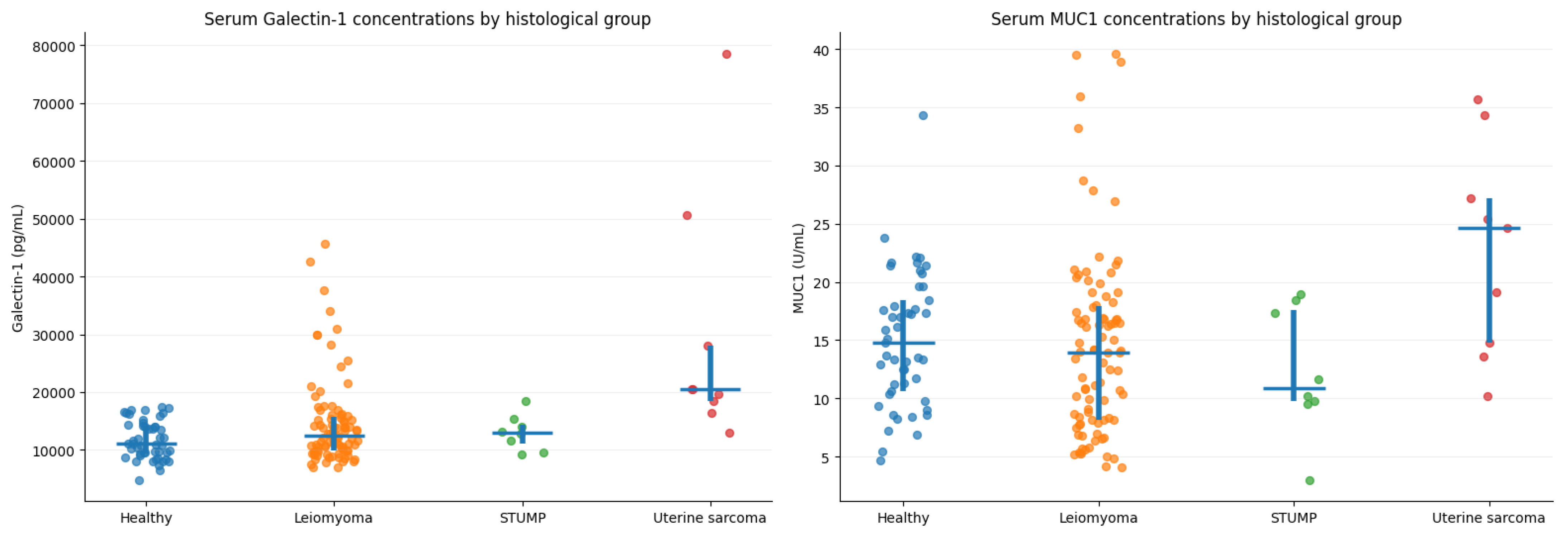
The present study evaluated serum galectin-1 and MUC1 in women with leiomyoma, STUMP, uterine sarcoma, and healthy controls. Galectin-1 concentrations differed significantly across the four histological groups, with the highest median levels observed in uterine sarcoma and significant pairwise differences between uterine sarcoma and both healthy controls and leiomyoma. MUC1 also differed overall across the four groups, with higher concentrations in uterine sarcoma than in leiomyoma, although its discriminatory performance in the primary leiomyoma versus sarcoma/STUMP ROC analysis was weak. In exploratory adjusted analysis, higher galectin-1 concentrations were associated with sarcoma/STUMP status.
Differentiating between benign uterine fibroids from malignant uterine sarcomas and intermediate entities such as STUMP remains one of the most challenging aspects of gynecologic practice [
28]. Although myomas are highly prevalent and benign, uterine sarcomas are rare but aggressive malignancies with poor prognosis [
11]. STUMP is a rare and heterogeneous diagnosis positioned between leiomyoma and leiomyosarcoma in classification systems, typically established only after postoperative histopathologic evaluation using criteria centered on cytologic atypia, mitotic activity, and coagulative tumor necrosis [
29]. Despite their typically low malignant potential, these tumors may occasionally recur or metastasize [
30]. Gupta et al. recently proposed updated diagnostic criteria for STUMP, based on WHO guidelines and multicenter experience; these include borderline or difficult-to-define tumor necrosis, atypia with mitotic activity near malignant thresholds, high mitotic counts, focal coagulative necrosis in otherwise benign-appearing tumors, intermediate cytologic features, myometrial invasion without overt malignancy, and atypical mitotic figures in the absence of other malignant characteristics [
31].
Preoperative differentiation is often limited by overlapping clinical presentation and imaging characteristics, and definitive diagnosis is frequently established only after surgical removal and histopathological examination [
32]. From a clinical standpoint, the need for improved preoperative risk stratification is underscored by the well-recognized, low but consequential risk of occult uterine sarcoma at surgery for presumed fibroids and the potential for iatrogenic dissemination with power morcellation [
33]. The FDA’s 2014 Safety Communication discouraged the use of laparoscopic power morcellation in most women undergoing hysterectomy or myomectomy for fibroids, and updated 2020 guidance recommends that, when morcellation is appropriate, it should be performed only with a containment system and in carefully selected patients [
20,
34]. Therefore, the identification of reliable, non-invasive biomarkers capable of improving preoperative risk stratification remains an important unmet clinical need.
Multiple studies across malignancies suggest that circulating galectin-1 can reflect tumor aggressiveness and microenvironmental remodeling, but diagnostic performance is context-dependent; as a β-galactoside-binding lectin, galectin-1 is involved in several processes central to tumor development and progression, including modulation of the tumor microenvironment, angiogenesis, and immune evasion [
35,
36]. In ovarian tumor cohorts, serum galectin-1 measured by ELISA has shown excellent discrimination in some settings (e.g., AUC 0.936 with a reported cutoff of 15.9 ng/mL achieving 88.9% sensitivity and 93% specificity), while other programs—particularly those focused on high-grade serous ovarian carcinoma—reported minimal case–control separation in plasma despite stromal galectin-1 being prognostically informative in tissue [
37,
38]. This variability is consistent with galectin-1 acting as a stromal/immune biomarker: in pancreatic cancer, where galectin-1 is strongly linked to desmoplastic stroma, plasma galectin-1 is elevated versus healthy controls but can also rise in benign inflammatory fibrosis (chronic pancreatitis), and performance improves when combined with an established marker (CA19-9) [
39,
40,
41]. Published tissue-based analyses have reported lower galectin-1 expression in leiomyosarcomas compared to leiomyomas [
42]. This finding is not directly comparable with the present study because we measured circulating serum galectin-1 rather than tumor-tissue expression. In our cohort, galectin-1 was higher in uterine sarcomas than in myomas and controls and remained associated with sarcoma after adjustment for age and menopausal status.
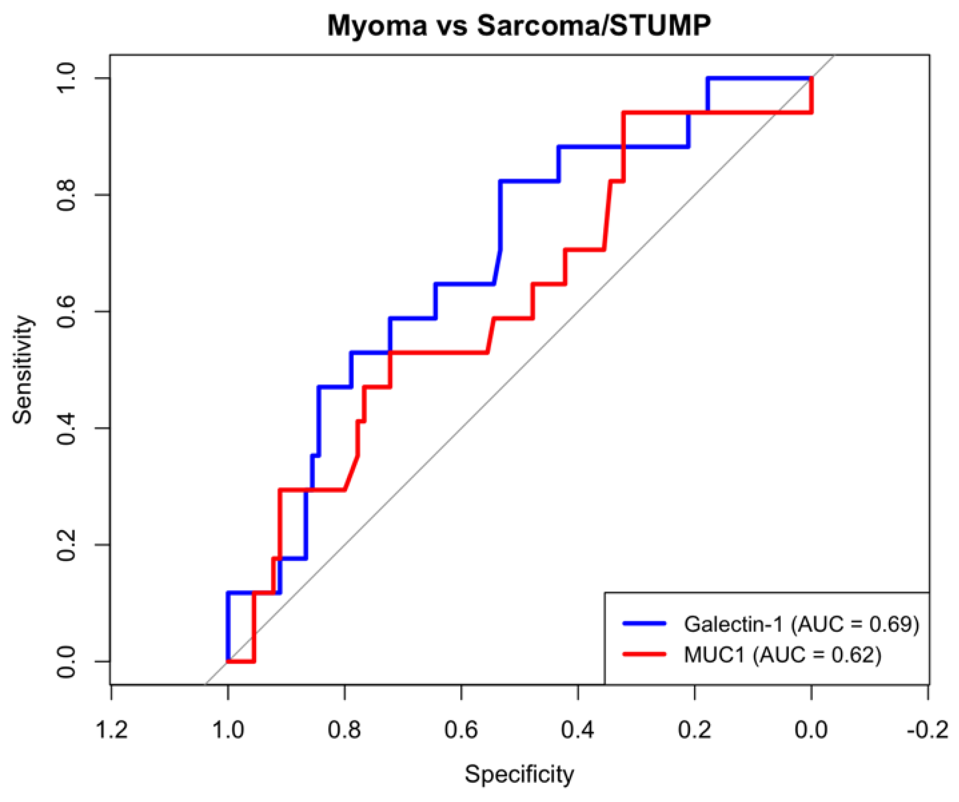
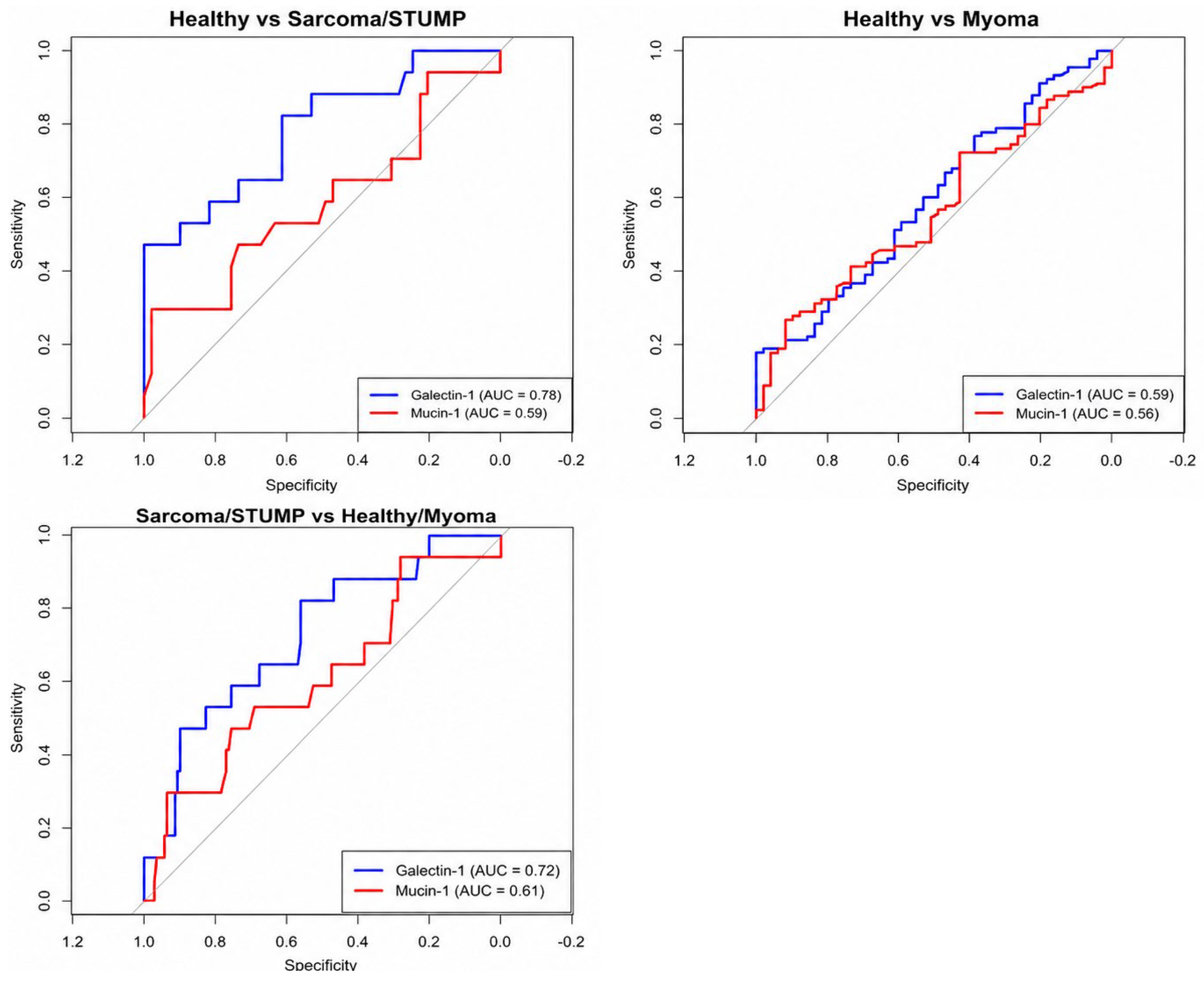
From a clinical perspective, the most relevant diagnostic challenge is the preoperative distinction between presumed leiomyoma and sarcoma/STUMP. In this setting, galectin-1 demonstrated only modest discrimination, with an AUC of 0.69. Although performance was higher when sarcoma/STUMP cases were compared with healthy controls, this contrast is less representative of the real-world diagnostic setting. Internal bootstrap validation yielded an AUC estimate similar to the apparent analysis, but cutoff-dependent sensitivity and specificity showed considerable variability. Taken together, these findings do not support the use of galectin-1 as a standalone diagnostic test. Its potential value may instead lie as an adjunct to existing clinical and imaging assessment, provided that its performance is confirmed in larger, clinically representative cohorts.
Circulating MUC1 levels are highly epitope- and assay-dependent and can differ between benign and malignant disease but often show high inter-individual variability and limited differential diagnostic utility, whereas tumor-epitope-specific MUC1 assays can achieve higher discrimination [
43]. Furthermore, variations in glycosylation and the release of soluble MUC1 fragments into circulation may influence its detectability and clinical utility as a serum biomarker [
26]. Consistent with these findings, our study demonstrated significant overall differences in circulating MUC1 levels between groups, with higher concentrations in uterine sarcoma than in myoma; however, MUC1 showed weak discrimination in the primary myoma versus sarcoma/STUMP comparison. Although MUC1 plays a well-established role in tumor progression in epithelial malignancies, its expression in mesenchymal tumors such as uterine sarcomas may follow different biological patterns [
44]. In ovarian tumor patients, CA 15-3 and CA 27.29 (MUC-derived assays) can be higher in malignancy but show substantial overlap and high variability [
45]. Similarly, MUC1 can behave paradoxically (lower median in ovarian cancer than benign disease) and achieves only modest sensitivity at high specificity (AUC 0.784 with 37% sensitivity at 95% specificity) [
46]. In uterine tumors, the role of MUC1 appears to be less straightforward; while MUC1 is associated with the presence of tumor tissue, it may not reliably distinguish between benign and malignant uterine lesions at the tissue level [
42].
Strengths of this study include preoperative serum sampling and histopathological confirmation of tumor diagnoses. Several limitations should be acknowledged. First, the small numbers of STUMP (n = 8) and uterine sarcoma (n = 9) cases limited statistical power, and the number of covariates relative to outcome events introduced a risk of overfitting; accordingly, adjusted analyses should be regarded as exploratory. Second, the sarcoma/STUMP group was histologically heterogeneous, limiting subtype-specific conclusions. The prolonged accrual period (2013–2024) may also have introduced temporal heterogeneity, because diagnostic and clinical practices evolved over time, and cases were not uniformly re-evaluated according to the most recent WHO classification. Another limitation concerns the control group: although controls were asymptomatic and had normal transvaginal ultrasound findings, predefined exclusion criteria for inflammatory, autoimmune, or other systemic conditions were unavailable, and controls were not spectrum-matched to women presenting with uterine masses, introducing potential spectrum bias. Comparisons with healthy controls should therefore be interpreted primarily as reference information rather than estimates of real-world diagnostic performance. Preanalytical factors and preoperative hormonal treatments were not systematically documented. Menopausal status, alcohol consumption, and hypertension differed among study groups. Although age and menopausal status were included in exploratory adjusted models, the limited number of sarcoma/STUMP events precluded stable adjustment for a larger set of covariates. Residual confounding related to alcohol use, hypertension, unrecorded inflammatory conditions, other comorbidities, and unavailable tumor-size information cannot be excluded. Internal bootstrap validation yielded AUC estimates broadly consistent with the apparent analyses; however, cutoff-dependent sensitivity and specificity showed considerable variability, and the derived thresholds should not be regarded as clinically validated cutoffs. Finally, the single-center design, absence of external validation, and potential residual confounding limit generalizability.
Future studies could integrate serum proteomic profiling with paired tumor-tissue expression analyses to determine whether circulating biomarker signals reflect tumor production, stromal responses, or systemic host processes, and to identify multimarker panels with greater discriminatory performance.





 ,
,