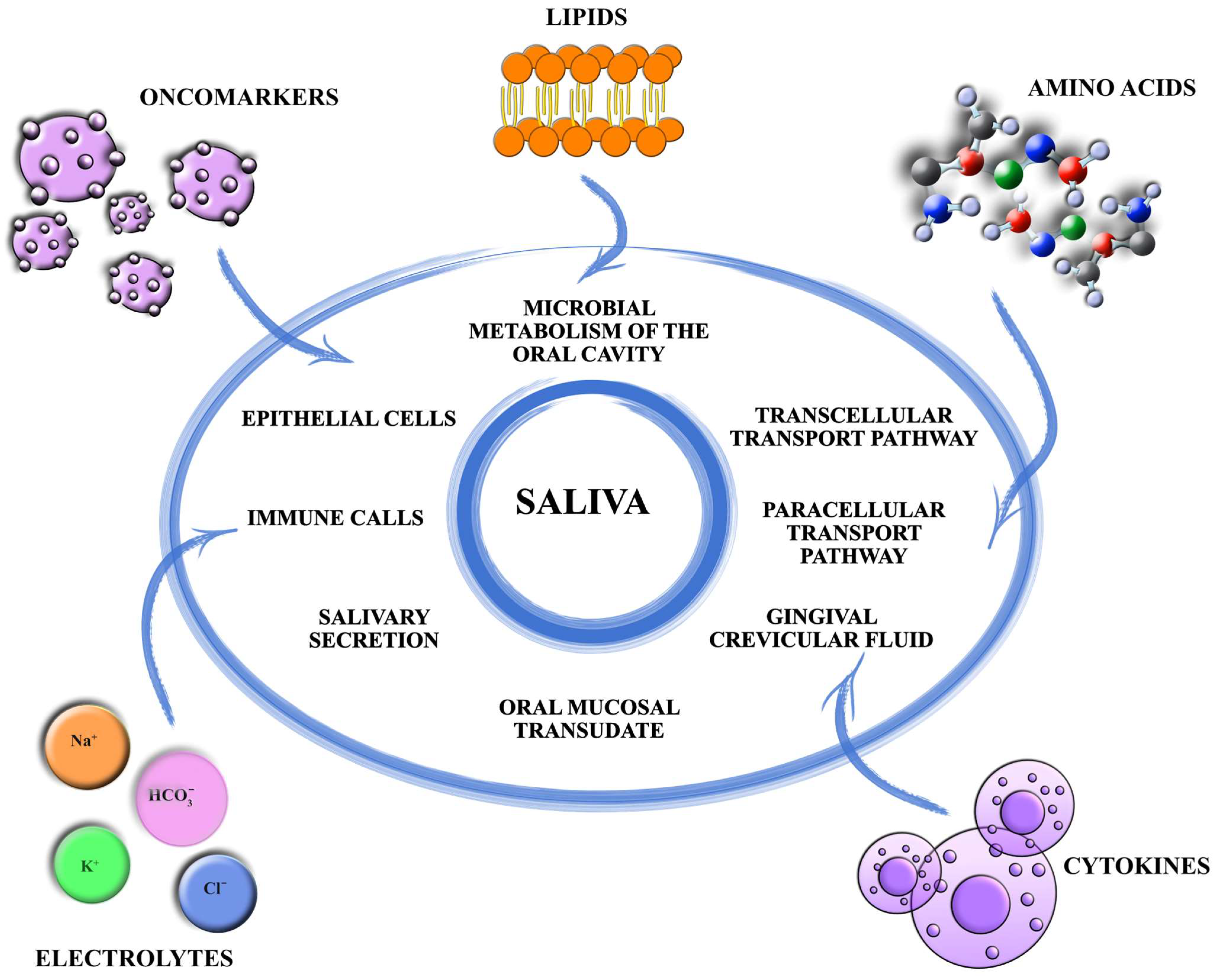
The Mechanism and Pathways of Formation and Modification of Salivary Metabolic Profile in Cancer



Abstract
1. Introduction
2. Electrolytes
3. Amino Acids
4. Lipids
5. Cytokines
6. Tumor Markers
7. Microbiome
8. Preliminary Conclusions on the Most Promising Candidates for Salivary Biomarkers and Priority Areas for Future Research
9. Conclusions
Author Contributions
Funding
Institutional Review Board Statement
Informed Consent Statement
Data Availability Statement
Conflicts of Interest
References
- Gröschl, M.; Rauh, M.; Wagner, R.; Neuhuber, W.; Metzler, M.; Tamgüney, G.; Zenk, J.; Schoof, E.; Dörr, H.G.; Blum, W.F.; et al. Identification of leptin in human saliva. J. Clin. Endocrinol. Metab. 2001, 86, 5234–5239. [Google Scholar] [CrossRef] [PubMed]
- Padalkar, P.; Yadadi, S.S.; Vivekanandan, G.; Shetty, S.R.; Andhare, M.; Pashine, A.; Vinay, V.; Desai, V.; Shetty, R.M. Salivary periostin levels as a non-invasive biomarker and their clinical correlates among healthy and periodontitis patients—A cross-sectional analytical study. Front. Dent. Med. 2025, 6, 1512252. [Google Scholar] [CrossRef] [PubMed]
- Al-Rawi, N.H.; Atiyah, K.M. Salivary neuron specific enolase: An indicator for neuronal damage in patients with ischemic stroke and stroke-prone patients. Clin. Chem. Lab. Med. 2009, 47, 1519–1524. [Google Scholar] [CrossRef] [PubMed]
- Karataş, A.; Ömercikoğlu, Z.; Öz, B.; Dağlı, A.F.; Çatak, O.; Erman, F.; Şahin, K.; Gözel, N.; Koca, S.S. Wnt signaling pathway activities may be altered in primary Sjogren’s syndrome. Turk. J. Med. Sci. 2021, 51, 2015–2022. [Google Scholar] [CrossRef] [PubMed]
- Lindell, S.G.; Suomi, S.J.; Shoaf, S.E.; Linnoila, M.; Higley, J.D. Salivary prolactin as a marker for central serotonin turnover. Biol. Psychiatry 1999, 46, 568–572. [Google Scholar] [CrossRef] [PubMed]
- Elgamal, A.A.; Ectors, N.L.; Sunardhi-Widyaputra, S.; Van Poppel, H.P.; Van Damme, B.J.; Baert, L.V. Detection of prostate specific antigen in pancreas and salivary glands: A potential impact on prostate cancer overestimation. J. Urol. 1996, 156, 464–468. [Google Scholar] [CrossRef] [PubMed]
- Shiiki, N.; Tokuyama, S.; Sato, C.; Kondo, Y.; Saruta, J.; Mori, Y.; Shiiki, K.; Miyoshi, Y.; Tsukinoki, K. Association between saliva PSA and serum PSA in conditions with prostate adenocarcinoma. Biomarkers 2011, 16, 498–503. [Google Scholar] [CrossRef] [PubMed]
- Turan, T.; Demir, S.; Aybek, H.; Atahan, O.; Tuncay, O.L.; Aybek, Z. Free and total prostate-specific antigen levels in saliva and the comparison with serum levels in men. Eur. Urol. 2000, 38, 550–554. [Google Scholar] [CrossRef] [PubMed]
- Yio, X.Y.; Jiang, J.; Yin, F.Z.; Ruan, K.H. Highly sensitive sandwich enzyme immunoassay for alpha-fetoprotein in human saliva. Ann. Clin. Biochem. 1992, 29, 519–522. [Google Scholar] [CrossRef] [PubMed]
- Recker, E.N.; Brogden, K.A.; Avila-Ortiz, G.; Fischer, C.L.; Pagan-Rivera, K.; Dawson, D.V.; Smith, K.M.; Elangovan, S. Novel biomarkers of periodontitis and/or obesity in saliva—An exploratory analysis. Arch. Oral Biol. 2015, 60, 1503–1509. [Google Scholar] [CrossRef] [PubMed]
- Mahajan, M.; Belgaumi, U.I.; Baad, R.; Vibhute, N.; Kadashetti, V.; Bommanavar, S.; Kamate, W. Salivary Human Chorionic Gonadotropin as a Novel Biomarker for Early Detection of Pregnancy: A Pilot Study. Gynecol. Minim. Invasive Ther. 2019, 8, 59–61. [Google Scholar] [CrossRef] [PubMed]
- Sireesha, D.; Reginald, B.A.; Reddy, B.S.; Samatha, M. Expression of human chorionic gonadotropin-β in tissue specimens, saliva and urine of oral squamous cell carcinoma patients. J. Oral Maxillofac. Pathol. 2021, 25, 417–422. [Google Scholar] [CrossRef] [PubMed]
- Selby, C.; Lobb, P.A.; Jeffcoate, W.J. Salivary albumin and sex hormone binding globulin (SHBG): Concentration and origin. Steroids 1988, 52, 373–374. [Google Scholar] [CrossRef] [PubMed]
- Hammond, G.L.; Langley, M.S. Identification and measurement of sex hormone binding globulin (SHBG) and corticosteroid binding globulin (CBG) in human saliva. Acta Endocrinol. 1986, 112, 603–608. [Google Scholar] [CrossRef]
- Hanif, H.; Ali, M.J.; Susheela, A.T.; Khan, I.W.; Luna-Cuadros, M.A.; Khan, M.M.; Lau, D.T. Update on the applications and limitations of alpha-fetoprotein for hepatocellular carcinoma. World J. Gastroenterol. 2022, 28, 216–229. [Google Scholar] [CrossRef] [PubMed]
- Ogino, M.H.; Tadi, P. Physiology, Chorionic Gonadotropin. In StatPearls; StatPearls Publishing: Treasure Island, FL, USA, 2026. Available online: https://www.ncbi.nlm.nih.gov/books/NBK556118/ (accessed on 10 July 2026).
- Balk, S.P.; Ko, Y.-J.; Bubley, G.J. Biology of prostate-specific antigen. J. Clin. Oncol. 2003, 21, 383–391. [Google Scholar] [CrossRef] [PubMed]
- Pugeat, M.; Nader, N.; Hogeveen, K.; Raverot, G.; Déchaud, H.; Grenot, C. Sex hormone-binding globulin gene expression in the liver: Drugs and the metabolic syndrome. Mol. Cell. Endocrinol. 2010, 316, 53–59. [Google Scholar] [CrossRef] [PubMed]
- Ayatollahi, H.; Darabi Mahboub, M.R.; Mohammadian, N.; Parizadeh, M.R.; Kianoosh, T.; Khabbaz Khoob, M.; Kamalian, F. Ratios of free to total prostate-specific antigen and total prostate specific antigen to protein concentrations in saliva and serum of healthy men. Urol. J. 2007, 4, 238–241. [Google Scholar] [PubMed]
- Nonaka, T.; Wong, D.T.W. Saliva diagnostics: Salivaomics, saliva exosomics, and saliva liquid biopsy. J. Am. Dent. Assoc. 2023, 154, 696–704. [Google Scholar] [CrossRef] [PubMed]
- Mai, S.; Mauger, M.T.; Niu, L.N.; Barnes, J.B.; Kao, S.; Bergeron, B.E.; Ling, J.Q.; Tay, F.R. Potential applications of antimicrobial peptides and their mimics in combating caries and pulpal infections. Acta Biomater. 2017, 49, 16–35. [Google Scholar] [CrossRef] [PubMed]
- Rosier, B.T.; Moya-Gonzalvez, E.M.; Corell-Escuin, P.; Mira, A. Isolation and Characterization of Nitrate-Reducing Bacteria as Potential Probiotics for Oral and Systemic Health. Front. Microbiol. 2020, 11, 555465. [Google Scholar] [CrossRef] [PubMed]
- Takahashi, N. Oral Microbiome Metabolism: From “Who Are They?” to “What Are They Doing?”. J. Dent. Res. 2015, 94, 1628–1637. [Google Scholar] [CrossRef] [PubMed]
- Efenberger, M.; Agier, J.; Pawłowska, E.; Brzezińska-Błaszczyk, E. Archaea prevalence in inflamed pulp tissues. Cent. Eur. J. Immunol. 2015, 40, 194–200. [Google Scholar] [CrossRef] [PubMed]
- Kistler, J.O.; Booth, V.; Bradshaw, D.J.; Wade, W.G. Bacterial community development in experimental gingivitis. PLoS ONE 2013, 8, e71227. [Google Scholar] [CrossRef] [PubMed]
- Abusleme, L.; Hoare, A.; Hong, B.Y.; Diaz, P.I. Microbial signatures of health, gingivitis, and periodontitis. Periodontology 2021, 86, 57–78. [Google Scholar] [CrossRef]
- Lin, D.; Yang, L.; Wen, L.; Lu, H.; Chen, Q.; Wang, Z. Crosstalk between the oral microbiota, mucosal immunity, and the epithelial barrier regulates oral mucosal disease pathogenesis. Mucosal Immunol. 2021, 14, 1247–1258. [Google Scholar] [CrossRef] [PubMed]
- Takahashi, N.; Sulijaya, B.; Yamada-Hara, M.; Tsuzuno, T.; Tabeta, K.; Yamazaki, K. Gingival epithelial barrier: Regulation by beneficial and harmful microbes. Tissue Barriers 2019, 7, e1651158. [Google Scholar] [CrossRef] [PubMed]
- Lin, Y.; Liang, X.; Li, Z.; Gong, T.; Ren, B.; Li, Y.; Peng, X. Omics for deciphering oral microecology. Int. J. Oral Sci. 2024, 16, 2. [Google Scholar] [CrossRef] [PubMed]
- Knight, R.; Vrbanac, A.; Taylor, B.C.; Aksenov, A.; Callewaert, C.; Debelius, J.; Gonzalez, A.; Kosciolek, T.; McCall, L.I.; McDonald, D.; et al. Best practices for analysing microbiomes. Nat. Rev. Microbiol. 2018, 6, 410–422. [Google Scholar] [CrossRef]
- Hillmann, B.; Al-Ghalith, G.A.; Shields-Cutler, R.R.; Zhu, Q.; Gohl, D.M.; Beckman, K.B.; Knight, R.; Knights, D. Evaluating the Information Content of Shallow Shotgun Metagenomics. mSystems 2018, 3, e00069-18. [Google Scholar] [CrossRef] [PubMed]
- Aguiar-Pulido, V.; Huang, W.; Suarez-Ulloa, V.; Cickovski, T.; Mathee, K.; Narasimhan, G. Metagenomics, Metatranscriptomics, and Metabolomics Approaches for Microbiome Analysis. Evol. Bioinform. Online 2016, 12, 5–16. [Google Scholar] [CrossRef] [PubMed]
- Baker, J.L. Illuminating the oral microbiome and its host interactions: Recent advancements in omics and bioinformatics technologies in the context of oral microbiome research. FEMS Microbiol. Rev. 2023, 47, fuad051. [Google Scholar] [CrossRef] [PubMed]
- Salter, S.J.; Cox, M.J.; Turek, E.M.; Calus, S.T.; Cookson, W.O.; Moffatt, M.F.; Turner, P.; Parkhill, J.; Loman, N.J.; Walker, A.W. Reagent and laboratory contamination can critically impact sequence-based microbiome analyses. BMC Biol. 2014, 12, 87. [Google Scholar] [CrossRef] [PubMed]
- Fierer, N.; Leung, P.M.; Lappan, R.; Eisenhofer, R.; Ricci, F.; Holland, S.I.; Dragone, N.; Blackall, L.L.; Dong, X.; Dorador, C.; et al. Guidelines for preventing and reporting contamination in low-biomass microbiome studies. Nat. Microbiol. 2025, 10, 1570–1580. [Google Scholar] [CrossRef] [PubMed]
- Davis, N.M.; Proctor, D.M.; Holmes, S.P.; Relman, D.A.; Callahan, B.J. Simple statistical identification and removal of contaminant sequences in marker-gene and metagenomics data. Microbiome 2018, 6, 226. [Google Scholar] [CrossRef] [PubMed]
- Lu, H.; Zou, P.; Zhang, Y.; Zhang, Q.; Chen, Z.; Chen, F. The sampling strategy of oral microbiome. Imeta 2022, 1, e23. [Google Scholar] [CrossRef] [PubMed]
- Yano, Y.; Hua, X.; Wan, Y.; Suman, S.; Zhu, B.; Dagnall, C.L.; Hutchinson, A.; Jones, K.; Hicks, B.D.; Shi, J.; et al. Comparison of Oral Microbiota Collected Using Multiple Methods and Recommendations for New Epidemiologic Studies. mSystems 2020, 5, e00156-20. [Google Scholar] [CrossRef] [PubMed]
- Lim, Y.; Totsika, M.; Morrison, M.; Punyadeera, C. The saliva microbiome profiles are minimally affected by collection method or DNA extraction protocols. Sci. Rep. 2017, 7, 8523. [Google Scholar] [CrossRef] [PubMed]
- Ruan, X.; Luo, J.; Zhang, P.; Howell, K. The salivary microbiome shows a high prevalence of core bacterial members yet variability across human populations. npj Biofilms Microbiomes 2022, 8, 85. [Google Scholar] [CrossRef] [PubMed]
- Collado, M.C.; Engen, P.A.; Bandín, C.; Cabrera-Rubio, R.; Voigt, R.M.; Green, S.J.; Naqib, A.; Keshavarzian, A.; Scheer, F.A.J.L.; Garaulet, M. Timing of food intake impacts daily rhythms of human salivary microbiota: A randomized, crossover study. FASEB J. 2018, 32, 2060–2072. [Google Scholar] [CrossRef] [PubMed]
- Kashyap, B.; Kullaa, A.M. Salivary Metabolites Produced by Oral Microbes in Oral Diseases. Metabolites 2024, 14, 277. [Google Scholar] [CrossRef] [PubMed]
- Basic, A.; Dahlén, G. Microbial metabolites in the pathogenesis of periodontal diseases: A narrative review. Front. Oral Health 2023, 4, 1210200. [Google Scholar] [CrossRef] [PubMed]
- Seshadri, R.; Myers, G.S.; Tettelin, H.; Eisen, J.A.; Heidelberg, J.F.; Dodson, R.J.; Davidsen, T.M.; DeBoy, R.T.; Fouts, D.E.; Haft, D.H.; et al. Comparison of the genome of the oral pathogen Treponema denticola with other spirochete genomes. Proc. Natl. Acad. Sci. USA 2004, 101, 5646–5651. [Google Scholar] [CrossRef] [PubMed]
- Watanabe, T.; Matsuura, M.; Seto, K. Proteolytic activities of Mycoplasma salivarium. Microbiol. Immunol. 1988, 32, 113–122. [Google Scholar]
- Barile, M.F.; Schimke, R.T.; Riggs, D.B. Presence of the arginine dihydrolase pathway in Mycoplasma. J. Bacteriol. 1966, 91, 189–192. [Google Scholar] [CrossRef] [PubMed]
- Barbour, A.; Philip, K. Metabolites of the oral microbiome: Important mediators of multikingdom interactions. FEMS Microbiol. Rev. 2022, 46, fuab039. [Google Scholar] [PubMed]
- Bostanghadiri, N.; Kouhzad, M.; Taki, E.; Elahi, Z.; Khoshbayan, A.; Navidifar, T.; Darban-Sarokhalil, D. Oral microbiota and metabolites: Key players in oral health and disorder, and microbiota-based therapies. Front. Microbiol. 2024, 15, 1431785. [Google Scholar] [CrossRef] [PubMed]
- Nani, B.D.; Lima, P.O.; Marcondes, F.K.; Groppo, F.C.; Rolim, G.S.; Moraes, A.B.A.; Cogo-Müller, K. Changes in salivary microbiota increase volatile sulfur compounds production in healthy male subjects with academic-related chronic stress. PLoS ONE 2017, 12, e0173686. [Google Scholar] [CrossRef] [PubMed]
- Fernández-Reina, A.; Urdiales, J.L.; Sánchez-Jiménez, F. What We Know and What We Need to Know about Aromatic and Cationic Biogenic Amines in the Gastrointestinal Tract. Foods 2018, 7, 145. [Google Scholar] [CrossRef] [PubMed]
- Persson, S.; Edlund, M.B.; Claesson, R.; Carlsson, J. The formation of hydrogen sulfide and methyl mercaptan by oral bacteria. Oral Microbiol. Immunol. 1990, 5, 195–201. [Google Scholar] [CrossRef] [PubMed]
- Vasquez, A.M.; Mouchlis, V.D.; Dennis, E.A. Review of four major distinct types of human phospholipase A2. Adv. Biol. Regul. 2018, 67, 212–218. [Google Scholar] [CrossRef] [PubMed]
- Shah, D.S.H.; Russell, R.R.B. A novel glucan-binding protein with lipase activity from the oral pathogen Streptococcus mutans. Microbiology 2004, 150, 1947–1956. [Google Scholar] [CrossRef] [PubMed]
- Sato, H.; Taketomi, Y.; Murakami, M. Metabolic regulation by secreted phospholipase A2. Inflamm. Regen. 2016, 36, 7. [Google Scholar] [CrossRef] [PubMed]
- Reisenberg, M.; Singh, P.K.; Williams, G.; Doherty, P. The diacylglycerol lipases: Structure, regulation and roles in and beyond endocannabinoid signalling. Philos. Trans. R. Soc. B 2012, 367, 3264–3275. [Google Scholar] [CrossRef]
- Fernandez-Gutierrez, M.M.; Imangaliyev, S.; Prodan, A.; Loos, B.G.; Keijser, B.J.F.; Kleerebezem, M. A salivary metabolite signature that reflects gingival host-microbe interactions: Instability predicts gingivitis susceptibility. Sci. Rep. 2020, 10, 3008. [Google Scholar] [CrossRef] [PubMed]
- Liu, T.; Zhang, L.; Joo, D.; Sun, S.-C. NF-κB signaling in inflammation. Signal Transduct. Target. Ther. 2017, 2, 17023. [Google Scholar] [CrossRef] [PubMed]
- Schwandner, R.; Dziarski, R.; Wesche, H.; Rothe, M.; Kirschning, C.J. Peptidoglycan- and lipoteichoic acid-induced cell activation is mediated by Toll-like receptor 2. J. Biol. Chem. 1999, 274, 17406–17409. [Google Scholar] [CrossRef] [PubMed]
- Bauer, S.; Kirschning, C.J.; Häcker, H.; Redecke, V.; Hausmann, S.; Akira, S.; Wagner, H.; Lipford, G.B. Human TLR9 confers responsiveness to bacterial DNA via species-specific CpG motif recognition. Proc. Natl. Acad. Sci. USA 2001, 98, 9237–9242. [Google Scholar] [CrossRef] [PubMed]
- Do, T.T.; Nguyen, V.T.; Nguyen, N.T.N.; Duong, K.T.T.; Nguyen, T.T.M.; Le, D.N.T.; Nguyen, T.H. A Review of a Breakdown in the Barrier: Tight Junction Dysfunction in Dental Diseases. Clin. Cosmet. Investig. Dent. 2024, 16, 513–531. [Google Scholar] [CrossRef] [PubMed]
- Zhang, W.; Luo, J.; Dong, X.; Zhao, S.; Hao, Y.; Peng, C.; Shi, H.; Zhou, Y.; Shan, L.; Sun, Q.; et al. Salivary Microbial Dysbiosis is Associated with Systemic Inflammatory Markers and Predicted Oral Metabolites in Non-Small Cell Lung Cancer Patients. J. Cancer 2019, 10, 1651–1662. [Google Scholar] [CrossRef] [PubMed]
- Devaraja, K.; Aggarwal, S. Dysbiosis of Oral Microbiome: A Key Player in Oral Carcinogenesis? A Critical Review. Biomedicines 2025, 13, 448. [Google Scholar] [CrossRef] [PubMed]
- Boksa, F.A.; Leinbach, L.I.; Mody, D.P.; Ganesan, S.M.; Mays, J.W. Oral microbiome alterations after cancer treatment: A scoping review and analysis. Med. Oncol. 2025, 42, 432. [Google Scholar] [CrossRef] [PubMed]
- Klymiuk, I.; Bilgilier, C.; Mahnert, A.; Prokesch, A.; Heininger, C.; Brandl, I.; Sahbegovic, H.; Singer, C.; Fuereder, T.; Steininger, C. Chemotherapy-associated oral microbiome changes in breast cancer patients. Front. Oncol. 2022, 12, 949071. [Google Scholar] [CrossRef] [PubMed]
- Laheij, A.M.G.A.; Raber-Durlacher, J.E.; Koppelmans, R.G.A.; Huysmans, M.-C.D.N.J.M.; Potting, C.; van Leeuwen, S.J.M.; Hazenberg, M.D.; Brennan, M.T.; von Bültzingslöwen, I.; Johansson, J.-E.; et al. Microbial changes in relation to oral mucositis in autologous hematopoietic stem cell transplantation recipients. Sci. Rep. 2019, 9, 16929. [Google Scholar] [CrossRef] [PubMed]
- Zaura, E.; Brandt, B.W.; Teixeira de Mattos, M.J.; Buijs, M.J.; Caspers, M.P.; Rashid, M.U.; Weintraub, A.; Nord, C.E.; Savell, A.; Hu, Y.; et al. Same Exposure but Two Radically Different Responses to Antibiotics: Resilience of the Salivary Microbiome versus Long-Term Microbial Shifts in Feces. mBio 2015, 6, e01693-15. [Google Scholar] [CrossRef] [PubMed]
- Hosseini, M.S.; Sanaie, S.; Mahmoodpoor, A.; Jabbari Beyrami, S.; Jabbari Beyrami, H.; Fattahi, S.; Jahanshahlou, F.; Zarei, M.; Rahimi Mamaghani, A.; Kuchaki Rafsanjani, M. Cancer treatment-related xerostomia: Basics, therapeutics, and future perspectives. Eur. J. Med. Res. 2024, 29, 571. [Google Scholar] [CrossRef] [PubMed]
- PDQ Supportive and Palliative Care Editorial Board. Oral Complications of Cancer Therapies (PDQ®): Patient Version. In PDQ Cancer Information Summaries; National Cancer Institute: Bethesda, MD, USA, 2024. Available online: https://www.ncbi.nlm.nih.gov/books/NBK65725/ (accessed on 10 July 2026).
- Hong, B.Y.; Sobue, T.; Choquette, L.; Dupuy, A.K.; Thompson, A.; Burleson, J.A.; Salner, A.L.; Schauer, P.K.; Joshi, P.; Fox, E.; et al. Chemotherapy-induced oral mucositis is associated with detrimental bacterial dysbiosis. Microbiome 2019, 7, 66. [Google Scholar] [CrossRef] [PubMed]
- Staruch, M.; Speth, M.M.; Neyer, P.; Riesterer, O.; Aebersold, D.M.; Stieb, S. Radiation-associated changes in saliva composition of head and neck cancer patients: A systematic review. Radiother. Oncol. 2024, 196, 110279. [Google Scholar] [CrossRef] [PubMed]
- Ingrosso, G.; Saldi, S.; Marani, S.; Wong, A.Y.W.; Bertelli, M.; Aristei, C.; Zelante, T. Breakdown of Symbiosis in Radiation-Induced Oral Mucositis. J. Fungi 2021, 7, 290. [Google Scholar] [CrossRef]
- Bruno, J.S.; Al-Qadami, G.H.; Laheij, A.M.G.A.; Bossi, P.; Fregnani, E.R.; Wardill, H.R. From Pathogenesis to Intervention: The Importance of the Microbiome in Oral Mucositis. Int. J. Mol. Sci. 2023, 24, 8274. [Google Scholar] [CrossRef] [PubMed]
- Klimiuk, A.; Zalewska, A.; Knapp, M.; Skutnik-Radziszewska, A.; Maciejczyk, M. Could inflammation contribute to salivary gland dysfunction in patients with chronic heart failure? Front. Immunol. 2022, 13, 1005981. [Google Scholar] [CrossRef] [PubMed]
- Vieira, G.S.; Kimura, T.C.; Scarini, J.F.; de Lima-Souza, R.A.; Lavareze, L.; Emerick, C.; Gonçalves, M.T.; Damas, I.I.; Figueiredo-Maciel, T.; Sales de Sá, R.; et al. Hematopoietic colony-stimulating factors in head and neck cancers: Recent advances and therapeutic challenges. Cytokine 2024, 173, 156417. [Google Scholar] [CrossRef] [PubMed]
- Aldahlawi, S.; Youssef, A.R.; Shahabuddin, S. Evaluation of chemokine CXCL10 in human gingival crevicular fluid, saliva, and serum as periodontitis biomarker. J. Inflamm. Res. 2018, 11, 389–396. [Google Scholar] [CrossRef] [PubMed]
- Silva, T.A.; Garlet, G.P.; Fukada, S.Y.; Silva, J.S.; Cunha, F.Q. Chemokines in Oral Inflammatory Diseases: Apical Periodontitis and Periodontal Disease. J. Dent. Res. 2007, 86, 306–319. [Google Scholar] [CrossRef] [PubMed]
- Aggor, F.E.Y.; Bertolini, M.; Coleman, B.M.; Taylor, T.C.; Ponde, N.O.; Gaffen, S.L. Combinatorial actions of IL-22 and IL-17 drive optimal immunity to oral candidiasis through SPRRs. PLoS Pathog. 2024, 20, e1012302. [Google Scholar] [CrossRef] [PubMed]
- Jiménez, C.; Carvajal, D.; Hernández, M.; Valenzuela, F.; Astorga, J.; Fernández, A. Levels of the interleukins 17A, 22, and 23 and the S100 protein family in the gingival crevicular fluid of psoriatic patients with or without periodontitis. An. Bras. Dermatol. 2021, 96, 163–170. [Google Scholar] [CrossRef] [PubMed]
- Hotamisligil, G.S. Inflammation and metabolic disorders. Nature 2006, 444, 860–867. [Google Scholar] [CrossRef] [PubMed]
- Rysman, E.; Brusselmans, K.; Scheys, K.; Timmermans, L.; Derua, R.; Munck, S.; Van Veldhoven, P.P.; Waltregny, D.; Daniëls, V.W.; Machiels, J.; et al. De novo lipogenesis protects cancer cells from free radicals and chemotherapeutics by promoting membrane lipid saturation. Cancer Res. 2010, 70, 8117–8126. [Google Scholar] [CrossRef] [PubMed]
- Lan, T.; Chen, L.; Wei, X. Inflammatory cytokines in cancer: Comprehensive understanding and clinical progress in gene therapy. Cells 2021, 10, 100. [Google Scholar] [CrossRef] [PubMed]
- Bunte, K.; Beikler, T. Th17 Cells and the IL-23/IL-17 Axis in the Pathogenesis of Periodontitis and Immune-Mediated Inflammatory Diseases. Int. J. Mol. Sci. 2019, 20, 3394. [Google Scholar] [PubMed]
- Abusleme, L.; Moutsopoulos, N.M. IL-17: Overview and role in oral immunity and microbiome. Oral Dis. 2017, 23, 854–865. [Google Scholar] [PubMed]
- Chen, C.; Zhang, Q.; Yu, W.; Chang, B.; Le, A.D. Oral Mucositis: An Update on Innate Immunity and New Interventional Targets. J. Dent. Res. 2020, 99, 1122–1130. [Google Scholar] [CrossRef] [PubMed]
- Damgaard, C.; Massarenti, L.; Danielsen, A.K.; Graversen, J.H.; Holmstrup, P.; Nielsen, C.H.; Palarasah, Y. Complement component 3 and its activation split-products in saliva associate with periodontitis. J. Periodontol. 2022, 93, 1294–1301. [Google Scholar] [CrossRef] [PubMed]
- Bhalla, S.P.; Shaju, A.M.; Figueredo, C.M.d.S.; Miranda, L.A. Increased Levels of C5a in Gingival Crevicular Fluid and Saliva of Patients with Periodontal Disease. Pathogens 2022, 11, 983. [Google Scholar] [CrossRef] [PubMed]
- Brandtzaeg, P. Secretory immunity with special reference to the oral cavity. J. Oral Microbiol. 2013, 5, 20401. [Google Scholar] [CrossRef]
- Vila, T.; Rizk, A.M.; Sultan, A.S.; Jabra-Rizk, M.A. The power of saliva: Antimicrobial and beyond. PLoS Pathog. 2019, 15, e1008058. [Google Scholar] [CrossRef] [PubMed]
- Oppenheim, F.G.; Xu, T.; McMillian, F.M.; Levitz, S.M.; Diamond, R.D.; Offner, G.D.; Troxler, R.F. Histatins, a novel family of histidine-rich proteins in human parotid secretion. Isolation, characterization, primary structure, and fungistatic effects on Candida albicans. J. Biol. Chem. 1988, 263, 7472–7477. [Google Scholar] [CrossRef] [PubMed]
- Greer, A.; Zenobia, C.; Darveau, R.P. Defensins and LL-37: A review of function in the gingival epithelium. Periodontology 2013, 63, 67–79. [Google Scholar] [CrossRef]
- Gorr, S.U. Antimicrobial peptides in periodontal innate defense. Front. Oral Biol. 2012, 15, 84–98. [Google Scholar] [PubMed]
- Johnstone, K.F.; Wei, Y.; Bittner-Eddy, P.D.; Vreeman, G.W.; Stone, I.A.; Clayton, J.B.; Reilly, C.S.; Walbon, T.B.; Wright, E.N.; Hoops, S.L.; et al. Calprotectin (S100A8/A9) Is an Innate Immune Effector in Experimental Periodontitis. Infect. Immun. 2021, 89, e0012221. [Google Scholar] [CrossRef] [PubMed]
- Kim, H.D.; Karna, S.; Shin, Y.; Vu, H.; Cho, H.J.; Kim, S. S100A8 and S100A9 in saliva, blood and gingival crevicular fluid for screening established periodontitis: A cross-sectional study. BMC Oral Health 2021, 21, 388. [Google Scholar] [CrossRef] [PubMed]
- Marcotte, H.; Lavoie, M.C. Oral microbial ecology and the role of salivary immunoglobulin A. Microbiol. Mol. Biol. Rev. 1998, 62, 71–109. [Google Scholar] [CrossRef] [PubMed]
- Gallo, M.; Ferrari, E.; Giovati, L.; Pertinhez, T.A.; Artesani, L.; Conti, S.; Ciociola, T. The Variability of the Salivary Antimicrobial Peptide Profile: Impact of Lifestyle. Int. J. Mol. Sci. 2024, 25, 11501. [Google Scholar] [CrossRef] [PubMed]
- Di Spirito, F.; Di Palo, M.P.; De Benedetto, G.; Piedepalumbo, F.; Galdi, M.; Cannatà, D.; Cafà, N.; Contaldo, M. Periodontal Microbial Profiles Across Periodontal Conditions in Pediatric Subjects: A Narrative Review. Microorganisms 2025, 13, 1813. [Google Scholar] [CrossRef] [PubMed]
- Kaczor-Urbanowicz, K.E.; Martin Carreras-Presas, C.; Aro, K.; Tu, M.; Garcia-Godoy, F.; Wong, D.T. Saliva diagnostics: Current views and directions. Exp. Biol. Med. 2017, 242, 459–472. [Google Scholar]
- Kumari, S.; Samara, M.; Ampadi Ramachandran, R.; Gosh, S.; George, H.; Wang, R.; Pesavento, R.P.; Mathew, M.T. A review on saliva-based health diagnostics: Biomarker selection and future directions. Biomed. Mater. Devices 2024, 2, 121–138. [Google Scholar] [CrossRef]
- Humphrey, S.P.; Williamson, R.T. A review of saliva: Normal composition, flow, and function. J. Prosthet. Dent. 2001, 85, 162–169. [Google Scholar] [CrossRef] [PubMed]
- Simińska, E.; Koba, M. Amino acid profiling as a method of discovering biomarkers for early diagnosis of cancer. Amino Acids 2016, 48, 1339–1345. [Google Scholar] [CrossRef] [PubMed]
- Han, Y.; Jia, L.; Zheng, Y.; Li, W. Salivary exosomes: Emerging roles in systemic disease. Int. J. Biol. Sci. 2018, 14, 633–643. [Google Scholar] [CrossRef] [PubMed]
- Bastías, D.; Maturana, A.; Marín, C.; Martínez, R.; Niklander, S.E. Salivary biomarkers for oral cancer detection: An exploratory systematic review. Int. J. Mol. Sci. 2024, 25, 2634. [Google Scholar] [CrossRef] [PubMed]
- Sarkar, A.; Kuehl, M.N.; Alman, A.C.; Burkhardt, B.R. Linking the oral microbiome and salivary cytokine abundance to circadian oscillations. Sci. Rep. 2021, 11, 2658. [Google Scholar] [CrossRef] [PubMed]
- Kashyap, B.; Debnath, N.; Hsu, Y.-H.; Hu, H.-C.; Chen, H.-Y. Salivary metabolites produced by oral microbes in oral cancer and oral potentially malignant disorder. Metabolites 2024, 14, 277. [Google Scholar] [CrossRef] [PubMed]
- Cleaver, L.M.; Carda-Diéguez, M.; Moazzez, R.; Carpenter, G.H. Novel bacterial proteolytic and metabolic activity associated with dental erosion-induced oral dysbiosis. Microbiome 2023, 11, 69. [Google Scholar] [CrossRef] [PubMed]
- Gonzalez Agurto, M.; Olivares, N.; Canedo-Marroquin, G.; Espinoza, D.; Tortora, S.C. The intersection of the oral microbiome and salivary metabolites in head and neck cancer: From diagnosis to treatment. Cancers 2024, 16, 3545. [Google Scholar] [CrossRef] [PubMed]
- Radaic, A.; Kapila, Y.L. The oralome and its dysbiosis: New insights into oral microbiome-host interactions. Comput. Struct. Biotechnol. J. 2021, 19, 1335–1360. [Google Scholar] [CrossRef] [PubMed]
- Li, Y.; Ou, Y.; Fan, K.; Liu, G. Salivary diagnostics: Opportunities and challenges. Theranostics 2024, 14, 6969–6990. [Google Scholar] [CrossRef] [PubMed]
- Lee, M.G.; Ohana, E.; Park, H.W.; Yang, D.; Muallem, S. Molecular mechanism of pancreatic and salivary gland fluid and HCO3− secretion. Physiol. Rev. 2012, 92, 39–74. [Google Scholar] [CrossRef] [PubMed]
- Zhao, H.; Xu, X.; Diaz, J.; Muallem, S. Na+, K+, and H+/HCO3− transport in submandibular salivary ducts: Membrane localization of transporters. J. Biol. Chem. 1995, 270, 19599–19605. [Google Scholar] [CrossRef] [PubMed]
- Carpenter, G.H. The secretion, components, and properties of saliva. Annu. Rev. Food Sci. Technol. 2013, 4, 267–276. [Google Scholar] [CrossRef] [PubMed]
- Ogobuiro, I.; Tuma, F. Physiology, Renal. In StatPearls; StatPearls Publishing: Treasure Island, FL, USA, 2026. Available online: https://www.ncbi.nlm.nih.gov/books/NBK538339/ (accessed on 10 July 2026).
- Hall, J.E.; do Carmo, J.M.; da Silva, A.A.; Wang, Z.; Hall, M.E. Obesity, kidney dysfunction and hypertension: Mechanistic links. Nat. Rev. Nephrol. 2019, 15, 367–385. [Google Scholar] [CrossRef] [PubMed]
- Ferrannini, E. Sodium-glucose co-transporters and their inhibition: Clinical physiology. Cell Metab. 2017, 26, 27–38. [Google Scholar] [CrossRef] [PubMed]
- Scott, J.H.; Menouar, M.A.; Dunn, R.J. Physiology, Aldosterone. In StatPearls; StatPearls Publishing: Treasure Island, FL, USA, 2026. Available online: https://www.ncbi.nlm.nih.gov/books/NBK470339/ (accessed on 10 July 2026).
- Proctor, G.B. The physiology of salivary secretion. Periodontology 2016, 70, 11–25. [Google Scholar]
- Kubala, E.; Strzelecka, P.; Grzegocka, M.; Lietz-Kijak, D.; Gronwald, H.; Skomro, P.; Kijak, E. A review of selected studies that determine the physical and chemical properties of saliva in the field of dental treatment. BioMed Res. Int. 2018, 2018, 6572381. [Google Scholar] [CrossRef] [PubMed]
- Imenez Silva, P.H.; Mohebbi, N. Kidney metabolism and acid-base control: Back to the basics. Pflüg. Arch. Eur. J. Physiol. 2022, 474, 919–934. [Google Scholar] [CrossRef]
- Hopkins, E.; Sanvictores, T.; Sharma, S. Physiology, Acid Base Balance. In StatPearls; StatPearls Publishing: Treasure Island, FL, USA, 2026. Available online: https://www.ncbi.nlm.nih.gov/books/NBK507807/ (accessed on 10 July 2026).
- Bardow, A.; Moe, D.; Nyvad, B.; Nauntofte, B. The buffer capacity and buffer systems of human whole saliva measured without loss of CO2. Arch. Oral Biol. 2000, 45, 1–12. [Google Scholar] [CrossRef] [PubMed]
- Hogan, P.G.; Lewis, R.S.; Rao, A. Molecular basis of calcium signaling in lymphocytes: STIM and ORAI. Annu. Rev. Immunol. 2010, 28, 491–533. [Google Scholar] [CrossRef] [PubMed]
- Vig, M.; Kinet, J.P. Calcium signaling in immune cells. Nat. Immunol. 2009, 10, 21–27. [Google Scholar] [CrossRef] [PubMed]
- Romani, A.M.P. Intracellular Magnesium Homeostasis. In Magnesium in the Central Nervous System; Vink, R., Nechifor, M., Eds.; University of Adelaide Press: Adelaide, Australia, 2011. Available online: https://www.ncbi.nlm.nih.gov/books/NBK507258/ (accessed on 10 July 2026).
- Singhal, I.; Arora, M.; Dave, A.; Bansal, S.K.; Saluja, P.; Rai, R. Evaluation of magnesium levels in serum and saliva by calmagite method in individuals with tobacco habits with or without potentially malignant disorders. J. Oral Maxillofac. Pathol. 2023, 27, 425–426. [Google Scholar] [CrossRef] [PubMed]
- Lima, F.D.S.; Fock, R.A. A review of the action of magnesium on several processes involved in the modulation of hematopoiesis. Int. J. Mol. Sci. 2020, 21, 7084. [Google Scholar] [CrossRef] [PubMed]
- Dongiovanni, P.; Meroni, M.; Casati, S.; Goldoni, R.; Thomaz, D.V.; Kehr, N.S.; Galimberti, D.; Del Fabbro, M.; Tartaglia, G.M. Salivary biomarkers: Novel noninvasive tools to diagnose chronic inflammation. Int. J. Oral Sci. 2023, 15, 27. [Google Scholar] [CrossRef] [PubMed]
- Berardi, R.; Torniai, M.; Lenci, E.; Pecci, F.; Morgese, F.; Rinaldi, S. Electrolyte disorders in cancer patients: A systematic review. J. Cancer Metastasis Treat. 2019, 5, 79. [Google Scholar]
- Munsif, A.; Nowaczyk, A.; Fijałkowski, Ł.; Riaz, S.; Jamil, A. Salivary biomarkers in cancer detection and management. Acta Biochim. Pol. 2025, 72, 14410. [Google Scholar] [CrossRef] [PubMed]
- Bennet, D.; Khorsandian, Y.; Pelusi, J.; Mirabella, A.; Pirrotte, P.; Zenhausern, F. Molecular and physical technologies for monitoring fluid and electrolyte imbalance: A focus on cancer population. Clin. Transl. Med. 2021, 11, e461. [Google Scholar] [CrossRef] [PubMed]
- Dong, Y.; Wang, T.; Wu, H. The role of cytokines from salivary gland epithelial cells in the immunopathology of Sjögren’s syndrome. Front. Immunol. 2024, 15, 1443455. [Google Scholar] [CrossRef] [PubMed]
- D’Agostino, C.; Elkashty, O.A.; Chivasso, C.; Perret, J.; Tran, S.D.; Delporte, C. Insight into salivary gland aquaporins. Cells 2020, 9, 1547. [Google Scholar] [CrossRef] [PubMed]
- Baker, O.J.; Camden, J.M.; Redman, R.S.; Jones, J.E.; Seye, C.I.; Erb, L.; Weisman, G.A. Proinflammatory cytokines tumor necrosis factor-alpha and interferon-gamma alter tight junction structure and function in the rat parotid gland Par-C10 cell line. Am. J. Physiol.-Cell Physiol. 2008, 295, C1191–C1201. [Google Scholar] [CrossRef] [PubMed]
- Owecki, W.; Wojtowicz, K.; Nijakowski, K. Salivary extracellular vesicles in detection of cancers other than head and neck: A systematic review. Cells 2025, 14, 411. [Google Scholar] [CrossRef] [PubMed]
- Shetty, S.R.; Babu, S.; Kumari, S.; Shetty, P.; Hegde, S.; Karikal, A. Status of trace elements in saliva of oral precancer and oral cancer patients. J. Cancer Res. Ther. 2015, 11, 146–149. [Google Scholar] [CrossRef] [PubMed]
- Saavedra, J.A.; Novo, D.R.; Mesko, M.F.; Uchoa Vasconcellos, A.C.; Duarte Da Silva, K.; Rojas Zuñiga, G.; Ramires, R.F.; Tarquinio, S.B.C. Comparison of salivary electrolytes profile in oral potentially malignant disorders and oral squamous cell carcinoma. Asian Pac. J. Cancer Prev. 2022, 23, 1031–1039. [Google Scholar] [CrossRef] [PubMed]
- Nijakowski, K.; Zdrojewski, J.; Nowak, M.; Gruszczyński, D.; Knoll, F.; Surdacka, A. Salivary metabolomics for systemic cancer diagnosis: A systematic review. Metabolites 2022, 13, 28. [Google Scholar] [CrossRef] [PubMed]
- Aps, J.K.M.; Martens, L.C. Review: The physiology of saliva and transfer of drugs into saliva. Forensic Sci. Int. 2005, 150, 119–131. [Google Scholar] [CrossRef] [PubMed]
- Al Habobe, H.; Haverkort, E.B.; Nazmi, K.; Van Splunter, A.P.; Pieters, R.H.H.; Bikker, F.J. The impact of saliva collection methods on measured salivary biomarker levels. Clin. Chim. Acta 2024, 552, 117628. [Google Scholar] [CrossRef] [PubMed]
- Wei, Z.; Liu, X.; Cheng, C.; Yu, W.; Yi, P. Metabolism of amino acids in cancer. Front. Cell Dev. Biol. 2021, 8, 603837. [Google Scholar] [CrossRef] [PubMed]
- Sirniö, P.; Väyrynen, J.P.; Klintrup, K.; Mäkelä, J.; Karhu, T.; Herzig, K.H.; Minkkinen, I.; Mäkinen, M.J.; Karttunen, T.J.; Tuomisto, A. Alterations in serum amino-acid profile in the progression of colorectal cancer: Associations with systemic inflammation, tumour stage and patient survival. Br. J. Cancer 2019, 120, 238–246. [Google Scholar] [PubMed]
- Martindale, R.G.; Heyland, D.K.; Rugeles, S.J.; Wernerman, J.; Weijs, P.J.; Patel, J.J.; McClave, S.A. Protein kinetics and metabolic effects related to disease states in the intensive care unit. Nutr. Clin. Pract. 2017, 32, 21S–29S. [Google Scholar] [CrossRef] [PubMed]
- Durham, W.J.; Dillon, E.L.; Sheffield-Moore, M. Inflammatory burden and amino acid metabolism in cancer cachexia. Curr. Opin. Clin. Nutr. Metab. Care 2009, 12, 72–77. [Google Scholar] [CrossRef] [PubMed]
- Lieu, E.L.; Nguyen, T.; Rhyne, S.; Kim, J. Amino acids in cancer. Exp. Mol. Med. 2020, 52, 15–30. [Google Scholar] [CrossRef] [PubMed]
- Dyachenko, E.I.; Bel’skaya, L.V. Transmembrane amino acid transporters in shaping the metabolic profile of breast cancer cell lines: The focus on molecular biological subtype. Curr. Issues Mol. Biol. 2024, 47, 4. [Google Scholar] [CrossRef] [PubMed]
- Chen, C.-L.; Hsu, S.-C.; Chung, T.-Y.; Chu, C.-Y.; Wang, H.-J.; Hsiao, P.-W. Arginine signaling and cancer metabolism. Cancers 2021, 13, 3541. [Google Scholar] [CrossRef] [PubMed]
- Gallo, O.; Fini-Storchi, I.; Vergari, W.A.; Masini, E.; Morbidelli, L.; Ziche, M.; Franchi, A. Role of nitric oxide in angiogenesis and tumor progression in head and neck cancer. JNCI J. Natl. Cancer Inst. 1998, 90, 587–596. [Google Scholar] [CrossRef] [PubMed]
- Lian, G.; Gnanaprakasam, J.R.; Wang, T.; Wu, R.; Chen, X.; Liu, L.; Shen, Y.; Yang, M.; Yang, J.; Chen, Y.; et al. Glutathione de novo synthesis but not recycling process coordinates with glutamine catabolism to control redox homeostasis and directs murine T cell differentiation. eLife 2018, 7, e36158. [Google Scholar] [CrossRef] [PubMed]
- Hasan, T.; Arora, R.; Bansal, A.K.; Bhattacharya, R.; Sharma, G.S.; Singh, L.R. Disturbed homocysteine metabolism is associated with cancer. Exp. Mol. Med. 2019, 51, 1–13. [Google Scholar] [CrossRef] [PubMed]
- Akbari, Z.; Dijojin, R.T.; Zamani, Z.; Hosseini, R.H.; Arjmand, M. Aromatic amino acids play a harmonizing role in prostate cancer: A metabolomics-based cross-sectional study. Int. J. Reprod. Biomed. 2021, 19, 741–750. [Google Scholar] [CrossRef] [PubMed]
- Opitz, C.A.; Somarribas Patterson, L.F.; Mohapatra, S.R.; Dewi, D.L.; Sadik, A.; Platten, M.; Trump, S. The therapeutic potential of targeting tryptophan catabolism in cancer. Br. J. Cancer 2020, 122, 30–44. [Google Scholar] [PubMed]
- Helmerhorst, E.J.; Dawes, C.; Oppenheim, F.G. The complexity of oral physiology and its impact on salivary diagnostics. Oral Dis. 2018, 24, 363–371. [Google Scholar] [PubMed]
- Hyvärinen, E.; Kashyap, B.; Kullaa, A.M. Oral sources of salivary metabolites. Metabolites 2023, 13, 498. [Google Scholar] [CrossRef] [PubMed]
- Yoshizawa, J.M.; Schafer, C.A.; Schafer, J.J.; Farrell, J.J.; Paster, B.J.; Wong, D.T. Salivary biomarkers: Toward future clinical and diagnostic utilities. Clin. Microbiol. Rev. 2013, 26, 781–791. [Google Scholar] [CrossRef] [PubMed]
- Syrjänen, S.M.; Alakuijala, L.; Alakuijala, P.; Markkanen, S.O.; Markkanen, H. Free amino acid levels in oral fluids of normal subjects and patients with periodontal disease. Arch. Oral Biol. 1990, 35, 189–193. [Google Scholar] [CrossRef] [PubMed]
- Subbarao, K.C.; Nattuthurai, G.S.; Sundararajan, S.K.; Sujith, I.; Joseph, J.; Syedshah, Y.P. Gingival crevicular fluid: An overview. J. Pharm. BioAllied Sci. 2019, 11, S135–S139. [Google Scholar] [CrossRef] [PubMed]
- Balci, N.; Kurgan, Ş.; Çekici, A.; Çakır, T.; Serdar, M.A. Free amino acid composition of saliva in patients with healthy periodontium and periodontitis. Clin. Oral Investig. 2021, 25, 4175–4183. [Google Scholar] [CrossRef] [PubMed]
- Hasan, M.M.; Razu, M.H.; Akter, S.; Mou, S.A.; Islam, M.; Khan, M. Development and validation of a non-invasive method for quantifying amino acids in human saliva. RSC Adv. 2024, 14, 22292–22303. [Google Scholar] [CrossRef] [PubMed]
- Di Pietro, L.; Boroumand, M.; Lattanzi, W.; Manconi, B.; Salvati, M.; Cabras, T.; Olianas, A.; Flore, L.; Serrao, S.; Calò, C.M.; et al. A catalog of coding sequence variations in salivary proteins’ genes occurring during recent human evolution. Int. J. Mol. Sci. 2023, 24, 15010. [Google Scholar] [CrossRef] [PubMed]
- Schwerdt, G.; Schulz, M.C.; Kopf, M.; Mildenberger, S.; Reime, S.; Gekle, M. Physiological regulation of oral saliva ion composition and flow rate are not coupled in healthy humans—Partial revision of our current knowledge required. Pflüg. Arch. Eur. J. Physiol. 2025, 477, 55–65. [Google Scholar]
- Kaufman, E.; Lamster, I.B. The diagnostic applications of saliva—A review. Crit. Rev. Oral Biol. Med. 2002, 13, 197–212. [Google Scholar] [PubMed]
- Ferrari, E.; Gallo, M.; Spisni, A.; Antonelli, R.; Meleti, M.; Pertinhez, T.A. Human serum and salivary metabolomes: Diversity and closeness. Int. J. Mol. Sci. 2023, 24, 16603. [Google Scholar] [CrossRef] [PubMed]
- Haeckel, R.; Hanecke, P. The application of saliva, sweat and tear fluid for diagnostic purposes. Ann. Biol. Clin. 1996, 54, 319–327. [Google Scholar]
- Messana, I.; Manconi, B.; Cabras, T.; Boroumand, M.; Sanna, M.T.; Iavarone, F.; Olianas, A.; Desiderio, C.; Rossetti, D.V.; Vincenzoni, F.; et al. The post-translational modifications of human salivary peptides and proteins evidenced by top-down platforms. Int. J. Mol. Sci. 2023, 24, 12776. [Google Scholar] [CrossRef] [PubMed]
- Vitorino, R.; Alves, R.; Barros, A.; Caseiro, A.; Ferreira, R.; Lobo, M.C.; Bastos, A.; Duarte, J.; Carvalho, D.; Santos, L.L.; et al. Finding new posttranslational modifications in salivary proline-rich proteins. Proteomics 2010, 10, 3732–3742. [Google Scholar] [CrossRef] [PubMed]
- Ruhl, S. The scientific exploration of saliva in the post-proteomic era: From database back to basic function. Expert Rev. Proteom. 2012, 9, 85–96. [Google Scholar] [CrossRef]
- Nijakowski, K.; Gruszczyński, D.; Kopała, D.; Surdacka, A. Salivary metabolomics for systemic cancer diagnosis. Int. J. Mol. Sci. 2022, 23, 15430. [Google Scholar]
- Wang, X.; Kaczor-Urbanowicz, K.E.; Wong, D.T.W. Salivary biomarkers in cancer detection. Med. Oncol. 2017, 34, 7. [Google Scholar] [PubMed]
- Lai, H.S.; Lee, J.C.; Lee, P.H.; Wang, S.T.; Chen, W.J. Plasma free amino acid profile in cancer patients. Semin. Cancer Biol. 2005, 15, 267–276. [Google Scholar] [CrossRef] [PubMed]
- Lau, C.; Kim, Y.; Chia, D.; Spielmann, N.; Eibl, G.; Elashoff, D.; Wei, F.; Lin, Y.L.; Moro, A.; Grogan, T.; et al. Role of pancreatic cancer-derived exosomes in salivary biomarker development. J. Biol. Chem. 2013, 288, 26888–26897. [Google Scholar] [CrossRef] [PubMed]
- Nonaka, T.; Wong, D.T.W. Saliva-exosomics in cancer: Molecular characterization of cancer-derived exosomes in saliva. Enzymes 2017, 42, 125–151. [Google Scholar] [PubMed]
- Setiawan, T.; Sari, I.N.; Wijaya, Y.T.; Julianto, N.M.; Muhammad, J.A.; Lee, H.; Chae, J.H.; Kwon, H.Y. Cancer cachexia: Molecular mechanisms and treatment strategies. J. Hematol. Oncol. 2023, 16, 54. [Google Scholar] [CrossRef] [PubMed]
- Woo, J.S.; Hwang, S.H.; Yang, S.C.; Lee, K.H.; Lee, Y.S.; Choi, J.W.; Park, J.S.; Jhun, J.Y.; Park, S.H.; Cho, M.L. Lactobacillus acidophilus and propionate attenuate Sjögren’s syndrome by modulating the STIM1-STING signaling pathway. Cell Commun. Signal. 2023, 21, 135. [Google Scholar] [CrossRef] [PubMed]
- Kim, D.S.; Woo, J.S.; Min, H.K.; Choi, J.W.; Moon, J.H.; Park, M.J.; Kwok, S.K.; Park, S.H.; Cho, M.L. Short-chain fatty acid butyrate induces IL-10-producing B cells by regulating circadian-clock-related genes to ameliorate Sjögren’s syndrome. J. Autoimmun. 2021, 119, 102611. [Google Scholar] [CrossRef] [PubMed]
- Cong, X.; Mao, X.D.; Wu, L.L.; Yu, G.Y. The role and mechanism of tight junctions in the regulation of salivary gland secretion. Oral Dis. 2024, 30, 3–22. [Google Scholar] [PubMed]
- Solomatin, D.V.; Sarf, E.A.; Bel’skaya, L.V. Metabolic Features of Saliva Before and After Breast Cancer Surgery. Metabolites 2025, 15, 693. [Google Scholar] [CrossRef] [PubMed]
- Ishikawa, S.; Sugimoto, M.; Konta, T.; Kitabatake, K.; Ueda, S.; Edamatsu, K.; Okuyama, N.; Yusa, K.; Iino, M. Salivary Metabolomics for Prognosis of Oral Squamous Cell Carcinoma. Front. Oncol. 2022, 11, 789248. [Google Scholar] [CrossRef] [PubMed]
- Neyraud, E.; Cabaret, S.; Brignot, H.; Chabanet, C.; Labouré, H.; Guichard, E.; Berdeaux, O. The basal free fatty acid concentration in human saliva is related to salivary lipolytic activity. Sci. Rep. 2017, 7, 5969. [Google Scholar] [CrossRef] [PubMed]
- Wu, J.; Liu, G.; Jia, R.; Guo, J. Salivary extracellular vesicles: Biomarkers and beyond in human diseases. Int. J. Mol. Sci. 2023, 24, 17328. [Google Scholar] [CrossRef] [PubMed]
- Cui, L.; Zheng, J.; Lu, Y.; Lin, P.; Lin, Y.; Zheng, Y.; Xu, R.; Mai, Z.; Guo, B.; Zhao, X. New frontiers in salivary extracellular vesicles: Transforming diagnostics, monitoring, and therapeutics in oral and systemic diseases. J. Nanobiotechnol. 2024, 22, 171. [Google Scholar] [CrossRef]
- Caterino, M.; Fedele, R.; Carnovale, V.; Castaldo, A.; Gelzo, M.; Iacotucci, P.; Ruoppolo, M.; Castaldo, G. Lipidomic alterations in human saliva from cystic fibrosis patients. Sci. Rep. 2023, 13, 600. [Google Scholar] [CrossRef] [PubMed]
- Agatonovic-Kustrin, S.; Morton, D.W.; Smirnov, V.; Petukhov, A.; Gegechkori, V.; Kuzina, V.; Gorpinchenko, N.; Ramenskaya, G. Analytical strategies in lipidomics for discovery of functional biomarkers from human saliva. Dis. Markers 2019, 2019, 6741518. [Google Scholar] [CrossRef] [PubMed]
- Matczuk, J.; Żendzian-Piotrowska, M.; Maciejczyk, M.; Kurek, K. Salivary lipids: A review. Adv. Clin. Exp. Med. 2017, 26, 1023–1029. [Google Scholar] [CrossRef]
- Verstappen, G.M.; Pringle, S.; Bootsma, H.; Kroese, F.G.M. Epithelial-immune cell interplay in primary Sjögren syndrome salivary gland pathogenesis. Nat. Rev. Rheumatol. 2021, 17, 333–348. [Google Scholar] [CrossRef] [PubMed]
- Liao, R.; Yang, H.T.; Li, H.; Liu, L.X.; Li, K.; Li, J.J.; Liang, J.; Hong, X.P.; Chen, Y.L.; Liu, D.Z. Recent advances of salivary gland biopsy in Sjögren’s syndrome. Front. Med. 2022, 8, 792593. [Google Scholar] [CrossRef]
- Fineide, F.; Chen, X.; Bjellaas, T.; Vitelli, V.; Utheim, T.P.; Jensen, J.L.; Galtung, H.K. Characterization of lipids in saliva, tears and minor salivary glands of Sjögren’s syndrome patients using an HPLC/MS-based approach. Int. J. Mol. Sci. 2021, 22, 8997. [Google Scholar] [CrossRef] [PubMed]
- Gao, K.; Zhou, H.; Zhang, L.; Lee, J.W.; Zhou, Q.; Hu, S.; Wolinsky, L.E.; Farrell, J.; Eibl, G.; Wong, D.T.W. Systemic disease-induced salivary biomarker profiles in mouse models of melanoma and non-small cell lung cancer. PLoS ONE 2009, 4, e5875. [Google Scholar] [CrossRef] [PubMed]
- Klein, A.; Klein, J.; Chacham, M.; Kleinman, S.; Shuster, A.; Peleg, O.; Ianculovici, C.; Kaplan, I. Acinar atrophy, fibrosis and fatty changes are significantly more common than Sjogren’s syndrome in minor salivary gland biopsies. Medicina 2022, 58, 175. [Google Scholar] [CrossRef] [PubMed]
- Skarstein, K.; Aqrawi, L.A.; Øijordsbakken, G.; Jonsson, R.; Jensen, J.L. Adipose tissue is prominent in salivary glands of Sjögren’s syndrome patients and appears to influence the microenvironment in these organs. Autoimmunity 2016, 49, 338–346. [Google Scholar] [CrossRef] [PubMed]
- Yin, H.; Pranzatelli, T.J.F.; French, B.N.; Zhang, N.; Warner, B.M.; Chiorini, J.A. Sclerosing sialadenitis is associated with salivary gland hypofunction and a unique gene expression profile in Sjögren’s syndrome. Front. Immunol. 2021, 12, 699722. [Google Scholar] [CrossRef] [PubMed]
- Shikama, Y.; Ishimaru, N.; Kudo, Y.; Bando, Y.; Aki, N.; Hayashi, Y.; Funaki, M. Effects of free fatty acids on human salivary gland epithelial cells. J. Dent. Res. 2013, 92, 540–546. [Google Scholar] [CrossRef] [PubMed]
- Mena, S.J.; Manosalva, C.; Carretta, M.D.; Teuber, S.; Olmo, I.; Burgos, R.A.; Hidalgo, M.A. Differential free fatty acid receptor-1 (FFAR1/GPR40) signalling is associated with gene expression or gelatinase granule release in bovine neutrophils. Innate Immun. 2016, 22, 479–489. [Google Scholar] [CrossRef] [PubMed]
- Hidalgo, M.A.; Carretta, M.D.; Burgos, R.A. Long chain fatty acids as modulators of immune cells function: Contribution of FFA1 and FFA4 receptors. Front. Physiol. 2021, 12, 668330. [Google Scholar] [CrossRef] [PubMed]
- Korbecki, J.; Bajdak-Rusinek, K. The effect of palmitic acid on inflammatory response in macrophages: An overview of molecular mechanisms. Inflamm. Res. 2019, 68, 915–932. [Google Scholar] [CrossRef] [PubMed]
- Belstrøm, D.; Damgaard, C.; Könönen, E.; Gürsoy, M.; Holmstrup, P.; Gürsoy, U.K. Salivary cytokine levels in early gingival inflammation. J. Oral Microbiol. 2017, 9, 1364101. [Google Scholar] [CrossRef] [PubMed]
- Diesch, T.; Filippi, C.; Fritschi, N.; Filippi, A.; Ritz, N. Cytokines in saliva as biomarkers of oral and systemic oncological or infectious diseases: A systematic review. Cytokine 2021, 143, 155506. [Google Scholar] [CrossRef] [PubMed]
- Ewert, P.; Aguilera, S.; Alliende, C.; Kwon, Y.J.; Albornoz, A.; Molina, C.; Urzúa, U.; Quest, A.F.G.; Olea, N.; Pérez, P.; et al. Disruption of tight junction structure in salivary glands from Sjögren’s syndrome patients is linked to proinflammatory cytokine exposure. Arthritis Rheum. 2010, 62, 1280–1289. [Google Scholar] [CrossRef] [PubMed]
- Mao, X.; Li, H.; Min, S.; Su, J.; Wei, P.; Zhang, Y.; He, Q.; Wu, L.; Yu, G.; Cong, X. Loss of tricellular tight junction tricellulin leads to hyposalivation in Sjögren’s syndrome. Int. J. Oral Sci. 2025, 17, 22. [Google Scholar] [CrossRef] [PubMed]
- Fung, K.Y.Y.; Fairn, G.D.; Lee, W.L. Transcellular vesicular transport in epithelial and endothelial cells: Challenges and opportunities. Traffic 2018, 19, 5–18. [Google Scholar] [PubMed]
- Qi, W.; Tian, J.; Wang, G.; Yan, Y.; Wang, T.; Wei, Y.; Wang, Z.; Zhang, G.; Zhang, Y.; Wang, J. Advances in cellular and molecular pathways of salivary gland damage in Sjögren’s syndrome. Front. Immunol. 2024, 15, 1405126. [Google Scholar] [CrossRef] [PubMed]
- Zhao, T.; Zhang, R.; Li, Z.; Qin, D.; Wang, X. A comprehensive review of Sjögren’s syndrome: Classification criteria, risk factors, and signaling pathways. Heliyon 2024, 10, e36220. [Google Scholar] [CrossRef] [PubMed]
- Porcheri, C.; Mitsiadis, T.A. Physiology, Pathology and Regeneration of Salivary Glands. Cells 2019, 8, 976. [Google Scholar] [CrossRef] [PubMed]
- Chibly, A.M.; Aure, M.H.; Patel, V.N.; Hoffman, M.P. Salivary gland function, development, and regeneration. Physiol. Rev. 2022, 102, 1495–1552. [Google Scholar] [CrossRef] [PubMed]
- Bibi, T.; Khurshid, Z.; Rehman, A.; Imran, E.; Srivastava, K.C.; Shrivastava, D. Gingival Crevicular Fluid (GCF): A Diagnostic Tool for the Detection of Periodontal Health and Diseases. Molecules 2021, 26, 1208. [Google Scholar] [CrossRef]
- Yue, Y.; Liu, Q.; Xu, C.; Loo, W.T.; Wang, M.; Wen, G.; Cheung, M.N.; Bai, L.J.; Dou, Y.D.; Chow, L.W.; et al. Comparative evaluation of cytokines in gingival crevicular fluid and saliva of patients with aggressive periodontitis. Int. J. Biol. Markers 2013, 28, 108–112. [Google Scholar] [CrossRef] [PubMed]
- Liu, Y.; Zhao, R.; Reda, B.; Yang, W.; Hannig, M.; Qu, B. Profiling of cytokines, chemokines and growth factors in saliva and gingival crevicular fluid. Cytokine 2021, 142, 155504. [Google Scholar] [CrossRef] [PubMed]
- Paganini, A.; Fritschi, N.; Filippi, C.; Ritz, N.; Simmen, U.; Scheinemann, K.; Filippi, A.; Diesch-Furlanetto, T. Comparative analysis of salivary cytokine profiles in newly diagnosed pediatric patients with cancer and healthy children. Sci. Rep. 2025, 15, 3544. [Google Scholar] [CrossRef] [PubMed]
- Chauhan, A.; Yadav, S.S.; Dwivedi, P.; Lal, N.; Usman, K.; Khattri, S. Correlation of Serum and Salivary Cytokines Level with Clinical Parameters in Metabolic Syndrome with Periodontitis. J. Clin. Lab. Anal. 2016, 30, 649–655. [Google Scholar] [CrossRef] [PubMed]
- Parkin, G.M.; Kim, S.; Mikhail, A.; Malhas, R.; McMillan, L.; Hollearn, M.; Granger, D.A.; Mapstone, M.; Yassa, M.A.; Thomas, E.A. Associations between saliva and plasma cytokines in cognitively normal, older adults. Aging Clin. Exp. Res. 2023, 35, 117–126. [Google Scholar] [PubMed]
- Metgud, R.; Bajaj, S. Altered serum and salivary C-reactive protein levels in patients with oral premalignant lesions and oral squamous cell carcinoma. Biotech. Histochem. 2016, 91, 96–101. [Google Scholar] [PubMed]
- Shan, J.; Sun, Z.; Yang, J.; Xu, J.; Shi, W.; Wu, Y.; Fan, Y.; Li, H. Discovery and preclinical validation of proteomic biomarkers in saliva for early detection of oral squamous cell carcinomas. Oral Dis. 2019, 25, 97–107. [Google Scholar] [PubMed]
- Deepthi, G.; Nandan, S.R.K.; Kulkarni, P.G. Salivary tumour necrosis factor-α as a biomarker in oral leukoplakia and oral squamous cell carcinoma. Asian Pac. J. Cancer Prev. 2019, 20, 2087–2093. [Google Scholar] [CrossRef]
- Li, M.; Li, M.; Qiao, L.; Wu, C.; Xu, D.; Zhao, Y.; Zeng, X. Role of JAK-STAT signaling pathway in pathogenesis and treatment of primary Sjögren’s syndrome. Chin. Med. J. 2023, 136, 2297–2306. [Google Scholar] [CrossRef] [PubMed]
- Spielmann, N.; Wong, D.T. Saliva: Diagnostics and therapeutic perspectives. Oral Dis. 2011, 17, 345–354. [Google Scholar] [PubMed]
- Cristaldi, M.; Mauceri, R.; Di Fede, O.; Giuliana, G.; Campisi, G.; Panzarella, V. Salivary Biomarkers for Oral Squamous Cell Carcinoma Diagnosis and Follow-Up: Current Status and Perspectives. Front. Physiol. 2019, 10, 1476. [Google Scholar] [CrossRef] [PubMed]
- Matsuoka, M.; Soria, S.A.; Pires, J.R.; Sant’Ana, A.C.P.; Freire, M. Natural and induced immune responses in oral cavity and saliva. BMC Immunol. 2025, 26, 34. [Google Scholar] [CrossRef] [PubMed]
- Chiabotto, G.; Gai, C.; Deregibus, M.C.; Camussi, G. Salivary Extracellular Vesicle-Associated exRNA as Cancer Biomarker. Cancers 2019, 11, 891. [Google Scholar] [CrossRef] [PubMed]
- Derakhshan, S.; Poosti, A.; Razavi, A.E.; Moosavi, M.A.; Mahdavi, N.; Naieni, F.B.; Hesari, K.K.; Rahpeima, A. Evaluation of squamous cell carcinoma antigen 1 expression in oral squamous cell carcinoma (tumor cells and peritumoral T-lymphocytes) and verrucous carcinoma and comparison with normal oral mucosa. J. Appl. Oral Sci. 2021, 29, e20210374. [Google Scholar] [CrossRef] [PubMed]
- Rajguru, J.P.; Mouneshkumar, C.D.; Radhakrishnan, I.C.; Negi, B.S.; Maya, D.; Hajibabaei, S.; Rana, V. Tumor markers in oral cancer: A review. J. Fam. Med. Prim. Care 2020, 9, 492–496. [Google Scholar] [CrossRef]
- Geng, X.F.; Du, M.; Han, J.X.; Zhang, M.; Tang, X.F.; Xing, R.D. Saliva CA125 and TPS levels in patients with oral squamous cell carcinoma. Int. J. Biol. Markers 2013, 28, 216–220. [Google Scholar] [CrossRef] [PubMed]
- Nagler, R.; Bahar, G.; Shpitzer, T.; Feinmesser, R. Concomitant analysis of salivary tumor markers--a new diagnostic tool for oral cancer. Clin. Cancer Res. 2006, 12, 3979–3984. [Google Scholar] [CrossRef] [PubMed]
- Rancourt, C.; Matte, I.; Lane, D.; Piché, A. The Role of MUC16 Mucin (CA125) in the Pathogenesis of Ovarian Cancer. In Ovarian Cancer—Basic Science Perspective; InTech: Rijeka, Croatia, 2012; pp. 67–85. [Google Scholar]
- Kim, S.; Park, B.K.; Seo, J.H.; Choi, J.; Choi, J.W.; Lee, C.K.; Chung, J.B.; Park, Y.; Kim, D.W. Carbohydrate antigen 19-9 elevation without evidence of malignant or pancreatobiliary diseases. Sci. Rep. 2020, 10, 8820. [Google Scholar] [CrossRef] [PubMed]
- Lee, T.; Teng, T.Z.J.; Shelat, V.G. Carbohydrate antigen 19-9—Tumor marker: Past, present, and future. World J. Gastrointest. Surg. 2020, 12, 468–490. [Google Scholar] [CrossRef] [PubMed]
- Galgano, M.; Hampton, G.; Frierson, H. Comprehensive analysis of HE4 expression in normal and malignant human tissues. Mod. Pathol. 2006, 19, 847–853. [Google Scholar] [CrossRef] [PubMed]
- Bingle, L.; Cross, S.S.; High, A.S.; Wallace, W.A.; Rassl, D.; Yuan, G.; Hellstrom, I.; Campos, M.A.; Bingle, C.D. WFDC2 (HE4): A potential role in the innate immunity of the oral cavity and respiratory tract and the development of adenocarcinomas of the lung. Respir. Res. 2006, 7, 61. [Google Scholar] [CrossRef] [PubMed]
- Wolber, P.; Mayer, M.; Nachtsheim, L.; Prinz, J.; Klußmann, J.P.; Quaas, A.; Arolt, C. Expression of Mucins in Different Entities of Salivary Gland Cancer: Highest Expression of Mucin-1 in Salivary Duct Carcinoma: Mucin-1—Highest expression in Salivary Duct Carcinoma. Head Neck Pathol. 2022, 16, 792–801. [Google Scholar] [CrossRef] [PubMed]
- Khurshid, Z.; Zafar, M.S.; Khan, R.S.; Najeeb, S.; Slowey, P.D.; Rehman, I.U. Role of Salivary Biomarkers in Oral Cancer Detection. Adv. Clin. Chem. 2018, 86, 23–70. [Google Scholar] [CrossRef] [PubMed]
- Uhlén, M.; Fagerberg, L.; Hallström, B.M.; Lindskog, C.; Oksvold, P.; Mardinoglu, A.; Sivertsson, Å.; Kampf, C.; Sjöstedt, E.; Asplund, A.; et al. Tissue-based map of the human proteome. Science 2015, 347, 1260419. [Google Scholar] [CrossRef] [PubMed]
- Human Protein Atlas. Available online: https://www.proteinatlas.org (accessed on 17 May 2026).
- Hernández, M.; Baeza, M.; Contreras, J.; Sorsa, T.; Tervahartiala, T.; Valdés, M.; Chaparro, A.; Hernández-Ríos, P. MMP-8, TRAP-5, and OPG Levels in GCF Diagnostic Potential to Discriminate between Healthy Patients’, Mild and Severe Periodontitis Sites. Biomolecules 2020, 10, 1500. [Google Scholar] [CrossRef] [PubMed]
- Tatullo, M.; Codispoti, B.; Makeeva, I.; Benincasa, C.; Spagnuolo, G. From Mouth to Brain: Neuroendocrine Markers Play as a Crosstalk Among Oral and Neurodegenerative Diseases. Front. Endocrinol. 2019, 10, 378. [Google Scholar] [CrossRef]
- Cobo, T.; Obaya, A.; Cal, S.; Solares, L.; Cabo, R.; Vega, J.A.; Cobo, J. Immunohistochemical localization of periostin in human gingiva. Eur. J. Histochem. 2015, 59, 2548. [Google Scholar] [CrossRef] [PubMed]
- Liu, Y.; Wei, C.; Wang, S.; Ding, S.; Li, Y.; Li, Y.; Zhang, D.; Zhu, G.; Meng, Z. Role of prognostic gene DKK1 in oral squamous cell carcinoma. Oncol. Lett. 2023, 27, 52. [Google Scholar] [CrossRef] [PubMed]
- Kawai, R.; Sugita, Y.; Suzumura, T.; Hattori, T.; Yoshida, W.; Kubo, K.; Maeda, H. Melanoma Inhibitory Activity and Melanoma Inhibitory Activity 2 as Novel Immunohistochemical Markers of Oral Epithelial Dysplasia. J. Clin. Med. 2021, 10, 3661. [Google Scholar] [CrossRef] [PubMed]
- Al-Chalabi, M.; Bass, A.N.; Alsalman, I. Physiology, Prolactin. In StatPearls; StatPearls Publishing: Treasure Island, FL, USA, 2026. Available online: https://www.ncbi.nlm.nih.gov/books/NBK507829/ (accessed on 10 July 2026).


| Biomarker/Pathway | Oral Cancer Evidence | Distant Cancer Evidence | Clinical Maturity |
|---|---|---|---|
| Electrolytes | Weak | Weak | Not diagnostic |
| Total amino acids/α-AAs | Weak | Moderate/uncertain | Not diagnostic |
| Arginine—NO-VEGF pathway | Moderate | Emerging | Research |
| Glutamine/Glutamate/Aspartate metabolism | Moderate | Moderate | Research |
| TCA cycle amino acid input | Moderate | Moderate | Research |
| Redox-related amino acids: Cys, Met, Gln, Glu | Moderate | Moderate | Research |
| Tryptophan–kynurenine pathway | Emerging/moderate | Moderate | Research/experimental |
| BCAA metabolism: Val, Leu, Ile | Emerging | Emerging/moderate | Research |
| Phenylalanine/Tyrosine metabolism | Emerging | Emerging | Experimental |
| Proline/Histidine and ROS/H2O2 regulation | Emerging | Emerging | Experimental |
| FTIR lipid CH2/CH3 profile: 1396, 1458, 2853, 2923, 2957 cm−1 | Moderate | Emerging | Research |
| CH2/CH3 ratios: 2923/2957 and 1458/1396 | Moderate | Emerging | Research |
| Phospholipids: phosphatidylcholine, phosphatidylethanolamine, etc. | Moderate | Emerging | Research |
| Sphingolipids/sphingomyelin | Moderate | Emerging/moderate | Research |
| Cholesterol/membrane rigidity pathway | Emerging/moderate | Emerging | Research |
| Diacylglycerols and triacylglycerols | Emerging | Emerging/moderate | Research |
| Free fatty acids | Moderate | Moderate | Research |
| Saturated fatty acids/palmitate—NF—κB—MAPK pathway | Moderate | Emerging/moderate | Research |
| Oleic acid and omega-3 fatty acid anti-inflammatory pathway | Emerging | Emerging | Experimental |
| FFA receptors: FFA1/GPR40 and FFA4/GPR120 | Emerging | Emerging | Experimental |
| IL-8 | Strong | Moderate/uncertain | Research |
| IL-6/JAK–STAT3 pathway | Moderate/strong | Moderate/uncertain | Research |
| TNF-α/NF-κB pathway | Moderate/strong | Moderate | Research |
| IL-1β/inflammasome-associated inflammation | Moderate/strong | Emerging/moderate | Research |
| IL-10/immunoregulatory pathway | Moderate | Moderate/uncertain | Research |
| IL-17A/IL-17F pathway | Moderate | Emerging | Research |
| IFN-γ/Th1–cytotoxic immunity | Emerging/moderate | Emerging/moderate | Research |
| IFN-α/type I interferon response | Emerging | Emerging | Experimental |
| VEGF/angiogenic inflammatory pathway | Moderate/strong | Moderate | Research |
| HGF/tissue remodeling pathway | Moderate | Emerging | Research |
| CRP/acute-phase inflammatory signal | Emerging/moderate | Moderate | Research |
| Tricellulin/tight junction barrier pathway | Emerging | Emerging | Experimental |
| Transcellular/vesicular cytokine transport | Emerging | Emerging | Experimental |
| Receptor-mediated epithelial signaling | Moderate | Moderate | Research |
| JAK/STAT signaling | Moderate | Moderate | Research |
| NF-κB signaling | Strong | Moderate/ Strong | Research |
| MAPK/p38 signaling | Moderate | Moderate | Research |
| CEA | Moderate | Emerging/uncertain | Research |
| CA15-3/MUC1 | Emerging/moderate | Moderate/uncertain | Research |
| CA125/MUC16 | Emerging | Moderate/uncertain | Research |
| CA19-9/sialyl Lewis A | Emerging | Moderate/uncertain | Research |
| CYFRA 21-1/cytokeratin 19 fragment | Moderate | Emerging/uncertain | Research |
| TPS/cytokeratin turnover | Emerging/moderate | Emerging | Research |
| SCCA1/SCCA2 | Moderate | Weak/emerging | Research |
| HE4/WFDC2 | Emerging | Moderate/uncertain | Research |
| CD44/epithelial adhesion pathway | Moderate/strong | Emerging | Research |
| EpCAM/epithelial tumor-cell marker | Emerging/moderate | Emerging | Research |
| MMP-2/MMP-11/matrix remodeling | Moderate | Emerging | Research |
| HGF/tissue remodeling and invasion | Moderate | Emerging | Research |
| FGF-2, TGF-α, SCF growth-factor pathway | Emerging/moderate | Emerging | Research |
| LDH/tissue damage and glycolytic metabolism | Moderate | Emerging/moderate | Research (nonspecific) |
| 8-OHdG/oxidative DNA damage | Moderate | Emerging/moderate | Research |
| CA9/hypoxia-associated pathway | Emerging/moderate | Emerging | Research |
| ALDH1A1/cancer stem-like phenotype | Emerging | Emerging | Experimental/research |
| Mesothelin | Emerging | Moderate/uncertain | Research |
| DKK1/WNT signaling pathway | Emerging/moderate | Emerging/moderate | Research |
| Periostin/stromal and periodontal remodeling | Moderate but confounded | Emerging | Research (strong periodontal confounder) |
| NSE/neural-neuroendocrine marker | Emerging | Emerging/uncertain | Research (nonspecific) |
| TRAP5/osteoclast activity | Emerging | Emerging | Research (periodontal/bone confounder) |
| Leptin | Emerging/moderate | Emerging/moderate | Research (metabolic confounder) |
| Prolactin | Emerging | Emerging/moderate | Research (systemic and glandular source) |
| PSA (free/total) | Not relevant except systemic transfer | Moderate/uncertain | Research (limited diagnostic value in saliva) |
| AFP | Not relevant except systemic transfer | Emerging/moderate | Research (limited diagnostic value in saliva) |
| HCGβ | Not relevant except systemic transfer | Emerging/moderate | Research (limited diagnostic value in saliva) |
| Exosomal RNA | Emerging | Emerging | Experimental |
| Biomarker | Cancer Type | Biological Source | Evidence Level | Clinical Readiness |
|---|---|---|---|---|
| Electrolytes (Na+, K+, Cl−, HCO3−) | Multiple | Salivary glands, ductal transport, plasma transudate | Weak | Not diagnostic |
| Total amino acids/α-amino acids | Multiple | Salivary glands, epithelial turnover, microbiome, plasma | Weak–moderate | Not diagnostic |
| Free fatty acids | Multiple | Cell membranes, salivary glands, microbiome | Moderate | Research |
| IL-1β | Oral SCC, multiple | Immune cells, epithelial cells | Moderate–strong | Research |
| IL-6 | Oral SCC, breast, ovarian | Immune cells, salivary glands | Moderate–strong | Research |
| IL-8 | Oral SCC | Epithelial cells, neutrophils, gingival crevicular fluid | Strong | Research |
| IL-10 | Multiple | Immune cells | Moderate | Research |
| IL-17A | Oral SCC, multiple | Th17 lymphocytes | Moderate | Research |
| TNF-α | Multiple | Macrophages, neutrophils, epithelial cells | Moderate–strong | Research |
| IFN-γ | Multiple | T lymphocytes, NK cells | Emerging–moderate | Research |
| VEGF | Oral SCC, multiple | Epithelial cells, endothelial cells, inflammatory cells | Moderate–strong | Research |
| HGF | Oral SCC, multiple | Fibroblasts, stromal cells, salivary glands | Moderate | Research |
| CRP | Multiple | Serum transudate | Emerging–moderate | Research |
| CEA | GI, lung, oral | Epithelial cells, serum transudate | Moderate | Research |
| CA15-3 (MUC1) | Breast | Epithelial cells, serum transudate | Emerging–moderate | Research |
| CA125 (MUC16) | Ovarian | Serum transudate, salivary glands, epithelium | Emerging | Research |
| CA19-9 (Sialyl Lewis A) | Pancreatic, GI | Serum transudate, glandular secretion | Emerging | Research |
| HE4 (WFDC2) | Ovarian | Serum, glandular secretion | Emerging | Research |
| CYFRA 21-1 | Oral SCC | Epithelial turnover | Moderate | Research |
| TPS | Multiple epithelial cancers | Cytokeratin turnover | Emerging–moderate | Research |
| SCCA1/SCCA2 | Oral SCC | Squamous epithelium | Moderate | Research |
| CD44 | Oral SCC | Tumor epithelium, extracellular vesicles | Moderate–strong | Research |
| EpCAM | Oral SCC | Tumor epithelial cells | Emerging–moderate | Research |
| MMP-2/MMP-11 | Oral SCC | Stromal cells, inflammatory cells | Moderate | Research |
| FGF-2 | Multiple | Stromal cells, salivary glands | Emerging–moderate | Research |
| TGF-α | Multiple | Epithelial cells | Emerging–moderate | Research |
| SCF | Multiple | Stromal cells | Emerging | Research |
| LDH | Multiple | Damaged epithelial cells | Moderate | Research (nonspecific) |
| 8-OHdG | Multiple | Oxidative DNA damage | Moderate | Research |
| CA9 | Multiple | Hypoxic tumor cells | Emerging–moderate | Research |
| ALDH1A1 | Multiple | Cancer stem-like cells | Emerging | Experimental/Research |
| Mesothelin | Ovarian, mesothelioma | Tumor cells, serum | Emerging | Research |
| DKK1 | Multiple | Tumor cells | Emerging–moderate | Research |
| Periostin | Oral SCC | Stromal cells, periodontal tissues | Moderate (confounded) | Research |
| NSE | Neuroendocrine tumors | Neuroendocrine cells | Emerging | Research (nonspecific) |
| TRAP5 | Bone-related tumors | Osteoclasts | Emerging | Research (bone/periodontal confounder) |
| Leptin | Multiple | Adipose tissue, salivary glands | Emerging–moderate | Research |
| Prolactin | Breast, multiple | Pituitary, salivary glands | Emerging | Research |
| PSA (free/total) | Prostate | Serum transudate | Moderate (systemic transfer) | Research (limited diagnostic value in saliva) |
| AFP | Hepatocellular carcinoma | Serum transudate | Emerging | Research (limited diagnostic value in saliva) |
| β-hCG | Germ-cell tumors | Serum transudate | Emerging | Research (limited diagnostic value in saliva) |
| Exosomal RNA | Multiple | Tumor-derived extracellular vesicles | Emerging | Experimental |
Disclaimer/Publisher’s Note: The statements, opinions and data contained in all publications are solely those of the individual author(s) and contributor(s) and not of MDPI and/or the editor(s). MDPI and/or the editor(s) disclaim responsibility for any injury to people or property resulting from any ideas, methods, instructions or products referred to in the content. |
© 2026 by the authors. Licensee MDPI, Basel, Switzerland. This article is an open access article distributed under the terms and conditions of the Creative Commons Attribution (CC BY) license.
Share and Cite
Dyachenko, E.I.; Bel’skaya, L.V. The Mechanism and Pathways of Formation and Modification of Salivary Metabolic Profile in Cancer. Curr. Issues Mol. Biol. 2026, 48, 728. https://doi.org/10.3390/cimb48070728
Dyachenko EI, Bel’skaya LV. The Mechanism and Pathways of Formation and Modification of Salivary Metabolic Profile in Cancer. Current Issues in Molecular Biology. 2026; 48(7):728. https://doi.org/10.3390/cimb48070728
Chicago/Turabian StyleDyachenko, Elena I., and Lyudmila V. Bel’skaya. 2026. "The Mechanism and Pathways of Formation and Modification of Salivary Metabolic Profile in Cancer" Current Issues in Molecular Biology 48, no. 7: 728. https://doi.org/10.3390/cimb48070728
APA StyleDyachenko, E. I., & Bel’skaya, L. V. (2026). The Mechanism and Pathways of Formation and Modification of Salivary Metabolic Profile in Cancer. Current Issues in Molecular Biology, 48(7), 728. https://doi.org/10.3390/cimb48070728