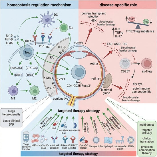
Research Progress on the Regulatory Role of Treg Cells in Inflammatory Eye Diseases

 , , , , and
Abstract
1. Introduction
2. The Physiological Distribution of Treg Cells in the Eye and the Mechanism of Maintaining Immune Homeostasis
2.1. Immunologic Characteristics of Ocular Microenvironment
2.2. The Specific Distribution of Treg Cells in Ocular Tissues
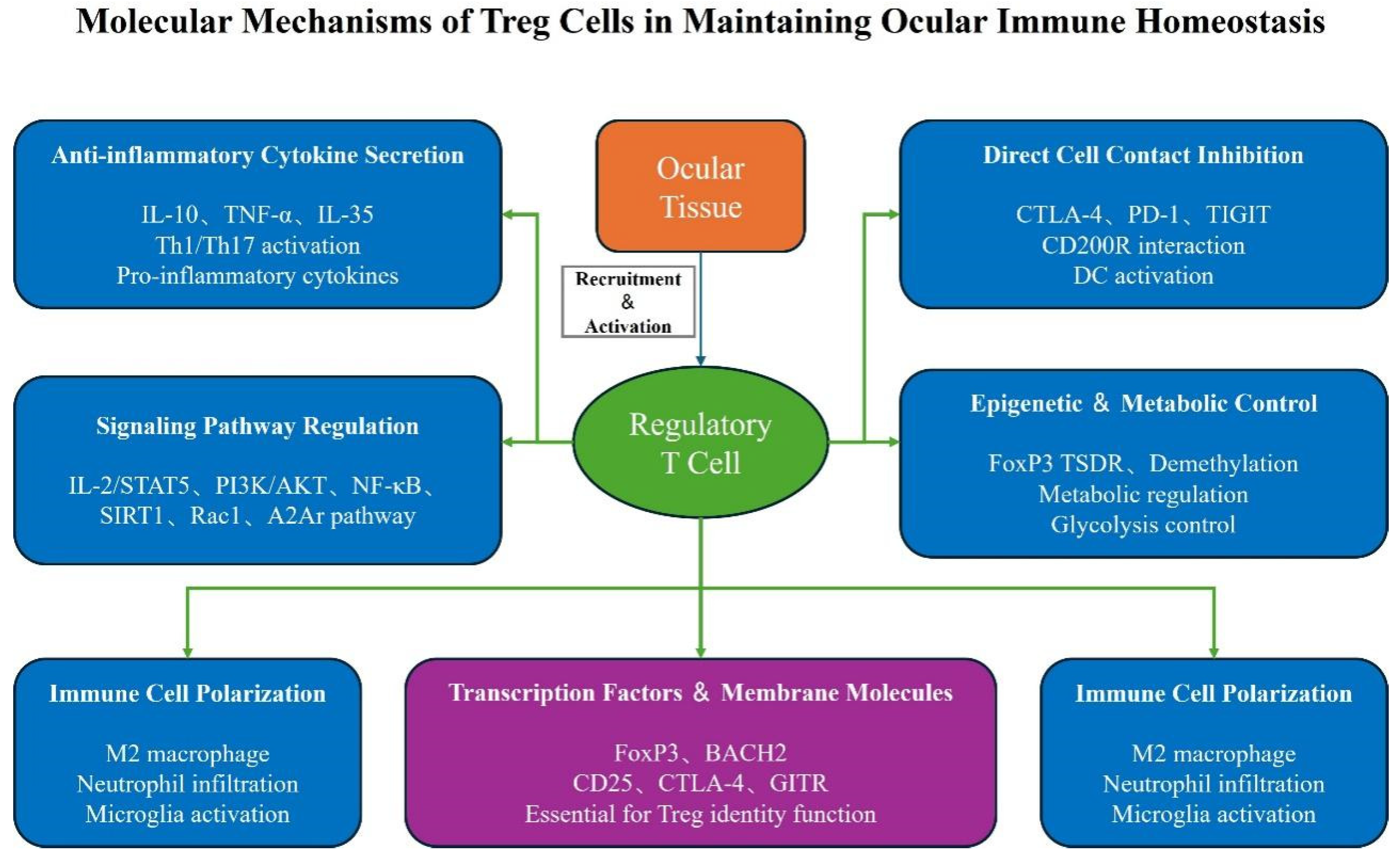
2.3. The Molecular Mechanism of Treg Cells in Maintaining Ocular Immune Homeostasis
2.3.1. Anti-Inflammatory Cytokine Secretion
2.3.2. Direct Contact Inhibition of Cells
2.3.3. Signal Pathway Regulation
2.3.4. Epigenetics and Metabolic Regulation
2.3.5. Immune Cell Polarization Regulation
2.3.6. Cytokine Networks and Chemokine Receptor-Mediated
2.3.7. Transcription Factors and Membrane Molecules Regulation
3. The Regulatory Role of Treg Cells in Different Inflammatory Eye Diseases and Experimental Evidence
3.1. Corneal Inflammatory Diseases
3.1.1. Corneal Transplant Rejection Regulatory Mechanism
3.1.2. Herpes Simplex Keratitis (HSK)
3.1.3. Other Corneal Inflammations
3.2. Uveitis
3.2.1. Experimental Autoimmune Uveitis (EAU)
3.2.2. Tuberculous Uveitis
3.2.3. Chronic Autoimmune Uveitis (CAU)
3.3. Conjunctivitis
3.3.1. Allergic Conjunctivitis (AC)
3.3.2. Conjunctival Squamous Cell Carcinoma-Related Conjunctivitis
3.4. Retinal Choroiditis
3.4.1. Wet Age-Related Macular Degeneration (AMD)
3.4.2. Retinal Detachment
3.4.3. Oxygen-Induced Retinopathy (OIR)
3.4.4. Retinal I/R Injury
3.5. Other Inflammatory Eye Diseases
3.5.1. Dry Eye Disease (DED)
3.5.2. Autoimmune Dacryoadenitis
3.5.3. Uveal Melanoma-Associated Ocular Inflammation
4. Key Factors Influencing the Function of Treg Cells in Inflammatory Eye Diseases
4.1. Drug-Related Factors Influencing Treg Function
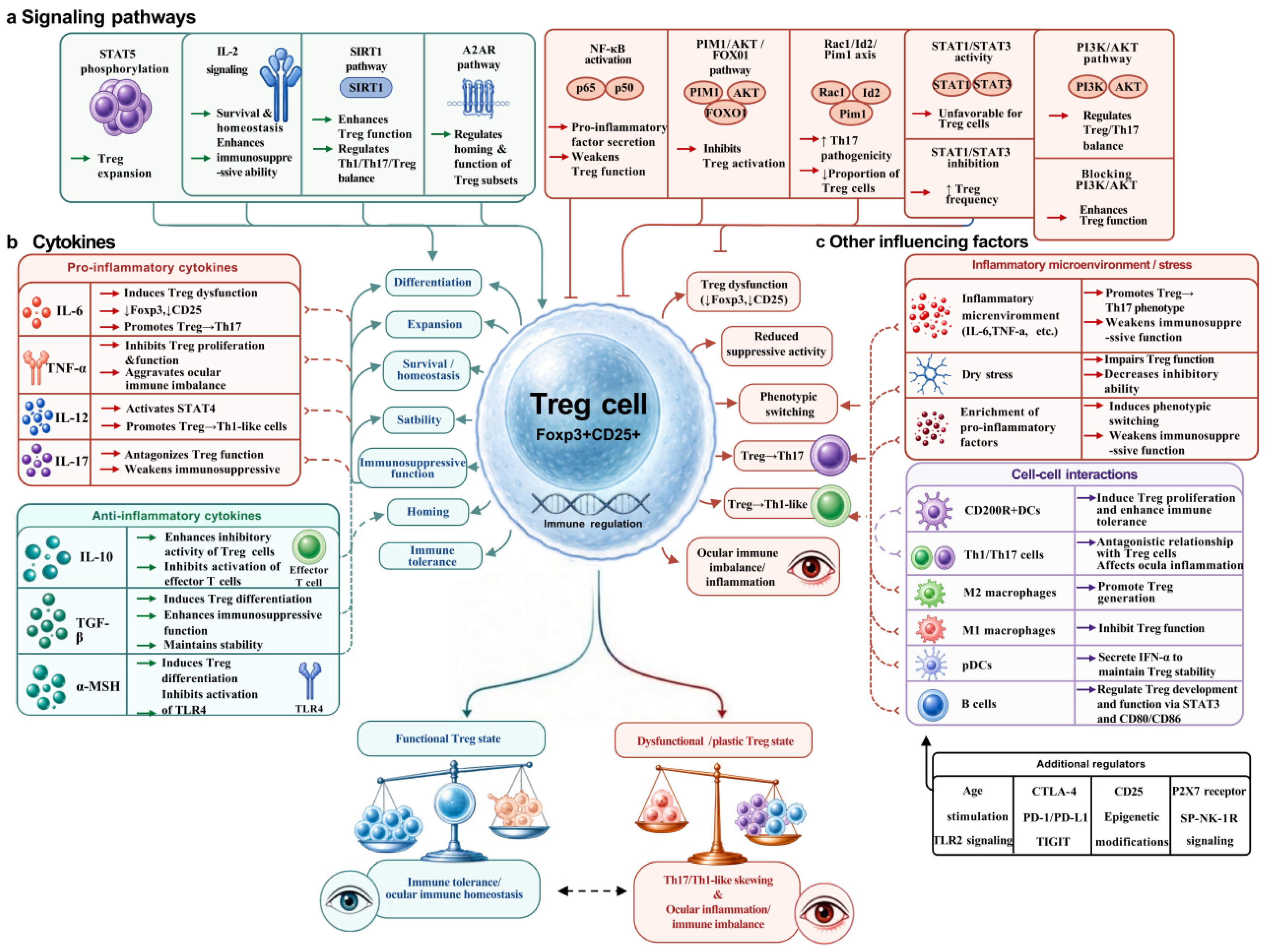
4.2. Signaling Pathways
4.3. Cytokines
4.4. Other Influencing Factors
5. Treg Cell-Based Therapeutic Strategies for Inflammatory Eye Diseases
5.1. Targeted Interventions to Enhance Treg Cell Function
5.1.1. Cell Therapy
5.1.2. Pharmacological Interventions
5.2. Therapeutic Approaches for Regulating Treg-Related Signaling Pathways
5.2.1. Targeting the P2X7 Receptor
5.2.2. Targeting the Cytokine Network
5.2.3. Comparative Analysis: Cell Therapy vs. P2X7R Antagonism
5.3. Combined Treatment Strategy
5.3.1. Immunomodulation + Anti-Inflammatory Therapy
5.3.2. Targeted Delivery Systems
6. Discussion and Outlook
6.1. Core Regulatory Characteristics and Mechanism Correlation of Treg Cells
6.2. Research Limitations
- (1)
- Insufficient analysis of Treg cell heterogeneity: Treg cells exhibit significant subpopulation heterogeneity (such as CD25lo/hi, antigen-specific Treg, effector memory Treg), and the functional differences among different subpopulations in different inflammatory eye diseases have not been fully clarified [24,31]. For instance, CD25loTreg cells are prone to transform into ex-Treg cells and promote inflammation after HSV-1 infection [24], while antigen-specific Treg cells show stronger targeting in the treatment of uveitis [3]. However, there is currently a lack of systematic comparative studies on the phenotypes and functions of Treg subpopulations in different eye diseases, which makes it impossible to realize the precise regulation of Treg subpopulations.
- (2)
- Disconnection between basic research and clinical translation: Most existing studies are based on animal models (such as mouse EAU models, corneal transplantation models, OIR models), and human clinical data is limited [3,14]. For example, low-dose IL-2 can effectively expand Treg cells and alleviate retinal inflammation in animal models [1], but its efficacy and safety in human non-infectious uveitis still require large-scale clinical trials for verification; Treg cell adoptive transfer shows good anti-inflammatory effects in animal experiments [10,54], but faces technical bottlenecks such as cell source, in vitro expansion efficiency, and in vivo homing specificity in clinical application [3,5]. In addition, the lack of standardized clinical detection methods for Treg cells also restricts the clinical application of Treg cell-based therapies.
- (3)
- Insufficient research on the mechanisms of some diseases: The regulatory role and mechanism of Treg cells in some inflammatory eye diseases such as endophthalmitis and retinal chorioretinitis remain unclear. Existing research mainly focuses on corneal inflammation, uveitis and dry eye [39,74], lacking original experimental evidence for these diseases, which limits the application scope of Treg-related treatment strategies.
- (4)
- Imperfect targeted delivery system: The current drug interventions (such as sCD83, NAD+ and IL-2) are mostly administered systemically or delivered locally in a simple manner, which leads to problems such as poor ocular targeting, insufficient local drug concentration, and systemic side effects [7,13,29]. Although delivery carriers such as liposomes, nanoparticles, and hydrogels have shown potential [30,59,71], it is still necessary to optimize the particle size, biocompatibility, and drug release kinetics of the carriers to enhance the targeting and efficacy of Treg-related interventions.
6.3. Future Research Directions
- (1)
- Systematically analyze the disease-specific functions of Treg subgroups by multi-omics technologies: Utilize single-cell RNA sequencing, flow cytometry, spatial transcriptomics and other multi-omics technologies to systematically analyze the phenotypic characteristics (surface markers, transcription factor expression, metabolic characteristics) of and functional differences in Treg subgroups in different inflammatory eye diseases, clarify the regulatory roles of antigen-specific Tregs, effector memory Tregs and other subgroups in different ocular inflammatory pathologies, and screen the specific surface markers of functional Treg subgroups. On this basis, we aim to develop precise targeted intervention strategies for Treg subgroups, realize the precise regulation of Treg cells, and improve the therapeutic specificity and efficiency.
- (2)
- Optimize Treg-related targeted delivery systems based on ocular tissue specificity: Based on the anatomical characteristics and tissue specificity of different ocular tissues (cornea, retina, uvea, etc.), develop novel ocular targeted delivery carriers with good biocompatibility, high targeting and sustained release, such as cornea-penetrating nanoparticles, retinal-targeted liposomes, injectable hydrogels and biodegradable microneedle patches. Realize the local efficient delivery of Treg cells and related drugs, increase the concentration of drugs/cells in the target ocular tissue, prolong the retention time, and reduce systemic side effects. In addition, we aim to develop personalized targeted delivery systems according to the different pathological characteristics of patients, further improve the therapeutic effect.
- (3)
- Accelerate the clinical translational research of Treg cell-based therapeutic strategies: Carry out small-sample clinical trials to evaluate the safety, efficacy, and optimal dosage of Treg-related interventions (e.g., low-dose IL-2, Treg adoptive transfer, and STAT3 inhibitors) in patients with human inflammatory eye diseases such as non-infectious uveitis and high-risk corneal transplant rejection. Establish a standardized clinical sample bank to systematically collect clinical data (such as disease severity, treatment response, and prognosis) and biological samples (peripheral blood, aqueous humor, vitreous fluid) from patients, analyze the correlation between Treg cell proportion, functional status, and disease progression/prognosis, and screen potential Treg-related biomarkers for disease diagnosis, efficacy evaluation, and prognosis prediction [37,80]. Strengthen the cooperation between basic research institutions and clinical institutions to promote the two-way translation of basic research results and clinical needs.
- (4)
- Develop novel regulatory targets based on multi-omics technologies: Combine genomics, transcriptomics, proteomics, metabolomics, and epigenomics technologies to comprehensively analyze the molecular mechanisms of Treg cell function regulation in inflammatory eye diseases. Focus on exploring the epigenetic regulatory mechanisms (such as TSDR methylation [24], TET2/NT5E axis [35]) and metabolic reprogramming characteristics (such as glycolysis, oxidative phosphorylation, amino acid metabolism) of Treg cells in different ocular inflammatory microenvironments and identify new key regulatory molecules and signaling pathways (such as PIM1, Rac1 [33,63]). On this basis, we aim to develop novel targeted drugs with higher specificity and stronger efficacy, and provide new ideas and targets for the precise treatment of inflammatory eye diseases.
- (5)
- Optimize combined treatment strategies based on precision medicine: According to the different pathological types, severity degrees, and patient individual characteristics of inflammatory eye diseases, develop personalized combined treatment strategies. Explore the optimal combination of “immune regulation + anti-inflammatory treatment” (such as Treg adoptive transfer + local anti-inflammatory drugs, IL-6 antagonists + ocular surface anti-inflammatory preparations [60,63]), and achieve synergistic enhancement of Treg function, rapid control of acute inflammation, reducing the dose-dependent toxicity of single treatment, and achieving both symptomatic and root-cause treatment [2,42].
Author Contributions
Funding
Data Availability Statement
Acknowledgments
Conflicts of Interest
References
- Deliyanti, D.; Suphapimol, V.; Joglekar, A.; Jayasimhan, A.; Wilkinson-Berka, J.L. Immunotherapy with low-dose IL-2 attenuates vascular injury in mice with diabetic and neovascular retinopathy by restoring the balance between Foxp3+ Tregs and CD8+ T cells. Diabetologia 2025, 68, 1559–1573. [Google Scholar] [CrossRef] [Scilit] [PubMed] [PubMed Central]
- Duan, R.; Xie, L.; Li, H.; Wang, R.; Liu, X.; Tao, T.; Yang, S.; Gao, Y.; Lin, X.; Su, W. Insights gained from Single-Cell analysis of immune cells on Cyclosporine A treatment in autoimmune uveitis. Biochem. Pharmacol. 2022, 202, 115116. [Google Scholar] [CrossRef] [Scilit] [PubMed]
- Sun, J.; Wang, T.; Bian, J.; Shi, W.; Ruan, Q. Immune tolerance induced in the anterior chamber ameliorates corneal transplant rejection. Clin. Immunol. 2023, 257, 109797. [Google Scholar] [CrossRef] [Scilit] [PubMed]
- Fan, N.W.; Li, J.; Mittal, S.K.; Foulsham, W.; Elbasiony, E.; Huckfeldt, R.M.; Chauhan, S.K.; Chen, Y. Characterization of Clinical and Immune Responses in an Experimental Chronic Autoimmune Uveitis Model. Am. J. Pathol. 2021, 191, 425–437. [Google Scholar] [CrossRef] [Scilit] [PubMed] [PubMed Central]
- Scholand, K.K.; Schaefer, L.; Govindarajan, G.; Yu, Z.; Galletti, J.G.; de Paiva, C.S. Aged regulatory T cells fail to control autoimmune lacrimal gland pathogenic CD4+ T cells. Geroscience 2025, 47, 4219–4240. [Google Scholar] [CrossRef] [Scilit] [PubMed] [PubMed Central]
- Li, N.; Gao, Z.; Zhao, L.; Du, B.; Ma, B.; Nian, H.; Wei, R. MSC-Derived Small Extracellular Vesicles Attenuate Autoimmune Dacryoadenitis by Promoting M2 Macrophage Polarization and Inducing Tregs via miR-100-5p. Front. Immunol. 2022, 13, 888949. [Google Scholar] [CrossRef] [Scilit] [PubMed] [PubMed Central]
- Peckert-Maier, K.; Schönberg, A.; Wild, A.B.; Royzman, D.; Braun, G.; Stich, L.; Hadrian, K.; Tripal, P.; Cursiefen, C.; Steinkasserer, A.; et al. Pre-incubation of corneal donor tissue with sCD83 improves graft survival via the induction of alternatively activated macrophages and tolerogenic dendritic cells. Am. J. Transplant. 2022, 22, 438–454. [Google Scholar] [CrossRef] [Scilit] [PubMed]
- Peng, Z.; Nagarajan, V.; Horai, R.; Jittayasothorn, Y.; Mattapallil, M.J.; Caspi, R.R. Ocular immune privilege in action: The living eye imposes unique regulatory and anergic gene signatures on uveitogenic T cells. Cell Rep. 2025, 44, 115780. [Google Scholar] [CrossRef] [Scilit] [PubMed] [PubMed Central]
- Neelam, S.; Niederkorn, J.Y. Corneal Nerve Ablation Abolishes Ocular Immune Privilege by Downregulating CD103 on T Regulatory Cells. Investig. Ophthalmol. Vis. Sci. 2020, 61, 25. [Google Scholar] [CrossRef] [Scilit] [PubMed] [PubMed Central]
- Agrawal, M.; Rasiah, P.K.; Bajwa, A.; Rajasingh, J.; Gangaraju, R. Mesenchymal Stem Cell Induced Foxp3(+) Tregs Suppress Effector T Cells and Protect against Retinal Ischemic Injury. Cells 2021, 10, 3006. [Google Scholar] [CrossRef] [Scilit] [PubMed] [PubMed Central]
- Deliyanti, D.; Talia, D.M.; Zhu, T.; Maxwell, M.J.; Agrotis, A.; Jerome, J.R.; Hargreaves, E.M.; Gerondakis, S.; Hibbs, M.L.; Mackay, F.; et al. Foxp3+ Tregs are recruited to the retina to repair pathological angiogenesis. Nat. Commun. 2017, 8, 748. [Google Scholar] [CrossRef] [Scilit] [PubMed] [PubMed Central]
- Li, S.; Zhang, P.; Li, A.; Bao, J.; Pan, Z.; Jie, Y. Rho-kinase inhibitor alleviates CD4+T cell mediated corneal graft rejection by modulating its STAT3 and STAT5 activation. Exp. Eye Res. 2024, 242, 109857. [Google Scholar] [CrossRef] [Scilit] [PubMed]
- Guo, J.; Li, B.; Wang, J.; Guo, R.; Tian, Y.; Song, S.; Guo, L. Protective effect and mechanism of nicotinamide adenine dinucleotide against optic neuritis in mice with experimental autoimmune encephalomyelitis. Int. Immunopharmacol. 2021, 98, 107846. [Google Scholar] [CrossRef] [Scilit] [PubMed]
- Lee, A.Y.; Foulsham, W. Regulatory T Cells: Therapeutic Opportunities in Uveitis. Front. Ophthalmol. 2022, 2, 901144. [Google Scholar] [CrossRef] [Scilit] [PubMed] [PubMed Central]
- Tagami, M.; Kakehashi, A.; Katsuyama-Yoshikawa, A.; Misawa, N.; Sakai, A.; Wanibuchi, H.; Azumi, A.; Honda, S. FOXP3 and CXCR4-positive regulatory T cells in the tumor stroma as indicators of tumor immunity in the conjunctival squamous cell carcinoma microenvironment. PLoS ONE 2022, 17, e0263895. [Google Scholar] [CrossRef] [Scilit] [PubMed] [PubMed Central]
- Ni, Y.; Wang, L.; Liu, C.; Li, Z.; Yang, J.; Zeng, J. Gene expression profile analyses to identify potential biomarkers for myopia. Eye 2023, 37, 1264–1270. [Google Scholar] [CrossRef] [Scilit] [PubMed] [PubMed Central]
- Zhang, W.; Schönberg, A.; Bock, F.; Cursiefen, C. Posttransplant VEGFR1R2 Trap Eye Drops Inhibit Corneal (Lymph)angiogenesis and Improve Corneal Allograft Survival in Eyes at High Risk of Rejection. Transl. Vis. Sci. Technol. 2022, 11, 6. [Google Scholar] [CrossRef] [Scilit] [PubMed] [PubMed Central]
- Stepp, M.A.; Menko, A.S. Immune responses to injury and their links to eye disease. Transl. Res. 2021, 236, 52–71. [Google Scholar] [CrossRef] [Scilit] [PubMed] [PubMed Central]
- Yi, W.; Wang, H.; Liu, S.; Zhang, J.; Ji, Y.; Wang, H.; Zhao, X.; Chen, J.; Luo, W. 6-Shogaol Alleviates Endotoxin-Induced Uveitis via Activation of HIF-1α to Regulate Inflammatory Responses and Endoplasmic Reticulum Stress. Investig. Ophthalmol. Vis. Sci. 2025, 66, 43. [Google Scholar] [CrossRef] [Scilit] [PubMed] [PubMed Central]
- Trujillo-Vargas, C.M.; Mauk, K.E.; Hernandez, H.; de Souza, R.G.; Yu, Z.; Galletti, J.G.; Dietrich, J.; Paulsen, F.; de Paiva, C.S. Immune phenotype of the CD4+ T cells in the aged lymphoid organs and lacrimal glands. Geroscience 2022, 44, 2105–2128. [Google Scholar] [CrossRef] [Scilit] [PubMed] [PubMed Central]
- Cai, Y.; Chen, J.; Sun, H.; Zhou, T.; Cai, X.; Fu, Y. Crosstalk between TRPV1 and immune regulation in Fuchs endothelial corneal dystrophy. Clin. Immunol. 2023, 254, 109701. [Google Scholar] [CrossRef] [Scilit] [PubMed]
- Xiao, S.; Yang, Y.; Miao, W.; Lyu, C.; Tao, J.; Yu, Y. Activation of the STAT5 Signaling Pathway by Yiqi Jiedu Formula Induces Regulatory T Cell-Mediated Alleviation of Corneal Immunopathological Damage in Mice With Recurrent Herpes Simplex Keratitis. Front. Pharmacol. 2022, 12, 790787. [Google Scholar] [CrossRef] [Scilit] [PubMed] [PubMed Central]
- Jamali, A.; Hu, K.; Sendra, V.G.; Blanco, T.; Lopez, M.J.; Ortiz, G.; Qazi, Y.; Zheng, L.; Turhan, A.; Harris, D.L.; et al. Characterization of Resident Corneal Plasmacytoid Dendritic Cells and Their Pivotal Role in Herpes Simplex Keratitis. Cell Rep. 2020, 32, 108099. [Google Scholar] [CrossRef] [Scilit] [PubMed] [PubMed Central]
- Bhela, S.; Varanasi, S.K.; Jaggi, U.; Sloan, S.S.; Rajasagi, N.K.; Rouse, B.T. The Plasticity and Stability of Regulatory T Cells during Viral-Induced Inflammatory Lesions. J. Immunol. 2017, 199, 1342–1352. [Google Scholar] [CrossRef] [Scilit] [PubMed] [PubMed Central]
- Taketani, Y.; Marmalidou, A.; Dohlman, T.H.; Singh, R.B.; Amouzegar, A.; Chauhan, S.K.; Chen, Y.; Dana, R. Restoration of Regulatory T-Cell Function in Dry Eye Disease by Antagonizing Substance P/Neurokinin-1 Receptor. Am. J. Pathol. 2020, 190, 1859–1866. [Google Scholar] [CrossRef] [Scilit] [PubMed] [PubMed Central]
- Corliss, B.A.; Ray, H.C.; Mathews, C.; Fitzgerald, K.; Doty, R.W.; Smolko, C.M.; Shariff, H.; Peirce, S.M.; Yates, P.A. Myh11 Lineage Corneal Endothelial Cells and ASCs Populate Corneal Endothelium. Investig. Ophthalmol. Vis. Sci. 2019, 60, 5095–5103. [Google Scholar] [CrossRef] [Scilit] [PubMed]
- Fang, F.; A, T.; Chen, J.; Li, S.; Zhou, T.; Chen, L.; Fu, Y.; Shao, C. Therapeutic potential of regulatory T cells for stem cell regulation: Insights from Treg-mediated enhancement of limbal stem cell functions. iScience 2025, 28, 112515. [Google Scholar] [CrossRef] [Scilit] [PubMed]
- Shi, W.H.; Wang, L.M.; Yan, H.J.; Liu, S.L.; Yang, X.; Yang, X.J.; Che, C.Y. CD3ε of a pan T cell marker involved in mouse Aspergillus fumigatus keratitis. Int. J. Ophthalmol. 2024, 17, 616–624. [Google Scholar] [CrossRef] [Scilit] [PubMed] [PubMed Central]
- Singh, R.B.; Blanco, T.; Mittal, S.K.; Alemi, H.; Chauhan, S.K.; Chen, Y.; Dana, R. Pigment Epithelium-Derived Factor Enhances the Suppressive Phenotype of Regulatory T Cells in a Murine Model of Dry Eye Disease. Am. J. Pathol. 2021, 191, 720–729. [Google Scholar] [CrossRef] [Scilit] [PubMed] [PubMed Central]
- Zhang, T.; Han, X.; Zhong, Y.; Kam, H.T.; Qiao, D.; Chen, Z.; Chan, K.W.Y.; Chong, W.P.; Chen, J. Regulatory T cell intravitreal delivery using hyaluronan methylcellulose hydrogel improves therapeutic efficacy in experimental autoimmune uveitis. Biomater. Adv. 2023, 151, 213496. [Google Scholar] [CrossRef] [Scilit] [PubMed]
- Silver, P.B.; Horai, R.; Chen, J.; Jittayasothorn, Y.; Chan, C.C.; Villasmil, R.; Kesen, M.R.; Caspi, R.R. Retina-specific T regulatory cells bring about resolution and maintain remission of autoimmune uveitis. J. Immunol. 2015, 194, 3011–3019, Erratum in J. Immunol. 2015, 195, 393. https://doi.org/10.4049/jimmunol.1501038. [Google Scholar] [CrossRef] [Scilit] [PubMed] [PubMed Central]
- Bowers, C.E.; Calder, V.L.; Greenwood, J.; Eskandarpour, M. Experimental Autoimmune Uveitis: An Intraocular Inflammatory Mouse Model. J. Vis. Exp. 2022, 179, e61832. [Google Scholar] [CrossRef] [Scilit] [PubMed]
- Li, H.; Xie, L.; Zhu, L.; Li, Z.; Wang, R.; Liu, X.; Huang, Z.; Chen, B.; Gao, Y.; Wei, L.; et al. Multicellular immune dynamics implicate PIM1 as a potential therapeutic target for uveitis. Nat. Commun. 2022, 13, 5866. [Google Scholar] [CrossRef] [Scilit] [PubMed] [PubMed Central]
- Livne-Bar, I.; Maurya, S.; Gronert, K.; Sivak, J.M. Lipoxins A4 and B4 inhibit glial cell activation via CXCR3 signaling in acute retinal neuroinflammation. J. Neuroinflamm. 2024, 21, 18. [Google Scholar] [CrossRef] [Scilit] [PubMed] [PubMed Central]
- Ku, L.C.; Sheu, M.L.; Cheng, H.H.; Lee, C.Y.; Tsai, Y.C.; Tsai, C.Y.; Lin, K.H.; Lai, L.C.; Lai, D.W. Melatonin protects retinal integrity through mediated immune homeostasis in the sodium iodate-induced mouse model of age-related macular degeneration. BioMed. Pharmacother. 2023, 161, 114476. [Google Scholar] [CrossRef] [Scilit] [PubMed]
- Li, J.; Ren, Y.; Li, H.; Zheng, Z. Rac1 overexpression promotes Treg-derived cytokines to mediate choroidal neovascularization in wet age-related macular degeneration. Braz. J. Med. Biol. Res. 2025, 58, e14187. [Google Scholar] [CrossRef] [Scilit] [PubMed] [PubMed Central]
- Sharma, R.K.; Gupta, A.; Kamal, S.; Bansal, R.; Singh, N.; Sharma, K.; Virk, S.; Sachdeva, N. Role of Regulatory T Cells in Tubercular Uveitis. Ocul. Immunol. Inflamm. 2018, 26, 27–36. [Google Scholar] [CrossRef] [Scilit] [PubMed]
- Nagarkatti-Gude, N.; Bronkhorst, I.H.; van Duinen, S.G.; Luyten, G.P.; Jager, M.J. Cytokines and chemokines in the vitreous fluid of eyes with uveal melanoma. Investig. Ophthalmol. Vis. Sci. 2012, 53, 6748–6755. [Google Scholar] [CrossRef] [Scilit] [PubMed]
- Déchelle-Marquet, P.A.; Che, Y.; Roux, C.; Blond, F.; Ronning, K.E.; Augustin, S.; Lagouge-Roussey, P.; Nous, C.; Touhami, S.; Bodaghi, B.; et al. Macrophage expression of P2X7 controls autoimmune uveitis. J. Neuroinflamm. 2025, 22, 212. [Google Scholar] [CrossRef] [Scilit] [PubMed] [PubMed Central]
- Matas, J.; Llorenç, V.; Fonollosa, A.; Díaz-Valle, D.; Esquinas, C.; de la Maza, M.T.S.; Figueras-Roca, M.; Artaraz, J.; Berasategui, B.; Mesquida, M.; et al. Systemic Regulatory T Cells and IL-6 as Prognostic Factors for Anatomical Improvement of Uveitic Macular Edema. Front. Immunol. 2020, 11, 579005. [Google Scholar] [CrossRef] [Scilit] [PubMed] [PubMed Central]
- Ratay, M.L.; Balmert, S.C.; Bassin, E.J.; Little, S.R. Controlled release of an HDAC inhibitor for reduction of inflammation in dry eye disease. Acta Biomater. 2018, 71, 261–270. [Google Scholar] [CrossRef] [Scilit] [PubMed] [PubMed Central]
- Surico, P.L.; Lee, S.; Singh, R.B.; Naderi, A.; Bhullar, S.; Blanco, T.; Chen, Y.; Dana, R. Local administration of myeloid-derived suppressor cells prevents progression of immune-mediated dry eye disease. Exp. Eye Res. 2024, 242, 109871. [Google Scholar] [CrossRef] [Scilit] [PubMed] [PubMed Central]
- Nieto, J.E.; Casanova, I.; Serna-Ojeda, J.C.; Graue-Hernández, E.O.; Quintana, G.; Salazar, A.; Jiménez-Martinez, M.C. Increased Expression of TLR4 in Circulating CD4+T Cells in Patients with Allergic Conjunctivitis and In Vitro Attenuation of Th2 Inflammatory Response by Alpha-MSH. Int. J. Mol. Sci. 2020, 21, 7861. [Google Scholar] [CrossRef] [Scilit] [PubMed] [PubMed Central]
- Dou, X.; Zhang, W.; Zhang, X. Exploring the immunological underpinnings of allergic conjunctivitis: The role of CD4 + T Cells and SLAM expression in ocular surface disease. Int. Ophthalmol. 2025, 45, 77. [Google Scholar] [CrossRef] [Scilit] [PubMed]
- Zhang, J.; Wu, J.; Lu, D.; To, C.H.; Lam, T.C.; Lin, B. Retinal Proteomic Analysis in a Mouse Model of Endotoxin-Induced Uveitis Using Data-Independent Acquisition-Based Mass Spectrometry. Int. J. Mol. Sci. 2022, 23, 6464. [Google Scholar] [CrossRef] [Scilit] [PubMed] [PubMed Central]
- Li, D.; Ou, Q.; Shen, Q.; Lu, M.M.; Xu, J.Y.; Jin, C.; Gao, F.; Wang, J.; Zhang, J.; Zhang, J.; et al. Subconjunctival injection of human umbilical cord mesenchymal stem cells alleviates experimental allergic conjunctivitis via regulating T cell response. Stem Cell Res. Ther. 2023, 14, 281. [Google Scholar] [CrossRef] [Scilit] [PubMed] [PubMed Central]
- Coco, G.; Foulsham, W.; Nakao, T.; Yin, J.; Amouzegar, A.; Taketani, Y.; Chauhan, S.K.; Dana, R. Regulatory T cells promote corneal endothelial cell survival following transplantation via interleukin-10. Am. J. Transplant. 2020, 20, 389–398. [Google Scholar] [CrossRef] [Scilit] [PubMed] [PubMed Central]
- Muhammad, F.; Wang, D.; Montieth, A.; Lee, S.; Preble, J.; Foster, C.S.; Larson, T.A.; Ding, K.; Dvorak, J.D.; Lee, D.J. PD-1+ melanocortin receptor dependent-Treg cells prevent autoimmune disease. Sci. Rep. 2019, 9, 16941. [Google Scholar] [CrossRef] [Scilit] [PubMed] [PubMed Central]
- Mbanefo, E.C.; Yan, M.; Kang, M.; Alhakeem, S.A.; Jittayasothorn, Y.; Yu, C.R.; Parihar, A.; Singh, S.; Egwuagu, C.E. STAT3-Specific Single Domain Nanobody Inhibits Expansion of Pathogenic Th17 Responses and Suppresses Uveitis in Mice. Front. Immunol. 2021, 12, 724609. [Google Scholar] [CrossRef] [Scilit] [PubMed] [PubMed Central]
- Peters, K.; McDonald, T.; Muhammad, F.; Walsh, M.; Drenen, K.; Montieth, A.; Stephen Foster, C.; Lee, D.J. A2Ar-dependent PD-1+ and TIGIT+ Treg cells have distinct homing requirements to suppress autoimmune uveitis in mice. Mucosal Immunol. 2023, 16, 422–431. [Google Scholar] [CrossRef] [Scilit] [PubMed] [PubMed Central]
- Polansky, J.K.; Schreiber, L.; Thelemann, C.; Ludwig, L.; Krüger, M.; Baumgrass, R.; Cording, S.; Floess, S.; Hamann, A.; Huehn, J. Methylation matters: Binding of Ets-1 to the demethylated Foxp3 gene contributes to the stabilization of Foxp3 expression in regulatory T cells. J. Mol. Med. 2010, 88, 1029–1040. [Google Scholar] [CrossRef] [Scilit] [PubMed]
- Gong, X.; Fan, J.; Huang, H.; Xu, F.; Hu, K.; Liu, J.; Tan, Y.; Chen, F. Plasma Metabolic Profiles of Chronic and Recurrent Uveitis Treated by Artesunate in Lewis Rats. Biomedicines 2025, 13, 821. [Google Scholar] [CrossRef] [Scilit] [PubMed] [PubMed Central]
- Kang, M.; Lee, H.S.; Choi, J.K.; Yu, C.R.; Egwuagu, C.E. Deletion of Irf4 in T Cells Suppressed Autoimmune Uveitis and Dysregulated Transcriptional Programs Linked to CD4+ T Cell Differentiation and Metabolism. Int. J. Mol. Sci. 2021, 22, 2775. [Google Scholar] [CrossRef] [Scilit] [PubMed] [PubMed Central]
- Yan, D.; Yu, F.; Chen, L.; Yao, Q.; Yan, C.; Zhang, S.; Wu, N.; Gong, D.; Sun, H.; Fu, Y.; et al. Subconjunctival Injection of Regulatory T Cells Potentiates Corneal Healing Via Orchestrating Inflammation and Tissue Repair After Acute Alkali Burn. Investig. Ophthalmol. Vis. Sci. 2020, 61, 22. [Google Scholar] [CrossRef] [Scilit] [PubMed] [PubMed Central]
- Liu, X.; Gu, C.; Lv, J.; Jiang, Q.; Ding, W.; Huang, Z.; Liu, Y.; Su, Y.; Zhang, C.; Xu, Z.; et al. Progesterone attenuates Th17-cell pathogenicity in autoimmune uveitis via Id2/Pim1 axis. J. Neuroinflamm. 2023, 20, 144, Erratum in J. Neuroinflamm. 2025, 22, 99. https://doi.org/10.1186/s12974-025-03412-8. [Google Scholar] [CrossRef] [Scilit] [PubMed] [PubMed Central]
- Bian, J.; Wang, T.; Sun, J.; He, X.; Wu, Z.; Zhang, S.; Chi, H.; Fan, T.; Wang, S.; Shi, W.; et al. Targeting NF-κB c-Rel in regulatory T cells to treat corneal transplantation rejection. Am. J. Transplant. 2021, 21, 3858–3870. [Google Scholar] [CrossRef] [Scilit] [PubMed]
- Fu, T.; Lu, W.; Wu, D.; Xu, L.; Wu, G. Exploring the immunomodulatory mechanism of total glucosides of paeony on Sjögren’s syndrome dry eye disease based on the “gut-eye axis” pathway. Int. Ophthalmol. 2025, 45, 230. [Google Scholar] [CrossRef] [Scilit] [PubMed]
- Yang, Y.; Yin, X.; Yi, J.; Peng, X. MiR-146a overexpression effectively improves experimental allergic conjunctivitis through regulating CD4+CD25-T cells. BioMed. Pharmacother. 2017, 94, 937–943. [Google Scholar] [CrossRef] [Scilit] [PubMed]
- A, T.X.; Shao, C.Y.; Fu, Y. Research progress on the role of regulatory T cells in ocular surface diseases. J. Shanghai Jiaotong Univ. 2022, 42, 1145–1150. [Google Scholar]
- Ortiz, G.; Blanco, T.; Singh, R.B.; Kahale, F.; Wang, S.; Chen, Y.; Dana, R. IL-6 induces Treg dysfunction in desiccating stress-induced dry eye disease. Exp. Eye Res. 2024, 246, 110006. [Google Scholar] [CrossRef] [Scilit] [PubMed] [PubMed Central]
- Bing, S.J.; Shemesh, I.; Chong, W.P.; Horai, R.; Jittayasothorn, Y.; Silver, P.B.; Sredni, B.; Caspi, R.R. AS101 ameliorates experimental autoimmune uveitis by regulating Th1 and Th17 responses and inducing Treg cells. J. Autoimmun. 2019, 100, 52–61. [Google Scholar] [CrossRef] [Scilit] [PubMed] [PubMed Central]
- Llorenç, V.; Nakamura, Y.; Metea, C.; Karstens, L.; Molins, B.; Lin, P. Antimetabolite Drugs Exhibit Distinctive Immunomodulatory Mechanisms and Effects on the Intestinal Microbiota in Experimental Autoimmune Uveitis. Investig. Ophthalmol. Vis. Sci. 2022, 63, 30. [Google Scholar] [CrossRef] [Scilit] [PubMed] [PubMed Central]
- Gu, C.; Liu, Y.; Lv, J.; Zhang, C.; Huang, Z.; Jiang, Q.; Gao, Y.; Tao, T.; Su, Y.; Chen, B.; et al. Kurarinone regulates Th17/Treg balance and ameliorates autoimmune uveitis via Rac1 inhibition. J. Adv. Res. 2025, 69, 381–398. [Google Scholar] [CrossRef] [Scilit] [PubMed] [PubMed Central]
- Tahvildari, M.; Dana, R. Low-Dose IL-2 Therapy in Transplantation, Autoimmunity, and Inflammatory Diseases. J. Immunol. 2019, 203, 2749–2755. [Google Scholar] [CrossRef] [Scilit] [PubMed] [PubMed Central]
- Gao, Q.; Peng, J.; Xiong, M.; Zhou, S.; Wang, X.; Lu, J.; Guo, Y.; Peng, Q.; Zeng, M.; Song, H. Sinomenine alleviates experimental autoimmune uveitis in rats: Possible involvement of PI3K/AKT and NF-κB signaling pathways. Eur. J. Pharmacol. 2025, 996, 177571. [Google Scholar] [CrossRef] [Scilit] [PubMed]
- Alam, K.; Raina, A.; Das, B.; Bhanja, S.; Ghosh, S.; Forrester, J.V.; Basu, S. Antigen-specific polyfunctional cytotoxic T cells differentiate intraocular from peripheral blood immune responses in posterior uveitis. Clin. Transl. Immunol. 2025, 14, e70036. [Google Scholar] [CrossRef] [Scilit] [PubMed] [PubMed Central]
- Guedes, M.C.E.; Arroz, M.J.; Martins, C.; Angelo-Dias, M.; Proença, R.D.; Borrego, L.M. Regulatory T cells and IL-17A levels in noninfectious uveitis. Graefes Arch. Clin. Exp. Ophthalmol. 2020, 258, 1269–1278. [Google Scholar] [CrossRef] [Scilit] [PubMed]
- Oladipupo, F.O.; Yu, C.R.; Olumuyide, E.; Jittaysothorn, Y.; Choi, J.K.; Egwuagu, C.E. STAT3 deficiency in B cells exacerbates uveitis by promoting expansion of pathogenic lymphocytes and suppressing regulatory B cells (Bregs) and Tregs. Sci. Rep. 2020, 10, 16188. [Google Scholar] [CrossRef] [Scilit] [PubMed] [PubMed Central]
- Ko, J.H.; Lee, H.J.; Kim, H.J.; Ryu, J.S.; Choi, Y.R.; Yoon, C.H.; Song, H.B.; Kim, D.; Lee, R.H.; Oh, J.Y. Toll-like receptor 2 deficiency exacerbates corneal angiogenesis in injury by impairing regulatory T cells. Theranostics 2025, 15, 6082–6099. [Google Scholar] [CrossRef] [Scilit] [PubMed] [PubMed Central]
- Ahn, M.; Dostal, J.; Hegde, P.; Lee, D.J. CTLA-4 expression on tregs is needed for suppression of autoimmune uveitis. Sci. Rep. 2025, 15, 17745. [Google Scholar] [CrossRef] [Scilit] [PubMed] [PubMed Central]
- Cao, Y.; Peng, X.; Luo, S.; Du, Y.; Zhao, R. Reverse Regulation of Th1 and Treg Immune Responses by P2X7 Receptor: A Possible Cause of the Progression of Experimental Autoimmune Uveitis. Curr. Eye Res. 2025, 51, 147–156. [Google Scholar] [CrossRef] [Scilit] [PubMed]
- Peters, K.; McDonald, T.; Muhammad, F.; Brady, A.; Dostal, J.; Lee, D.J. TIGIT stimulation suppresses autoimmune uveitis by inhibiting Th17 cell infiltration. J. Leukoc. Biol. 2024, 116, 1054–1060. [Google Scholar] [CrossRef] [Scilit] [PubMed] [PubMed Central]
- Hedley, R.; Ward, A.; Chu, C.J.; Coupland, S.E.; Kiriakidis, S.; Taylor, P.C.; Dakin, S.G.; ORBIT Research Consortium; Buckley, C.D.; Sherlock, J.; et al. IL-23 drives uveitis by acting on a population of tissue-resident entheseal T cells. JCI Insight 2025, 10, e182616. [Google Scholar] [CrossRef] [Scilit] [PubMed] [PubMed Central]
- Tahvildari, M.; Me, R.; Setia, M.; Gao, N.; Suvas, P.; McClellan, S.A.; Suvas, S. Foxp3+ regulatory T cells reside within the corneal epithelium and co-localize with limbal stem cells. Exp. Eye Res. 2024, 249, 110123. [Google Scholar] [CrossRef] [Scilit] [PubMed] [PubMed Central]
- Cheng, Y.; Chen, H.; Gu, S.; Li, W.; Yu, H.; Zhang, J.; Zhang, H.; Lin, J.; Zhu, H.; Liu, Y.; et al. TLR3-overexpressing umbilical cord mesenchymal stromal cells suppress immune responses to attenuate high-risk corneal transplantation rejection. Stem Cell Res. Ther. 2025, 16, 370. [Google Scholar] [CrossRef] [Scilit] [PubMed] [PubMed Central]
- Huang, C.; Zhang, Z.; Gu, J.; Li, D.; Gao, S.; Zhang, R.; Shi, R.; Sun, J. Combined Therapy of Experimental Autoimmune Uveitis by a Dual-Drug Nanocomposite Formulation with Berberine and Dexamethasone. Int. J. Nanomed. 2023, 18, 4347–4363. [Google Scholar] [CrossRef] [Scilit] [PubMed] [PubMed Central]
- Huang, Z.; Li, W.; Su, W. Tregs in Autoimmune Uveitis. Adv. Exp. Med. Biol. 2021, 1278, 205–227. [Google Scholar] [CrossRef] [Scilit] [PubMed]
- He, Y.; Zhang, D.; Liu, J.; Ouyang, J.; Zhang, C.; Tan, Q.; Jiang, J.; Hu, K. Ocular Soluble Microneedle Patch Loaded with Immunogenic Cell Death Inducer PKHB1 Peptide for Alleviating Herpes Simplex Keratitis. ACS Appl. Mater. Interfaces 2025, 17, 42824–42837. [Google Scholar] [CrossRef] [Scilit] [PubMed]
- Vazirani, J.; Shukla, S.; Chhawchharia, R.; Sahu, S.; Gokhale, N.; Basu, S. Allergic conjunctivitis in children: Current understanding and future perspectives. Curr. Opin. Allergy Clin. Immunol. 2020, 20, 507–515. [Google Scholar] [CrossRef] [Scilit] [PubMed]
- Dehghani, S.; Chacon, M.; Forouzanfar, K.; Lee, S.; Narimatsu, A.; Singh, R.B.; Musayeva, A.; Kahale, F.; Anchouche, S.; Heydarian, N.; et al. Role of PD-L1 in Regulatory T Cell-Mediated Suppression of Corneal Neovascularization. Am. J. Pathol. 2025, 195, 2463–2476. [Google Scholar] [CrossRef] [Scilit] [PubMed] [PubMed Central]


| Ocular Tissues/Sites | Distribution Characteristics | Associated Inflammatory Eye Diseases | Core Regulatory Mechanisms | Detection Methods | References |
|---|---|---|---|---|---|
| Cornea | Steady state: Anterior stroma near epithelial layer (peripheral density > central); Inflammation: Infiltration in graft/host stroma; HSV-1 infection: Increased CD25loTreg; FECD: Negatively correlated with TRPV1 | Corneal transplant rejection, HSK, Aspergillus fumigatus keratitis, FECD, corneal nerve injury-related inflammation | 1. Corneal transplant rejection: Inhibit (lymph)angiogenesis, secrete IL-10 to protect CEnCs; 2. HSK: CD25loTreg → ex-Treg, pDCs-TLR9-IFN-α maintains Treg stability; 3. Others: CD3ε upregulates IL-10, TRPV1 regulates Treg function, substance P downregulates Treg CD103 | Flow cytometry, histological analysis, immunohistochemistry, qPCR | [16,17,21,22,23,24,25,26,27,28,29] |
| Retina | Steady state: Few in blood vessels and GCL; Inflammation: Enriched in neovascularization/inflammatory sites, co-localized with Th17; Melatonin treatment: 6-fold increase in middle/deep retina | EAU, OIR, AMD, retinal I/R injury | 1. EAU: Th17/Treg balance, PIM1/AKT/FOXO1 pathway, A2Ar-dependent Treg homing; 2. OIR: Low-dose IL-2 restores Treg function, CTLA-4 inhibits microglia; 3. AMD: Rac1 signaling pathway, TET2/NT5E-CCR4 axis; 4. Retinal I/R injury: iMSC mitochondrial transport promotes Treg differentiation | Flow cytometry, immunofluorescence, histological analysis | [2,10,11,14,30,31,32,33,34,35,36] |
| Uvea | Steady state: Few distribution; Inflammation: Increased recruitment; Uveal melanoma: Increased Foxp3+ Treg; Tuberculous uveitis: Decreased Treg infiltration | Uveitis (EAU, tuberculous, chronic autoimmune), uveal melanoma-associated inflammation | 1. Tuberculous uveitis: Decreased Treg frequency, downregulated TGF-β/IL-2Rα, weakened inhibition on Th1/Th17; 2. Uveal melanoma: IL-6/IP-10 regulates Treg infiltration | Flow cytometry, histological analysis, clinical sample detection | [37,38,39,40] |
| Lacrimal Gland | Steady state: CD4+ CD25+ Foxp3+ Treg exists; CD25KO mice: Significantly reduced Treg; Aged mice: Increased Treg proportion with activation markers; Autoimmune dacryoadenitis: Decreased Treg proportion | Dry eye, immune aging, autoimmune dacryoadenitis | 1. Autoimmune dacryoadenitis: Downregulated Foxp3/Nurr1, Th17/Treg imbalance, TNF-α/IL-1β inhibits Treg; 2. Dry eye: Age-related Treg functional decline | Flow cytometry, histological analysis, qPCR | [6,20,41,42] |
| Conjunctiva | Steady state: Detectable Treg in peripheral blood/tear of healthy donors; Inflammation: Dynamic migration to lesions; Dry eye (SS-related): Higher Treg proportion; Conjunctival squamous cell carcinoma: Foxp3+ Treg in stroma/tumor, higher in Tadv than Tis group | Allergic conjunctivitis, dry eye, conjunctival squamous cell carcinoma | 1. Allergic conjunctivitis: SLAM-mediated Treg/effector T-cell imbalance, miR-146a regulates NF-κB, α-MSH induces Treg; 2. Conjunctival squamous cell carcinoma: Foxp3/CXCR4 axis mediates Treg infiltration | Flow cytometry, immunohistochemistry, clinical sample detection | [15,25,43,44] |
| Aqueous Humor | Corneal transplantation: Detectable CD4+ Foxp3+ Treg; Treg-related cytokines (IL-10, TGF-β) detectable | Corneal graft rejection, uveitis | 1. Corneal transplant tolerance: ACAID-induced antigen-specific Treg; 2. Uveitis: Cytokine network regulates Treg function | Flow cytometry, cytokine detection | [3,37,38] |
| Iris-Ciliary Body | Enriched CD4+ Foxp3+ Treg; ACAID-mediated immune tolerance induction | Corneal transplant tolerance, uveitis | ACAID: APCs capture antigens to induce Treg, maintaining immune homeostasis | Flow cytometry, histological analysis | [3,18] |
| Vitreous Body | Inflammation: Increased infiltration; Human vitreous: CD3+ CD4+ CD25+ CD127−Treg exists; B-VRL: Significantly decreased Treg | Uveitis, B-VRL | Uveitis: Treg functional impairment contributes to chronic inflammation | Flow cytometry, clinical sample detection | [17,37,45] |
| Draining Lymph Nodes (Neck /Submandibular) | Main site of Treg proliferation/differentiation; Inflammation (corneal transplantation, allergic conjunctivitis, EAU, dry eye): Significantly increased Treg proportion | Corneal graft rejection, allergic conjunctivitis, EAU, dry eye | 1. Corneal transplantation: Treg expansion inhibits allogeneic immune response; 2. Dry eye: SP-NK-1R signaling impairs Treg function | Flow cytometry, qPCR | [2,3,4,9,17,20,25,33,39,46,47] |
| Lens | Epithelial cells: Resident immune cells with Treg characteristics; Cataract surgery: Activated and aggregated at injury sites | Inflammation after cataract surgery | Trauma-induced Treg activation contributes to tissue repair | Histological analysis | [18] |
| Types of Factors | Specific Factors | Mechanism of Action | Related Diseases | References |
|---|---|---|---|---|
| Pharmacological Interventions | CsA | Enhance IL-2 signaling pathway, reverse Treg inflammatory phenotype, promote Treg proliferation | Autoimmune uveitis | [2] |
| VEGFR1R2Trap | Increase Treg frequency in draining lymph nodes, inhibit DCs activation | Corneal transplant rejection | [17] | |
| Apumilast | Block PI3K/AKT pathway, increase Treg numbers | EAU | [14] | |
| AS101 | Inhibit phosphorylation of AKT and STAT3/4, promote naive T cells to differentiate into Treg | EAU | [14,61] | |
| MTX | Inhibit purine synthesis, regulate Treg/Teff balance | EAU | [62] | |
| MMF | Inhibit guanine nucleotide synthesis, promote Treg activation | EAU | [62] | |
| Rac1 inhibitors (1A-116, NSC23766) | Inhibit Rac1 signaling pathway | AMD, EAU | [36,63] | |
| Low-dose IL-2 | Bind IL-2 receptor, promote Treg proliferation, upregulate inhibitory molecules (CTLA-4, PD-1, TIGIT) | Diabetic retinopathy, OIR, corneal transplantation | [1,64] | |
| Sinomenine | Inhibit PI3K/AKT and NF-κB signaling pathways, restore Th17/Treg balance | EAU | [65] | |
| Yiqi Jiedu Prescription (YQJD) | Activate STAT5 signaling pathway | Recurrent HSK | [22] | |
| Progesterone | Upregulate Treg functional molecules, inhibit Id2/Pim1 axis | EAU | [55] | |
| Melatonin | Activate TET2/NT5E axis, promote CCR4-mediated Treg recruitment | AMD | [35] | |
| Signaling Pathways | PI3K/AKT pathway | Regulate Treg/Th17 balance, pathway blockade enhances Treg function | EAU, uveitis | [14,65] |
| STAT pathway | STAT5 phosphorylation induces Treg expansion; STAT1/3 inhibition increases Treg frequency; STAT3 maintains Treg quiescence | EAU, uveitis | [14,49] | |
| IL-2 signaling pathway | Maintain Treg survival and homeostasis, enhance immunosuppressive capacity | Autoimmune uveitis | [2] | |
| NF-κB pathway | Affect Treg immunosuppressive activity, promote pro-inflammatory factor secretion, weaken Treg function | Uveitis | [65] | |
| SIRT1 pathway | Activate to enhance Treg function, regulate Th1/Th17/Tregs balance | Optic neuritis | [13] | |
| A2Ar pathway | Regulate homing and function of Treg subsets | EAU | [50] | |
| PIM1/AKT/FOXO1 pathway | Inhibit Treg activation | EAU | [33] | |
| Rac1/Id2/Pim1 axis | Promote Th17 pathogenicity, reduce Treg proportion | EAU | [63] | |
| Cytokines | IL-6 | Induce Treg dysfunction, downregulate Foxp3/CD25, promote Treg → Th17 transformation | Dry eye, uveitis, myopia | [14,16,37,43,60] |
| IL-10 | Enhance Treg inhibitory activity, inhibit effector T cell activation | Uveitis, HSK, dry eye | [2,14,22,65,66,67] | |
| TNF-α | Inhibit Treg proliferation and function, aggravate immune imbalance | Uveitis, dry eye, autoimmune dacryoadenitis | [6,14,16,65,66] | |
| TGF-β | Induce Treg differentiation, enhance immunosuppressive function, maintain Treg stability | Multiple inflammatory eye diseases | [3,8,14,31] | |
| IL-12 | Activate STAT4 pathway, promote Treg → Th1-like cell transformation | HSK | [24] | |
| IL-17 | Antagonize Treg function, weaken immunosuppressive effect | Uveitis | [2,33] | |
| α-MSH | Induce Treg differentiation, inhibit TLR4 activation | Allergic conjunctivitis | [43] | |
| Microbiome | Gut microbiome disturbance | Affect butyrate production, regulate Treg differentiation and function | EAU | [14] |
| Vancomycin/metronidazole | Increase Treg number in retina and lymph nodes, reduce EAU severity | EAU | [14] | |
| Akkermansia | Inhibit inflammation, inversely correlated with intraocular TNF-α+ T cells | EAU | [62] | |
| LachnospiraceaeNK4A136 | Promote Treg activation | EAU | [62] | |
| Inflammatory Microenvironment | IL-6, TNF-α | Promote Treg→Th17 phenotype transformation, weaken immunosuppressive function | Uveitis, myopia | [14,16] |
| IL-17 signaling pathway | Antagonize Treg function | Autoimmune uveitis | [2] | |
| Dry stress | Cause Treg dysfunction, reduce inhibitory capacity | Dry eye | [25,60] | |
| Pro-inflammatory factor enrichment | Induce Treg phenotype switching, loss of immunosuppressive function | Multiple inflammatory eye diseases | [18,24] | |
| Cell–Cell Interaction | DC phenotypes | CD200R+ DCs induce Treg proliferation and enhance immune tolerance; Tolerogenic DCs promote Treg differentiation | Corneal graft rejection, EAU | [7,14,17] |
| Effector T cells | Th1/Th17 cells antagonize Treg, affect inflammation outcome | Uveitis | [2,14] | |
| Macrophages | M2 macrophages secrete Arg1/IL-10 to promote Treg generation; M1 macrophages secrete TNF-α/IL-1β to inhibit Treg function | Autoimmune dacryoadenitis, AMD | [6,11,54] | |
| pDCs | Secrete IFN-α, maintain Treg stability | HSK | [23] | |
| B cells | STAT3 deficiency inhibits Treg development; CD80/CD86 expression regulates Treg function | EAU | [68] | |
| Other Factors | Age | Aged Treg shows activated effector memory phenotype, decreased inhibitory function; non-Treg cell depletion | Dry eye, immune aging | [5,20] |
| Antigen-specific stimulation | Retinal autoantigen (IRBP) and Mycobacterium tuberculosis antigen induce Treg multifunctional response | Uveitis | [66] | |
| TLR2 signaling pathway | Promote Treg proliferation and IL-10 secretion, enhance anti-angiogenic and anti-inflammatory functions | Corneal inflammation | [69] | |
| CTLA-4 molecule | Regulate tissue-specific Treg function; Splenic Treg requires CTLA-4, ocular Treg is CTLA-4-independent | EAU | [70] | |
| PD-1/PD-L1 | PD-L1 binds to Treg surface PD-1, enhances Treg activity | EAU | [48,71] | |
| TIGIT | Positively correlated with Foxp3, enhances Treg inhibitory function on Th17 cells after stimulation | EAU | [50,72] | |
| CD25 molecules | CD25 deficiency leads to reduced Treg number and functional defects | Autoimmune dacryoadenitis | [5,6] | |
| Epigenetic modifications (TSDR methylation) | Demethylation maintains stable Foxp3 expression; Methylation leads to Treg functional instability | HSK, EAU | [24,31] | |
| P2X7 receptor | Macrophage P2X7 activation promotes IL-1β release, indirectly weakens Treg suppressive function | EAU | [39,73] | |
| SP-NK-1R signal | SP binds to Treg surface NK-1R, downregulates Foxp3 and CTLA-4 expression | Dry eye, corneal nerve injury | [9,25] |
| Therapeutic Type | Specific Strategy | Route of Administration | Target Pathway/Mechanism | Experimental Model | Efficacy Outcomes | Technical Limitations | References |
|---|---|---|---|---|---|---|---|
| Cell Therapy | MSC Transplantation | Intraperitoneal injection | Induce antigen-specific Treg generation | EAU mouse model | Prevent EAU recurrence, long-term Treg survival | May exert pro-inflammatory effects in inflammatory microenvironment | [14] |
| hAEC Transplantation | Subretinal injection | Increase Treg/Th17 ratio | EAU rat model | Reduce pathological score | Therapeutic effect depends on administration time | [14] | |
| IL-35+ Bregs Adoptive Transfer | Intraperitoneal injection | Promote Treg expansion, inhibit Th1/Th17 response | EAU mouse model | Alleviate inflammation severity | Need to generate autologous Bregs in vitro | [14] | |
| Treg Adoptive Transfer | Subconjunctival injection, anterior chamber injection, tail vein injection | Directly supplement functional Treg, inhibit immune response | Mouse corneal transplantation model, EAU mouse model, corneal mechanical injury model | Improve graft survival rate, reduce rejection index, accelerate corneal wound healing, reduce retinal inflammation | Difficulty in preparing antigen-specific Treg; low in vitro expansion efficiency; poor ocular targeting of systemic administration | [3,48,71,74] | |
| A2Ar-Dependent Treg Subset Adoptive Transfer | Tail vein injection | Regulate Treg homing and function | EAU mouse model | Reduce disease recurrence rate, alleviate ocular inflammatory infiltration | Complex and expensive clinical-grade Treg subset sorting technology | [50] | |
| hUC-MSC-sEVs Treatment | Subconjunctival injection | Promote M2 macrophage polarization, induce Treg via miR-100-5p | Rabbit model of autoimmune dacryoadenitis | Increase lacrimal gland Treg proportion, improve tear secretion, reduce inflammatory infiltration | Long-term efficacy needs verification; administration frequency needs optimization | [6] | |
| iMSC Transplantation | Intravitreal injection | Promote Treg differentiation via mitochondrial transport | Mouse retinal I/R injury model | Increase retinal Foxp3+ Treg number, improve b-wave amplitude | Unclear Treg source (local upregulation or peripheral recruitment); unelucidated downstream molecules | [10] | |
| MDSCs Adoptive Transfer | Subconjunctival injection | Enhance Treg functional stability via IL-10 secretion | Mouse dry eye model | Reduce corneal fluorescein staining score, enhance Treg function | Complex in vitro cell expansion technology; difficult clinical translation | [42] | |
| hUCMSCs Transplantation | Subconjunctival injection | Regulate T cell response, increase Treg proportion | Mouse experimental allergic conjunctivitis model | Reduce conjunctival inflammation, decrease Th2/Th17 ratio | Intravenous administration has no significant effect | [46] | |
| Drug Intervention (Small Molecule Drugs) | CsA | Intraperitoneal injection (20 mg/kg/day for 2 weeks) | Enhance IL-2 signaling pathway, inhibit NF-κB pathway | EAU mouse model | Reduce Th1/Th17 ratio, increase Treg number, decrease clinical/histological scores | Systemic side effects with long-term use | [2] |
| VEGFR1R2Trap Eye Drops | Topical (10 mg/mL, 3 times/day for 2 weeks) | Inhibit VEGF signaling pathway | Mouse corneal transplantation model | Increase Treg frequency in draining lymph nodes, improve graft survival | Local irritation may occur | [17] | |
| Apumilast | In vitro intervention + in vivo administration | Block PI3K/AKT/FOXO1 pathway | EAU mouse model | Reduce disease severity, increase Treg number, decrease Th17 cells | Potential gastrointestinal side effects | [14] | |
| AS101 | In vitro (5 μg/mL) + intraperitoneal injection (27 μg/rat for 14 days) | Inhibit AKT, STAT3/4 phosphorylation | EAU mouse model | Promote naive T cell → Treg transformation, increase splenic Treg proportion | Need to optimize in vivo administration dose | [14,61] | |
| Low-dose IL-2 | Intraperitoneal injection (25,000 units/dose) | Bind IL-2 receptor, promote Treg proliferation | Diabetic retinopathy mice, OIR mice | Restore Treg/CD8+ T ratio, reduce vascular injury and neovascularization | Risk of activating effector T cells at high doses | [1,64] | |
| 1A-116 | Intraperitoneal injection (3 mg/kg, twice a week) | Inhibit Rac1 signaling pathway | Laser-induced AMD mouse model | Reduce choroidal neovascularization area and microvessel density | Need to improve ocular targeting | [36] | |
| Drug Intervention (Plant Extracts/Prescriptions) | Sinomenine | Oral (25 mg/kg, 50 mg/kg for 12 days) | Inhibit PI3K/AKT and NF-κB signaling pathways | EAU rat model | Decrease ocular inflammation score, increase IL-10 level | Low bioavailability | [65] |
| Yiqi Jiedu Prescription (YQJD) | Gavage (1100 mg/mL) | Activate STAT5 signaling pathway | Mice with recurrent HSK | Reduce corneal injury score, increase Treg proportion and IL-10/TGF-β levels | Complex composition, unclear active ingredients | [22] | |
| Drug Intervention (Biological Agents) | Anti-IL-6R Antibody (Tocilizumab) | Clinical routine dose | Inhibit IL-6 signaling pathway | Uveitis patients | Enhance Treg function, improve anatomical outcome of macular edema | Risk of infection with long-term use | [40] |
| IL-10 Monoclonal Antibody (JES5-2A5) | Intraperitoneal injection (1 mg/kg, twice a week) | Neutralize Treg-derived IL-10 | Laser-induced AMD mouse model | Reduce intraocular VEGFA/Ang2 levels, inhibit choroidal neovascularization | May affect normal immune tolerance | [36] | |
| Drug Intervention (Others) | Progesterone | Intraperitoneal injection (50 mg/kg, days 2–14 after immunization) | Inhibit Id2/Pim1 axis, upregulate Treg functional molecules | EAU mouse model | Decrease clinical/histological scores, reduce Th17 cell proportion | Hormonal side effects (e.g., irregular menstruation) | [55] |
| Melatonin | Intraperitoneal injection (10 mg/kg, twice a week for 4 weeks) | Activate TET2/NT5E axis, promote CCR4-mediated Treg recruitment | AMD mouse model | Increase retinal thickness, decrease apoptosis, increase M2 macrophage proportion | Need to optimize administration frequency | [35] | |
| SAHA | Lacrimal gland injection (10 mg/mL, PLGA microsphere-loaded) | Promote Foxp3 acetylation | Concanavalin A-induced DED mice | Restore tear secretion, reduce pro-inflammatory factors | Invasive administration | [41] | |
| miR-146a Mimics | Tail vein injection (lentiviral vector packaging) | Inhibit NF-κB signaling pathway | Allergic conjunctivitis mice | Decrease IgE, IL-5/IL-13 levels and eosinophil infiltration | Potential off-target effects | [58] |
| Therapeutic Category | Specific Strategy | Key Components/Carriers | Experimental Model | Therapeutic Advantages | Technical Parameters | References |
|---|---|---|---|---|---|---|
| Combined Treatment (Immunomodulation + Anti-Inflammatory Therapy) | CsA + Topical/Systemic Steroids | CsA + Glucocorticoids | EAU mouse model + clinical patients | Synergistically inhibit inflammation; rescue steroid-resistant cases | No serious complications | [2] |
| Low-dose IL-2 + Immunomodulators | IL-2 + Immunomodulators | Behcet’s disease patients | Specifically expand Treg; avoid systemic immunosuppression | No obvious adverse reactions | [14] | |
| MDSCs + Topical Anti-Inflammatory Drugs | MDSCs + Ocular surface anti-inflammatory preparations | Mouse dry eye model (in vitro validation) | Enhance Treg function; rapidly control ocular surface inflammation; protect corneal epithelium | No reported complications | [42] | |
| Anti-IL-6 Antibody + Ocular Surface Anti-Inflammatory Preparations | Anti-IL-6 antibody + Ocular surface anti-inflammatory drugs | Mouse dry eye model (in vitro validation) | Restore Treg function; reduce corneal fluorescein staining; improve tear film stability | No reported complications | [60] | |
| UC-MSC Lenses + Low-dose Corticosteroids | UC-MSC-loaded silicone hydrogel lenses + Low-dose glucocorticoids | Rabbit model of high-risk corneal transplant rejection | Synergistically enhance immunosuppression; reduce hormone dosage and side effects | Reduce complication rate by >30% | [75] | |
| Spantide I + Th17 Pathway Inhibitors | NK-1R antagonist + Th17 pathway inhibitors | Mouse DED model | Restore Treg function; directly inhibit pathogenic Th17 response | Significantly reduce corneal inflammation and epithelial injury | [25] | |
| Berberine + Dexamethasone | Berberine + Dexamethasone | EAU rats | Sustained drug release; prolong anti-inflammatory effect; reduce single-drug dose dependence | No obvious complications | [76] | |
| Targeted Delivery Systems | Nanocarriers | Everolimus-loaded nanocarriers | EAU mouse model | Extend ocular retention time; improve drug bioavailability | Particle size: 50–100 nm; Ocular retention: Extended to 72 h | [77] |
| Eye Drop Carriers | VEGFR1R2Trap-loaded eye drops | Mouse corneal transplantation model | Non-invasive administration; maintain local drug concentration | Detectable for 14 days after transplantation | [17] | |
| Silk Fibroin Nanoparticles (SFNPs) | Dexamethasone-loaded SFNPs | SD rat retinal detachment model | Sustained drug release; increase local drug concentration | Particle size: 100.22 ± 2.41 nm; Release duration: 12 h | [59] | |
| Hyaluronan Methylcellulose (HAMC) | Treg-loaded HAMC | EAU mouse model | Enhance Treg retention and survival in the eye | Retention time: 24 h (high proportion of Treg detectable) | [30] | |
| hUC-MSC-sEVs | miR-100-5p-loaded hUC-MSC-sEVs | Rabbit model of autoimmune dacryoadenitis | Targeted delivery to lacrimal gland; enhance miR-100-5p expression | Particle size: 50–150 nm (peak 113 nm); Retention time: ≥7 days | [6] | |
| HAMA Soluble Microneedle Patch | PKHB1 peptide-loaded microneedle patch | HSK mouse model | Enhance local drug concentration; minimally invasive administration | Microneedle height: 200 μm; Base diameter: 100 μm; Retention time: 12 h | [78] | |
| Collagen Scaffold Carriers | Treg-loaded collagen scaffolds | Mouse corneal alkali burn model | Enhance Treg local retention; improve cell survival rate | Retention time: 48 h; Cell survival rate increased by 40% | [54] | |
| Mesoporous Silica Nanoparticles (MSNs) + Thermal Gels | Berberine + Dexamethasone-loaded MSNs + thermal gels | EAU rats | Prolong drug release; increase intraocular drug concentration | Particle size: 33.5 ± 5.2 nm; Retention time: Up to 4 weeks | [76] | |
| Polylactic Acid-Co-Glycolic Acid (PLGA) Microspheres | SAHA-loaded PLGA microspheres | Concanavalin A-induced DED mice | Sustained local drug release; avoids systemic side effects of invasive administration | Particle size: ~17 μm; In vitro cumulative release: ~50 ng/mg over 5–6 days | [41] | |
| Poly(Lactic Acid-Caprolactone) Micromembrane | Tacrolimus-loaded micromembrane | Allergic conjunctivitis model | Inhibits eosinophil infiltration; improves ocular surface immune microenvironment | No quantified concentration data; biocompatible with ocular surface | [79] | |
| Liposome Carriers | PEDF-loaded liposomes | Dry eye model | Enhances local bioavailability of PEDF; promotes Treg suppressive phenotype | Particle size: 80–120 nm; Ocular retention: Extended to 48 h; Concentration enhancement: 2.8-fold | [29] | |
| Extracellular Vesicles/mPEG-hexPLA Nanocarriers | Everolimus-loaded nanocarriers | EAU mouse model | Prolongs drug residence time; reduces systemic immunosuppression | Particle size: 50–100 nm; Ocular retention: Extended to 72 h; Concentration enhancement: 3.2-fold | [77] | |
| In Situ Gel Carriers | Sinomenine-loaded in situ gel | EAU rat model | Extends ocular retention time; improves bioavailability of lipophilic drugs | No quantified particle size; sustained release for 72 h in vitro | [65] | |
| Silicone Hydrogel Lenses | UC-MSC-loaded silicone hydrogel lenses | Rabbit model of high-risk corneal transplant rejection | Adaptable to ocular surface curvature; maintains local cell concentration | Ocular retention: Above 4 days; Local cell concentration increased by >10 times | [75] | |
| Combined Treatment (Immunomodulation + Anti-Inflammatory Therapy) | 6-Shogaol + Anti-Inflammatory Effects | 6-Shogaol + HIF-1α pathway targeting | Endotoxin-induced uveitis (EIU) mice, BV2 cells | Synergistically inhibits endoplasmic reticulum stress and inflammation; targets HIF-1α pathway | No obvious complications; reduces pro-inflammatory cytokine release | [19] |
| Lipoxin A4/B4 + CXCR3 Antagonism | Lipoxin A4/B4 + CXCR3 antagonist | LPS-induced uveitis mice | Synergistically inhibits glial cell activation; enhances inflammation resolution | No obvious complications; reduces retinal inflammatory infiltration | [34] | |
| Artesunate + Metabolic Regulation | Artesunate + metabolic microenvironment modulation | Chronic recurrent EAU (tEAU) rats | Improves Treg metabolic microenvironment; reduces disease recurrence rate | No obvious complications; enhances Treg functional stability | [52] | |
| IL-2 + Rapamycin | Low-dose IL-2 + Rapamycin | Mouse skin transplantation model (corneal transplantation-related) | Synergistically expands Treg cells; inhibits effector T cell activation | Superior to single-drug therapy in delaying graft rejection; no clear ocular-specific data | [64] | |
| PKHB1-MN + Antiviral Drugs | PKHB1 peptide-loaded soluble microneedle patch + Ganciclovir | HSV keratitis (HSK) mouse model | Enhances antiviral immunity; reduces drug resistance risk; improves Treg function | Microneedle height: 200 μm; Base diameter: 100 μm; Drug retention: 12 h | [78] |
Disclaimer/Publisher’s Note: The statements, opinions and data contained in all publications are solely those of the individual author(s) and contributor(s) and not of MDPI and/or the editor(s). MDPI and/or the editor(s) disclaim responsibility for any injury to people or property resulting from any ideas, methods, instructions or products referred to in the content. |
© 2026 by the authors. Licensee MDPI, Basel, Switzerland. This article is an open access article distributed under the terms and conditions of the Creative Commons Attribution (CC BY) license.
Share and Cite
Pan, Z.; Wang, Y.; Zhang, J.; Bian, X.; Zhang, H.; Pan, J.; Wang, X.; Guo, D. Research Progress on the Regulatory Role of Treg Cells in Inflammatory Eye Diseases. Curr. Issues Mol. Biol. 2026, 48, 555. https://doi.org/10.3390/cimb48060555
Pan Z, Wang Y, Zhang J, Bian X, Zhang H, Pan J, Wang X, Guo D. Research Progress on the Regulatory Role of Treg Cells in Inflammatory Eye Diseases. Current Issues in Molecular Biology. 2026; 48(6):555. https://doi.org/10.3390/cimb48060555
Chicago/Turabian StylePan, Zitong, Yi Wang, Jieya Zhang, Xiaoran Bian, Huaxue Zhang, Jiahao Pan, Xinyu Wang, and Dadong Guo. 2026. "Research Progress on the Regulatory Role of Treg Cells in Inflammatory Eye Diseases" Current Issues in Molecular Biology 48, no. 6: 555. https://doi.org/10.3390/cimb48060555
APA StylePan, Z., Wang, Y., Zhang, J., Bian, X., Zhang, H., Pan, J., Wang, X., & Guo, D. (2026). Research Progress on the Regulatory Role of Treg Cells in Inflammatory Eye Diseases. Current Issues in Molecular Biology, 48(6), 555. https://doi.org/10.3390/cimb48060555