Review

Advances in the Treatment of Autosomal Dominant Polycystic Kidney Disease and Novel Therapeutic Targets


School of Basic Medical Sciences, Beihua University, Jilin 132013, China
*
Author to whom correspondence should be addressed.
These authors contributed equally to this work.
Curr. Issues Mol. Biol. 2026, 48(5), 468; https://doi.org/10.3390/cimb48050468
Submission received: 1 April 2026 / Revised: 23 April 2026 / Accepted: 26 April 2026 / Published: 30 April 2026
(This article belongs to the Special Issue Molecular Mechanisms and Treatment of Kidney Diseases)

Abstract

Autosomal dominant polycystic kidney disease (ADPKD), with a prevalence of approximately 1 in 1000, is the most common inherited cause of end-stage renal disease (ESRD). It is primarily caused by mutations in the PKD1 or PKD2 genes. Multiple studies have demonstrated that deficiency of polycystin proteins, dysregulation of signaling pathways, and activation of inflammatory factors contribute to the progression of ADPKD. The cAMP-targeting drug tolvaptan is currently the only approved therapy for ADPKD; however, its side effects and high cost have limited its widespread use. Meanwhile, mTOR inhibitors, AMPK-targeting agents, anti-inflammatory agents, and dietary interventions have shown promising results in treating ADPKD. Furthermore, the emergence of novel targets such as Notch3 and AURKA offers new directions for ADPKD therapy. This article aims to review the pathogenesis of ADPKD and current treatment advances, while exploring potential new targets for future research, hoping to provide a scientific theoretical foundation for disease management.

1. Introduction

Autosomal dominant polycystic kidney disease (ADPKD) is the most common inherited renal disorder, with an estimated incidence ranging from 1 in 1000 to 1 in 2500 live births [1]. It is primarily caused by mutations in the PKD1 or PKD2 genes, which encode polycystin-1 (PC1) and polycystin-2 (PC2), respectively. These proteins are involved in regulating key cellular processes, including differentiation, proliferation, apoptosis, and cell adhesion [2]. The hallmark of ADPKD is the progressive development of numerous fluid-filled cysts within the kidneys, which ultimately disrupts normal renal architecture and function, leading to end-stage renal disease (ESRD). In addition to renal manifestations, ADPKD is associated with various extrarenal complications, such as hypertension, hepatic cysts, intracranial aneurysms, and cardiac valve abnormalities [3]. Although the condition follows an autosomal dominant inheritance pattern, cyst formation typically requires a somatic “second-hit” mutation that further reduces or abolishes the functional dosage of the corresponding polycystin in renal tubular or biliary epithelial cells [4]. Once formed, cyst expansion—driven by epithelial proliferation and fluid secretion—exerts pressure on the surrounding parenchyma, resulting in local injury, inflammation, and fibrosis. This review aims to synthesize recent advances in the pathophysiology, genetics, and clinical management of ADPKD, incorporating evidence from key trials, systematic reviews, and translational studies published in the past three years. We also highlight emerging therapeutic strategies beyond tolvaptan, including metabolic interventions, gene editing, and organoid-based disease modeling, to inform future research directions and clinical decision-making.

2. Pathogenesis of ADPKD

Mutations in PKD1 (located on chromosome 16p13.3) account for approximately 78–85% of ADPKD cases, while mutations in PKD2 (chromosome 4q22.1) are responsible for about 15–20% of cases. The remaining 5–10% of cases are genetically unresolved or attributed to rare mutations in other loci [5]. PC1 is a large integral membrane protein involved in the modulation of multiple signaling cascades. Its C-terminal tail mediates interaction with PC2, a transient receptor potential channel that regulates ion transport and indirectly influences intracellular Ca2+ signaling [6]. Both polycystins are primarily localized to the primary cilium—a microtubule-based sensory organelle that extends from the apical surface of renal epithelial cells into the tubular lumen. At the ciliary membrane, polycystins form multiprotein complexes that coordinate key signaling pathways—including Ca2+, cAMP, mTOR, Wnt, vascular endothelial growth factor (VEGF), and Hippo signaling—to establish and maintain the differentiated state of renal epithelial cells. Accumulating evidence supports a central role for the primary cilium in ADPKD pathogenesis [7,8,9]; notably, ciliary ablation in the setting of polycystin deficiency has been shown to suppress cyst growth [10]. As the disease progresses, reduced intracellular Ca2+ influx, elevated cAMP levels, and aberrant activation of the RAS–RAF–ERK pathway in renal epithelial cells are critical drivers of cyst expansion. Other signaling mechanisms implicated in disease progression include dysregulation of the MAPK/ERK pathway, heterotrimeric G proteins, mTOR, phosphoinositide 3-kinase (PI3K)/AKT, AMP-activated protein kinase (AMPK), nuclear factor of activated T-cells (NFAT), JAK/STAT, and nuclear factor-κB (NF-κB) [11]. In addition, cyst growth is accompanied by profound metabolic reprogramming, characterized by increased glucose uptake, enhanced aerobic glycolysis, and impaired fatty acid oxidation [12,13].
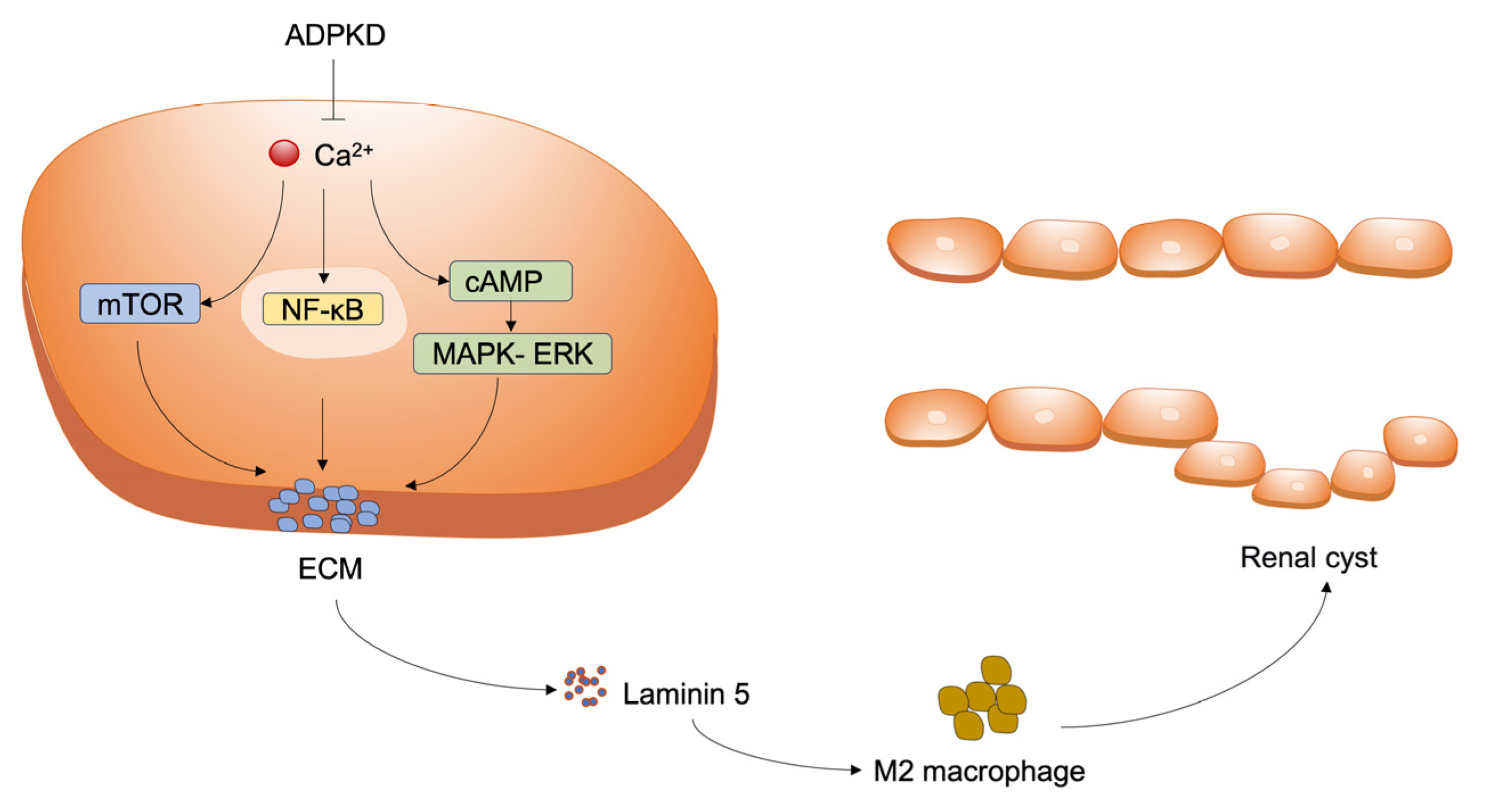
Accumulation of extracellular matrix (ECM) components and infiltration of inflammatory cells are observed in the early stages of ADPKD. In ADPKD kidneys, laminin-5 expression is upregulated within the ECM. Purified laminin-5 has been shown to activate extracellular signal-regulated kinase (ERK), thereby stimulating renal epithelial cell proliferation and promoting cystogenesis [14]. As cysts expand and compress the surrounding parenchyma and vasculature, obstructed glomeruli eventually give rise to atubular glomeruli, and apoptotic cell death becomes evident in proximal tubules [15]. Renal tubular epithelial cells, along with adjacent interstitial fibroblasts and infiltrating inflammatory cells, secrete a variety of chemokines, cytokines, and growth factors [16]. Among these, TGF-β promotes tubular cell dedifferentiation, recruits inflammatory cells to cystic sites, and activates α-smooth muscle actin (αSMA)-positive myofibroblasts, leading to increased ECM deposition. M2-like macrophages, in turn, secrete anti-inflammatory and pro-fibrotic mediators that stimulate tubular cell proliferation and myofibroblast activation. Pro-inflammatory stimuli and fibrotic accumulation also contribute to pericyte detachment, resulting in microvascular rarefaction and local tissue hypoxia, which further exacerbate the fibrotic response [1]. Pro-inflammatory molecules attract and activate more inflammatory cells, aggravating local injury and ultimately worsening cystic disease. The associated regulatory mechanism is given below (Figure 1).

3. Existing Pharmacological Therapies

3.1. Angiotensin-Converting Enzyme Inhibitors (ACEIs)

Current management of ADPKD primarily focuses on symptomatic control of hypertension and proteinuria, with particular attention to the evaluation and treatment of ADPKD-related complications, including cyst hemorrhage, cyst infection, nephrolithiasis, and chronic pain.
Cyst expansion in ADPKD compresses adjacent renal parenchyma and vasculature, leading to intrarenal ischemia and activation of the renin–angiotensin–aldosterone system (RAAS). This, in turn, increases aldosterone, promotes renal parenchymal fibrosis, and ultimately contributes to cyst enlargement and hypertension. Histological studies have found components of the RAAS—including angiotensinogen, renin, angiotensin-converting enzyme, angiotensin II, and angiotensin AT1 receptors—within cysts and dilated tubules of ADPKD kidneys, indicating a significant role for this system in the disease [17]. Strict blood pressure control, particularly via angiotensin blockade, remains a cornerstone of ADPKD management [1]. Recent studies show that ACEI treatment in ADPKD patients for 6 weeks significantly reduces renal vascular resistance and slows the rate of kidney disease progression [18]. However, ACEIs have potential side effects, including reduced renal blood flow leading to renal ischemia, hyperkalemia, and renal failure, necessitating judicious use and careful monitoring.

3.2. Vasopressin V2 Receptor Antagonists

Vasopressin acts on V2 receptors on the basolateral membrane of collecting duct cells to promote water reabsorption. Elevated vasopressin levels activate V2 receptors, increasing intracellular cyclic adenosine 3′,5′-monophosphate (cAMP), which influences cell proliferation and fluid secretion, thereby promoting cyst growth and renal function decline [19]. To date, vasopressin V2 receptor antagonists are the only therapy proven to have renoprotective effects [20].

3.2.1. Tolvaptan

Historically, treatment for ADPKD was limited to symptom and complication management. Recently, the U.S. Food and Drug Administration (FDA) approved tolvaptan, the first pharmacotherapy indicated to slow the decline of kidney function in adults at risk of rapidly progressing ADPKD [21]. Tolvaptan is an oral, selective vasopressin V2 receptor antagonist [22] that reduces cAMP levels in the collecting duct and thick ascending limb [23] at cyst sites. By binding to V2 receptors on the vascular side of the collecting duct, it causes aquaporin-2 internalization from the apical membrane, blocking water reabsorption and increasing water excretion. This reduces the annual rate of kidney volume growth and slows renal function decline. Initial studies indicate that split daily dosing of tolvaptan is necessary to maintain effective V2 receptor inhibition, as evidenced by sustained suppression of urine osmolality to ~300 mOsm/kg [24], balancing efficacy and tolerability. Tolvaptan also shows benefit in patients with later-stage ADPKD [25]. Despite its approval, some patients experience adverse effects, such as polyuria and thirst, leading to intolerance [1]. In addition, a clinical trial with Somatostatin analogue Octreotide and Tolvaptan combinations has also been conducted. In patients with ADPKD, octreotide-LAR added-on tolvaptan reduced GFR effectively. Octreotide-LAR also reduced total and cystic kidney volumes and attenuated the aquaretic effect of tolvaptan [26].

3.2.2. Lixivaptan

Lixivaptan is a potent, selective vasopressin V2 receptor antagonist. Wang et al. [27] fed ADPKD rats a lixivaptan-containing diet for 8 weeks and found a >50% reduction in cyst volume, a 23% decrease in intracellular cAMP levels compared to controls, and preliminary in vitro data suggesting a lower risk of hepatotoxicity than tolvaptan. These results provide the first evidence of lixivaptan’s potential therapeutic effect in ADPKD.

3.2.3. Somatostatin Analogues

In ADPKD, mutations in PKD1 or PKD2 disrupt the normal differentiated phenotype of tubular epithelial cells, leading to increased intracellular cAMP. cAMP signaling plays a central role in ADPKD pathophysiology; its upregulation activates protein kinase A (PKA), driving cell proliferation via the cystic fibrosis transmembrane conductance regulator (CFTR) and promoting cystogenesis [28]. cAMP and PKA also lead to activation of the mitogen-activated protein kinase (MAPK) cascade and mammalian target of rapamycin (mTOR) [29,30], Wnt-dependent microtubule formation [31], increased cilium length [32], and centrosome amplification [33]. Cyst growth displaces and destroys normal kidney tissue, leading to renal function decline. Given the key role of elevated intracellular cAMP, therapies aimed at reducing cAMP have been investigated.
Somatostatin binds to five G protein-coupled receptors, directly or indirectly inhibiting cAMP production in hepatic and renal cells. This inhibition suppresses fluid secretion, cell proliferation, and can induce apoptosis. Natural somatostatin has a very short in vivo half-life and is rapidly cleared. Therefore, longer-acting synthetic analogues have been developed, such as octreotide, lanreotide, and pasireotide [34].

3.3. mTOR Inhibitors

mTOR is a serine/threonine protein kinase of the PI3K-related kinase (PIKK) family and the catalytic subunit of the protein complexes mTORC1 and mTORC2. It integrates signals from growth factors, ATP, oxygen, and amino acids to regulate cell growth and metabolism. PC-1 inhibits mTORC1 via interaction with the protein TSC2 [35]. In ADPKD, loss of this regulation results in hyperactive mTORC1 signaling, increasing protein translation and leading to cell proliferation, growth, and cystogenesis [35,36]. Rapamycin, a macrolide, exerts antiproliferative, growth-inhibitory, and antifibrotic effects by inhibiting the mTOR enzyme [37]. Experimental inhibition of mTOR with rapamycin slows PKD progression in rats. Sirolimus, a natural macrolide antibiotic produced by Streptomyces hygroscopicus, is a specific inhibitor of the mTOR pathway [38]. Sirolimus significantly delays disease progression in Han:SPRD (cy) rats, reducing proliferation in cystic and non-cystic tubules, inhibiting kidney enlargement and cyst formation, and preserving renal function. However, the effective dose in Pkd1 mutant rats far exceeds what is safely tolerable in humans [39], and adverse effects such as chronic proteinuria and hyperlipidemia are concerns.

3.4. AMPK Agonists

mTOR and AMPK are opposing energy metabolism sensors regulating cell growth and proliferation. AMPK is a key cellular sensor and regulator of energy homeostasis, highly expressed in the kidney and modulating various physiological and pathological processes [40,41]. It plays a central pathogenic role in conditions like obesity, metabolic syndrome, diabetes, inflammation, and cancer [42]. ADPKD is characterized by increased aerobic glycolysis, where glucose is preferentially converted to lactate rather than being fully oxidized in mitochondria [43]. In ADPKD mouse models, moderate dietary restriction slows cyst growth by activating AMPK and inhibiting mTOR signaling [43]. Therapeutic AMPK activation can mitigate the severity of cystic kidney disease by improving mitochondrial biogenesis and reducing tissue inflammation [22].

3.4.1. Metformin

Type 2 diabetes medications like pioglitazone and metformin have recently shown positive effects in ADPKD. However, pioglitazone appears effective only when combined with tolvaptan [44]. Metformin, a biguanide, activates AMPK, which phosphorylates and inhibits CFTR, thereby inhibiting fluid and electrolyte secretion in epithelial cells [45]. Furthermore, AMPK phosphorylates tuberin, indirectly inhibiting the mTOR pathway. Fatty acid oxidation is also impaired in PKD, and metformin can positively influence this [13]. Based on observational studies, metformin is a promising drug for early-stage ADPKD.

3.4.2. Statins

Statins, or 3-hydroxy-3-methylglutaryl-coenzyme A (HMG-CoA) reductase inhibitors, possess antiproliferative, anti-inflammatory, and antioxidant properties [46]. Statins activate AMPK and have been shown to ameliorate cystic proliferation in ADPKD animal models [47]. Gile et al. [48] demonstrated that lovastatin significantly reduced cystic kidney size, cyst volume density, and serum urea nitrogen levels in heterozygous male Han:SPRD rats. A clinical trial in 110 children and young adults with ADPKD showed a benefit of pravastatin on total kidney volume [49]. However, whether statins can be a standard treatment for ADPKD requires confirmation through large-scale randomized controlled trials.

3.5. Anti-Inflammatory Agents

3.5.1. IL-37b

Interstitial inflammation, attributed largely to macrophage infiltration, is considered a feature that exacerbates disease progression. Studies by Zylberberg et al. [50] and Yang et al. [51] demonstrated, in transgenic mice expressing human IL-37b and in early-onset ADPKD mice injected with recombinant human IL-37b, that IL-37b promotes interferon signaling in kidney-resident macrophages, thereby inhibiting cyst formation. These findings suggest IL-37 as a feasible immunomodulatory therapy for ADPKD.

3.5.2. TWEAK

TWEAK is a TNF superfamily cytokine that regulates inflammatory responses. Its receptor, Fn14, is expressed on glomerular epithelial cells. Cordido et al. [52] showed that anti-TWEAK treatment slows cyst growth and improves renal function and survival in models. Anti-TWEAK antibody restored several ADPKD-related pathways (e.g., proliferation, NF-κB), mildly reduced fibrosis and apoptosis, and indirectly decreased macrophage recruitment. These results identify the TWEAK signaling pathway as a novel disease mechanism and a potential therapeutic target in ADPKD. Blocking TWEAK signaling slows cyst growth and disease progression [52].

3.6. MicroRNA Blockers

MicroRNAs (miRNAs) are short, non-coding RNAs that bind target mRNAs with sequence specificity, inhibiting their translation. Recent findings indicate a common set of dysregulated miRNAs in PKD mouse models, particularly the miR-17 family and miR-21, both upregulated in renal cysts and promoting ADPKD progression in mice. MiR-17 promotes cyst proliferation, while miR-21 suppresses pro-apoptotic genes, inhibiting cyst apoptosis [53]. RGLS4326 is a short oligonucleotide inhibitor of miR-17. Studies found that subcutaneous administration of RGLS4326 was safe and effective in ADPKD animal models [54]. Conversely, in later stages of cyst formation, miR-192 and miR-194 are downregulated due to hypermethylation, suggesting a potential role in limiting cyst expansion. The therapeutic effect of miR-192 and -194 precursors has been demonstrated in vivo and in vitro. Injection of miRNA precursors slowed cyst growth in Pkd1 knockout mice [55]. miRNAs have emerged as new regulators of ADPKD pathogenesis, and anti-miRs represent a viable novel drug class for treatment [53].

3.7. Dietary Interventions

General recommendations for ADPKD patients include a healthy diet and lifestyle, smoking cessation, maintaining optimal weight, and regular cardiovascular check-ups [1]. Additionally, salt intake should be limited to no more than 5 g per day [56].

3.7.1. Caloric Restriction and Ketogenic Diet

Recently, dietary interventions such as caloric restriction (CR), time-restricted feeding (TRF), and the ketogenic diet (KD) have emerged as potential strategies to induce metabolic reprogramming and delay ADPKD progression. These interventions hold promise for controlling ADPKD by improving metabolism and reducing oxidative stress.
In animal models, Kipp et al. [57] found that CR slows cyst growth and prevents ESRD by inhibiting mTORC1 signaling. Furthermore, CR reduced the expression of pro-inflammatory cytokines (MCP-1, IL-6, TNF-α) and restored the expression of the glycolytic enzyme HK2 to normal levels. Studies show that a 30–50% reduction in caloric intake without malnutrition slows cyst growth in animal models [57,58]. Cyst cells are glucose-dependent; a sustained shift from mitochondrial oxidative phosphorylation to aerobic glycolysis is a factor driving cyst growth, making these cells sensitive to glucose availability. Preclinical studies targeting glucose metabolism via intermittent fasting are promising [42]. Torres et al. [59] experimentally confirmed that KD dramatically improved the phenotype in Han: SPRD rat models, reducing kidney weight, cyst index, and epithelial proliferation. KD also attenuated fibrosis and mTOR signaling and improved creatinine clearance. KD can also reduce kidney and cyst size in established disease, suggesting that low-calorie or ketogenic diets may slow ADPKD progression [60]. In human data, obesity is a strong independent predictor of ADPKD progression [61]. CR benefits overweight or obese patients by promoting weight loss and improving metabolic parameters [62]. KD reduces body weight and fat mass while improving glomerular filtration rate in ADPKD patients [63].

3.7.2. 2-Deoxyglucose (2DG)

PKD1 mutation leads to enhanced glycolysis in the kidneys of ADPKD mice and patients [43]. Increased aerobic glycolysis becomes dominant in cyst cells. 2DG, transported into cyst cells, acts as a competitive inhibitor of the glycolytic pathway. Magistroni et al. [64] experimentally confirmed that 2DG reverses the glycolytic shift in ADPKD kidneys, improving kidney weight, volume, cyst index, and proliferation rate in ADPKD mice [65]. These studies demonstrate that metabolic regulation is a potentially important factor in ADPKD progression and a possible therapeutic target [43,66]. Multiple studies indicate 2DG is a safe and effective drug in animal models, with no toxic effects observed even with long-term, low-dose administration [67,68].

4. Novel Therapeutic Targets

4.1. Modulating Intracellular Calcium and the Cell Cycle

4.1.1. TMEM16A Inhibitors

Research has found that the calcium-activated chloride channel TMEM16A is crucial for fluid secretion into renal cysts in vitro [69]. TMEM16A inhibitors reduce chloride channel activity. Niclosamide and benzbromarone, which inhibit TMEM16A, are associated with reduced cyst growth [70]. The TMEM16A-specific small molecule Ani9 also inhibits cystogenesis in vivo [71]. TMEM16A inhibitors not only block chloride current but also suppress TMEM16A expression during long-term treatment. TMEM16A inhibitors appear promising for future ADPKD therapy.

4.1.2. Calcimimetics

Calcimimetics activate the calcium-sensing receptor, leading to decreased cAMP and increased intracellular calcium. The calcimimetic R568 significantly reduced kidney cyst volume in mice [72]. In ADPKD rats and mice, combined treatment with lixivaptan and the calcimimetic R568 was more effective than either alone in reducing kidney weight, cyst and fibrosis volume [73].

4.1.3. Triptolide

Triptolide, a natural compound extracted from the traditional Chinese herb Tripterygium wilfordii, has been shown to restore cell membrane Ca2+ release and induce growth arrest in Pkd1 mouse renal epithelial cells. Leuenroth et al. [74] confirmed that triptolide reduced cyst formation from youth to adulthood in a Pkd1 ADPKD model.

4.1.4. R-Roscovitine

Pharmacological inhibition of cell cycle progression using the cyclin-dependent kinase (CDK) inhibitor R-roscovitine in animal models showed that effectively inhibiting the cell cycle, proliferation, and apoptosis suppresses the progression of renal cystic disease and hepatic cystogenesis [75].

4.2. Histone Deacetylase (HDAC) Inhibitors

Benzothiazole Derivatives

Emerging evidence indicates that HDACs are important regulators in ADPKD. Cao et al. [76] designed and synthesized a series of benzothiazole-containing compounds as potential HDAC inhibitors. Given the multifaceted involvement of HDACs in cyst progression, they performed target-based screening using HeLa cell nuclear extracts to identify potent pan-HDAC inhibitors. Compound 26 emerged as the most effective candidate. Subsequent pharmacological characterization revealed it effectively inhibited multiple HDACs (especially HDAC1, HDAC2, and HDAC6, IC50 all < 150 nM), with particular sensitivity to HDAC6 (IC50 = 11 nM). This compound significantly inhibited cyst formation and expansion in an in vitro cyst model and effectively slowed cyst growth in an embryonic kidney cyst model and an ADPKD mouse model in vivo.

4.3. Novel Targets

4.3.1. Notch3

Recent research indicates that the Notch signaling pathway plays an indispensable role in the pathogenesis of various kidney diseases, including ADPKD. Notch3 receptor is overexpressed in kidney tissues from ADPKD mice and patients. The Notch3 intracellular domain (N3ICD) and Hes1 can bind to the PTEN promoter, leading to transcriptional repression of PTEN. This further activates the downstream PI3K-AKT-mTOR pathway, promoting renal epithelial cell proliferation. γ-Secretase inhibitors, which block the proteolytic cleavage required for Notch3 activation, and shRNA-mediated knockdown of Notch3 significantly delayed renal cyst growth in vitro and in vivo. Therefore, Notch3 is a novel driver of renal epithelial cell proliferation and cyst formation in ADPKD, presenting a promising therapeutic target [77].

4.3.2. AURKA

Aurora kinase A (AURKA) promotes cell proliferation and is overexpressed in different types of PKD. Tham et al. [78] deleted the Aurka gene in mouse models of ADPKD and Joubert syndrome (caused by mutations in Pkd1 and Inpp5e, respectively) and found that Aurka deletion prevented cyst formation. They also identified an association with AKT signaling in cyst prevention. Further experiments established AURKA as a master regulator of cyst development across different PKD types, functioning in part through AKT. Therefore, anti-AURKA therapy may become a new research focus.

4.3.3. LncRNA

Long non-coding RNAs (lncRNAs) are a class of non-protein-coding RNAs with key functions in development and disease. Weisser et al. [79] confirmed in a new study that lncRNAs are involved in ADPKD pathogenesis. The lncRNA Hoxb3os is downregulated in ADPKD and regulates the mammalian target of rapamycin (mTOR)/Akt pathway in mouse kidneys in vivo. Reducing Hoxb3os expression in ADPKD mice activated mTOR complex 2 (mTORC2) signaling and exacerbated cyst formation.

4.3.4. Glis2

Following PC1 inactivation, cyst growth requires intact primary cilia, a process termed cilia-dependent cyst activation (CDCA). Zhang et al. [80] studied kidneys from mice with cilia inactivated prior to cyst formation and identified Glis2 as a functional effector of polycystin signaling and CDCA. Changes in Glis2 expression in vitro mirrored observed CDCA changes in renal tissue, validating its association with cyst formation. In an ADPKD mouse model, Glis2 inactivation suppressed polycystic kidney disease, and pharmacological targeting of Glis2 with antisense oligonucleotides slowed disease progression, identifying Glis2 as a potential therapeutic target.

4.3.5. Thiamet G

Su et al. [81] found that O-GlcNAcylation and O-GlcNAc transferase (OGT) were downregulated in kidney tissues of Pkd1-silenced mice. Furthermore, O-GlcNAcylation was shown to regulate the stability and function of the PC1 protein’s C-terminal cytoplasmic tail (CTT). Treatment of PKD mice with Thiamet G significantly reduced cystogenesis. These results reveal a unique role for O-GlcNAcylation in PKD cyst formation, positioning Thiamet G as a potential therapeutic target.

4.3.6. RAGE

The RAGE gene, the receptor for advanced glycation end products, is involved in inflammatory responses and cell proliferation. Park et al. [82] confirmed that blocking RAGE function inhibits ADPKD cyst growth in vitro. Building on this, their team investigated the impact of RAGE on cyst expansion in vivo using a severe ADPKD mouse model (PC2R). Intravenous injection of adenovirus carrying anti-RAGE short interfering RNA led to RAGE knockdown, resulting in reduced kidney weight and volume. Furthermore, cystic area derived from different nephron segments decreased due to RAGE downregulation. Blood urea nitrogen and creatinine levels showed a declining trend after RAGE inhibition. Based on these results, the RAGE gene is a valid therapeutic target for ADPKD.

4.3.7. Adeno-Associated Virus Serotype 1 (AAV1)

Ciobanu et al. [83] experimentally found sialic acid residues on ADPKD cyst epithelial cells that enhance transduction by adeno-associated virus serotype 1 (AAV1). AAV1 also exhibited tropism for cyst epithelium. They further observed that intervention targeting the cystic fibrosis transmembrane conductance regulator (CFTR) via AAV1 significantly reduced cyst area and volume in mice, suggesting promising prospects for AAV1 and CFTR-targeted therapy in ADPKD.

4.3.8. CD8+ T Cells

Kleczko et al. [84] used flow cytometry to find that the numbers of CD8+ and CD4+ T cells in the kidney progressively increased with ADPKD severity, but only CD8+ T cells were selectively activated. Immunofluorescence analysis showed T cells specifically localized to cyst lesions, and qPCR/ in situ hybridization detected elevated levels of T cell-recruiting chemokines (CXCL9/CXCL10) in mouse kidneys, patient tissues, and ADPKD epithelial cell lines. The team also found that sustained CD8+ T cell immune depletion for 1–3 months in C57Bl/6 Pkd1 mice worsened ADPKD pathology, reduced apoptosis, and increased proliferation (compared to IgG controls), confirming a renoprotective role for CD8+ T cells. This reveals a functional role for T cells, particularly CD8+ T cells, in ADPKD progression. Therefore, targeting this pathway with immuno-oncology drugs may offer a novel therapeutic strategy.

4.3.9. Dihydrotanshinone I (DHTS)

Mahendran et al. [85] found that dihydrotanshinone I (DHTS) inhibits ADPKD cell proliferation by inducing G1 phase cell cycle arrest and enhancing necroptosis. DHTS treatment may also modulate enhanced immune surveillance observed in untreated ADPKD cells. After elucidating related signaling pathways and ensuring safety for normal cells, the antiproliferative effect of DHTS on ADPKD cells demonstrates its potential as an alternative therapy.

4.3.10. Aquaporins

Studies suggest that aquaporins—transmembrane channel proteins including AQP-1, AQP-2, AQP-3, and AQP-11—are involved in the pathogenesis of ADPKD. Aquaporins may represent potential new therapeutic targets [86].

4.3.11. Super-Enhancers and Metabolic Reprogramming

Super-enhancers (SEs) are large clusters of transcriptional enhancers that drive robust expression of cell identity and disease genes. Mi et al. [87] experimentally revealed extensive SE remodeling during cystogenesis, with SE-associated transcripts most enriched in metabolic processes in cyst cells. Inhibiting the transcriptional kinase CDK7 (cyclin-dependent kinase 7), involved in SE assembly and maintenance, or targeting the CDK7-regulated metabolic target gene AMP deaminase 3 (AMPD3, slowed cyst growth in an ADPKD mouse model. The study also found that CDK7 expression is generally elevated in ADPKD patients and positively correlates with AMPD3 expression and disease severity, elucidating the mechanism by which SEs regulate metabolic gene transcription during cyst formation. Therefore, SE-driven metabolic reprogramming is a potential therapeutic target.

4.3.12. Epigenetics

Zhou et al. [88] showed that DNA methyltransferase 1 (DNMT1) expression is upregulated in cystic renal epithelial cells and tissues. Knockout of the Dnmt1 gene or targeted inhibition of DNMT1 using demethylating agents delayed cyst growth in Pkd1-mutant kidneys and extended the lifespan of Pkd1 conditional knockout mice. The study also identified novel DNMT1 targets—PTPRM and PTPN22 (both belonging to the protein tyrosine phosphatase family)—which are involved in regulating the phosphorylation and activation of PKD-related signaling pathways (including ERK, mTOR, and STAT3). The researchers also found methylation dysregulation of epigenetic clock-related genes in ADPKD patients, confirming epigenetic age acceleration in their kidneys. Furthermore, the team identified five epigenetics-related genes (Hsd17b14, Itpkb, Mbnl1, Rassf5, and Plk2), with biological functions indicating their methylation status participates in ADPKD progression. Additionally, the epigenetic regulator BRD4 has been confirmed to be involved in ADPKD pathogenesis. Yang et al. [89] found that a BRD4 inhibitor exhibited high efficacy and selectivity in ADPKD cell and embryonic kidney models, potentially becoming a future therapeutic target.

4.3.13. Post-Translational Modifications

Recently, post-translational modifications (PTMs) have been recognized as key factors in cystogenesis. Ren et al. [90] found that the E3 ubiquitin ligase TRAF6 is significantly upregulated in ADPKD cells, mouse models, and patient tissues. TRAF6 interacts with the transcription factor STAT3, selectively enhancing its K63-linked ubiquitination, promoting its activation and transcriptional activity, thereby cooperatively promoting renal cyst growth in ADPKD cells and mouse models. Inhibiting TRAF6 significantly suppressed STAT3 transcriptional activity and slowed cyst growth in vitro and in vivo. Therefore, targeting TRAF6 may provide a novel strategy.
Furthermore, Ren et al. [91] found that the deubiquitinase USP28, abnormally upregulated in ADPKD patients, specifically removes K48-linked polyubiquitin chains, thereby counteracting the protein degradation of signal transducer and activator of transcription 3 (STAT3). They also observed that USP28 directly interacts with and stabilizes c-Myc, a transcriptional target of STAT3. These two mechanisms synergistically promote renal cystogenesis. Pharmacological inhibition of USP28 significantly attenuated cyst formation in vitro and in vivo. Thus, USP28 participates in ADPKD pathogenesis by regulating STAT3 trafficking and its transcriptional target c-Myc, making its inhibition a potential novel strategy.
Additionally, Lv et al. [92] found that enhancer of zeste homolog 2 (EZH2) is significantly upregulated in Pkd1 cells, Pkd1 mice, and human ADPKD kidneys. Inhibiting EZH2 alleviated cyst development in MDCK cells and mouse embryonic kidney cyst models, and both conditional knockout of Ezh2 and treatment with GSK126 inhibited renal cyst growth and protected renal function in Pkd1 mice. The study showed that the cAMP/PKA/CREB pathway promotes EZH2 expression. EZH2 mediates cyst formation through three pathways: enhancing STAT3 methylation and activation, inhibiting p21 to promote the cell cycle, and activating non-phosphorylated β-catenin in the Wnt pathway. Moreover, EZH2 enhances ferroptosis in ADPKD by inhibiting SLC7A11 and GPX4. This study clarifies the critical role of EZH2 in promoting renal cyst growth via epigenetic mechanisms and suggests that inhibiting or degrading EZH2 could be a novel therapeutic strategy. All targets are summarized in the table below (Table 1).

5. Discussion

ADPKD, as a common inherited renal disorder, involves multiple layers of pathogenesis including loss of polycystin function, dysregulated signaling pathways, and metabolic abnormalities [2]. With deepening research into its molecular mechanisms, treatment strategies have gradually shifted from traditional symptomatic management towards targeted interventions addressing core disease processes. Based on current research, this article systematically reviews advances in ADPKD treatment and explores the potential of emerging targets, aiming to provide a theoretical foundation for clinical practice and future research.
ADPKD is primarily caused by mutations in PKD1 or PKD2, leading to defective function of the polycystin complex and subsequently affecting primary cilium-mediated cell signaling. Polycystin deficiency triggers pathological changes such as disrupted intracellular calcium homeostasis, elevated cAMP levels, abnormal mTOR pathway activation, and metabolic reprogramming [7,8]. These changes collectively promote proliferation of renal tubular epithelial cells and cyst fluid secretion, ultimately forming cysts that destroy kidney structure. Furthermore, inflammatory responses and fibrosis play significant roles in disease progression. Macrophage infiltration, cytokine release (e.g., TGF-β, TWEAK), and extracellular matrix remodeling further accelerate loss of renal function. These mechanisms also provide multiple potential targets for drug development. For example, aberrant activation of the cAMP signaling pathway is a core driver of cyst growth, making cAMP pathway inhibitors like tolvaptan and somatostatin analogues hotspots for clinical research. Concurrently, metabolic abnormalities (e.g., enhanced aerobic glycolysis) and dysregulation of energy-sensing pathways (e.g., AMPK/mTOR) provide a theoretical basis for metabolic interventions like metformin and ketogenic diets.
Current pharmacological treatments for ADPKD mainly focus on: (1) cAMP pathway inhibitors; (2) mTOR signaling inhibitors; (3) AMPK agonists and metabolic modulators; (4) anti-inflammatory and immunomodulatory agents; and (5) dietary and metabolic interventions. (1) Among cAMP pathway inhibitors, tolvaptan, as the first approved targeted drug for ADPKD, delays renal function decline and kidney volume growth by antagonizing the vasopressin V2 receptor to lower cAMP levels. However, its risk of hepatotoxicity, side effects like polyuria, and high cost limit its widespread use [93,94]. Emerging therapeutic strategies beyond tolvaptan are under active investigation, including combination therapies, metabolic interventions, and gene editing. For instance, glucagon-like peptide-1 (GLP-1) receptor agonists have shown potential protective effects through metabolic modulation and anti-inflammatory actions [95,96]. Ketogenic dietary interventions have demonstrated reduced cyst growth in animal models, but clinical evidence remains limited and long-term safety concerns exist [97]. CRISPR-Cas9 gene editing has successfully corrected pathogenic mutations in in vitro models, though delivery efficiency and in vivo application require further refinement [98]. International and regional guidelines, including those from KDIGO and Taiwanese consensus statements, emphasize risk stratification using imaging, genotyping, and height-adjusted total kidney volume (htTKV), alongside individualized management [99,100,101]. Future directions integrating organoid models, systematic reviews, real-world studies, and precision medicine approaches will likely advance ADPKD care from slowing progression toward targeted repair [102,103,104]. Lixivaptan, another V2 receptor antagonist, shows similar efficacy and potentially lower hepatotoxicity in preclinical studies, but its clinical value requires further validation. Somatostatin analogues (e.g., octreotide) inhibit cAMP generation and have shown effects in slowing kidney growth in some clinical trials, but their long-term efficacy and safety need more evidence. (2) mTOR signaling inhibitors: Rapamycin and its derivatives (e.g., sirolimus) effectively inhibit cyst growth in animal models, but human clinical trial results are inconsistent, and high doses may cause adverse effects like proteinuria and hyperlipidemia. This suggests caution in using mTOR inhibitors for ADPKD; they may be more suitable for specific subgroups or combination therapies. (3) AMPK agonists and metabolic modulators: Drugs like metformin activate AMPK to inhibit CFTR and the mTOR pathway, showing potential in early-stage ADPKD patients. Statins, beyond lipid-lowering effects, can inhibit cell proliferation and inflammation via AMPK-dependent pathways. Due to their safety and accessibility, these drugs are important candidates for adjuvant therapy in ADPKD. (4) Anti-inflammatory and immunomodulatory agents: Agents like IL-37b and anti-TWEAK antibodies can reduce cyst growth and fibrosis by modulating macrophage polarization and NF-κB signaling. Additionally, MicroRNA blockers (e.g., RGLS4326) effectively inhibit cyst proliferation and promote apoptosis in animal models by targeting the miR-17 family and miR-21, opening a new direction for gene therapy in ADPKD. (5) Dietary interventions: Low-calorie diets, intermittent fasting, and ketogenic diets significantly delay disease progression in animal models by improving mitochondrial function, inhibiting glycolysis, and reducing oxidative stress. 2-Deoxyglucose (2DG), as a glycolytic inhibitor, can reverse metabolic abnormalities in cyst cells, but its long-term safety requires further evaluation.
Simultaneously, with deeper understanding of ADPKD pathogenesis, numerous novel targets have emerged. Ion channel and calcium modulators: TMEM16A inhibitors (e.g., niclosamide, Ani9) inhibit cyst expansion by blocking chloride secretion. Calcimimetics (e.g., R568) and triptolide exhibit anti-cyst effects in animal models by restoring intracellular calcium homeostasis. These drugs target the core abnormality of ion transport in ADPKD and have high translational potential. Epigenetic regulators: Histone deacetylase inhibitors (e.g., benzothiazole derivatives) can influence multiple pathogenic pathways by modulating chromatin structure. Long non-coding RNAs (e.g., Hoxb3os) and Glis2, as new epigenetic regulators, show promising effects in animal models when targeted. Furthermore, adeno-associated virus serotype 1 (AAV1)-mediated gene delivery systems offer the possibility of restoring polycystin function. Novel signaling pathway targets: Overexpression of Notch3 and AURKA promotes cell proliferation and cyst formation in ADPKD; their inhibitors or gene silencing strategies significantly delay disease progression in preclinical studies. Additionally, CDK inhibitors (e.g., R-roscovitine) and natural compounds like dihydrotanshinone I (DHTS) exhibit antiproliferative activity by inducing cell cycle arrest and apoptosis. Emerging immune and inflammatory targets: Recent studies found CD8+ T cells specifically accumulate in ADPKD kidneys and inhibit cyst growth through immune surveillance. This finding suggests immunomodulatory drugs (e.g., checkpoint inhibitors) could become a new therapeutic strategy. Meanwhile, RAGE receptor antagonists alleviate cyst burden in animal models by blocking inflammatory and proliferative signals.
Despite significant progress in ADPKD treatment research, many challenges remain. First, individual genetic differences demand more precise treatment strategies. Variations in mutation type (PKD1 vs. PKD2), mutation site, genetic background, and environmental factors collectively determine disease progression rate and treatment response. For example, PKD1 mutation patients typically progress faster than PKD2 patients [105], and even patients with the same mutation can have significantly different clinical presentations. Tolvaptan is effective only in rapidly progressing patients, while metabolic interventions may be more suitable for early-stage or overweight patients. Second, most novel target drugs are still in the preclinical stage, and their safety, administration routes, and long-term efficacy require validation through large-scale clinical trials. Additionally, combination therapies (e.g., tolvaptan combined with metformin may simultaneously target the cAMP pathway and metabolic abnormalities; mTOR inhibitors combined with AMPK agonists may more effectively balance cell proliferation and energy metabolism regulation) may improve efficacy and reduce side effects through multi-target synergy. Treatment for ADPKD has entered an era coexisting with targeted interventions and metabolic regulation. The continuous emergence of new targets brings new hope for ADPKD treatment. From ion channel regulation to epigenetic control, from cell cycle inhibition to immune microenvironment modulation, targets like Notch3, AURKA, and TMEM16A provide abundant options. In the future, by deeply analyzing the molecular network of ADPKD and integrating the concept of precision medicine, effective control and potentially functional cure of the disease may be achievable.

Author Contributions

Conceptualization, W.Z., T.S. and X.W.; investigation, W.Z.; resources, T.J.; writing—original draft preparation, W.Z.; writing—review and editing, T.S. and T.J.; funding acquisition, T.J. All authors have read and agreed to the published version of the manuscript.

Funding

Project of Jilin Provincial Department of science and technology. Number: YDZJ202401061ZYTS.

Institutional Review Board Statement

Not applicable.

Informed Consent Statement

Not applicable.

Data Availability Statement

No new data were created or analyzed in this study. Data sharing is not applicable to this article.

Acknowledgments

Thanks to the department of Beihua University; thanks to Tao Jang for his invaluable guidance; thanks to Tanze Sun for his patient mentorship.

Conflicts of Interest

We confirm that neither the manuscript nor any parts of its content are currently under consideration or published in another journal. All authors have approved the manuscript and agree with its submission to CIMB and declare no competing financial interests.

Abbreviations

The following abbreviations are used in this manuscript:
ADPKDAutosomal dominant polycystic kidney disease
PC1Polycystin-1
PC2Polycystin-2
ESRDEnd-stage renal disease
VEGFVascular endothelial growth factor
PI3KPhosphoinositide 3-kinase
NFATNuclear factor of activated T-cells
ECMExtracellular matrix
ERKExtracellular signal-regulated kinase
RAASRenin–angiotensin–aldosterone system
ACEIsAngiotensin-Converting Enzyme Inhibitors
cAMPCyclic adenosine 3′,5′-monophosphate
FDAFood and Drug Administration
CFTRCystic fibrosis transmembrane conductance regulator
PIKKPI3K-related kinase
CRCaloric restriction
TRFTime-restricted feeding
KDKetogenic diet
CDKCyclin-dependent kinase
AURKAAurora kinase A
lncRNAsLong non-coding RNAs
AAV1Adeno-Associated Virus Serotype 1
DHTSDihydrotanshinone I
DNMT1DNA methyltransferase 1
PTMsPost-translational modifications

References

  1. Reiterová, J.; Tesař, V. Autosomal Dominant Polycystic Kidney Disease: From Pathophysiology of Cystogenesis to Advances in the Treatment. Int. J. Mol. Sci. 2022, 23, 3317. [Google Scholar] [CrossRef]
  2. Igarashi, P.; Somlo, S. Genetics and pathogenesis of polycystic kidney disease. J. Am. Soc. Nephrol. JASN 2002, 13, 2384–2398. [Google Scholar] [CrossRef] [PubMed]
  3. Cornec-Le Gall, E.; Alam, A.; Perrone, R.D. Autosomal dominant polycystic kidney disease. Lancet 2019, 393, 919–935. [Google Scholar] [CrossRef] [PubMed]
  4. Lanktree, M.B.; Haghighi, A.; Di Bari, I.; Song, X.; Pei, Y. Insights into Autosomal Dominant Polycystic Kidney Disease from Genetic Studies. Clin. J. Am. Soc. Nephrol. CJASN 2021, 16, 790–799. [Google Scholar] [CrossRef] [PubMed]
  5. Cornec-LEGall, E.; Torres, V.E.; Harris, P.C. Genetic Complexity of Autosomal Dominant Polycystic Kidney and Liver Diseases. J. Am. Soc. Nephrol. JASN 2018, 29, 13–23. [Google Scholar] [CrossRef]
  6. Tsiokas, L.; Kim, E.; Arnould, T.; Sukhatme, V.P.; Walz, G. Homo- and heterodimeric interactions between the gene products of PKD1 and PKD 2. Proc. Natl. Acad. Sci. USA 1997, 94, 6965–6970. [Google Scholar] [CrossRef]
  7. Pazour, G.J.; Quarmby, L.; Smith, A.O.; Desai, P.B.; Schmidts, M. Cilia in cystic kidney and other diseases. Cell. Signal. 2020, 69, 109519. [Google Scholar] [CrossRef]
  8. Ha, K.; Nobuhara, M.; Wang, Q.; Walker, R.V.; Qian, F.; Schartner, C.; Cao, E.; Delling, M. The heteromeric PC-1/PC-2 polycystin complex is activated by the PC-1 N-terminus. eLife 2020, 9, e60684. [Google Scholar] [CrossRef]
  9. Ta, C.M.; Vien, T.N.; Ng, L.C.T.; DeCaen, P.G. Structure and function of polycystin channels in primary cilia. Cell. Signal. 2020, 72, 109626. [Google Scholar] [CrossRef]
  10. Ma, M.; Tian, X.; Igarashi, P.; Pazour, G.J.; Somlo, S. Loss of cilia suppresses cyst growth in genetic models of autosomal dominant polycystic kidney disease. Nat. Genet. 2013, 45, 1004–1012. [Google Scholar] [CrossRef]
  11. Bergmann, C.; Guay-Woodford, L.M.; Harris, P.C.; Horie, S.; Peters, D.J.M.; Torres, V.E. Polycystic kidney disease. Nat. Rev. Dis. Prim. 2018, 4, 50. [Google Scholar] [CrossRef]
  12. Chiaravalli, M.; Rowe, I.; Mannella, V.; Quilici, G.; Canu, T.; Bianchi, V.; Gurgone, A.; Antunes, S.; D’Adamo, P.; Esposito, A.; et al. 2-Deoxy-d-Glucose Ameliorates PKD Progression. J. Am. Soc. Nephrol. 2016, 27, 1958–1969. [Google Scholar] [CrossRef]
  13. Menezes, L.F.; Lin, C.C.; Zhou, F.; Germino, G.G. Fatty Acid Oxidation is Impaired in An Orthologous Mouse Model of Autosomal Dominant Polycystic Kidney Disease. EBioMedicine 2016, 5, 183–192. [Google Scholar] [CrossRef] [PubMed]
  14. Joly, D.; Berissi, S.; Bertrand, A.; Strehl, L.; Patey, N.; Knebelmann, B. Laminin 5 regulates polycystic kidney cell proliferation and cyst formation. J. Biol. Chem. 2006, 281, 29181–29189. [Google Scholar] [CrossRef]
  15. Liu, D.; Wang, C.J.; Judge, D.P.; Halushka, M.K.; Ni, J.; Habashi, J.P.; Moslehi, J.; Bedja, D.; Gabrielson, K.L.; Xu, H.; et al. A Pkd1-Fbn1 genetic interaction implicates TGF-β signaling in the pathogenesis of vascular complications in autosomal dominant polycystic kidney disease. J. Am. Soc. Nephrol. JASN 2014, 25, 81–91. [Google Scholar] [CrossRef]
  16. Swenson-Fields, K.I.; Vivian, C.J.; Salah, S.M.; Peda, J.D.; Davis, B.M.; van Rooijen, N.; Wallace, D.P.; A Fields, T. Macrophages promote polycystic kidney disease progression. Kidney Int. 2013, 83, 855–864. [Google Scholar] [CrossRef]
  17. Cadnapaphornchai, M.A. Hypertension in Children with Autosomal Dominant Polycystic Kidney Disease (ADPKD). Curr. Hypertens. Rev. 2013, 9, 21–26. [Google Scholar] [CrossRef]
  18. Kazancioglu, R.; Gursu, M. New options in the treatment of autosomal dominant polycystic kidney disease. Ren. Fail. 2015, 37, 535–541. [Google Scholar] [CrossRef][Green Version]
  19. Meijer, E.; Gansevoort, R.T. Emerging non-pharmacological interventions in ADPKD: An update on dietary advices for clinical practice. Curr. Opin. Nephrol. Hypertens. 2021, 30, 482–492. [Google Scholar] [CrossRef] [PubMed]
  20. Edwards, M.E.; Chebib, F.T.; Irazabal, M.V.; Ofstie, T.G.; Bungum, L.A.; Metzger, A.J.; Senum, S.R.; Hogan, M.C.; El-Zoghby, Z.M.; Kline, T.L.; et al. Long-Term Administration of Tolvaptan in Autosomal Dominant Polycystic Kidney Disease. Clin. J. Am. Soc. Nephrol. CJASN 2018, 13, 1153–1161, Correction in Clin. J. Am. Soc. Nephrol. 2019, 14, 910. https://doi.org/10.2215/CJN.02810319. [Google Scholar] [CrossRef] [PubMed]
  21. Chebib, F.T.; Perrone, R.D.; Chapman, A.B.; Dahl, N.K.; Harris, P.C.; Mrug, M.; Mustafa, R.A.; Rastogi, A.; Watnick, T.; Yu, A.S.; et al. A Practical Guide for Treatment of Rapidly Progressive ADPKD with Tolvaptan. J. Am. Soc. Nephrol. 2018, 29, 2458–2470. [Google Scholar] [CrossRef]
  22. Song, X.; Tsakiridis, E.; Steinberg, G.R.; Pei, Y. Targeting AMP-activated protein kinase (AMPK) for treatment of autosomal dominant polycystic kidney disease. Cell. Signal. 2020, 73, 109704. [Google Scholar] [CrossRef]
  23. Juul, K.V.; Bichet, D.G.; Nielsen, S.; Nørgaard, J.P. The physiological and pathophysiological functions of renal and extrarenal vasopressin V2 receptors. Am. J. Physiol. –Ren. Physiol. 2014, 306, F931–F940. [Google Scholar] [CrossRef]
  24. Shoaf, S.E.; Chapman, A.B.; Torres, V.E.; Ouyang, J.; Czerwiec, F.S. Pharmacokinetics and Pharmacodynamics of Tolvaptan in Autosomal Dominant Polycystic Kidney Disease: Phase 2 Trials for Dose Selection in the Pivotal Phase 3 Trial. J. Clin. Pharmacol. 2017, 57, 906–917. [Google Scholar] [CrossRef] [PubMed]
  25. Torres, V.E.; Chapman, A.B.; Devuyst, O.; Gansevoort, R.T.; Perrone, R.D.; Koch, G.; Ouyang, J.; McQuade, R.D.; Blais, J.D.; Czerwiec, F.S.; et al. Tolvaptan in Later-Stage Autosomal Dominant Polycystic Kidney Disease. N. Engl. J. Med. 2017, 377, 1930–1942. [Google Scholar] [CrossRef]
  26. Trillini, M.; Caroli, A.; Perico, N.; Remuzzi, A.; Brambilla, P.; Villa, G.; Perna, A.; Peracchi, T.; Rubis, N.; Martinetti, D.; et al. Effects of Octreotide-Long-Acting Release Added-on Tolvaptan in Patients with Autosomal Dominant Polycystic Kidney Disease: Pilot, Randomized, Placebo-Controlled, Cross-Over Trial. Clin. J. Am. Soc. Nephrol. 2023, 18, 223–233. [Google Scholar] [CrossRef]
  27. Wang, X.; Constans, M.M.; Chebib, F.T.; Torres, V.E.; Pellegrini, L. Effect of a Vasopressin V2 Receptor Antagonist on Polycystic Kidney Disease Development in a Rat Model. Am. J. Nephrol. 2019, 49, 487–493. [Google Scholar] [CrossRef]
  28. Saigusa, T.; Bell, P.D. Molecular pathways and therapies in autosomal-dominant polycystic kidney disease. Physiology 2015, 30, 195–207. [Google Scholar] [CrossRef]
  29. Distefano, G.; Boca, M.; Rowe, I.; Wodarczyk, C.; Ma, L.; Piontek, K.B.; Germino, G.G.; Pandolfi, P.P.; Boletta, A. Polycystin-1 regulates extracellular signal-regulated kinase-dependent phosphorylation of tuberin to control cell size through mTOR and its downstream effectors S6K and 4EBP1. Mol. Cell. Biol. 2009, 29, 2359–2371. [Google Scholar] [CrossRef] [PubMed]
  30. Spirli, C.; Okolicsanyi, S.; Fiorotto, R.; Fabris, L.; Cadamuro, M.; Lecchi, S.; Tian, X.; Somlo, S.; Strazzabosco, M. Mammalian target of rapamycin regulates vascular endothelial growth factor-dependent liver cyst growth in polycystin-2-defective mice. Hepatology 2010, 51, 1778–1788. [Google Scholar] [CrossRef] [PubMed]
  31. Gallegos, T.F.; Kouznetsova, V.; Kudlicka, K.; Sweeney, D.E.; Bush, K.T.; Willert, K.; Farquhar, M.G.; Nigam, S.K. A protein kinase A and Wnt-dependent network regulating an intermediate stage in epithelial tubulogenesis during kidney development. Dev. Biol. 2012, 364, 11–21. [Google Scholar] [CrossRef] [PubMed]
  32. Besschetnova, T.Y.; Kolpakova-Hart, E.; Guan, Y.; Zhou, J.; Olsen, B.R.; Shah, J.V. Identification of Signaling Pathways Regulating Primary Cilium Length and Flow-Mediated Adaptation. Curr. Biol. 2010, 20, 182–187. [Google Scholar] [CrossRef] [PubMed]
  33. Ahmed, A.A.; Lu, Z.; Jennings, N.B.; Etemadmoghadam, D.; Capalbo, L.; Jacamo, R.O.; Barbosa-Morais, N.; Le, X.-F.; Vivas-Mejia, P.; Lopez-Berestein, G.; et al. SIK2 is a centrosome kinase required for bipolar mitotic spindle formation that provides a potential target for therapy in ovarian cancer. Cancer Cell. 2010, 18, 109–121. [Google Scholar] [CrossRef]
  34. Hogan, M.C.; Masyuk, T.V.; Page, L.J.; Kubly, V.J.; Bergstralh, E.J.; Li, X.; Kim, B.; King, B.F.; Glockner, J.; Holmes, D.R.; et al. Randomized clinical trial of long-acting somatostatin for autosomal dominant polycystic kidney and liver disease. J. Am. Soc. Nephrol. JASN 2010, 21, 1052–1061. [Google Scholar] [CrossRef] [PubMed]
  35. Dere, R.; Wilson, P.D.; Sandford, R.N.; Walker, C.L. Carboxy terminal tail of polycystin-1 regulates localization of TSC2 to repress mTOR. PLoS ONE 2010, 5, E9239. [Google Scholar] [CrossRef]
  36. Braun, W.E. Autosomal dominant polycystic kidney disease: Emerging concepts of pathogenesis and new treatments. Cleve Clin. J. Med. 2009, 76, 97–104. [Google Scholar] [CrossRef]
  37. Kou, P.; Wei, S.; Xiong, F. Recent Advances of mTOR Inhibitors Use in Autosomal Dominant Polycystic Kidney Disease: Is the Road Still Open? Curr. Med. Chem. 2019, 26, 2962–2973. [Google Scholar] [CrossRef]
  38. Ma, M.K.M.; Yung, S.; Chan, T.M. mTOR Inhibition and Kidney Diseases. Transplantation 2018, 102, S32–S40. [Google Scholar] [CrossRef]
  39. Watnick, T.; Germino, G.G. mTOR inhibitors in polycystic kidney disease. N. Engl. J. Med. 2010, 363, 879–881. [Google Scholar] [CrossRef]
  40. Nowak, K.L.; Hopp, K. Metabolic Reprogramming in Autosomal Dominant Polycystic Kidney Disease: Evidence and Therapeutic Potential. Clin. J. Am. Soc. Nephrol. CJASN 2020, 15, 577–584. [Google Scholar] [CrossRef]
  41. Rajani, R.; Pastor-Soler, N.M.; Hallows, K.R. Role of AMP-activated protein kinase in kidney tubular transport, metabolism, and disease. Curr. Opin. Nephrol. Hypertens. 2017, 26, 375–383. [Google Scholar] [CrossRef]
  42. Steinberg, G.R.; Carling, D. AMP-activated protein kinase: The current landscape for drug development. Nat. Rev. Drug Discov. 2019, 18, 527–551. [Google Scholar] [CrossRef]
  43. Rowe, I. Defective glucose metabolism in polycystic kidney disease identifies a new therapeutic strategy. Nat. Med. 2013, 19, 488–493. [Google Scholar] [CrossRef]
  44. Kanhai, A.A.; Bange, H.; Verburg, L.; Dijkstra, K.L.; Price, L.S.; Peters, D.J.M.; Leonhard, W.N. Renal cyst growth is attenuated by a combination treatment of tolvaptan and pioglitazone, while pioglitazone treatment alone is not effective. Sci. Rep. 2020, 10, 1672. [Google Scholar] [CrossRef] [PubMed]
  45. Takiar, V.; Nishio, S.; Seo-Mayer, P.; King, J.D., Jr.; Li, H.; Zhang, L.; Karihaloo, A.; Hallows, K.R.; Somlo, S.; Caplan, M.J. Activating AMP-activated protein kinase (AMPK) slows renal cystogenesis. Proc. Natl. Acad. Sci. USA 2011, 108, 2462–2467. [Google Scholar] [CrossRef] [PubMed]
  46. Ecder, T. Statins in the treatment of autosomal dominant polycystic kidney disease. Nephrol. Dial. Transplant. 2016, 31, 1194–1196. [Google Scholar] [CrossRef] [PubMed]
  47. Fassett, R.G.; Coombes, J.S.; Packham, D.; Fairley, K.F.; Kincaid-Smith, P. Effect of pravastatin on kidney function and urinary protein excretion in autosomal dominant polycystic kidney disease. Scand. J. Urol. Nephrol. 2010, 44, 56–61. [Google Scholar] [CrossRef]
  48. Gile, R.D.; Cowley, B.D., Jr.; Gattone, V.H., II; O’Donnell, M.P.; Swan, S.K.; Grantham, J.J. Effect of lovastatin on the development of polycystic kidney disease in the Han:SPRD rat. Am. J. Kidney Dis. 1995, 26, 501–507. [Google Scholar] [CrossRef]
  49. Cadnapaphornchai, M.A.; George, D.M.; Mcfann, K.; Wang, W.; Gitomer, B.; Strain, J.D.; Schrier, R.W. Effect of pravastatin on total kidney volume, left ventricular mass index, and microalbuminuria in pediatric autosomal dominant polycystic kidney disease. Clin. J. Am. Soc. Nephrol. CJASN 2014, 9, 889–896. [Google Scholar] [CrossRef]
  50. Zylberberg, A.K.; Cottle, D.L.; Runting, J.; Rodrigues, G.; Tham, M.S.; Jones, L.K.; Cumming, H.E.; Short, K.M.; Zaph, C.; Smyth, I.M. Modulating inflammation with interleukin 37 treatment ameliorates murine Autosomal Dominant Polycystic Kidney Disease. Kidney Int. 2024, 105, 731–743. [Google Scholar] [CrossRef]
  51. Yang, B.; Crowley, S.D. Interleukin-37: A new therapeutic target in autosomal dominant polycystic kidney disease. Kidney Int. 2024, 105, 661–663. [Google Scholar] [CrossRef] [PubMed]
  52. Cordido, A.; Nuñez-Gonzalez, L.; Martinez-Moreno, J.M.; Lamas-Gonzalez, O.; Rodriguez-Osorio, L.; Perez-Gomez, M.V.; Martin-Sanchez, D.; Outeda, P.; Chiaravalli, M.; Watnick, T.; et al. TWEAK Signaling Pathway Blockade Slows Cyst Growth and Disease Progression in Autosomal Dominant Polycystic Kidney Disease. J. Am. Soc. Nephrol. JASN 2021, 32, 1913–1932. [Google Scholar] [CrossRef]
  53. Yheskel, M.; Patel, V. Therapeutic microRNAs in polycystic kidney disease. Curr. Opin. Nephrol. Hypertens. 2017, 26, 282–289. [Google Scholar] [CrossRef] [PubMed]
  54. Lee, E.C.; Valencia, T.; Allerson, C.; Schairer, A.; Flaten, A.; Yheskel, M.; Kersjes, K.; Li, J.; Gatto, S.; Takhar, M.; et al. Discovery and preclinical evaluation of anti-miR-17 oligonucleotide RGLS4326 for the treatment of polycystic kidney disease. Nat. Commun. 2019, 10, 4148. [Google Scholar] [CrossRef]
  55. Kim, D.Y.; Woo, Y.M.; Lee, S.; Oh, S.; Shin, Y.; Shin, J.-O.; Park, E.Y.; Ko, J.Y.; Lee, E.J.; Bok, J.; et al. Impact of miR-192 and miR-194 on cyst enlargement through EMT in autosomal dominant polycystic kidney disease. FASEB J. 2019, 33, 2870–2884. [Google Scholar] [CrossRef]
  56. Torres, V.E.; Abebe, K.Z.; Schrier, R.W.; Perrone, R.D.; Chapman, A.B.; Yu, A.S.; Braun, W.E.; Steinman, T.I.; Brosnahan, G.; Hogan, M.C.; et al. Dietary salt restriction is beneficial to the management of autosomal dominant polycystic kidney disease. Kidney Int. 2017, 91, 493–500. [Google Scholar] [CrossRef] [PubMed]
  57. Kipp, K.R.; Rezaei, M.; Lin, L.; Dewey, E.C.; Weimbs, T.; Kruger, S.L.; Schimmel, M.F.; Parker, N.; Shillingford, J.M.; Leamon, C.P.; et al. A mild reduction of food intake slows disease progression in an orthologous mouse model of polycystic kidney disease. Am. J. Physiol. –Ren. Physiol. 2016, 310, F726–F731. [Google Scholar] [CrossRef]
  58. Warner, G.; Hein, K.Z.; Nin, V.; Edwards, M.; Chini, C.C.; Hopp, K.; Harris, P.C.; Torres, V.E.; Chini, E.N. Food Restriction Ameliorates the Development of Polycystic Kidney Disease. J. Am. Soc. Nephrol. JASN 2016, 27, 1437–1447. [Google Scholar] [CrossRef]
  59. Torres, J.A.; Kruger, S.L.; Broderick, C.; Amarlkhagva, T.; Agrawal, S.; Dodam, J.R.; Mrug, M.; Lyons, L.A.; Weimbs, T. Ketosis Ameliorates Renal Cyst Growth in Polycystic Kidney Disease. Cell. Metab. 2019, 30, 1007–1023.e5. [Google Scholar] [CrossRef]
  60. Carriazo, S.; Perez-Gomez, M.V.; Cordido, A.; García-González, M.A.; Sanz, A.B.; Ortiz, A.; Sanchez-Niño, M.D. Dietary Care for ADPKD Patients: Current Status and Future Directions. Nutrients 2019, 11, 1576. [Google Scholar] [CrossRef]
  61. Nowak, K.L.; You, Z.; Gitomer, B.; Brosnahan, G.; Torres, V.E.; Chapman, A.B.; Perrone, R.D.; Steinman, T.I.; Abebe, K.Z.; Rahbari-Oskoui, F.F.; et al. Overweight and Obesity Are Predictors of Progression in Early Autosomal Dominant Polycystic Kidney Disease. J. Am. Soc. Nephrol. JASN 2018, 29, 571–578. [Google Scholar] [CrossRef] [PubMed]
  62. Chebib, F.T.; Nowak, K.L.; Chonchol, M.B.; Bing, K.; Ghanem, A.; Rahbari-Oskoui, F.F.; Dahl, N.K.; Mrug, M. Polycystic Kidney Disease Diet: What is Known and What is Safe. Clin. J. Am. Soc. Nephrol. CJASN 2024, 19, 664–682. [Google Scholar] [CrossRef]
  63. Cukoski, S.; Lindemann, C.H.; Arjune, S.; Todorova, P.; Brecht, T.; Kühn, A.; Oehm, S.; Strubl, S.; Becker, I.; Kämmerer, U.; et al. Feasibility and impact of ketogenic dietary interventions in polycystic kidney disease: KETO-ADPKD—A randomized controlled trial. Cell. Rep. Med. 2023, 4, 101283. [Google Scholar] [CrossRef]
  64. Magistroni, R.; Boletta, A. Defective glycolysis and the use of 2-deoxy-d-glucose in polycystic kidney disease: From animal models to humans. J. Nephrol. 2017, 30, 511–519. [Google Scholar] [CrossRef] [PubMed]
  65. Riwanto, M.; Kapoor, S.; Rodriguez, D.; Edenhofer, I.; Segerer, S.; Wüthrich, R.P. Inhibition of Aerobic Glycolysis Attenuates Disease Progression in Polycystic Kidney Disease. PLoS ONE 2016, 11, e0146654. [Google Scholar] [CrossRef]
  66. Rowe, I.; Boletta, A. Defective metabolism in polycystic kidney disease: Potential for therapy and open questions. Nephrol. Dial. Transplant. 2014, 29, 1480–1486. [Google Scholar] [CrossRef][Green Version]
  67. Ockuly, J.C.; Gielissen, J.M.; Levenick, C.V.; Zeal, C.; Groble, K.; Munsey, K.; Sutula, T.P.; Stafstrom, C.E. Behavioral, cognitive, and safety profile of 2-deoxy-2-glucose (2DG) in adult rats. Epilepsy Res. 2012, 101, 246–252. [Google Scholar] [CrossRef]
  68. Stafstrom, C.E.; Roopra, A.; Sutula, T.P. Seizure suppression via glycolysis inhibition with 2-deoxy-D-glucose (2DG). Epilepsia 2008, 49, 97–100. [Google Scholar] [CrossRef]
  69. Cabrita, I.; Kraus, A.; Scholz, J.K.; Skoczynski, K.; Schreiber, R.; Kunzelmann, K.; Buchholz, B. Cyst growth in ADPKD is prevented by pharmacological and genetic inhibition of TMEM16A in vivo. Nat. Commun. 2020, 11, 4320. [Google Scholar] [CrossRef]
  70. Miner, K.; Labitzke, K.; Liu, B.; Wang, P.; Henckels, K.; Gaida, K.; Elliott, R.; Chen, J.J.; Liu, L.; Leith, A.; et al. Drug Repurposing: The Anthelmintics Niclosamide and Nitazoxanide Are Potent TMEM16A Antagonists That Fully Bronchodilate Airways. Front. Pharmacol. 2019, 10, 51. [Google Scholar] [CrossRef] [PubMed]
  71. Seo, Y.; Kim, J.; Chang, J.; Kim, S.S.; Namkung, W.; Kim, I. Synthesis and biological evaluation of novel Ani9 derivatives as potent and selective ANO1 inhibitors. Eur. J. Med. Chem. 2018, 160, 245–255. [Google Scholar] [CrossRef]
  72. Gattone, V.H.; Chen, N.X.; Sinders, R.M.; Seifert, M.F.; Duan, D.; Martin, D.; Henley, C.; Moe, S.M. Calcimimetic Inhibits Late-Stage Cyst Growth in ADPKD. J. Am. Soc. Nephrol. 2009, 20, 1527–1532. [Google Scholar] [CrossRef] [PubMed]
  73. Di Mise, A.; Wang, X.; Ye, H.; Pellegrini, L.; Torres, V.E.; Valenti, G. Pre-clinical evaluation of dual targeting of the GPCRs CaSR and V2R as therapeutic strategy for autosomal dominant polycystic kidney disease. FASEB J. 2021, 35, e21874. [Google Scholar] [CrossRef] [PubMed]
  74. Leuenroth, S.J.; Bencivenga, N.; Chahboune, H.; Hyder, F.; Crews, C.M. Triptolide reduces cyst formation in a neonatal to adult transition Pkd1 model of ADPKD. Nephrol. Dial. Transplant. 2010, 25, 2187–2194. [Google Scholar] [CrossRef] [PubMed]
  75. Bukanov, N.O.; Moreno, S.E.; Natoli, T.A.; Rogers, K.A.; Smith, L.A.; Ledbetter, S.R.; Oumata, N.; Galons, H.; Meijer, L.; Ibraghimov-Beskrovnaya, O. CDK inhibitors R-roscovitine and S-CR8 effectively block renal and hepatic cystogenesis in an orthologous model of ADPKD. Cell. Cycle 2012, 11, 4040–4046. [Google Scholar] [CrossRef]
  76. Cao, X.; Fan, Z.; Xu, L.; Zhao, W.; Zhang, H.; Yang, Y.; Ren, Y.; Xiao, Y.; Zhou, N.; Yin, L.; et al. Benzothiazole derivatives as histone deacetylase inhibitors for the treatment of autosomal dominant polycystic kidney disease. Eur. J. Med. Chem. 2024, 271, 116428. [Google Scholar] [CrossRef]
  77. Su, L.; Chen, T.; Hu, H.; Xu, Z.; Luan, X.; Fu, K.; Ren, Y.; Sun, D.; Sun, Y.; Guo, D. Notch3 as a novel therapeutic target for the treatment of ADPKD by regulating cell proliferation and renal cyst development. Biochem. Pharmacol. 2024, 224, 116200. [Google Scholar] [CrossRef]
  78. Tham, M.S.; Cottle, D.L.; Zylberberg, A.K.; Short, K.M.; Jones, L.K.; Chan, P.; Conduit, S.E.; Dyson, J.M.; Mitchell, C.A.; Smyth, I.M. Deletion of Aurora kinase A prevents the development of polycystic kidney disease in mice. Nat. Commun. 2024, 15, 371. [Google Scholar] [CrossRef]
  79. Weisser, I.; Eckberg, K.; D’amico, S.; Buttram, D.; Aboudehen, K. Ablation of Long Noncoding RNA Hoxb3os Exacerbates Cystogenesis in Mouse Polycystic Kidney Disease. J. Am. Soc. Nephrol. JASN 2024, 35, 41–55. [Google Scholar] [CrossRef]
  80. Zhang, C.; Rehman, M.; Tian, X.; Pei, S.L.C.; Gu, J.; Bell, T.A.; Dong, K.; Tham, M.S.; Cai, Y.; Wei, Z.; et al. Glis2 is an early effector of polycystin signaling and a target for therapy in polycystic kidney disease. Nat. Commun. 2024, 15, 3698. [Google Scholar] [CrossRef]
  81. Su, W.C.; Hung, C.F.; Wang, Y.C.; Peng, H.; Huang, W.-H.; Lo, Y.-L.; Lo, Y.-H.; Chen, Y.-C.; Su, H.-H.; Chen, Y.-L. Thiamet G as a Potential Treatment for Polycystic Kidney Disease. In Vivo 2023, 37, 2524–2532. [Google Scholar] [CrossRef]
  82. Park, E.Y.; Kim, B.H.; Lee, E.J.; Chang, E.; Kim, D.W.; Choi, S.Y.; Park, J.H. Targeting of receptor for advanced glycation end products suppresses cyst growth in polycystic kidney disease. J. Biol. Chem. 2014, 289, 9254–9262. [Google Scholar] [CrossRef]
  83. Ciobanu, C.; Cebotaru, L. AAV1-CFTR preferentially transduces cysts and reduces cyst size in a mouse model of ADPKD. Am. J. Physiol. –Cell. Physiol. 2025, 328, C1783–C1792. [Google Scholar] [CrossRef]
  84. Kleczko, E.K.; Marsh, K.H.; Tyler, L.C.; Furgeson, S.B.; Bullock, B.L.; Altmann, C.J.; Miyazaki, M.; Gitomer, B.Y.; Harris, P.C.; Weiser-Evans, M.C.; et al. CD8+ T cells modulate autosomal dominant polycystic kidney disease progression. Kidney Int. 2018, 94, 1127–1140. [Google Scholar] [CrossRef]
  85. Mahendran, R.; Lim, S.K.; Ong, K.C.; Chua, K.H.; Chai, H.C. Antiproliferative effects of dihydrotanshinone I on autosomal dominant polycystic kidney disease via immunomodulation. J. Pharmacol. Exp. Ther. 2025, 392, 103655. [Google Scholar] [CrossRef] [PubMed]
  86. Lan, Q.; Li, J.; Zhang, H.; Zhou, Z.; Fang, Y.; Yang, B. Mechanistic complement of autosomal dominant polycystic kidney disease: The role of aquaporins. J. Mol. Med. 2024, 102, 773–785. [Google Scholar] [CrossRef] [PubMed]
  87. Mi, Z.; Song, Y.; Cao, X.; Lu, Y.; Liu, Z.; Zhu, X.; Geng, M.; Sun, Y.; Lan, B.; He, C.; et al. Super-enhancer-driven metabolic reprogramming promotes cystogenesis in autosomal dominant polycystic kidney disease. Nat. Metab. 2020, 2, 717–731. [Google Scholar] [CrossRef] [PubMed]
  88. Zhou, J.X.; Li, L.X.; Zhang, H.; Agborbesong, E.; Harris, P.C.; Calvet, J.P.; Li, X. DNA methyltransferase 1 (DNMT1) promotes cyst growth and epigenetic age acceleration in autosomal dominant polycystic kidney disease. Kidney Int. 2024, 106, 258–272. [Google Scholar] [CrossRef]
  89. Yang, Y.; Liu, H.; Yuan, H.; Lyu, K.; Zhong, H.; Li, Y.; Cao, D.; Zhao, W.; Zhang, H.; Xiong, B.; et al. Design of Selective BRD4 Inhibitors for the Treatment of Autosomal Dominant Polycystic Kidney Disease. J. Med. Chem. 2025, 68, 5257–5274. [Google Scholar] [CrossRef]
  90. Ren, Y.; Pan, Y.; Zhu, X.; Li, L.; Zhao, W.; Fu, K.; Chen, Y.; Sun, Y.; Guo, D. Targeting TRAF6 inhibits cystogenesis in autosomal dominant polycystic kidney disease. Biochem. Pharmacol. 2025, 242, 117222. [Google Scholar] [CrossRef]
  91. Ren, Y.; Zhu, X.; Fu, K.; Zhang, H.; Zhao, W.; Lin, Y.; Fang, Q.; Wang, J.; Chen, Y.; Guo, D. Inhibition of deubiquitinase USP28 attenuates cyst growth in autosomal dominant polycystic kidney disease. Biochem. Pharmacol. 2023, 207, 115355. [Google Scholar] [CrossRef]
  92. Lv, J.; Lan, B.; Fu, L.; He, C.; Zhou, W.; Wang, X.; Zhou, C.; Mao, Z.; Chen, Y.; Mei, C.; et al. EZH2 inhibition or genetic ablation suppresses cyst growth in autosomal dominant polycystic kidney disease. J. Transl. Med. 2024, 22, 979. [Google Scholar] [CrossRef]
  93. Gordon, C.E.; Garimella, P.S.; Perrone, R.D.; Miskulin, D.C. Autosomal Dominant Polycystic Kidney Disease: Core Curriculum 2025. Am. J. Kidney Dis. 2025, 86, 525–542. [Google Scholar] [CrossRef] [PubMed]
  94. Gittus, M.; Haley, H.; Harris, T.; Borrows, S.; Padmanabhan, N.; Gale, D.; Simms, R.; Williams, T.; Acquaye, A.; Wong, A.; et al. Commentary: Tolvaptan for Autosomal Dominant Polycystic Kidney Disease (ADPKD)—An update. BMC Nephrol. 2025, 26, 79. [Google Scholar] [CrossRef] [PubMed] [PubMed Central]
  95. Ali, H.; Alahmad, B.; Al-Mulla, F. Can GLP-1 receptor agonists slow the progression of Autosomal Dominant Polycystic Kidney Disease? BMC Nephrol. 2025, 26, 279. [Google Scholar] [CrossRef] [PubMed] [PubMed Central]
  96. Fang, C.; Norouzi, S.; Garimella, P.S. Therapies in autosomal dominant polycystic kidney disease: Beyond tolvaptan. Curr. Opin. Nephrol. Hypertens. 2025, 34, 368–374. [Google Scholar] [CrossRef] [PubMed] [PubMed Central]
  97. Li, D.; Dawson, J.; Gunton, J.E. Therapeutic Potential of Ketogenic Interventions for Autosomal-Dominant Polycystic Kidney Disease: A Systematic Review. Nutrients 2024, 17, 145. [Google Scholar] [CrossRef] [PubMed] [PubMed Central]
  98. D’Ambrosio, V.; Huimei, C.; Vo, N.; Siew, K.; Evans, R.D.R.; Freedman, B.; Pesce, F. CRISPR and gene editing for kidney diseases: Where are we? Clin. Kidney J. 2025, 18, sfaf246. [Google Scholar] [CrossRef] [PubMed] [PubMed Central]
  99. Ong, A.C.M.; Carriazo, S.; Ma, B.M.; Torra, R.; Pei, Y. Autosomal dominant polycystic kidney disease. Lancet 2026, 407, 1289–1302. [Google Scholar] [CrossRef] [PubMed]
  100. Yen, P.W.; Chen, Y.A.; Wang, W.; Mao, F.S.; Chao, C.T.; Chiang, C.K.; Lin, S.H.; Tarng, D.C.; Chiu, Y.W.; Wu, M.J.; et al. The screening, diagnosis, and management of patients with autosomal dominant polycystic kidney disease: A national consensus statement from Taiwan. Nephrology 2024, 29, 245–258. [Google Scholar] [CrossRef] [PubMed]
  101. Leemans, C.; Massoz, L.; Gillard, R.; Dideberg, V.; Jouret, F. À propos d’une maladie rare. Polykystose hépatorénale autosomique dominante: Actualités et recommandations KDIGO pour la prise en charge [Autosomal dominant polycystic kidney disease]. Rev. Med. Liege 2026, 81, 171–177. (In French) [Google Scholar] [PubMed]
  102. Chebib, F.T.; Hanna, C.; Harris, P.C.; Torres, V.E.; Dahl, N.K. Autosomal Dominant Polycystic Kidney Disease: A Review. JAMA 2025, 333, 1708–1719. [Google Scholar] [CrossRef] [PubMed] [PubMed Central]
  103. Jdiaa, S.S.; Mustafa, R.A.; Yu, A.S.L. Treatment of Autosomal-Dominant Polycystic Kidney Disease. Am. J. Kidney Dis. 2025, 85, 491–500. [Google Scholar] [CrossRef] [PubMed]
  104. St Pierre, K.; Cashmore, B.A.; Bolignano, D.; Zoccali, C.; Ruospo, M.; Craig, J.C.; Strippoli, G.F.; Mallett, A.J.; Green, S.C.; Tunnicliffe, D.J. Interventions for preventing the progression of autosomal dominant polycystic kidney disease. Cochrane Database Syst. Rev. 2024, 10, CD010294. [Google Scholar] [CrossRef] [PubMed] [PubMed Central]
  105. Cornec-Le Gall, E.; Audrézet, M.P.; Chen, J.M.; Hourmant, M.; Morin, M.-P.; Perrichot, R.; Charasse, C.; Whebe, B.; Renaudineau, E.; Jousset, P.; et al. Type of PKD1 mutation influences renal outcome in ADPKD. J. Am. Soc. Nephrol. 2013, 24, 1006–1013. [Google Scholar] [CrossRef]
Figure 1. Pathogenesis of ADPKD. In ADPKD, polycystin deficiency leads to downregulation of intracellular calcium signaling, which releases the inhibition of the mTOR and NF-κB pathways, driving cystic epithelial cell proliferation and hypersecretion, and triggering basement membrane degradation and abnormal ECM deposition (including laminin 5 disorder). These changes recruit and polarize M2 macrophages, which further deteriorate the interstitial environment through pro-fibrotic effects, ultimately constituting a self-sustaining malignant cycle that drives the occurrence, expansion, and progression of renal cysts.
Cimb 48 00468 g001
Table 1. ADPKD Existing Targets And Research Results. According to the corresponding chart compiled in the previous text, it includes target category, Specific target/pathway, intervention/drug name, core Mechanism and research results.
Target CategorySpecific Target/PathwayIntervention/Drug NameCore MechanismResearch Status
Clinical researchRAAS Pathway (ACE/AT1 Receptor)Angiotensin-Converting Enzyme Inhibitors (ACEI)Block RAAS activation, reduce renal vascular resistance, slow renal disease progression; inhibit aldosterone elevation and renal parenchymal fibrosisRoutinely used clinically (for hypertension/proteinuria); monitoring required for side effects such as renal ischemia and hyperkalemia
Vasopressin V2 ReceptorTolvaptanSelectively bind to V2 receptors, decrease cAMP levels in collecting ducts/thick ascending limbs of Henle’s loop, block water reabsorption, and reduce annual renal volume growthApproved by FDA for adult ADPKD (at risk of rapid progression); some patients intolerant due to polyuria/edema
Vasopressin V2 ReceptorLixivaptanPotently inhibit V2 receptors, reduce cAMP levels in cyst cells; lower hepatotoxicity risk than tolvaptanPreclinical (animal study: >50% cyst volume reduction in ADPKD rats)
cAMP Regulatory PathwaySomatostatin Analogues (Octreotide, Lanreotide, etc.)Inhibit cAMP production in liver/kidney cells, suppress fluid secretion and cell proliferation, induce apoptosisPreclinical and preliminary clinical exploration; targeting cAMP-related pathological pathways
mTOR PathwayRapamycin, SirolimusInhibit mTOR enzyme activity, reduce protein translation, and suppress cell proliferation and cyst formationEffective in animal studies (slowing rat PKD progression); insufficient safe dosage for humans; side effects include chronic proteinuria
AMPK PathwayStatins (Lovastatin, Pravastatin, etc.)Activate AMPK, exert antiproliferative, anti-inflammatory, and antioxidant effects; improve cystic hyperplasiaEffective in animal studies (reducing rat cyst volume); clinical trial trends show renal protection in children/adolescents
Inflammatory PathwayIL-37b (Interleukin-37b)Promote interferon signaling in renal resident macrophages and inhibit cyst formationValidated in transgenic mouse studies; potential immunomodulatory therapy
Inflammatory Pathway (TWEAK-Fn14)Anti-TWEAK AntibodiesBlock TWEAK signaling, restore proliferation/NF-κB pathway, reduce fibrosis, and decrease macrophage recruitmentPreclinical (slowing cyst growth and improving renal function in animal models)
miRNA RegulationRGLS4326 (miR-17 Inhibitor)Inhibit miR-17 expression and block its cyst-promoting proliferative effectPreclinically safe and effective (tolerated by subcutaneous injection in animal models)
miRNA RegulationmiRNA-192/194 PrecursorsSupplement downregulated miRNAs and inhibit cyst expansionValidated in vitro and in vivo; not entered clinical trials
Glycolytic Pathway2-Deoxyglucose (2DG)Competitively inhibit glycolytic pathway and reverse enhanced glycolysis in ADPKD kidneysPreclinically safe and effective (improving renal weight and cyst index in mice); no obvious toxicity with long-term low-dose administration
Preclinical
Research
Dietary InterventionCaloric Restriction (CR), Time-Restricted Feeding (TRF)Inhibit mTORC1 signaling, reduce pro-inflammatory cytokine expression, and restore normal glycolytic enzyme levelsPreclinically proven to slow cyst growth; clinically recommended as adjuvant intervention
Dietary InterventionKetogenic Diet (KD)Reduce body weight and fat mass, inhibit mTOR signaling, and improve creatinine clearanceEffective in animal studies (improving ADPKD phenotype in rats); clinically explored as metabolic intervention strategy
Calcium Channel/Cell CycleTMEM16A (Calcium-Activated Chloride Channel)Niclosamide, Benzbromarone, Ani9 (inhibitors): Inhibit chloride channel activity and reduce cyst fluid secretionValidated in vitro and in vivo; potential clinical translation value
Calcium-Sensing PathwayCalcimimetics (R568)Activate calcium-sensing receptors, decrease cAMP, and increase intracellular calcium concentrationEffective as monotherapy in animal studies; synergistic effect with lixivaptan
Cell Membrane Ca2+ RegulationTriptolideRestore cell membrane Ca2+ release and induce growth arrest of renal epithelial cellsPreclinical (reducing cyst formation in Pkd1 mice)
Cell Cycle (CDK)R-roscovitine (CDK Inhibitor)Inhibit cell cycle progression, reduce cell proliferation and apoptosis, and suppress renal/hepatic cyst formationPreclinically effective (slowing cyst disease progression in animal models)
Histone Deacetylases (HDACs)Benzothiazole Derivatives (Compound 26)Potently inhibit HDAC1/2/6 and suppress cyst formation and expansionValidated in vitro and in animal models (IC50 < 150 nM); not entered clinical trials
Notch3 Pathwayγ-Secretase Inhibitors, Notch3 shRNABlock Notch3 activation, relieve its transcriptional inhibition of PTEN, and suppress PI3K-AKT-mTOR pathwayValidated in vitro and in vivo to slow cyst growth; novel driver target
Aurora Kinase A (AURKA)AURKA Gene Knockout/Inhibitors (Under Development)Inhibit AURKA-mediated cell proliferation and block AKT signaling transductionPreclinical (preventing cyst formation in PKD mouse models); potential research hotspot
Long Non-Coding RNA (lncRNA)Hoxb3os Modulators (Under Development)Regulate Hoxb3os expression and inhibit mTORC2 signaling transductionPreclinically proven to be associated with cyst formation; targeted intervention alleviates disease
Cilia-Dependent Cyst Activation (CDCA)Glis2 Antisense OligonucleotidesInhibit Glis2 expression, block CDCA process, and suppress cyst growthPreclinical (slowing ADPKD progression in animal models)
O-Glycosylation (OGT)Thiamet GUpregulate O-GlcNAcylation, stabilize PC1 protein function, and reduce renal cell productionValidated in PKD mouse models; novel metabolic modification target
Receptor for Advanced Glycation End Products (RAGE)Anti-RAGE siRNA (Adenoviral Vector)Downregulate RAGE gene, inhibit inflammation and cell proliferation, and reduce cyst areaPreclinically effective (decreasing renal weight/volume and improving renal function indices)
Cyst Epithelium-Targeted DeliveryAdeno-Associated Virus Type 1 (AAV1) + CFTR InterventionAAV1 tropism for cyst epithelium; combined with CFTR regulation to reduce cyst area and volumePreclinically validated; targeted delivery system under exploration
Immune RegulationCD8+ T Cell Activators (Under Development)Enhance renoprotective effects of CD8+ T cells, inhibit cyst proliferation, and promote apoptosisPreclinically proven CD8+ T cell function; immuno-oncology strategy under exploration
Cell Cycle/Immune SurveillanceDihydrotanshinone I (DHTS)proliferation, and promote apoptosis
Induce G1 phase cell cycle arrest, enhance necrosis, and regulate immune surveillance
In vitro proven antiproliferative effect on ADPKD cells; safety validation in normal cells required
Aquaporins (AQP1/2/3/11)Aquaporin Inhibitors (Under Development)Block aquaporin-mediated water transport and inhibit cyst fluid accumulationMechanistic studies confirm involvement in pathogenesis; potential novel target with no specific drugs available
Super-Enhancers (SEs)/Metabolic ReprogrammingCDK7 Inhibitors, AMPD3 InhibitorsInhibit SE assembly or their regulated metabolic target genes, and slow cyst growthPreclinically effective; CDK7/AMPD3 expression positively correlated with disease severity in ADPKD patients
Epigenetic RegulationDNMT1 Inhibitors (Demethylating Agents)Downregulate DNMT1, restore PTPRM/PTPN22 expression, and inhibit ERK/mTOR/STAT3 pathwaysProlonged survival and slowed cyst growth in Pkd1-knockout mice
Epigenetic Regulation (BRD4)BRD4 Inhibitors (Under Development)Inhibit BRD4-mediated transcriptional regulation and block cyst formationHighly selective and effective in cell and embryonic kidney models; potential target
Post-Translational Modification (Ubiquitination)TRAF6 Inhibitors (Under Development)Inhibit TRAF6-mediated K63 ubiquitination and activation of STAT3, and reduce cyst growthValidated in vitro and in vivo
Post-Translational Modification (Deubiquitination)USP28 Inhibitors (Under Development)Inhibit USP28-mediated stabilization of STAT3/c-Myc and block cyst formationSignificantly reduced cystogenesis in vitro and in vivo
Post-Translational Modification (Methylation)EZH2 Inhibitors (GSK126, etc.)Inhibit EZH2-mediated STAT3 methylation, Wnt pathway activation, and enhance ferroptosisInhibited cyst growth and preserved renal function in Pkd1delta/delta mice
Disclaimer/Publisher’s Note: The statements, opinions and data contained in all publications are solely those of the individual author(s) and contributor(s) and not of MDPI and/or the editor(s). MDPI and/or the editor(s) disclaim responsibility for any injury to people or property resulting from any ideas, methods, instructions or products referred to in the content.