Article

The Anti-Tumor and Bortezomib-Sensitizing Effects of Apigenin in Multiple Myeloma

School of Life Sciences, Zhejiang Chinese Medical University, Hangzhou 310053, China
*
Author to whom correspondence should be addressed.
These authors contributed equally to this work.
Curr. Issues Mol. Biol. 2025, 47(9), 717; https://doi.org/10.3390/cimb47090717
Submission received: 27 June 2025 / Revised: 26 August 2025 / Accepted: 2 September 2025 / Published: 3 September 2025
(This article belongs to the Section Molecular Pharmacology)

Abstract

Multiple myeloma (MM) is a kind of plasma cell neoplasm, accounting for approximately 10% of hematologic malignancies, with a high mortality rate. Apigenin (APG), a flavonoid, has been reported to have antiviral, antibacterial, antioxidant, and anticancer properties. However, the impact of APG on MM and bortezomib (BTZ) sensitization has not been investigated. The effects of APG on the proliferation, cell cycle, apoptosis, and oxidative stress of RPMI-8226 and U266 cells were investigated using CCK-8 assay, crystal violet staining, flow cytometry, Western blot, and PCR. It was observed that APG treatment increased the G1 phase cells, by which the expression of P21 increased, and the expression of CDK2 and Cyclin D1 decreased. Even though Necrostatin-1 (a potent necroptosis inhibitor) and Fer-1 (a ferroptosis inhibitor) could attenuate the effect of APG, the effect of Z-VAD-FMK (a pan-caspase inhibitor) was more significant. APG treatment increased the transcription of P53 and BAX, and the level of cleaved-PARP1 and cleaved-Caspase 3 in two MM cell strains. In addition, the APG application could dose-dependently increase the ROS, MDA, and GSSH levels, and decrease the GSH level in both cell strains, by which the transcription of GCLC, NQO1, GSTM2, NRF2, and GPX4 were attenuated. Finally, APG enhances the inhibitory effect of BTZ on MM cell growth. This study provides a potential therapeutic approach of APG on MM.

1. Introduction

Multiple myeloma (MM) is a heterogeneous B-cell malignancy characterized by clonal plasma cell proliferation in the bone marrow. Its complex biological mechanisms lead to profound immunosuppression, end organ damage, and high mortality [1,2]. Although immunomodulators, proteasome inhibitors, and anti-CD38 monoclonal antibodies have significantly improved survival, drug resistance and relapse remain major challenges [3]. Thus, discovering novel therapeutic agents and developing complementary strategies are urgent unmet needs in MM therapy.
Proteasome inhibitors (PIs) represent the most commonly used class of targeted therapeutics for MM [4]. The induction of oxidative stress has been recognized as a crucial mechanism underlying PI-induced apoptosis in MM cells [5]. Under oxidative stress conditions [6], excessive intracellular reactive oxygen species (ROS) levels can result in lipid peroxidation and damage of proteins and DNA [7]. The predominant mechanism, by which ROS exerts cytotoxic effects on tumor cells, involves the activation of programmed cell death (PCD), which is executed by a family of cysteine-dependent aspartate-directed proteases known as caspases. Through either extrinsic or intrinsic pathways, caspase-mediated PCD ultimately leads to the execution of apoptosis [8]. Cysteine protease 3 (Caspase 3) serves as the pivotal enzyme in apoptosis signaling cascades, and its activation constitutes a critical step in apoptotic signal generation [9]. During caspase-dependent apoptosis, poly (ADP-ribose) polymerase 1 (PARP1) undergoes cleavage by Caspase 3 into a DNA-binding fragment and a catalytic fragment. The DNA-binding fragment translocates into the nucleus, where it irreversibly binds to DNA, thereby inhibiting the repair function of PARP1. Simultaneously, the catalytic fragment, a cytoplasmic poly (ADP-ribose) (PAR) carrier, facilitates apoptosis-inducing factor (AIF)-mediated apoptosis [10]. Additionally, ROS can trigger apoptosis through inactivating or enhancing ubiquitination of the key anti-apoptotic protein B-cell lymphoma 2 (BCL2) [11].
Apigenin (APG) (Figure 1), a natural flavonoid compound, exhibits multifaceted therapeutic potential through diverse biological mechanisms, including the induction of cell cycle arrest, promotion of apoptosis, anti-inflammatory effects, and antioxidant activity. APG induces cell cycle arrest at various proliferative phases (G1/S or G2/M) by modulating the expression of Cyclin-dependent kinases (CDKs) [12]. As established by previous research, APG regulates the intrinsic apoptotic pathway through Caspase 3 activation and subsequent initiation of programmed cell death [13]. Extensive studies have demonstrated APG’s antitumor efficacy in multiple cancer types, including bladder cancer [14], breast cancer [15], ovarian cancer [16], colon cancer [17], hepatocellular carcinoma [18], prostate cancer [19], and melanoma [20], by inhibiting proliferation of tumor cells and inducing apoptosis. However, the potential therapeutic application of APG in MM and its ability to sensitize cells to bortezomib (BTZ) remains unexplored. This study investigates the antitumor effects of APG against MM.

2. Materials and Methods

2.1. Reagents

APG was purchased from TargetMol Chemicals Inc. (Boston, MA, USA) and dissolved in DMSO to obtain gradually increased concentrations of 5, 10, 20, 40, 80 μM BTZ. chloroquine (CQ), necrostatin-1, ferrostatin-1 (Fer-1), and Z-VAD-FMK were purchased from MedChem Express (Monmouth Junction, NJ, USA).

2.2. Cell Culture

Human MM cell lines RPMI-8226 and U266, purchased from Meisen Chinese Tissue Culture Collections (Hangzhou, China), were cultured in RPMI 1640 medium (R8758, Sigma-Aldrich, Merck KGaA, Darmstadt, Germany) supplemented with 10% (v/v) fetal bovine serum (FBS, SA211.02, Biological Industrie, Beit Haemek, ISR) at 37 °C in a humidified incubator (Thermo Fisher Scientific, Marietta, GA, USA).

2.3. CCK-8 Assay

The Cell Counting Kit-8 (CCK-8, K1018, APExBIO, Shanghai, China) was used to assess the viability of MM cells. Briefly, 2 × 10⁴ cells were seeded in a 96-well plate.
APG Inhibits MM Cell Growth: Following treatment with various concentrations of APG for 24 h, 10 μL of CCK-8 solution was added to each well. The plate was then incubated at 37 °C for 20 min, and absorbance was measured at 450 nm using a microplate reader (Omega, model 415-2180, Deckenpfronn, Germany).
APG Potentiates the Anti-Tumor Effects of Bortezomib (BTZ): RPMI-8226 and U266 cells were treated with increasing concentrations of BTZ alone for 6 h. After BTZ removal, cells were either cultured for an additional 42 h or treated with 20 μM APG and cultured for a further 42 h. Subsequently, 10 μL of CCK-8 solution was added to each well. The plate was incubated at 37 °C for 20 min, and absorbance was measured at 450 nm using the microplate reader.

2.4. Crystal Violet Staining

Crystal violet staining solution (C0121, Beyotime, Shanghai, China) was also applied to assess the viability of MM cells. The MM cells’ treatment was identical to that described in 2.3. Briefly, the 20 μM and 40 μM APG-treated MM cells in a 96-wells plate were fixed with 4% paraformaldehyde for 10 min. After washing twice with distilled water for 2 min, the cells were stained with crystal violet solution for 15 min. After staining, the cells were washed with distilled water, and the cells were photographed under a microscope (SOPTOP, E31SPM, Ningbo, China).

2.5. Flow Cytometry for Cell Cycle Analysis

The Cell Cycle Assay Kit (C543, Dojindo, Shanghai, China) was used to determine the cell cycle. Briefly, MM cells were seeded in 6-well plates with a density of 5 × 10⁶ cells per well to grow overnight, and then treated with 40 μM APG for 24 h. Then, the cells were collected by centrifugation. After washing with phosphate-buffered saline (PBS), the cells were resuspended in 70% ethanol and fixed at 4 °C for 2 h. Again, the fixed cells were washed with PBS and centrifuged. The cells were finally resuspended with the working solution (500 μL Assay Buffer containing 25 μL PI Solution and 2.5 μL RNase Solution), and incubated at 37 °C and 4 °C in the dark for 30 min each, and then analyzed using a BD Accuri C6 flow cytometer (BD Biosciences, Franklin Lakes, NJ, USA).

2.6. Flow Cytometer Detection of Reactive Oxygen Species (ROS)

The ROS Detection Kit (S0033S, Beyotime, Shanghai, China) was used to assess the intracellular ROS levels. Briefly, MM cells were, respectively, treated with 20 μM and 40 μM APG for 24 h. After that, the cells were collected and resuspended with DCFH-DA solution. After incubation at 37 °C for 20 min, the cells were washed three times with serum-free culture medium and immediately analyzed with a BD Accuri C6 flow cytometer (BD Biosciences, Franklin Lakes, NJ, USA), and the ROS levels were determined using FlowJo software (version 10.0.7r2).

2.7. Determination of Apoptosis

Apoptotic cells were stained with annexin V-FITC/propyridine iodide (PI) and identified using an annexin V-FITC apoptosis detection kit (Beyotime, Shanghai, China). Then, 3 × 10⁵ MM cells per well were cultured in six-well plates and treated with corresponding concentrations of BTZ and APG. After 48 h, the cells were collected, washed with PBS, and suspended in 150 μL annexin V binding buffer. Then, they were fixed in 1.25 μL of annexin V-FITC and 1.25 μL of PI at 24 °C for 15 min. Apoptosis in each well was detected using FlowJo software (version 10.0.7r2).

2.8. Western Blot (WB) Analysis

Cells were lysed with RIPA lysis buffer containing phosphatase and protease inhibitors (AR0105-100, Boster, Wuhan, China). The lysates were centrifuged at 12,000 rpm for 15 min to collect the supernatants. The protein concentration of the supernatants was determined with the BCA Protein Assay Kit (P0009, Beyotime, Shanghai, China). The identity amount proteins of different cells assays were separated by 10% or 12% PAGE gels (PG112, PG113, Yanmei, Shanghai, China) and then transferred onto PVDF membranes (IPVH00010, Immobilon-P, Carrigtwohill, Ireland). After blocking with a protein-free blocking solution (AR0041, Boster, Wuhan, China) for 30 min, the membranes were sheared according to the molecular mass of the detected proteins and incubated with the specific primary antibodies overnight at 4 °C. In this study, the following antibodies have been engaged: anti-Cyclin-dependent kinase inhibitor 1A (P21) (1:1000, A19094, ABclonal, Wuhan, China), anti-Cyclin-dependent kinase 2 (CDK2) (1:1000, A18000, ABclonal), anti-Cyclin D1 (1:2000, ab40754, Abcam), Caspase 3 (1:3000, ab32351, Abcam), PARP1 (1:2500, ab191217, Abcam), anti-GAPDH (1:50,000, 60004-1-Ig, Proteintech), and β-actin (1:20,000, 4970s, Cell Signaling Technology). On the next day, the membranes were washed three times with TBS containing 0.05% Tween-20 (GC204002, Servicebio, Wuhan, China), and incubated with the corresponding secondary antibodies at room temperature for 1 h. After washing three times, E-ECL Oxidant (SQ203L-2, Yamei, Shanghai, China) and E-ECL (SQ203L-1, Yamei, Shanghai, China) Substrate solutions were pipetted onto the membranes for the chemiluminescence signal development.

2.9. Total RNA Preparation and RT-qPCR

The total RNA was extracted from MM cells with TRIzol reagent (Invitrogen, Carlsbad, CA, USA) and the concentration was determined with a Nanodrop (Thermo Scientific, Waltham, MA, USA). Subsequently, the mRNA was reverse transcribed into cDNA with ReverTra Ace® qPCR RT Master Mix with gDNA Remover (TOYOBO, Osaka, Japan). The specific genes were amplified and real-time monitored by the StepOnePlus Real-Time PCR System (Applied Biosystems, Foster City, CA, USA) and relative transcriptional expression was calculated with the 2−∆∆CT method. All primers applied in this study were synthesized by Sangon Biotech (Shanghai, China), and the primer sequences are listed in Table 1.

2.10. Determination of Oxidative Stress-Related Indicators

The RPMI-8226 and U266 cells were, respectively, treated with 20 μM or 40 μM APG for 24 h. The MDA and oxidized GSH/GSSG concentrations in cells were measured using the MDA or GSH/GSSG assay kits (Beyotime Biotechnology, Shanghai, China), respectively, following the manufacturer’s instructions.

3. Results

3.1. APG Inhibits MM Cell Growth

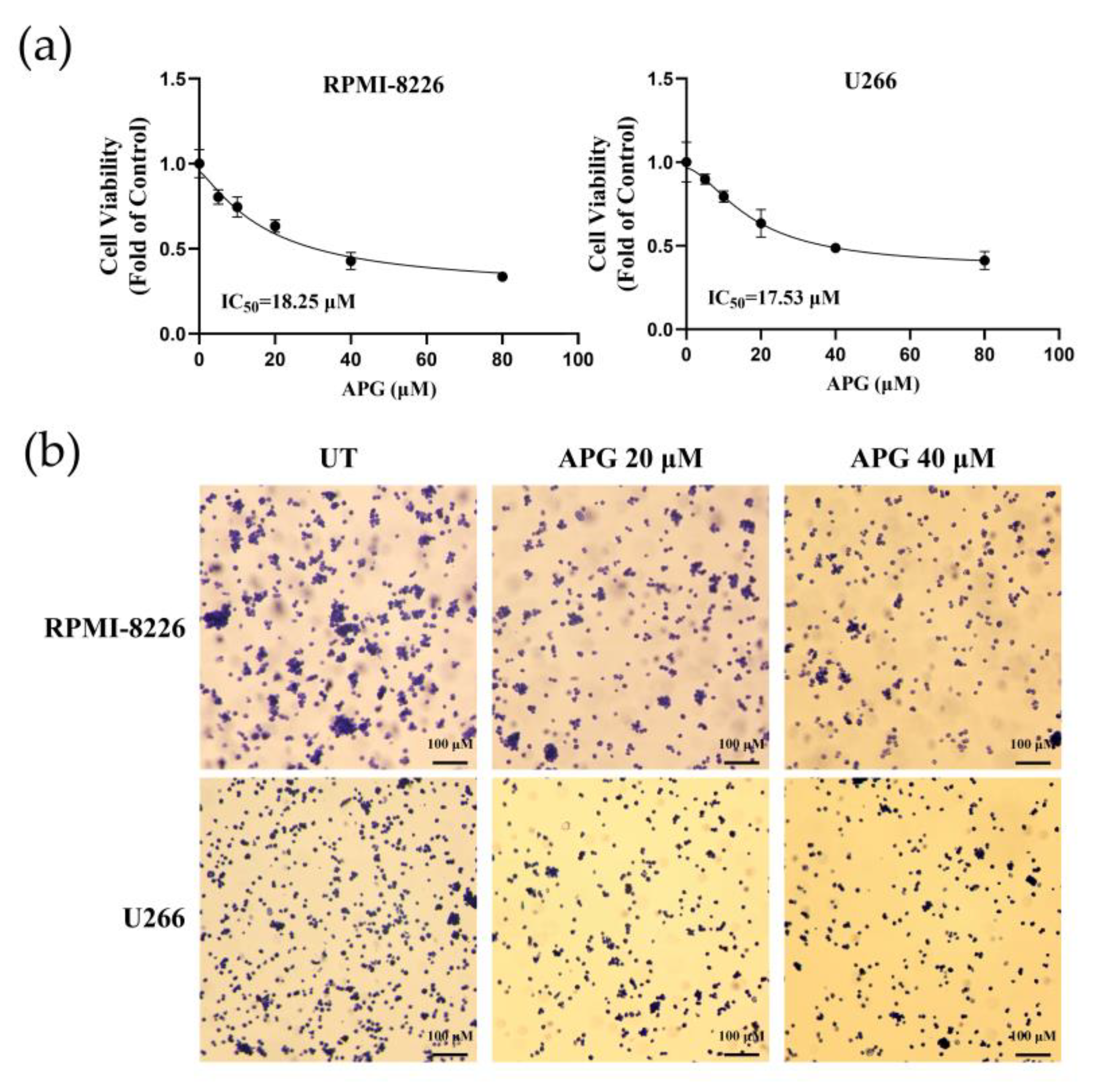
Two MM cell lines, RPMI-8226 and U266, were used to evaluate the effect of APG on the proliferation of MM cells. In this work, RPMI-8226 and U266 cells were incubated with gradually increased concentrations of APG and cell viability was then measured using CCK-8 assays. The results of CCK-8 assays showed that APG inhibited the growth of MM cells in a dose-dependent manner. And the IC50 (50% inhibitory concentration) of APG on RPMI-8226 and U266 was 18.25 μM and 17.53 μM, respectively, which was calculated with the cell viability curve (Figure 2a). To further validate the anti-myeloma activity of APG, we performed crystal violet staining assays. Compared with the control group, treatment of 20 μM and 40 μM APG significantly reduced crystal violet-stained cells (Figure 2b), indicating that APG markedly inhibits the growth of MM cells.

3.2. APG Induces G0/G1 Cell Cycle Arrest of MM Cells

To investigate whether the inhibitory effect of APG on MM cell proliferation was mediated through cell cycle arrest, we performed cell cycle analysis by flow cytometry. The results showed that APG treatment significantly increased the proportion of MM cells in G1 phase while decreasing the population in G2 phase (Figure 3a). We assessed the protein level of G1/S-Specific Cyclin D1 (Cyclin D1) and Cyclin-dependent kinase 2 (CDK2), two key regulators improving G1 to S phase transition (Figure 3b,c). Notably, APG treatment downregulated the expression of both Cyclin D1 and CDK2 and, on the contrary, stimulated Cyclin-dependent kinase inhibitor 1A (P21) expression. These findings indicated that APG suppressed MM cell proliferation by inducing G0/G1 phase arrest.

3.3. APG Induces Oxidative Stress in MM Cells

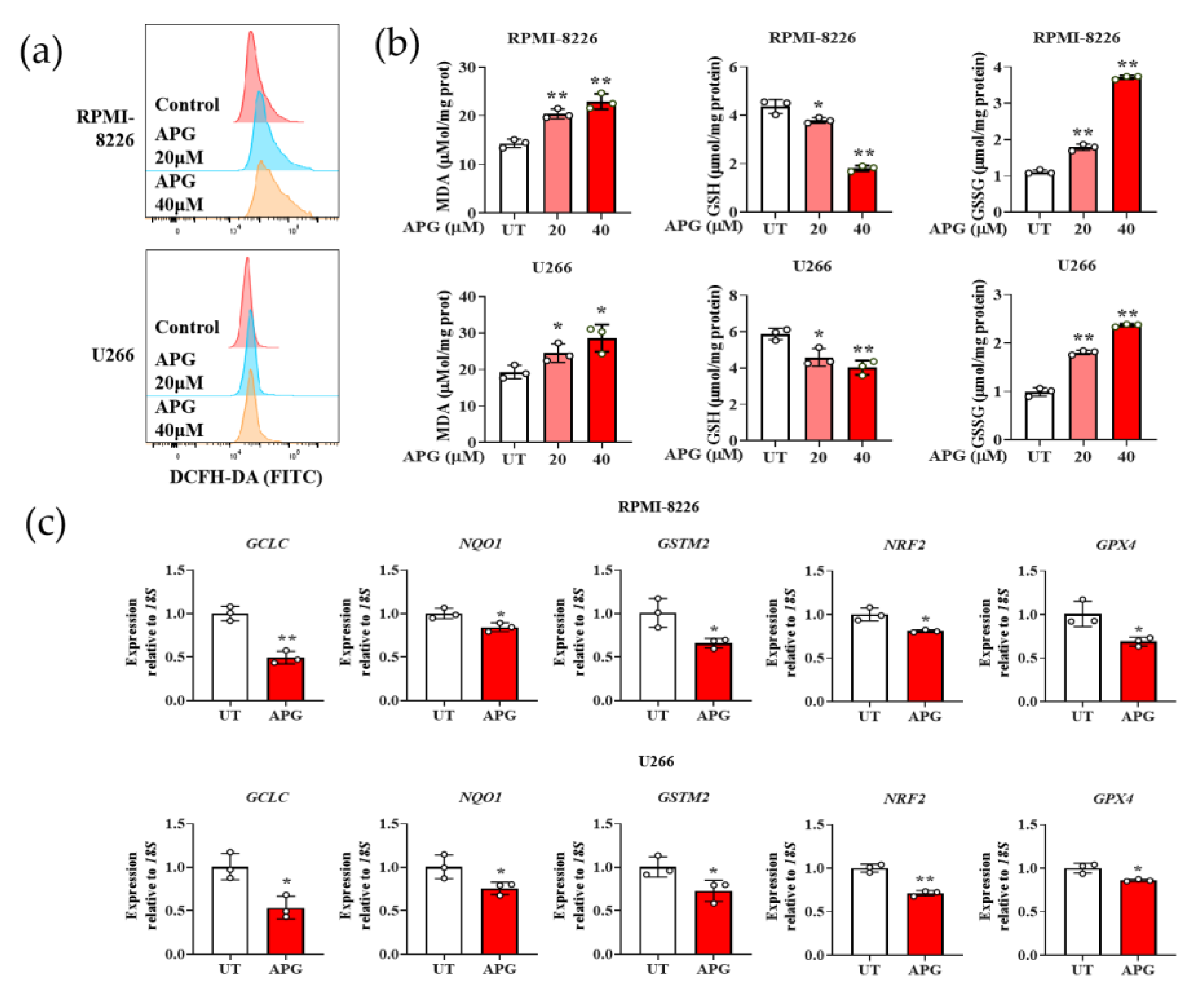
We evaluated the oxidative stress of APG treated MM cells with the DCFH-DA probe. The results showed that APG treatment stimulated ROS levels in both two types of MM cell strains in a dose-dependent manner (Figure 4a). Subsequently, we investigated the levels of Malondialdehyde (MDA), Glutathione (GSH), and Glutathione Disulfide (GSSG)—some of the hallmarks of cellular oxidative stress—in MM cells. The results indicated that the GSH content in APG-treated MM cells was significantly decreased. In contrast, the levels of MDA and GSSG were statistically increased (Figure 4b). In addition, we detected the transcriptional expression of proteins GCLC, NQO1, GSTM2, NRF2, and GPX4 in APG-treated MM cells. The results showed that the transcription of these proteins in MM cells were reduced by APG treatment (Figure 4c). In summary, these results demonstrate that APG induces MM cells apoptosis by stimulating oxidative stress.

3.4. APG Promotes Apoptosis in MM Cells

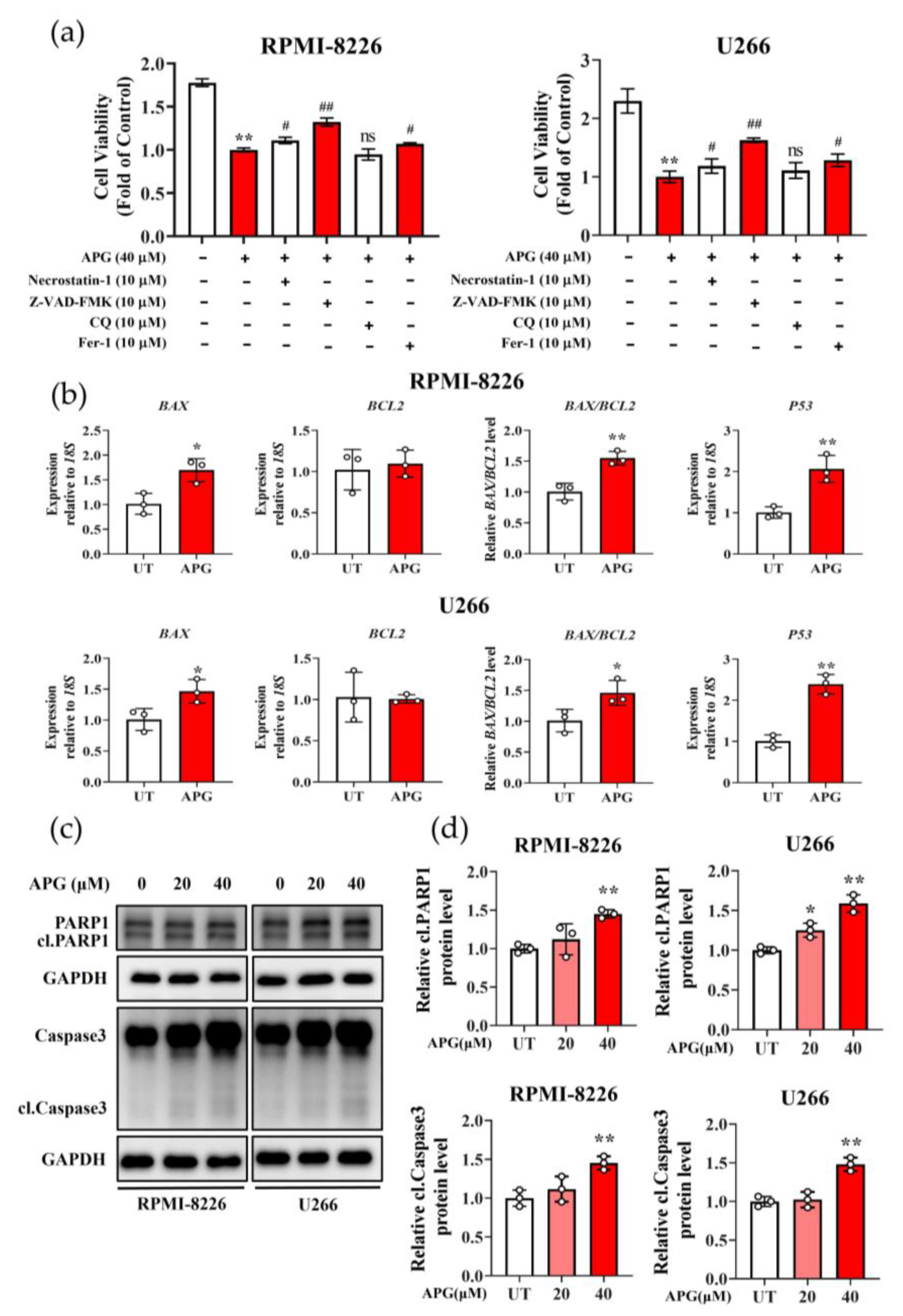
We employed various inhibitors, Necrostatin-1 (a potent necroptosis inhibitor), ZVAD-FMK (a pan-caspase inhibitor), chloroquine (CQ, an autophagy inhibitor), and ferrostatin-1 (Fer-1, a ferroptosis inhibitor) to characterize the APG-induced cell death. Although Necrostatin-1 (cell recovery rate was 10.86%) and Fer-1 (cell recovery rate was 12.17%) could reduce the damaging effect of APG on MM cells viability, strikingly, ZVAD-FMK (cell recovery rate was 21.35%) showed the more pronounced protective effect against APG-induced cytotoxicity (Figure 5a). This indicated that APG predominantly triggers apoptotic cell death in MM cells. Since the ratio of pro-apoptotic protein BCL2-associated X (BAX) to anti-apoptotic protein BCL2 can indicate apoptosis, we detected the transcription of BAX, BCL2, and P53 in both APG-treated MM cell strains with RT-qPCR. The results showed that following treatment with APG, BAX levels increased to 1.5-fold of control values, while BCL2 expression showed no significant alteration; the BAX/BCL2 ratio increased to 1.5-fold relative to controls. Separately, P53 levels increased to 2.0-fold of control values, compared with the untreated cell assays (Figure 5b). Furthermore, the result of Western blot (WB) analysis demonstrated the significantly increased levels of the cleaved/activated forms of both Caspase 3 and PARP1 (Figure 5c,d). All these results suggested that APG could promote apoptosis in MM cells. But the underlying mechanism was still not discovered.

3.5. APG Potentiates the Anti-Tumor Effects of Bortezomib (BTZ)

RPMI-8226 and U266 cells were treated with increasing concentrations of BTZ alone for 6 h. After BTZ removal, cells were either cultured for 42 h or treated with 20 μM APG and cultured for 42 h. Cell viability was then determined using the CCK-8 assay. The results demonstrated that at equivalent BTZ concentrations, the combination treatment (BTZ followed by APG) significantly reduced the viability of MM cells compared to BTZ alone (Figure 6a). Next, we analyzed the synergism between APG and BTZ by applying CompuSyn (version 1.0). The combination of APG and BTZ showed synergistic anti-MM activity with a combination index (CI) < 1.0 (Figure 6b). Subsequently, apoptosis was analyzed by flow cytometry, and the results showed that compared with BTZ monotherapy, the combination treatment significantly enhanced cell apoptosis (Figure 6c). Additionally, transcriptional levels of apoptosis-associated genes BAX, BCL2, and P53, along with expression levels of apoptosis-related proteins Caspase 3 and PARP1 were examined in RPMI-8226 cells. The combination treatment significantly upregulated transcription of BAX and P53, downregulated BCL2 transcription compared to BTZ monotherapy (Figure 6d), and increased cleavage of Caspase 3 and PARP1 (Figure 6e). These findings indicate that APG acts as a sensitizer, potentiating the anti-myeloma activity of BTZ.

4. Discussion

MM represents a clinically and genetically heterogeneous hematologic malignancy [21], exhibiting the high incidence and mortality rates among blood cancers, surpassed only by non-Hodgkin lymphoma [22]. Despite advances in proteasome inhibitors (such as BTZ) and chimeric antigen receptor (CAR) T-cell therapies, MM remains incurable due to drug resistance and toxicity [23,24,25]. It has been recognized that drug resistance is one of the major mortality factors in the clinic [22]. Notably, small molecules, such as flavonoids in herbs, have been increasingly applied in anticancer drug development and therapeutic applications due to their non-mutagenic properties in humans. Extensive studies have demonstrated that APG exhibits minimal toxicity to normal cells while displaying significant cytotoxicity against various cancer cell types. This selective anticancer activity enables the effective suppression of cancer stem cells in multiple malignancies [26]. Our results demonstrate that APG not only displays anti-MM activity but also can serve as a chemosensitizer for BTZ (Figure 6), effectively reducing drug resistance in MM.
The cell cycle represents a conserved mechanism for eukaryotic cell self-replication. Cyclin D/Cyclin-dependent kinase (Cdk) complexes play a pivotal role in cell cycle progression by coupling extracellular signals to the cell cycle machinery [27]. Cyclin D1 could complex with Cyclin-dependent kinase 4 (CDK4) and Cyclin-dependent kinase 6 (CDK6) to generate active kinase complexes which act early in the G1 phase, while Cyclin E is transcribed, translated, and complexes with CDK2 kinase, leading to progression through late G1 and entry into S phase [28]. This G1/S transition process is inhibited by P21 (the CDK inhibitor) through suppression of Cyclin E/CDK2 [29]. Our results demonstrate that APG treatment significantly downregulated Cyclin D1 and CDK2 expression while upregulating P21 in multiple myeloma (MM) cells (Figure 2b,c). This was concomitant with an increased proportion of cells in the G0 phase and decreased proportion in the S phase (Figure 2a), indicating that APG induces G0/G1 phase cell cycle arrest in MM cells, consistent with its reported effects in neuroblastoma and cervical cancer [30,31]. Notably, the cell cycle is closely associated with apoptotic processes, and several genes involved in cell cycle regulation, such as P53, also participate in apoptosis. The P53 tumor suppressor primarily functions during the G1 phase of the cell cycle, influencing cellular proliferation through its roles in cell cycle arrest and apoptosis [32]. Previous studies have demonstrated that APG induces P53 accumulation [33]. Activated P53 inhibits the G1-to-S phase transition by upregulating P21 expression. Additionally, it enhances the transcriptional level of BAX (a transcriptional target of P53) to promote apoptosis. As Schneider et al. reported [34], P53 can also modulate Cdk-activating kinase (CAK) activity, leading to CDK2 downregulation.
Excessive intracellular ROS generation frequently accompanies malignant transformation, leading to oxidative stress that may exert cytotoxic effects if left uncontrolled. Due to oncogene activation and enhanced metabolic activity, tumor cells typically exhibit elevated ROS levels compared to normal cells, rendering them more susceptible to chemotherapy-induced oxidative stress. An excessive ROS production leads to disruption of intracellular redox homeostasis, which can induce cellular apoptosis [35]. Our data show that APG increases intracellular ROS and MDA while reducing GSH (a key antioxidant) and increasing GSSG (oxidized GSH) in MM cells (Figure 4a,b). Concomitantly, APG downregulates the transcription of antioxidant proteins, including nuclear factor erythroid 2-related factor 2 (NRF2, a master regulator of redox homeostasis [36]), Glutathione peroxidase 4 (GPX4, an essential role in eliminating lipid peroxidation and exerting antioxidant effects [37]), and GCLC, NQO1, and GSTM2 (enzymes involved in GSH synthesis and detoxification) (Figure 4c). These findings demonstrate that APG can effectively induce oxidative stress in MM cells. The excessive accumulation of oxidative stress subsequently triggers apoptosis, thereby providing a promising therapeutic strategy for MM management.
The apoptotic program requires precise activation of multiple signaling cascades that are commonly disrupted in malignant cells. As such, pharmacological induction of apoptosis constitutes the predominant therapeutic mechanism for most clinically employed antitumor agents. Apoptosis, the primary mode of APG-induced cell death in MM, is supported by our inhibitor studies (Figure 5a). Molecularly, APG upregulates the pro-apoptotic protein BAX and downregulates the anti-apoptotic BCL2, increasing the BAX/BCL2 ratio (Figure 5b). This imbalance precipitates mitochondrial outer membrane permeabilization (MOMP), leading to the release of cytochrome c and subsequent activation of caspase cascades that execute the apoptotic program [38]. Poly (ADP-ribose) polymerase1 (PARP1), a nuclear enzyme, undergoes rapid activation in response to genotoxic stressors, including reactive oxygen species (ROS)-induced DNA damage [39]. During caspase-dependent apoptosis, PARP1 undergoes proteolytic cleavage, which simultaneously abrogates its DNA repair function and amplifies the apoptotic signal through multiple downstream effectors [40,41]. In our experimental results, the increased cleavage of Caspase 3 and PARP1, the key effectors of apoptotic execution, is consistent with the above. These results indicate that APG-induced oxidative stress activates apoptosis in MM cells.
Although APG has been shown to induce ferroptosis through mitochondrial pathways [42], trigger necrosis via mitochondrial dysfunction, ATP depletion, and elevated mitochondrial ROS production, and mediate autophagic cell death in human papillary thyroid carcinoma BCPAP cells [43], our findings indicate that these mechanisms are not predominant in MM (Figure 5a). Interestingly, APG-induced autophagy appears to exert a cytoprotective effect against apoptosis. Autophagy inhibition enhanced apoptotic cell death, thereby potentiating antitumor activity [44]. These findings suggest that a combination therapy strategy employing APG with autophagy inhibitors may represent a novel and effective approach for cancer treatment.
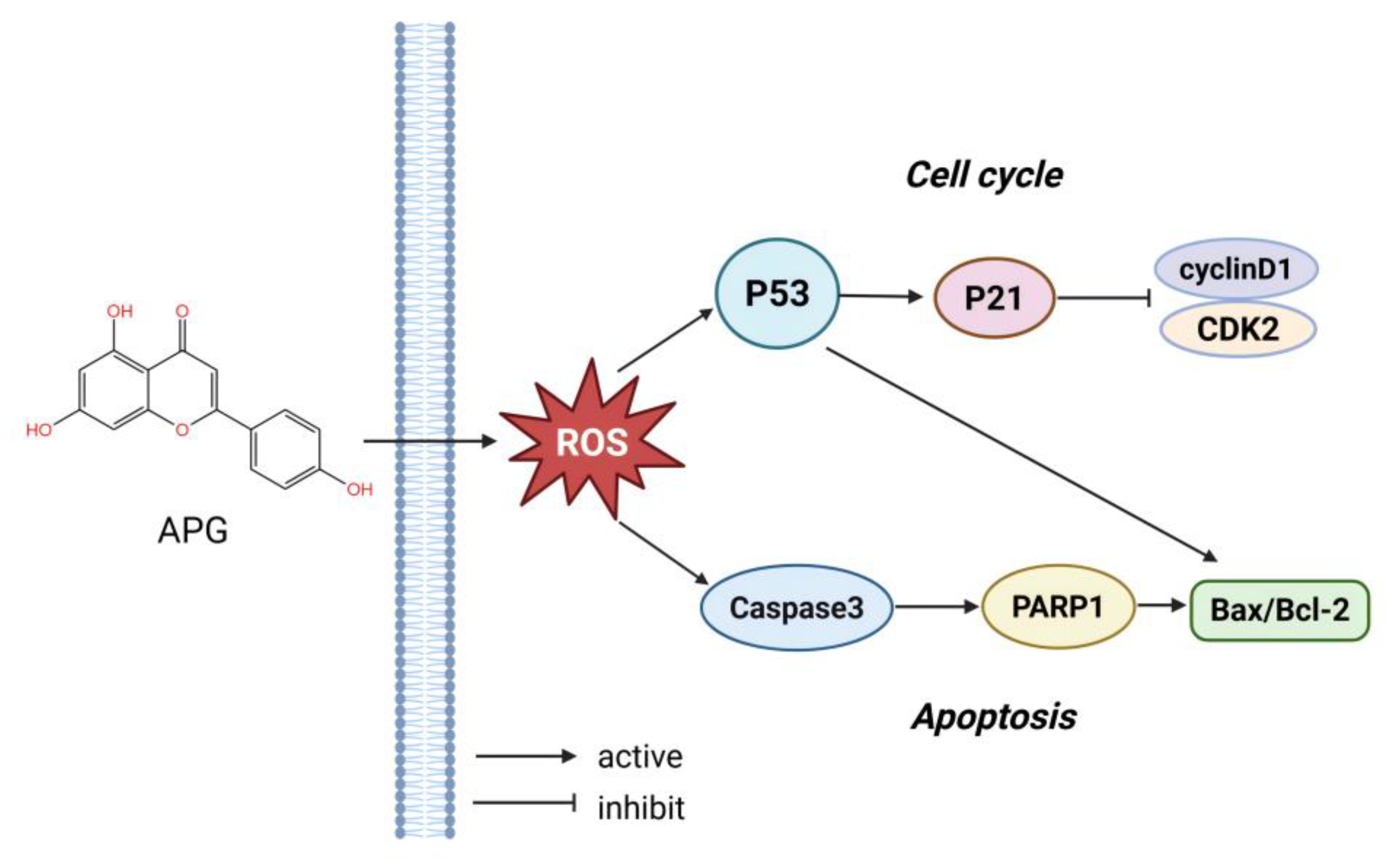
In short, our study demonstrates that APG exerts anti-myeloma effects through multiple mechanisms, including proliferation suppression, cell cycle arrest induction, oxidative stress generation, and apoptosis promotion, while simultaneously potentiating the therapeutic efficacy of BTZ in MM (Figure 7). However, several limitations should be acknowledged. First, the precise molecular mechanisms underlying APG-induced apoptosis in MM cells remain to be fully elucidated. Second, the absence of in vivo validation through animal models represents a significant limitation of our current findings. Finally, clinical data are lacking to substantiate the potential therapeutic efficacy of APG in MM patients. These limitations highlight important directions for future research to further validate and expand upon our findings.

5. Conclusions

In this study, we investigated the therapeutic potential of APG in MM. APG treatment significantly inhibited proliferation, induced G1/S phase cell cycle arrest, triggered oxidative stress, promoted apoptosis, and enhanced BTZ sensitivity in MM cell lines. Notably, we provide the first experimental evidence demonstrating APG’s ability to effectively induce apoptosis in MM cells, underscoring its potential as a novel therapeutic agent for MM. However, the clinical translatability of these findings requires further validation. Additional studies are necessary to comprehensively evaluate the anti-myeloma efficacy of APG and its potential clinical applications.

Author Contributions

Conceptualization, B.D. and Y.H.; methodology, Y.C. and L.W.; investigation, Y.C., L.W., H.C., M.C. and S.W.; data curation, Y.C. and L.W.; writing—original draft preparation, Y.C.; writing—review and editing, B.D. and Y.C.; visualization, Y.C. and L.W.; supervision, B.D. and Y.H.; funding acquisition, B.D. All authors have read and agreed to the published version of the manuscript.

Funding

This work was supported by grants from the Zhejiang Province Traditional Chinese Medicine science and technology plan project (2023ZF011).

Institutional Review Board Statement

Not applicable.

Informed Consent Statement

Not applicable.

Data Availability Statement

Data are contained within the article.

Acknowledgments

We appreciate the great help from Medical Research Center, Academy of Chinese Medical Sciences, and Zhejiang Chinese Medical University.

Conflicts of Interest

The authors declare no conflicts of interest.

Abbreviations

The following abbreviations are used in this manuscript:
AIFApoptosis-inducing factor
APGApigenin
BAXBCL2-associated X
BCL2B-cell lymphoma 2
BTZBortezomib
CAKCdk-activating kinase
Caspase 3Cysteine protease 3
CCK-8Cellcountingkit-8
CDK2Cyclin-dependent kinase 2
CDK4Cyclin-dependent kinase 4
CDK6Cyclin-dependent kinase 6
CDKsCyclin-dependent kinases
CQChloroquine
CyclinD1G1/S-Specific Cyclin-D1
GAPDHGlyceraldehyde-3-Phosphate Dehydrogenase
GCLCGlutamate-Cysteine Ligase Catalytic Subunit
GPX4Glutathione peroxidase 4
GSHGlutathione
GSSGGlutathione Disulfide
GSTM2Glutathione S-Transferase Mu 2
MDAMalondialdehyde
MMMultiple myeloma
NQO1NAD(P)H Quinone Dehydrogenase 1
NRF2Nuclear factor erythroid 2-related factor 2
P21Cyclin-dependent kinase inhibitor 1A
P53Tumor Protein p53
PARP1Poly (ADP-ribose) polymerase 1
PCDProgrammed cell death
PIProteasome inhibitor
RNA18SN5RNA, 18S ribosomal N5
ROSReactive oxygen species
WBWestern blot

References

  1. Palumbo, A.; Anderson, K. Multiple myeloma. N. Engl. J. Med. 2011, 364, 1046–1060. [Google Scholar] [CrossRef] [PubMed]
  2. Landgren, O. Monoclonal gammopathy of undetermined significance and smoldering multiple myeloma: Biological insights and early treatment strategies. Hematol. Am. Soc. Hematol. Educ. Program 2013, 2013, 478–487. [Google Scholar] [CrossRef] [PubMed]
  3. McCurdy, A.; Seow, H.; Pond, G.P.; Gayowsky, A.; Chakraborty, R.; Visram, A.; Kaedbey, R.; D’Souza, A.; Mohyuddin, G.R.; Wildes, T.M.; et al. Cancer-specific mortality in multiple myeloma: A population-based retrospective cohort study. Haematologica 2023, 108, 3384–3391. [Google Scholar] [CrossRef] [PubMed]
  4. Plakoula, E.; Kalampounias, G.; Alexis, S.; Verigou, E.; Kourakli, A.; Zafeiropoulou, K.; Symeonidis, A. Prognostic value of psmb5 and correlations with lc3ii and reactive oxygen species levels in the bone marrow mononuclear cells of bortezomib-resistant multiple myeloma patients. Curr. Issues Mol. Biol. 2025, 47, 32. [Google Scholar] [CrossRef]
  5. Nerini-Molteni, S.; Ferrarini, M.; Cozza, S.; Caligaris-Cappio, F.; Sitia, R. Redox homeostasis modulates the sensitivity of myeloma cells to bortezomib. Br. J. Haematol. 2008, 141, 494–503. [Google Scholar] [CrossRef]
  6. Murphy, M.P.; Bayir, H.; Belousov, V.; Chang, C.J.; Davies, K.J.A.; Davies, M.J.; Dick, T.P.; Finkel, T.; Forman, H.J.; Janssen-Heininger, Y.; et al. Guidelines for measuring reactive oxygen species and oxidative damage in cells and in vivo. Nat. Metab. 2022, 4, 651–662. [Google Scholar] [CrossRef]
  7. Juan, C.A.; de la Lastra, J.M.P.; Plou, F.J.; Perez-Lebena, E. The chemistry of reactive oxygen species (ros) revisited: Outlining their role in biological macromolecules (DNA, lipids and proteins) and induced pathologies. Int. J. Mol. Sci. 2021, 22, 4642. [Google Scholar] [CrossRef]
  8. Chandra, J.; Samali, A.; Orrenius, S. Triggering and modulation of apoptosis by oxidative stress. Free Radic. Biol. Med. 2000, 29, 323–333. [Google Scholar] [CrossRef]
  9. Asadi, M.; Taghizadeh, S.; Kaviani, E.; Vakili, O.; Taheri-Anganeh, M.; Tahamtan, M.; Savardashtaki, A. Caspase-3: Structure, function, and biotechnological aspects. Biotechnol. Appl. Biochem. 2022, 69, 1633–1645. [Google Scholar] [CrossRef]
  10. Mashimo, M.; Onishi, M.; Uno, A.; Tanimichi, A.; Nobeyama, A.; Mori, M.; Yamada, S.; Negi, S.; Bu, X.; Kato, J.; et al. The 89-kda parp1 cleavage fragment serves as a cytoplasmic par carrier to induce aif-mediated apoptosis. J. Biol. Chem. 2021, 296, 100046. [Google Scholar] [CrossRef]
  11. Li, D.; Ueta, E.; Kimura, T.; Yamamoto, T.; Osaki, T. Reactive oxygen species (ros) control the expression of bcl-2 family proteins by regulating their phosphorylation and ubiquitination. Cancer Sci. 2004, 95, 644–650. [Google Scholar] [CrossRef] [PubMed]
  12. Maggioni, D.; Garavello, W.; Rigolio, R.; Pignataro, L.; Gaini, R.; Nicolini, G. Apigenin impairs oral squamous cell carcinoma growth in vitro inducing cell cycle arrest and apoptosis. Int. J. Oncol. 2013, 43, 1675–1682. [Google Scholar] [CrossRef] [PubMed]
  13. Seo, H.S.; Ku, J.M.; Choi, H.S.; Woo, J.K.; Jang, B.H.; Shin, Y.C.; Ko, S.G. Induction of caspase-dependent apoptosis by apigenin by inhibiting stat3 signaling in her2-overexpressing mda-mb-453 breast cancer cells. Anticancer Res. 2014, 34, 2869–2882. [Google Scholar] [PubMed]
  14. Zhu, Y.; Mao, Y.; Chen, H.; Lin, Y.; Hu, Z.; Wu, J.; Xu, X.; Xu, X.; Qin, J.; Xie, L. Apigenin promotes apoptosis, inhibits invasion and induces cell cycle arrest of t24 human bladder cancer cells. Cancer Cell Int. 2013, 13, 54. [Google Scholar] [CrossRef]
  15. Yin, F.; Giuliano, A.E.; Law, R.E.; Van Herle, A.J. Apigenin inhibits growth and induces g2/m arrest by modulating cyclin-cdk regulators and erk map kinase activation in breast carcinoma cells. Anticancer Res. 2001, 21, 413–420. [Google Scholar]
  16. Rossi, M.; Negri, E.; Lagiou, P.; Talamini, R.; Maso, L.D.; Montella, M.; Franceschi, S.; La Vecchia, C. Flavonoids and ovarian cancer risk: A case-control study in Italy. Int. J. Cancer 2008, 123, 895–898. [Google Scholar] [CrossRef]
  17. Wang, W.; Heideman, L.; Chung, C.S.; Pelling, J.C.; Koehler, K.J.; Birt, D.F. Cell-cycle arrest at g2/m and growth inhibition by apigenin in human colon carcinoma cell lines. Mol. Carcinog. 2000, 28, 102–110. [Google Scholar] [CrossRef]
  18. Yang, J.; Pi, C.; Wang, G. Inhibition of pi3k/akt/mtor pathway by apigenin induces apoptosis and autophagy in hepatocellular carcinoma cells. Biomed. Pharmacother. 2018, 103, 699–707. [Google Scholar] [CrossRef]
  19. Zhu, Y.; Wu, J.; Li, S.; Wang, X.; Liang, Z.; Xu, X.; Xu, X.; Hu, Z.; Lin, Y.; Chen, H.; et al. Apigenin inhibits migration and invasion via modulation of epithelial mesenchymal transition in prostate cancer. Mol. Med. Rep. 2015, 11, 1004–1008. [Google Scholar] [CrossRef]
  20. Caltagirone, S.; Rossi, C.; Poggi, A.; Ranelletti, F.O.; Natali, P.G.; Brunetti, M.; Aiello, F.B.; Piantelli, M. Flavonoids apigenin and quercetin inhibit melanoma growth and metastatic potential. Int. J. Cancer 2000, 87, 595–600. [Google Scholar] [CrossRef]
  21. Gordon, M.J.; Smith, M.R.; Nastoupil, L.J. Follicular lymphoma: The long and winding road leading to your cure? Blood Rev. 2023, 57, 100992. [Google Scholar] [CrossRef]
  22. van de Donk, N.; Pawlyn, C.; Yong, K.L. Multiple myeloma. Lancet 2021, 397, 410–427. [Google Scholar] [CrossRef]
  23. Hu, A.; Chen, H.; Liang, J.; Liu, C.; Li, F.; Mu, C. Cell-based therapeutics for the treatment of hematologic diseases inside the bone marrow. J. Control Release 2021, 339, 1–13. [Google Scholar] [CrossRef]
  24. Trendowski, M. Pu-h71: An improvement on nature’s solutions to oncogenic hsp90 addiction. Pharmacol. Res. 2015, 99, 202–216. [Google Scholar] [CrossRef] [PubMed]
  25. Li, Q.; Yue, Y.; Chen, L.; Xu, C.; Wang, Y.; Du, L.; Xue, X.; Liu, Q.; Wang, Y.; Fan, F. Resveratrol sensitizes carfilzomib-induced apoptosis via promoting oxidative stress in multiple myeloma cells. Front. Pharmacol. 2018, 9, 334. [Google Scholar] [CrossRef] [PubMed]
  26. Ketkaew, Y.; Osathanon, T.; Pavasant, P.; Sooampon, S. Apigenin inhibited hypoxia induced stem cell marker expression in a head and neck squamous cell carcinoma cell line. Arch. Oral Biol. 2017, 74, 69–74. [Google Scholar] [CrossRef] [PubMed]
  27. MacLachlan, T.K.; Sang, N.; Giordano, A. Cyclins, cyclin-dependent kinases and cdk inhibitors: Implications in cell cycle control and cancer. Crit. Rev. Eukaryot. Gene Expr. 1995, 5, 127–156. [Google Scholar] [CrossRef]
  28. Li, Y.; Cheng, X.; Chen, C.; Huijuan, W.; Zhao, H.; Liu, W.; Xiang, Z.; Wang, Q. Apigenin, a flavonoid constituent derived from p. Villosa, inhibits hepatocellular carcinoma cell growth by cyclind1/cdk4 regulation via p38 mapk-p21 signaling. Pathol. Res. Pract. 2020, 216, 152701. [Google Scholar] [CrossRef]
  29. Karimian, A.; Ahmadi, Y.; Yousefi, B. Multiple functions of p21 in cell cycle, apoptosis and transcriptional regulation after DNA damage. DNA Repair 2016, 42, 63–71. [Google Scholar] [CrossRef]
  30. Chakrabarti, M.; Banik, N.L.; Ray, S.K. Sequential htert knockdown and apigenin treatment inhibited invasion and proliferation and induced apoptosis in human malignant neuroblastoma sk-n-dz and sk-n-be2 cells. J. Mol. Neurosci. 2013, 51, 187–198. [Google Scholar] [CrossRef]
  31. Zheng, P.W.; Chiang, L.C.; Lin, C.C. Apigenin induced apoptosis through p53-dependent pathway in human cervical carcinoma cells. Life Sci. 2005, 76, 1367–1379. [Google Scholar] [CrossRef] [PubMed]
  32. Levine, A.J. P53, the cellular gatekeeper for growth and division. Cell 1997, 88, 323–331. [Google Scholar] [CrossRef]
  33. Liu, R.; Ji, P.; Liu, B.; Qiao, H.; Wang, X.; Zhou, L.; Deng, T.; Ba, Y. Apigenin enhances the cisplatin cytotoxic effect through p53-modulated apoptosis. Oncol. Lett. 2017, 13, 1024–1030. [Google Scholar] [CrossRef] [PubMed]
  34. Schneider, E.; Montenarh, M.; Wagner, P. Regulation of cak kinase activity by p53. Oncogene 1998, 17, 2733–2741. [Google Scholar] [CrossRef]
  35. Circu, M.L.; Aw, T.Y. Reactive oxygen species, cellular redox systems, and apoptosis. Free Radic. Biol. Med. 2010, 48, 749–762. [Google Scholar] [CrossRef]
  36. Fu, C.; Wu, Y.; Liu, S.; Luo, C.; Lu, Y.; Liu, M.; Wang, L.; Zhang, Y.; Liu, X. Rehmannioside a improves cognitive impairment and alleviates ferroptosis via activating pi3k/akt/nrf2 and slc7a11/gpx4 signaling pathway after ischemia. J. Ethnopharmacol. 2022, 289, 115021. [Google Scholar] [CrossRef]
  37. Friedmann Angeli, J.P.; Schneider, M.; Proneth, B.; Tyurina, Y.Y.; Tyurin, V.A.; Hammond, V.J.; Herbach, N.; Aichler, M.; Walch, A.; Eggenhofer, E.; et al. Inactivation of the ferroptosis regulator gpx4 triggers acute renal failure in mice. Nat. Cell Biol. 2014, 16, 1180–1191. [Google Scholar] [CrossRef]
  38. Kashyap, D.; Garg, V.K.; Goel, N. Intrinsic and extrinsic pathways of apoptosis: Role in cancer development and prognosis. Adv. Protein Chem. Struct. Biol. 2021, 125, 73–120. [Google Scholar] [CrossRef]
  39. Spiegel, J.O.; Van Houten, B.; Durrant, J.D. Parp1: Structural insights and pharmacological targets for inhibition. DNA Repair 2021, 103, 103125. [Google Scholar] [CrossRef]
  40. Soldani, C.; Scovassi, A.I. Poly(adp-ribose) polymerase-1 cleavage during apoptosis: An update. Apoptosis 2002, 7, 321–328. [Google Scholar] [CrossRef]
  41. Soldani, C.; Lazze, M.C.; Bottone, M.G.; Tognon, G.; Biggiogera, M.; Pellicciari, C.E.; Scovassi, A.I. Poly(adp-ribose) polymerase cleavage during apoptosis: When and where? Exp. Cell Res. 2001, 269, 193–201. [Google Scholar] [CrossRef]
  42. Xie, Y.; Zhou, X.; Li, J.; Yao, X.C.; Liu, W.L.; Kang, F.H.; Zou, Z.X.; Xu, K.P.; Xu, P.S.; Tan, G.S. Identification of a new natural biflavonoids against breast cancer cells induced ferroptosis via the mitochondrial pathway. Bioorg. Chem. 2021, 109, 104744. [Google Scholar] [CrossRef]
  43. Zhang, L.; Cheng, X.; Gao, Y.; Zheng, J.; Xu, Q.; Sun, Y.; Guan, H.; Yu, H.; Sun, Z. Apigenin induces autophagic cell death in human papillary thyroid carcinoma bcpap cells. Food Funct. 2015, 6, 3464–3472. [Google Scholar] [CrossRef]
  44. Uberti, F.; Lattuada, D.; Morsanuto, V.; Nava, U.; Bolis, G.; Vacca, G.; Squarzanti, D.F.; Cisari, C.; Molinari, C. Vitamin d protects human endothelial cells from oxidative stress through the autophagic and survival pathways. J. Clin. Endocrinol. Metab. 2014, 99, 1367–1374. [Google Scholar] [CrossRef]
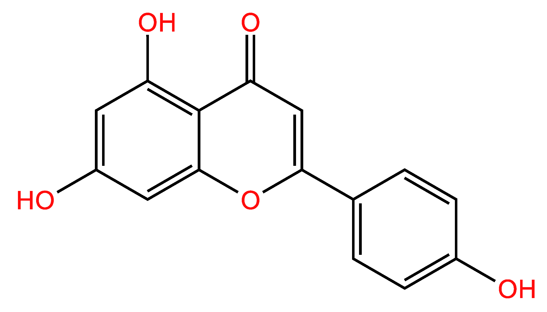
Figure 1. Chemical structure of apigenin (APG).
Cimb 47 00717 g001
Figure 2. APG inhibited multiple myeloma (MM) growth: (a) CCK-8 assay showed that APG inhibited the proliferation of RPMI-8226 and U266 cells after continuous incubation for 24 h. (b) Crystal violet staining of MM cells treated with APG for 24 h. UT: untreated; APG: APG-treated.
Cimb 47 00717 g002
Figure 3. APG induces G0/G1 phase arrest in MM cells: (a) The APG treated MM cells, in the G1, S, and G2 phases, was detected by flow cytometry. (b,c) The levels of cell-cycle-regulating proteins were detected by Western blot (WB). All values are expressed as the mean ± SD of three independent replicates. * p < 0.05, ** p < 0.01.
Cimb 47 00717 g003
Figure 4. APG triggered oxidative stress in MM cells: (a) The effect of APG on ROS levels in MM cells was detected by DCFH-DA. (b) The effects of APG on the levels of MDA, GSH, and GSSG in MM cells. (c) The effects of APG on the transcription of GCLC, NQO1, GSTM2, NRF2, and GPX4 in MM cells. All values are expressed as the mean ± SD of three independent replicates. *: compared with the untreated group. * p < 0.05, ** p < 0.01.
Cimb 47 00717 g004
Figure 5. APG promotes apoptosis in MM cells: (a) The beneficial effects of Necrostatin-1, ZVAD-FMK, CQ, and Fer-1 on APG-induced cytotoxicity in MM cells. (b) The transcription of BAX, BCL2, and P53 in APG-treated and untreated MM cells were determined by RT-qPCR. (c,d) The WB detection of the expression of PARP1, cleaved-PARP1 (cl. PARP1), Caspase 3, and cleaved-Caspase 3 (cl. Caspase 3) in APG-treated and untreated MM cells, which was quantified with ImageJ 1.54d. All values are expressed as the mean ± SD of three independent replicates. #: compared with the APG-treated group. *: compared with the untreated group. ns: not significant. * p < 0.05, ** p < 0.01, # p < 0.05, ## p < 0.01.
Cimb 47 00717 g005
Figure 6. Apigenin increases multiple myeloma cell sensitivity to bortezomib: (a) CCK-8 assay demonstrating the viability of RPMI-8226 and U266 cells following treatment with BTZ alone or in combination with APG. (b) Combination treatments were performed in RPMI-8226 and U266 cells maintaining a constant ratio between the dose of the APG and BTZ, and cell viability was assessed at 48 h by CCK-8 assay. The combination index (CI) value and the relative fraction affected (FA) were determined at each dose combination (actual), and a simulation was run to estimate the CI value and confidence interval (—) across the entire FA range (simulation). CI < 1, CI = 1, and CI > 1 indicate synergistic, additive, and antagonistic effects, respectively. CI was calculated by the CompuSyn software program. (c) Apoptosis in RPMI-8226 cells was analyzed by flow cytometry. (d) The transcription of BAX, BCL2, and P53 of RPMI-8226 cells following treatment with BTZ alone or in combination with APG were determined by RT-qPCR. (e) The WB detection of the expression of PARP1, cleaved-PARP1 (cl. PARP1), Caspase 3 and cleaved-Caspase 3 (cl. Caspase 3) in RPMI-8226 cells following treatment with BTZ alone or in combination with APG, which was quantified with ImageJ. All values are expressed as the mean ± SD of three independent replicates. ns: not significant. * p < 0.05, ** p < 0.01, *** p < 0.001.
Cimb 47 00717 g006
Figure 7. APG exerts anti-myeloma effects by triggering oxidative stress, inducing cell cycle arrest and apoptosis in MM cells.
Cimb 47 00717 g007
Table 1. Primer sets for qPCR.
Gene NameAccession NumberAmplicon SizeSequence (5′-3′)
BCL2NM_00065789Forward: GGTGGGGTCATGTGTGTGG
Reverse: CGGTTCAGGTACTCAGTCATCC
BAXNM_138763155Forward: CCCGAGAGGTCTTTTTCCGAG
Reverse: CCAGCCCATGATGGTTCTGAT
P53NM_001126118125Forward: CAGCACATGACGGAGGTTGT
Reverse: TCATCCAAATACTCCACACGC
GCLCNM_00119711579Forward: GGAGGAAACCAAGCGCCAT
Reverse: CTTGACGGCGTGGTAGATGT
NQO1NM_001025433196Forward: GAAGAGCACTGATCGTACTGGC
Reverse: GGATACTGAAAGTTCGCAGGG
GSTM2NM_00084899Forward: TGTGCGGGGAATCAGAAAAGG
Reverse: CTGGGTCATAGCAGAGTTTGG
NRF2NM_001145412174Forward: TCAGCGACGGAAAGAGTATGA
Reverse: CCACTGGTTTCTGACTGGATGT
GPX4NM_001039847100Forward: GAGGCAAGACCGAAGTAAACTAC
Reverse: CCGAACTGGTTACACGGGAA
RNA18SN5NR_003286158Forward: ACCCGTTGAACCCCATTCGTGA
Reverse: GCCICACAAACCACCAATCGG
Abbreviation: BCL2, B-cell lymphoma 2; BAX, BCL2-associated X; P53, Tumor Protein p53; GCLC, Glutamate-Cysteine Ligase Catalytic Subunit; NQO1, NAD(P)H Quinone Dehydrogenase 1; GSTM2, Glutathione S-Transferase Mu 2; NRF2, nuclear factor erythroid 2-relatedfactor 2; GPX4, Glutathione peroxidase 4; RNA18SN5, RNA, 18S Ribosomal N5.
Disclaimer/Publisher’s Note: The statements, opinions and data contained in all publications are solely those of the individual author(s) and contributor(s) and not of MDPI and/or the editor(s). MDPI and/or the editor(s) disclaim responsibility for any injury to people or property resulting from any ideas, methods, instructions or products referred to in the content.