
Fruit Peels: Food Waste as a Valuable Source of Bioactive Natural Products for Drug Discovery †

 ,
,  ,
,  ,
,
Abstract
1. Introduction
2. Traditional Uses of Fruit Peels
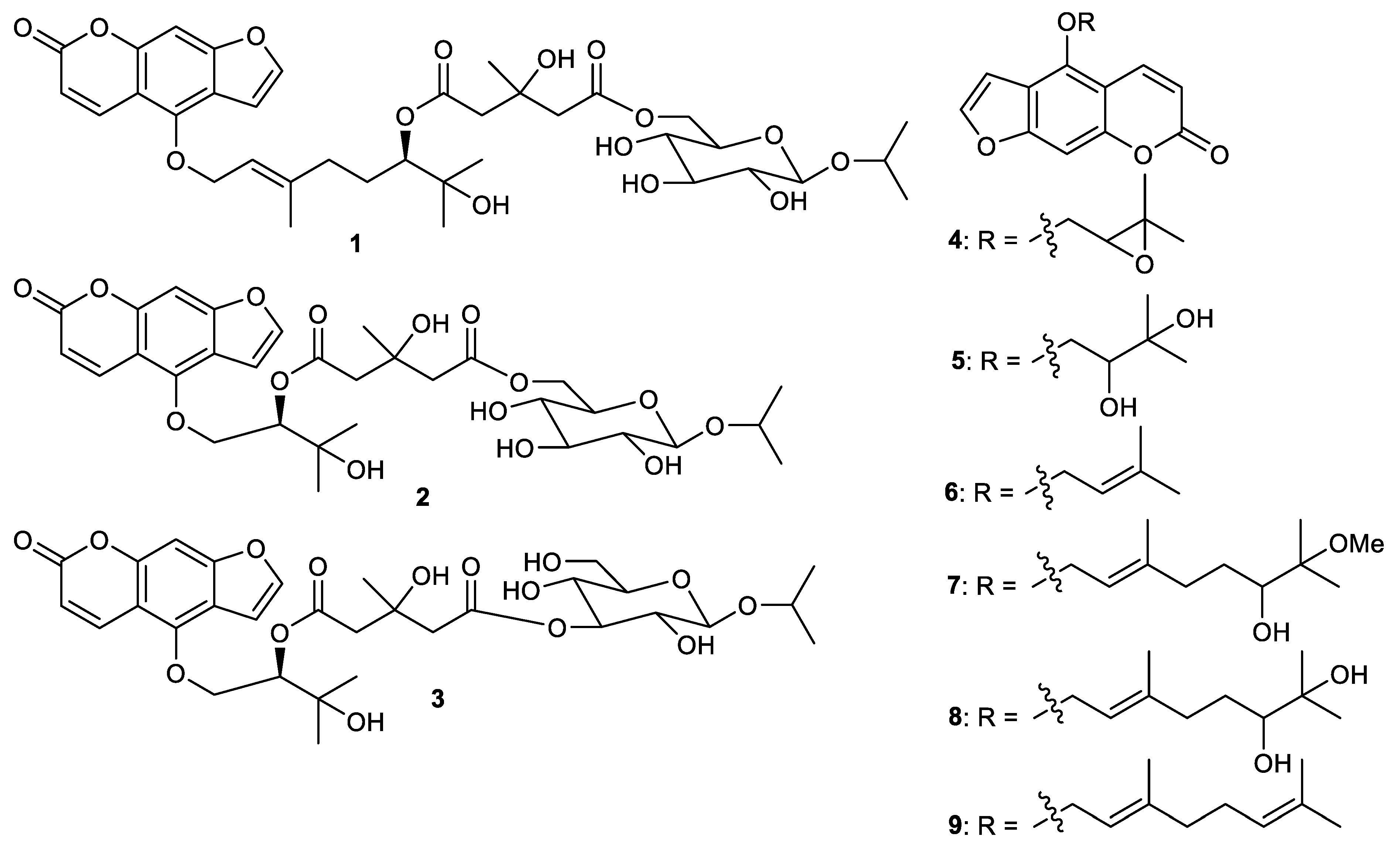
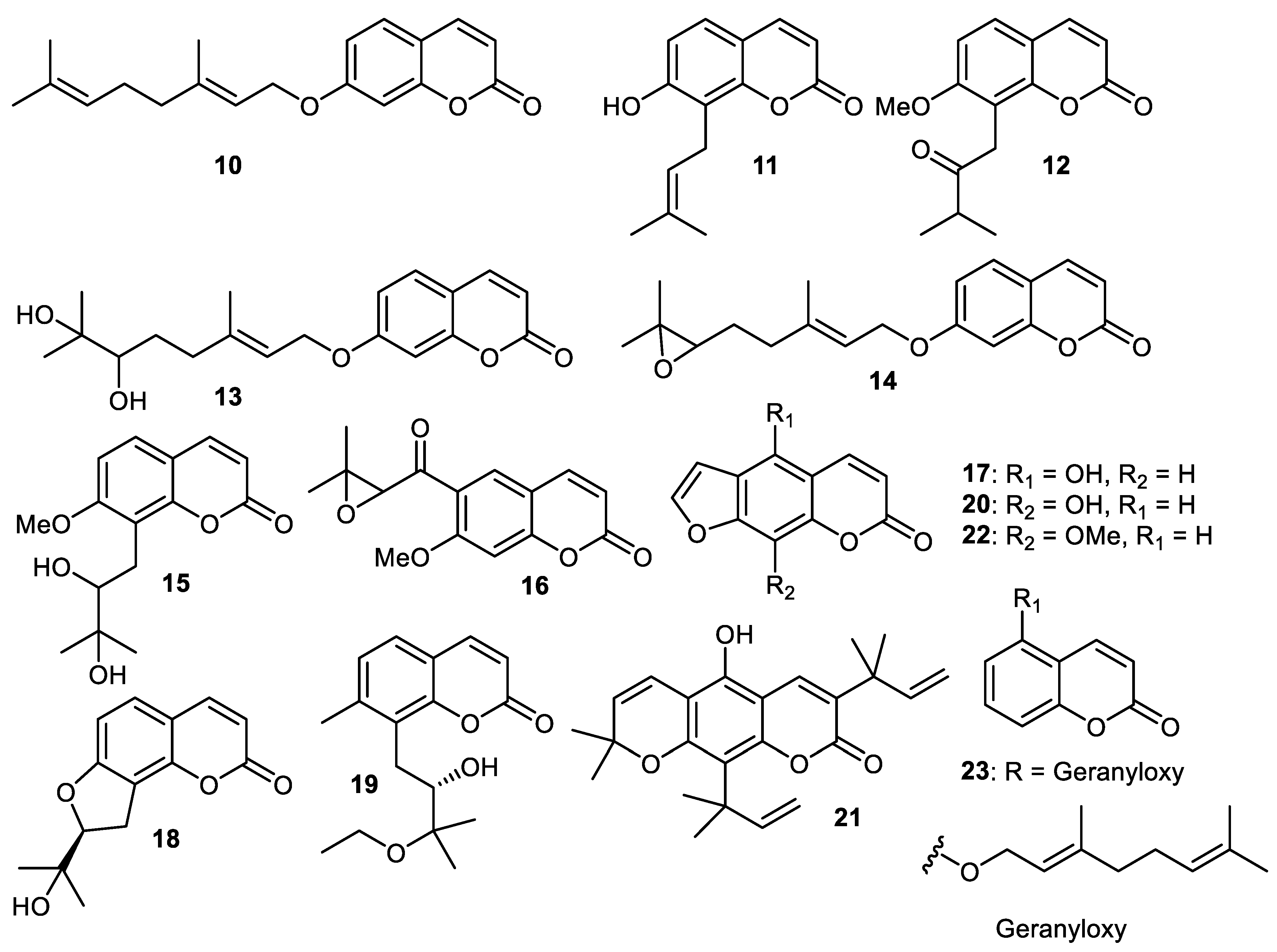
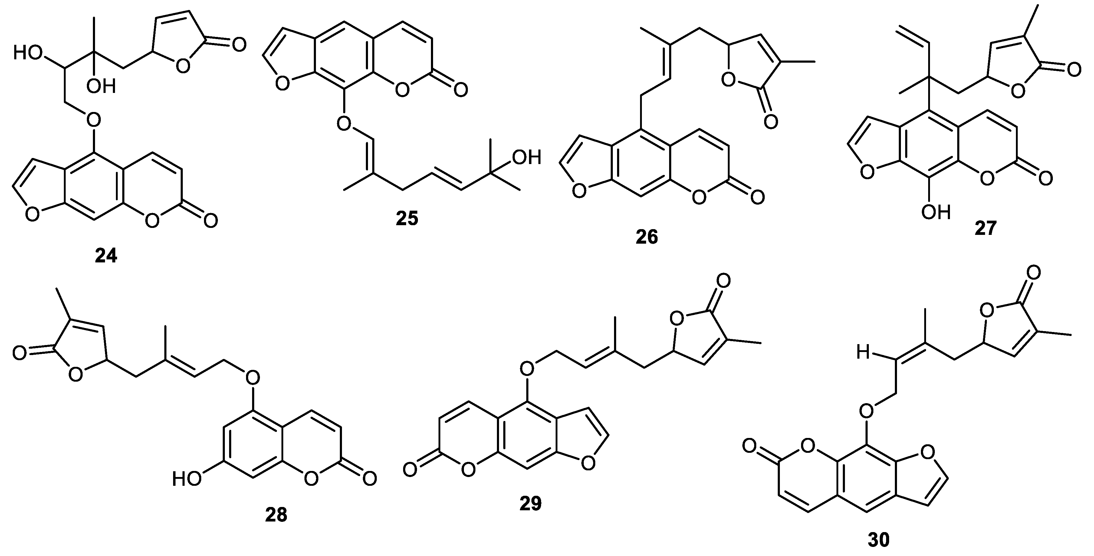
3. Coumarins
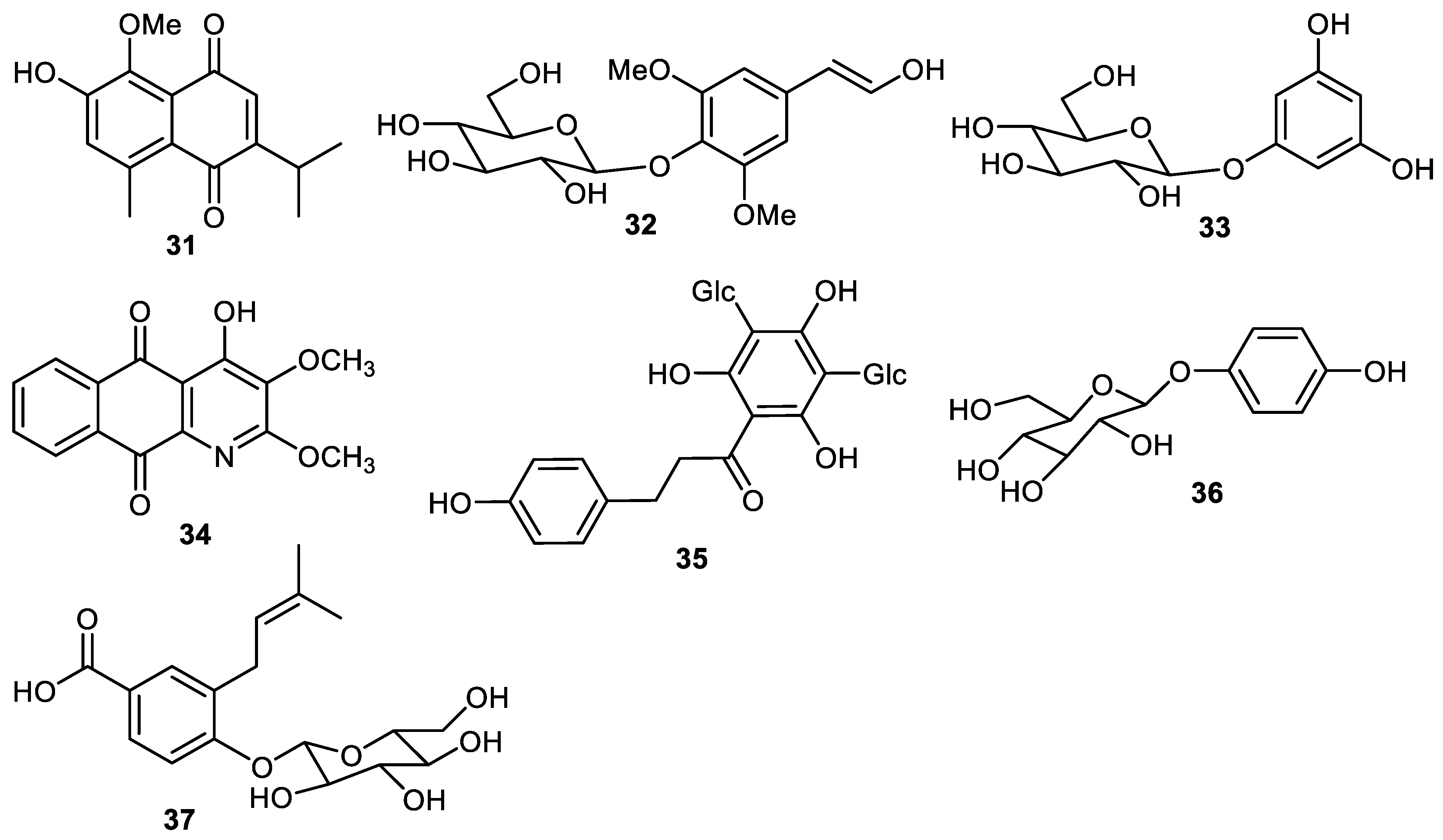
4. Quinone and Phenolic Glycosides
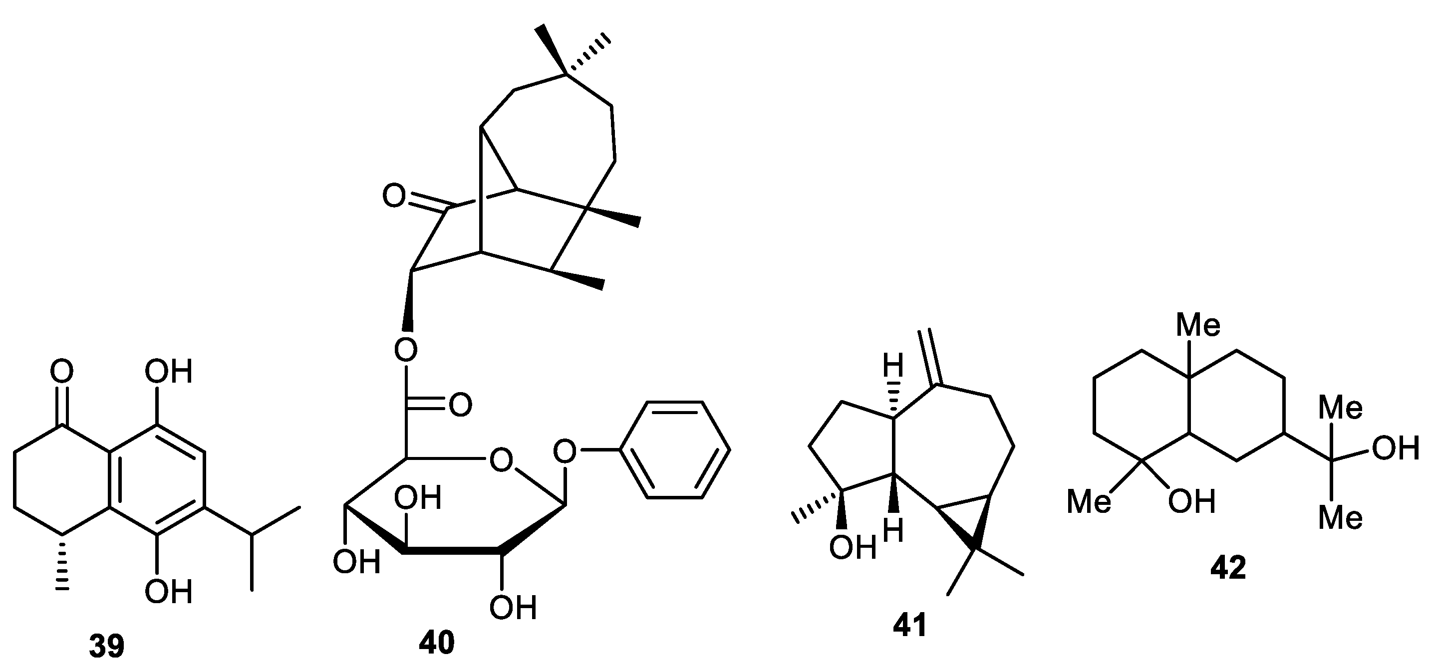
5. Sesquiterpenes
6. Naphthalene and Betacyanins
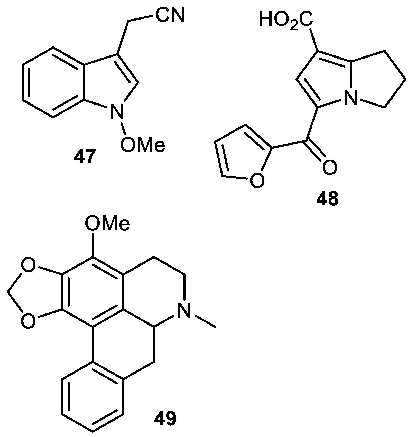
7. Alkaloids
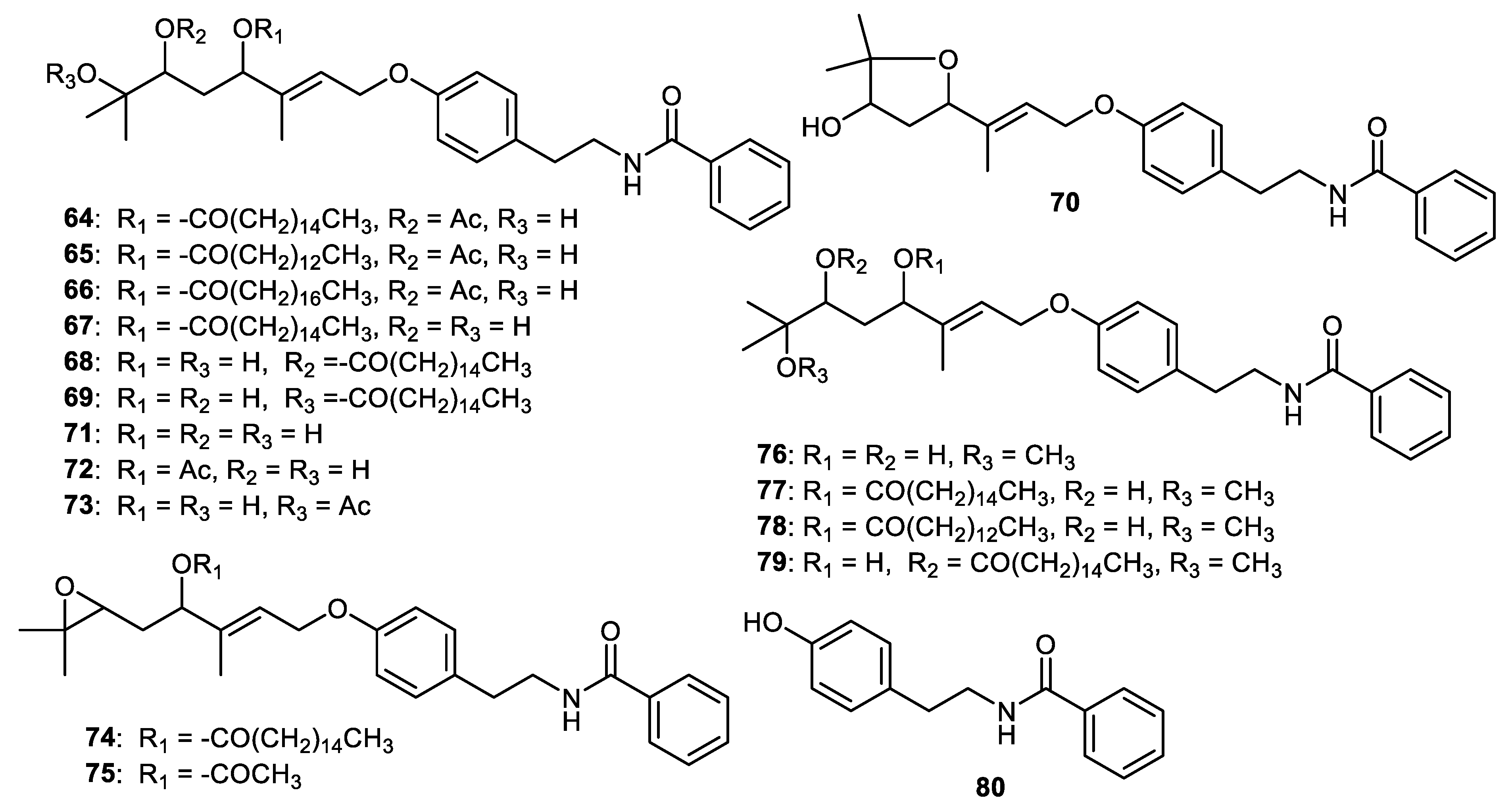
8. Benzoyltyramines
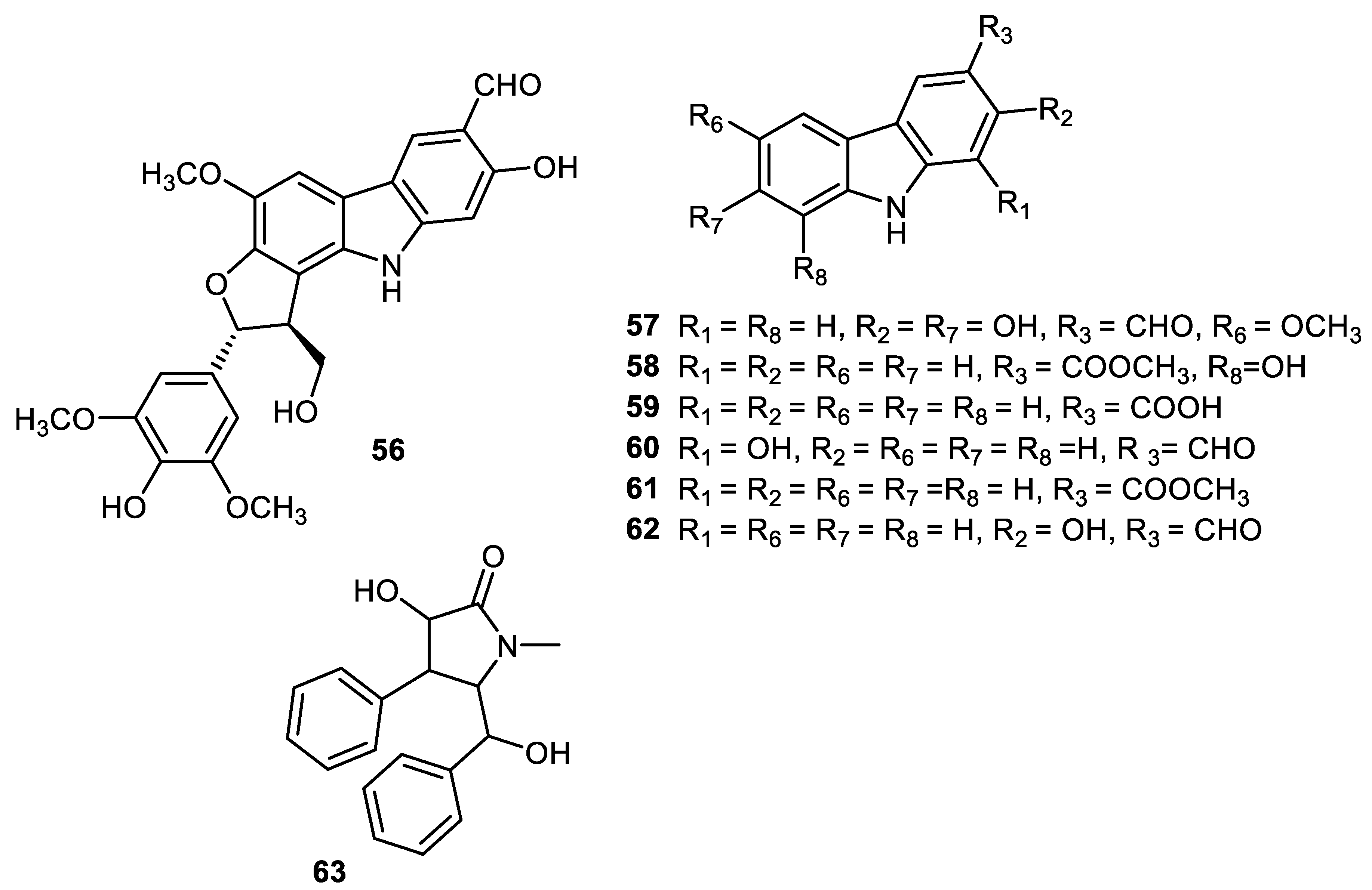
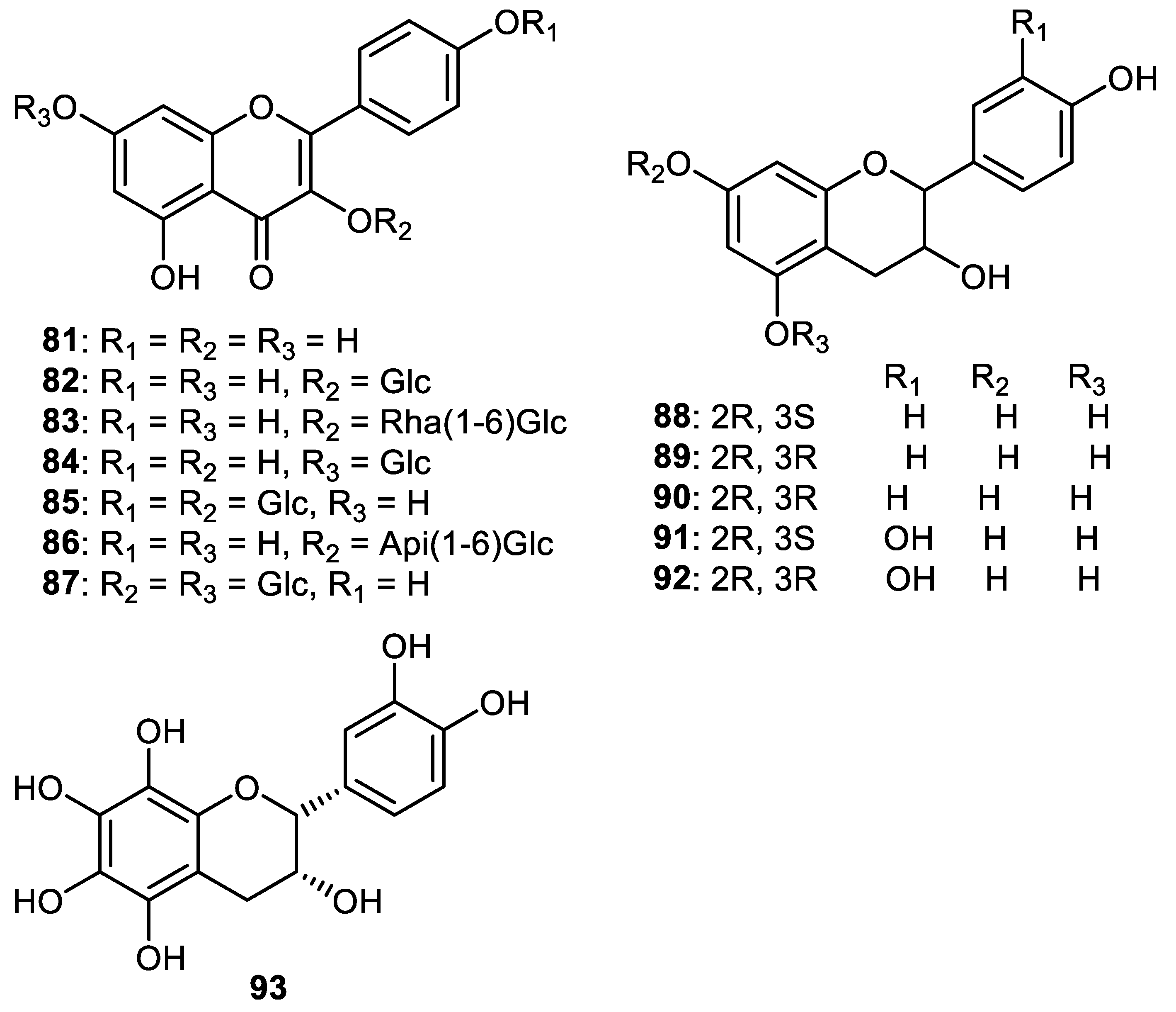
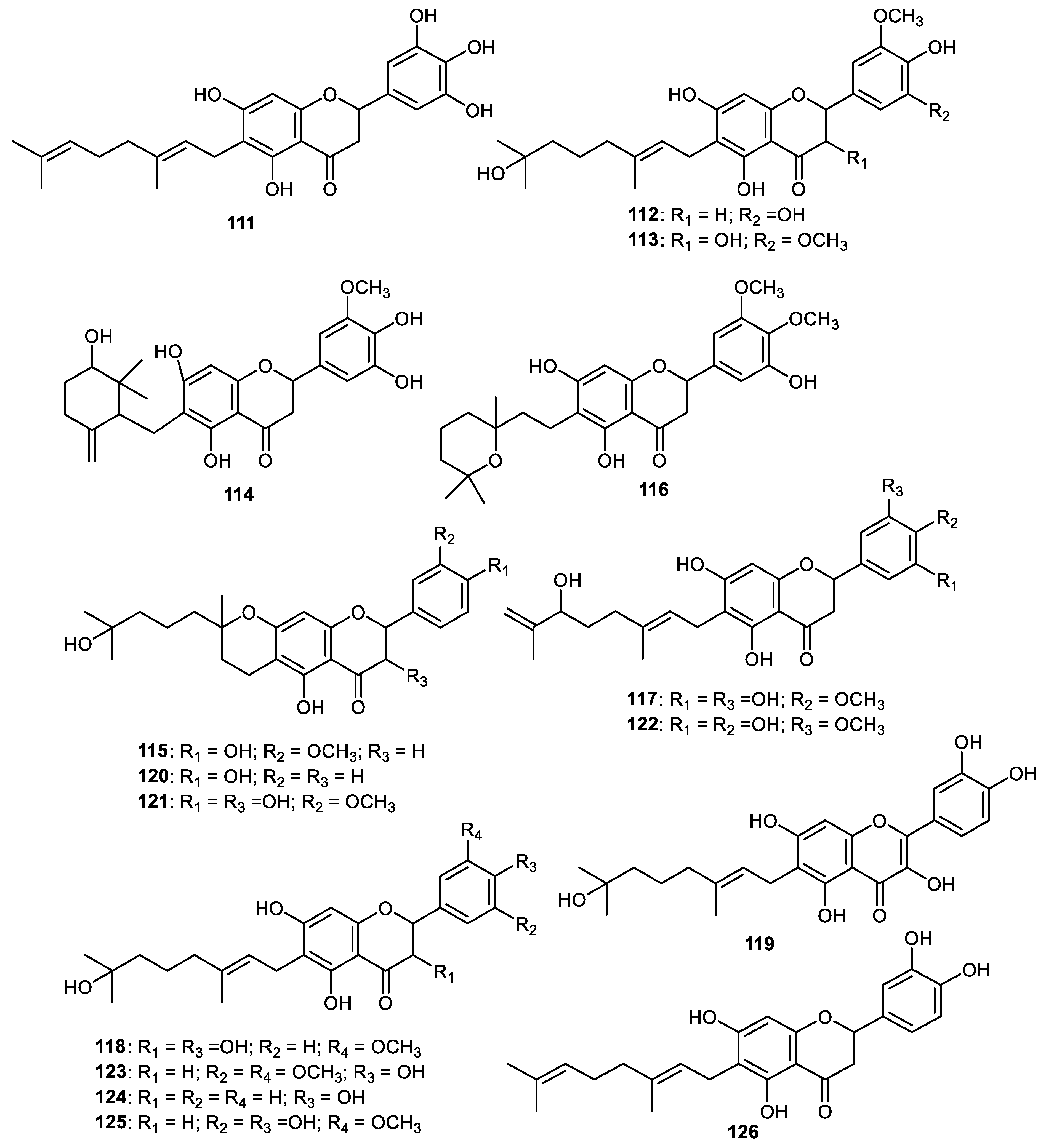
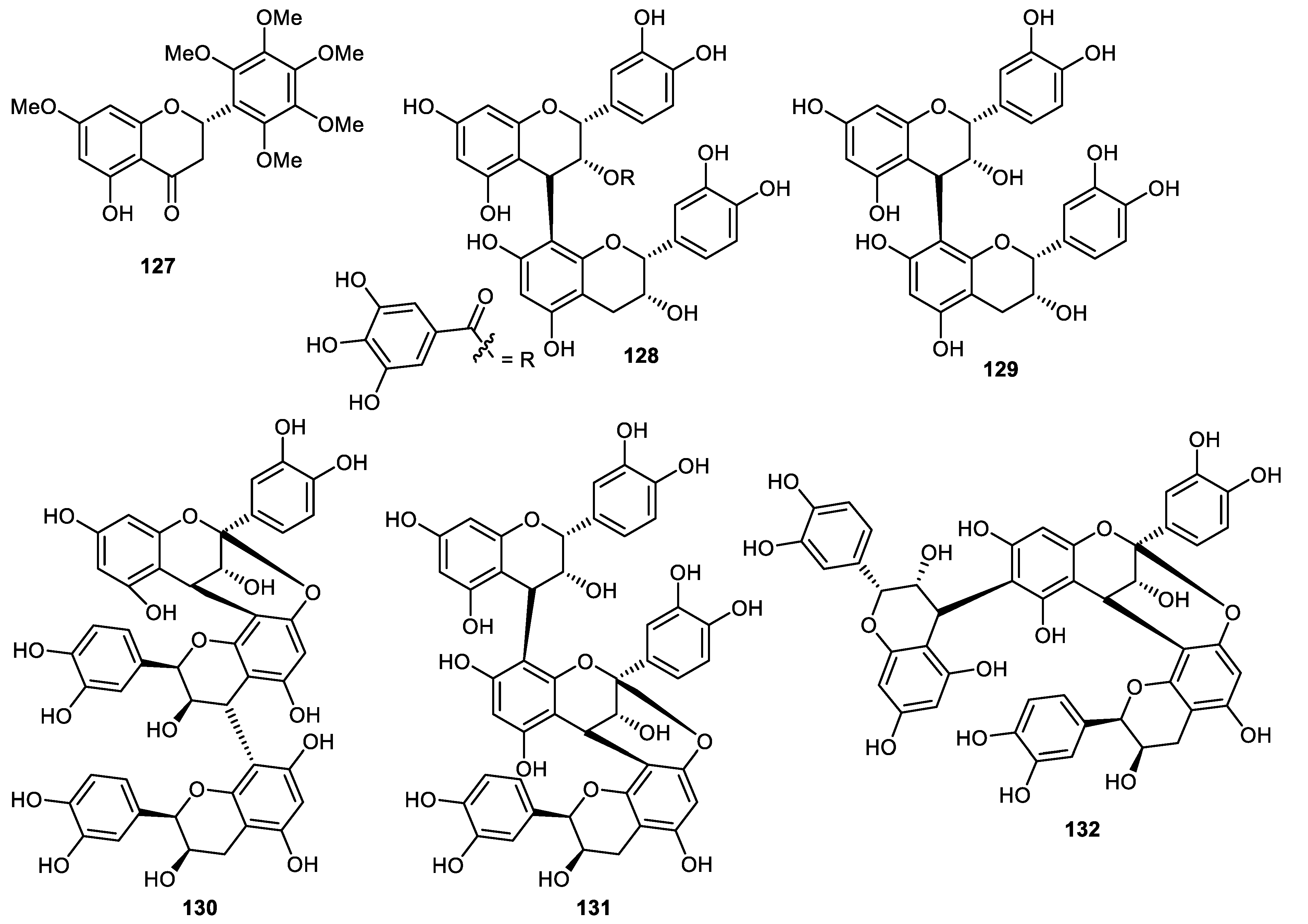
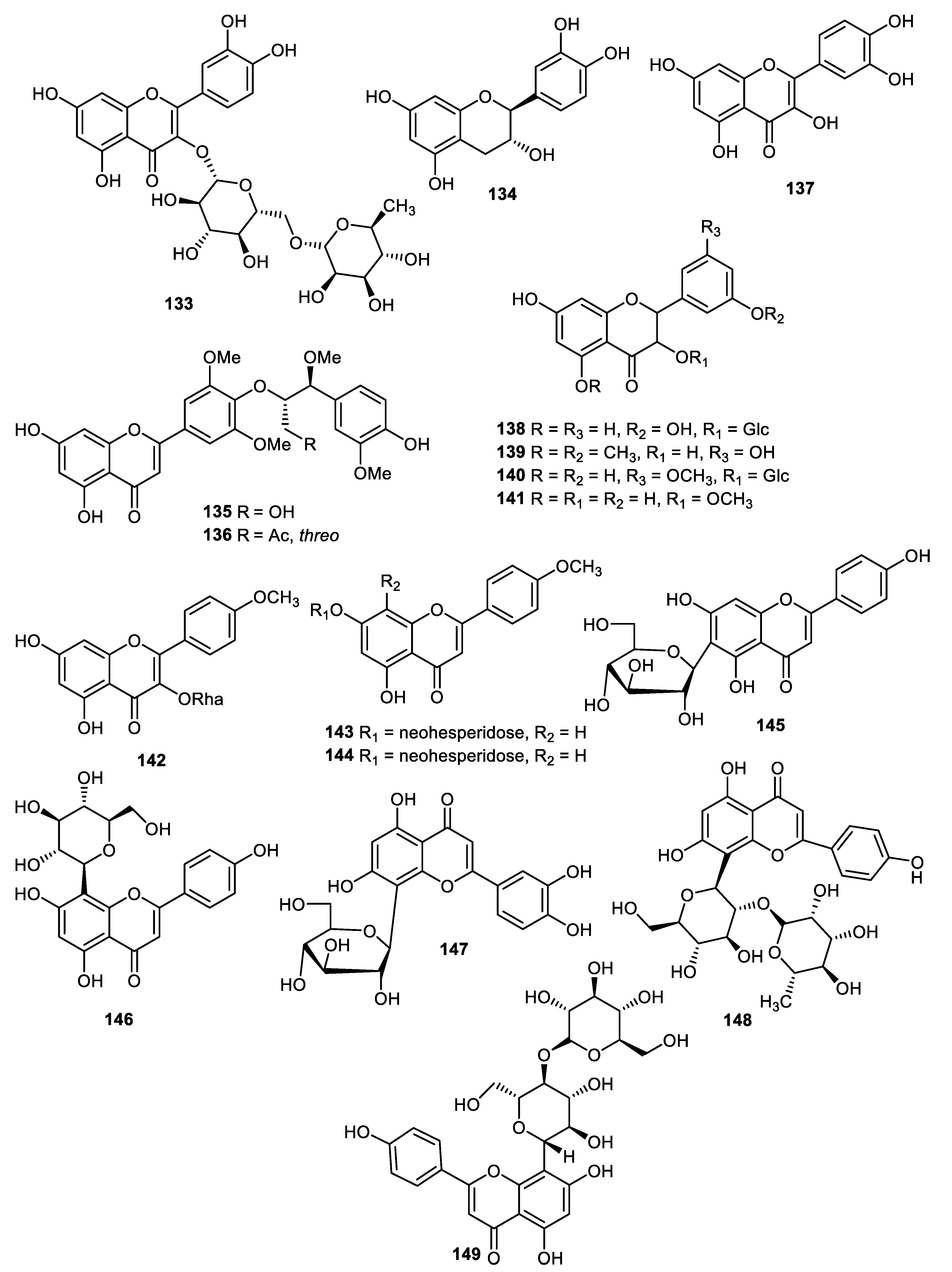
9. Flavones, Flavanone and Condensed Tannins
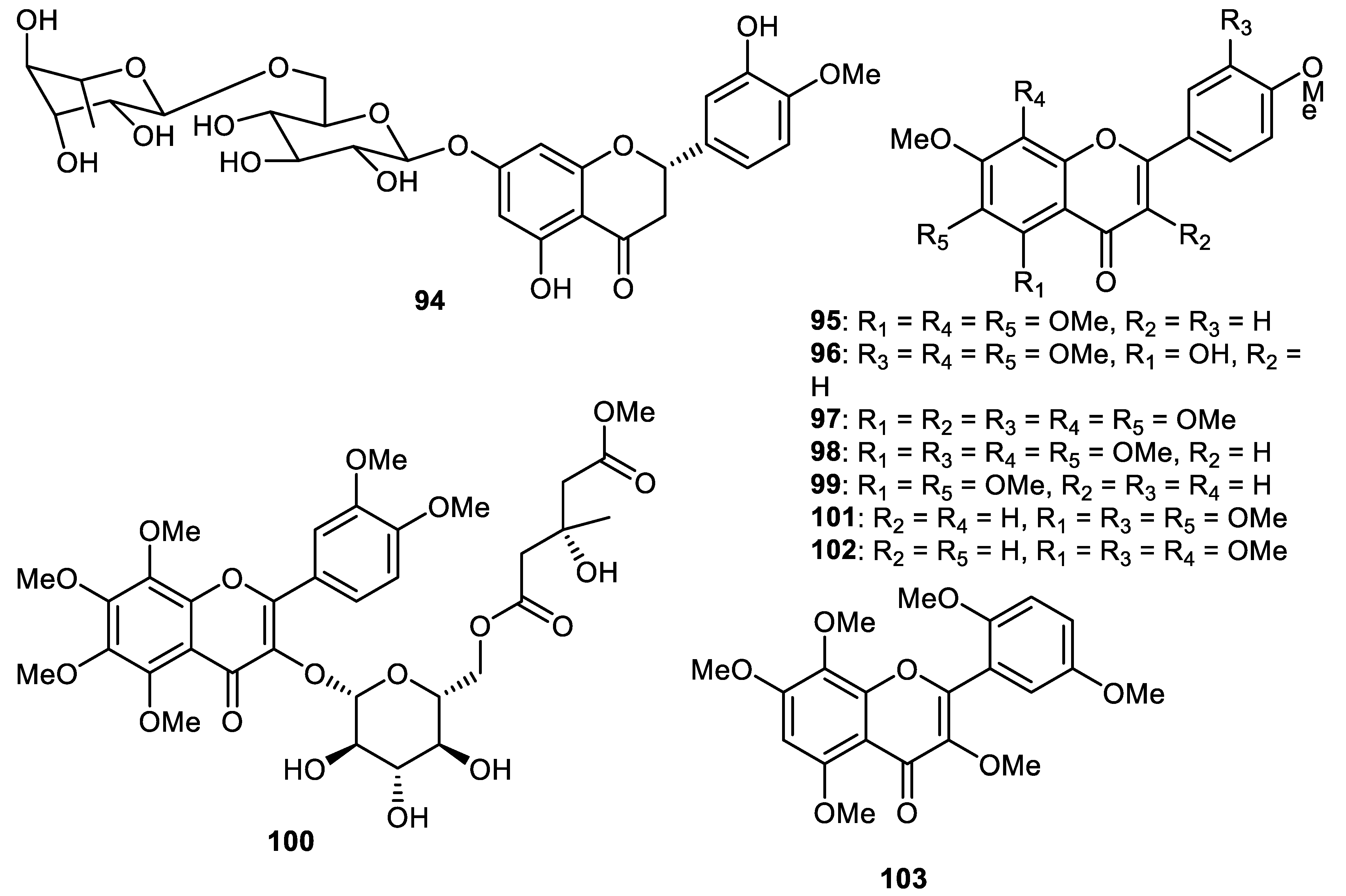
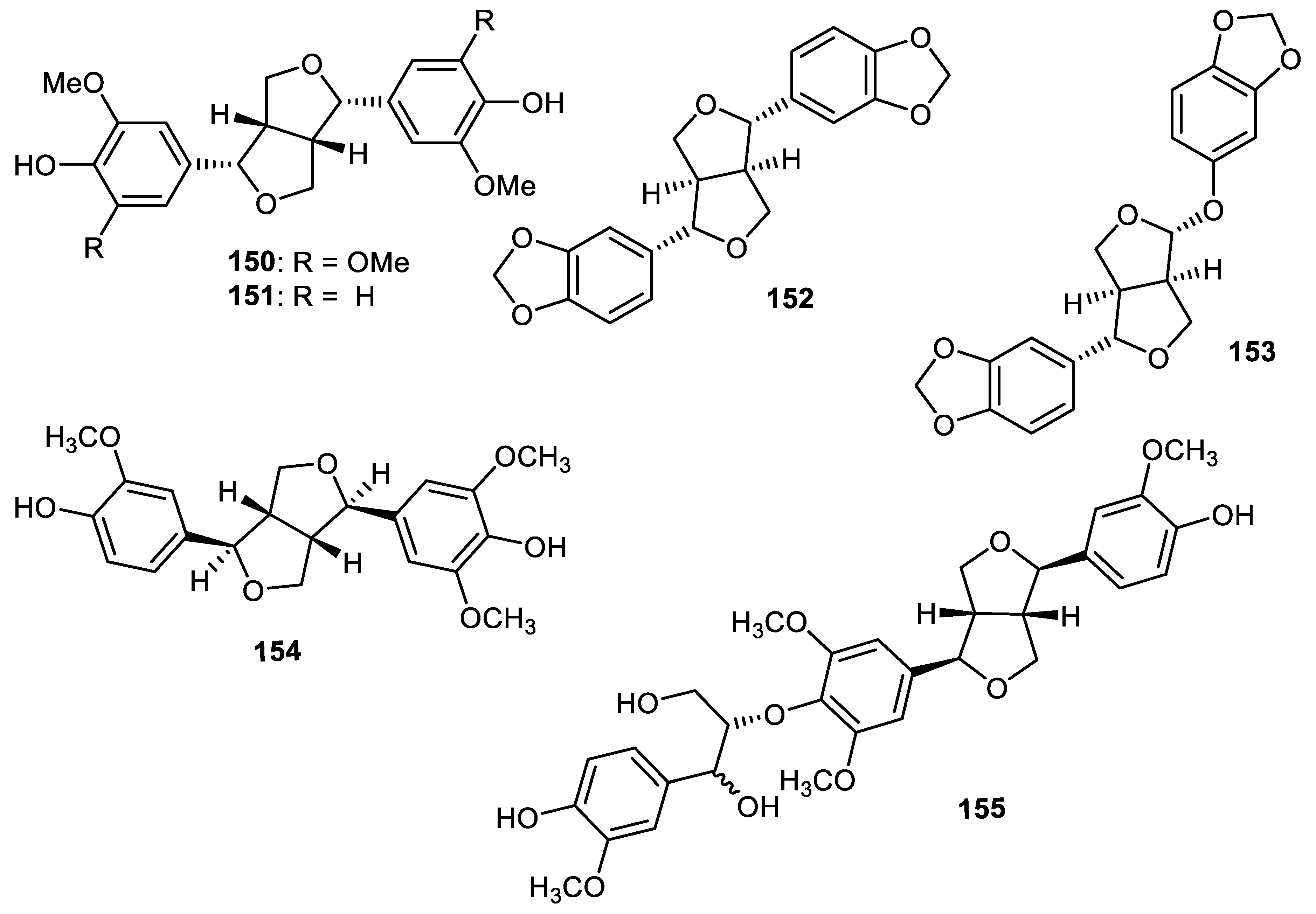
10. Lignans
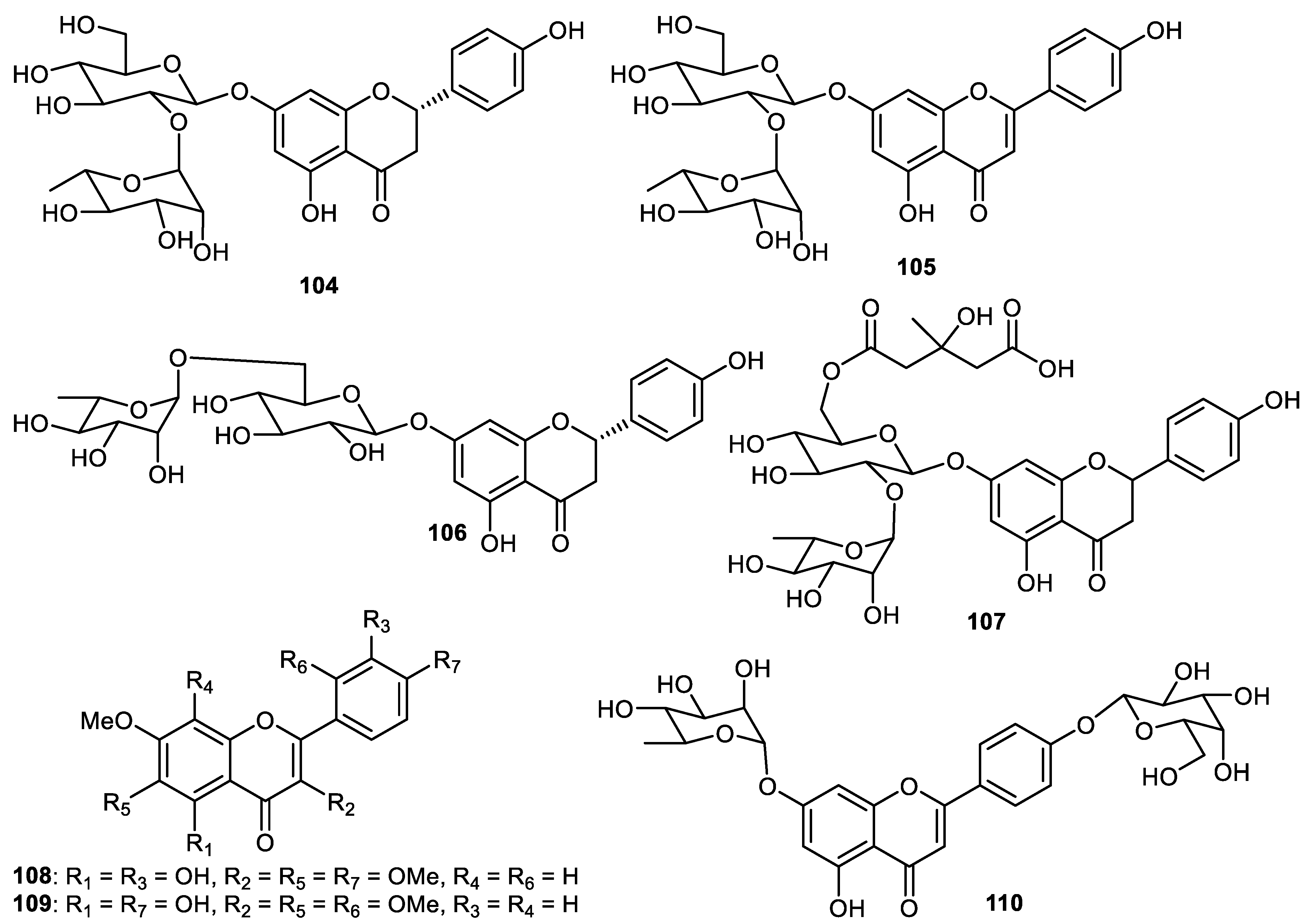
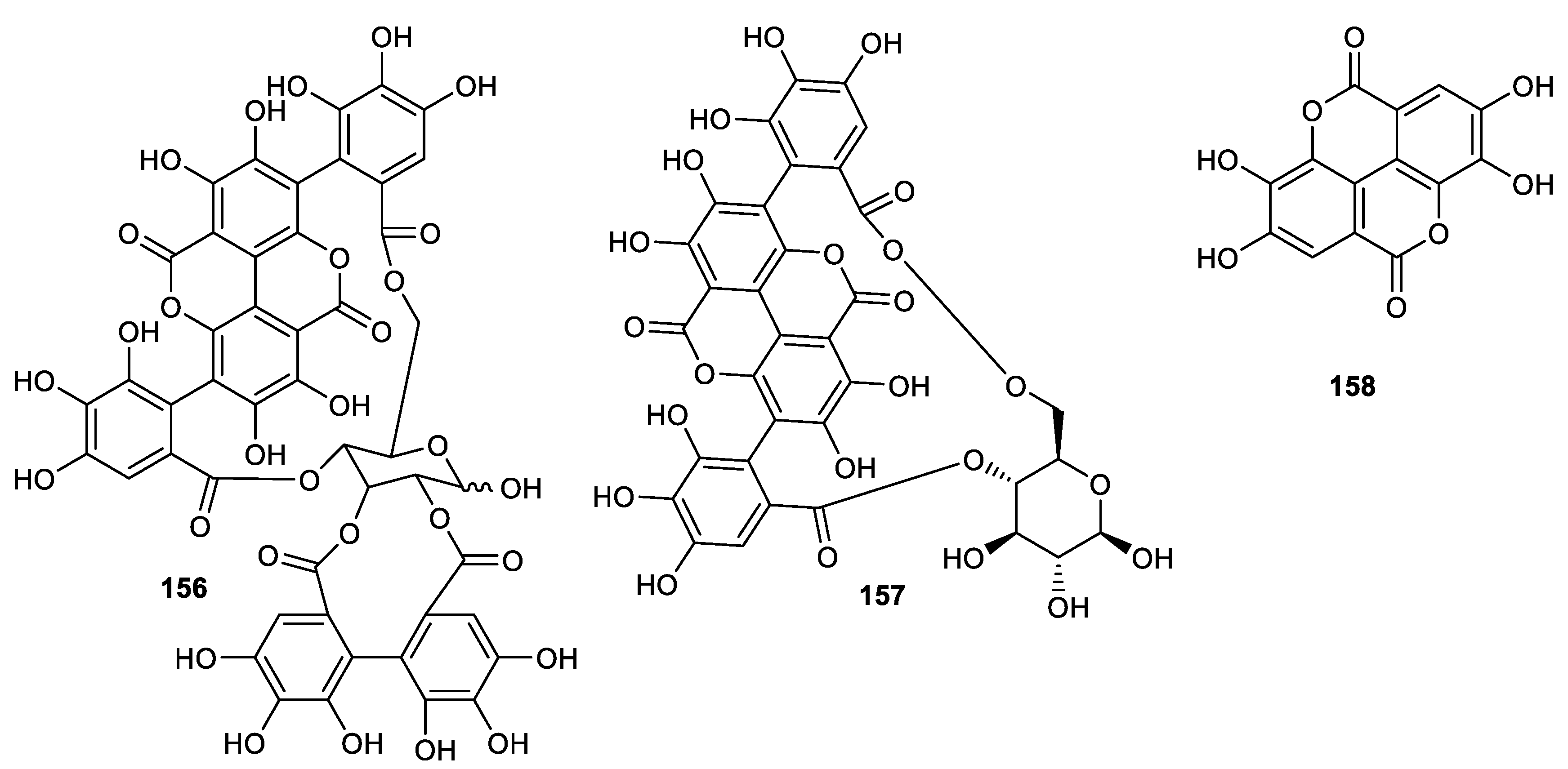
11. Hydrolyzable Tannins
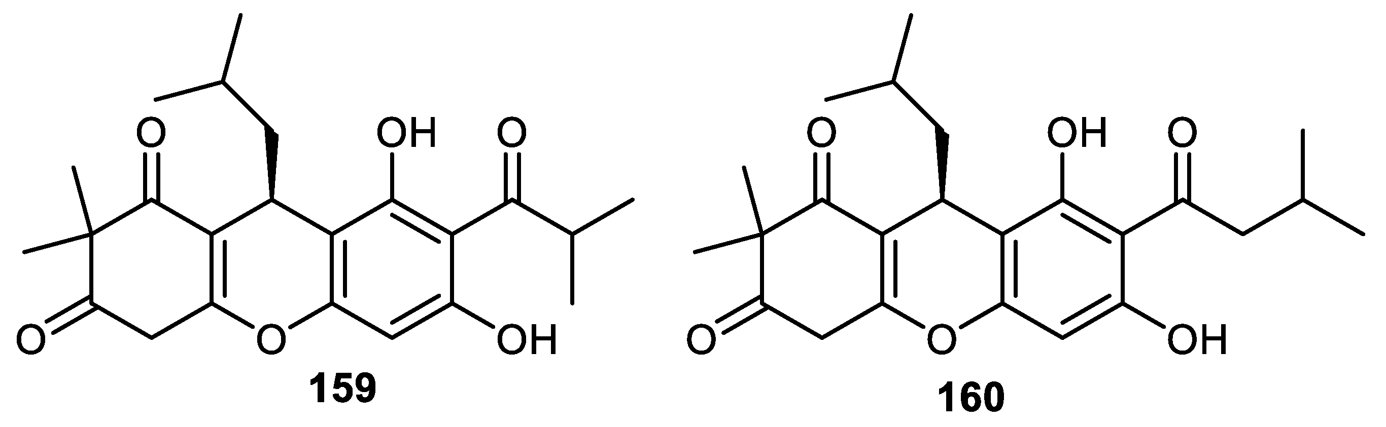
12. Phloroglucinol
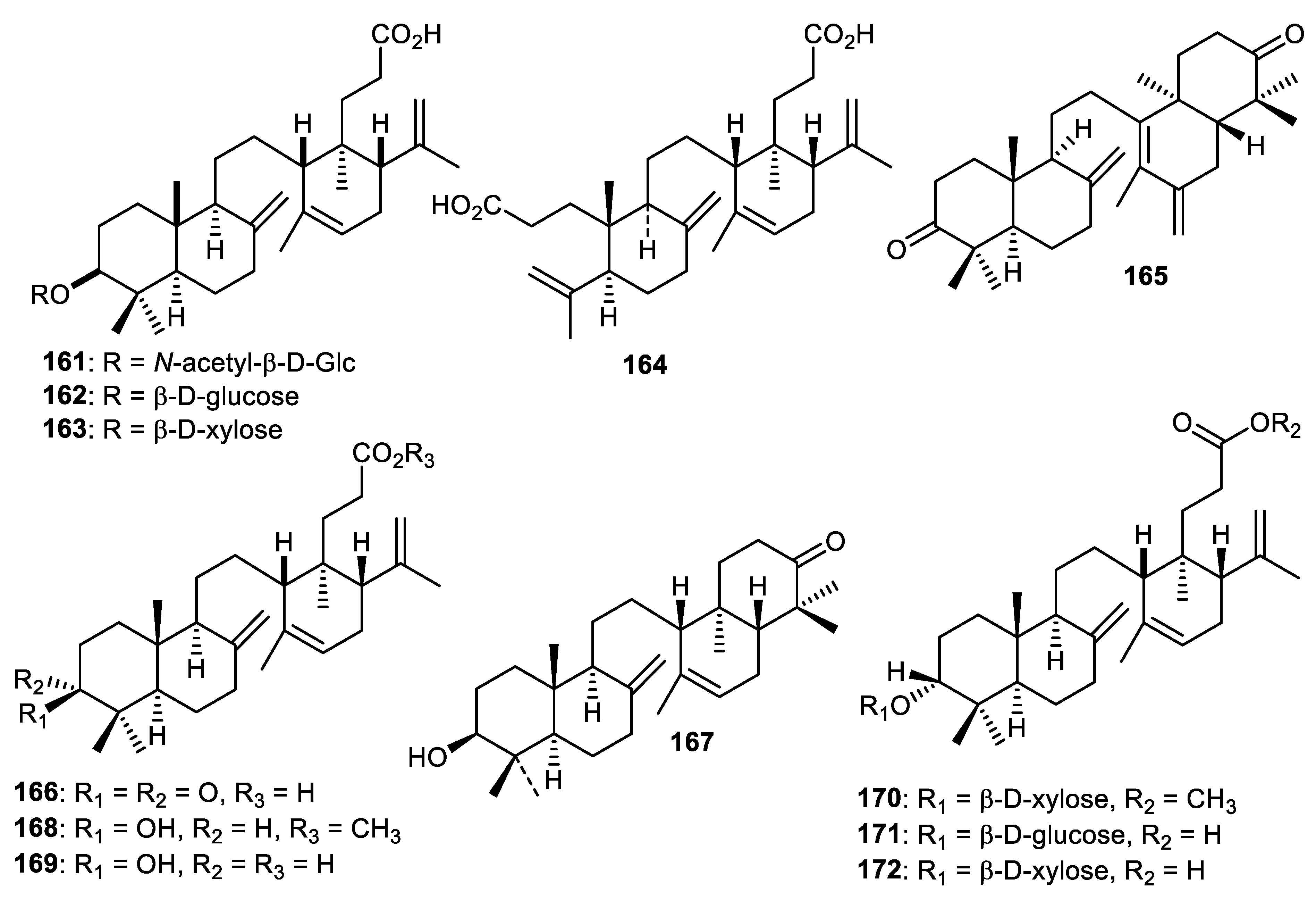
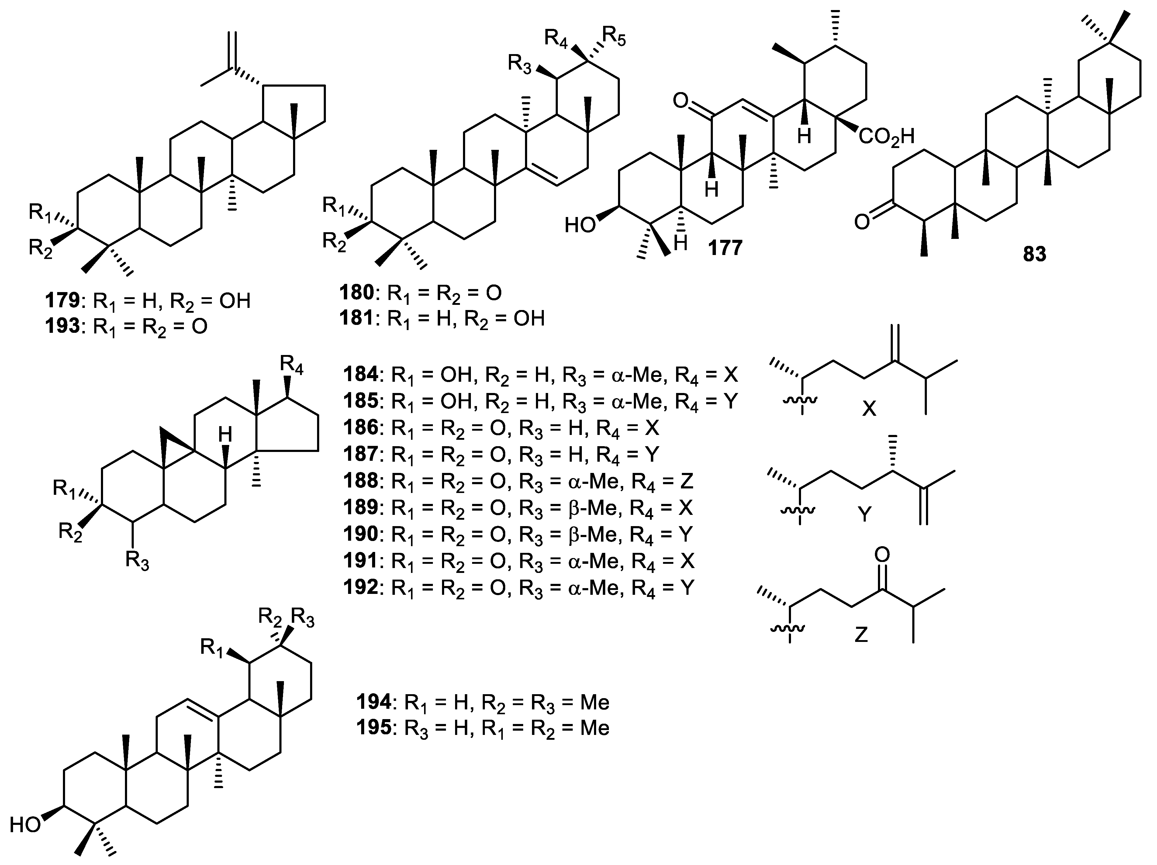
13. Triterpenoids
13.1. Onocerane Triterpenoids
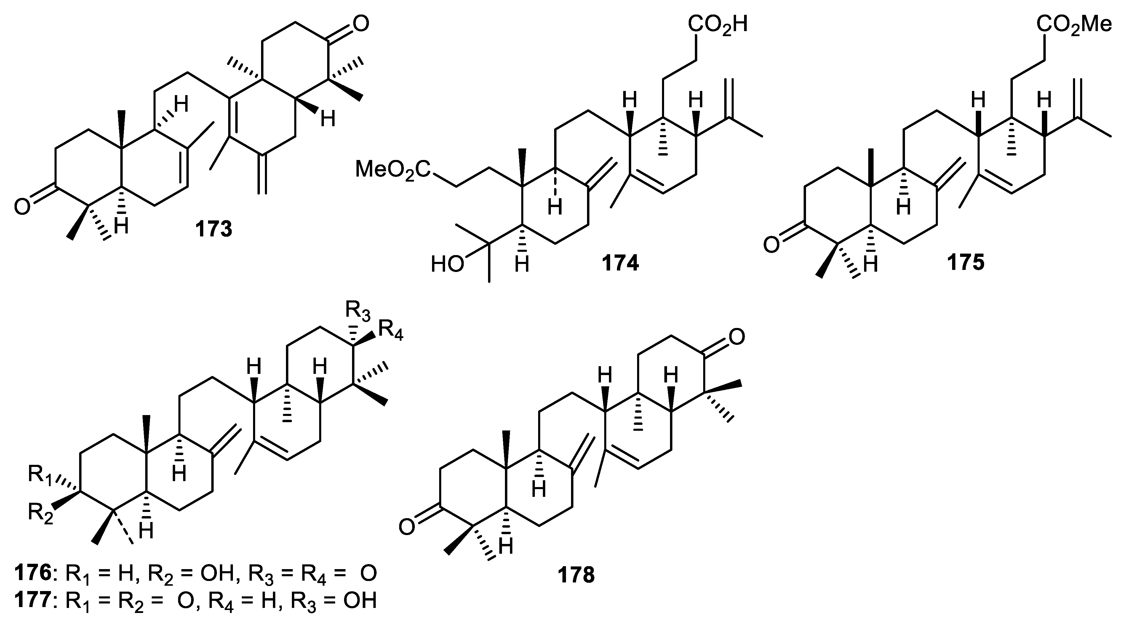
13.2. Miscellaneous Triterpenoids
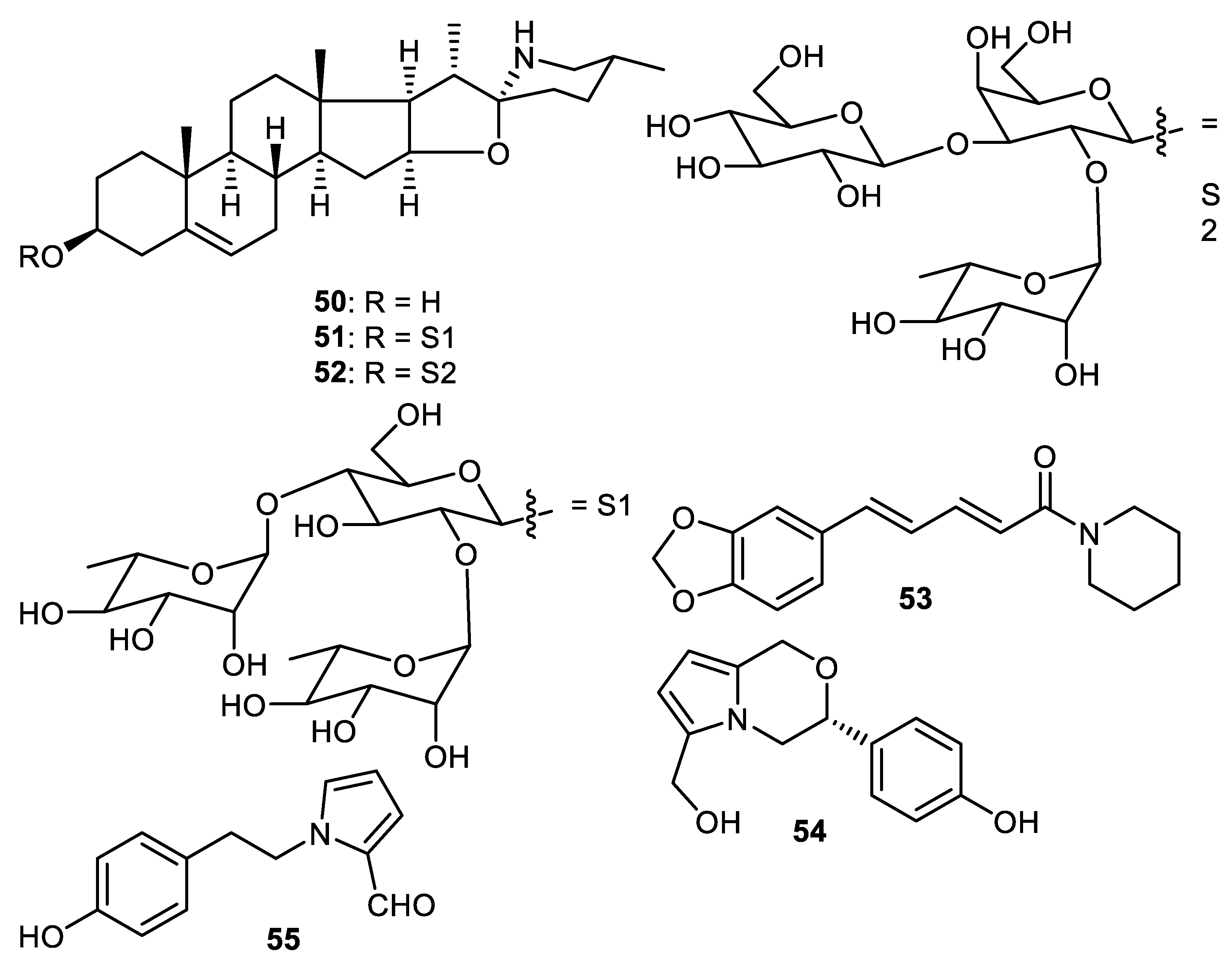
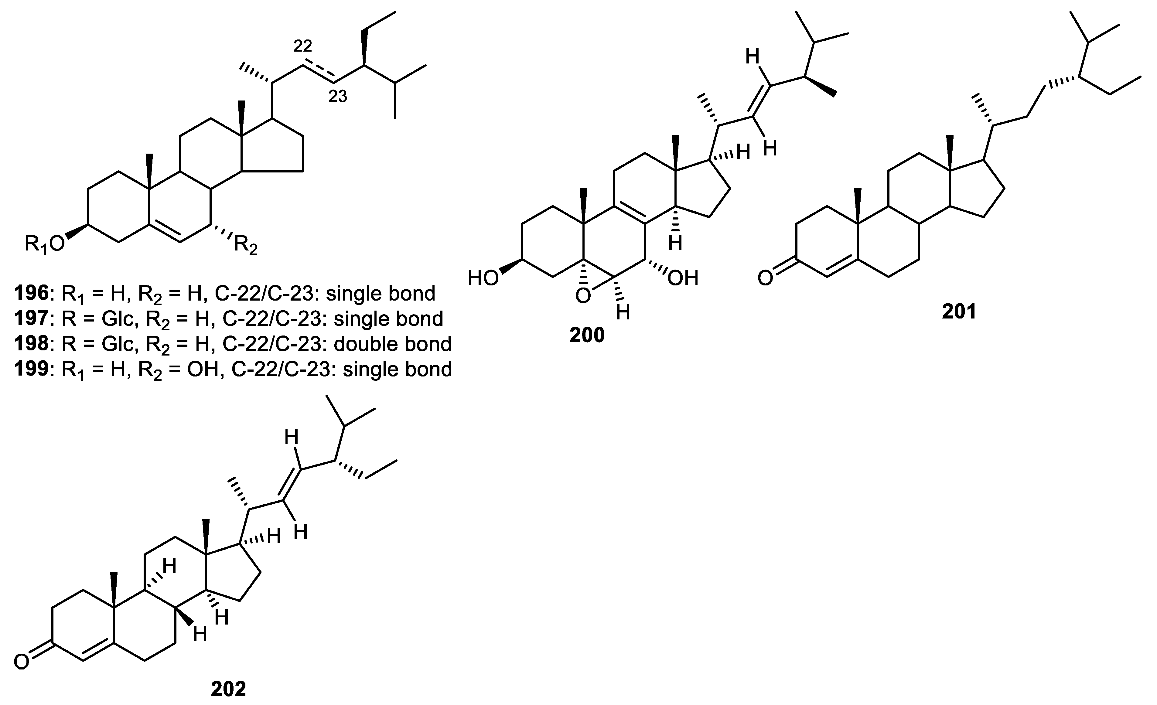
14. Steroids
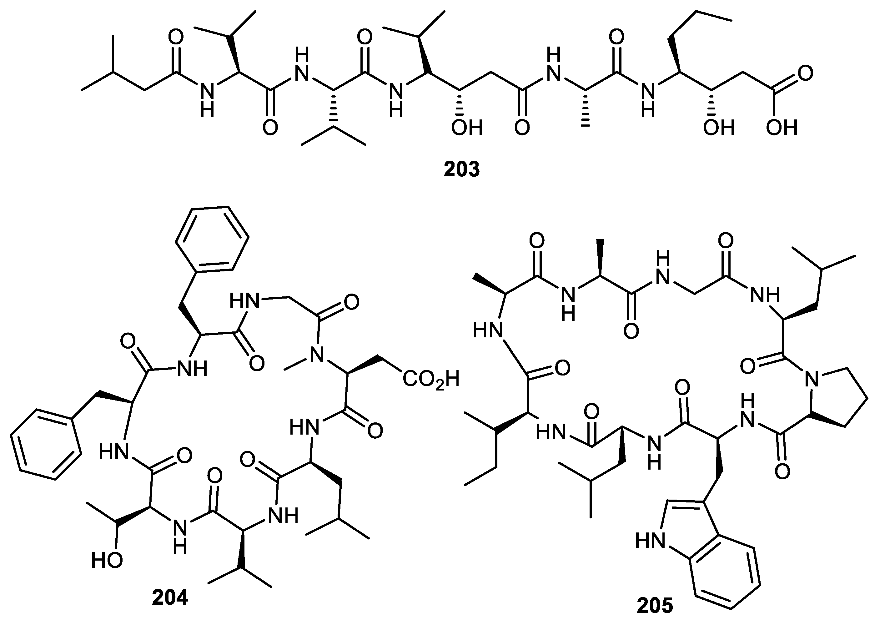
15. Peptides
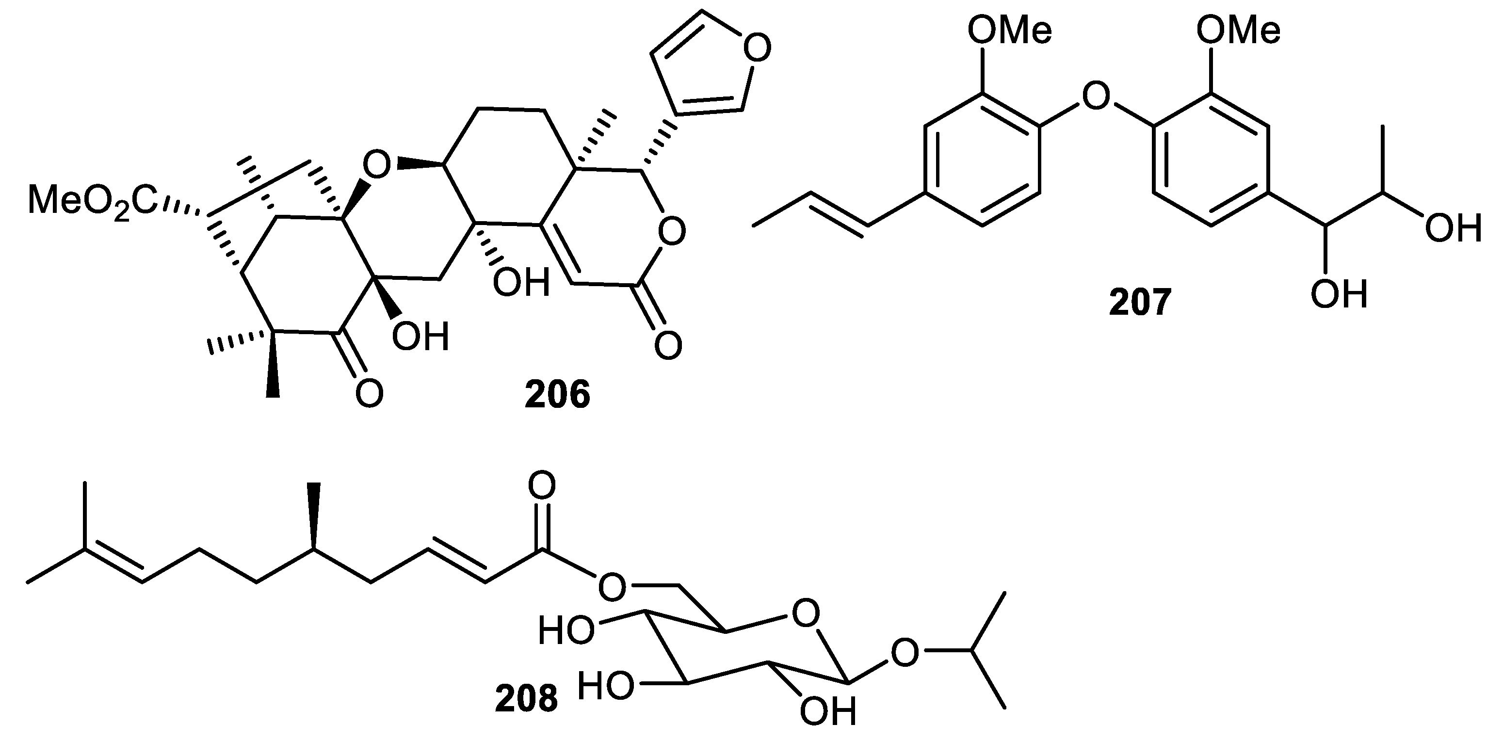
16. Miscellaneous
17. Fruit Peels and Food Industries

| Fruit Peels | Antimicrobial and Antioxidant Effects | Ref. |
|---|---|---|
| Wampee (Clausena lansium) | Antioxidant: Extract(s) showed better effect than BHT | [29] |
| Red dragon fruit (Hylocereus polyrhizus) E | Antibacterial: Staphylococcus aureus; Streptococcus mutans; Antifungal: Candida albicans;Aspergillus fumigatus (EO); Antioxidant: E showed good effects | [125,126,127,128,129,130] |
| Melons (Cucumis melo L.) E | Antibacterial: Staphylococcus epidermidis; Streptococcus pyogenes | [130] |
| Passion (Passiflora edulis) E | Antibacterial: Staphylococcus epidermidis; Streptococcus pyogenes; Antioxidant: E showed good effects | [130,131] |
| Pineapple (Ananas comosus) E | Antibacterial: Staphylococcus epidermidis; Streptococcus pyogenes; Pseudomonas aeruginosa; Antioxidant: E showed good effects | [130,132] |
| Carica papaya | Antibacterial: Pseudomonas aeruginosa; Antioxidant: E showed good effects | [132] |
| Dragon Fruit (Hylocereus undatus) E | Antibacterial: Staphylococcus epidermidis; Streptococcus pyogenes | [130] |
| Watermelon (Citrullus lanatus) E | Antibacterial: Staphylococcus epidermidis; Streptococcus pyogenes; Bacillus subtilis; Pseudomonas species; Staphylococcus aureus; Klebsiella pneumoniae; Protieus mirabilis; Antioxidant: E showed significant effects | [130,133] |
| Mango (Mangifera indica) E | Antibacterial: Staphylococcus epidermidis; Streptococcus pyogenes;Antioxidant: E showed significant effects | [130,134] |
| Mangifera pajang | Antioxidant: E showed significant effects | [135] |
| Citrus reticulata | Antioxidant: EO showed good effects; Antibacterial: Escherichia coli; Staphylococcus aureus; Enterococcus faecalis; Salmonella typhi; Klebsiella pneumoniae; Pseudomonas aeruginosa;Antifungal: Candida albicans | [136,137] |
| Pomelo (Citrus maxima) | Antioxidant: E showed significant effects | [138] |
| Citrus aurantifolia (EO) | Antibacterial: Streptococcus mutans;Lactobacillus casei | [139] |
| Citrus sinensis | Antibacterial: Escherichia coli | [140] |
| Citrus reticulate | Antibacterial: Escherichia coli | [140] |
| Citrus limetta | Antibacterial: Escherichia coli | [140] |
| Citrus medica | Antibacterial: Pseudomonas aeruginosa | [141] |
| Citrus karna EO | Antibacterial: Bacillus subtilis; Pseudomonas aeruginosa | [142] |
| Annona squamosa | Antioxidant: E showed potent effects | [143] |
| Annona reticulate | Antioxidant: E showed potent effects | [143] |
| Apple (Malus domestica) | Antioxidant: E showed good effects | [134] |
| Green sugar apple (Annona squamosa) | Antioxidant: E showed significant effects | [144] |
| Purple sugar apple (Annona squamosa) | Antioxidant: E showed significant effects | [144] |
| Green star apple (Chrysophyllum cainito) | Antioxidant: E showed significant effects | [144] |
| Apricot (Prunus armeniaca) | Antioxidant: E showed good effects | [134] |
| Avocado (Persea americana) | Antioxidant: E showed significant effects | [134] |
| Grapefruit (Citrus x paradisi) | Antioxidant: E showed good effects | [134] |
| Kiwi (Actinidia deliciosa) | Antioxidant: E showed good effects | [134,145] |
| Pomegranate (Punica granatum) | Antibacterial: Escherichia coli; Proteus vulgaris; Pseudomonas aeruginosa; Klebsiella pneumonia; Staphylococcus saprophyticus; Enterococcus faecalis; Streptococcus agalactiae; Klebsiella pneumoniae; Antioxidant: E showed good effects | [146,147] |
| Banana (Musa sp.) | Antibacterial: Escherichia coli; Proteus vulgaris; Pseudomonas aeruginosa; Klebsiella pneumonia; Staphylococcus saprophyticus; Enterococcus faecalis; Streptococcus agalactiae;Antioxidant: E showed significant effects | [146,147] |
| lemon (Citrus limon) | Antibacterial: Escherichia coli; Proteus vulgaris; Pseudomonas aeruginosa; Klebsiella pneumonia; Staphylococcus saprophyticus; Enterococcus faecalis; Streptococcus agalactiae | [146] |
| Solanum melongena | Antioxidant: E showed significant effects | [148] |
| Putranjiva roxburghii | Antibacterial: Bacillus subtelis; Enterobacter xiangfangensis; Antioxidant: E showed significant effects | [149] |
| Pouteria caimito | Antibacterial: Staphylococcus epidermidis; Escherichia coli | [150] |
| Saskatoon berry (Amelanchier alnifolia) | Antioxidant: E showed significant effects | [151] |
| Rambutan (Nephelium lappaceum) | Antibacterial: Salmonella enteritidis; Vibrio parahaemolyticus; Antioxidant: E showed significant effects | [152,153] |
| Ficus carica | Antibacterial: Micrococcus luteus; Proteus vulgaris; Antioxidant: E showed significant effects | [154] |
| Jaboticaba (Plinia peruviana) | Antioxidant: E showed significant effects | [30] |
| Peach (Prunus persica) | Antioxidant: E showed significant effects | [155] |
| Garcia mangostana | Antioxidant: E showed significant effects | [156] |
| Persea americana | Antioxidant: E showed significant effects | [156] |
| Mangifera odorata | Antioxidant: E showed significant effects | [156] |
| Dimocarpus longan | Antioxidant: E showed significant effects | [156] |
| Solanum betaceum | Antioxidant: E showed significant effects | [156] |
| Annona squamosa | Antioxidant: E showed significant effects | [156] |
| Archidendron pauciflorum | Antioxidant: E showed significant effects | [156] |
| Parkia speciosa | Antioxidant: E showed significant effects | [156] |
| Caryocar brasiliense | Antioxidant: E showed significant effects | [157] |
| Leucaena leucocephala | Antioxidant: E showed significant effects | [158] |
| Loquat (Eriobotrya japonica) | Antioxidant: E showed significant effects | [159] |
| Mangosteen (Garcinia mangostana) | Antioxidant: E showed significant effects | [160] |
| Rambutan (Nephelium lappaceum) | Antioxidant: E showed significant effects | [161] |
| Tea (Camellia sinensis) | Antioxidant: E showed significant effects | [162] |
| Syzygium cumini | Antibacterial: Staphylococcus aureus; Enterococcus faecalis; Escherichia coli; Pseudomonas aeruginosa; Proteus vulgaris; Serratia marcescens; Bacillus subtilis; Bacillus cereus; Salmonella typhimurium; Enterobacter aerogenes; Antifungal: Candida albicans; Aspergillus niger | [163] |
| Coconut (Cocos nucifera) | Antioxidant: E showed significant effects | [147] |
18. Fruit-Peel-Based Edible Coatings/Film and Probiotics
19. Conclusions and Future Perspective
Author Contributions
Funding
Conflicts of Interest
Abbreviation
| iNOS | Inducible nitric oxide synthase |
| COX-2 | Cyclooxygenase-2 |
| qPCR | Quantitative polymerase chain reaction |
| HCV 2 | Herpes simplex virus 2 |
| VSMC | Vascular smooth muscle cells |
| PDGF | Platelet-derived growth factor |
| NOX | (NADPH Oxidase) |
| NF-κB | Nuclear factor kappa B |
| IL-1β | Interleukin 1 beta |
| TNF-α | Tumor necrosis factor-alpha |
| PGE2 | Prostaglandin E2 |
| IL-6 | Interleukin 6 |
References
- Kumar, H.; Bhardwaj, K.; Sharma, R.; Nepovimova, E.; Kuca, K.; Dhanjal, D.S.; Verma, R.; Bhardwaj, P.; Sharma, S.; Kumar, D. Fruit and Vegetable Peels: Utilization of High Value Horticultural Waste in Novel Industrial Applications. Molecules 2020, 25, 2812. [Google Scholar] [CrossRef] [PubMed]
- Galanakis, C.M. Recovery of high added-value components from food wastes: Conventional, emerging technologies and commercialized applications. Trends Food Sci. Technol. 2012, 26, 68–87. [Google Scholar] [CrossRef]
- Gorinstein, S.; Martín-Belloso, O.; Park, Y.-S.; Haruenkit, R.; Lojek, A.; Cíž, M.; Caspi, A.; Libman, I.; Trakhtenberg, S. Comparison of some biochemical characteristics of different citrus fruits. Food Chem. 2001, 74, 09–315. [Google Scholar] [CrossRef]
- Soong, Y.-Y.; Barlow, P.J. Antioxidant activity and phenolic content of selected fruit seeds. Food Chem. 2004, 88, 411–417. [Google Scholar] [CrossRef]
- Fahmy, H.A.; Farag, M.A. Ongoing and potential novel trends of pomegranate fruit peel; a comprehensive review of its health benefits and future perspectives as nutraceutical. J. Food Biochem. 2022, 46, e14024. [Google Scholar] [CrossRef]
- Chukwuma, C.I.; Izu, G.O.; Chukwuma, M.S.; Samson, M.S.; Makhafola, T.J.; Erukainure, O.L. A review on the medicinal potential, toxicology, and phytochemistry of litchi fruit peel and seed. J. Food Biochem. 2021, 45, e13997. [Google Scholar] [CrossRef]
- Furukawa, Y.; Okuyama, S.; Amakura, Y.; Sawamoto, A.; Nakajima, M.; Yoshimura, M.; Igase, M.; Fukuda, N.; Tamai, T.; Yoshida, T. Isolation and characterization of neuroprotective components from citrus peel and their application as functional food. Chem. Pharm. Bull. 2021, 69, 2–10. [Google Scholar] [CrossRef]
- Jiang, H.; Zhang, W.; Li, X.; Shu, C.; Jiang, W.; Cao, J. Nutrition, phytochemical profile, bioactivities and applications in food industry of pitaya (Hylocereus spp.) peels: A comprehensive review. Trends Food Sci. Technol. 2021, 116, 199–217. [Google Scholar] [CrossRef]
- Umamahesh, K.; Gandhi, A.D.; Reddy, O.V.S. Ethnopharmacological Applications of Mango (Mangiferaindica, L.) Peel—A Review. Curr. Pharm. Biotechnol. 2020, 21, 1298–1303. [Google Scholar] [CrossRef]
- Gullon, P.; Astray, G.; Gullon, B.; Tomasevic, I.; Lorenzo, J.M. Pomegranate peel as suitable source of high-added value bioactives: Tailored functionalized meat products. Molecules 2020, 25, 2859. [Google Scholar] [CrossRef]
- Singh, B.; Singh, J.P.; Kaur, A.; Singh, N. Phenolic composition, antioxidant potential and health benefits of citrus peel. Food Res. Int. 2020, 132, 109114. [Google Scholar] [CrossRef] [PubMed]
- Karle, P.P.; Dhawale, S.C. Manilkara zapota (L.) royen fruit peel: A phytochemical and pharmacological review. Sys. Rev. Pharm. 2019, 10, 11–14. [Google Scholar]
- Choubey, S. Extraction & evaluation of anti-oxidants from different fruit peels: A review. World J. Pharm. Res. 2018, 7, 363–376. [Google Scholar]
- Singh, B.; Singh, J.P.; Kaur, A.; Singh, N. Antimicrobial potential of pomegranate peel: A review. Int. J. Food Sci. Technol. 2019, 54, 959–965. [Google Scholar] [CrossRef]
- Karasawa, M.M.G.; Mohan, C. Fruits as Prospective Reserves of bioactive Compounds: A Review. Nat. Prod. Bioprospect. 2018, 8, 335–346. [Google Scholar] [CrossRef]
- Singh, B.; Singh, J.P.; Kaur, A.; Singh, N. Phenolic compounds as beneficial phytochemicals in pomegranate (Punica granatum L.) peel: A review. Food Chem. 2018, 261, 75–86. [Google Scholar] [CrossRef]
- Rodrigo, M.J.; Alquezar, B.; Alos, E.; Lado, J.; Zacarias, L. Biochemical bases and molecular regulation of pigmentation in the peel of Citrus fruit. Sci. Hortic. 2013, 163, 46–62. [Google Scholar] [CrossRef]
- Belgacem, I.; Li, D.N.M.G.; Pangallo, S.; Agosteo, G.E.; Schena, L.; Abdelfattah, A.; Benuzzi, M. Pomegranate Peel Extracts as Safe Natural Treatments to Control Plant Diseases and Increase the Shelf-Life and Safety of Fresh Fruits and Vegetables. Plants 2021, 10, 453. [Google Scholar] [CrossRef]
- Parmar, H.S.; Dixit, Y.; Kar, A. Fruit and vegetable peels: Paving the way towards the development of new generation therapeutics. Drug Discov. Ther. 2010, 4, 314–325. [Google Scholar]
- Adhikari-Devkota, A.; Kurauchi, Y.; Yamada, T.; Katsuki, H.; Watanabe, T.; Devkota, H.P. Anti-neuroinflammatory activities of extract and polymethoxyflavonoids from immature fruit peels of Citrus ‘Hebesu’. J. Food Biochem. 2019, 43, e12813. [Google Scholar] [CrossRef]
- Gao, Z.; Gao, W.; Zeng, S.L.; Li, P.; Liu, E.H. Chemical structures, bioactivities and molecular mechanisms of citrus polymethoxyflavones. J. Funct. Foods 2018, 40, 498–509. [Google Scholar] [CrossRef]
- Matsumoto, T.; Nishikawa, T.; Furukawa, A.; Itano, S.; Tamura, Y.; Hasei, T.; Watanabe, T. Antimutagenic effects of polymethoxylated flavonoids of Citrus unshiu. Nat. Prod. Commun. 2017, 12, 23–26. [Google Scholar] [PubMed]
- Zhang, C.; Bucheli, P.; Liang, X.; Lu, Y. Citrus flavonoids as functional ingredients and their role in traditional Chinese medicine. Food 2007, 1, 287–296. [Google Scholar]
- Ihara, H.; Yamamoto, H.; Ida, T.; Tsutsuki, H.; Sakamoto, T.; Fujita, T.; Kozaki, S. Inhibition of nitric oxide production and inducible nitric oxide synthase expression by a polymethoxyflavone from young fruits of Citrus unshiu in rat primary astrocytes. Biosci. Biotechnol. Biochem. 2012, 76, 1843–1848. [Google Scholar] [CrossRef] [PubMed]
- Guo, J.; Tao, H.; Cao, Y.; Ho, C.T.; Jin, S.; Huang, Q. Prevention of Obesity and Type 2 Diabetes with Aged Citrus Peel (Chenpi) Extract. J. Agricult. Food Chem. 2016, 64, 2053–2061. [Google Scholar] [CrossRef] [PubMed]
- Choi, M.Y.; Chai, C.; Park, J.H.; Lim, J.; Lee, J.; Kwon, S.W. Effects of storage period and heat treatment on phenolic compound composition in dried Citrus peels (Chenpi) and discrimination of Chenpi with different storage periods through targeted metabolomic study using HPLC-DAD analysis. J. Pharm. Biomed. Anal. 2011, 54, 638–645. [Google Scholar] [CrossRef] [PubMed]
- Tang, K.; He, S.; Zhang, X.; Guo, J.; Chen, Q.; Yan, F.; Banadyga, L.; Zhu, W.; Qiu, X.; Guo, Y. Tangeretin, an extract from Citrus peels, blocks cellular entry of arenaviruses that cause viral hemorrhagic fever. Antivir. Res. 2018, 160, 87–93. [Google Scholar] [CrossRef]
- Hyun, J.M.; Jo, Y.J.; Kim, J.E.; An, H.J.; Choi, Y.H.; Hyun, C.G.; Lee, N.H. Tetramethyl-O-scutellarin isolated from peels of immature Shiranuhi fruit exhibits anti-inflammatory effects on LPS-induced RAW264.7 cells. Trop. J. Pharm. Res. 2017, 16, 2197–2205. [Google Scholar] [CrossRef]
- Prasad, K.N.; Xie, H.; Hao, J.; Yang, B.; Qiu, S.; Wei, X.; Chen, F.; Jiang, Y. Antioxidant and anticancer activities of 8-hydroxypsoralen isolated from wampee [Clausena lansium (Lour.) Skeels] peel. Food Chem. 2010, 118, 62–66. [Google Scholar] [CrossRef]
- Pitz, H.d.S.; Pereira, A.; Blasius, M.B.; Voytena, A.P.L.; Affonso, R.C.L.; Fanan, S.; Trevisan, A.C.D.; Ribeiro-do-Valle, R.M.; Maraschin, M. In Vitro evaluation of the antioxidant activity and wound healing properties of jaboticaba (Plinia peruviana) fruit peel hydroalcoholic extract. Oxid. Med. Cell. Longev. 2016, 2016, 3403586. [Google Scholar] [CrossRef]
- Youkwan, J.; Sutthivaiyakit, S.; Sutthivaiyakit, P. Citrusosides A-D and Furanocoumarins with Cholinesterase Inhibitory Activity from the Fruit Peels of Citrus hystrix. J. Nat. Prod. 2010, 73, 1879–1883. [Google Scholar] [CrossRef] [PubMed]
- Kidarn, S.; Saenjum, C.; Hongwiset, D.; Phrutivorapongkul, A. Furanocoumarins from Kaffir lime and their inhibitory effects on inflammatory mediator production. Cogent Chem. 2018, 4, 1529259. [Google Scholar] [CrossRef]
- Zhang, T.; Peng, S. Introduction to the origin and evolution of Pomelo and its distribution in China. Chin. J. Ecol. 2000, 19, 58–61. [Google Scholar]
- Kuo, P.C.; Liao, Y.R.; Hung, H.Y.; Chuang, C.W.; Hwang, T.L.; Huang, S.C.; Shiao, Y.J.; Kuo, D.H.; Wu, T.S. Anti-Inflammatory and Neuroprotective Constituents from the Peels of Citrus grandis. Molecules 2017, 22, 967. [Google Scholar] [CrossRef]
- Wang, D.; Liu, B.; Ma, Z.; Feng, J.; Yan, H. Reticine A, a new potent natural elicitor: Isolation from the fruit peel of Citrus reticulate and induction of systemic resistance against tobacco mosaic virus and other plant fungal diseases. Pest Manag. Sci. 2021, 77, 354–364. [Google Scholar] [CrossRef]
- Saleem, M.; Afza, N.; Anwar, M.A.; Ali, M.S. Aromatic constituents from fruit peels of Citrus reticulata. Nat. Prod. Res. 2005, 19, 633–638. [Google Scholar] [CrossRef]
- Deng, H.D.; Cai, C.H.; Liu, S.; Zeng, Y.B.; Mei, W.L.; He, F.; Hua, M.; Dai, H.F.; Li, S.P. A New Monoterpenoid and a New Flavonoid Glycoside from the Peels of Clausena lansium. Nat. Prod. Commun. 2016, 11, 573–575. [Google Scholar] [CrossRef]
- Deng, H.D.; Mei, W.L.; Guo, Z.K.; Liu, S.; Zuo, W.J.; Dong, W.H.; Li, S.P.; Dai, H.F. Monoterpenoid Coumarins from the Peels of Clausena lansium. Planta Med. 2014, 80, 955–958. [Google Scholar] [CrossRef]
- Rédei, D.; Kúsz, N.; Rafai, T.; Bogdanov, A.; Burián, K.; Csorba, A.; Mándi, A.; Kurtán, T.; Vasas, A.; Hohmann, J. 14-Noreudesmanes and a phenylpropane heterodimer from sea buckthorn berry inhibit Herpes simplex type 2 virus replication. Tetrahedron 2019, 75, 1364–1370. [Google Scholar] [CrossRef]
- Orban-Gyapai, O.; Liktor-Busa, E.; Kúsz, N.; Stefko, D.; Urban, E.; Hohmann, J.; Vasas, A. Antibacterial screening of Rumex species native to the Carpathian Basin and bioactivity-guided isolation of compounds from Rumex aquaticus. Fitoterapia 2017, 118, 101–106. [Google Scholar] [CrossRef]
- Abdullah, A.; Zakaria, Z.; Ahmad, F.B.; Mat-Salleh, K.; Din, L.B. Chemical Constituents from the Fruit Peel of Goniothalamus scortechinii. Sains Malays. 2009, 38, 365–369. [Google Scholar]
- Cho, J.Y.; Kawazoe, K.; Moon, J.H.; Park, K.H.; Murakami, K.; Takaishi, Y. Chemical Constituents from the fruit peels of Fortunella japonica. Food Sci. Biotechnol. 2005, 14, 599–603. [Google Scholar]
- Lee, K.H.; Cho, J.Y.; Lee, H.J.; Park, K.Y.; Ma, Y.K.; Lee, S.H.; Cho, J.A.; Kim, W.S.; Park, K.H.; Moon, J.H. Isolation and Identification of Phenolic Compounds from an Asian Pear (Pyrus pyrifolia Nakai) Fruit Peel. Food Sci. Biotechnol. 2011, 20, 1539–1545. [Google Scholar] [CrossRef]
- Sultana, S.; Ali, M.; Ansari, S.H.; Bagri, P. A new sesquiterpene derivatives from the fruit peels of Citrus limon (Linn.) Burm.f. Sci. Pharm. 2007, 75, 165–170. [Google Scholar] [CrossRef][Green Version]
- Dai, W.; Zhang, Y.; Liu, Y.; Jiao, S.; Zhang, M. Chemical Constituents with nitric oxide inhibition from the fruit peel of Annona squamosa. Chem. Nat. Comp. 1983, 57, 1153–1156. [Google Scholar] [CrossRef]
- Wybraniec, S.; Nowak-Wydra, B.; Mitka, K.; Kowalski, P.; Mizrahi, Y. Minor betalains in fruits of Hylocereus species. Phytochemistry 2007, 68, 251–259. [Google Scholar] [CrossRef]
- Gescher, K.; Hensel, A.; Hafezi, W.; Derksen, A.; Kühn, J. Oligomeric proanthocyanidins from Rumex acetosa L. inhibit the attachment of herpes simplex virus type-1. Antivir. Res. 2011, 89, 9–18. [Google Scholar] [CrossRef]
- Pedras, M.S.C.; Sarwar, M.G.; Suchy, M.; Adio, A.M. The phytoalexins from cauliflower, caulilexins A, B and C: Isolation, structure determination, syntheses and antifungal activity. Phytochemistry 2006, 67, 1503–1509. [Google Scholar] [CrossRef]
- Wu, Q.; Bang, M.H.; Lee, D.Y.; Cho, J.G.; Jeong, R.H.; Shrestha, S.; Lee, K.T.; Chung, H.G.; Ahn, E.M.; Baek, N.I. New indoles from the roots of Brassica rapa ssp. Campestris. Chem. Nat. Comp. 2012, 48, 281–284. [Google Scholar] [CrossRef]
- Sun, H.Y.; Ma, N.; Pan, T.; Du, C.L.; Sun, J.-Y. Punicagranine, a new pyrrolizine alkaloid with anti-inflammatory activity from the peels of Punica granatum. Tetrahedron Lett. 2019, 60, 1231–1233. [Google Scholar] [CrossRef]
- Justino, A.B.; Pereira, M.N.; Peixoto, L.G.; Vilela, D.D.; Caixeta, D.C.; de Souza, A.V.; Teixeira, R.R.; Silva, H.C.G.; de Moura, F.B.R.; Moraes, I.B.; et al. Hepatoprotective Properties of a Polyphenol-Enriched Fraction from Annona crassiflora Mart. Fruit Peel against Diabetes-Induced Oxidative and Nitrosative Stress. J. Agricult. Food Chem. 2017, 65, 4428–4438. [Google Scholar] [CrossRef] [PubMed]
- Pereira, M.N.; Justino, A.B.; Martins, M.M.; Peixoto, L.G.; Vilela, D.D.; Santos, P.S.; Teixeira, T.L.; da Silva, C.V.; Goulart, L.R.; Pivatto, M.; et al. Stephalagine, an alkaloid with pancreatic lipase inhibitory activityisolated from the fruit peel of Annona crassiflora Mart. Ind. Crops Prod. 2017, 97, 324–329. [Google Scholar] [CrossRef]
- Justino, A.B.; Barbosa, M.F.; Neves, T.V.; Silva, H.C.G.; Brum, E.S.; Fialho, M.F.P.; Couto, A.C.; Saraiva, A.L.; Avila, V.M.R.; Oliveira, S.M.; et al. Stephalagine, an aporphine alkaloid from Annona crassiflora fruit peel, induces antinociceptive effects by TRPA1 and TRPV1 channels modulation in mice. Bioorg. Chem. 2020, 96, 103562. [Google Scholar] [CrossRef] [PubMed]
- Shabana, M.M.; Salama, M.M.; Ezzat, S.M.; Ismail, L.R. In Vitro and In Vivo Anticancer Activity of the Fruit Peels of Solanum melongena L. against Hepatocellular Carcinoma. J. Carcinog. Mutagen. 2013, 4, 149. [Google Scholar]
- Fekry, M.I.; Ezzat, S.M.; Salama, M.M.; Alshehri, O.Y.; Al-Abd, A.M. Bioactive glycoalkaloides isolated from Solanum melongena fruit peels with potential anticancer properties against hepatocellular carcinoma cells. Sci. Rep. 2019, 9, 1–11. [Google Scholar] [CrossRef] [PubMed]
- Chaturvedi, A.K.; Luqman, S.; Dubey, V.; Thakur, J.P.; Saikia, D.; Chanotiya, C.S.; Shanker, K.; Negi, A.S. Inhibition of Cathepsin D protease activity by Punica granatum fruit peel extracts, isolates, and semisynthetic analogs. Med. Chem. Res. 2013, 22, 3953–3958. [Google Scholar] [CrossRef]
- Sichaem, J.; Ingkaninan, K.; Tip-pyang, S. A novel pyrrole alkaloid from the fruit peels of Strychnos nux-blanda. Nat. Prod. Res. 2017, 31, 149–154. [Google Scholar] [CrossRef]
- Deng, H.D.; Mei, W.L.; Wang, H.; Guo, Z.K.; Dong, W.H.; Wang, H.; Li, S.P.; Dai, H.F. Carbazole alkaloids from the peels of Clausena lansium. J. Asian Nat. Prod. Res. 2014, 16, 1024–1028. [Google Scholar] [CrossRef]
- Sribuhom, T.; Boueroy, P.; Hahnvajanawong, C.; Phatchana, R.; Yenjai, C. Benzoyltyramine Alkaloids Atalantums A−G from the Peels of Atalantia monophylla and Their Cytotoxicity against Cholangiocarcinoma Cell Lines. J. Nat. Prod. 2017, 80, 403–408. [Google Scholar] [CrossRef]
- Sombatsri, A.; Thummanant, Y.; Sribuhom, T.; Wongphakham, P.; Senawong, T.; Yenjai, C. Atalantums H-K from the peels of Atalantia monophylla and their cytotoxicity. Nat. Prod. Res. 2020, 34, 2124–2130. [Google Scholar] [CrossRef]
- Tai, B.H.; Trung, T.N.; Nhiem, N.X.; Ha, D.T.; Phuong, T.; Thu, N.B.; Luong, H.V.; Bae, K.H.; Kim, Y.H. Chemical Components from the Fruit Peels of Wisteria floribunda and their Effects on Rat Aortic Vascular Smooth Muscle Cells. Bull. Korean Chem. Soc. 2011, 32, 2079–2082. [Google Scholar] [CrossRef][Green Version]
- Aghel, N.; Ramezani, Z.; Beiranvand, S. Hesperidin from Citrus sinensis cultivated in Dezful, Iran. Pak. J. Biol. Sci. 2008, 11, 2451–2453. [Google Scholar] [CrossRef] [PubMed]
- Lahmer, N.; Belboukhari, N.; Cheriti, A.; Sekkoum, K. Hesperidin and hesperitin preparation and purification from Citrus sinensis Peels. Pharm. Chem. 2015, 7, 1–4. [Google Scholar]
- Nguyen, V.S.; Li, W.; Li, Y.; Wang, Q. Synthesis of citrus polymethoxyflavonoids and their antiproliferative activities on Hela cells. Med. Chem. Res. 2017, 26, 1585–1592. [Google Scholar] [CrossRef]
- Chen, X.M.; Tait, A.R.; Kitts, D.D. Flavonoid composition of orange peel and itsassociation with antioxidant and anti-inflammatory activities. Food Chem. 2017, 218, 15–21. [Google Scholar] [CrossRef] [PubMed]
- Cui, Y.; Wu, J.; Jung, S.C.; Park, D.B.; Maeng, Y.H.; Hong, J.Y.; Eun, S.Y. Anti-neuroinflammatory activity of nobiletin on suppression of microglial activation. Biol. Pharm. Bull. 2010, 33, 1814–1821. [Google Scholar] [CrossRef]
- Lin, Y.S.; Li, S.; Ho, C.T.; Lo, C.Y. Simultaneous analysis of six polymethoxyflavones and six 5- hydroxyl-polymethoxyflavones by high performance liquid chromatography combined with linear ion trap mass spectrometry. J. Agricult. Food Chem. 2012, 60, 12082–12087. [Google Scholar] [CrossRef]
- Tang, K.; Zhang, X.; Guo, Y. Identification of the dietary supplement capsaicin as an inhibitor of Lassa virus entry. Acta Pharm. Sin. B. 2020, 10, 789–798. [Google Scholar] [CrossRef]
- Uckoo, R.M.; Jayaprakasha, G.; Vikram, A.; Patil, B.S. Polymethoxyflavones isolated from the peel of Miaray Mandarin (Citrus miaray) have biofilm inhibitory activity in Vibrio harveyi. J. Agricult. Food Chem. 2015, 63, 7180–7189. [Google Scholar] [CrossRef]
- Itoh, N.; Iwata, C.; Toda, H. Molecular cloning and characterization of a flavonoid-O-methyltransferase with broad substrate specificity and regioselectivity from Citrus depressa. BMC Plant Biol. 2016, 16, 180. [Google Scholar] [CrossRef]
- Lee, Y.-H.; Charles, A.L.; Kung, H.-F.; Ho, C.-T.; Huang, T.-C. Extraction of nobiletin and tangeretin from Citrus depressa Hayata by supercritical carbon dioxide with ethanol as modifier. Ind. Crops Prod. 2010, 31, 59–64. [Google Scholar] [CrossRef]
- Chen, S.; Cai, D.; Pearce, K.; Sun, P.Y.; Roberts, A.C.; Glanzman, D.L. Reinstatement of long-term memory following erasure of its behavioral and synaptic expression in Aplysia. eLife 2014, 3, e03896. [Google Scholar] [CrossRef] [PubMed]
- Chiou, H.; Yoshitani, S.-I.; Tsukio, Y.; Murakami, A.; Koshimizu, K.; Yano, M.; Tokuda, H.; Nishino, H.; Ohigashi, H.; Tanaka, T. Dietary administration of citrus nobiletin inhibits azoxymethane-induced colonic aberrant crypt foci in rats. Life Sci. 2001, 69, 901–913. [Google Scholar]
- Murakami, A.; Nakamura, Y.; Torikai, K.; Tanaka, T.; Koshiba, T.; Koshimizu, K.; Kuwahara, S.; Takahashi, Y.; Ogawa, K.; Yano, M. Inhibitory effect of citrus nobiletin on phorbol ester-induced skin inflammation, oxidative stress, and tumor promotion in mice. Cancer Res. 2000, 60, 5059–5066. [Google Scholar] [PubMed]
- Uckoo, R.M.; Jayaprakasha, G.K.; Patil, B.S. Rapid separation method of polymethoxyflavones from citrus using flash chromatography. Sep. Purif. Technol. 2011, 81, 151–158. [Google Scholar] [CrossRef]
- Teruya, T.; Teruya, Y.; Sueyoshi, K.; Yamano, A.; Jitai, Y. Manufacturing Method of Fermentation Treated Products Containing High-Content Nobiletin and Tangeretin. Patent JP 2015202065, 16 November 2015. [Google Scholar]
- Zhang, N.; Wei, W.Y.; Yang, Z.; Che, Y.; Jin, Y.G.; Liao, H.H.; Wang, S.S.; Deng, W.; Tang, Q.Z. Nobiletin, a Polymethoxy Flavonoid, Protects Against Cardiac Hypertrophy Induced by Pressure-Overload via Inhibition of NAPDH Oxidases and Endoplasmic Reticulum Stress. Cell. Phys. Biochem. 2017, 42, 1313–1325. [Google Scholar] [CrossRef]
- Choi, S.Y.; Hwang, J.H.; Ko, H.C.; Park, J.G.; Kim, S.J. Nobiletin from citrus fruit peel inhibits the DNA-binding activity of NF-κB and ROS production in LPS-activated RAW 264.7 cells. J. Ethnopharmacol. 2007, 113, 149–155. [Google Scholar] [CrossRef]
- Wu, Y.Q.; Zhou, C.H.; Tao, J.; Li, S.N. Antagonistic effects of nobiletin, a polymethoxyflavonoid, on eosinophilic airway inflammation of asthmatic rats and relevant mechanisms. Life Sci. 2006, 78, 2689–2696. [Google Scholar] [CrossRef]
- Murakami, A.; Shigemori, T.; Ohigashi, H. Zingiberaceous and citrus constituents, 1’-acetoxychavicol acetate, zerumbone, auraptene, and nobiletin, suppress lipopolysaccharide-induced cyclooxygenase-2 expression in RAW 264.7 murine macrophages through different modes of action. J. Nutr. 2005, 135, 2987S–2992S. [Google Scholar] [CrossRef]
- Manthey, J.A.; Guthrie, N. Antiproliferative activities of citrus flavonoids against six human cancer cell lines. J. Agricult. Food Chem. 2002, 50, 5837–5843. [Google Scholar] [CrossRef]
- Chiou, Y.-S.; Zheng, Y.-N.; Tsai, M.-L.; Lai, C.-S.; Ho, C.-T.; Pan, M.-H. 5-Demethylnobiletin more potently inhibits colon cancer cell growth than nobiletin in vitro and in vivo. J. Food Bioact. 2018, 2, 91–97. [Google Scholar] [CrossRef]
- Wu, X.; Song, M.; Wang, M.; Zheng, J.; Gao, Z.; Xu, F.; Zhang, G.; Xiao, H. Chemopreventive effects of nobiletin and its colonic metabolites on colon carcinogenesis. Mol. Nutr. Food Res. 2015, 59, 2383–2394. [Google Scholar] [CrossRef] [PubMed]
- Wu, X.; Song, M.; Gao, Z.; Sun, Y.; Wang, M.; Li, F.; Zheng, J.; Xiao, H. Nobiletin and its colonic metabolites suppress colitis-associated colon carcinogenesis by down-regulating iNOS, inducing antioxidative enzymes and arresting cell cycle progression. J. Nutr. Biochem. 2017, 42, 17–25. [Google Scholar] [CrossRef] [PubMed]
- Suzuki, R.; Kohno, H.; Murakami, A.; Koshimizu, K.; Ohigashi, H.; Yano, M.; Tokuda, H.; Nishino, H.; Tanaka, T. Citrus nobiletin inhibits azoxymethane-induced large bowel carcinogenesis in rats. Biofactors 2004, 21, 111–114. [Google Scholar] [CrossRef]
- Miyamoto, S.; Yasui, Y.; Ohigashi, H.; Tanaka, T.; Murakami, A. Dietary flavonoids suppress azoxymethane-induced colonic preneoplastic lesions in male C57BL/KsJ-db/db mice. Chem.-Biol. Interact. 2010, 183, 276–283. [Google Scholar] [CrossRef]
- Miyamoto, S.; Yasui, Y.; Tanaka, T.; Ohigashi, H.; Murakami, A. Suppressive e_ects of nobiletin on hyperleptinemia and colitis-related colon carcinogenesis in male ICR mice. Carcinogenesis 2008, 29, 1057–1063. [Google Scholar] [CrossRef]
- Zhong, W.J.; Luo, Y.J.; Li, J.; Wu, Y.P.; Gao, Y.J.; Luo, H.J.; Yang, Y.T.; Jiang, L. Polymethoxylated flavonoids from Citrus reticulata Blanco. Biochem. Syst. Ecol. 2016, 68, 11–14. [Google Scholar] [CrossRef]
- Wang, Y.; Xue, J.; Jia, X.H.; Du, C.L.; Tang, W.Z.; Wang., X.J. New antioxidant C-geranylated flavonoids from the fruit peels of Paulownia catalpifolia T. Gong ex D.Y. Hong. Phytochem. Lett. 2017, 21, 169–173. [Google Scholar] [CrossRef]
- Wang, Y.A.; Guo, X.; Jia, X.H.; Xue, J.; Du, H.F.; Du, C.L.; Tang, W.Z.; Wang, X.J.; Zhao, Y.X. Undescribed C-geranylflavonoids isolated from the fruit peel of Paulownia catalpifolia T. Gong ex D.Y. Hong with their protection on human umbilical vein endothelial cells injury induced by hydrogen peroxide. Phytochemistry 2019, 158, 126–134. [Google Scholar]
- Chen, C.Y.; Wang, J.J.; Kao, C.L.; Yeh, H.C.; Song, P.L.; Liu, S.L.; Wu, H.M.; Li, H.T.; Li, W.J. A new flavanone from Citrus reticulate. Chem. Nat. Comp. 2021, 57, 277–279. [Google Scholar] [CrossRef]
- Prior, R.L.; Gu, L. Occurrrence and biological significance of proanthocyanidins in the American diet. Phytochemistry 2005, 66, 2264–2280. [Google Scholar] [CrossRef] [PubMed]
- Zhang, Y.B.; Choi, H.J.; Han, H.S.; Park, J.H.; Son, J.H.; Bae, J.H.; Seung, T.S.; An, B.J.; Kim, H.G.; Choi, C. Chemical structure of polyphenol isolated from Korean pear (Pyrus pyrifolia Nakai). Korean J. Food Sci. Technol. 2003, 35, 959–967. [Google Scholar]
- Hamauzu, Y.; Sakai, I. Change in polyphenolic compounds and antioxidant functions in ‘Bartlett’ pear fruit during storage and postharvest ripening. Food Preserv. Sci. 2002, 28, 25–32. [Google Scholar] [CrossRef][Green Version]
- Jeong, D.E.; Cho, J.Y.; Lee, Y.G.; Jeong, H.Y.; Lee, H.J.; Moon, J.H. Isolation of five proanthocyanidins from pear (Pyrus pyrifolia Nakai) fruit peels. Food Sci. Biotechnol. 2017, 26, 1209–1215. [Google Scholar] [CrossRef] [PubMed]
- Idowe, T.O.; Ogundaini, A.O.; Salau, A.O.; Obuotor, E.M.; Bezabih, M.; Abegaz, M. Doubly linked, A-type proanthocyanidin trimer and other constituents of Ixora coccinea leaves and their antioxidant and antibacterial properties. Phytochemistry 1993, 71, 2092–2098. [Google Scholar] [CrossRef]
- Morimoto, S.; Nonaka, G.; Nishioka, I. Tannins and related compounds. LIX. Aesculitannins, novel proanthocyanidins with doubly-bonded structures from Aesculus hippocastanum L. Chem. Pharm. Bull. 1987, 35, 4717–4729. [Google Scholar] [CrossRef]
- Foo, L.Y.; Lu, Y.; Howell, A.B.; Vorsa, N. A-Type proanthocyanidin trimers from cranberry that inhibit adherence of uropathogenic P-fimbriated Escherichia coli. J. Nat. Prod. 2000, 63, 1225–1228. [Google Scholar] [CrossRef]
- Neto, C.C. Cranberry and its phytochemicals: A review of in vitro anticancer studies. J. Nutr. 2007, 137, 186s–193s. [Google Scholar] [CrossRef]
- Qiu, D.; Guo, J.; Yu, H.; Yane, J.; Yang, S.; Li, X.; Zhang, Y.; Sun, J.; Cong, J.; He, S.; et al. Antioxidant phenolic compounds isolated from wild Pyrus ussuriensis Maxim. fruit peels and leaves. Food Chem. 2018, 241, 182–187. [Google Scholar]
- Elkady, W.M.; Bishr, M.M.; Abdel-Aziz, M.M.; Salama, O.M. Identification and isolation of anti-pneumonia bioactive compounds from Opuntia ficus-indica fruit waste peels. Food Funct. 2020, 11, 5275–5283. [Google Scholar] [CrossRef]
- Ryu, H.S.; Lee, S.J.; Whang, W.K. Isolation of Anti-Diabetic Active Compounds from Benincasae exocarpium (Benincasa cerifera) and Development of Simultaneous Analysis by HPLC-PDA. Molecules 2022, 27, 9. [Google Scholar] [CrossRef] [PubMed]
- Foss, S.R.; Nakamura, C.V.; Ueda-Nakamura, T.; Cortez, D.A.G.; Endo, E.H.; Filho, B.P.D. Antifungal activity of pomegranate peel extract and isolated compound punicalagin against dermatophytes. Ann. Clin. Microbiol. Antimicrob. 2014, 13, 32. [Google Scholar] [CrossRef] [PubMed]
- Reddy, B.U.; Mullick, R.; Kumar, A.; Sudha, G.; Srinivasan, N.; Das, S. Small molecule inhibitors of HCV replication from Pomegranate. Sci. Rep. 2014, 4, 5411. [Google Scholar] [CrossRef] [PubMed]
- Haslam, E. Natural polyphenols (vegetable tannins) as drugs: Possible modes of action. J. Nat. Prod. 1996, 59, 205–215. [Google Scholar] [CrossRef] [PubMed]
- Vanella, L.; Giacomo, C.D.; Acquaviva, R.; Barbagallo, T.; Cardile, V.; Kim, D.H.; Abraham, N.G.; Sorrenti, V. Apoptotic markers in a prostate cancer cell line: Effect of ellagic acid. Oncol. Rep. 2013, 30, 2804–2810. [Google Scholar] [CrossRef] [PubMed]
- Edderkaoui, M.; Odinokova, I.; Ohno, I.; Gukovsky, I.; Go, V.L.W.; Pandol, S.J.; Gukovskaya, A.S. Ellagic acid induces apoptosis through inhibition of nuclear factor kappa B in pancreatic cancer cells. World J. Gastroenterol. 2008, 14, 672–3680. [Google Scholar] [CrossRef] [PubMed]
- Mishra, S.; Vinayak, M. Anti-carcinogenic action of ellagic acid mediated via modulation of oxidative stress regulated genes in Dalton lymphoma bearing mice. Leuk. Lymphoma 2011, 52, 2155–2161. [Google Scholar] [CrossRef]
- Kaneshima, T.; Myoda, T.; Toeda, K.; Fujimori, T.; Nishizawa, M. Antimicrobial constituents of peel and seeds of camu-camu (Myrciaria dubia). Biosci. Biotechnol. Biochem. 2017, 81, 1461–1465. [Google Scholar] [CrossRef]
- Nishizawa, M.; Nishide, H.; Kosela, S.; Hayashi, Y. Structure of Lansiosides: Biologically Active New Triterpene Glycosides from Lansium domesticum. J. Org. Chem. 1983, 48, 4462–4466. [Google Scholar] [CrossRef]
- Potipiranun, T.; Worawalai, W.; Phuwapraisirisan, P.; Lamesticumin, G. A new α-glucosidase inhibitor from the fruit peels of Lansium parasiticum. Nat. Prod. Res. 2018, 32, 1881–1886. [Google Scholar] [CrossRef]
- Ramadhan, R.; Worawalai, W.; Phuwapraisirisan, P. New onoceranoid xyloside from Lansium parasiticum. Nat. Prod. Res. 2019, 33, 2917–2924. [Google Scholar] [CrossRef] [PubMed]
- Labibah, Q.; Tun, K.N.W.; Aminah, N.S.; Kristanti, A.N.; Ramadhan, R.; Takaya, Y.; Abdullah, C.A.C.; Masarudin, M.J. Cytotoxic constituent in the fruit peel of Lansium domesticum. Rasayan J. Chem. 2021, 14, 1336–1340. [Google Scholar] [CrossRef]
- Fadhilah, K.; Wahyuono, S.; Astuti, P. A bioactive compound isolated from Duku (Lansium domesticum Corr) fruit peels exhibits cytotoxicity against T47D cell line. F1000Research 2021, 9, 3. [Google Scholar] [CrossRef] [PubMed]
- Tanaka, T.; Ishibashi, M.; Fujimoto, H.; Okuyama, E.; Koyano, T.; Kowithayakorn, T.; Hayashi, M.; Komiyama, K. New Onoceranoid Triterpene Constituents from Lansium domesticum. J. Nat. Prod. 2002, 65, 1709–1711. [Google Scholar] [CrossRef] [PubMed]
- Ragasa, C.Y.; Labrador, P.; Rideout, J.A. Antimicrobial Terpenoids from Lansium domesticum. Philipp. Agricult. Sci. 2006, 89, 101–105. [Google Scholar]
- Akihisa, T.; Kimura, Y.; Tamura, T. Cycloartane triterpenes from the fruit peel of Musa sapientum. Phytochemistry 1998, 47, 1107–1110. [Google Scholar] [CrossRef]
- Akihisa, T.; Kimura, T.; Kokke, W.C.M.C.; Takase, S.I.; Yasukawa, K.; Jin-Nai, A.; Tamura, T. 4-Epicycloeucalenone and 4-Epicyclomusalenone: Two 3-Oxo-28-norcycloartanes from the Fruit Peel of Musa sapientum L. Chem. Pharm. Bull. 1997, 45, 744–746. [Google Scholar] [CrossRef][Green Version]
- Motwani, H.V.; De Rosa, M.; Odell, L.R.; Hallberg, A.; Larhed, M. Aspartic protease inhibitors containing tertiary alcoholtransition-state mimics. Eur. J. Med. Chem. 2015, 90, 462–490. [Google Scholar] [CrossRef]
- Matsumoto, T.; Nishimura, K.; Takeya, K. New Cyclic Peptides from Citrus medica var. sarcodactylis SWINGLE. Chem. Pharm. Bull. 2002, 50, 857–860. [Google Scholar] [CrossRef][Green Version]
- Fauzi, F.M.; Meilanie, S.R.; Zulfikar; Farabi, K.; Herlina, T.; Anshori, J.A.; Mayanti, T. Kokosanolide D: A New Tetranortriterpenoid from Fruit Peels of Lansium domesticum Corr. cv Kokossan. Molbank 2021, 2021, M1232. [Google Scholar] [CrossRef]
- Lewis, B.J.; Herrlinger, K.A.; Craig, T.A.; Mehring-Franklin, C.E.; DeFreitas, Z.; Hinojosa-Laborde, C. Antihypertensive effect of passion fruit peel extract and its major bioactive components following acute supplementation in spontaneously hypertensive rats. J. Nutr. Biochem. 2013, 24, 1359–1366. [Google Scholar] [CrossRef] [PubMed]
- Cho, J.Y.; Lee, Y.G.; Lee, S.H.; Kim, W.S.; Park, K.H.; Moon, J.H. An Ether and Three Ester Derivatives of Phenylpropanoid from Pear (Pyrus pyrifolia Nakai cv. Chuhwangbae) Fruit and Their Radical-Scavenging Activity. Food Sci. Biotechnol. 2014, 23, 253–259. [Google Scholar] [CrossRef]
- Adikaram, N.K.B.; Ewing, D.F.; Karunaratne, M.A.; Wijeratne, M.K. Antifungal compounds from immature Avocado fruit Peel. Phytochemistry 1992, 31, 93–96. [Google Scholar] [CrossRef]
- Aulia, S.H.; Setiawati, Y.; Koendhori, E.B. Antibacterial activity of methanol extract of red dragonfruit peel (Hylocereus polyrhizus) against methicillin-susceptible Staphylococcus aureus (MSSA) ATCC 25923 andmethicillin resistant Staphylococcus aureus (MRSA) in vitro. Indian J. Forensic Med. Toxicol. 2021, 15, 4269–4273. [Google Scholar]
- Rahayu, Y.C.; Sabir, A.; Setyorini, D. Antibacterial activity of red dragon fruit extract (Hylocereuspolyrhizus) on Streptococcus mutans. Int. J. Appl. Pharm. 2019, 11, 60–63. [Google Scholar]
- Hendra, R.; Masdeatresa, L.; Almurdani, M.; Abdulah, R.; Haryani, Y. Antifungal activity of red dragon peel (Hylocereus polyrhizus). In Proceedings of the 2nd International Conference on Chemistry and Material Science, Malanga, Indonesia, 2–3 November 2019; Volume 833. [Google Scholar]
- Febrianti, N.; Purbosari, P.P.; Hertiani, T.; Moeljopawiro, S.; Haryana, S.M. 2020. Antioxidant Potency of Red Dragon Fruit Flesh and Peel Prepared by Different Methods. Curr. Nutr. Food Sci. 2020, 16, 1106–1111. [Google Scholar] [CrossRef]
- Li, R.; Yang, J.J.; Song, X.Z.; Wang, Y.F.; Corlett, R.T.; Xu, Y.K.; Hu, H.B. Chemical composition and the cytotoxic, antimicrobial, and anti-inflammatory activities of the fruit peel essential oil from Spondias pinnata (Anacardiaceae) in Xishuangbanna, southwest China. Molecules 2020, 25, 343. [Google Scholar] [CrossRef]
- Musika, S.; Pokratok, N.; Pliankratoke, J.; Khongla, C.; Kupradit, C.; Ranok, A.; Mangkalanan, S. Antioxidant, antityrosinase and antibacterial activities of fruit peel extracts. Int. J. Agricult. Technol. 2021, 17, 1447–1460. [Google Scholar]
- Shuai, L.; Liao, L.Y.; Duan, Z.H.; Song, M.B.; Liang, Y.L.; Xie, Y.H.; Liu, Y.F. Optimization of Extraction Technology of Polysaccharides from Passion Fruit Peel and Its Antioxidant Activity. Sci. Technol. Food Ind. 2020, 41, 156–162. [Google Scholar]
- Gnanasaraswathi, M.; Lakshmipraba, S.; Rajadurai, J.R.P.; Abhinayashree, M.; Fathima, B.M.; Aarthi, L.V.; Kamatchi, S. Potent anti-oxidant behaviour of citrus fruit peels and their bactericidal activity against multi drug resistant organism Pseudomonas aeruginosa. J. Chem. Pharm. Sci. 2014, 2, 139–144. [Google Scholar]
- Gladvin, G.; Santhi, S.K.V. Evaluation of antibacterial and antioxidant property of active ingredient of watermelon peel extract. Res. J. Pharm. Biol. Chem. Sci. 2020, 11, 25–33. [Google Scholar]
- Suleria, H.A.R.; Barrow, C.J.; Dunshea, F.R. Screening and Characterization of Phenolic Compounds and Their Antioxidant Capacity in Different Fruit Peels. Foods 2020, 9, 1206. [Google Scholar] [CrossRef] [PubMed]
- Hassan, F.A.; Ismail, A.; Hamid, A.A.; Azlan, A.; Al-sheraji, S.H. Characterization of fibre-rich powder and antioxidant capacity of Mangifera pajang K. fruit peels. Food Chem. 2011, 126, 283–288. [Google Scholar] [CrossRef]
- Ishfaq, M.; Akhtar, B.; Muhammad, F.; Sharif, A.; Akhtar, M.F.; Hamid, I.; Sohail, K.; Muhammad, H. Antioxidant and Wound Healing Potential of Essential Oil from Citrus reticulata Peel and Its Chemical Characterization. Curr. Pharm. Biotechnol. 2021, 22, 1114–1121. [Google Scholar] [CrossRef] [PubMed]
- Shahzad, K.; Nawaz, S.; Ahmad, R.; Akram, N.; Iqbal, Z. Evaluation of antibacterial, antifungal and antioxidant activity of essential oil of Citrus reticulata fruit (tangerine fruit peel). Pharmacologyonline 2009, 3, 614–622. [Google Scholar]
- Abudayeh, Z.H.; Al Khalifa, I.I.; Mohammed, S.M.; Ahmad, A.A. Phytochemical content and antioxidant activities of pomelo peel extract. Pharmacogn. Res. 2019, 11, 244–247. [Google Scholar] [CrossRef]
- Lemes, R.S.; Alves, C.C.F.; Estevam, E.B.B.; Santiago, M.B.; Martins, C.H.G.; Dossantos, T.C.L.; Crotti, A.E.M.; Miranda, M.L.D. Chemical composition and antibacterial activity of essential oils from Citrus aurantifolia leaves and fruit peel against oral pathogenic bacteria. An. Acad. Bras. Cienc. 2018, 90, 1285–1292. [Google Scholar] [CrossRef]
- Kumar, S.; Sharma, R. Phytochemical and antimicrobial activity of different citrus fruit peels. Eur. J. Biomed. Pharm. Sci. 2017, 4, 804–806. [Google Scholar]
- Deng, G.; Craft, J.D.; Steinberg, K.M.; Li, P.L.; Pokharel, S.K.; Setzer, W.N. Influence of different isolation methods on chemical composition and bioactivities of the fruit peel oil of Citrus medica L. var. sarcodactylis (Noot.) Swingle. Medicines 2017, 4, 1. [Google Scholar] [CrossRef]
- Dar, M.S.; Lawrence, R.; Khan, A.N. Evaluation of the chemical constituents and the antibacterial activity of essential oil of Citrus karna fruit peel. Int. J. Pharm. Sci. Res. 2016, 7, 1245–1250. [Google Scholar]
- Aggarwal, V.; Varghese, J.; Joshi, N. Antioxidant potential of fruit peel waste of two species of annonaceae and detection of spathulenol and β-pimaric acid as major bioactive compounds by GC-MS. J. Curr. Pharm. Res. 2018, 9, 2695–2715. [Google Scholar]
- Can-Cauich, C.A.; Sauri-Duch, E.; Betancur-Ancona, D.; Chel-Guerrero, L.; Gonzalez-Aguilar, G.A.; Cuevas-Glory, L.F.; Perez-Pacheco, E.; Moo-Huchin, V.M. Tropical fruit peel powders as functional ingredients: Evaluation of their bioactive compounds and antioxidant activity. J. Funct. Foods 2017, 37, 501–506. [Google Scholar] [CrossRef]
- Wang, Y.; Li, L.; Liu, H.; Zhao, T.; Meng, C.; Liu, Z.; Liu, X. Bioactive compounds and in vitro antioxidant activities of peel, flesh and seed powder of kiwi fruit. Int. J. Food Sci. Technol. 2018, 53, 2239–2245. [Google Scholar] [CrossRef]
- Al-Zehry, H.H.; Al-Hazmi, N.E. Antibacterial effect of some fruit peels against bacteria causing urinary tract infection. Res. J. Pharm. Biol. Chem. Sci. 2020, 11, 37–42. [Google Scholar]
- Okonogi, S.; Duangrat, C.; Anuchpreeda, S.; Tachakittirungrod, S.; Chowwanapoonpohn, S. Comparison of antioxidant capacities and cytotoxicities of certain fruit peels. Food Chem. 2007, 103, 839–846. [Google Scholar] [CrossRef]
- Sarkar, J.; Garg, A.; Gupta, P. Evaluation of antioxidant activity of Solanum melongena fruit peel extract. Int. J. Pharm. Biol. Sci. 2019, 9, 190–196. [Google Scholar]
- Hasan, M.M.; Tasmin, M.S.; Reza, M.A.; Haque, A. Evaluation of antioxidant and antibacterial activity of Putranjiva roxburghii Wall. fruit peel. Int. J. Biosci. 2019, 15, 396–402. [Google Scholar]
- Abreu, M.M.; De, P.; Nobrega, A.; Sales, P.F.; de Oliveira, F.R.; Nascimento, A.A. Antimicrobial and antidiarrheal activities of methanolic fruit peel extract of Pouteria caimito. Pharmacogn. J. 2019, 11, 944–950. [Google Scholar] [CrossRef]
- Lachowicz, S.; Seliga, L.; Pluta, S. Distribution of phytochemicals and antioxidative potency in fruit peel, flesh, and seeds of Saskatoon berry. Food Chemistry 2020, 305, 125430. [Google Scholar] [CrossRef]
- Phuong, N.N.M.; Le Thien, T.; Van Camp, J.; Katleen, K.R. 2020. Evaluation of antimicrobial activity of rambutan (Nephelium lappaceum L.) peel extracts. Int. J. Food Microbiol. 2020, 321, 108539. [Google Scholar] [CrossRef]
- Nguyen, N.M.P.; Le, T.T.; Vissenaekens, H.; Gonzales, G.B.; Van-Camp, J.; Smagghe, G.; Raes, K. In vitro antioxidant activity and phenolic profiles of tropical fruit by-products. Int. J. Food Sci. Technol. 2019, 54, 1169–1178. [Google Scholar] [CrossRef]
- Arumugam, P.; Haritha, M.; Keerthana, R.; Vijayalakshmi, M.; Saraswathi, K. Comparative antioxidant and antimicrobial activities of peel and pulp of fruits of Ficus carica L. World J. Pharm. Res. 2018, 7, 1–20. [Google Scholar]
- Stojanovic, B.T.; Mitic, S.S.; Stojanovic, G.S.; Mitic, M.N.; Kostic, D.A.; Paunovic, D.D.; Arsic, B.B. Phenolic profile and antioxidant activity of pulp and peel from peach and nectarine fruits. Not. Bot. Hort. Agrobot. Cluj-Napoca 2016, 44, 175–182. [Google Scholar] [CrossRef]
- Sihombing, J.R.; Dharma, A.; Chaidir, Z.; Fachrial, E.; Munaf, E. Phytochemical screening and antioxidant activities of 31 fruit peel extract from Sumatera, Indonesia. J. Chem. Pharm. Res. 2015, 7, 190–196. [Google Scholar]
- Monteiro, S.S.; da Silva, R.R.; Martins, S.C.d.; Barin, J.S.; da Rosa, C.S. Phenolic compounds and antioxidant activity of extracts of pequi peel (Caryocar brasiliense Camb.). Int. Food Res. J. 2015, 22, 1985–1992. [Google Scholar]
- Yang, J.; Yu, L.; Wu, M.; Liu, J.; Zhang, Y.; Jiang, K.; Wang, B. Antioxidant activities and total flavonoid content in the peel of Leucaena leucocephala fruits. Shipin Kexue 2015, 36, 187–190. [Google Scholar]
- Delfanian, M.; Kenari, R.E.; Sahari, M.A. Antioxidant Activity of Loquat (Eriobotrya japonica Lindl.) Fruit Peel and Pulp Extracts in Stabilization of Soybean Oil During Storage Conditions. Int. J. Food Prop. 2015, 18, 2813–2824. [Google Scholar] [CrossRef]
- Suttirak, W.; Manurakchinakorn, S. In vitro antioxidant properties of mangosteen peel extract. J. Food Sci. Technol. 2014, 51, 3546–3558. [Google Scholar] [CrossRef]
- Nurhuda, H.H.; Maskat, M.Y.; Mamot, S.; Afiq, J.; Aminah, A. Effect of blanching on enzyme and antioxidant activities of rambutan (Nephelium lappaceum) peel. Int. Food Res. J. 2013, 20, 1725–1730. [Google Scholar]
- Wang, Y.F.; Wang, J.; Wu, J.; Xu, P.; Wang, Y.Q.; Gao, J.J.; Hochstetter, D. In vitro antioxidant activity and potential inhibitory action against α-glucosidase of polysaccharides from fruit peel of tea (Camellia sinensis L.). J. Zhejiang Univ. Sci. B 2014, 15, 173–180. [Google Scholar] [CrossRef]
- Sathyan, S.L.P.; Ponnusamy, R.D.; Palanisami, E.; Kingsley, J. In vitro antimicrobial activity of Syzygium cumini fruit peel and identification of anthocyanins. Afr. J. Pharm. Pharmacol. 2013, 7, 1719–1728. [Google Scholar]
- Lucarini, M.; Durazzo, A.; Bernini, R.; Campo, M.; Vita, C.; Souto, E.B.; Lombardi-Boccia, G.; Ramadan, M.F.; Santini, A. Fruit Wastes as a Valuable Source of Value-Added Compounds: A Collaborative Perspective. Molecules 2021, 26, 6338. [Google Scholar] [CrossRef] [PubMed]
- Shin, S.-H.; Chang, Y.; Lacroix, M.; Han, J. Control of microbial growth and lipid oxidation on beef product using an apple peel-based edible coating treatment. LWT 2017, 84, 183–188. [Google Scholar] [CrossRef]
- Al-Anbari, I.H.; Dakhel, A.M.; Adnan, A. The effect of adding local orange peel powder to microbial inhibition and oxidative reaction within edible film component. Plant Arch. 2019, 19, 1006–1012. [Google Scholar]
- Moghadam, M.; Salami, M.; Mohammadian, M.; Khodadadi, M.; Emam-Djomeh, Z. Development of antioxidant edible films based on mung bean protein enriched with pomegranate peel. Food Hydrocoll. 2020, 104, 105735. [Google Scholar] [CrossRef]
- Alparslan, Y.; Baygar, T. Effect of Chitosan Film Coating Combined with Orange Peel Essential Oil on the Shelf Life of Deepwater Pink Shrimp. Food Bioprocess Technol. 2017, 10, 842–853. [Google Scholar] [CrossRef]
- Rahmawati, D.; Chandra, M.; Santoso, S.; Puteri, M.G. Application of lemon peel essential oil with edible coating agent to prolong shelf life of tofu and strawberry. AIP Conf. Proc. 2017, 1803, 020037. [Google Scholar]
- Radi, M.; Akhavan, H.-R.; Amiri, S.; Akhavan-Darabi, S. The use of orange peel essential oil microemulsion and nanoemulsion in pectin-based coating to extend the shelf life of fresh-cut orange. J. Food Process. Preserv. 2018, 42, e13441. [Google Scholar] [CrossRef]
- Alparslan, Y.; Metin, C.; Yapıcı, H.H.; Baygar, T.; Günlü, A.; Baygar, T. Combined effect of orange peel essential oil and gelatin coating on the quality and shelf life of shrimps. J. Food Saf. Food Qual. 2017, 68, 69–78. [Google Scholar]
- Sah, B.N.P.; Vasiljevic, T.; McKechnie, S.; Donkor, O. Effect of pineapple waste powder on probiotic growth, antioxidant and antimutagenic activities of yogurt. J. Food Sci. Technol. 2015, 53, 1698–1708. [Google Scholar] [CrossRef]
- Santo, A.P.D.E.; Cartolano, N.S.; Silva, T.F.; Soares, F.A.S.D.M.; Gioielli, L.; Perego, P.; Converti, A.; de Oliveira, M.N. Fibers from fruit by-products enhance probiotic viability and fatty acid profile and increase CLA content in yoghurts. Int. J. Food Microbiol. 2012, 154, 135–144. [Google Scholar] [CrossRef] [PubMed]
- Vicenssuto, G.M.; De Castro, R.J.S. Development of a novel probiotic milk product with enhanced antioxidant properties using mango peel as a fermentation substrate. Biocatal. Agricult. Biotechnol. 2020, 24, 101564. [Google Scholar] [CrossRef]
- Pgi, D.; Jwa, S.; Rmusk, R. Formulation and development of composite fruit peel powder incorporated fat and sugar-free probiotic set yogurt. GSC Biol. Pharm. Sci. 2020, 11, 93–99. [Google Scholar]


Publisher’s Note: MDPI stays neutral with regard to jurisdictional claims in published maps and institutional affiliations. |
© 2022 by the authors. Licensee MDPI, Basel, Switzerland. This article is an open access article distributed under the terms and conditions of the Creative Commons Attribution (CC BY) license (https://creativecommons.org/licenses/by/4.0/).
Share and Cite
Hussain, H.; Mamadalieva, N.Z.; Hussain, A.; Hassan, U.; Rabnawaz, A.; Ahmed, I.; Green, I.R. Fruit Peels: Food Waste as a Valuable Source of Bioactive Natural Products for Drug Discovery. Curr. Issues Mol. Biol. 2022, 44, 1960-1994. https://doi.org/10.3390/cimb44050134
Hussain H, Mamadalieva NZ, Hussain A, Hassan U, Rabnawaz A, Ahmed I, Green IR. Fruit Peels: Food Waste as a Valuable Source of Bioactive Natural Products for Drug Discovery. Current Issues in Molecular Biology. 2022; 44(5):1960-1994. https://doi.org/10.3390/cimb44050134
Chicago/Turabian StyleHussain, Hidayat, Nilufar Z. Mamadalieva, Amjad Hussain, Uzma Hassan, Aisha Rabnawaz, Ishtiaq Ahmed, and Ivan R. Green. 2022. "Fruit Peels: Food Waste as a Valuable Source of Bioactive Natural Products for Drug Discovery" Current Issues in Molecular Biology 44, no. 5: 1960-1994. https://doi.org/10.3390/cimb44050134
APA StyleHussain, H., Mamadalieva, N. Z., Hussain, A., Hassan, U., Rabnawaz, A., Ahmed, I., & Green, I. R. (2022). Fruit Peels: Food Waste as a Valuable Source of Bioactive Natural Products for Drug Discovery. Current Issues in Molecular Biology, 44(5), 1960-1994. https://doi.org/10.3390/cimb44050134