
Palladium Phthalocyanines Varying in Substituents Position for Photodynamic Inactivation of Flavobacterium hydatis as Sensitive and Resistant Species

 ,
, 

Abstract
:1. Introduction
2. Material and Methods
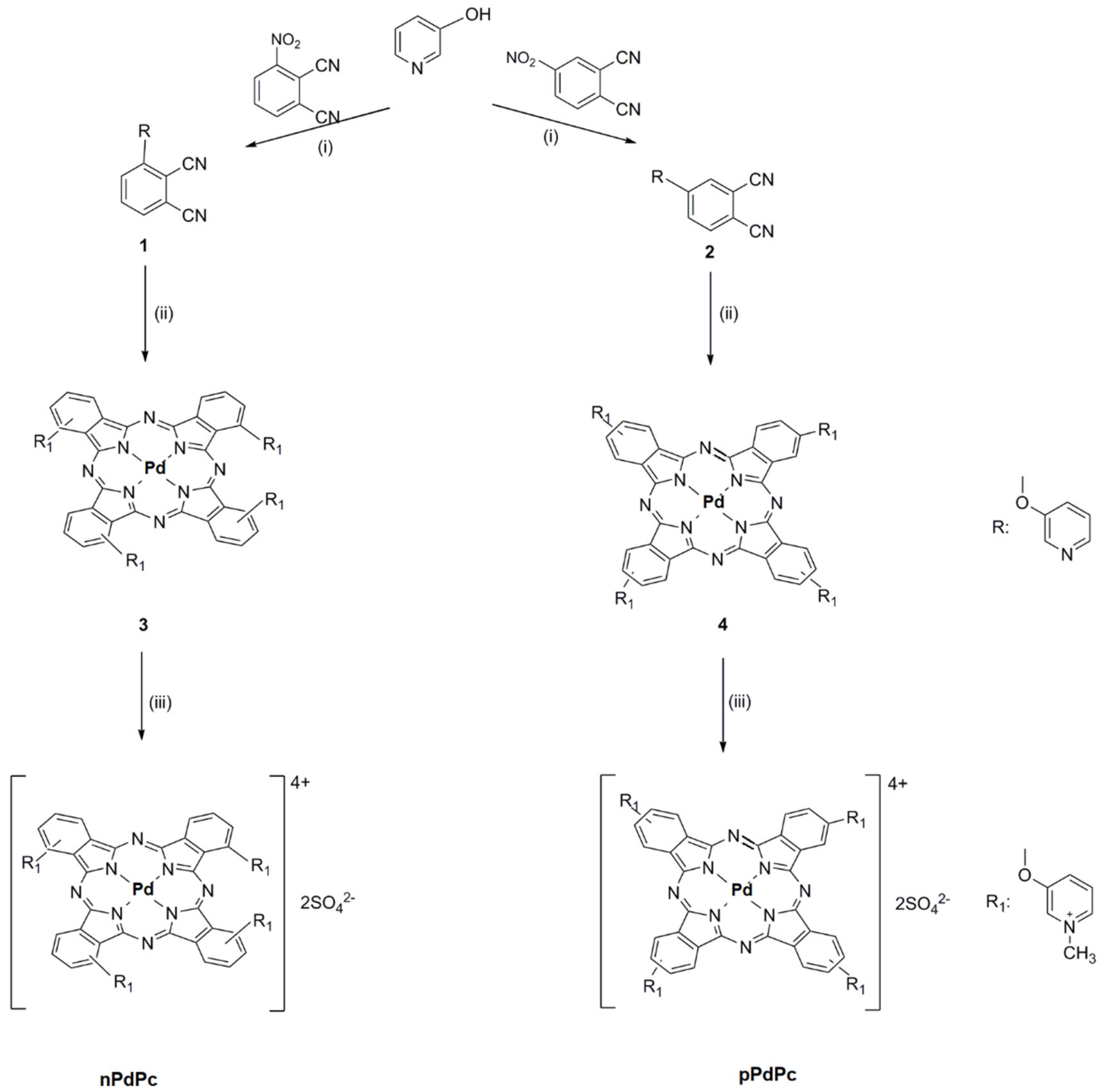
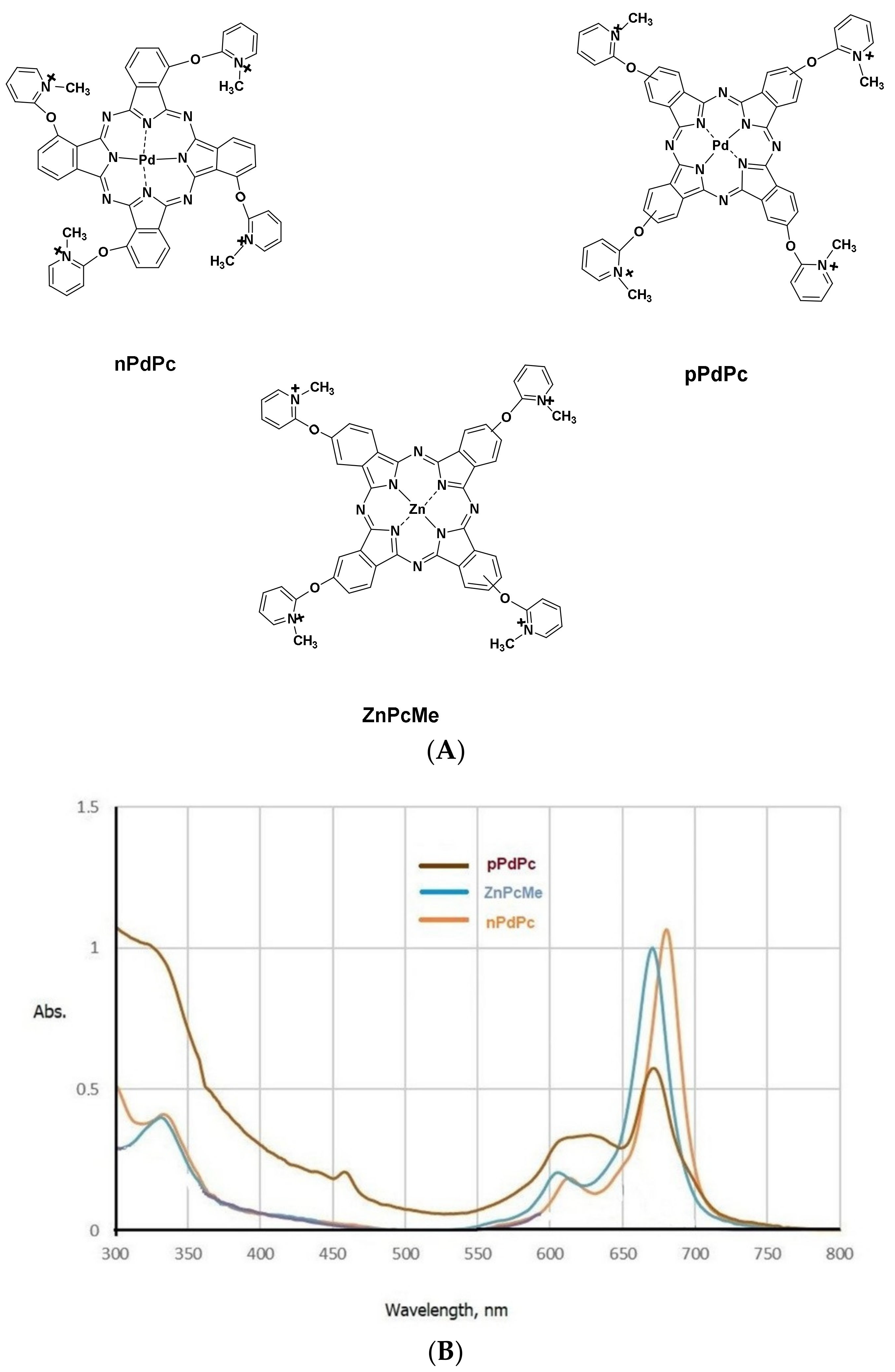
2.1. Phthalocyanines and Other Chemicals
2.2. Bacterial Strains and Cultivation
2.3. Antimicrobial Susceptibility
2.4. Photodynamic Inactivation Study
2.5. Statistics
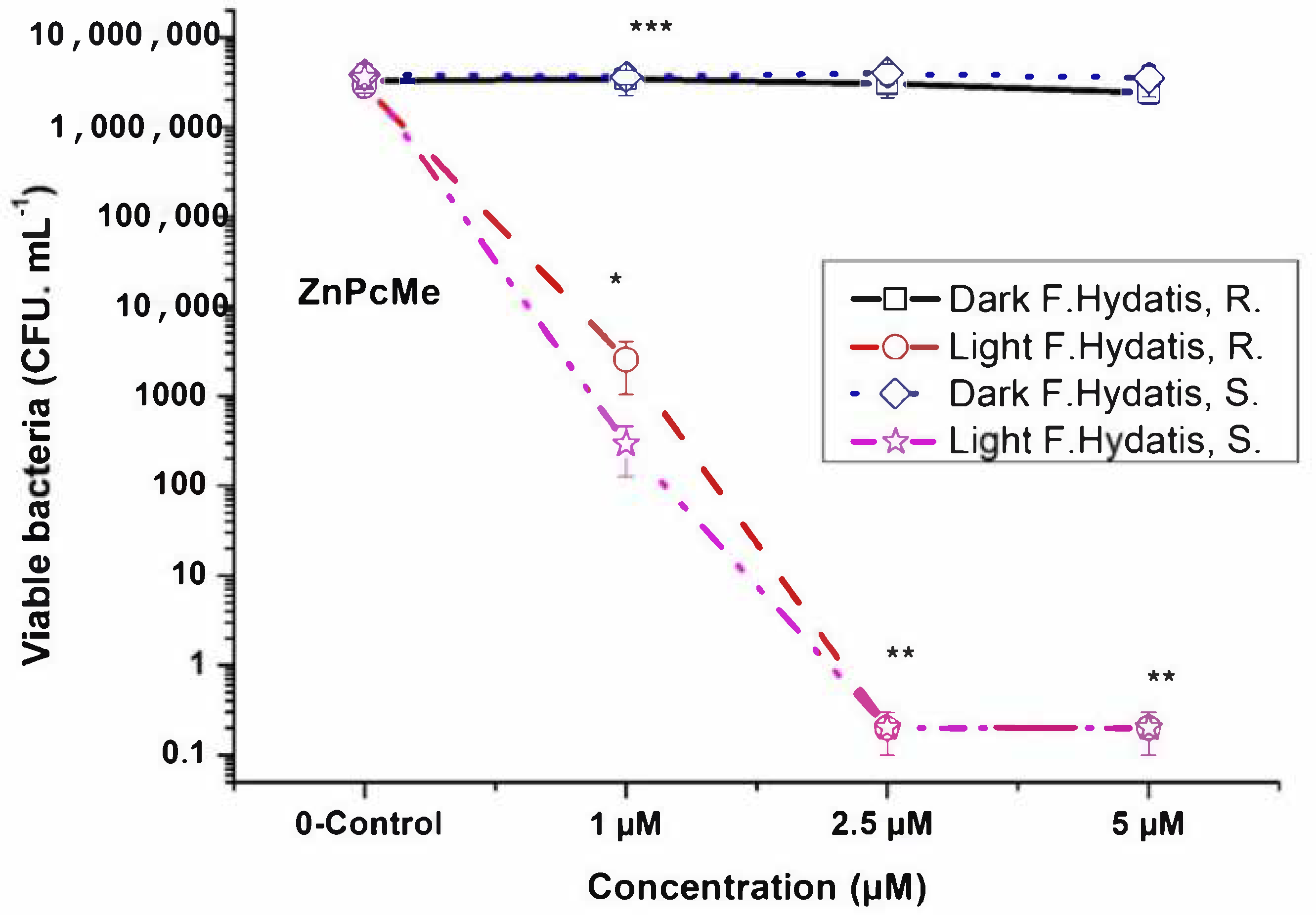
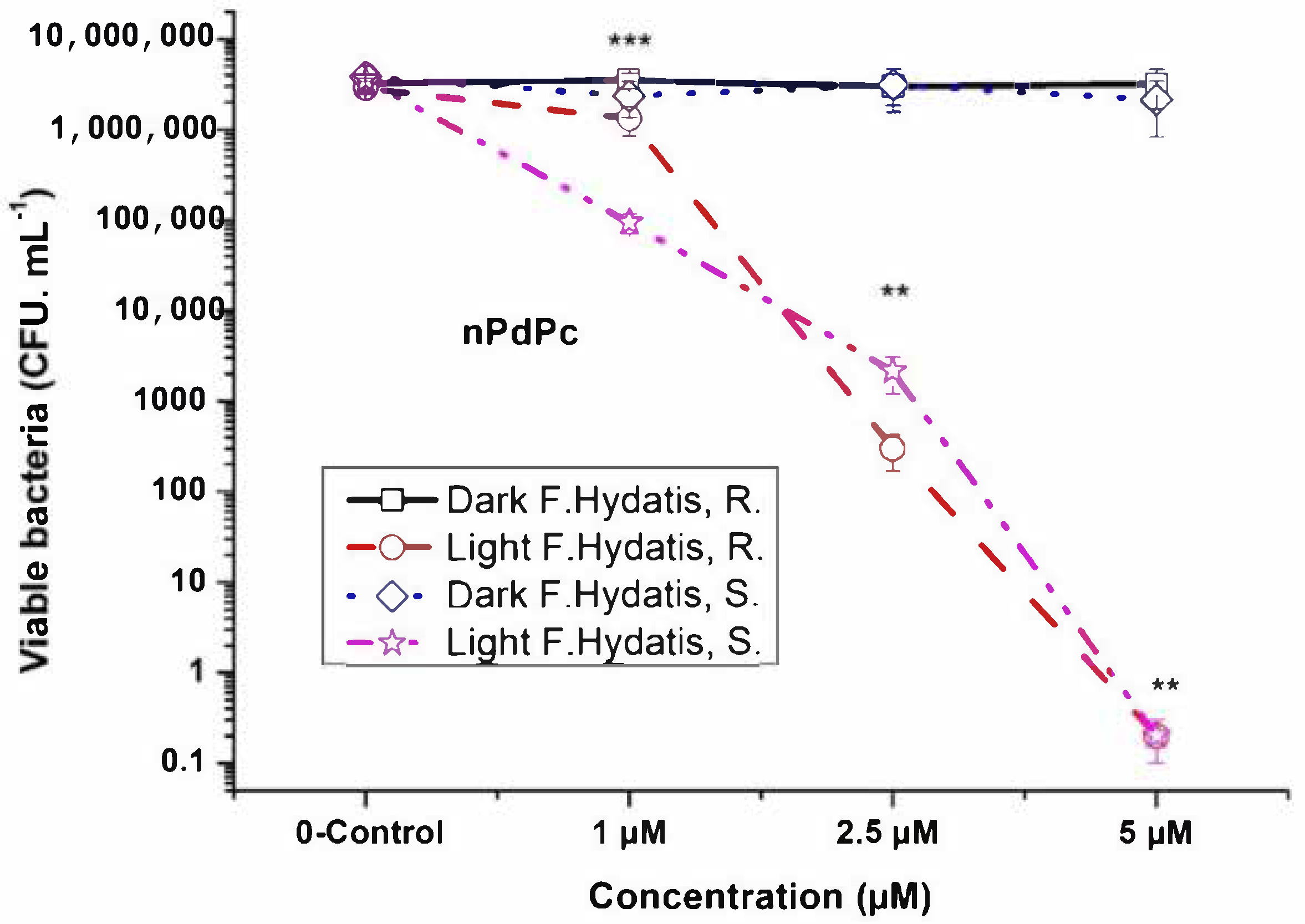
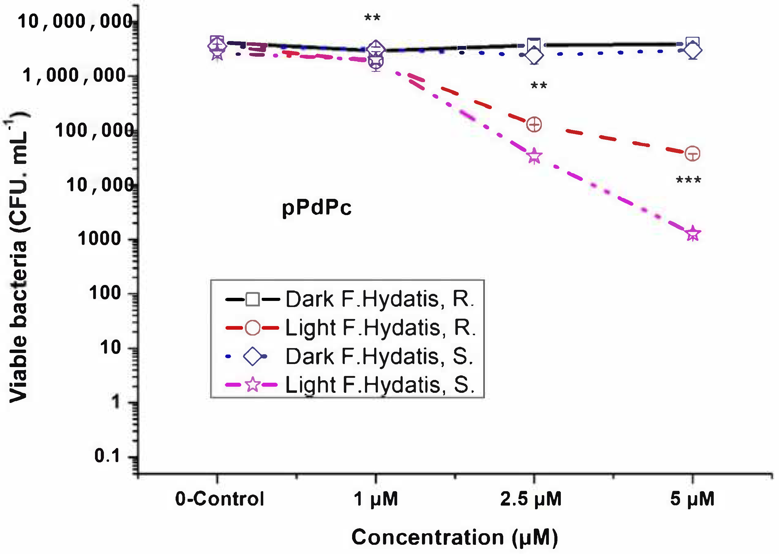
3. Results
4. Discussion
5. Conclusions
Author Contributions
Funding
Institutional Review Board Statement
Informed Consent Statement
Conflicts of Interest
Ethics Approval Statement
References
- Fossmark, R.O.; Vadstein, O.; Rosten, T.W.; Bakke, I.; Košeto, D.; Bugten, A.V.; Helberg, G.A.; Nesje, J.; Jørgensen, N.O.; Raspati, G.; et al. Effects of reduced organic matter loading through membrane filtration on the microbial community dynamics in recirculating aquaculture systems (RAS) with Atlantic salmon parr (Salmo salar). Aquaculture 2020, 524, 735268. [Google Scholar] [CrossRef]
- Roh, H.J.; Kang, G.S.; Kim, A.; Kim, N.E.; Nguyen, T.L.; Kim, D.-H. Blue light-emitting diode photoinactivation inhibits edwardsiellosis in fancy carp (Cyprinus carpio). Aquaculture 2018, 483, 1–7. [Google Scholar] [CrossRef]
- Vestrum, R.I.; Luef, B.; Forberg, T.; Bakke, I.; Vadstein, O. Investigating Fish Larvae-Microbe Interactions in the 21st Century: Old Questions Studied with New Tools. In Emerging Issues in Fish Larvae Research; Yúfera, M., Ed.; Springer: Cham, Switzerland, 2018. [Google Scholar] [CrossRef]
- Starliper, C.E. Bacterial coldwater disease of fishes caused by Flavobacterium psychrophilum. J. Adv. Res. 2011, 2, 97–108. [Google Scholar] [CrossRef] [Green Version]
- Akinola, O.G.; Olakunbi, O.O. Antibiotic sensitivity and sodium chloride susceptibility patterns of Flavobacterium columnare isolated from clinical columnaris in cultured Clarias gariepinus. J. Vet. Med. Anim. Health 2019, 11, 59–63. [Google Scholar]
- Wahli, T.; Madsen, L. Flavobacteria, a never-ending threat for fish: A review. Curr. Clin. Microbiol. Rep. 2018, 5, 26–37. [Google Scholar] [CrossRef]
- Austin, B.; Austin, D.A. Bacterial Fish Pathogens: Diseases of Farmed and Wild Fish, 4th ed.; Praxis Publishing Ltd.: Omaha, NE, USA, 2007. [Google Scholar]
- Sitjà-Bobadilla, A.; Oidtmann, B. Integrated pathogen management strategies in fish farming. In Fish Diseases; Academic Press: London, UK, 2017; pp. 119–144. [Google Scholar]
- Almeida, A.; Cunha, A.; Gomes, N.; Alves, E.; Costa, L.; Faustino, M. Phage therapy and photodynamic therapy: Low environmental impact approaches to inactivate microorganisms in fish farming plants. Mar. Drugs 2009, 7, 268–313. [Google Scholar] [CrossRef] [Green Version]
- Ashok, A.; Arshad, E.; Jasmin, C.; Pai, S.S.; Singh, I.S.B.; Mohandas, A.; Anas, A. Reducing Vibrio load in Artemia nauplii using antimicrobial photodynamic therapy: A promising strategy to reduce antibiotic application in shrimp larviculture. Microb. Biotechnol. 2012, 5, 59–68. [Google Scholar] [CrossRef] [Green Version]
- Lo, P.C.; Rodríguez-Morgade, M.S.; Pandey, R.K.; Ng, D.K.P.; Torres, T.; Dumoulin, F. The unique features and promises of phthalocyanines as advanced photosensitisers for photodynamic therapy of cancer. Chem. Soc. Rev. 2020, 49, 1041–1056. [Google Scholar] [CrossRef]
- Alves, E.; Costa, L.; Carvalho, C.M.; Tomé, J.P.; Faustino, M.A.; Neves, M.G.; Tomé, A.C.; Cavaleiro, J.A.; Cunha, Â.; Almeida, A. Charge effect on the photoinactivation of Gram-negative and Gram-positive bacteria by cationic meso-substituted porphyrins. BMC Microbiol. 2009, 9, 70. [Google Scholar] [CrossRef] [Green Version]
- Li, X.; Zheng, B.-D.; Peng, X.-H.; Li, S.-Z.; Ying, J.-W.; Zhao, Y.; Huang, J.-D.; Yoon, J. Phthalocyanines as medicinal photosensitizers: Developments in the last five years. Coord. Chem. Rev. 2019, 379, 147–160. [Google Scholar] [CrossRef]
- Galstyan, A. Turning Photons into Drugs: Phthalocyanine-Based Photosensitizers as Efficient Photoantimicrobials. Chem.—A Eur. J. 2021, 27, 1903–1920. [Google Scholar] [CrossRef] [PubMed]
- Ribeiro, C.P.; Lourenço, L.M. Overview of cationic phthalocyanines for effective photoinactivation of pathogenic microorganisms. J. Photochem. Photobiol. C Photochem. Rev. 2021, 48, 100422. [Google Scholar] [CrossRef]
- Mantareva, V.N.; Kussovski, V.; Orozova, P.; Dimitrova, L.; Kulu, I.; Angelov, I.; Durmus, M.; Najdenski, H. Photodynamic Inactivation of Antibiotic-Resistant and Sensitive Aeromonas hydrophila with Peripheral Pd(II)- vs. Zn(II)-Phthalocyanines. Biomedicines 2022, 10, 384. [Google Scholar] [CrossRef] [PubMed]
- Kulu, I.; Mantareva, V.; Kussovski, V.; Angelov, I.; Durmuş, M. Effects of metal ion in cationic Pd(II) and Ni(II) phthalocyanines on physicochemical and photodynamic inactivation properties. J. Mol. Struct. 2022, 1247, 131288. [Google Scholar] [CrossRef]
- Lähteenmäki, H.; Pätilä, T.; Räisänen, I.T.; Kankuri, E.; Tervahartiala, T.; Sorsa, T. Repeated Home-Applied Dual-Light Antibacterial Photodynamic Therapy Can Reduce Plaque Burden, Inflammation, and aMMP-8 in Peri-Implant Disease—A Pilot Study. Curr. Issues Mol. Biol. 2022, 44, 1273–1283. [Google Scholar] [CrossRef]
- Karges, J.; Goldner, P.; Gasser, G. Synthesis, Characterization, and Biological Evaluation of Red-Absorbing Fe(II) Polypyridine Complexes. Inorganics 2019, 7, 4. [Google Scholar] [CrossRef] [Green Version]
- Tomić, S.L.; Vuković, J.S. Antimicrobial Activity of Silver, Copper, and Zinc Ions/Poly(Acrylate/Itaconic Acid) Hydrogel Matrices. Inorganics 2022, 10, 38. [Google Scholar] [CrossRef]
- Maldonado-Carmona, N.; Ouk, T.S.; Calvete, M.J.F.; Pereira, M.M.; Villandier, N.; Leroy-Lhez, S. Conjugating biomaterials with photosensitizers: Advances and perspectives for photodynamic antimicrobial chemotherapy. Photochem. Photobiol. Sci. 2020, 19, 445–461. [Google Scholar] [CrossRef]
- Jenni, S.; Sour, A. Molecular Theranostic Agents for Photodynamic Therapy (PDT) and Magnetic Resonance Imaging (MRI). Inorganics 2019, 7, 10. [Google Scholar] [CrossRef] [Green Version]
- Bauer, A.W.; Kirby, W.M.; Sherris, J.C.; Turck, M. Antibiotic susceptibility testing by a standardized disk method. Am. J. Clin. Pathol. 1966, 45, 493–496. [Google Scholar] [CrossRef]
- Kanyal, P.; Rameez Roshan, P.M.; Danish, M.; Anita, A.; Khati, A.; Chauhan, R. Antimicrobial photodynamic therapy and its applicability in aquaculture systems and aquatic animal health management: An overview. J. Appl. Nat. Sci. 2016, 8, 506–514. [Google Scholar] [CrossRef]
- Pedigo, L.A.; Gibbs, A.J.; Scott, R.J.; Street, C.N. Absence of bacterial resistance following repeat exposure to photodynamic therapy. In Photodynamic Therapy: Back to the Future; International Society for Optics and Photonics: Seattle, WA, USA, 2009; Volume 7380. [Google Scholar]
- Tavares, A.; Carvalho, C.M.B.; Faustino, M.A.; Neves, M.G.P.M.S.; Tomé, J.P.C.; Tomé, A.C.; Cavaleiro, J.A.S.; Cunha, Â.; Gomes, N.C.M.; Alves, E.; et al. Antimicrobial Photodynamic Therapy: Study of Bacterial Recovery Viability and Potential Development of Resistance after Treatment. Mar. Drugs 2010, 8, 91–105. [Google Scholar] [CrossRef] [Green Version]
- Paronyan, M.H.; Koloyan, H.O.; Avetisyan, S.V.; Aganyants, H.A.; Hovsepyan, A. Study of the possible development of bacterial resistance to photodynamic in- activation. Biol. J. Armen. 2019, 71, 17–22. [Google Scholar]
- Alves, E.; Faustino, M.A.F.; Tome, J.P.C.; Neves, M.G.P.M.S.; Tomé, A.C.; Cavaleiro, J.A.S.; Cunha, Â.; Gomes, N.C.M.; Almeida, A. Photodynamic antimicrobial chemotherapy in aquaculture: Photoinactivation studies of Vibrio fischeri. PLoS ONE 2011, 6, e20970. [Google Scholar] [CrossRef] [PubMed] [Green Version]
- Malara, D.; Mielke, C.; Oelgemöller, M.; Senge, M.O.; Heimann, K. Sustainable water treatment in aquaculture—Photolysis and photodynamic therapy for the inactivation of Vibrio species. Aquac. Res. 2017, 48, 2954–2962. [Google Scholar] [CrossRef]
- Wong, P.N.; Mak, S.K.; Lo, M.W.; Lo, K.-Y.; Tong, G.M.-W.; Wong, Y.; Wong, A.K.-M. Vibrio vulnificus peritonitis after handling of seafood in a patient receiving CAPD. Am. J. Kidney Dis. 2005, 46, e87–e90. [Google Scholar] [CrossRef]
- Magaraggia, M.; Faccenda, F.; Gandolfi, A.; Jori, G. Treatment of microbiologically polluted aquaculture waters by a novel photochemical technique of potentially low environmental impact. J. Environ. Monit. 2006, 8, 923. [Google Scholar] [CrossRef]


| Antibiotics | Flavobacterium hydatis (Sensitive) Zone of Inhibition (mm) | Flavobacterium hydatis (Resistant) Zone of Inhibition (mm) |
|---|---|---|
| Tob10 Tobramycin | 0 (R) | 0 (R) |
| Nb Novobiocin 5 µg | 0 (R) | 0 (R) |
| Nx Nalidixic acid 30 | 30 (S) | 0 (R) |
| FUR Ceftiofur 30 | 0 (R) | 0 (R) |
| EX Enrofloxacin 5 | >30 (S) | 0 (R) |
| GEN Gentamicin 10 | 12 (I) | 0 (R) |
| NX Norfloxacin 10 | 25 (S) | 0 (R) |
| AMP Ampicillin 10 | 0 (R) | 0 (R) |
| OA Oxolinic acid 2 | 15 (I) | 0 (R) |
| APR Apramycin 15 | 27 (S) | 0 (R) |
| SPX Sparfloxacin 5 | >30 (S) | 5 (R) |
| FLM Flumequine 30 | >30 (S) | 0 (R) |
| O Oxytetracycline 30 | 18 (I) | 0 (R) |
| COT Co-Trimoxazole 25 | 5 (R) | 0 (R) |
| FFC Florfenicol 30 | 28 (S) | 0 (R) |
Publisher’s Note: MDPI stays neutral with regard to jurisdictional claims in published maps and institutional affiliations. |
© 2022 by the authors. Licensee MDPI, Basel, Switzerland. This article is an open access article distributed under the terms and conditions of the Creative Commons Attribution (CC BY) license (https://creativecommons.org/licenses/by/4.0/).
Share and Cite
Mantareva, V.; Kussovski, V.; Orozova, P.; Angelov, I.; Durmuş, M.; Najdenski, H. Palladium Phthalocyanines Varying in Substituents Position for Photodynamic Inactivation of Flavobacterium hydatis as Sensitive and Resistant Species. Curr. Issues Mol. Biol. 2022, 44, 1950-1959. https://doi.org/10.3390/cimb44050133
Mantareva V, Kussovski V, Orozova P, Angelov I, Durmuş M, Najdenski H. Palladium Phthalocyanines Varying in Substituents Position for Photodynamic Inactivation of Flavobacterium hydatis as Sensitive and Resistant Species. Current Issues in Molecular Biology. 2022; 44(5):1950-1959. https://doi.org/10.3390/cimb44050133
Chicago/Turabian StyleMantareva, Vanya, Vesselin Kussovski, Petya Orozova, Ivan Angelov, Mahmut Durmuş, and Hristo Najdenski. 2022. "Palladium Phthalocyanines Varying in Substituents Position for Photodynamic Inactivation of Flavobacterium hydatis as Sensitive and Resistant Species" Current Issues in Molecular Biology 44, no. 5: 1950-1959. https://doi.org/10.3390/cimb44050133
APA StyleMantareva, V., Kussovski, V., Orozova, P., Angelov, I., Durmuş, M., & Najdenski, H. (2022). Palladium Phthalocyanines Varying in Substituents Position for Photodynamic Inactivation of Flavobacterium hydatis as Sensitive and Resistant Species. Current Issues in Molecular Biology, 44(5), 1950-1959. https://doi.org/10.3390/cimb44050133