Article

Phytochemical Screening, Nutritional Value, Anti-Diabetic, Anti-Cancer, and Anti-Bacterial Assessment of Aqueous Extract from Abelmoschus esculentus Pods

1
Nanotechnology and Nanomedicine Lab-6 (IIRC), Department of Biosciences, Integral University, Lucknow 226026, India
2
Department of Clinical Pharmacy, College of Pharmacy, Jazan University, P.O. Box 114, Jazan 45142, Saudi Arabia
3
Department of Clinical Laboratory Sciences, College of Applied Medical Sciences, King Khalid University, P.O. Box 61413, Abha 61421, Saudi Arabia
4
Respiratory Therapy Department, Faculty of Applied Medical Sciences, Jazan University, Jazan 45142, Saudi Arabia
5
Department of Biology, College of Sciences, University of Hail, P.O. Box 2240, Hail 81451, Saudi Arabia
6
Department of Research, Jawaharlal Nehru Cancer Hospital and Research Centre, Bhopal 462001, India
*
Authors to whom correspondence should be addressed.
Processes 2022, 10(2), 183; https://doi.org/10.3390/pr10020183
Submission received: 9 December 2021 / Revised: 11 January 2022 / Accepted: 12 January 2022 / Published: 18 January 2022

Abstract

:
Known for its high nutritional and medicinal value, okra (Abelmoschus esculentus) is commonly used for replacing plasma and expanding blood volume in humans. It is a major economic crop cultivated in tropical and subtropical regions worldwide. The present study aimed to investigate and evaluate the nutritional properties and prospective applications of the consumable parts of okra. The total ash content (mineral content), carbohydrate, crude fiber, fat, protein, and moisture fractions of okra pod aqueous extract were determined. The results show that okra aqueous extract contained 84.670–87.650% moisture, 1.514–1.197% ash, 7.857–8.261% carbohydrate, 2.367–3.410% crude protein, and 6.781–8.314% crude fiber. Okra was determined to have high nutritional value, with γ-tocopherol and α-tocopherol contents about 2.67 mg/100 g and 1.62 mg/100 g, respectively. High-performance liquid chromatography (HPLC) was performed to determine the sugars present in okra aqueous extract. The water-soluble polysaccharide content was 10.22–16.45 g/100 g. The tested aqueous extract was a rich source of total phenolic compounds in gallic acid equivalents (288.2–3426.2 mg/100 g), chlorophyll a (3.53 mg/100), chlorophyll b (2.43 mg/100), and carotenoids (1.3 mg/100 g). The detected minerals were Ca, Mg, Cu, Zn, Fe, K, Na, and Mn. Atomic absorption spectrometry analysis of these ashed minerals was performed. In addition to the nutritional benefits, okra pods exhibited antimicrobial, anticancer, and antioxidant properties. The aqueous extract was found to be potentially active against bacterial strains of Staphylococcus aureus (MIC value = 21.8 mg/mL), Escherichia coli (MIC value = 18.7 mg/mL), Bacillus cereus (MIC value = 20.7 mg/mL), and Klebsiella pneumoniae (MIC value = 20.2 mg/mL). Okra aqueous extract exhibited inhibitory activity against α-amylase (IC50 = 120 µg/mL) and α-glucosidase (IC50 = 115 µg/mL). The okra extract exhibited high anticancer activity, concentration-dependent and with an IC50 value of about 158.3 mg/mL. The results indicated that okra pods have nutritional and medicinal properties and, hence, can be used as a functional food and broad-spectrum nutraceutical supplement.

1. Introduction

The botanical name of okra is Abelmoschus esculentus, which is derived from the original Arabic term “Abual-misk”, meaning “Father of Musk and Kaab-el-misk”, a reference to the musky fragrance of its seeds. It belongs to the family Malvaceae, genus Abelmoschus, species esculentus, and consists of edible green pods, seeds, and fiber [1]. Okra is a common edible vegetable valued for its flavor in West Africa, North America, Asia, and Arab countries and is widely distributed in tropical and subtropical regions. However, its diversity is richest in South Asia and southwest Pacific regions [2]. In different regions, okra is known by a few common names, such as ladyfingers, Bamya (in Iraq), and bhindi (in India) [3]. In India, this vegetable is commonly consumed and cultivated, and favorable climatic conditions have made this region an epicenter of diversity for this crop. It is widely found in evergreen forests and wastelands and is also grown commercially on farms. Four varieties of Abelmoschus, including 11 species and three subspecies, are found from the Himalayan region [4] to the south peninsular region of India [5]. Owing to its notable nutritional value, okra is considered beneficial as a supplement to improve human nutrition.
The high nutritional value of okra is attributed to its richness in vitamins A, E, and C. It also contains essential minerals, such as sodium, potassium, magnesium, and calcium, and trace elements, such as zinc, iron, and nickel, in minor proportions [6]. Okra seeds contain phenolic compounds, such as flavonoids and catechins, and oligomeric derivatives and antioxidants [6]. Okra seeds are covered by a hard coat that contains a high percentage of raw fiber. The seeds also have essential nutrients; for example, they have higher protein content compared to other plant crops. The seeds are also rich in oil, carbohydrates, dietary fiber, vitamins, and minerals [7,8,9]. The physiological and biological activities of okra, such as antimicrobial, antioxidant, and anti-inflammatory properties, are attributed to its phenolic compounds, chlorophyll, and carotene, which promote health and protect the body from free radical damage [10].
The biological properties of okra are due to the presence of phenolic compounds, carotenoids, tocopherols, and minerals. Carotenoids are biologically active fat-soluble compounds found in plants and include beta-carotene, lycopene, lutein, and xanthophane [11]. They are yellow, orange, or red, and nearly 600 known carotenoid species are found in vegetables and fruits. The phenolic compounds present in okra fruits vary; for example, 167.62 mg/100 g are present in Anamika species, 112.27 mg/100 g in the Sinnova type, and 106.26 mg/100 g in Shagun [12]. Analyses of okra revealed relatively high chlorophyll levels of 0.141 mg/100 g in Anamika species and 0.135 mg/100 g in Sadahar species. Another important component for nutrition is vitamin E and tocopherols. Tocopherols are natural antioxidants and exist in four forms: α, β, Y, and δ. δ-Tocopherol is considered the most important, as it reduces fat adhesion to cell walls, reduces cholesterol in the blood, and captures cell tissue peroxide radicals [13]. Tocopherols are fat-soluble compounds; they are unaffected by acids and are resistant to high temperatures in the absence of oxygen. Tocopherols are potent reducing agents and, hence, are extensively used as antioxidants. Okra pods are effective in lowering cholesterol due to their hypocholesterolemic properties [14].
Minerals are essential components of human nutrition and necessary during all growth stages of the human body. In total, 22 metal elements are needed for the growth and maintenance of the human body [15]. They contribute to physical and mental well-being, as they are involved in the formation of bones, teeth, muscles, tissues, blood, and nerve cells [16]. These elements are also required for the maintenance of acid–base balance, stimulation of nerve responses, and blood clotting [5]. Calcium, potassium, sodium, and magnesium are the major elements in okra, whereas iron, nickel, manganese, zinc, and chlorides are present in lower amounts [11].
The antidiabetic activity of okra was reported by Sabitha and coworkers [9], who demonstrated the in vitro inhibitory effect of okra peel and seed aqueous extract on α-glucosidase and α-amylase, indicating that okra peel can help to maintain blood sugar levels. Because of the potential applications of okra in food and medicine, its cultivation has increased in recent years [8,9,17]. Abelmoschus is useful in papermaking, fiber production, jute substitution, wastewater treatment, and the textile industry [5]. Abelmoschus is also a reservoir of essential micronutrients that have applications in the fortification process.
In the present study, we aimed to determine the phytochemical and nutritional properties of Abelmoschus esculentus aqueous extract and its dietary and medicinal applications. We evaluated these applications both qualitatively and quantitatively. The results of this study highlight the dietary and pharmacological (antidiabetic, anticancer, and antimicrobial) importance of okra pods.

2. Materials and Methods

2.1. Chemical, Reagents, and Cell Line

All reagents and chemicals were purchased from Himedia, India. Unless otherwise specified, all solvents and chemicals were of analytical grade and procured from Merck and Sigma-Aldrich (St. Louis, MO, USA). Cell lines were purchased from NCCS Pune India, and all chemicals and media used in cell culture were procured from Himedia (West Chester, PA, USA).

2.2. Collection and Drying of Okra Pods

Fresh okra pods Abelmoschus esculentus were obtained from the herbarium garden at Integral University Campus, Lucknow, India in November 2020. The plant specimen was authenticated by the taxonomist at the Department of Pharmacognosy, Faculty of Pharmacy, Integral University, Lucknow, U.P., India, with voucher specimen reference no. (IU/PHAR/HRB/20/11). The color of the okra pods was green to dark green, and the average length of pods was nearly 8–10 cm with a pentagonal cross-section containing numerous seeds. The type of okra used in the study was locally available, namely, Okra–Komal. After collection, okra pods were washed thoroughly and dried at room temperature for 72 h. After 72 h, they were dried at 30–40 °C until a constant weight was obtained. After drying, they were ground to powder using an electric blender, sieved using sieve #22, and stored in an air-tight jar for further use.

2.3. Preparation of Extract

For cold aqueous extraction of okra pods, 300 mL of double-distilled water was added to 100 g of powdered okra and homogenized at 2000 rpm at 4 °C in a homogenizer (Science-Tech, Lucknow, India; motor specifications: Prompt-FHP, AC/DC-ACW, S.NO.99085, RPM-4000, Uttam Electrical Industries, Varansi, Uttar Pradesh, India). The mixture was filtered through Whatman No. 1 filter paper. The residue was dried at a temperature of approximately 40 °C to obtain the dried form of okra crude extract. The dried residue of aqueous extract was stored in a refrigerator for further use.

2.4. Phytochemical Estimations of Okra Pods Extract

2.4.1. Assessment of Moisture, Ash, Crude Protein, Fiber, and Fat Content

The standard procedures described in the Association of Official Analytical Chemists (AOAC) manual (2005) were followed to evaluate nutrient constituents such as moisture, ash, protein, fiber, and fat in okra pods. The sample was kept in an oven (Universal, Gurugram, India) overnight at 100–110 °C and was cooled in a desiccator (Tarson-402020, Vacuum Desiccator Size 200 mm, India). The weight loss was measured and expressed as moisture content. To analyze the ash content of okra pods, the sample was subjected to a temperature of 550 °C in a muffle furnace (Thermotech, Hyderabad, India) to obtain grayish-white ash. The weight of this ash was measured to determine the ash content (Method No. 930.05, AOAC 2005). To analyze the crude fiber content of okra pods, the sample was treated with 1.25% H2SO4, 1.25% NaOH, and 1% HNO3. It was then filtered and washed with hot water. The residue obtained was dried in an oven at 130 °C. It was then ignited at 550 °C in a furnace. The weight loss in the ignition was considered to be crude fiber content. The micro Kjeldahl method was employed to determine the nitrogen content of the sample, whereas protein content was determined by multiplying the value of nitrogen content by 6.25. Total carbohydrate content was calculated using the formula given below:
100 − (percentage of ash + percentage of total lipid + percentage of protein + percentage of crude fiber) (AOAC, 2000)
The calorific value was evaluated according to Guil-Guerrero et al., 1998, using the formula given below:
(Total carbohydrate × 4) + (lipid × 9) + (protein × 4)
The resultant value is expressed in kilocalories. The Soxhlet extraction method was used to quantify crude fat content. In this method, the extracting agent was petroleum ether, and the temperature was 60–80 °C, as reported in Method No. 930.09 (AOAC 2005).

2.4.2. Mineral Determination

The ashed sample was dissolved in 100 mL of HNO3 (2%), and double-distilled water was added to obtain the final volume. Ca, Mg, Cu, Zn, Fe, Pb, As, Se, Cr, Cd, and Mn were detected in the ashed sample. Each mineral was separately measured using an Atomic Absorption Spectrometer (Perkin Elmer, Analyst 800). For phosphorus content, a Spectrum spectrophotometer (754 PC, Shanghai, China) was used, and the absorbance was measured at 440 nm.

2.4.3. Determination of Total Phenolic Content

The Folin–Ciocalteu method was employed to estimate the total phenolic content of okra extract [18]. One gram of okra extract was mixed with 46 mL of double-distilled water and 1 mL of Folin–Ciocalteu reagent. It was mixed well, and 3 mL of sodium carbonate (2% solution) was added after 3 min. The solution was shaken intermittently and was kept stable for 2 h. The absorbance was measured at 760 nm.

2.4.4. Determination of Chlorophyll and Carotenoids

The chlorophyll and carotenoid content of okra pod extract was determined by the method of Nagata and Yamashita [19] with some modifications. 150 mg of fine dried aqueous okra powder extract was mixed with 10 mL of acetone–hexane (4:6) solution for 1 min and filtered using Whatman No. 4 filter paper. The absorbance of the filtrate was measured at 470, 645, and 663 nm for carotene, chlorophyll b, and chlorophyll a content, respectively. The assessment of chlorophyll content was based on α-pheophytin, and that of carotene content was based on lutein. Chlorophyll and carotenoid contents were estimated using the formulas given below [20]:
Chlorophyll a and b were calculated by the following equations:
Chlorophyll   a = [ 12.7   A   ( 663 ) 2.63   A   ( 645 ) ] 1000 × W × V Chlorophyll   b = [ 22.9   A   ( 645 ) 2.63   A   ( 663 ) ] 1000 × W × V
where A is absorbance, W is the weight of the sample, and V is the final extract volume. Carotenoid was calculated by the following equation:
Carotenoid (mg/kg) = (A470 × 106)/(2000 × 100 × d)
where 613 is molecular absorption (ε) of the compound α-pheophytin, 2000 is molecular absorption (ε) of lutein, and d is cell thickness (1 cm).
α-Carotene and lycopene were calculated by the following equations:
α-Carotene = 0.216 × A663 − 1.220 × A645 − 0.304 × A505 + 0.452 × A453
Lycopene = −0.0458 × A663 + 0.204 × A645 − 0.304 × A505 + 0.452 × A453

2.4.5. Diphenyl-1-Picrylhydrazyl Assay

DPPH radical scavenging activities of okra pods were determined by the method of Blois (1958) with some modifications [21]. The working reagent of 0.1 mM DPPH was prepared in methanol. A 1 mL sample of the aqueous extract was mixed with 2 mL of prepared DPPH solution and incubated for 30 min at room temperature. After incubation, absorbance was measured at 517 nm. Synthetic ascorbic acid was used as the reference antioxidant, and methanol was used as a negative control. The antioxidant capacity for scavenging DPPH free radicals was determined by the equation given below. The results are expressed as percentage inhibition [22].
Scavenging activity (%) = (Abscontrol − Abssample/Abscontrol) × 100

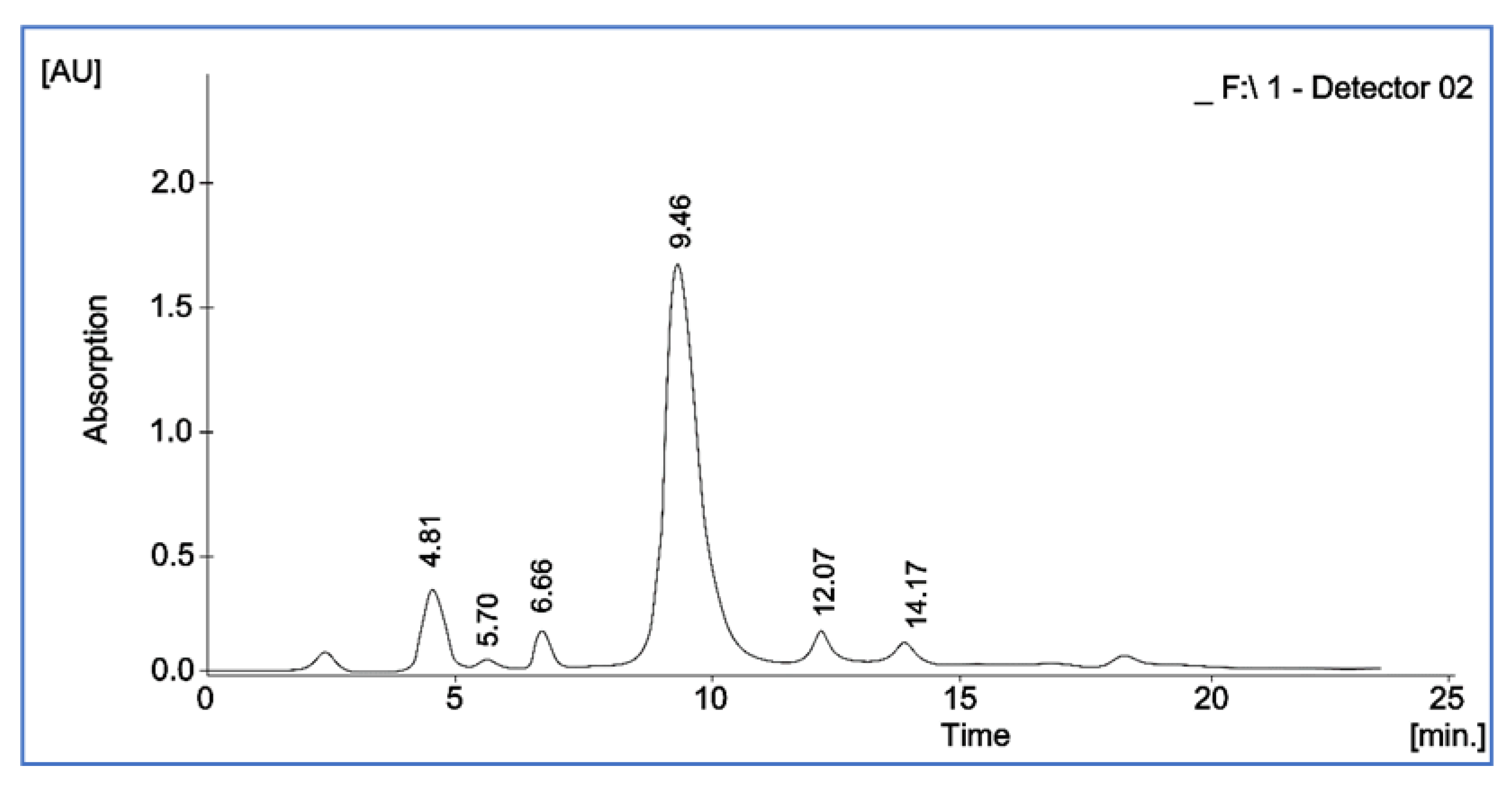
2.4.6. Estimation of Vitamin E (Tocopherol) by High-Performance Liquid Chromatography

The vitamin E content of okra pod extract was estimated by HPLC (Shimadzu-model HPLC) equipped with an LC-20 AT pump, a SPD-20 A UV/VIS detector, and a 20-ll loop Rheodyne injector. The okra pod extract, 20 µL of the standard solution, and 25 ppm vitamin E were loaded using the 20-ll loop Rheodyne injector. The liquid phase was water/methanol (90:10, v/v) at a rate of 1 mL/min and a wavelength of 294 nm. The vitamin E content was calculated using the formula given below:
Response factor = Standard size of the surface material AS/Concentration of the standard material (CS)
Concentration of sample (Cu) = Top size of the sample (Au)/Response factor

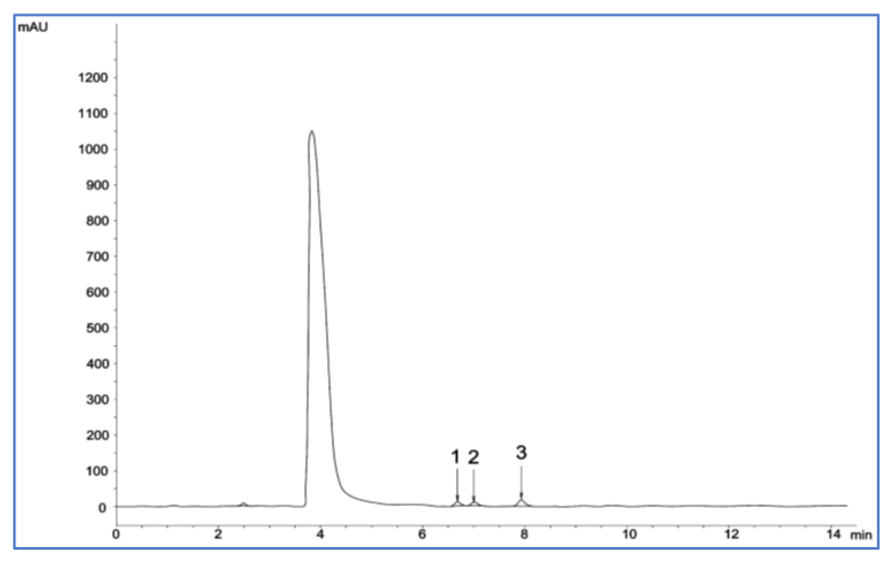
2.4.7. Analysis of Sugars by High-Performance Liquid Chromatography

Okra contains reducing and non-reducing sugars. Analysis of sugar content was performed in triplicate by high-performance liquid chromatography (HPLC). Ultrapure water–ethanol (100 mL, 80/20, v/v) was used to reflux the requisite amount of dried sample. It was boiled for 30 min, centrifuged at 6000 rpm for 20 min, and filtered through a 0.45 µm membrane filter. The sugars were separated by liquid chromatography using a Knauer (Berlin, Germany) carbohydrate column with a particle size of 5 µm and 250 mm × 4.6 mm i.d. at 35 °C. The solvents obtained after separation were filtered using a 0.45 µm membrane filter. An ultrasonic bath (Ultrasonic Cleaner Device, Model HZSH, CSF-1A, Shanghai, China) was used to obtain the filtrate. Acetonitrile and ultrapure water (75:25% v/v) were used as the mobile phase with a 1.0 mL/min flow rate. The injection volume was adjusted to 20 µL. An RI detector (K-230 l, Knauer, Berlin, Germany) was used. The peaks obtained were scaled with the specific concentrations of three sugars, namely, glucose, sucrose, and fructose, using external standards.

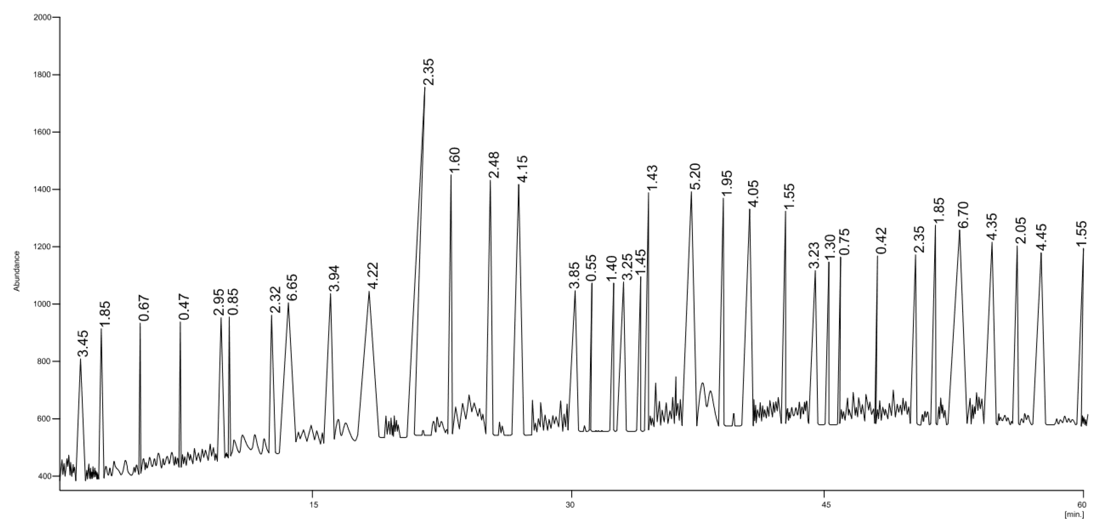
2.4.8. Gas Chromatography–Mass Spectrometry Analysis of Okra Pod Extract

The gas chromatography–mass spectrometry analysis of okra pod extract was performed using a Shimadzu chromatograph (model GC-MS QP 2010) and capillary column (Durabond) having a stationary phase (DB-5HT 30 mx) with a phase thickness of 0.319 mm × 0.10 µm. Helium gas was used as the carrier gas at a flow rate of 43.7 cm·s−1. In 1:50 split mode and at an injector temperature of 290 °C, 1 µL of the sample was loaded using an injector. Initially, the column temperature was 80 °C and was subject to two heating steps: 10 °C min−1 to 150 °C and 6 °C min−1 to 230 °C. The column was maintained at this temperature for 50 min. The temperature of the mass detector and interface was 250 °C. The m/z ratios at the start and end of the process were 40 and 1000, respectively. The standards present in a software library (Mass Spectral Database NIST/EPA/NIH) were used for the characterization. The mass spectra obtained were compared with those of the standards. The total area of the identified peaks was determined, and the percentages were measured using the relative area of each peak.

2.5. In Vitro Antidiabetic Activity of Aqueous Extract of Okra Pods

The antidiabetic activity of okra pods was determined by performing amylase activity inhibition and glucose diffusion inhibition assays.

2.5.1. Amylase Inhibition Assay

Aqueous extracts of okra pods with four concentrations of 25, 50, 75, and 100 mg/mL were prepared using double-distilled water. The aqueous extract (500 µL) was mixed with the same quantity of 0.02 M sodium phosphate buffer (pH 6.9) and 0.006 M sodium chloride, followed by the addition of 0.5 mg/mL α-amylase solution and incubation at 25 °C for 10 min. Subsequently, 1% starch solution was prepared in 0.02 M sodium phosphate buffer (pH 6.9) and 0.006 M sodium chloride in four different tubes. The solution (500 µL) was added to each tube at an interval of 5 s. This reaction mixture was incubated for 10 min. To terminate this reaction, 1 mL of 3,5-dinitrosalicylic acid reagent was added. All tubes containing the reaction mixture were incubated in a water bath for 5 min. They were then cooled to room temperature. The reaction mixture was diluted with 10 mL of double-distilled water. The absorbance was measured at 540 nm [23]. The % inhibition was calculated using the formula given below:
% inhibition = (Abscontrol − Absextract/Abscontrol) × 100

2.5.2. Glucose Diffusion Inhibition Assay

The dialysis membrane (Dialysis Membrane-70, approximate capacity of 2.41 mL/cm) containing okra pod aqueous extract and glucose solution (0.2 mM in 0.15 M sodium chloride) was kept in a beaker containing 40 mL of 0.15 M sodium chloride and 10 mL of double-distilled water. The beaker was kept in an orbital shaker at room temperature, and the diffusion of glucose into the external solution was measured. The assay was performed in triplicate [7].

2.6. Anticancer Activity of Okra Pod Extract against HepG2 Cells

The human liver cell line HepG2 (hepatocellular carcinoma) and normal rat kidney (NRK) cells were obtained from the National Centre for Cell Sciences, Pune, India. Dulbecco’s Modified Eagle Medium (DMEM) and DMEM-F12 supplemented with 10% fetal bovine serum and 1% antimicrobial cocktail containing antibiotics, specifically streptomycin (10 mg), penicillin (10,000 units), and amphotericin (25 mg), were used. The cells were separately cultured in these media at 37 °C in a CO2 humidified incubator and grown as a monolayer in 75 cm2 tissue culture flasks.

2.6.1. Determination of Cell Viability by 3-(4,5-Dimethylthiazol-2-yl)-2,5-diphenyl

Tetrazolium Bromide Assay

A stock solution of the extract with a concentration of 8 mg/mL was prepared in double-distilled water. The stock solution was diluted to obtain dilutions of 0.005–0.4 mg/mL. The effect of the extract on the human tumor cell line HepG2 and normal NRK cells was evaluated. The cells were grown in 96-well plates to obtain a final cell density of 5000 cells/well. These cells were left for 24 h to allow their attachment. After 24 h, the cells were treated with the extract, incubated again for 24 h, and assessed for cytotoxicity. The 3-(4,5-dimethylthiazol-2-yl)-2,5-diphenyl tetrazolium bromide assay was performed to quantify the cytotoxicity of okra pod extract in human HepG2 and NRK cell lines [24]. The absorbance was measured at 570 nm. The untreated cell lines (controls) were used to determine the live-cell percentage (%). The half-maximal inhibitory concentration (IC50) values were computed using the Origin 6.0 Professional software.

2.6.2. Morphological Analysis by Phase-Contrast Microscopy

Variations in cell morphology were noted when HepG2 and NRK cells were treated with the okra pod extract at IC50 concentration for 48 h [25]. The morphology of treated cells was compared with that of untreated cells. The morphological changes in treated cells were visualized using an inverted phase-contrast microscope (Labomed, Los Angeles, CA, USA).

2.6.3. Nuclear Condensation Detection by 4,6-Diamidino-2-phenylindole Staining

The apoptotic effect of okra extract at IC50 concentration on the HepG2 cell line was analyzed. The fluorescent nuclear dye 4,6-diamidino-2-phenylindole (DAPI) was used at a concentration (300 nm). Cellular internalization and chromatin interaction were assessed by DAPI staining. Okra extract-treated and DAPI-stained HepG2 cells were incubated for 24 h at 37 °C, and untreated and DAPI dye-stained cells were used as the controls. The stained cells were visualized using an inverted Nikon ECLIPSE Ti-S fluorescence magnifying instrument from Japan.

2.6.4. Quantification of Intracellular Reactive Oxygen Species

The dichloro-dihydro-fluorescein diacetate assay was performed to measure the level of reactive oxygen species (ROS) in the HepG2 cell line treated with okra pod extract at IC50 concentration [26]. The working concentration of ROS is 10 µM. A fluorescence microscope (Nikon ECLIPSE Ti-S, Tokyo, Japan) and multiwall smaller-scale plate reader (Synergy H1 Hybrid Multi-Mode Microplate Reader, BioTek, Winooski, VT, USA) were used to detect fluorescence. The excitation and emission wavelengths were 485 nm and 528 nm, respectively. The change in fluorescence intensity of treated cells was measured by comparing it with the untreated cells.

2.7. Antimicrobial Activity of Okra Pod Extract

The antibacterial activity of okra pod extract was primarily assessed using the disc diffusion method [19,27]. The microdilution method was used to determine the minimum inhibitory concentration required to inhibit 50% growth of organisms (MIC50) of the extract [17]. Strains of Staphylococcus aureus (NICM 2079), Escherichia coli (ATCC 25923), Bacillus cereus (NCIM 2156), and Klebsiella pneumoniae (NCIM 2957) in the midlogarithmic stage were used. The cells were treated with 10 mM sodium phosphate buffer (SPB) at pH 7.4 and diluted to 2 × 10⁵ colony-forming units (CFUs)/mL in SPB with 0.03% Luria–Bertani (LB) broth. Different concentrations of okra extract varying from 5 to 35 mg/mL were added to a bacterial inoculum of 5 × 10⁴ CFU/well and diluted in 96-well microtiter plates with 50 µL of LB medium. This setup was incubated overnight at 37 °C. The MIC50 of okra pod extract was determined by using the formula listed below:
MIC 50 = ( AControl A   Ttreatement ) × 100

2.8. Statistical Analysis

The analysis of variance and Tukey’s honestly significant difference tests were performed using SPSS Statistics Software Version 20 (IBM, New York, NY, USA). The data are expressed as mean values and standard deviations (SD) of three repeated readings of the samples and concentrations. A p-value of <0.05 was considered statistically significant.

3. Results

3.1. Proximate Nutritional Composition

Okra is a staple food and an established component of a complete diet in various parts of the world. The results show that the okra fruit had dietary fiber content of 6.781–8.314 g/100 g, moisture content of 84.67–87.65 g/100 g, fat content of 0.066–0.1 g/100 g, carbohydrate content of 7.857–8.261 g/100 g, and protein content of 2.367–3.41 g/100 g, and its calorific value was 51.258 kcal/100 g. Table 1 presents the proximate nutritional composition of okra pods.

3.2. Proximate Mineral Composition

The proximate mineral composition of okra pods is presented in Table 2. Vital constituents of human nutrition include both macroelements and microelements. Okra is nutritionally rich because it contains abundant minerals [28]. Mineral composition analysis showed that the most abundant mineral in okra was potassium (330–350 mg/100 g), followed by calcium (260–270 mg/100 g). The microelements detected in okra were zinc (1.8–2.2 mg/100 g), iron (1.5–1.7 mg/100 g), copper (0.48–0.5 mg/100 g), and manganese (0.12–0.15 mg/100 g).

3.3. Determination of Phenolic Compounds

The total phenolic content of okra pod extract was 4.75 mg/g. The phenolic compounds in okra seeds and podless flours were reported previously [19] and exhibited total phenolic content values (gallic acid equivalents) ranging from 288.2 to 3426.2 mg/100 g. Because reducing sugars are abundant in vegetable extracts, they can reduce Folin–Ciocalteu reagent, resulting in exaggerated values of total phenolic content [27].

3.4. Estimation of Chlorophyll and Carotenoid Content

Naturally occurring plant pigments, including chlorophylls and carotenoids, are commonly found in organic food components. The α-carotene and lycopene contents of okra were 0.83 mg/100 g and 0.55 mg/100 g, respectively. Although chlorophyll is not a nutritional compound, it is responsible for the green color of vegetables, indicates senescence to consumers through its oxidation effects, and contributes to the healing of wounds and inflammation [27]. The chlorophyll a and chlorophyll b contents were determined to be 3.53 mg/100 and 2.43 mg/100 g, respectively.

3.5. Antioxidant Potential of Okra

Free radicals are reduced by hydrogen atoms or electrons donated by antioxidants or free radical scavengers. DPPH is a stable free radical and was used to quantify the free radical scavenging activity by okra pods in this study. The DPPH radical scavenging assay showed that okra exhibits antioxidant potential, and the IC50 value was 1.05 mg/mL.

3.6. Estimation of Vitamin E (Tocopherol) Content

Okra contains a high amount of tocopherol, which is involved in growth and bone synthesis. The major tocopherol found in okra was γ-tocopherol (2.67 mg/100 g), followed by α-tocopherol (1.62 mg/100 g) (Figure 1).

3.7. Determination of Sugar Content

The principal sugar detected in the pods was sucrose (Peak 3) with a concentration of 105–115 mg/100 g. The other two sugars detected were fructose and glucose (Peaks 1 and 2) with concentrations of 33–35 and 28–32 mg/100 g, respectively.
The carbohydrate constituents of okra are affected by reaping conditions, soil characteristics, climatic conditions, and the genotype of the plant. The sugar content of okra pods is presented in Figure 2 and Table 3.

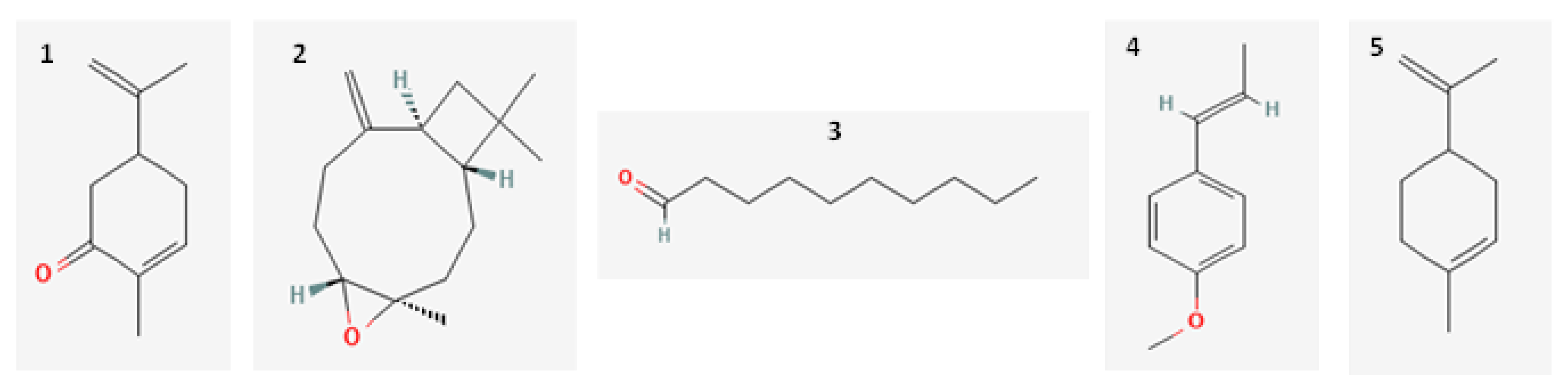
3.8. GC–MS Analysis of Okra Pods

GC–MS analysis of okra extract was performed, and a total of 35 compounds were detected. The detected compounds accounted for 91.63% of the total (Figure 3).
The percentage of compounds identified in okra pods is presented in Table 4. The compound profiles showed the presence of non-terpene derivatives (37.53%). Quantitative analysis of compounds indicated that oxygenated monoterpenes, sesquiterpene hydrocarbons, phenylpropanoids, and monoterpene hydrocarbons were present in moderate amounts of 21.1%, 10.5%, 10.3%, and 8.5%, respectively. Apocarotenes were present in small amounts (3.7%). The main constituents of okra pods were (E)-anethole (6.7%), limonene (6.65%), β-caryophyllene (5.2%), decanal (4.45%), and carvone (4.35%). The chemical structure of the main compounds is listed in Figure 4.

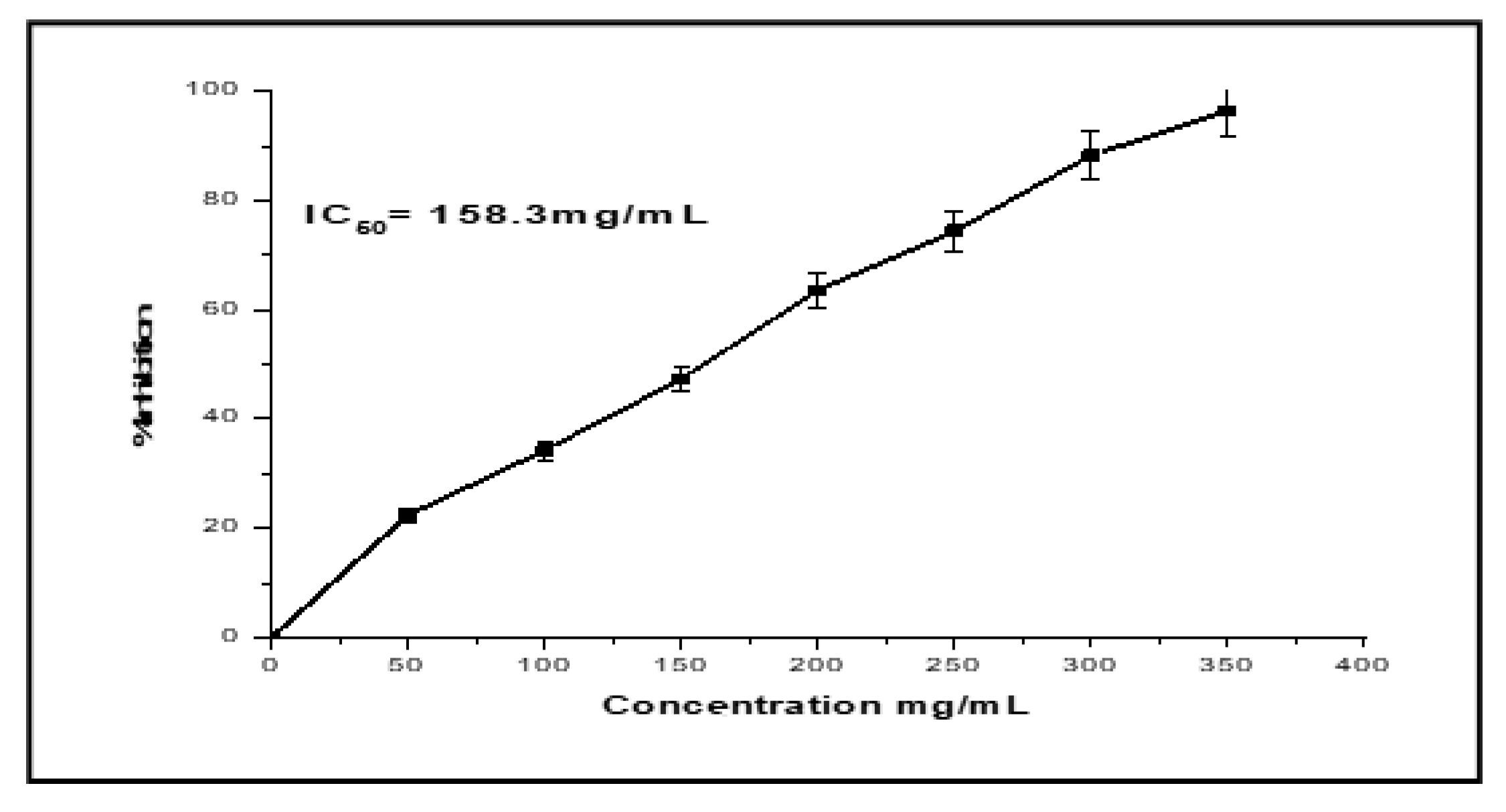
3.9. α-Amylase Inhibition and Glucose Diffusion Inhibition Activity

Okra exhibited inhibitory activity against α-amylase (IC50 = 120 µg/mL) and α-glucosidase (IC50 = 115 µg/mL). The high inhibitory potential of okra might be due to the synergic effect of its polysaccharides and flavonoids. This finding contradicts the results of Yuang et al., who attributed the high inhibitory potential of okra only to polysaccharides [17]. Flavonoids inhibit glucose transporters and consequently inhibit α-amylase and α-glucosidase activities.

3.10. In Vitro Anticancer Activity of Okra Extract

The anticancer activity of okra extract was analyzed in vitro against the HepG2 cell line. The okra extract exhibited high anticancer activity, with an IC50 value of 158.3 mg/mL. The anticancer effect was dose-dependent. Okra extract was nontoxic to NRK cells up to a concentration of 500 mg/mL (Figure 5).

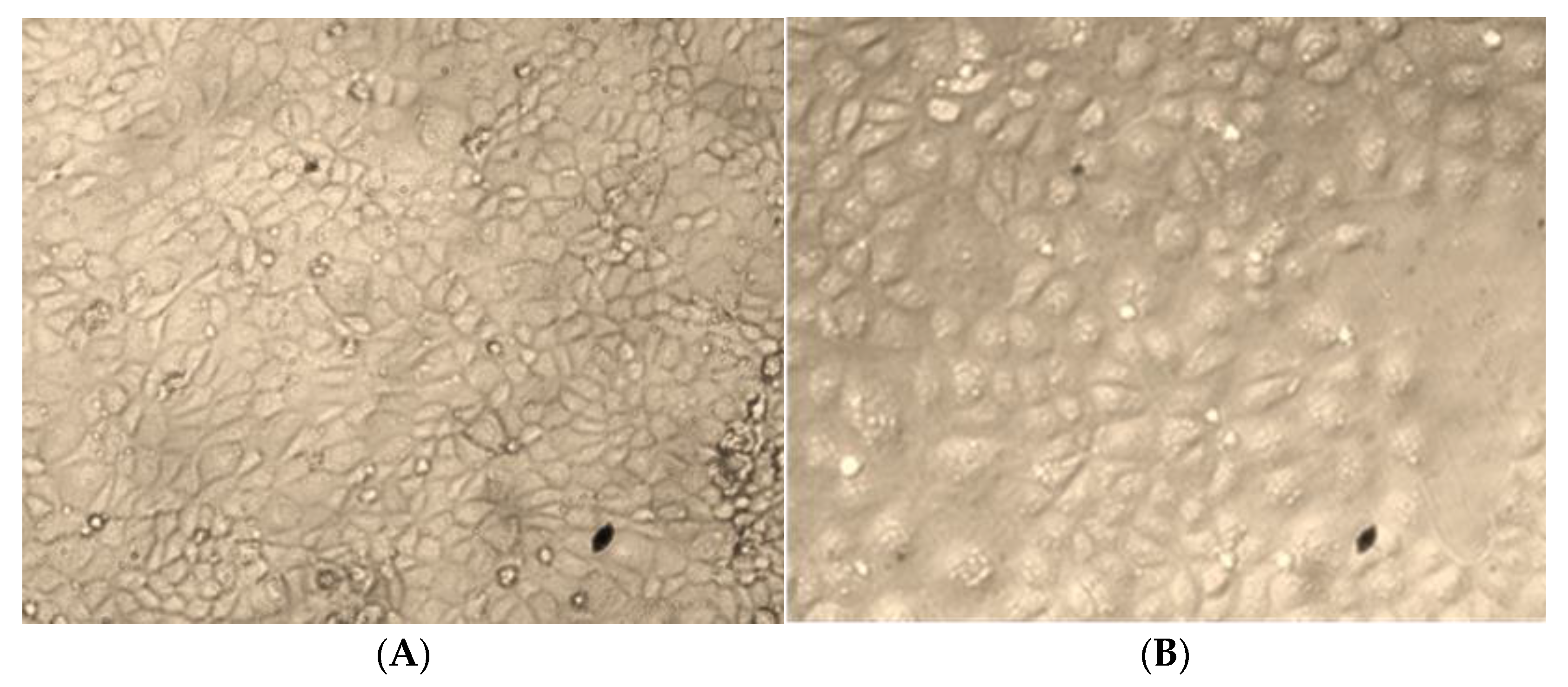
3.10.1. Morphological Changes in HepG2 Cells

Phase-contrast microscopy showed morphological changes in HepG2 cells treated with okra extract at the IC50 concentration (Figure 6). No notable morphological changes were observed in control (untreated) cells after incubation for 48 h, and they remained uniformly distributed (Figure 6A). However, after 48 h of incubation, HepG2 cells treated with okra extract showed a shrunken cell morphology and were irregular, necrotic, and detached from the well surface. Some cells had an intact (unblemished) plasma membrane. This indicated that the cells had initiated apoptosis.

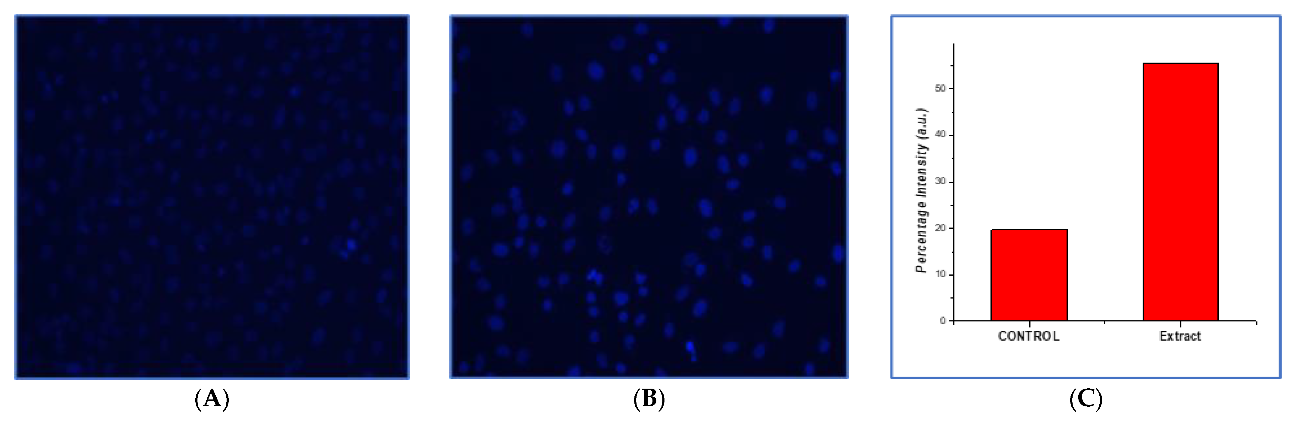
3.10.2. Analysis of Morphological and Nuclear Structural Changes

Incubation of HepG2 cells with okra extract inhibited the enzyme thymidylate synthase, which is involved in the nucleic acid synthesis. The effect of okra extract on nucleic acids led to considerable morphological and nuclear structural changes in HepG2 cells. Increased cell membrane penetrability and condensed chromatin were observed in okra extract-treated cells stained with DAPI (Figure 7B) compared to untreated cells (Figure 7A), indicating the apoptotic effect of okra extract. The condensation of the nucleus confirms the cytotoxic effect of okra extract. A graph of the fluorescence intensity, which was estimated by Image-J software, is given below and the graph was plotted with the help of Origin 6.0 Professional software (Figure 7C).

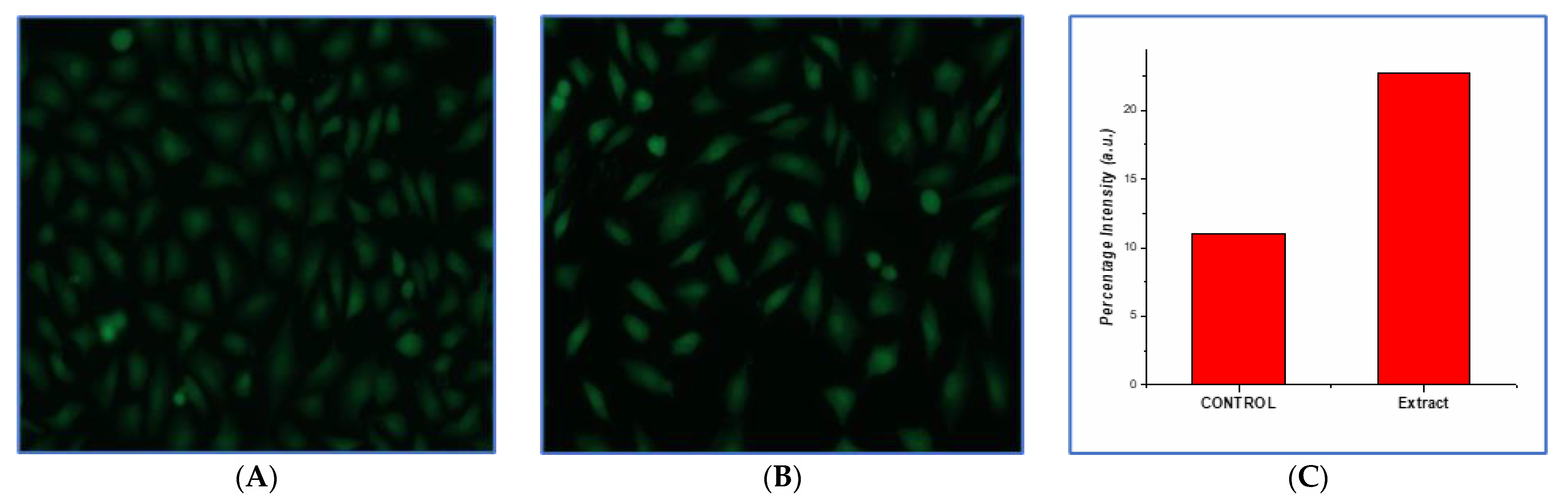
3.10.3. Production of Reactive Oxygen Species

Increased intracellular ROS were observed in okra extract-treated HepG2 cells compared with untreated cells (Figure 8A), which exhibited lower fluorescence than okra extract-treated HepG2 cells (Figure 8B). The generated ROS disturbed the compactness or structure of the plasma membrane, whereas no disorganization was observed in untreated cells, and their plasma membranes remained intact. Cytotoxicity due to oxidative pressure and apoptosis might have produced ROS. A graph of the fluorescence intensity, which was estimated by Image-J software, is given below and the graph was plotted with the help of Origin 6.0 Professional software (Figure 8C).

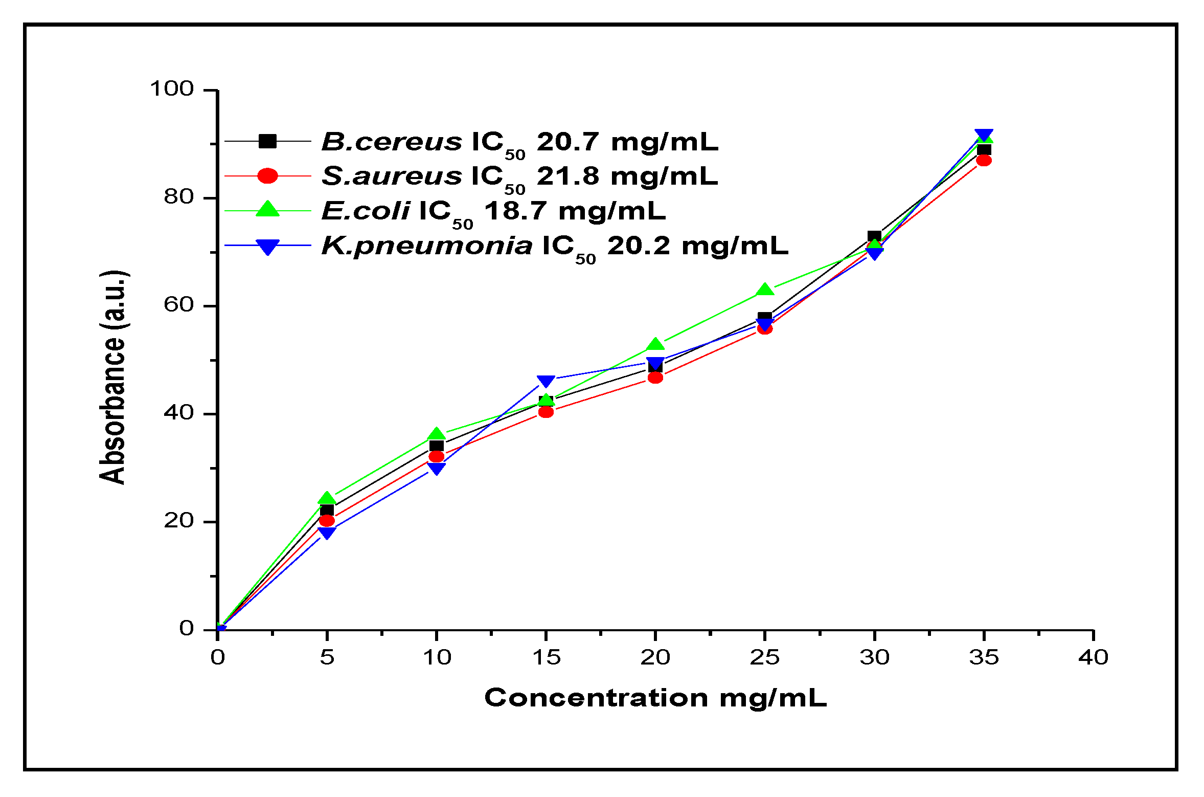
3.11. Antimicrobial Activity of Okra Extract

The present study assessed the antimicrobial activity of okra extract. Okra extract exhibited microbicidal activity against multiple pathogenic micro-organisms (Figure 9), indicating its broad-spectrum nature. The IC50 values obtained were 21.8 mg/mL for S. aureus, 18.7 mg/mL for E. coli, 20.7 mg/mL for B. cereus, and 20.2 mg/mL for K. pneumoniae.

4. Discussion

Okra is an edible vegetable crop that is commonly consumed and readily available in local markets. The present study evaluated the nutritional composition and the antimicrobial, antioxidant, and cytotoxic potential of okra pods. The nutritional composition of fresh okra is 90% water, 2% protein, and fiber, and it is rich in fat, pectin, hemicellulose, alpha-cellulose, and lignin. Okra is also rich in vitamins and minerals, such as calcium, iron, magnesium, phosphorus, potassium, and zinc [1]. The okra analyzed in the present study had significant dietary fiber, moisture, carbohydrate, and protein content. The calorific value was 51.258 kcal/100 g. The results of our study indicate that this plant species is potentially as nutritious as other species.
Spineless and dwarf long green varieties have higher protein contents than that of okra [29]. Okra pods were determined to have a lower calorific value than quinoa from Brazil and Chile because of their low carbohydrate, fat, and protein contents [30]. The moisture, fiber, protein, and fat contents of some Amaranthus species are similar to those of okra pods. These nutrient fractions have several benefits for human health. Insoluble dietary fiber contributes to the normal function of the intestinal tract and the prevention of gastrointestinal disorders. Gut micro-organisms can utilize soluble dietary fiber and regulate lipid metabolism in humans, contributing to the hypocholesterolemic properties of dietary fiber. Okra also has hypolipidemic properties, which potentially make it effective in controlling genetic metabolic disorders, including hyperlipidemia [31]. Because okra has higher crude fiber content than quinoa and Amaranthus, it could be a potential alternative to other nutrient sources in the daily diet. Micronutrients, including zinc, iron, copper, magnesium, calcium, sodium, potassium, and manganese, which play important roles in human diet and health, were also detected in okra. A comparative study showed proximate composition values of 8.1–8.9%, 8.4–9.0%, 14.3–15.3%, 1.4–2.1%, 16.9–18.2%, and 47.1–49.4% for moisture, ash, crude protein, crude fat, crude fiber, and carbohydrate content, respectively, and the mineral content values were 7.6–8.7 mg/100 g, 35.7–41.2 mg/100 g, 26.5–28.1 mg/100 g, 93.2–95.8 mg/100 g, 1.6–1.8 mg/100 g, and 5.2–5.7 mg/100 g for sodium, magnesium, potassium, calcium, iron, and zinc, respectively [32].
Phenolic compounds are considered major bioactive molecules in okra pods, as reported previously [19,33]. In the present study, total phenolic content (gallic acid equivalents) ranged from 288.2 to 3426.2 mg/100 g. The reducing sugars in okra reduce Folin–Ciocalteu reagent, resulting in exaggerated values of total phenolic content [27]. The plant matric flavonoids, including quercetin and kaempferol, are attached to sugar moieties. These flavonoid glycosides have many broad-spectrum biological activities, including antimicrobial, antioxidant, and antiproliferative properties [34]. Other compounds estimated in okra include chlorophyll and carotenoids, the values of which were higher than those in other okra genotypes, such as Lassithi, Pylaea, and Veloudo. Though chlorophyll is not a nutritional compound, it is responsible for the green color of vegetables, indicates senescence to consumers through oxidation effects, and contributes to the healing of wounds and inflammation [27]. The obtained values of chlorophyll a and b were equivalent to those reported in other studies. Okra is rich in vitamin E and tocopherols, which were estimated in our study using HPLC. Tocopherols are natural antioxidants, and vitamin E is an important component of nutrition. Four types of tocopherols are recognized, α, β, Y, and δ; the most important of which is α-tocopherol, as it reduces fat adhesion to the cell wall and reduces the amount of cholesterol in the blood by capturing peroxide radicals in cell tissues [13]. The GC–MS analysis showed the presence of 35 compounds in okra pods. The main constituents were (E)-anethole (6.7%), limonene (6.65%), β-caryophyllene (5.2%), decanal (4.45%), and carvone (4.35%). The main constituents found in okra pods have been studied previously for their antimicrobial and cytotoxic activities. Anethole has been analyzed for its antimicrobial, antifungal, anti-inflammatory, mutagenic, and cytotoxic activities [35,36]. Limonene exhibits antibacterial and cytotoxic activities [37]. Similarly, β-caryophyllene also possesses antibacterial, antioxidant, anti-inflammatory, and anticarcinogenic activities [36].
Among nutritional components, phenolic and bioactive compounds are responsible for the biological activities of okra. Okra has also been reported as a remedy for diabetic patients. Around 5% of the global population suffers from diabetes mellitus; however, all therapies have secondary effects. The disease is caused by faulty communication between insulin action and its secretion. This results in high blood glucose levels that damage blood vessels and impair body functions over time. The progressive weakening of body cells is a hallmark of the disease. Type 2 diabetes is a metabolic disorder in which the affected person is insulin-resistant and requires a combination of medications for dyslipidemia and arterial hypertension [38]. A conventional approach to treating diabetes mellitus or type 2 diabetes is reducing postprandial glucose levels. This can be achieved by slowing glucose absorption by targeting the activity of hydrolyzing enzymes associated with carbohydrates. Hydrolyzing enzymes, including α-amylase and α-glucosidase, naturally reside in the brush borders of the small intestine. The enzymes are required to absorb and break down oligosaccharides, polysaccharides, and disaccharides into monosaccharides [8,39,40]. Okra contains polysaccharides, which have several roles [35]. Okra exhibited inhibitory activity against α-amylase (IC50 = 120 µg/mL) and α-glucosidase (IC50 = 115 µg/mL). The high inhibitory potential of okra might be because of the synergic effect of its polysaccharides and flavonoids. This finding contradicts the results of Yuang et al., who attributed the high inhibitory potential of okra only to polysaccharides [35]. Flavonoids inhibit glucose transporters and consequently inhibit α-amylase and α-glucosidase activities. A recent study reported that the inhibitory potential of okra is associated with the units of hydroxyl groups on the flavonoid backbone’s B ring. An interlinkage is formed between the hydrogen bonds of the hydroxyl group of ring A at positions R6 and R7 and the polyphenol ligands of ring B at positions R40 and R50 and attachment sites of the catalytic portion. The interlinkage is also formed by the conjugated π-system, which maintains the interaction between active sites [39].
The antioxidant potential was determined by the widely used DPPH method. The antioxidant potential of okra is due to the presence of vitamin E and tocopherols. Tocopherols reduce oxidation and work as natural antioxidants and prevent low-density lipoprotein (LDL) oxidation in coronary arteries, which leads to atherosclerosis and heart attacks; thus, tocopherols have protective effects against heart attacks. Tocopherols protect cells from the effects of free radicals [1,14]. The phenolic content in okra is responsible for its antioxidant properties. Many studies have correlated total phenolic content with antioxidant potential in okra pods, as phenolic compounds have hydroxyl groups that donate protons to free radicals to scavenge them; DPPH values of EC50 = 1.03 mg/mL were previously reported [28]. In other studies, okra extract prepared in methanol, ethyl acetate, and n-hexane showed varying DPPH antioxidant activity, with IC50 values of 35.21 µg/mL, 181.09 µg/mL, and 104.06 µg/mL, respectively. This variation is due to the presence of active compounds such as quercetin, catechin, and vitamin E, which are more soluble in methanol than other solvent systems [27].
Okra showed potent antimicrobial activity against common pathogenic micro-organisms that frequently cause nosocomial infections: S. aureus, E. coli, B. cereus, and K. pneumoniae. Several researchers have shown the antimicrobial activity of okra against different pathogenic strains. One of the studies showed antimicrobial activity in both lyophilized and freshwater extracts of okra pods against R. opacus, Mycobacterium sp., M. aurum, S. aureus, E. coli, and Xanthobacter Py2; these effects were attributed to the presence of lipids, namely, palmitic and stearic acids [41]. Seed extracts exhibited antimicrobial properties against Listeria monocytogenes, Salmonella enteritidis, and S. typhimurium [42].
Cancer is one of the global leading causes of death. Despite advances in drug development, there is a need to develop new plant-derived medicines. Such plant-derived agents include vinca alkaloids and taxanes [43,44]. Flavonoids and phenols in okra flowers have antitumor effects on colorectal cancer cells both in vitro and in vivo, antiproliferative effects, and strong antioxidant potency. Flavonoids induced the activation of p53 and suppressed mitochondrial functions in colorectal tumor cells, resulting in apoptosis and inhibiting autophagy [45]. The anticancer effect of okra extracts was studied in vitro in several cell lines. One flavonoid in okra is Hyperin, also known as quercetin-3-O-β-D-galactoside. It is reported to have anticancer potential in gastric cancer cells (CHI) by producing antiproliferative, antimigratory, and anti-invasive effects and results in apoptosis by blocking the Wnt/β-catenin signal pathway [46]. Flavonoids in okra extracts were observed to exert cytotoxic effects in breast cancer cells (MCF-7), hepatoma cells (HepG2), and human cervical cancer (HeLa) cells in a dose-dependent manner [47]. Lectins from okra caused caspase-mediated downstream signaling in MCF7 cells and normal fibroblasts (CCD-1059 SK) 42 [48]. In the present study, the okra extract was observed to exert anticancer effects against the HepG2 cell line in a dose-dependent manner. When treated with okra extract, the cells underwent morphological changes and apoptosis and contained intracellular ROS.

5. Conclusions

Okra (Abelmoschus esculentus) is high in fiber, and our investigations revealed that it also contains significant quantities of proteins and carbohydrates, as well as minerals, such as Ca, Mg, Cu, Zn, Fe, K, Na, and Mn. Our results indicate that the aqueous extract of okra exhibited potent antimicrobial activity against common bacterial strains that are responsible for nosocomial infections. The antioxidant activity measured in the DPPH assay demonstrated promising results, and morphological changes and ROS generation in HepG2 cells suggest potential anticancer activity. These biological activities are due to the presence of tocopherols and other bioactive compounds detected in the aqueous extract of okra. Therefore, on the basis of key findings of this research, the aforementioned vegetable is effective as a nutritional supplement for cancer patients. However, before its proper application as an additional supplement in the treatment of cancer patients, further in vivo investigations are required.

Author Contributions

Conceptualization, S.K. and M.S.; methodology, S.M., Z.R. and A.B.; software, I.A. and A.H.; validation, Y.A., M.Y.A. and Z.R.; formal analysis, Z.R. and A.B.; investigation, S.K. and A.S.; resources, A.G.A.; data curation, M.A.; writing—original draft preparation, M.S. and S.K.; writing—review and editing, A.S. and I.A.; visualization, S.K. and Y.A.; supervision, S.K.; project administration, A.B.; funding acquisition, M.Y.A. All authors have read and agreed to the published version of the manuscript.

Funding

This research was funded by the Ministry of Education in KSA, grant number IFP-KKU-2020/14.

Institutional Review Board Statement

Not Applicable.

Informed Consent Statement

Not applicable.

Data Availability Statement

Not applicable.

Acknowledgments

The authors extend their appreciation to the Ministry of Education in KSA for funding this research through project number IFP-KKU-2020/14.

Conflicts of Interest

The authors declare no conflict of interest.

References

  1. Alshahrani, M.Y.; Rafi, Z.; Alabdallah, N.M.; Shoaib, A.; Ahmad, I.; Asiri, M.; Zaman, G.S.; Wahab, S.; Saeed, M.; Khan, S. A Comparative Antibacterial, Antioxidant, and Antineoplastic Potential of Rauwolfia serpentina (L.) Leaf Extract with Its Biologically Synthesized Gold Nanoparticles (R-AuNPs). Plants 2021, 10, 2278. [Google Scholar] [CrossRef]
  2. Kumar, S.; Dagnoko, S.; Haougui, A.; Ratnadass, A.; Pasternak, N.; Kouame, C. Okra (Abelmoschus spp.) in West and Central Africa: Potential and progress on its improvement. Afr. J. Agric. Res. 2010, 5, 3590–3598. [Google Scholar]
  3. Al-Shawi, A.A.; Hameed, M.F.; Hussein, K.A.; Thawini, H.K. Review on the “Biological Applications of Okra Polysaccharides and Prospective Research”. Future J. Pharm. Sci. 2021, 7, 102. [Google Scholar] [CrossRef]
  4. Bhat, U.R.; Tharanathan, R.N. Functional properties of okra (Hibiscus esculentus) mucilage. Starch-Stärke 1987, 39, 165–167. [Google Scholar] [CrossRef]
  5. Somasundaram, G.; Rajan, J. Effectual Role of Abelmoschus esculentus (Okra) extract on morphology, microbial and photocatalytic activities of CdO Tetrahedral Clogs. J. Inorg. Organomet. Polym. Mater. 2018, 28, 152–167. [Google Scholar] [CrossRef]
  6. Prommakool, A.; Sajjaanantakul, T.; Janjarasskul, T.; Krochta, J.M. Whey protein-okra polysaccharide fraction blend edible films: Tensile properties, water vapor permeability and oxygen permeability. J. Sci. Food Agric. 2011, 91, 362–369. [Google Scholar] [CrossRef]
  7. Lee, C.S.; Chong, M.F.; Robinson, J.; Binner, E. Optimisation of extraction and sludge dewatering efficiencies of bio-flocculants extracted from Abelmoschus esculentus (okra). J. Environ. Manag. 2015, 157, 320–325. [Google Scholar] [CrossRef] [Green Version]
  8. Sabitha, V.; Panneerselvam, K.; Ramachandran, S. In vitro α-glucosidase and α-amylase enzyme inhibitory effects in aqueous extracts of Abelmoscus esculentus (L.) Moench. Asian Pac. J. Trop. Biomed. 2012, 2, S162–S164. [Google Scholar] [CrossRef]
  9. Sabitha, V.; Ramachandran, S.; Naveen, K.R.; Panneerselvam, K. Antidiabetic and antihyperlipidemic potential of Abelmoschus esculentus (L.) Moench. in streptozotocin-induced diabetic rats. J. Pharm. Bioallied. Sci. 2011, 3, 397–402. [Google Scholar]
  10. Durazzo, A.; Lucarini, M.; Novellino, E.; Souto, E.B.; Daliu, P.; Santini, A. Abelmoschus esculentus (L.): Bioactive Components’ Beneficial Properties-Focused on Antidiabetic Role-For Sustainable Health Applications. Molecules 2018, 24, 38. [Google Scholar] [CrossRef] [Green Version]
  11. Whistler, R.L.; Conrad, H. 2-O-(D-Galactopyranosyluronic Acid)-L-rhamnose from Okra Mucilage1. J. Am. Chem. Soc. 1954, 76, 3544–3546. [Google Scholar] [CrossRef]
  12. Ameena, K.; Dilip, C.; Saraswathi, R.; Krishnan, P.; Sankar, C.; Simi, S. Isolation of the mucilages from Hibiscus rosasinensis linn. and Okra (Abelmoschus esculentus linn.) and studies of the binding effects of the mucilages. Asian Pac. J. Trop. Med. 2010, 3, 539–543. [Google Scholar] [CrossRef] [Green Version]
  13. Ogungbenle, H.; Omosola, S. The comparative assessment of nutritive values of dry Nigerian okra (Abelmoschus esculentus) fruit and oil. Int. J. Food Sci. Nutr. Eng. 2015, 5, 8–14. [Google Scholar]
  14. Al-Kanani, E.A.S.; Al-Hilifi, S.A.H.; Al-Kareem, A.H. The nutritional composition and vitamin E of three Iraqi okra (Abelmoschus esculentus L.) seeds oil. In IOP Conference Series: Earth and Environmental Science; IOP Publishing: Bristol, UK, 2019. [Google Scholar]
  15. Kaur, G.; Singh, D.; Brar, V. Bioadhesive okra polymer based buccal patches as platform for controlled drug delivery. Int. J. Biol. Macromol. 2014, 70, 408–419. [Google Scholar] [CrossRef]
  16. Freitas, T.; Oliveira, V.; de Souza, M.; Geraldino, H.; Almeida, V.; Fávaro, S.; Garcia, J. Optimization of coagulation-flocculation process for treatment of industrial textile wastewater using okra (A. esculentus) mucilage as natural coagulant. Ind. Crops Prod. 2015, 76, 538–544. [Google Scholar] [CrossRef]
  17. Adamma, E.P.; Sani, S.M.; Israel, O.K.; Yusuf, Z.S. Proximate and anti-nutritional constituents of Abelmoschus esculentus grown in Fadaman Kubanni, Zaria, Kaduna State, Nigeria. J. Sci. Res. Rep. 2014, 3, 2015–2027. [Google Scholar] [CrossRef] [PubMed]
  18. Bessada, S.M.; Barreira, J.C.M.; Barros, L.; Ferreira, I.C.F.R.; Oliveira, M.B.P.P. Phenolic profile and antioxidant activity of Coleostephus myconis (L.) Rchb. f.: An underexploited and highly disseminated species. Ind. Crops Prod. 2016, 89, 45–51. [Google Scholar] [CrossRef] [Green Version]
  19. Nagata, M.; Yamashita, I. Simple method for simultaneous determination of chlorophyll and carotenoids in tomato fruit. Nippon Shokuhin Kogyo Gakkaishi 1992, 39, 925–928. [Google Scholar] [CrossRef] [Green Version]
  20. Mounir, S.; Ghandour, A.; Téllez-Pérez, C.; Aly, A.A.; Mujumdar, A.S.; Allaf, K. Phytochemicals, chlorophyll pigments, antioxidant activity, relative expansion ratio, and microstructure of dried okra pods: Swell-drying by instant controlled pressure drop versus conventional shade drying. Dry. Technol. 2020, 39, 2145–2159. [Google Scholar] [CrossRef]
  21. Blois, M.S. Antioxidant determinations by the use of a stable free radical. Nature 1958, 181, 1199–1200. [Google Scholar] [CrossRef]
  22. Mansoor, S.; Khan, I.; Fatima, J.; Saeed, M.; Mustafa, H. Anti-bacterial, anti-oxidant and cytotoxicity of aqueous and organic extracts of Ricinus communis. Afr. J. Microbiol. Res. 2016, 10, 260–270. [Google Scholar]
  23. Lengsfeld, C.; Titgemeyer, F.; Faller, G.; Hensel, A. Glycosylated compounds from okra inhibit adhesion of Helicobacter pylori to human gastric mucosa. J. Agric. Food Chem. 2004, 52, 1495–1503. [Google Scholar] [CrossRef] [PubMed]
  24. Iram, S.; Zahera, M.; Khan, S.; Khan, I.; Syed, A.; Ansary, A.A.; Ameen, F.; Shair, O.H.; Khan, M.S. Gold nanoconjugates reinforce the potency of conjugated cisplatin and doxorubicin. Colloids Surf. B Biointerfaces 2017, 160, 254–264. [Google Scholar] [CrossRef] [PubMed]
  25. Pakos, E.E.; Nearchou, A.D.; Grimer, R.J.; Koumoullis, H.D.; Abudu, A.; Bramer, J.A.; Jeys, L.M.; Franchi, A.; Scoccianti, G.; Campanacci, D.; et al. Prognostic factors and outcomes for osteosarcoma: An international collaboration. Eur. J. Cancer 2009, 45, 2367–2375. [Google Scholar] [CrossRef] [PubMed]
  26. Hasan, A.; Haque, E.; Hameed, R.; Maier, P.N.; Irfan, S.; Kamil, M.; Nazir, A.; Mir, S.S. Hsp90 inhibitor gedunin causes apoptosis in A549 lung cancer cells by disrupting Hsp90:Beclin-1:Bcl-2 interaction and downregulating autophagy. Life Sci. 2020, 256, 118000. [Google Scholar] [CrossRef]
  27. Wahyuningsih, S.P.A.; Savira, N.I.I.; Anggraini, D.W.; Winarni, D.; Suhargo, L.; Kusuma, B.W.A.; Nindyasari, F.; Setianingsih, N.; Mwendolwa, A.A. Antioxidant and Nephroprotective Effects of Okra Pods Extract (Abelmoschus esculentus L.) against Lead Acetate-Induced Toxicity in Mice. Scientifica 2020, 2020, 4237205. [Google Scholar] [CrossRef]
  28. Romdhane, M.H.; Al-Mohammadi, A.R.; Mahgoub, S.; Abdel-Shafi, S.; Askar, E.; Ghaly, M.F.; Taha, M.A.; El-Gazzar, N. Chemical Composition, Nutritional Value, and Biological Evaluation of Tunisian Okra Pods (Abelmoschus esculentus L. Moench). Molecules 2020, 25, 4739. [Google Scholar] [CrossRef]
  29. Ilodibia, C.V.; Achebe, U.A.; Chiafor, C. Nutrient characteristics assessment of two variants of okra (Abelmoschus esculentus L. Moench.) found in Anambra State, Nigeria. Arch. Agric. Environ. Sci. 2017, 2, 298–300. [Google Scholar] [CrossRef]
  30. Miranda, M.; Vega-Gálvez, A.; López, J.; Parada, G.; Sanders, M.; Aranda, M.; Uribe, E.; Di Scala, K. Impact of air-drying temperature on nutritional properties, total phenolic content and antioxidant capacity of quinoa seeds (Chenopodium quinoa Willd.). Ind. Crops Prod. 2010, 32, 258–263. [Google Scholar] [CrossRef]
  31. Ciudad-Mulero, M.; Fernández-Ruiz, V.; Matallana-González, M.C.; Morales, P. Dietary fiber sources and human benefits: The case study of cereal and pseudocereals. Adv. Food Nutr. Res. 2019, 90, 83–134. [Google Scholar]
  32. Omoniyi, S.A.; Muhammad, A.M.; Ayuba, R. Nutrient composition and anti-nutritional properties of okra (Abelmoschus esculentus) calyx flour. Nutr. Food Sci. 2021, 51, 30–40. [Google Scholar] [CrossRef]
  33. Hussain, M.S.; Fareed, S.; Saba Ansari, M.; Rahman, A.; Ahmad, I.Z.; Saeed, M. Current approaches toward production of secondary plant metabolites. J. Pharm. Bioallied. Sci. 2012, 4, 10–20. [Google Scholar] [CrossRef] [PubMed]
  34. Yang, B.; Liu, H.; Yang, J.; Gupta, V.K.; Jiang, Y. New insights on bioactivities and biosynthesis of flavonoid glycosides. Trends Food Sci. Technol. 2018, 79, 116–124. [Google Scholar] [CrossRef]
  35. Fujita, K.I.; Ishikura, T.; Jono, Y.; Yamaguchi, Y.; Ogita, A.; Kubo, I.; Tanaka, T. Anethole potentiates dodecanol’s fungicidal activity by reducing PDR5 expression in budding yeast. Biochim. Biophys. Acta Gen. Subj. 2017, 1861, 477–484. [Google Scholar] [CrossRef] [PubMed] [Green Version]
  36. Yoo, H.J.; Jwa, S.K. Inhibitory effects of beta-caryophyllene on Streptococcus mutans biofilm. Arch. Oral Biol. 2018, 88, 42–46. [Google Scholar] [CrossRef]
  37. Vieira, A.J.; Beserra, F.P.; Souza, M.; Totti, B.; Rozza, A.L. Limonene: Aroma of innovation in health and disease. Chem. Interact. 2018, 283, 97–106. [Google Scholar] [CrossRef] [Green Version]
  38. Tan, S.Y.; Wong, J.L.M.; Sim, Y.J.; Wong, S.S.; Elhassan, S.A.M.; Tan, S.H.; Lim, G.P.L.; Tay, N.W.R.; Annan, N.C.; Bhattamisra, S.K.; et al. Type 1 and 2 diabetes mellitus: A review on current treatment approach and gene therapy as potential intervention. Diabetes Metab. Syndr. 2019, 13, 364–372. [Google Scholar] [CrossRef]
  39. Hussain, T.; Tan, B.; Murtaza, G.; Liu, G.; Rahu, N.; Kalhoro, M.S.; Kalhoro, D.H.; Adebowale, T.; Mazhar, M.U.; Rehman, Z.U.; et al. Flavonoids and type 2 diabetes: Evidence of efficacy in clinical and animal studies and delivery strategies to enhance their therapeutic efficacy. Pharmacol. Res. 2020, 152, 104629. [Google Scholar] [CrossRef]
  40. Saeed, M.; Shoaib, A.; Tasleem, M.; Alabdallah, N.; Alam, J.; Asmar, Z.; Jamal, Q.; Bardakci, F.; Alqahtani, S.; Ansari, I.; et al. Assessment of Antidiabetic Activity of the Shikonin by Allosteric Inhibition of Protein-Tyrosine Phosphatase 1B (PTP1B) Using State of Art: An In Silico and In Vitro Tactics. Molecules 2021, 26, 3996. [Google Scholar] [CrossRef]
  41. de Carvalho, C.C.; Cruz, P.A.; da Fonseca, M.M.R.; Xavier-Filho, L. Antibacterial properties of the extract of Abelmoschus esculentus. Biotechnol. Bioprocess Eng. 2011, 16, 971–977. [Google Scholar] [CrossRef]
  42. Petropoulos, S.; Fernandes, Â.; Barros, L.; Ciric, A.; Sokovic, M.; Ferreira, I.C. The chemical composition, nutritional value and antimicrobial properties of Abelmoschus esculentus seeds. Food Funct. 2017, 8, 4733–4743. [Google Scholar] [CrossRef]
  43. Elkhalifa, A.E.O.; Alshammari, E.; Adnan, M.; Alcantara, J.C.C.; Awadelkareem, A.M.; Eltoum, N.E.; Mehmood, K.; Panda, B.P.; Ashraf, S.A. Okra (Abelmoschus Esculentus) as a Potential Dietary Medicine with Nutraceutical Importance for Sustainable Health Applications. Molecules 2021, 26, 696. [Google Scholar] [CrossRef] [PubMed]
  44. Saeed, M.; Shoaib, A.; Kandimalla, R.; Javed, S.; Almatroudi, A.; Gupta, R.; Aqil, F. Microbe-based therapies for colorectal cancer: Advantages and limitations. Semin. Cancer Biol. 2021. [Google Scholar] [CrossRef] [PubMed]
  45. Deng, Y.; Li, S.; Wang, M.; Chen, X.; Tian, L.; Wang, L.; Yang, W.; Chen, L.; He, F.; Yin, W. Flavonoid-rich extracts from okra flowers exert antitumor activity in colorectal cancer through induction of mitochondrial dysfunction-associated apoptosis, senescence and autophagy. Food Funct. 2020, 11, 10448–10466. [Google Scholar] [CrossRef]
  46. Ping, M.H. Hyperin Controls the Development and Therapy of Gastric Cancer via Regulating Wnt/beta-Catenin Signaling. Cancer Manag. Res. 2020, 12, 11773–11782. [Google Scholar] [CrossRef]
  47. Chaemsawang, W.; Prasongchean, W.; Papadopoulos, K.I.; Ritthidej, G.; Sukrong, S.; Wattanaarsakit, P. The Effect of Okra (Abelmoschus esculentus (L.) Moench) Seed Extract on Human Cancer Cell Lines Delivered in Its Native Form and Loaded in Polymeric Micelles. Int. J. Biomater. 2019, 2019, 9404383. [Google Scholar] [CrossRef] [Green Version]
  48. de Oliveira Figueiroa, E.; Da Cunha, C.R.A.; Albuquerque, P.B.; De Paula, R.A.; Aranda-Souza, M.A.; Alves, M.S.; Zagmignan, A.; Carneiro-Da-Cunha, M.G.; Da Silva, L.C.N.; Correia, M.T.D.S. Lectin-Carbohydrate Interactions: Implications for the Development of New Anticancer Agents. Curr. Med. Chem. 2017, 24, 3667–3680. [Google Scholar] [CrossRef] [PubMed]
Figure 1. HPLC chromatogram representing the tocopherol content in the aqueous extract of okra pods.
Processes 10 00183 g001
Figure 2. Reducing sugars (fructose, glucose, and saccharose as Peaks 1, 2, and 3, respectively) detected in the aqueous extract of okra pods by high-performance liquid chromatography.
Processes 10 00183 g002
Figure 3. GC–MS chromatogram representing the bioactive constituents of aqueous extract of okra pods.
Processes 10 00183 g003
Figure 4. Chemical structure of the main five constituents identified in Okra extract. (1) carvone; (2) β-caryophyllene; (3) decanal; (4) (E)-anethole; and (5) limonene.
Processes 10 00183 g004
Figure 5. The dose-dependent cytotoxic effect of okra extract against HepG2 cells. All tests were performed in triplicate, and data are expressed as mean ± standard deviation.
Processes 10 00183 g005
Figure 6. Images depicting the morphological changes in (A) untreated control cells and (B) HepG2 cells treated with okra extract at IC50 concentration at 20X magnification under phase-contrast microscopy.
Processes 10 00183 g006
Figure 7. Images depicting the apoptotic effect of okra extract on DAPI-stained HepG2 cells at 20X magnification under a phase-contrast microscope. (A) Untreated HepG2 cells stained with DAPI; (B) okra extract-treated HepG2 cells; (C) fluorescence intensity graph.
Processes 10 00183 g007
Figure 8. Images depict DCFDA-stained HepG2 cells after 48 h at 20X magnification under a phase-contrast microscope. (A) DCFDA experimental control; (B) okra extract-treated HepG2 cells; (C) fluorescence intensity graphs.
Processes 10 00183 g008
Figure 9. Graph showing antibacterial potential of okra extract against S. aureus (21.8 mg/mL), E. coli (18.7 mg/mL), B. cereus (20.7 mg/mL), and K. pneumoniae (20.2 mg/mL). The experiment was performed in triplicate, and the results are expressed as mean ± standard deviation.
Processes 10 00183 g009
Table 1. Proximate nutrient content of okra pods.
S.No.Nutrient CompositionContent
(g/100 g)
1Moisture84.67–87.65
2Total ash1.514–1.197
3Crude protein2.367–3.41
4Total fat0.066–0.1
5Total carbohydrate7.857–8.261
6Dietary fiber6.781–8.314
7Energy (kcal/100 g)51.258
Table 2. Proximate mineral content of okra pods.
S.No.Mineral CompositionContent
(mg/100 g)
1Iron1.5–1.7
2Copper0.48–0.5
3Manganese0.12–0.15
4Zinc1.8–2.2
5Magnesium100–104
6Calcium260–270
7Sodium120–130
8Potassium330–350
Table 3. Composition of sugars in okra pods.
S.No.Soluble SugarsContent mg/100 g
1Fructose33–35
2Glucose28–32
3Sucrose105–115
4Total sugars166–182
Table 4. GC–MS identification of phytoconstituents in Okra extract.
CompoundsAbundance/
Values
Chemical
Formula
Molecular Weight (g/mol)
4-Hydroxy-d-methyl-2-pentanone834; 3.45
α-Pinene939; 1.85C10H16136.23
(E)-2-Heptanal959; 0.67C7H12O112.17
Benzaldehyde961; 0.47C7H6O106.12
1-Octn-3-ol978; 2.95C8H16O128.21
β-Pinene980; 0.85C10H16136.23
6-Methyl-5-hepten-2-one984; 2.32C8H14O126.20
Limonene1030; 6.65C10H16136.24
(E)-3-Octen-1-ol1059; 3.94C8H16O128.21
(E)-2-Octen-1-ol1068; 4.22C8H16O128.21
1-Octanol1072; 3.85C8H18O130.23
n-Undecane1098; 0.55C11H24156.31
Linalool1098; 1.4C10H18O154.25
Nonanal1102; 3.25C9H18O142.23
cis-ρ-Menth-2-en-1-ol1120; 1.45C10H18O154.24
Camphor1142; 3.23C10H16O152.23
Menthol1172; 1.3C10H20O156.27
α-Terpineol1184; 0.75C10H18O154.25
cis-Dihydrocarvone1193; 0.42C10H16O152.23
n-Dodecane1197; 2.35C12H26170.33
Decanal1204; 4.45C10H20O156.2
β-Cyclocitral1219; 1.55C10H16O152.23
Exo-fenchyl acetate1228; 2.05C12H20O2196.29
Carvone1241; 4.35C10H14O150.22
(E)-Anethole1283; 6.7C10H12O148.20
n-Tridecane1297; 1.85C13H28184.37
α-Cubebene1349; 1.55C15H24204.35
Eugenol1356; 4.05C10H12O2164.2
n-Tetradecane1395; 1.95C14H30198.39
β-Caryophyllene1017; 5.2C15H24204.36
2,5-Dimethoxy-ρ-cymene1419; 1.43C12H18O2194.27
Δ8,9-Dehydro-4-hydroxythymol dimethyl ether1442; 4.15C12H16O2192.26
Δ8,9-Dehydro-4-hydroxythymol1457; 2.48C10H12O2164.20
γ-Muurolene1476; 1.6C15H24204.35
3,4-Dehydro-β-ionone1781; 2.35C13H22O194.31
Class of CompoundsPercentage
Monoterpene hydrocarbons8.50
Oxygenated monoterpenes21.10
Sesquiterpene hydrocarbons10.50
Phenylpropanoids10.30
Apocarotenes3.70
Non-terpene derivatives37.53
Total identified (%)91.63
Publisher’s Note: MDPI stays neutral with regard to jurisdictional claims in published maps and institutional affiliations.

