Article

In Silico Molecular Docking Studies and Xanthine Oxidase Inhibitory Activity of Abies kawakamii Leaf Extract and Its Constituent


1
Agricultural Technology Research Institute, Hsinchu 300110, Taiwan
2
Program in Specialty Crops and Metabolomics, Academy of Circular Economy, National Chung Hsing University, Nantou 54071, Taiwan
3
School of Forestry and Resource Conservation, National Taiwan University, Taipei 10617, Taiwan
4
Institute of Environmental and Occupational Health Sciences, National Taiwan University, Taipei 10055, Taiwan
5
Instrumentation Center, College of Science, National Taiwan University, Taipei 10617, Taiwan
*
Author to whom correspondence should be addressed.
Pharmaceuticals 2026, 19(7), 1100; https://doi.org/10.3390/ph19071100
Submission received: 4 June 2026 / Revised: 5 July 2026 / Accepted: 14 July 2026 / Published: 17 July 2026

Abstract

Gout is a metabolic disorder associated with abnormal purine metabolism and persistent hyperuricaemia, which promotes formation and deposition of monosodium urate crystal in joints, leading to acute arthritis and impaired quality of life. This study aimed to evaluate the xanthine oxidase inhibitory activity of ethanolic leaf extract of Abies kawakamii and its fractions. The protocol of bioassay-guided fractionation was employed to isolate active compounds from leaf extract. Maltol was identified as the major active compound from the ethyl acetate fraction (EAF). Maltol inhibited xanthine oxidase with IC50 values of 33.18 and 26.67 μg/mL against xanthine and hypoxanthine, respectively, significantly lower than the crude extract (152.04 and 136.57 μg/mL) and EAF (66.70 and 48.76 μg/mL). Enzyme kinetic analyses further showed that EAF inhibited xanthine oxidase through a competitive inhibition mechanism toward both substrates. Molecular docking analysis suggested that maltol may interact with the active-site region of xanthine oxidase, showing a binding affinity of −6.5 kcal/mol, slightly weaker than that of allopurinol, with no predicted hepatotoxicity based on in silico analysis. Our findings indicate that A. kawakamii leaf extract and its constituent, maltol, could be further explored as a natural approach for gout management.

1. Introduction

Gout is a disorder associated with abnormal purine metabolism caused by elevated uric acid levels (hyperuricemia), which promote monosodium urate crystals formation and deposition in joints [1]. The typical symptoms of acute gout are characterized by intense joint pain, accompanied by swelling and redness, thereby affecting patients’ quality of life. During purine catabolism, purines derived from dietary intake and endogenous metabolism are sequentially converted into hypoxanthine and xanthine through a series of enzymatic processes. Xanthine oxidase subsequently catalyzes the oxidation of hypoxanthine to xanthine and finally converts xanthine into uric acid [1,2,3,4,5]. Insulin resistance would increase the body weight and serum uric acid level. Gout can be classified into four clinical features, including asymptomatic hyperuricemia, acute gouty arthritis, chronic tophaceous gout, and intercritical gout. Modifications of the dietary and lifestyle of gout patients to reach the ideal weight, and both insulin resistance and serum uric acid levels could decrease [4,5,6,7]. In addition, allopurinol has been widely used as a xanthine oxidase inhibitor for controlling gout and hyperuricemia, while more recently developed medications include febuxostat, topiroxostat, and uricosuric agents such as probenecid. Certain anti-gout medications may be associated with adverse reactions; for example, febuxostat has been reported to induce diarrhea, elevation of liver enzymes, and nausea [8,9,10].
Many researchers have proven that plant natural products exhibited a lot of bioactivities, including antioxidant, antimelanogenic, antimicrobial, xanthine oxidase inhibitory, tyrosinase inhibitory, anti-tumor, anti-inflammatory, larvicidal, and antidepressant activities [11,12,13,14,15,16,17,18,19]. Researchers have increasingly investigated plant-derived natural products as potential sources of xanthine oxidase inhibitors [20,21,22,23,24,25]. The twig extract of Cinnamomum cassia demonstrated inhibitory activity against xanthine oxidase [26]. Extracts obtained from Tetracera scandens, Blumea balsamifera, Caesalpinia sappan, and Chrysanthemum sinense demonstrated inhibitory activity toward xanthine oxidase [27]. Phytic acid, which is widely distributed in cereals and nuts, can suppress uric acid formation using xanthine as the substrate, with an IC50 of nearly 30 mM [28].
The genus Abies (Pinaceae), Fir, includes 50–60 species is widely distributed in South Europe, North America and East Asia. Natural products from Fir have been used traditionally to support respiratory health, pulmonary diseases, colds, and rheumatic diseases antitumor activity against adenocarcinoma [29,30,31,32,33]. Abies kawakamii is known as Taiwanese Fir or Formosan Fir, is an evergreen coniferous tree and the endemic species in Taiwan. The genus Abies has a long history of ethnomedicinal use in traditional medicine. Its reported ethnomedicinal bioactivities include anti-inflammatory, analgesic, antitussive, antimicrobial, antioxidant, antidiabetic, etc., and have long histories of use in traditional medicine [30,31,33,34,35,36]. Its decoctions and extracts were traditionally used to treat rheumatism, joint inflammation, and symptoms associated with gout [30,31]. However, there is still very limited research on the bioactive effects of extracts from A. kawakamii, and no paper reported the xanthine oxidase inhibition potential of A. kawakamii leaf extract and its fractions.
This study investigated the xanthine oxidase inhibition potential of A. kawakamii leaf extract and its fractions. The phytochemical constituents present in leaf extract, active fractions, and active compound were isolated and identified by liquid chromatography and spectroscopies. An enzyme kinetic study and in silico molecular docking analyses were applied to elucidate the mechanism of active specimens against xanthine oxidase.

2. Results and Discussion

2.1. Xanthine Oxidase Inhibition Effects of A. kawakamii Leaf Extract and Its Fractions

A. kawakamii leaf extract was prepared by ambient solvent extraction and subsequently separated into four fractions (HXF, EAF, BUF, and HOF) through liquid–liquid partitioning. Table 1 summarizes the xanthine oxidase inhibitory activities of A. kawakamii leaf extract, its fractions, and allopurinol as the positive control at 100 μg/mL against hypoxanthine and xanthine substrates. Using hypoxanthine and xanthine as substrates, the leaf extract exhibited inhibition rates of 44.40% and 31.07%, respectively. Complete inhibition (100%) against both substrates was observed for allopurinol and the ethyl acetate fraction. The EAF showed inhibitory activities of 100% and 80.82% toward hypoxanthine and xanthine, respectively. BUF showed the lowest activity, whereas HXF displayed no detectable inhibition.
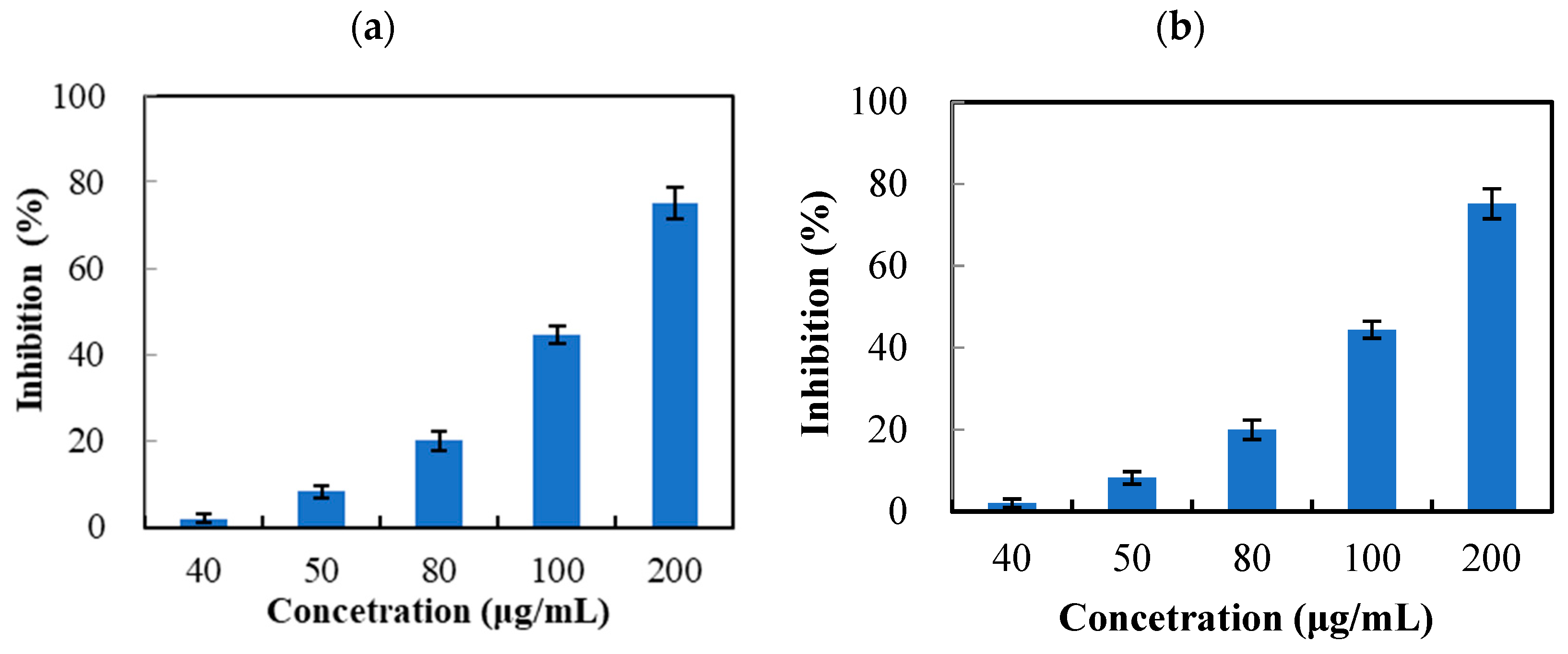
A. kawakamii leaf extract suppressed xanthine oxidase activity in a dose-dependent manner when hypoxanthine and xanthine were used as substrates (Figure 1), with the IC50 values of 136.57 and 152.04 μg/mL, respectively (Table 2). EAF showed the strongest inhibitory activity against xanthine oxidase, with the IC50 values of 48.76 μg/mL for hypoxanthine and 66.70 μg/mL for xanthine (Table 2). Results revealed that A. kawakamii leaf extract and EAF possess considerable xanthine oxidase inhibitory potential.
Abdulhafiz et al. reported Alocasia longiloba (Keladi Candik), traditional medicine in Malaysia, extracts were found to exhibit the high xanthine oxidase inhibitory activity, with IC50 values range of 42.71 to 51.32 µg/mL [25]. Xanthine oxidase inhibitory activities were reported in both aqueous and ethanolic extract of Pistacia integerrima leaves, with IC50 values of 85 and 60 μg/mL, respectively, when hypoxanthine served as the substrate. [37]. Xanthine oxidase inhibitory activities were observed in the bark and leaf extracts of Erythrina variegata, yielding IC50 values of 52.75 and 84.75 μg/mL, respectively, when xanthine served as the substrate. [38]. The present results demonstrated that A. kawakamii leaf extract and its EAF possessed the xanthine oxidase inhibitory activity. In addition, EAF showed significantly higher activity than the other tested specimens (p < 0.05).

2.2. Structural Characterization of Maltol

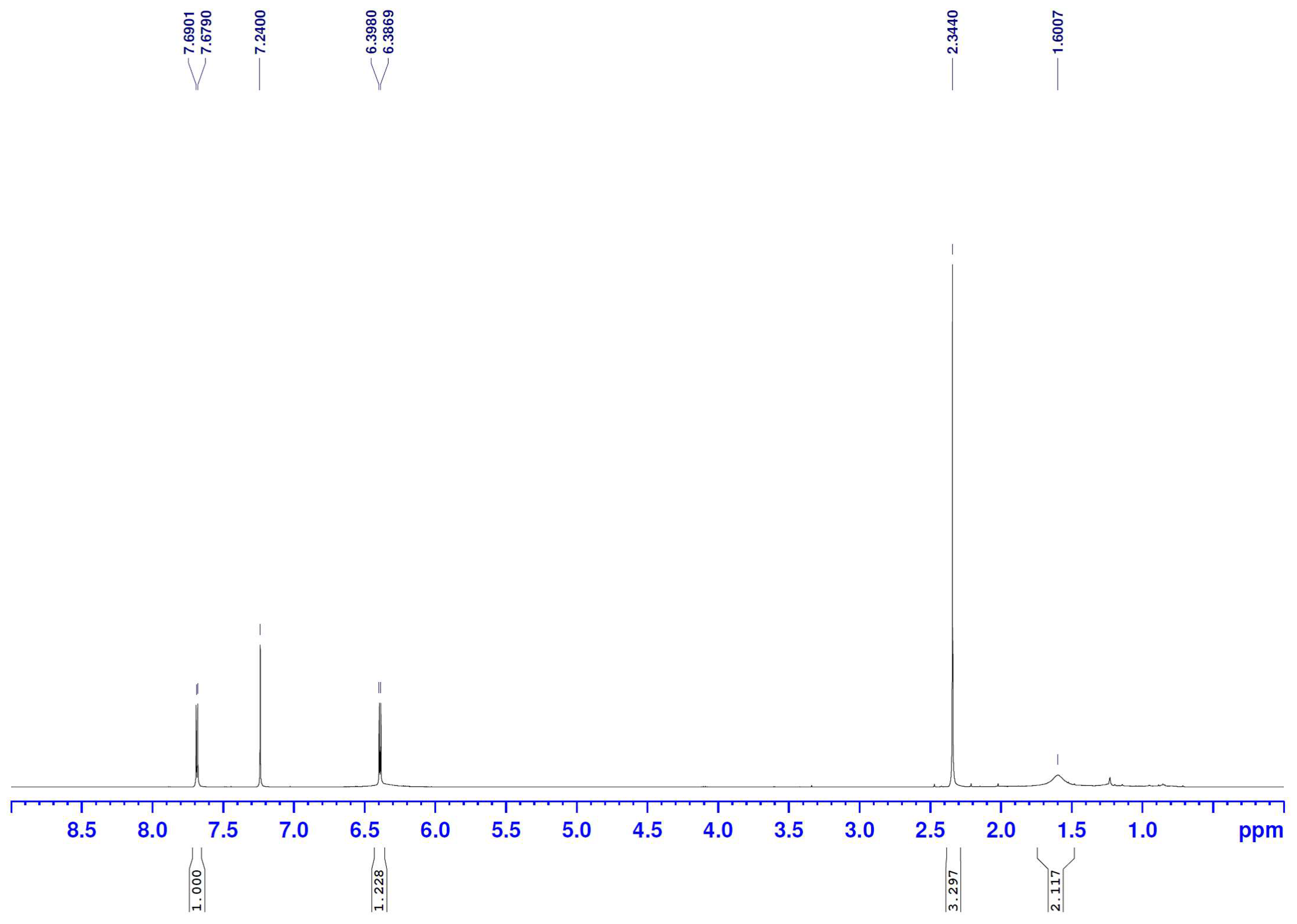
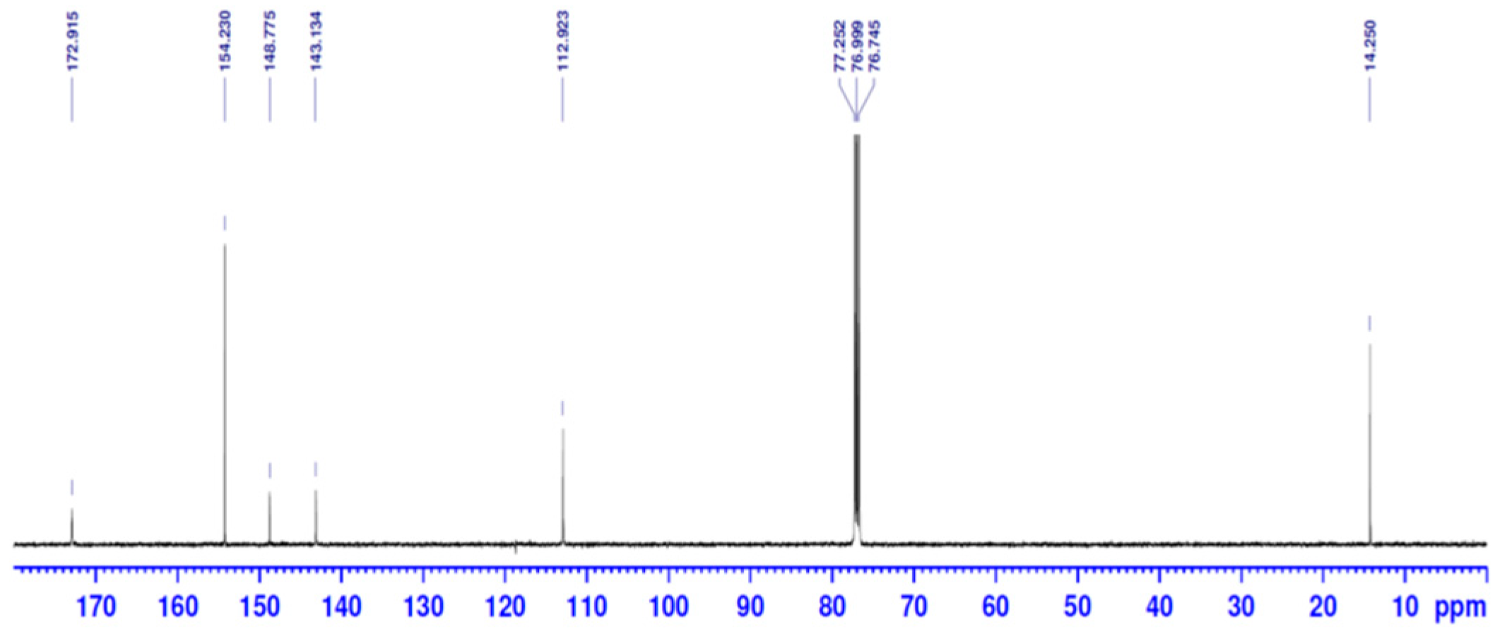
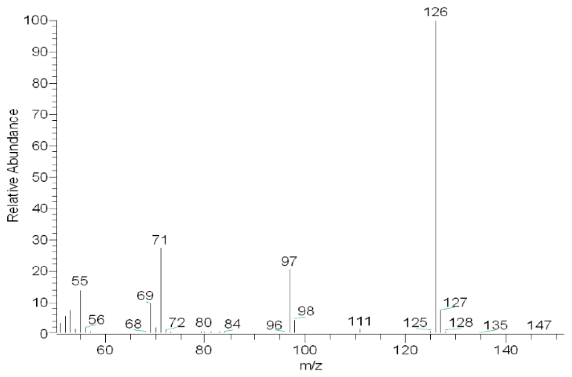
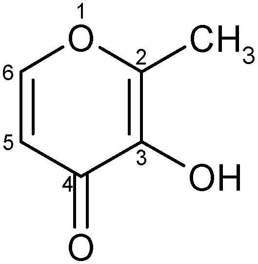
Compound M from EAF was characterized as maltol using NMR (Figure 2 and Figure 3) and MS spectroscopy (Figure 4). Table 3 presents the 1H and 13C NMR data of maltol. White crystal needle; mp: 161–163 °C; UV (MeOH) λmax (log ε): 275, and 322 nm; 1H NMR (CDCl3, 500 MHz) δ 7.69 (1H, d, J = 5.55 Hz), 6.39 (1H, d, J = 5.55 Hz), 2.34 (3H, s, C2-CH3). 13C NMR (CDCl3, 125 MHz) δc 172.9 (C-4), 154.2 (C-6), 148.8 (C-2), 143.1 (C-3), 112.9 (C-5). NMR spectra were in agreement with the literature [39,40]. Figure 4 was the mass spectrum of compound M. EI-MS m/z: 55 (14), 69 (9), 71 (27), 97 (20), 126 (M+, 100), molecular formula C6H6O3. Figure 5 shows the chemical structure of maltol (3-hydroxy-2methyl-4H-pyran-4-one).
Maltol was also isolated from the same genus, Abies sibirica (Siberian Fir), Abies pindrow, Abies fraseri, and Abies nebrodensis leaf extracts [40,41,42,43]. It is also found in other species and baked products. Maltol is a naturally occurring compound, with sweet, and caramel-like scents, resulting from the heterocyclic structure. The bioactivities of maltol include antimicrobial, antioxidant, ROS scavenging, anti-inflammatory, anticancer, etc. [42,44,45,46]. Maltol is used as a safe and reliable flavor enhancer, and is designated in the U.S. and Europe with the E number E636 and the CAS number 118-71-8 [42,44,45,46,47].

2.3. Xanthine Oxidase Inhibitory Activity and Enzyme Kinetic Study of Ethyl Acetate Fraction and Its Constituents

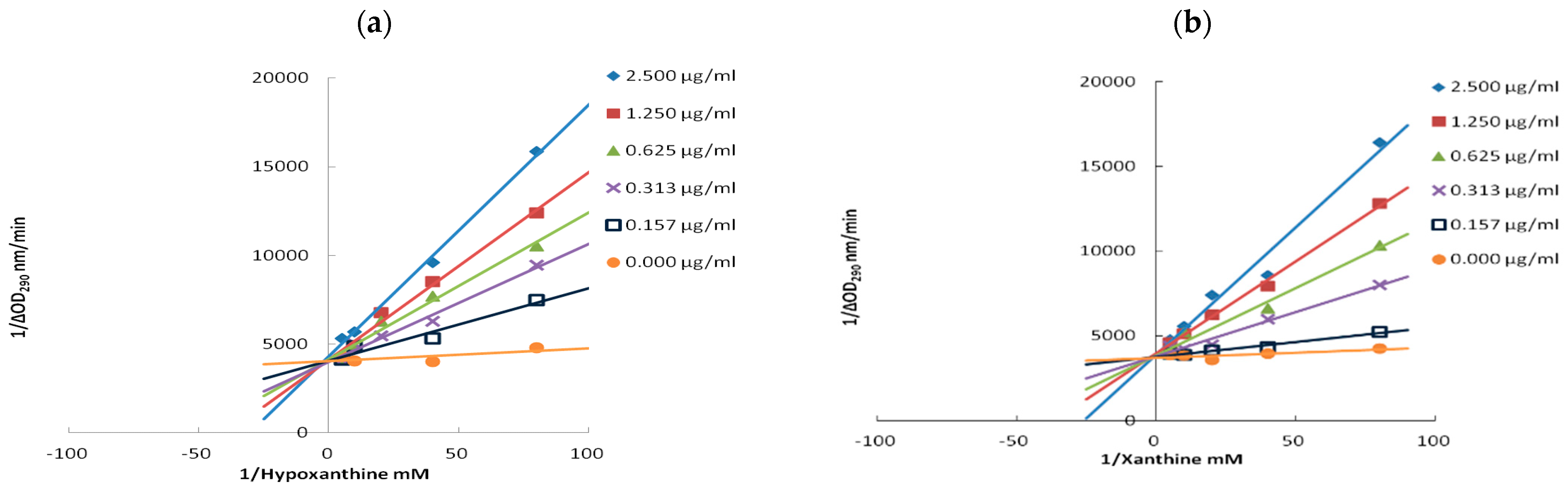
The major types of enzyme inhibition include noncompetitive, uncompetitive, competitive, and mixed types. To validate the experimental system, an enzyme kinetic study was first conducted using allopurinol as a positive control (Figure 6). With both hypoxanthine and xanthine as substrates, the Lineweaver–Burk plots of allopurinol displayed identical y-intercepts but progressively steeper slopes as the inhibitor concentration increased. Table 4 summarizes the kinetic parameters of allopurinol against xanthine oxidase, showing an increased Km while Vmax remained unchanged. Accordingly, competitive inhibition of xanthine oxidase by allopurinol contributed to the suppression of uric acid formation. Competitive-type inhibition by allopurinol has previously been reported by Huang et al. and Chen et al., which is consistent with our data [48,49]. It manifested that allopurinol showed a strong preference for interacting with free xanthine oxidase, consequently hindering substrate interaction with the enzyme.
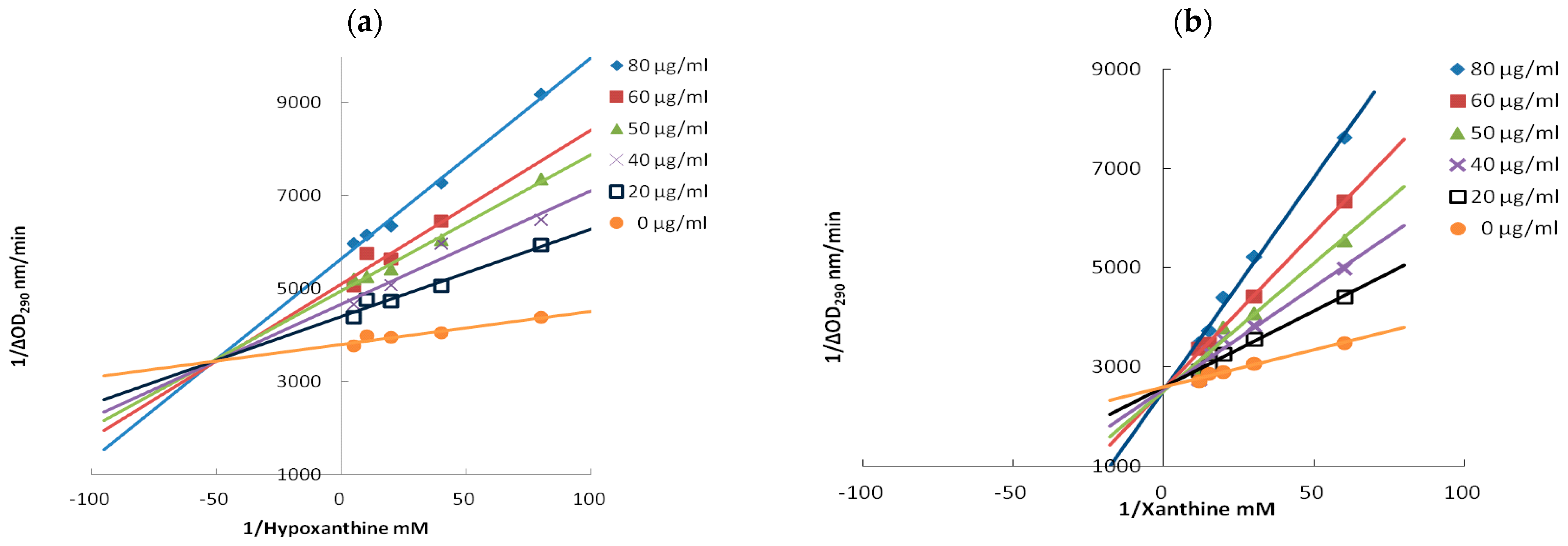
Figure 7 shows the Lineweaver–Burk plots of EAF with two substrates. Kinetic parameters of EAF were listed in Table 5. In the presence of EAF, Km increased while Vmax decreased when hypoxanthine was used as the substrate, indicating a mixed-type inhibition pattern. In contrast, the use of xanthine as the substrate resulted in an increased Km and without a change in Vmax, suggesting competitive inhibition.
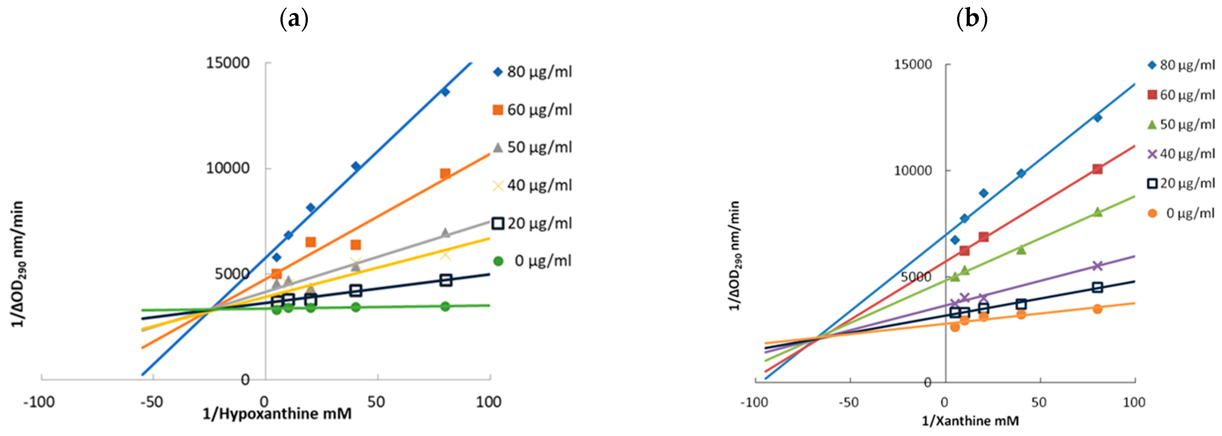
The inhibitory mode of maltol toward xanthine oxidase was further clarified by kinetic analysis, with the corresponding Lineweaver–Burk plots and kinetic constants presented in Figure 8 and Table 6. Maltol treatment altered both kinetic parameters, as reflected by an increased Km and a decreased Vmax. It demonstrated that maltol suppresses uric acid production through a mixed-type inhibition mechanism. Similar inhibitory behavior has been reported for quercetin, a multifunctional flavonoid, which inhibits xanthine oxidase through both competitive and noncompetitive components [48,49,50].
To further contextualize these findings, the inhibitory activity of the present samples was compared with previously reported natural products. Xanthine oxidase inhibitory activity was detected in Pistacia integerrima leaf extract and its fractions, where quercetin-3-O-D-glucopyranoside, rutin, and apigenin showed IC50 values ranging from 5.75 to 61 μg/mL for hypoxanthine as substrate [37]. Xanthine oxidase inhibitory activity was identified in the ethyl acetate extract of snake fruit (Salacca edulis), in which 2-metyl ester-1H-pyrrole-4-carboxilyc acid exhibited the strongest activity with an IC50 value of 48.86 μg/mL for xanthine as substrate [51]. In the present study, maltol showed IC50 values of 26.67 μg/mL and 33.18 μg/mL using hypoxanthine and xanthine as substrates, respectively, indicating a moderate inhibitory effect comparable to other natural products.

2.4. Molecular Docking of Maltol with Xanthine Oxidase

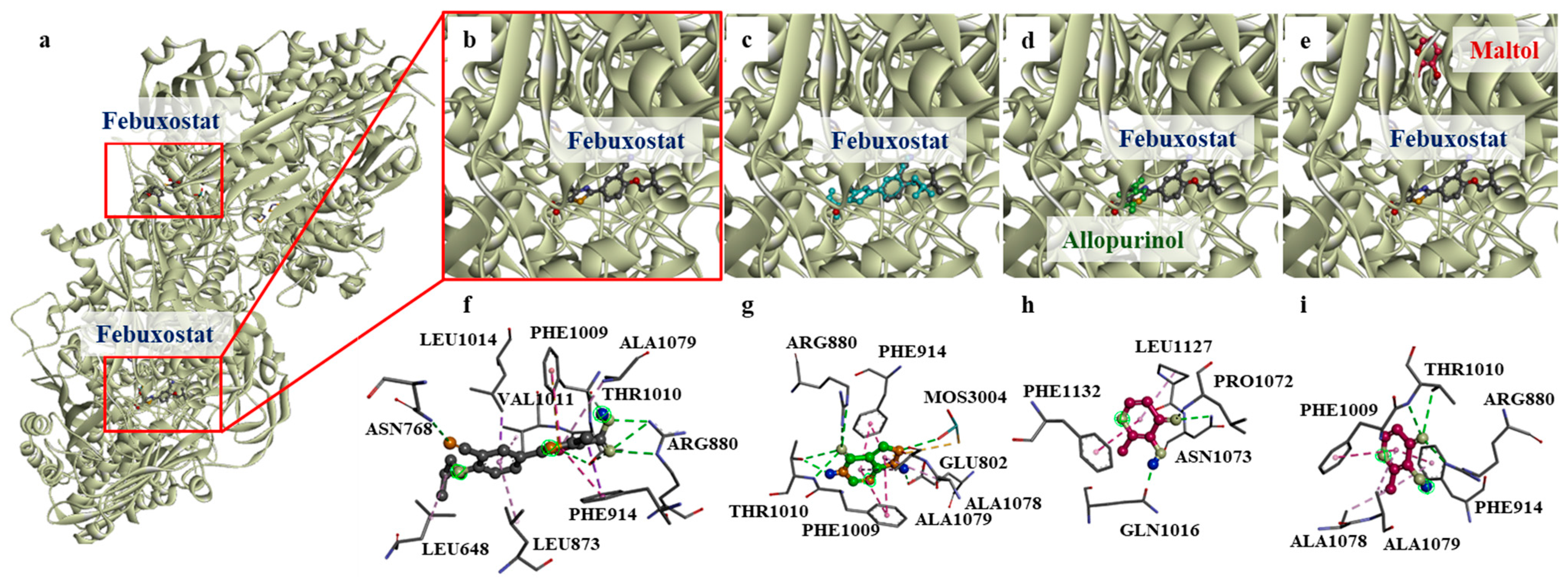
The mixed-type inhibition pattern observed for maltol suggests that its interaction with xanthine oxidase may involve more complex binding behavior than simple competitive inhibition [52]. However, enzyme kinetic analysis alone cannot definitively determine the underlying molecular interaction mechanism. Molecular docking analysis further indicated possible interactions between maltol and residues located within the active-site channel. The xanthine oxidase structure (PDB ID: 1N5X, Figure 9a,b) was used to predict the binding site of maltol. To validate the docking protocol, febuxostat was first removed from the co-crystallized structure and subsequently redocked into the active site. The redocked pose closely overlapped with the co-crystallized conformation, with only minor differences in the orientation of functional groups (Figure 9c). Among the 20 independent redocking runs performed for febuxostat, the pose with the lowest root-mean-square deviation (RMSD) relative to the crystallographic febuxostat pose was selected as the best redocked pose. This pose yielded an RMSD of 0.911 Å, supporting the validity of the docking protocol. Across the 20 independent redocking runs, febuxostat showed a mean binding affinity of −8.5 ± 0.3 kcal/mol. In the crystallographic binding pose, febuxostat interacted with ASN768, ARG880, and THR1010 through hydrogen bonding, along with π–alkyl interactions with LEU873 and VAL1011, and a π–sigma interaction with LEU1014 and PHE914 (Figure 9f and Figure S1a).
Allopurinol was readily accommodated within the same active site (Figure 9d), although its binding affinity (−7.0 ± 0.1 kcal/mol) was weaker than that of febuxostat (−8.5 ± 0.3 kcal/mol). It interacted with GLU802, ARG880, and THR1010 through hydrogen bonding and exhibited π–π interactions with PHE914 and PHE1009 (Figure 9g and Figure S1b).
For maltol, the predominant docking pose (11 of 20 runs) was in a peripheral region, involving hydrogen bonding with LEU1127, ASN1073, and GLN1016, along with π–π and π–alkyl interactions with PHE1132 and PRO1072, respectively (Figure 9h and Figure S1c). This binding mode was positioned farther from the co-crystallized febuxostat (Figure 9e), and an unfavorable acceptor–acceptor interaction was observed between the hydroxyl group of maltol and ASN1073. In the remaining poses, alternative hydrogen bonding with THR1010 and ARG880 was identified (Figure 9i and Figure S1d). Notably, these residues are key components of the canonical active site, suggesting that maltol may adopt a secondary conformation within the Mo-pterin active-site channel. Despite its shifted binding position, maltol still exhibited a stable interaction with a binding affinity of −6.5 ± 0.1 kcal/mol.
The Mo-pterin active channel of xanthine oxidase is a key substrate-access tunnel leading to the deep catalytic center and is lined by residues such as ARG880, PHE914, PHE1009, and THR1010 [53]. Our docking results suggest that maltol is capable of accessing this active-site channel, as approximately 50% of the docked poses were located within the Mo-pterin channel. However, compared with allopurinol, maltol showed weaker binding affinity and less consistent localization within this region. In contrast, allopurinol occupied the active-site channel in 19 of 20 runs and formed more favorable interactions with the residues in this key substrate-access tunnel. Febuxostat was also able to bind within the same site and displayed more extensive interactions with xanthine oxidase; however, it occupied this position in only 8 of 20 runs, likely reflecting conformational variability in docking rather than reduced binding capability. These observations are consistent with the higher IC50 and lower inhibitory potency of maltol. However, the docking results alone are insufficient to establish the inhibition mechanism.
Compared with flavonoids, stilbenes, or other synthetic xanthine oxidase inhibitors [54,55], maltol is structurally smaller and less capable of forming multiple stabilizing interactions with active-site residues. Nevertheless, maltol was predicted to interact with critical residues such as ARG880 and THR1010. These interaction patterns may partially explain the enzyme kinetic results, in which allopurinol exhibited competitive inhibition whereas maltol displayed a mixed-type inhibition pattern. Overall, the docking results suggest that allopurinol and febuxostat are more likely to occupy the canonical active-site channel of xanthine oxidase, whereas maltol may act as a milder modulator with a more complex interaction pattern than simple competitive inhibition, possibly due to weaker and less stable interactions within the active-site channel. Although these docking findings may help explain the observed inhibition patterns, they should still be interpreted with caution because they represent predicted binding modes rather than experimentally validated mechanisms.

2.5. In Silico Predictions of Physicochemical, Pharmacokinetic, and Toxicological Properties of Maltol

To evaluate the potency of maltol as a potential xanthine oxidase inhibitor, its physicochemical properties, pharmacokinetics, and toxicity were predicted using the SwissADME and ProTox 3.0 platforms, respectively, with allopurinol used as a reference compound. The results showed that the Log S (ESOL) and Log P (o/w) values of maltol were −1.17 and 0.55, respectively (Table S2), indicating high aqueous solubility and suggesting favorable bioavailability. Although its properties were slightly different, they remained comparable to those of allopurinol (Log S (ESOL) = −0.93; Log P (o/w) = 0.01). Consistently, both compounds exhibited an identical predicted bioavailability score of 0.55 in SwissADME.
Evaluation based on the Lipinski’s Rule of Five, Ghose filter, and Veber rule revealed three violations for maltol, including a molecular weight below 160, a molar refractivity of 31.97 (below 40, Table S2), and fewer than 20 atoms. Similarly, allopurinol also exhibited three violations, including a molecular weight below 160, a molar refractivity of 34.51 (below 40, Table S2), and fewer than 20 atoms. Although such violations may generally limit drug-likeness, these deviations are primarily associated with the small molecular size of both compounds. Notably, allopurinol, an approved drug, exhibits similar violations, suggesting that these features are unlikely to substantially impair the therapeutic efficacy of maltol as an orally active agent.
Maltol and allopurinol did not exhibit inhibitory activity against the five major cytochrome P450 isoforms (Table S3), suggesting a low potential for drug–drug interactions. In addition, it showed high gastrointestinal absorption, indicating suitability for systemic therapeutic applications. Unlike allopurinol, maltol is predicted to cross the blood–brain barrier (BBB); therefore, its potential central nervous system (CNS) effects require further investigation. Maltol and allopurinol were also predicted not to be a P-glycoprotein (P-gp) substrate, suggesting a higher likelihood of absorption and retention.
Toxicity predictions indicated that maltol is inactive for hepatotoxicity, neurotoxicity, respiratory toxicity, cardiotoxicity, immunotoxicity, and cytotoxicity, while showing weak activity in nephrotoxicity, carcinogenicity, and mutagenicity endpoints (Table S4). Compared with allopurinol, which has been associated with hepatotoxicity, maltol exhibited a lower predicted risk of hepatotoxicity. As most compounds typically exhibit at least one predicted toxicological liability [54], maltol showed a relatively favorable in silico toxicity profile. However, these ADMET and toxicity predictions should be interpreted as preliminary screening results rather than definitive evidence of safety, as they are based on computational models and require further experimental validation. Taken together, these findings suggest that maltol may warrant further investigation for its potential as an alternative candidate.

3. Materials and Methods

3.1. Plant Material and Extraction

Abies kawakamii, 76 years old, was collected from the Duigaoyue Forest Working Unit, Experimental Forest of National Taiwan University. The collection source and laboratory records of the plant materials followed our previously published report [56]. Fresh leaves were subjected to two rounds of extraction with 95% ethanol at room temperature for 7 days each. After filtration, the solvent was removed under vacuum using a rotary evaporator [57]. The 95% ethanolic extraction yield of A. kawakamii leaves was 9.24% on a dry weight basis.

3.2. Liquid–Liquid Partition

The extract was further separated by liquid–liquid partitioning with solvents of progressively increasing polarity. Four fractions were subsequently obtained, including the n-hexane fraction (HXF), ethyl acetate fraction (EAF), n-butanol fraction (BUF), and aqueous fraction (HOF) [23,57,58]. The four fractions exhibited a decreasing trend in content levels: EAF (31.40%) > HOF (20.90%) > HXF (14.43%) > BUF (7.96%).

3.3. High Performance Liquid Chromatography

Bioactive fractions were further purified using semi-preparative high-performance liquid chromatography (HPLC; L-2130, Hitachi, Tokyo, Japan) equipped with a 9.4 × 250 mm Zorbax Sil column (5 μm). Separation was carried out under isocratic conditions with an n-hexane–ethyl acetate–acetone mixture (1:1:1, v/v) as the mobile phase at a flow rate of 1 mL/min, and chromatographic signals were monitored using a refractive index (RI) detector [58,59].

3.4. Isolation and Identification of Compounds

The isolated constituents were structurally characterized through spectroscopic analyses, including mass spectroscopy (MS; MAT-958, Finnigan, MA, USA), nuclear magnetic resonance spectroscopy (NMR; Bruker AVIII, Bruker Avance, Rheinstetten, Germany). The 1H-NMR and 13C-NMR spectra were acquired at 500 MHz and 125 MHz, respectively [59,60,61,62].

3.5. Xanthine Oxidase Assay

Xanthine oxidase inhibition was determined using in vitro spectrophotometric method [23,37,49]. Hypoxanthine and xanthine were separately employed as substrates for the assay. Briefly, xanthine oxidase solution (60 µL, 0.025 unit/mL; EC 1.1.3.22), 50 mM potassium phosphate buffer (117 µL, pH 7.8), and test sample (3 µL) were introduced into the 96-well plate for 10 min at 25 °C. Subsequently, 100 µL of substrate (0.15 mM xanthine or hypoxanthine) was added and incubated at 37 °C in the dark for 30 min. 20 µL of 1 N HCl was used to stop the enzymatic reaction. Absorbance at 290 nm was monitored using a microplate spectrometer (SPECTROstar Nano, BMG LABTECH, Offenburg, Germany). Allopurinol was included as a positive control, and each experiment was conducted in triplicate. Xanthine oxidase inhibition was calculated using the following equation:
Inhibition (%) = {[(A_control − A_control blank) − (A_sample − A_sample blank)]/[(A_control − A_control blank)]} × 100.
The IC50 was obtained from the dose–response curve and defined as the concentration required to inhibit 50% of xanthine oxidase activity.

3.6. Enzyme Kinetic Study

Lineweaver–Burk plots were applied to estimate the kinetic behavior of xanthine oxidase and to clarify how the tested samples affected the enzyme–substrate interaction. The enzyme concentration was fixed at 0.025 unit/mL, while the substrate concentration was adjusted from 0.0125 to 0.20 mM using either hypoxanthine or xanthine. The assay mixture contained 60 µL of xanthine oxidase solution, 117 µL of potassium phosphate buffer, 3 µL of test sample, and 100 µL of substrate in a 96-well microplate. Immediately after mixing, changes in absorbance at 290 nm were recorded for 3 min at 37 °C. Michaelis–Menten constant (Km) and maximum velocity (Vmax) were calculated from the linear equation of the Lineweaver–Burk plot. The inhibition pattern was then classified as uncompetitive, noncompetitive, competitive, or mixed-type inhibition [38,49,63,64].

3.7. Molecular Docking

Three-dimensional ligand files for maltol and the positive controls, febuxostat and allopurinol, were downloaded as SDF format from PubChem. The xanthine oxidase crystal complex co-crystallized with febuxostat (PDB ID: 1N5X; resolution: 2.80 Å) was selected from the Protein Data Bank for docking analysis. The co-crystallized ligand was removed from the protein structure prior to docking. Polar hydrogens were then added using AutoDockTools (version 1.5.7). The prepared protein was then converted to PDBQT format. Because the molybdenum cofactor could not be properly recognized and parameterized during receptor preparation in AutoDockTools, it was excluded from the docking model. The grid box was centered at coordinates (96, 52, 38) with dimensions of 40 × 40 × 40 Å [53]. Docking simulations were conducted using AutoDock Vina (version 1.2.x) [65], with 20 independent runs for each ligand. The binding affinities were expressed as mean ± standard deviation (SD) based on the results from the 20 runs. The RMSD between the redocked and crystallographic poses was calculated in PyMOL (version 3.1.6.1) by pairwise fitting of 22 heavy atoms using the pair_fit command. Docking poses and interactions were visualized using Discovery Studio Visualizer (DSV).

3.8. SwissADME and ProTox 3.0 Analysis

SwissADME and ProTox 3.0 are useful web-based platforms for predicting the physicochemical properties, pharmacokinetics, and toxicity profiles of drug candidates. Both tools analyze the uploaded chemical structures of maltol and allopurinol to evaluate drug-likeness and ADME-related properties, while ProTox 3.0 provides more detailed toxicity endpoints. Allopurinol exists in multiple tautomeric forms, and the keto tautomer was selected in this study based on its representation in the prediction database.

3.9. Statistical Analysis

Experimental data were analyzed using SPSS (Chicago, IL, USA, Version 16). Group differences were further examined using Scheffe’s post hoc multiple comparison procedure at a 95% confidence level.

4. Conclusions

Gout is a metabolic disorder associated with disrupted purine metabolism, resulting in hyperuricemia and monosodium urate crystal deposition in the joints. A daily diet with purine-rich foods (shellfish, red meat, organ meats), beer and sugary drinks can easily lead to gout, and the development of anti-gout agents with fewer adverse effects is urgently needed. The effects of A. kawakamii leaf extract, ethyl acetate fraction, and its constituent on xanthine oxidase inhibition were investigated in this study. The EAF was the most effective at inhibiting xanthine oxidase activity among all tested fractions, showing IC50 values of 48.76 μg/mL for hypoxanthine and 66.70 μg/mL for xanthine as substrates. Maltol, obtained and characterized from the ethyl acetate fraction, exhibited the inhibitory of xanthine oxidase for both substrates. The IC50 values of maltol against xanthine oxidase were 26.67 μg/mL and 33.18 μg/mL when using hypoxanthine and xanthine as substrates, respectively. Enzyme kinetic analysis demonstrated that maltol exhibited mixed-type inhibition against xanthine oxidase using both substrates. In silico docking predicted that maltol can bind to xanthine oxidase with a binding affinity of −6.5 kcal/mol, which is comparable to that of allopurinol, while showing no predicted hepatotoxicity. The results revealed that A. kawakamii leaf extract, ethyl acetate fraction, and maltol have potential as potential natural agents targeting xanthine oxidase for gout treatment, although further research is required to confirm their clinical feasibility.

Supplementary Materials

The following supporting information can be downloaded at: https://www.mdpi.com/article/10.3390/ph19071100/s1, Figure S1. Two-dimensional interaction diagrams of the top-ranked docking poses of (a) febuxostat, (b) allopurinol, and (c, d) maltol with xanthine oxidase. Table S1. Binding affinity of febuxostat, allopurinol, and maltol to xanthine oxidase. Table S2. Prediction of the molecular descriptors of allopurinol and maltol. Table S3. Prediction of pharmacokinetic profile of allopurinol and maltol. Table S4. Prediction of toxicity of allopurinol and maltol.

Author Contributions

Conceptualization, H.-T.C., P.-L.Y. and C.-H.K.; methodology, C.-Y.H., L.-S.H. and H.-T.C.; software, C.-Y.H., J.-G.L. and L.-S.H.; formal analysis and investigation, C.-Y.H., S.-L.H., L.-S.H. and H.-T.C.; writing—original draft preparation, C.-Y.H., Y.-M.H., P.-L.Y. and H.-T.C.; writing—review and editing, P.-L.Y., Y.-M.H. and H.-T.C. All authors have read and agreed to the published version of the manuscript.

Funding

This research received financial support from the Ministry of Science and Technology, Taiwan.

Institutional Review Board Statement

Not applicable.

Informed Consent Statement

Not applicable.

Data Availability Statement

The data are available from the corresponding author on reasonable request.

Acknowledgments

The authors gratefully thank Shou-Ling Huang for the assistance in NMR experiments of the Instrumentation Center at NTU.

Conflicts of Interest

The authors declare no conflicts of interest.

References

  1. Ursini, F.; De Giorgi, A.; D’Onghia, M.; De Giorgio, R.; Fabbian, F.; Manfredini, R. Chronobiology and chronotherapy in inflammatory joint diseases. Pharmaceutics 2021, 13, 1832. [Google Scholar] [CrossRef] [PubMed]
  2. Shoji, A.; Yamanaka, H.; Kamatani, N. A etrospective study of the relationship between serum urate level and recurrent attacks of gouty arthritis: Evidence for reduction of recurrent gouty arthritis with antihyperuricemic therapy. Arthritis Rheum. 2004, 51, 321–325. [Google Scholar] [CrossRef] [PubMed]
  3. Whelton, A.; Macdonald, P.A.; Zhao, L.; Hunt, B.; Gunawardhana, L. Renal function in gout: Long-term treatment effects of febuxostat. J. Clin. Rheumatol. 2011, 17, 7–13. [Google Scholar] [CrossRef] [PubMed]
  4. Abhishek, A.; Roddy, E.; Doherty, M. Gout—A guide for the general and acute physicians. Clin. Med. 2017, 17, 54–59. [Google Scholar] [CrossRef] [PubMed]
  5. Ragab, G.; Elshahaly, M.; Bardin, T. Gout: An old disease in new perspective—A review. J. Adv. Res. 2017, 8, 495–511. [Google Scholar] [CrossRef] [PubMed]
  6. Zhang, Y.; Chen, S.; Yuan, M.; Xu, Y.; Xu, H. Gout and diet: A comprehensive review of mechanisms and management. Nutrients 2022, 14, 3525. [Google Scholar] [CrossRef] [PubMed]
  7. Muntiu, M.; Joosten, L.A.B.; Crişan, T.O. Gout basic research: 2023 in review. Gout Urate Cryst. Depos. Dis. 2024, 2, 220–235. [Google Scholar] [CrossRef]
  8. Sattui, S.E.; Gaffo, A.L. Treatment of hyperuricemia in gout: Current therapeutic options, latest developments and clinical implications. Ther. Adv. Musculoskelet. Dis. 2016, 8, 145–159. [Google Scholar] [CrossRef] [PubMed]
  9. Jordan, A.; Gresser, U. Side effects and interactions of the xanthine oxidase inhibitor febuxostat. Pharmaceuticals 2018, 11, 51. [Google Scholar] [CrossRef] [PubMed]
  10. Tátrai, P.; Erdő, F.; Dörnyei, G.; Krajcsi, P. Modulation of urate transport by drugs. Pharmaceutics 2021, 13, 899. [Google Scholar] [CrossRef] [PubMed]
  11. Owen, P.L.; Johns, T. Xanthine oxidase inhibitory activity of northeastern North American plant remedies used for gout. J. Ethnopharmacol. 1999, 64, 149–160. [Google Scholar] [CrossRef] [PubMed]
  12. Matsuura, R.; Ukeda, H.; Sawamura, M. Tyrosinase inhibitory activity of citrus essential oils. J. Agric. Food Chem. 2006, 54, 2309–2313. [Google Scholar] [CrossRef] [PubMed]
  13. Chen, C.H.; Chan, H.C.; Chu, Y.T.; Ho, H.Y.; Chen, P.Y.; Lee, T.H.; Lee, C.K. Antioxidant activity of some plant extracts towards xanthine oxidase, lipoxygenase and tyrosinase. Molecules 2009, 14, 2947–2958. [Google Scholar] [CrossRef] [PubMed]
  14. Ding, H.-Y.; Chou, T.-H.; Liang, C.-H. Antioxidant and antimelanogenic properties of rosmarinic acid methyl ester from Origanum vulgare. Food Chem. 2010, 123, 254–262. [Google Scholar] [CrossRef]
  15. Djenane, D.; Yangüela, J.; Montañés, L.; Djerbal, M.; Roncalés, P. Antimicrobial activity of Pistacia lentiscus and Satureja montana essential oils against Listeria monocytogenes CECT 935 using laboratory media: Efficacy and synergistic potential in minced beef. Food Control 2011, 22, 1046–1053. [Google Scholar] [CrossRef]
  16. Silvestre, G.F.G.; Lucena, R.P.; Oliveira, G.D.; Pereira, H.N.; Dias, J.A.B.; Souza, I.A.; Alves, H.S. Anti-tumor and anti-Inflammatory activity in vivo of Apodanthera congestiflora Cogn. (Cucurbitaceae). Pharmaceutics 2021, 13, 743. [Google Scholar] [CrossRef] [PubMed]
  17. Myint, P.P.; Dao, T.T.P.; Kim, Y.S. Anticancer activity of Smallanthus sonchifolius methanol extract against human hepatocellular carcinoma cells. Molecules 2019, 24, 3054. [Google Scholar] [CrossRef] [PubMed]
  18. Chang, H.T.; Chang, M.L.; Chen, Y.T.; Chang, S.T.; Hsu, F.L.; Wu, C.C.; Ho, C.K. Evaluation of motor coordination and antidepressant activities of Cinnamomum osmophloeum ct. linalool leaf oil in rodent model. Molecules 2021, 26, 3037. [Google Scholar] [CrossRef] [PubMed]
  19. Chang, J.Y.; Tsai, K.H.; Huang, Y.M.; Chang, Y.Y.; Huang, C.S.; Ho, Y.T.; Wang, S.Y.; Chang, M.L.; Chang, H.T. Larvicidal activities of Juniperus chinensis var. kaizuka leaf essential oil and its constituents against Dengue vector mosquitoes, Aedes aegypti and Ae. albopictus. Plants 2025, 14, 3321. [Google Scholar] [CrossRef] [PubMed]
  20. Kong, L.D.; Abliz, Z.; Zhou, C.X.; Li, L.J.; Cheng, C.H.; Tan, R.X. Glycosides and xanthine oxidase inhibitors from Conyza bonariensis. Phytochemistry 2001, 58, 645–651. [Google Scholar] [CrossRef] [PubMed]
  21. Medina-López, R.; Vara-Gama, N.; Soria-Arteche, O.; Moreno-Rocha, L.A.; López-Muñoz, F.J. Pharmacokinetics and pharmacodynamics of (S)-ketoprofen co-administered with caffeine: A preclinical study in arthritic rats. Pharmaceutics 2018, 10, 20. [Google Scholar] [CrossRef] [PubMed]
  22. Mohamed Isa, S.S.P.; Ablat, A.; Mohamad, J. The antioxidant and xanthine oxidase inhibitory activity of Plumeria rubra flowers. Molecules 2018, 23, 400. [Google Scholar] [CrossRef] [PubMed]
  23. Huang, C.Y.; Yeh, T.F.; Hsu, F.L.; Lin, C.Y.; Chang, S.T.; Chang, H.T. Xanthine oxidase inhibitory activity and thermostability of cinnamaldehyde-chemotype leaf oil of Cinnamomum osmophloeum microencapsulated with β-cyclodextrin. Molecules 2018, 23, 1107. [Google Scholar] [CrossRef] [PubMed]
  24. Mohos, V.; Fliszár-Nyúl, E.; Poór, M. Inhibition of xanthine oxidase-catalyzed xanthine and 6-mercaptopurine oxidation by flavonoid aglycones and some of their conjugates. Int. J. Mol. Sci. 2020, 21, 3256. [Google Scholar] [CrossRef] [PubMed]
  25. Abdulhafiz, F.; Mohammed, A.; Kayat, F.; Bhaskar, M.; Hamzah, Z.; Podapati, S.K.; Reddy, L.V. Xanthine oxidase inhibitory activity, chemical composition, antioxidant properties and GC-MS analysis of Keladi Candik (Alocasia longiloba Miq). Molecules 2020, 25, 2658. [Google Scholar] [CrossRef] [PubMed]
  26. Ngoc, T.M.; Khoi, N.M.; Ha, D.T.; Nhiem, N.X.; Tai, B.H.; Don, D.V.; Luong, H.V.; Son, D.C.; Bae, K. Xanthine oxidase inhibitory activity of constituents of Cinnamomum cassia twigs. Bioorg. Med. Chem. Lett. 2012, 22, 4625–4628. [Google Scholar] [CrossRef] [PubMed]
  27. Nguyen, M.T.; Awale, S.; Tezuka, Y.; Tran, Q.L.; Watanabe, H.; Kadota, S. Xanthine oxidase inhibitory activity of Vietnamese medicinal plants. Biol. Pharm. Bull. 2004, 27, 1414–1421. [Google Scholar] [CrossRef] [PubMed]
  28. Muraoka, S.; Miura, T. Inhibition of xanthine oxidase by phytic acid and its antioxidative action. Life Sci. 2004, 74, 1691–1700. [Google Scholar] [CrossRef] [PubMed]
  29. Yeşilada, E.; Honda, G.; Sezik, E.; Tabata, M.; Fujita, T.; Tanaka, T.; Takeda, Y.; Takaishi, Y. Traditional medicine in Turkey. V. folk medicine in the inner Taurus Mountains. J. Ethnopharmacol. 1995, 46, 133–152. [Google Scholar] [CrossRef] [PubMed]
  30. Wajs-Bonikowska, A.; Szoka, Ł.; Kwiatkowski, P.; Maciejczyk, E. Greek fir seeds and cones as underestimated source of essential oil: Composition and biological properties. Appl. Sci. 2023, 13, 13238. [Google Scholar] [CrossRef]
  31. Wajs-Bonikowska, A.; Szoka, Ł.; Karna, E.; Wiktorowska-Owczarek, A.; Sienkiewicz, M. Abies concolor seeds and cones as new source of essential oils-composition and biological activity. Molecules 2017, 22, 1880. [Google Scholar] [CrossRef] [PubMed]
  32. Lee, T.H.; Subedi, L.; Ha, Y.J.; Moon, G.; Kim, S.Y.; Kim, C.S. Glycosylated constituents isolated from the trunk of Abies holophylla and their anti-inflammatory and neurotrophic activity. Phytochemistry 2021, 192, 112962. [Google Scholar] [CrossRef] [PubMed]
  33. Chung, J.Y.; Park, N.; Kim, M.H.; Yang, W.M. Abies holophylla leaf essential oil alleviates allergic rhinitis based on network pharmacology. Pharmaceutics 2023, 15, 1195. [Google Scholar] [CrossRef] [PubMed]
  34. de Aguiar, J.C.R.d.O.F.; da Silva, A.C.; Santos, E.F.; Vieira, G.J.d.S.G.; Araújo, L.d.A.; de Andrade, J.J.A.; Anselmo, W.M.; da Rocha, S.K.L.; dos Santos, F.H.G.; Arruda, C.C.L.; et al. Larvicidal and oviposition activity of commercial essential oils of Abies sibirica Ledeb., Pogostemon cablin (Blanco) Benth., Juniperus communis L. and their combinations against Aedes aegypti. Molecules 2024, 29, 5921. [Google Scholar] [CrossRef] [PubMed]
  35. Vukić, M.D.; Vuković, N.L.; Jakovljević, M.R.; Ristić, M.S.; Kačániová, M. Antimicrobial effects of Abies alba essential oil and its application in food preservation. Plants 2025, 14, 2071. [Google Scholar] [CrossRef] [PubMed]
  36. Ayupova, R.; Svajdlenka, E.; Zemlicka, M.; Ibadullayeva, G.; Raganina, K.; Alimova, U.; Nokerbek, S.; Botabayeva, R.; Kiyekbayeva, L.; Mombekov, S. Comparative phytochemical profiling of essential oils from selected Abies species and analysis of their antifungal and antiradical activity. Pharmaceutics 2026, 18, 26. [Google Scholar]
  37. Wu, C.-C.; Huang, S.-L.; Ko, C.-H.; Chang, H.-T. Antifungal Sesquiterpenoids from Michelia formosana Leaf Essential Oil against Wood-Rotting Fungi. Molecules 2022, 27, 2136. [Google Scholar] [CrossRef] [PubMed]
  38. Vassallo, A.; Armentano, M.F.; Miglionico, R.; Caddeo, C.; Chirollo, C.; Gualtieri, M.J.; Ostuni, A.; Bisaccia, F.; Faraone, I.; Milella, L. Hura crepitans L. extract: Phytochemical characterization, antioxidant activity, and nanoformulation. Pharmaceutics 2020, 12, 553. [Google Scholar] [CrossRef] [PubMed]
  39. Yen, P.-L.; Wu, C.-L.; Chang, S.-T.; Huang, S.L.; Chang, H.-T. Antioxidative lignans from phytochemical extract of Calocedrus formosana Florin. BioResources 2012, 7, 4122–4131. [Google Scholar] [CrossRef]
  40. Santonocito, D.; Granata, G.; Geraci, C.; Panico, A.; Siciliano, E.A.; Raciti, G.; Puglia, C. Carob seeds: Food waste or source of bioactive compounds? Pharmaceutics 2020, 12, 1090. [Google Scholar] [CrossRef] [PubMed]
  41. Nunes, A.; Marto, J.; Gonçalves, L.M.; Simões, S.; Félix, R.; Ascenso, A.; Lopes, F.; Ribeiro, H.M. Novel and modified neutrophil elastase inhibitor loaded in topical formulations for psoriasis management. Pharmaceutics 2020, 12, 358. [Google Scholar] [CrossRef] [PubMed]
  42. Wu, C.-C.; Wu, C.-L.; Huang, S.-L.; Chang, H.-T. Antifungal activity of liriodenine from Michelia formosana heartwood against wood-rotting fungi. Wood Sci. Technol. 2012, 46, 737–747. [Google Scholar] [CrossRef]
  43. Gutiérrez-Macías, P.; Peralta-Cruz, J.; Borja-de-la-Rosa, A.; Barragán-Huerta, B.E. Peltomexicanin, a peltogynoid quinone methide from Peltogyne mexicana Martínez purple heartwood. Molecules 2016, 21, 186. [Google Scholar] [CrossRef] [PubMed]
  44. Ahmad, N.S.; Farman, M.; Najmi, M.H.; Mian, K.B.; Hasan, A. Pharmacological basis for use of Pistacia integerrima leaves in hyperuricemia and gout. J. Ethnopharmacol. 2008, 117, 478–482. [Google Scholar] [CrossRef] [PubMed]
  45. Huang, C.Y.; Chang, Y.Y.; Chang, S.T.; Chang, H.T. Xanthine oxidase inhibitory activity and chemical composition of Pistacia chinensis leaf essential oil. Pharmaceutics 2022, 14, 1982. [Google Scholar] [CrossRef] [PubMed]
  46. Nagao, A.; Seki, M.; Kobayashi, H. Inhibition of xanthine oxidase by flavonoids. Biosci. Biotechnol. Biochem. 1999, 63, 1787–1790. [Google Scholar] [CrossRef] [PubMed]
  47. Takahama, U.; Koga, Y.; Hirota, S.; Yamauchi, R. Inhibition of xanthine oxidase activity by an oxathiolanone derivative of quercetin. Food Chem. 2011, 126, 1808–1811. [Google Scholar] [CrossRef] [PubMed]
  48. Sowndhararajan, K.; Joseph, J.M.; Rajendrakumaran, D. In vitro xanthine oxidase inhibitory activity of methanol extracts of Erythrina indica Lam. leaves and stem bark. Asian Pac. J. Trop. Biomed. 2012, 2, S1415–S1417. [Google Scholar] [CrossRef]
  49. Zhao, Q.; Meng, Y.; Liu, J.; Hu, Z.; Du, Y.; Sun, J.; Mao, X. Separation, identification and docking analysis of xanthine oxidase inhibitory peptides from pacific cod bone-flesh mixture. LWT 2022, 167, 113862. [Google Scholar] [CrossRef]
  50. Trott, O.; Olson, A.J. AutoDock vina: Improving the speed and accuracy of docking with a new scoring function, efficient optimization, and multithreading. J. Comput. Chem. 2010, 31, 455–461. [Google Scholar] [CrossRef] [PubMed]
  51. Mukha, S.A.; Antipova, I.A.; Medvedeva, S.A.; Saraev, V.V.; Larina, L.I.; Tsyrenzhapov, A.V.; Sukhov, B.G. Synthesis and properties of metal chelates based on natural γ-pyrone maltol. Chem. Sustain. Dev. 2007, 15, 448–458. [Google Scholar]
  52. Koch, A.S.; Chimento, C.A.; Berg, A.N.; Mughal, F.D.; Spencer, J.-P.; Hovland, D.E.; Mbadugha, B.; Hovland, A.K.; Eller, L.R. Extraction of maltol from Fraser fir: A comparison of microwave-assisted extraction and conventional heating protocols for the organic chemistry laboratory. J. Chem. Educ. 2015, 92, 170–174. [Google Scholar] [CrossRef]
  53. Tyukavkina, N.A.; Medvedeva, S.A.; Ivanova, S.Z.; Voronov, V.K. Maltol from needles of Abies sibirica. Chem. Nat. Compd. 1972, 8, 661. [Google Scholar] [CrossRef]
  54. Samejo, M.Q.; Ndukwe, G.I.; Burdi, D.K.; Bhanger, M.I.; Khan, K.M. Isolation and crystal structure of maltol from Abies pindrow. J. Med. Plants Res. 2009, 3, 55–60. [Google Scholar]
  55. Schicchi, R.; Geraci, A.; Rosselli, S.; Spinella, A.; Maggio, A.; Bruno, M. Phytochemical investigation of the needles of Abies nebrodensis (Lojac.) Mattei. Nat. Prod. Res. 2020, 34, 2131–2136. [Google Scholar] [CrossRef] [PubMed]
  56. Han, N.R.; Park, H.J.; Ko, S.G.; Moon, P.D. Maltol has anti-cancer effects via modulating PD-L1 signaling pathway in B16F10 cells. Front. Pharmacol. 2023, 14, 1255586. [Google Scholar] [CrossRef] [PubMed]
  57. Macedi, E.; Paderni, D.; Formica, M.; Conti, L.; Fanelli, M.; Giorgi, L.; Amatori, S.; Ambrosi, G.; Valtancoli, B.; Fusi, V. Playing with structural parameters: Synthesis and characterization of two new maltol-based ligands with binding and antineoplastic properties. Molecules 2020, 25, 943. [Google Scholar] [CrossRef] [PubMed]
  58. Ahn, H.; Lee, G.; Han, B.-C.; Lee, S.-H.; Lee, G.-S. Maltol, a natural flavor enhancer, inhibits NLRP3 and non-canonical inflammasome activation. Antioxidants 2022, 11, 1923. [Google Scholar] [CrossRef] [PubMed]
  59. Lee, A.; Kwon, H.; Kim, S.; Jeong, Y.; Choi, B.T.; Kho, C. Protective effect of maltol on pathological response of cardiomyocyte in dystrophic mice. Korean J. Physiol. Pharmacol. 2025, 29, 235–244. [Google Scholar] [CrossRef] [PubMed]
  60. Chen, L.; Yin, H.; Lan, Z.; Ma, S.; Zhang, C.; Yang, Z.; Li, P.; Lin, B. Anti-hyperuricemic and nephroprotective effects of Smilax china L. J. Ethnopharmacol. 2011, 135, 399–405. [Google Scholar] [CrossRef] [PubMed]
  61. Umamaheswari, M.; Prabhu, P.; Asokkumar, K.; Sicashanmugam, T.; Subhadradevi, V.; Jagannath, P.; Madeswaran, A. In silico docking studies and in vitro xanthine oxidase inhibitory activity of commercially available terpenoids. Int. J. Phytopharm. 2012, 2, 135–142. [Google Scholar] [CrossRef]
  62. Priyatno, L.H.A.; Sukandar, E.Y.; Ibrahim, S.; Adnyana, I.K. Xanthine oxidase inhibitor activity of terpenoid and pyrrole compounds isolated from snake fruit (Salacca edulis Reinw.) cv. Bongkok. J. Appl. Sci. 2007, 7, 3127–3130. [Google Scholar] [CrossRef][Green Version]
  63. Pesaresi, A. Mixed and non-competitive enzyme inhibition: Underlying mechanisms and mechanistic irrelevance of the formal two-site model. J. Enzym. Inhib. Med. Chem. 2023, 38, 2245168. [Google Scholar] [CrossRef] [PubMed]
  64. Orhan, I.E.; Deniz, F.S.S. Natural products and extracts as xantine oxidase inhibitors—A hope for gout disease? Curr. Pharm. Des. 2021, 27, 143–158. [Google Scholar] [CrossRef] [PubMed]
  65. Antoniolli, G.; de Moraes, G.R.; da Costa, R.P.N.; de Campos, G.A.R.; Coelho, F. Advances in xanthine oxidase inhibition: A review of potential bioactive synthetic compounds. Arch. Pharm. 2025, 358, e70079. [Google Scholar] [CrossRef] [PubMed]
Figure 1. Xanthine oxidase inhibitory effects of A. kawakamii extract using (a) hypoxanthine and (b) xanthine as substrates. Data are presented as mean ± SD (n = 3).
Pharmaceuticals 19 01100 g001
Figure 2. 1H-NMR spectral data of maltol (CDCl3, 500 MHz).
Pharmaceuticals 19 01100 g002
Figure 3. 13C-NMR spectral data of maltol (CDCl3, 125 MHz).
Pharmaceuticals 19 01100 g003
Figure 4. Mass spectrum of compound M.
Pharmaceuticals 19 01100 g004
Figure 5. Chemical structure of maltol.
Pharmaceuticals 19 01100 g005
Figure 6. Kinetic inhibition profiles of allopurinol toward xanthine oxidase using (a) hypoxanthine and (b) xanthine as substrates.
Pharmaceuticals 19 01100 g006
Figure 7. Kinetic inhibition profiles of ethyl acetate fraction toward xanthine oxidase using (a) hypoxanthine and (b) xanthine as substrates.
Pharmaceuticals 19 01100 g007
Figure 8. Kinetic inhibition profiles of maltol toward xanthine oxidase using (a) hypoxanthine and (b) xanthine as substrates.
Pharmaceuticals 19 01100 g008
Figure 9. Molecular docking of maltol within the active site of xanthine oxidase. (a) Representation of xanthine oxidase (PDB ID: 1N5X), showing febuxostat located in both chain A and chain B of xanthine oxidase co-crystallized structure. (b) Enlarged view of the active site in chain A with the co-crystallized febuxostat. (ce) Overlay of docked poses and the co-crystallized febuxostat within the active site, showing (c) febuxostat (cyan), (d) allopurinol (green), and (e) maltol (red). (fi) Interaction of (f) febuxostat, (g) allopurinol, and (h,i) maltol with residues in the xanthine oxidase active site. Green dashed lines indicate hydrogen bonds; pink lines indicate π–π and π–alkyl interactions; purple lines indicate π–sigma interactions; gold lines indicate π–sulfur interactions; and red lines represent unfavorable donor–donor interactions.
Pharmaceuticals 19 01100 g009
Table 1. Inhibition rates of A. kawakamii extract and four fractions against xanthine oxidase.
SpecimenInhibition Rates (%)
Hypoxanthine as the SubstrateXanthine as the Substrate
ALE44.40 ± 2.0831.07 ± 3.60
HXF– *
EAF100.00 ± 0.0080.82 ± 1.98
BUF19.36 ± 0.9730.40 ± 8.99
HOF45.20 ± 5.5670.13 ± 7.81
Allopurinol **100.00 ± 0.00100.00 ± 0.00
ALE: A. kawakamii leaf extract, HXF: n-hexane fraction, EAF: ethyl acetate fraction, BUF: n-butanol fraction (BUF), and HOF: aqueous fraction; concentration: 100 μg/mL; results are mean ± SD (n = 3). – *: no effect; ** Positive control.
Table 2. IC50 values of leaf extract, ethyl acetate fraction (EAF), and maltol against xanthine oxidase.
SpecimenIC50 (μg/mL)
Hypoxanthine as the SubstrateXanthine as the Substrate
Leaf extract136.57 ± 1.79 a152.04 ± 6.92 A
EAF48.76 ± 1.68 b66.70 ± 1.64 B
Maltol26.67 ± 0.43 c33.18 ± 1.12 C
Allopurinol *0.23 ± 0.04 d0.30 ± 0.01 D
Data are presented as mean ± SD (n = 3). * Positive control. Values sharing different superscript letters (a–d; A–D) within the same substrate group differ significantly at p < 0.05 based on Scheffe’s test.
Table 3. 1H and 13C NMR data of maltol.
Position13C1H
2148.8
3143.1
4172.9
5112.96.39 (1H, d, J = 5.55 Hz)
6154.27.69 (1H, d, J = 5.55 Hz)
C2-CH314.32.34 (3H, s)
Table 4. Kinetic parameters of allopurinol-mediated xanthine oxidase inhibition.
SubstrateKinetic ParameterConcentration (μg/mL)PotentialInhibition
Type
0.0000.1560.3130.6251.2502.500
HypoxanthineVmax0.000250.000250.000250.000240.000240.00023― *Competitive
Km0.001780.010030.016550.020070.025760.03277↑ **
XanthineVmax0.000270.000270.000260.000260.000250.00026Competitive
Km0.001650.004670.013690.020600.027310.03867
*: constant; **: increasing.
Table 5. Kinetic parameters of ethyl acetate fraction-mediated xanthine oxidase inhibition.
SubstrateKinetic ParameterConcentration (μg/mL)PotentialInhibition
Type
02040506080
HypoxanthineVmax0.000300.000270.000250.000240.000210.00017↓ *Mixed
type
Km0.000470.003760.007050.007960.012390.01738↑ **
XanthineVmax0.000270.000270.000260.000260.000250.00026― ***Competitive
Km0.001650.004670.013690.020600.027310.03867
*: decreasing; **: increasing; ***: constant.
Table 6. Kinetic parameters of maltol-mediated xanthine oxidase inhibition.
SubstrateKinetic ParameterConcentration (μg/mL)PotentialInhibition
Type
01020406080
HypoxanthineVmax0.000300.000270.000250.000240.000210.00017↓ *Mixed
Km0.000470.003760.007050.007960.012390.01738↑ **
XanthineVmax0.000360.000320.000270.000210.000170.00014Mixed
Km0.003450.005070.006380.008250.009540.01025
*: decreasing; **: increasing.
Disclaimer/Publisher’s Note: The statements, opinions and data contained in all publications are solely those of the individual author(s) and contributor(s) and not of MDPI and/or the editor(s). MDPI and/or the editor(s) disclaim responsibility for any injury to people or property resulting from any ideas, methods, instructions or products referred to in the content.