


Abstract
Obesity is a complex, heterogeneous, chronic, and progressive disease, which correlates with an augmented risk of developing several comorbidities, including painful conditions, such as osteoarthritis. In this review, authors present for the first time the term meta-neuroinflammation for describing how the chronic, low-grade systemic inflammation, that occurs in obesity, may trigger oxidative stress and neuroinflammatory processes. Both the peripheral and the central nervous system are involved in neuroinflammation, leading to central sensitization and pain chronification, which leads to the observed increased incidence in obese patients of chronic pain syndromes, particularly osteoarthritis, low back pain, fibromyalgia, headache, and diabetic peripheral neuropathy. Possible mechanisms by which obesity may cause meta-neuroinflammation include adiposopathy, gut microbiota dysbiosis, and compromised integrity of blood–brain barrier, which could explain obesity-related depressive and neurodegenerative disorders. Preclinical data suggest the meta-neuroinflammation as a potential target of treatment in obese patients with degenerative joint disease. Based on these observations, targeted therapeutic strategies may include systemic administration of ultramicronized palmitoylethanolamide (um-PEA), well known for its neuroprotective, anti-neuroinflammatory, and analgesic actions, and comicronized PEA–rutin and hydroxytyrosol to restore intestinal eubiosis, with beneficial effects on body weight and mental disorders. Finally, Adelmidrol, as a PEA congener, could be considered for mitigating intra-articular meta-neuroinflammation in knee osteoarthritis.
1. Obesity and Chronic Pain
Obesity is a complex, heterogeneous, chronic, and progressive disease, which substantially affects more than 890 million (13%) adults [1]. In 2022, 2.5 billion adults worldwide were overweight and about 16% of the population were obese [2].
Worldwide, obesity is a major public health problem associated with increased morbidity and mortality for all-causes [3]. Although obesity is recognized as a high-risk condition for the development of other chronic degenerative non-communicable diseases, this pathology has been declared as a disease per se that leads to a reduction in the quality and expectancy of life [4].
1.1. Adiposopathy and Low-Grade Inflammation
In obesity patients, the adipocyte hypertrophy, visceral and ectopic adiposity, increased production of adipokines with anorexigenic function, such as leptin, and of a plethora of pro-inflammatory cytokines, such as tumor necrosis factor-α (TNF-α), interleukin (IL)-6, IL-1β, and resistin, which predisposes the organism to the adiposopathy (or “sick fat”) [5]. Adiposopathy is defined as a “pathologic adipose tissue (AT) anatomic/functional disturbances induced by positive caloric balance in genetically susceptible individuals that results in adverse endocrine and immune responses that may cause or worsen metabolic dysfunction” [3]. This condition predisposes the body to the chronic low-grade inflammation or meta-inflammation, which is observed in all tissues involved in energy homeostasis [6]. The low-grade inflammation is also sustained by infiltration of bone marrow-derived immune cells that signal via the production of cytokines and chemokines. Despite its low-grade nature, meta-inflammation negatively impacts remote organ function, a phenomenon that is considered causative of the complications of obesity. The visceral and ectopic fat, either in the liver, muscle or heart, can increase the risk of developing insulin resistance, type 2 diabetes mellitus (DM), and cardiovascular diseases (CVDs) [7], and finally increases the risk of all-cause mortality [8].
1.2. Inflammation, Oxidative Stress and Fibrosis in Obesity-Related Comorbidities
Notably, two-thirds of obesity-related mortality is attributable to CVDs. For every additional 2 years lived with obesity, the risk of cardio-vascular mortality rises significantly, by 7% [8]. CVDs are followed by type 2 DM, cancer (especially esophagus, colon, rectum, and liver), and chronic kidney disease (CKD) [9,10]. Obesity contributes to the development of CKD among 15–30% of patients, though direct and indirect mechanisms [11]. Among the direct mechanisms, the altered secretion of adipokines and the lipotoxicity lead to the accumulation of perirenal AT and of fatty acids in the renal parenchyma. The result is tubule-interstitial damage, which involves either the proximal tubular epithelial cells or endothelial cells [12]. The hemodynamic changes, such as glomerular hyperfiltration and microvascular stretching, and the hyperactivation of renin–angiotensin–aldosterone system are the other two direct mechanisms contributing to inflammation, oxidative stress, and fibrosis [13,14]. This triad is exacerbated if the patient presents type 2 DM, arterial hypertension, and atherosclerosis, namely comorbidities and indirect mechanisms of obesity itself that are responsible for the development and progression of CKD [11].
Obesity is also related to other structural and functional abnormalities that reduce patients’ quality of life (QoL). These include gastrointestinal reflux disease, gallbladder disease, osteoarthritis (OA), obstructive sleep apnea/obesity hypoventilation syndrome, psychological and eating behavior disorders, anxiety and depression, and impairment of physical performance [15,16].
Chronic pain is a widespread health issue, which affects over 20% of the adult population [17,18]. In obese patients, chronic pain syndromes are among the most common observed comorbidities, with their relevant psychosocial consequences. These two phenomena are closely related, with each condition adversely impacting the other, because of limited mobility, mood disorders, and common chemical mediators [19].
1.2.1. Obesity and Osteoarthritis
Numerous studies investigated the relationship between OA-related pain and obesity, which is the focus of this review. Among older people, long-term obesity has been identified as a significant predictor of pain, particularly with an increased risk of back, hip, and knee pain [20], which are the most common sites of OA. Among OA-related pain syndromes, low back pain (LBP) has the highest prevalence in the general population, affecting over 600 million people globally. LBP is supposed to be a major determinant of age-standardized disability-adjusted life-years (DALYs) in the next 25 years, while improvement of body mass index (BMI) has been proposed as one of the strategies for improving life expectancy [21]. Overweight and obesity have been recognized as risk factors for LBP. Obesity increased the incidence of LBP, with an odd ratio of 1.36 and 1.4, respectively in men and women [22]. In a recent cohort study conducted in Norway, higher values of BMI have been associated with higher incidence of LBP, particularly among very obese women [23]; however, the exact magnitude of this phenomenon is still under investigation.
Advancing age and adiposity may contribute to musculoskeletal degenerative diseases, which lead to sarcopenic obesity (SO), a condition that links osteopenia/osteoporosis, muscle loss, and obesity [24]. SO is associated with reduced physical activity, loss of independence among older adults and is a determinant risk factor for frailty [25]. In postmenopausal women, SO had greater effect on knee OA compared to obesity without sarcopenia and to sarcopenia without obesity [26].
1.2.2. Obesity and Other Chronic Pain Syndromes
The hypothesis that obesity is linked to chronic pain because of joint overload is currently overtaken by the evidence of many other pain syndromes, commonly associated with obesity, such as painful diabetic peripheral neuropathy [27], headache [28], and fibromyalgia [29].
Diabetic neuropathy is the most common complication of DM, affecting about 50% of DM patients and about 70% of patients with diabetic neuropathies receive medications for neuropathic pain [30]. Visceral obesity is an independent risk factor for diabetic peripheral neuropathy [31].
Different studies investigated obesity as a risk factor for headache/migraine. Although migraine frequency was not associated with BMI, obese patients seem to have a higher prevalence of frequent and severe headache/migraine [32,33,34,35]. The exact relationship between these two comorbidities is not fully understood; however, there is evidence that obesity could be a consequence of migraine, through the effect of inflammatory mediators, adipokines, and alterations of gut microbiota [36].
Finally, 35% of adults with fibromyalgia are obese and obesity impacts most of the clinical features of fibromyalgia, such as tenderness and stiffness, fatigue, physical functioning, sleep, and cognitive function, leading to a reduced QoL [37]. Among women suffering from fibromyalgia, obese patients displayed higher levels of anxiety and depression, compared with the normal-weight subjects [38]. Even in this case, it is not possible to understand whether obesity is a cause or a consequence of fibromyalgia [29].
Women exhibit a higher susceptibility to chronic diseases, such as osteoarthrosis and fibromyalgia. Moreover, they are more likely to be diagnosed with obesity. Therefore, we could suppose sex/gender differences in the development of meta-neuroinflammation. In particular, sex-dependent neuroinflammatory mechanisms have been identified as potential determinants of brain function and psychiatric diseases in women, where hormonal fluctuations represent the most powerful modulators of neuroimmune dynamics. Neuroinflammatory vulnerability in women arise also from enhanced microglial activity in response to cytokines, greater cortisol reactivity, and enhanced interactions between estradiol, glucocorticoid receptors, and neuroimmune signaling [39].
These gender differences should be taken into account when mechanistically driven approaches are considered for managing neuroinflammation-based chronic diseases. Obese women are more likely to be vulnerable to meta-neuroinflammation, but they could benefit from better results when treated with molecules targeting neuroinflammation, such as palmitoylethanolamide (PEA). However, currently the heterogeneity in study design, the lack of sex-stratified data and hormonal status, limit the generalizability of these hypotheses.
The main objective of this narrative review was to explore the hypothesis of meta-neuroinflammatory pathways underlying several disorders related to obesity. We suggested identifying the obesity-related disorders associated with meta-neuroinflammation—a neologism used for describing the systemic chronic low-grade inflammation—as a potential trigger of oxidative stress and neuroinflammatory processes. We highlighted the possible role of pathological AT in neuroinflammation, through the alterations of gut and blood–brain barrier (BBB) permeability and the sensitization of peripheral and central nervous system (CNS).
Additionally, we focused on potential targeted therapeutic agents for patients with obesity, namely autacoid local injury antagonist amides (ALIAmides). Among these, PEA and adelmidrol (ADM) were discussed in this review for their anti-inflammatory, analgesic, and immunomodulatory properties. Regarding PEA, micronized-PEA (m-PEA), ultramicronized PEA (um-PEA) and its co-micronized formulations with rutin and luteolin (Lut)- namely co-mPEA-rutin and hydroxytyrosol (HTyr), and co-ultraPEA-Lut, appear to act as powerful modulators of meta-neuroinflammation and oxidative stress pathways.
Moreover, these bioactive lipid compounds show therapeutic potential in regulating glucose and lipid metabolisms through the activation of peroxisome proliferator-activated receptor (PPAR)-α. PEA and ADM are increasingly recognized as valuable components of the therapeutic armamentarium for managing painful conditions, particularly OA, fibromyalgia, and neuropathic pain, which are commonly observed in obese patients.
2. Search Methods
To ensure a comprehensive and transparent approach, this narrative review followed a structured search strategy. An extensive literature analysis was conducted across PubMed/MEDLINE, Scopus, EMBASE, and the Cochrane Central Register of Controlled Trials (CENTRAL). The search covered the period from June 2005 to June 2025, capturing two decades of advancements in the field.
The search strategy employed Boolean operators (AND, OR) to combine keywords related to the three main pillars of the review: (1) obesity (“obesity”, “adiposopathy”, “sarcopenic obesity”); (2) pathophysiological mechanisms (“neuroinflammation”, “meta-inflammation”, “oxidative stress”, “blood–brain barrier”); and (3) therapeutic targets (“ALIAmides”, “palmitoylethanolamide”, “adelmidrol”, “nutraceuticals”).
Studies were selected according to the following inclusion criteria: (i) original research (both preclinical and clinical), (ii) systematic and narrative reviews, (iii) practice guidelines, and (iv) studies focusing on the interaction between obesity-related inflammation and chronic pain syndromes (e.g., osteoarthritis, fibromyalgia, neuropathy). The exclusion criteria were: (i) articles not published in English, (ii) case reports or small case series (n < 5), (iii) editorial letters or commentaries, and (iv) studies not directly relevant to meta-neuroinflammation or ALIAmides.
The quality of the included clinical studies was assessed on study design, sample size, and the relevance of outcomes to the article’s objectives. A total of 283 references was ultimately included to provide a comprehensive overview of the topic.
3. Mechanisms Underlying Obesity-Induced Oxidative Stress and Meta-Neuroinflammation
AT is an endocrine organ distributed throughout the body and is characterized by high metabolic and dynamic activity [40]. AT regulates several physiological mechanisms through the secretion of adipocytokines (also called adipokines) into the bloodstream, creating a communication with other tissues and organs [41]. In adult mammals, AT is classified in two different types: white and beige. White AT (WAT) accounts for the largest percentage of AT in the human body and is localized around the viscera, subcutis, and perivascular. WAT stores excess energy in the form of triglycerides and secretes adipokines and vasoactive factors. Its phenotype changes in patients affected by obesity, becoming hyperplasic and hypertrophic, suffering the infiltration of the immune cells and secreting vasoconstrictor factors. Beige AT (BAT) mainly surrounds the thoracic aorta. It possesses anti-inflammatory and cardioprotective properties and is involved in the thermogenesis, dissipating energy as heat. For this reason, BAT has anti-obesogenic and anti-diabetic properties, ensuring cardio-metabolic health [42]. Lean individuals with normo-metabolic function present an increased production of anti-inflammatory ILs (like IL-10, IL-5, IL-4, IL-13, IL-25, IL-33) and anti-inflammatory adipokines (such as adiponectin, omentin, apelin and secreted frizzled-related protein-Sfrp-5) [43]. Moreover, in a healthy AT, macrophages constitute 5–10% of the cells, of which only a small part is in a pro-inflammatory state (M1) because the remainder of the resident macrophages is in an anti-inflammatory state (M2) [44]. Patients with obesity are characterized by the meta-inflammation in which AT is mainly represented by hypertrophic adipocytes that accumulate lipid droplets, secrete pro-inflammatory adipokines (like leptin, resistin, and visfatin), and amplify the infiltration into AT itself of pro-inflammatory cells (such as M1 macrophages, T helper-TH 1 cells, natural killer-NK cells, CD8+ T cells, neutrophils, and mast cells) [45].
3.1. From Meta-Inflammation to Meta-Neuroinflammation
A growing amount of literature in the last few years is focusing on the systemic effects of adiposopathy, as a consequence of an increased production of inflammatory cytokines. Most comorbidities associated with obesity have been directly related to a chronic low-grade inflammatory state, well known as metabolic inflammation, or meta-inflammation. Metabolic dysfunction has been clearly associated with chronic pain; however, the exact mechanism leading to central sensitization has only been recently identified in the close interlink between persistent peripheral cytokine expression and neuroinflammation, which involves peripheral and CNS. Immune and metabolic challenges have been shown to induce changes in the gene networks associated with pain perception, dopaminergic synapses, and glutamate signaling pathways, in the hypothalamus [46].
On the other side, neuroinflammation is by itself a physiological reparative process, with a specific role in the evolutive phase of the CNS. In particular, activated microglia plays a key role in the regulation of neurogenesis, the promotion of neurons survival, synaptic pruning, and the phagocytosis of neuronal cells or debris that is no longer needed. In adults, neuroinflammation mainly protects the CNS against external agents; however, when chronically activated it may represent the basis of several neurological diseases and contribute to pain chronification [47] through central sensitization [48].
The evidence that obesity leads to meta-inflammation, oxidative stress, and BBB disruption strongly supports the hypothesis that neuroinflammation could explain the increased incidence in these patients of chronic pain syndromes and CNS degenerative diseases, as discussed in this review. The theory of an immune-metabolic network, as the physiopathological linking between meta-inflammation and neuroinflammatory-mediated diseases, has led authors to move from the concept of obesity and meta-inflammation to the evidence of obesity-induced meta-neuroinflammation.
3.2. Obesity and Neuroinflammation
The CNS requires a highly controlled microenvironment to support its physiological functioning. This is possible thanks to the presence of three biological barriers at the blood–brain interface that effectively separate the brain from the rest of the body [49]. These include the BBB, the blood–cerebrospinal fluid barrier, and the arachnoid barrier [50]. The BBB is an anatomo-functional structure that protects the CNS from systemic circulation, not allowing the pro-inflammatory factors, toxins, immune cells, and pathogens to be translocated into the brain [51].
The integrity of the BBB is compromised in patients with obesity. Therefore, the disruption of the basement membrane (BM) in the BBB allows the extravasation of leukocytes. Leukocytes express highly glycosylated molecules on their surface, namely the P-selectin glycoprotein ligand-1 (PSGL-1), consenting selectins adhesion receptors to bind them and triggering the neuroinflammation response through the activation of microglia. The interaction between PSGL-1 and P-selectin and E-selectin mediate the initial capture and the rolling of leukocytes on the vascular endothelium in search of a point for extravasation, which can occur by paracellular and transcellular diapedesis. Most transmigration into the perivascular space occurs via a paracellular mechanism. The immune cells extend pseudopods and pass through the endothelium, thanks to the interaction with platelet endothelial cell adhesion molecule (PECAM) and junctional adhesion molecule-A (JAM-A). When leucocytes cannot find an endothelial junction, transcellular diapedesis occurs [52,53]. The leukocyte extravasation into the brain parenchyma is also permitted by matrix metalloproteinase (MMP)-9, which removes away BM filaments [54]. The impaired BM also becomes thicker, leading to increased vascular permeability [55]. This process is favored by the activation of protein kinase C (PKC), advanced glycation end-products (AGEs), transforming growth factor-β (TGF-β) and connective tissue growth factor [55]. AGEs act on AGE receptor (RAGE) to intensify nuclear factor kappa β activation (NF-κβ), increasing pro-inflammatory gene expression, including RAGE itself and pro-inflammatory cytokines, like TNF-α [56].
Astrocyte endfeet wrap around the entire CNS vascular tree and perform important functions in regulating the BBB, through the cerebral blood flow, nutrient uptake, and waste elimination [57]. During meta-neuroinflammation, astrocytes produce and secrete a wide range of molecules and chemokines to attract circulating peripheral immune cells (CD8+ T cells, B cells, NK cells, monocytes, and macrophages) into the CNS [58]. Conversely, astrocytes can boost effector functions of peripheral immune cells through the production of IL-15. TH 17 cells promote pathogenic activities of astrocytes by expressing the receptor activator of nuclear factor-kappa β (RANK) ligand and granulocyte-macrophage colony-stimulating factor (GM-CSF). The RANK activation by TH 17 cell–expressed RANK ligand triggers the production of C-C motif chemokine ligand (CCL)-20, inducing the recruitment of effector T cells in the CNS [59]. In astrocytes, GM-CSF causes the expression of pro-inflammatory genes [60], creating a cytotoxic state with consequent BM destruction [55].
Chronic overexposure of vascular endothelial growth factor (VEGF) also increases the expression of intercellular adhesion molecule-1 (ICAM-1) and major histocompatibility complex (MHC) class I and II expression, modulating immune responses in the CNS through opening of the BBB and allowing contacts between CNS antigens and blood-borne immune mediators [61].
Activated microglia migrates to the injured area and releases proinflammatory cytokines, nitric oxide (NO), reactive oxygen species (ROS), prostaglandins, and chemokines, resulting in the additional chemoattraction of circulating leukocytes [62]. Moreover, leptin leads to the activation of the mechanistic target of rapamycin (mTOR) and hypoxia-inducible factor 1 (HIF-1) in the endothelial cells of the CNS, leading to VEGF production [63]. VEGF signaling triggers the activation PKC-β and Rho-kinase (ROCK), exacerbating neuroinflammation [64]. Activation of PKC-β is also due to increased diacylglycerol concentrations, typically observed in hyperglycemic conditions [65]. This pathway increases the activity of nicotinamide adenine dinucleotide phosphate (NADPH) oxidase, producing O2−. The latter mediates the phosphorylation of the inhibitor of kappa β kinase (IKK) and induces downstream degradation of Iκβα, leading to the nuclear localization and transcriptional activation of NF-κβ [66], disrupting the BBB [67].
ROCK-mediated cellular pathway inhibits the expression of endothelial nitric oxide synthase (NOS), which reduces the availability of NO, inducing endothelial dysfunction [68] and increased vascular stiffness [42]. Mediators of obesity-induced endothelial dysfunction also include an altered sirtuin 1 expression, oxidative stress, autophagy machinery, and endoplasmic reticulum stress [68].
Inactivation of endothelial NOS causes the activation of microglia, promoting a pro-inflammatory phenotype in the brain, downregulating the claudin-5 and occludin, and increasing the BBB permeability [69]. Moreover, endothelial NOS-deficient mice exhibit impaired cognitive performance, suggesting that loss of endothelial NO has a detrimental effect on the functions of neuronal cells [70]. At the same time, an increased NO production in the CNS is associated with the pathogenesis of neurodegenerative diseases, such as Parkinson’s disease (PD), and Alzheimer’s disease (AD) [71]. In fact, the pathological manifestations of AD include not only the accumulation of amyloidbeta-protein (Aβ) and hyperphosphorylated tau (pTau) in the brain, but also microgliosis, astrocytosis, and neurodegeneration mediated by meta-neuroinflammation [72].
Aβ also increases ROCK-1 activity in neurons [73] and, in turn, ROCK-1 enhances cleavage of the amyloid precursor protein, producing increased Aβ formation [74].
3.3. Obesity and Oxidative Stress
Meta-inflammation induces a reduction in endogenous antioxidants, namely superoxide dismutase (SOD), catalase, and glutathione peroxidase (GPx). In fact, the amplified production of adipokines by hypertrophic adipocytes causes an increase in ROS production, resulting in oxidative stress [75]. Enhancement levels of ROS contribute to oxide lipids and proteins, resulting in reduced SOD activity [76]. Moreover, in obese patients, the excessive caloric intake causes mitochondrial dysfunction, contributing to the formation of O2− [77].
At the same time, hyperglycemia leads to amplification of mitochondrial oxidative phosphorylation and ROS production. The latter react with NO to generate peroxynitrite, which mediates MMPs activation and tissue inhibitor of metalloproteinases inhibition, causing BM degradation [55,78]. The BM regeneration is unable to compensate the protease activity of the MMPs. In fact, the increase in fibronectin, collagen IV, and laminin compromises the attachment of cells to the BM and the downregulation of heparin-sulfated proteoglycans removes anionic protein binding sites, destabilizing the BM [79].
The mechanism underlying obesity-induced oxidative stress and meta-neuroinflammation are summarized in Figure 1.
Figure 1.
Mechanisms underlying obesity-induced oxidative stress and meta-neuroinflammation. Abbreviations: AT, adipose tissue; Aβ, beta-amyloid; BBB, blood–brain barrier; BM, basement membrane; CNS, central nervous system; eNOS, endothelial nitric oxide synthase; GPx, glutathione peroxidase; HIF-1, Hypoxia-Inducible Factor-1; ILs, interleukins; JAM-A, Junctional Adhesion Molecule-A; MMPs, matrix metalloproteinases; mTOR, mechanistic Target of Rapamycin; NK cells, Natural killer cells; NO, nitric oxide; PECAM, Platelet Endothelial Cell Adhesion Molecule; PKC-β, Protein Kinase C beta; PSGL-1, P-selectin glycoprotein ligand-1; ROCK, Rho-kinase; ROS, reactive oxygen species; Sfrp-5, secreted frizzled-related protein; SOD, superoxide dismutase; Th1 cells, T helper 1 cells; TIMPs, tissue inhibitors of metalloproteinases; VEGF, vascular endothelial growth factor.
3.4. Sarcopenic-Obesity and Irisin Pathway
Irisin is an adipo-myokine hormone produced during physical exercise through the expression of the peroxisome proliferative activated receptor-γ coactivator-1 α (PGC-1α) [80]. Irisin binds to its integrin αV/β5 receptor with these consequences (i) WAT browning; (ii) improving of insulin sensitivity and metabolic balance, by enhancing mitochondrial functions and by reducing oxidative stress; (iii) promoting osteogenesis and mitigating the bone loss; (iv) attenuating the cognitive dysfunction, by decreasing Aβ toxicity, neuroinflammation, and oxidative stress, and by improving brain-derived neurotrophic factor (BDNF) signaling, which rescues cognition and synaptic health; (v) regulating dopamine pathways, alleviating neuropsychiatric symptoms, like depression and apathy; and (vi) mitigating cardiac injury [81,82].
The levels of irisin are significantly lower in patients with obesity, osteoporosis, sarcopenia, AD, and CVDs [82]. The dysfunctional phenotype caused by low levels of irisin is exacerbated in patients with SO. In fact, SO compromises mitochondrial oxidative capacity and lipid oxidation in skeletal muscle and suppresses sarcolipin-induced sarcoplasmic reticulum calcium ATPase (SERCA) activation, impairing the ability to switch between glucose and lipid metabolism in response to nutrients and physical exercise. Moreover, this impairment results in reduced oxidative capacity, diminished energy expenditure, and increased adiposity [83]. SO patients display a smaller total gray matter volume [84] and show higher serum levels of IL-6, IL-18, TNF-α, TNF-like weak inducer of apoptosis (TWEAK), and leptin compared to non-sarcopenic patients; in contrast, the levels of insulin growth factor 1, insulin, and adiponectin are significantly lower [85]. For these reasons, irisin may represent a therapeutic potential biomarker for metabolic diseases, osteoporosis, sarcopenia, and neurodegenerative diseases [81].
3.5. Osteoarthritis and Biomarkers
Current diagnostic methods detect OA only in its advanced stages, thereby limiting prevention perspectives, and patients with this condition are treated symptomatically.
In the last few years, attention has been drawn to disease-associated molecular biomarkers, which can be identified in readily accessible biofluids, such as blood, urine, and the synovial fluid and may be useful for predicting OA progression. In particular, the BIPEDS system has been developed by the Food and Drug Administration for classifying biomarkers, according to B (burden of disease), I (investigative biomarkers still to be defined), P (prognostic), E (efficacy of intervention), D (Diagnostic), and S (Safety) [86].
Biomarkers of inflammation, such as IL-6, TNF-α, and myeloperoxidase, have been identified as relevant indicators of disease activity. IL-6 and TNF-α promote joint inflammation and synovial degradation. Elevated levels of these pro-inflammatory cytokines correlate with knee radiographic OA and cartilage loss. Normalizing mast cell activities in the OA joints significantly reduces the concentration of IL-6 and TNF-α [87].
Beyond traditional inflammatory markers, other emerging biomarkers are currently under investigation, including metabolites, noncoding RNAs, and cartilage degradation markers. Interestingly, although blood-derived biomarkers are nowadays the most commonly studied, other biofluids, such as urine and synovial fluid may express increased levels of biomarkers, such as various degradation products of type II collagen [88].
Specific indicators of activated macrophages (CD14 and CD163) and neutrophils (SF elastase) have been detected in the synovial fluid of OA patients [89].
Biomarkers have been used for stratifying OA patients in different subclasses and targeting treatment according to endotypes, or disease subgroups defined by a distinct pathophysiological mechanism. Inflammatory endotype is characterized by high levels of cytokines in the synovial fluid and nociceptive pain. Metabolic endotype is associated with dysmetabolic diseases, such as obesity and DM, which support the meta-inflammation and the nerve sensitization. In these patients, leptin and adiponectin are useful biomarkers.
Bone remodeling endotype can be detected by increased markers of subchondral bone turnover, while senescent endotype, mediated by cellular aging, can be monitored by cartilage matrix degradation products [90].
In a recent systematic review, no relevant circulating biomarkers in blood, urine, synovial, and cerebrospinal fluids have been found to be associated with OA-related pain. Serum total cholesterol was the only biomarker consistently associated with pain [91].
These results indirectly support the hypothesis that inflammation of metabolic origin contributes to nerve sensitization and neuroinflammation, which mediates pain amplification and chronification.
Specific markers of neuroinflammation have been identified for targeting different glial populations. Microglia cells and astrocytes, which are the main non-neuronal key actors in the CNS, express a wide range of inflammatory mediators and several receptors. However, nowadays, these have a role only in preclinical studies, where they are used to assess the activation state of cells involved in neuroinflammatory processes [92].
4. Meta-Neuroinflammation and Oxidative Stress in Osteoarthritis
In the last few years, OA has been identified as a degenerative disease, sustained by an inflammatory chronic condition, where neuroinflammation and oxidative stress probably play a key role. Obesity has been recognized as a predisposing factor, due to low-grade chronic inflammation. Meta-neuroinflammation could represent a possible common pathway for both obesity and OA.
4.1. Osteoarthritis and Neuroinflammation
In addition to being triggered and aggravated by biomechanical trauma, OA is known to be an inflammatory chronic condition [93,94]. As for other chronic inflammatory diseases, OA-related pain is sustained by inflammatory responses in peripheral tissues, e.g., joints, as well as in the peripheral and CNS. Such phenomena are described as “neuroinflammation”, and they rely on a bidirectional signaling between nervous structures and cells and the peripheral damaged tissues, possibly as a compensatory response against the peripheral damage [95]. Animal models of OA showed that joint neurons, especially high-threshold C and Aδ afferents, undergo plastic changes [96] and develop sensitization, hence mechanical stimuli are perceived as painful in behavioral tests [97,98]. After the induction of knee OA via intra-articular monosodium iodoacetate (MIA) injection, destabilization of the medial meniscus (DMM), or partial meniscectomy, animals display mechanical hyperalgesia [99]. Nociceptors that are initially responsive to certain stimuli, namely cold, heat, or chemicals, and silent to mechanical stimuli, become mechanosensitive, with a polymodal phenotype [100]. Higher responses to the same stimuli also occur in dorsal root ganglia (DRG) neurons [101]. Here, glial cells, such as astrocytes [102] and, especially, microglia, the resident macrophages of the CNS, which are activated during neuroinflammatory processes [103,104]. Microglia activation in the DRG occurs in animal models, with different timing based on the OA inducers, ranging from one week after MIA injection, and 8 to 16 weeks after DMM [105]. Related pain behaviors, thermal allodynia, mechanical allodynia, and hyperalgesia, are related to microglia activation, as they are reversible after glial inhibition [106,107]. Microglial cells produce and release cytokines and molecules, such as TNFα, IL-1, nerve growth factor (NGF), and substance P, which may further activate similar cells in a paracrine manner, and sustain their shift to a M1 proinflammatory phenotype [108].
Under physiological conditions, chondrocytes, fibroblast-like synoviocytes, synovial macrophages, and mast cells (MCs) are present in the joints in a quiet state, as “sentinels” against pathogens and possible injuries [109,110]. Particularly, MCs play a key role in OA-induced neuroinflammation. They secrete granules containing proinflammatory substances, such as histamine, proteinases (tryptases and chymases) [111], as well as chemokines and cytokines (TNF-α, IL-1β, IL-6, IL-8, CCL2, VEGF, and others), thus leading to vasodilatation, angiogenesis, and the recruitment of other inflammatory cells from the bloodstream [112]. MCs also produce NGF [113], which binds to neurotrophin p75 and tropomyosin-related kinase (TRK)-A receptors on several inflammatory cells, including other MCs, promoting their degranulation, as well as sprouting of pain fibers [114], microglia activation in the dorsal horn [105], and other structural changes leading to neuroinflammation and pain chronification [115]. Inhibition of both NGF and TRK-A induced analgesia in animals; therefore, it was applied for pain management in OA [116,117].
When inflammation occurs, the glycation of the extracellular matrix (ECM) allows for the release of collagen-derived AGEs, which may hinder neuronal cell attachment and neurite formation, and elicit neuronal excitation, neuropeptide and neurotransmitters release, and eventually peripheral sensitization. Such effects are counteracted by morphine administration, thus suggesting that glycated extracellular matrix (ECMGC) may represent a new target for chronic pain treatment [118]. A specific trigger for ECM degradation during joint inflammation is lipid peroxidation, which is enhanced in OA synovial cells compared to healthy controls [119]. NO, released by chondrocytes in inflamed joints [120], triggers the production of lipid peroxidation products, such as 8-isoprostane F2α [121], malondialdehyde, and 4-Hydroxy-2-nonenal [119], as well as ECM degradation by MMPs [120] and inhibition of proteoglycans and collagen synthesis [122]. On the other hand, a growing body of evidence suggests a role for NO as an inhibitor of the NF-κB pathway and a stimulator of collagen synthesis in vitro [120]. ECM degradation also leads to the activation of the complement cascade [123], and the release of damage-associated molecular patterns into the joint cavity, as well as inflammatory, catabolic, and chemoattractant factors. All of these are responsible for increased BBB permeability, spinal infiltration of monocytes and their differentiation into activated microglial cells, eventually leading to the sensitization and excitation of DRG neurons [124] and cerebral areas associated with pain perception, such as the thalamus. Similarly, cytokines and chemokines, namely prokineticin (PK)-1 and PK-2, were upregulated in brain areas that are typically related to mood control, such as the prefrontal cortex and the hippocampus, therefore explaining mood disorders, particularly anxiety and depression, associated with MIA-induced OA in mice, alongside allodynia, motor deficits, and fatigue. Interestingly, OA symptom burden seems to be more severe in women than in men in clinical practice [125,126], possibly due to higher inflammatory responses [127,128] and suggesting sex-related peculiarities in the crosstalk among peripheral damaged tissue and the CNS. Also, in animal models, older subjects display higher levels of inflammatory markers in the DRG and the spinal cord, and in peripheral tissues, counteracted by morphine administration [129].
4.2. Osteoarthritis, Oxidative Stress and Mitochondrial Dysfunction
Oxidative stress has been linked to various inflammatory and degenerative conditions [130], and may be a contributing factor for OA pathogenesis and progression [131,132], as demonstrated both in vitro and in vivo [133]. An imbalance between the production of ROS and antioxidant defensive mechanisms occurs, with negative impact on joints and pain development [134]. Overexpression of anti-inflammatory molecules, namely sestrin2 (Sesn2) [135], and the inhibition of pro-inflammatory ones, such as glycogen synthase kinase-3β (GSK-3β) [136,137], reduced ROS and cytokines levels in the spinal cord, in MIA- and complete Freund’s adjuvant (CFA)-induced OA, respectively, with analgesic effects. OA-related pain is often undertreated, and the inhibition of oxidative processes may be a new target for treatment [138]. Mitochondria are key players in the oxidation pathways. Damaged mitochondria are continuously replaced by new ones, in order to preserve mitochondrial function: this process is known as mitochondrial biogenesis. Mitochondria are highly dynamic entities [139], with damaged ones getting removed through autophagy [140]. When such mechanisms are impaired, a reduction in ATP generation occurs, alongside augmented ROS production, mitochondrial DNA mutations, and mitochondrial membrane dysfunction [141], all linked to a wide range of degenerative and inflammatory diseases, including onset and progression of OA [142], as assessed in animal models [143] and human chondrocytes [144].
Several transcription factors regulate mitochondrial renewal, namely PGC-1α [145], nuclear factor erythroid 2-related factor (Nrf)-1 and Nrf-2, and mitochondrial transcription factor A [146].
Nrf-2 positively modulates the expression of many endogenous antioxidants, namely NAD(P)H oxidoreductase 1 (NQO1), heme oxygenase-1 (HO-1), SOD, glutathione (GSH), and GPx [147,148], and inhibits pro-inflammatory pathways, such as NF-κB [149], hence reducing levels of inflammatory cytokines [150]. Nrf-2 also promotes macrophage differentiation to a M2 anti-inflammatory phenotype [151], modulates osteoclastogenesis [152], and inhibits the activation of inflammatory synovial fibroblasts [153], which are responsible for synovial and ECM degradation through the production of degrading factors, such as MMPs and disintegrin and metalloproteinase with thrombospondin motifs (ADAMTS) [154,155]. Nrf2 induction in models of surgically induced OA prevented OA progression via inhibition of NLR family pyrin domain containing 3 (NLRP3) inflammasome [156], a complex with a pivotal role in triggering inflammatory responses in several pathological conditions [157]. NLRP3 inflammasome is upregulated in the synovial tissue of mice with collagen-induced arthritis [158], as well as in sensory neurons in the DRG in MIA-induced OA. Accordingly, its inhibition prevented pain chronicization in such models [159].
Concentrations of N-acetylaspartate, a marker for neuronal integrity produced in oligodendrocytic and neuronal mitochondria, were reduced compared to healthy controls, but returned to normal-range levels after total knee arthroplasty, thus suggesting that surgical treatment may counteract the maladaptive mechanisms leading to pain sensitization and chronification in subjects with OA, possibly via ameliorated mitochondrial function [133].
GSH is a well-known antioxidant with pleiotropic effects, including the activation of Nrf2 [160]. GSH and its precursor molecule, N-acetylcysteine, have a role in resistance to oxidative stress [161], reducing inflammation markers and cartilage degradation, as well as better pain control and functionality [162]. A way to increase GSH levels is through hydrogen sulfide: administration of slow-releasing hydrogen sulfide donors, which are known to boost GHS, alleviated mechanical allodynia, grip strength, and memory deficits, and depressive-like behaviors accompanying OA through inhibition of activated microglia and downregulation of inflammation makers, namely inducible NOS, all while maintaining high levels of antioxidant/detoxicant molecules in central regions such as the hippocampus, the amygdala, periaqueductal gray matter, and infralimbic cortex [163,164,165].
5. Therapeutic Perspectives
In patients suffering from obesity, targeted treatment of disorders associated with meta-neuroinflammation using novel adjuvant therapies, in combination with lifestyle modifications and first-line pharmacological treatment, could represent an innovative approach to the management of chronic pain syndromes, as well as adiposopathy, gut microbiota dysbiosis, and neuroinflammation.
The modification of dietary habits and regular physical exercise, combined with traditional pharmacological interventions, can have a significant positive impact on the central nervous system–gut–AT axis [166,167,168]. The role of the Mediterranean diet (MedDiet) in improving metabolic health, alleviating meta-inflammation, and modulating neuroinflammation processes is well established [169]. The MedDiet is rich in fiber, polyunsaturated fatty acids (PUFAs), bioactive phytochemicals [170], and essential micronutrients, all of which contribute to its recognized benefits for both metabolic and neurological health. Moreover, the MedDiet plays a pivotal role in modulating host metabolism and in profoundly influencing the composition and function of the gut microbiota and organs with a metabolic function. Similarly, therapeutic ketogenic regimens offer neuroprotective benefits through anti-inflammatory and metabolic optimization mechanisms [171].
Physical activity can also modulate gut microbiota composition and optimize gut–brain axis signaling pathways, potentially mitigating neuroinflammation through its systemic metabolic effects [172].
Alongside lifestyle modifications, first-line pharmacological therapies are recommended due to their superior efficacy in promoting weight loss and improving cardiovascular and metabolic health in patients suffering from obesity [173].
The pleiotropic effects of glucagon-like peptide-1 receptor agonists (GLP-1RAs), already approved by the Food and Drug Administration for the treatment of type 2 DM and obesity [174], could be repurposed for the treatment of neuroinflammation and neurodegenerative pathways [175,176,177]. GLP-1RAs have been associated with anti-inflammatory, neurotrophic, and neuroprotective properties in preclinical models of neurodegenerative disorders, operating through a dual mechanism of action [178]. In fact, GLP-1RAs can mitigate insulin resistance and suppress the complex neuroinflammatory cascade [179]. There is growing evidence to suggest that GLP-1RAs are a promising and readily available therapeutic strategy for disrupting the core inflammatory and altered metabolic pathways that are common in many neurodegenerative conditions [180]. However, further research through human clinical trials, characterized by long-term follow-up, is required to confirm the safety, tolerability, and efficacy of GLP-1RAs in reducing neuroinflammation, and to clarify their potential in the treatment of neurodegenerative diseases [179].
Based on the preclinical and clinical evidence reported in the scientific literature, we propose a “think outside the box” approach that includes the use of nutraceuticals and dietary supplements belonging to the ALIAmide family, such as PEA and ADM. These molecules possess anti-inflammatory, analgesic, and immunomodulatory properties [181]. NAEs are therefore considered powerful modulators of meta-neuroinflammation and oxidative stress [182]. These bioactive lipid compounds have potential therapeutic perspectives for controlling glucose and lipid metabolism [183,184,185].
The activation of PPAR-α, directly by PEA and indirectly by ADM, may control hepatic lipid homeostasis by stimulating fatty acid oxidation and adapting the metabolic response to lipid overload and storage [186,187].
The close relationship between dysfunctional AT and innate immune cells in the brain (mainly microglia) and in periphery (mast cells) could be an intriguing target of intervention. PEA and ADM are becoming increasingly relevant molecules with a growing reality in the therapeutic armamentarium for managing painful conditions, particularly for OA, fibromyalgia, and neuropathic pain, which are commonly observed in obese patients [181,188,189,190].
5.1. Palmitoylethanolamide
PEA (N-hexadecanoylethanolamide) is a N-acylethanolamine (NAE), with anti-inflammatory and analgesic properties [191]. Due to its high lipophilicity, when orally administrated, PEA is only able to cross the BBB in small amounts [192]. Micronization and ultra-micronization are used to reduce its size and improve its uptake through the gastrointestinal tract [193,194].
After absorption, PEA is hydrolyzed to ethanolamine and palmitic acid by fatty acid amide hydrolase (FAAH), which is a membrane-bound enzyme with a heterodimeric structure, located in the endoplasmic reticulum and acting on multiple substrates, including N-acylamines, NAEs, and N-acyltaurines [195]. PEA is also metabolized by FAAH-2, which is localized in lipid droplets [195], and by lysosomal enzyme NAE acid amidase [196]. Such enzymes act on either endogenous [197,198,199] or exogenous [200,201] PEA, and are found in different tissues and cytotypes, ranging from the gastrointestinal tract [199,202], to joints [203], to the brain [204,205,206].
5.1.1. Preclinical Data
In the CNS, PEA is produced, released, and hydrolyzed by microglia [192]. Its involvement in neuroinflammatory processes has been thoroughly studied, and it may be mediated by its direct and indirect interaction with various receptors, in particular the PPAR-α [192], which is itself involved in several pathways [207], including proinflammatory responses [208].
The modulation of neuroinflammation by PEA is mediated by non-neuronal cells of the nervous system, particularly mast cells (both in the peripheral and CNS) and microglia (at spinal and supraspinal levels), through a close interaction with neurons located centrally, spinally, or peripherally [192].
The preclinical effects of PEA on neuroinflammation associated with neurodegenerative and other neurological disorders have been investigated in animal models of both PD and AD.
In PD mouse models, the PPAR-α receptor-mediated effects of m-PEA and of co-ultraPEA-Lut have been shown to reduce neuroinflammation by decreasing the expression of inducible NOS and cyclooxygenase (COX)-2 [209,210], as well as IL-1β and TNF-α [211].
In animal models of AD, chronic treatment with um-PEA reduced neuroinflammation and oxidative stress thorough PPAR-α receptor-mediated mechanisms, including the inhibition of IL-6 increase in the hippocampus, the reduction in inducible NOS and caspase-3 activation and the decrease in ROS production [212,213]. Additionally, chronic administration of co-ultraPEA-Lut prevented the upregulation of COX-2, IL-1β, and TNF-α gene expression, while restoring IL-10 mRNA levels [214].
Moreover, the um-PEA directly counteracts the inflammation and the mitochondrial dysfunction in a PPAR-α-dependent manner [215].
Epidemiological studies indicate that AD and PD risk positively correlate with metabolic diseases such as DM and metabolic syndrome (MetS) [216].
Meta-inflammation and neuroinflammation are closely interlinked, creating a vicious cycle between obesity and brain dysfunction. Saturated fatty acids and pro-inflammatory cytokines associated with obesity-induced meta-inflammation act as primary mediators, transmitting signals from dysfunctional AT to the brain, particularly to the hypothalamus [217]. The temporal course of meta-neuroinflammation following obesity onset, particularly at the hypothalamic level, can be conceptualized as a three-phase process: Initiation Spark phase (first 24 h), the Adaptive Transition phase (days to weeks), and the Dysfunctional Phase. The early phase is characterized by rapid increases in pro-inflammatory cytokines and glial activation. This is followed by transient gliosis, BBB alterations, and compensatory neuronal and structural responses, whereas the chronic phase involves persistent gliosis, reprogramming of neuroinflammatory signaling, structural deterioration, and progressive functional decline [218].
Obesity-driven neuroinflammation has been associated with disruption of the BBB in the hippocampus [219]. In an experimental model of high-fat diet (HFD)-induced obesity, um-PEA limited albumin extravasation and restored tight junction gene expression [215].
PEA has been extensively studied for its pleiotropic effects at both central and peripheral levels. Its ability to limit obesity-related disorders associated with meta-neuroinflammation has been demonstrated in an experimental model of HFD-induced obesity. um-PEA appears to modulate the inflammatory response by inhibiting the NF-κB signaling pathway in the hypothalamus, with a consequent reduction in pro-inflammatory cytokines such as IL-1β. In the hippocampus, it decreases TNF-α and IL-1β levels, and in the periphery, it reduces MCP-1 and LPS [215].
In addition to controlling meta-inflammation and neuroinflammation, PEA’s neuroprotective effects also lead to improvements in neurotransmitter imbalances associated with behavioral dysfunction [215].
The time-dependent effects of m-PEA formulations in modulating meta-neuroinflammation have been partially characterized in preclinical studies. The effects of PEA on restoration of inflammatory homeostasis can occur within days to weeks after treatment initiation, rather than leading to a rapid and complete resolution of inflammation. In animal models of chronic inflammation, sustained administration with m-PEA or um-PEA is required to achieve a more pronounced attenuation of meta-neuroinflammation [215,220,221,222,223].
Hence, PEA has potential benefits in several pathological and inflammatory conditions. Its ability to revert astrogliosis [224,225] and learning and memory impairments in mice [213], makes it a promising molecule for management of human neurodegenerative disorders [214,226].
In addition, PEA has an “entourage effect” on other receptors and pathways, such as cannabinoid receptors (CB)-1 and CB2 [227], as well as non-CB1 and non-CB2 [228], and transient receptor potential vanilloid 1 (TRPV1) channels [229] with paracrine activation of other microglial cells [230], as well as MCs [231]. Administration of exogenous PEA was found to be effective against MC-mediated acute and neurogenic inflammation [232], to reduce neuropathic pain [233], and to improve functionality and pain control in small animals with chronic OA [234].
PEA is present in high concentrations in healthy joints and acts as a MCs modulator [231]. Physiologically, MCs, alongside macrophages, represent about 3% of resident cellular cell population and act as sentinels for possible pathogens and injuries. In arthritic joints, the number of MCs increases via maturation and proliferation of resident cells, as well as recruitment of progenitors from the blood stream via paracrine mechanisms [112]. PEA reduces the degranulation of MCs in vitro [235] and in vivo [236], and was found to reverse histopathological changes in OA rat modals, alongside a reduction in joint swelling, levels of proinflammatory markers, and cartilage-degrading MMPs [237].
PEA may be beneficial in metabolic dysfunctions [238], since it was found to promote white-to-beige AT conversion [186] and reduce fat mass, especially when combined with other antioxidant and anti-inflammatory compounds, such as rutin [239].
In HFD-fed mice, the long-term administration of PEA (30 mg/kg/die per os) limited hepatic lipid accumulation, increased energy expenditure, and reduced insulin resistance. Moreover, mechanistic studies indicated that the effects of PEA on lipid metabolism are attenuated by AMP-activated protein kinase (AMPK) inhibition. All these findings identify PEA as a modulator of hepatic lipid and glucose homeostasis, limiting metabolic inflexibility induced by nutrient overload [183].
PEA may also have modulating effects in intestinal inflammatory conditions [240,241]. The bidirectional communication between the gastrointestinal tract and the CNS, defined as “gut microbiota–brain axis” [242], occurs through the neuroendocrine [243], autonomic and enteric nervous systems [244], with the activation of the immune system and the production of bacterial metabolites by the gut microbiota [245]. On the other hand, the gut microbiota exerts a significant influence on both human physical and mental health [246]. Gut dysbiosis may be a consequence of HFDs and Western diet [247], drugs, immune system dysfunction, and stress itself [248]. Particularly, gut dysbiosis is associated with a reduction in short-chain fatty acids (SCFAs), which are normally produced by bacterial fermentation in the gastrointestinal tract [249], and increase tight junctions protein expression both in the intestinal epithelial barrier [244] and the BBB [250]. A reduction in SCFAs occurs in gut dysbiosis, with translocation of bacterial lipopolysaccharide [245,247] through disrupted gut barrier into the bloodstream [251], hence triggering systemic inflammation [252], further hypothalamic–pituitary–adrenal axis activation [253], and eventually paving the way for chronic-metabolic diseases [254], such as type 2 DM, obesity, CKD, arterial hypertension, inflammatory bowel disease, as well as autoimmune, neoplastic, neurodegenerative disorders, and even chronic pain [255,256,257,258,259,260]. In chronic pain patients, gut microbiota may be affected by opioid administration [261] and drugs used for managing opioid induced constipation [262]. Restoring the eubiosis of gut microbiota could be beneficial to nervous peripheral and central disorders related to gut dysbiosis [263]. PEA has been shown to reduce the permeability of the human gastrointestinal tract in vitro, ex vivo, and in vivo [264], and to reduce gut inflammatory response and gut dysbiosis in HFD-fed mice, through a limitation in immune cell recruitment and activation of intestinal MCs and macrophages [223].
5.1.2. Clinical Data
As already suggested by preclinical studies, PEA has been investigated as add-on therapy for migraine [265] or for neurodegenerative disorders [266], as well as against adiposopathy. Regarding metabolic disorders, the co-mPEA–rutin and HTyr were proved as a potential conservative treatment for MetS patients. It has been shown that an eight-week treatment with co-mPEA–rutin and HTyr, combined with a tailored calorie-controlled MedDiet, significantly reduced the body weight, BMI, fat mass, and inflammation biomarkers (like C-reactive protein and erythrocyte sedimentation rate), compared to placebo-supplemented patients. At the same time, the fat-free mass, phase angle, and body cell mass were increased [267]. These preliminary results, obtained from 19 patients with MetS, are currently being confirmed by the authors in a larger study population, in order to evaluate the role of co-mPEA–rutin and HTyr also in the regulation of glucose and lipid metabolism.
Currently, PEA supplementation at a dose of 700 mg per day for 12 weeks has been shown to significantly reduce serum triglyceride levels in 58 overweight adults [184].
In the field of pain management, m-PEA administration was useful against neuropathic features in patients with LBP. Although m-PEA may not have a role on functional improvement when administrated alone at low doses (600 mg/die) [268], um-PEA showed promising results at a higher dosage (600 mg twice a day) as a support therapy during rehabilitation, with improvement in mental and physical components of QoL, and disability scores [269]. Add-on therapy with um-PEA was correlated with reduced intake of opioids in patients with chronic LBP, with significant reduction in pain perception and neuropathic manifestations, overall maintaining a good tolerability and safety profile [270,271], which makes um-PEA a good option even in older patients [272]. In addition, um-PEA ameliorated pain control in patients with failed back surgery syndrome as an add-on therapy, together with dual opioid tapentadol and anticonvulsant pregabalin [273].
It is well known that neuroinflammation plays a key role in the onset and evolution of chronic pain and probably also in its transition from acute to chronic phase. Supplementation with oral m-PEA formulations for the management of chronic pain appears to have time-dependent effects. As well as the advantage of early treatment, extending treatment beyond the first month has an overall beneficial effect, especially for those patients with incomplete pain management [274].
With regard to gut dysbiosis, only a randomized, placebo-controlled, double-blind study conducted by Batacan and co-authors investigated the role of PEA on the gut microbiome of overweight adults (BMI 30–40 kg/m2), at the dose of 700 mg/day for 12 weeks, with PEA reducing triglycerides and IL-2 levels. No significant differences were observed in overall microbiota composition after PEA administration; the microbiota richness and diversity remained constant for both groups [184].
In light of these results, PEA seems to be a promising agent for both obesity and chronic pain conditions as an add-on therapy; however, current clinical evidence is still weak, and further studies are needed to establish its clinical role in meta-neuroinflammatory processes. Further investigations are also warranted to better determine the action of PEA on gut dysbiosis in patients suffering from obesity [275,276].
PEA has most often been used by adults in doses of 300–1200 mg by mouth daily. No indications are available regarding the optimal dosage in obese patients. Clinical trials have always been conducted at a standard dosage, without dosing adjustments based on body weight. However, it is reasonable to believe that obese patients may require higher doses, because lipophilic drugs are usually administered according to the total body weight.
5.2. Adelmidrol
Being PEA insoluble in water, another member of the ALIAmides family, named ADM, a synthetic derivate of azelaic acid, has been investigated as particularly suitable for topical and intra-articular administration, because of its both amphipathic and amphiphilic properties [277]. Data are currently available for its use in mouse model of colitis [278], acute lung injury [279], and other skin and mucosal inflammatory conditions [280,281].
5.2.1. Preclinical Data
The protective effects of ADM on non-alcoholic steatohepatitis (NASH) were evaluated at three different doses (5, 10, and 20 mg/kg/die) in an HFD-induced mouse model, with intraperitoneal administration once daily for 7 weeks. ADM-treated mice showed a significant reduction in hepatic transaminase levels (aspartate aminotransferase and alanine aminotransferase), along with a dose-dependent improvement in liver histopathology compared to the control group. ADM administration also resulted in decreased levels of TNF-α, triglycerides, and total cholesterol, as well as increased levels of adiponectin, high-density lipoprotein cholesterol, and hepatic GSH. Moreover, a marked decline in MMP-1 levels was observed [185]. These findings suggest that ADM exerts its protective effects by modulating inflammatory pathways and enhancing metabolic processes, possibly through the activation of different receptors such as PPAR-α, PPAR-γ, and CB2. Overall, this study highlights the potential of ADM as protective agent in liver diseases associated with metabolic disorders but further research, aimed at exploring its efficacy and its mechanisms in humans, are necessary [185].
A combination of 2% ADM with 1% high molecular weight hyaluronic acid (HA) has been approved for intra-articular injections in knee OA, with a significant improvement in analgesia and functionality [277]. In OA joints, ADM acts as a PEA enhancer, leading to higher PEA levels [282]. Moreover, ADM leads to a reduction in inflammatory cytokines and cartilage degradation, through its effects on MCs [187].
The current unmet need in the use of intra-articular injections of HA is the duration of the therapeutic effect, which is linked to the time of degradation of HA. The intra-articular inflammatory niche accelerates this process. The number of MCs dramatically increases and the release of lytic enzymes by activated MCs contributes to HA degradation in OA joints. Therefore, ADM, by normalizing MCs activity, may contribute to preventing the degradation of HA and prolong the efficacy of exogenous HA. Moreover, it supports the phenotypical switch of MCs, from an hyperactivated to a physiological state, with consequent restoration of their function of heparin secretion. Since heparin is a precursor of HA, ADM displayed a visco-inductive effect in OA joints in preclinical models [277].
5.2.2. Clinical Data
Intra-articular administration of ADM-HA 2%/1% produced a significant improvement in analgesia, QoL, and functionality in patients suffering from Kellgren and Lawrence grade II-III OA of the knee. The covariates that significantly influenced the results over time were BMI and the presence of dysmetabolic disorders [277]. A retrospective clinical investigation comparing ADM-HA 2%/1% versus HA alone, showed that at 2-years follow-up, ADM improved all components of the WOMAC scale in the treatment of knee OA, physical function, stiffness, and pain, with an overall better result than HA alone [283].
These data suggest the possible combined use in obese patients of ADM-HA, together with other systemic strategies, for targeting meta-neuroinflammation, such as um-PEA and the association m-PEA–rutin and HTyr. Although data on combined effects of these approaches are currently not available, evidence suggests meta-neuroinflammation as an emerging target for optimizing BMI, reducing inflammation biomarkers, and avoiding pain chronification through modulation of primary afferent fibers and prevention of central sensitization (Figure 2).
Figure 2.
Mechanisms of obesity-related disorders associated with meta-neuroinflammation. Possible targets of treatment for managing obesity-related disorders associated with meta-neuroinflammation include: the pro-inflammatory state sustained by the adipose tissue, the microglial activation in the CNS, and peripheral hyperactivation of mast cells in the peripheral joints. Abbreviations: Ca2+, calcium; CCL2, C-C motif ligand 2; IL-1β, Interleukin-1β; IL-6, Interleukin-6; IL-8, Interleukin-8; NGF, Nerve Growth Factor; NO, nitric oxide; ROS, reactive oxygen species; SP, substance P; TNF-α, Tumor Necrosis Factor-α; TrkA, Tropomyosin-Related Kinase A; TRPV1, Transient receptor potential vanilloid 1; VEGF, Vascular Endothelial Growth Factor.
6. Conclusions
Globally, obesity is a serious public health problem associated with increased morbidity and mortality from all causes, including OA, which leads to a reduction in patients’ QoL and life expectancy.
The main findings of this review may be reviewed in the following take-home messages:
- Meta-neuroinflammation is a neologism proposed by authors for describing how the chronic, low-grade systemic inflammation, that occurs in obesity, may trigger oxidative stress and neuroinflammatory processes through the nervous system;
- Obesity could trigger meta-neuroinflammation through dysfunctional adipose tissue, gut dysbiosis and compromised integrity of BBB;
- Meta-neuroinflammation could explain chronic painful diseases, including OA, impaired cognitive function, and mood disorders, observed in obese patients;
- Authors propose a “think outside the box” approach for managing OA in obese patients, by targeting innate immune cells in the brain, mainly microglia, and in periphery, MCs;
- The m-PEA, um-PEA along with its co-micronized formulations, and ADM are promising agents for modulating neuroinflammation in obese patients, with particular benefits for those suffering from OA.
Clinical evidence is still weak, and further studies are warranted to support this hypothesis and open new perspectives for the future of obese patients suffering from painful OA.
Author Contributions
Conceptualization, F.C. and A.N.; writing—original draft preparation, K.C. and M.S.S.; writing—review and editing, F.C. and A.N.; visualization, F.C., K.C., M.S.S. and A.N.; supervision, F.C. and A.N. All authors have read and agreed to the published version of the manuscript.
Funding
The open access fee for publication of this article was funded by Epitech Group S.p.A. The funder had no role in the study design, data collection and analysis, decision to publish, or preparation of the manuscript.
Institutional Review Board Statement
Not applicable.
Informed Consent Statement
Not applicable.
Data Availability Statement
No new data were created or analyzed in this study. Data sharing is not applicable to this article.
Conflicts of Interest
Epitech Group S.p.A co-funded the PhD Program of Dr. Kevin Cornali. The Professor Flaminia Coluzzi and the Professor Annalisa Noce served as speakers and consultants for Epitech Group S.p.A.
Abbreviations
The following abbreviations are used in this manuscript:
| AD | Alzheimer’s disease |
| ADAMTS | Disintegrin and Metalloproteinase with Thrombospondin Motifs |
| ADM | Adelmidrol |
| AGEs | Advanced glycation end-products |
| AT | Adipose tissue |
| Aβ | Amyloidbeta-protein |
| BAT | Beige adipose tissue |
| BBB | Blood–brain barrier |
| BDNF | Brain-Derived Neurotrophic Factor |
| BM | Basement membrane |
| BMI | Body Mass Index |
| CB | Cannabinoid Receptors |
| CCL | C-C motif ligand |
| CFA | Complete Freund’s Adjuvant |
| CKD | Chronic kidney disease |
| CNS | Central nervous system |
| CVDs | Cardiovascular diseases |
| DALYs | Disability-Adjusted Life-Years |
| DM | Diabetes mellitus |
| DMM | Medial meniscus |
| DRG | Dorsal root ganglia |
| ECM | Extracellular matrix |
| ECMGC | Glycated extracellular matrix |
| FAAH | Fatty Acid Amide Hydrolase |
| GM-CSF | Granulocyte-Macrophage Colony-Stimulating Factor |
| GPx | Glutathione Peroxidase |
| GSH | Glutathione |
| GSK-3β | Glycogen synthase kinase-3β |
| HA | Hyaluronic acid |
| HIF-1 | Hypoxia-Inducible Factor 1 |
| HO-1 | Heme Oxygenase-1 |
| HTyr | Hydroxytyrosol |
| ICAM-1 | Intercellular Adhesion Molecule-1 |
| IKK | Inhibitor of Kappa β Kinase |
| IL | Interleukin |
| JAM-A | Junctional Adhesion Molecule-A |
| LBP | low back pain |
| m-PEA | Micronized palmitoylethanolamide |
| M1 | Pro-inflammatory macrophages |
| M2 | Anti-inflammatory macrophages |
| MCs | Mast cells |
| MHC | Major Histocompatibility Complex |
| MIA | Monosodium iodoacetate |
| MMPs | Matrix metalloproteinases |
| m-PEA–rutin | Comicronized palmitoylethanolamide with rutin |
| mTOR | Mechanistic Target of Rapamycin |
| NADPH | Nicotinamide Adenine Dinucleotide Phosphate |
| NAE | N-acylethanolamine |
| NF-κβ | Nuclear Factor kappa β |
| NGF | Nerve Growth Factor |
| NLRP3 | NLR Family Pyrin Domain Containing 3 |
| NO | Nitric oxide |
| NOS | Nitric oxide synthase |
| NQO1 | NAD(P)H Oxidoreductase 1 |
| Nrf | Nuclear Factor Erythroid 2-related Factor |
| OA | Osteoarthritis |
| PD | Parkinson’s disease |
| PEA | Palmitoylethanolamide |
| PECAM | Platelet Endothelial Cell Adhesion Molecule |
| PGC-1α | Peroxisome Proliferative Activated Receptor-γ Coactivator-1α |
| PK | Prokineticin |
| PKC | Protein Kinase C |
| PPAR | Peroxisome Proliferator-Activated Receptor |
| PSGL-1 | P-selectin glycoprotein ligand-1 |
| pTau | Hyperphosphorylated Tau |
| QoL | Quality of Life |
| RAGE | Advanced glycation end-products receptor |
| RANK | Receptor Activator of Nuclear Factor-kappa β |
| C | Rho-kinase |
| ROS | Reactive Oxygen Species |
| SCFAs | Short-Chain Fatty Acids |
| SERCA | Sarcoplasmic Reticulum Calcium ATPase |
| Sesn2 | Sestrin2 |
| SO | Sarcopenic obesity |
| SOD | Superoxide Dismutase |
| TGF-β | Transforming Growth Factor-β |
| TNF-α | Tumor Necrosis Factor-α |
| TRK | Tropomyosin-Related Kinase |
| TRPV1 | Transient receptor potential vanilloid 1 |
| TWEAK | Tumour Necrosis Factor-Like Weak Inducer of Apoptosis |
| um-PEA | Ultramicronized palmitoylethanolamide |
| VEGF | Vascular Endothelial Growth Factor |
| WAT | White adipose tissue |
References
- Lingvay, I.; Cohen, R.V.; Roux, C.W.L.; Sumithran, P. Obesity in adults. Lancet 2024, 404, 972–987. [Google Scholar] [CrossRef] [Scilit]
- Islam, A.; Sultana, H.; Nazmul Hassan Refat, M.; Farhana, Z.; Abdulbasah Kamil, A.; Meshbahur Rahman, M. The global burden of overweight-obesity and its association with economic status, benefiting from STEPs survey of WHO member states: A meta-analysis. Prev. Med. Rep. 2024, 46, 102882. [Google Scholar] [CrossRef] [Scilit]
- De Lorenzo, A.; Gratteri, S.; Gualtieri, P.; Cammarano, A.; Bertucci, P.; Di Renzo, L. Why primary obesity is a disease? J. Transl. Med. 2019, 17, 169. [Google Scholar] [CrossRef] [Scilit] [PubMed]
- Koskinas, K.C.; Van Craenenbroeck, E.M.; Antoniades, C.; Bluher, M.; Gorter, T.M.; Hanssen, H.; Marx, N.; McDonagh, T.A.; Mingrone, G.; Rosengren, A.; et al. Obesity and cardiovascular disease: An ESC clinical consensus statement. Eur. Heart J. 2024, 45, 4063–4098. [Google Scholar] [CrossRef] [Scilit]
- Elagizi, A.; Kachur, S.; Carbone, S.; Lavie, C.J.; Blair, S.N. A Review of Obesity, Physical Activity, and Cardiovascular Disease. Curr. Obes. Rep. 2020, 9, 571–581. [Google Scholar] [CrossRef] [Scilit]
- Russo, S.; Kwiatkowski, M.; Govorukhina, N.; Bischoff, R.; Melgert, B.N. Meta-Inflammation and Metabolic Reprogramming of Macrophages in Diabetes and Obesity: The Importance of Metabolites. Front. Immunol. 2021, 12, 746151. [Google Scholar] [CrossRef] [Scilit]
- Favaretto, F.; Bettini, S.; Busetto, L.; Milan, G.; Vettor, R. Adipogenic progenitors in different organs: Pathophysiological implications. Rev. Endocr. Metab. Disord. 2022, 23, 71–85. [Google Scholar] [CrossRef] [Scilit]
- Abdullah, A.; Wolfe, R.; Stoelwinder, J.U.; de Courten, M.; Stevenson, C.; Walls, H.L.; Peeters, A. The number of years lived with obesity and the risk of all-cause and cause-specific mortality. Int. J. Epidemiol. 2011, 40, 985–996. [Google Scholar] [CrossRef] [Scilit] [PubMed]
- Kloock, S.; Ziegler, C.G.; Dischinger, U. Obesity and its comorbidities, current treatment options and future perspectives: Challenging bariatric surgery? Pharmacol. Ther. 2023, 251, 108549. [Google Scholar] [CrossRef] [Scilit]
- Basilicata, M.; Pieri, M.; Marrone, G.; Nicolai, E.; Di Lauro, M.; Paolino, V.; Tomassetti, F.; Vivarini, I.; Bollero, P.; Bernardini, S.; et al. Saliva as Biomarker for Oral and Chronic Degenerative Non-Communicable Diseases. Metabolites 2023, 13, 889. [Google Scholar] [CrossRef] [Scilit] [PubMed]
- Rhee, C.M.; Ahmadi, S.F.; Kalantar-Zadeh, K. The dual roles of obesity in chronic kidney disease: A review of the current literature. Curr. Opin. Nephrol. Hypertens. 2016, 25, 208–216. [Google Scholar] [CrossRef] [Scilit]
- Czaja-Stolc, S.; Potrykus, M.; Stankiewicz, M.; Kaska, L.; Malgorzewicz, S. Pro-Inflammatory Profile of Adipokines in Obesity Contributes to Pathogenesis, Nutritional Disorders, and Cardiovascular Risk in Chronic Kidney Disease. Nutrients 2022, 14, 1457. [Google Scholar] [CrossRef] [Scilit] [PubMed]
- Martin-Taboada, M.; Vila-Bedmar, R.; Medina-Gomez, G. From Obesity to Chronic Kidney Disease: How Can Adipose Tissue Affect Renal Function? Nephron 2021, 145, 609–613. [Google Scholar] [CrossRef] [Scilit] [PubMed]
- Wang, M.; Wang, Z.; Chen, Y.; Dong, Y. Kidney Damage Caused by Obesity and Its Feasible Treatment Drugs. Int. J. Mol. Sci. 2022, 23, 747. [Google Scholar] [CrossRef] [Scilit] [PubMed]
- Jastreboff, A.M.; Kotz, C.M.; Kahan, S.; Kelly, A.S.; Heymsfield, S.B. Obesity as a Disease: The Obesity Society 2018 Position Statement. Obesity 2019, 27, 7–9. [Google Scholar] [CrossRef] [Scilit]
- Ullah, M.I.; Tamanna, S. Obesity: Clinical Impact, Pathophysiology, Complications, and Modern Innovations in Therapeutic Strategies. Medicines 2025, 12, 19. [Google Scholar] [CrossRef] [Scilit]
- Zimmer, Z.; Fraser, K.; Grol-Prokopczyk, H.; Zajacova, A. A global study of pain prevalence across 52 countries: Examining the role of country-level contextual factors. Pain 2022, 163, 1740–1750. [Google Scholar] [CrossRef] [Scilit]
- Coluzzi, F. The Unserved Majority: The Urgent Need to Address the Mild-Impact Chronic Pain Population. Eur. J. Pain 2026, 30, e70189. [Google Scholar] [CrossRef] [Scilit]
- Okifuji, A.; Hare, B.D. The association between chronic pain and obesity. J. Pain Res. 2015, 8, 399–408. [Google Scholar] [CrossRef] [Scilit]
- Chen, S.; Min, M.; Du, L.; Gao, Y.; Xie, L.; Gao, J.; Li, L.; Zhong, Z. Trajectories of obesity indices and their association with pain in community-dwelling older adults: Findings from the English longitudinal study of ageing. Arch. Gerontol. Geriatr. 2025, 129, 105690. [Google Scholar] [CrossRef] [Scilit]
- GBD 2021 US Burden of Disease and Forecasting Collaborators. Burden of disease scenarios by state in the USA, 2022–2050: A forecasting analysis for the Global Burden of Disease Study 2021. Lancet 2024, 404, 2341–2370. [Google Scholar] [CrossRef] [Scilit]
- Zhang, T.T.; Liu, Z.; Liu, Y.L.; Zhao, J.J.; Liu, D.W.; Tian, Q.B. Obesity as a Risk Factor for Low Back Pain: A Meta-Analysis. Clin. Spine Surg. 2018, 31, 22–27. [Google Scholar] [CrossRef] [Scilit]
- Heuch, I.; Heuch, I.; Hagen, K.; Zwart, J.A. Overweight and obesity as risk factors for chronic low back pain: A new follow-up in the HUNT Study. BMC Public Health 2024, 24, 2618. [Google Scholar] [CrossRef] [Scilit]
- Vincent, H.K.; Raiser, S.N.; Vincent, K.R. The aging musculoskeletal system and obesity-related considerations with exercise. Ageing Res. Rev. 2012, 11, 361–373. [Google Scholar] [CrossRef] [Scilit] [PubMed]
- Liu, Y.; Hao, Q.; Zhou, J.; Wu, J. A comprehensive meta-analysis of risk factors associated with osteosarcopenic obesity: A closer look at gender, lifestyle and comorbidities. Osteoporos. Int. 2024, 35, 759–773. [Google Scholar] [CrossRef] [Scilit]
- Kim, H.I.; Ahn, S.H.; Kim, Y.; Lee, J.E.; Choi, E.; Seo, S.K. Effects of sarcopenia and sarcopenic obesity on joint pain and degenerative osteoarthritis in postmenopausal women. Sci. Rep. 2022, 12, 13543. [Google Scholar] [CrossRef] [Scilit]
- Lawal, Y.; Mshelia-Reng, R.; Omonua, S.O.; Odumodu, K.; Shuaibu, R.; Itanyi, U.D.; Abubakar, A.I.; Kolade-Yunusa, H.O.; David, Z.S.; Ogunlana, B.; et al. Predictors of Peripheral Neuropathy Among Persons with Diabetes Mellitus: A Multicenter Cross-Sectional Study. J. Am. Podiatr. Med. Assoc. 2025, 115, 1–7. [Google Scholar] [CrossRef] [Scilit] [PubMed]
- Chai, N.C.; Scher, A.I.; Moghekar, A.; Bond, D.S.; Peterlin, B.L. Obesity and headache: Part I—A systematic review of the epidemiology of obesity and headache. Headache 2014, 54, 219–234. [Google Scholar] [CrossRef] [Scilit] [PubMed]
- Ursini, F.; Naty, S.; Grembiale, R.D. Fibromyalgia and obesity: The hidden link. Rheumatol. Int. 2011, 31, 1403–1408. [Google Scholar] [CrossRef] [Scilit]
- Zhang, E.X.; Yazdi, C.; Islam, R.K.; Anwar, A.I.; Alvares-Amado, A.; Townsend, H.; Allen, K.E.; Plakotaris, E.; Hirsch, J.D.; Rieger, R.G.; et al. Diabetic Neuropathy: A Guide to Pain Management. Curr. Pain Headache Rep. 2024, 28, 1067–1072. [Google Scholar] [CrossRef] [Scilit]
- Wu, R.L.; Chen, N.; Chen, Y.; Wu, X.; Ko, C.Y.; Chen, X.Y. Visceral Adiposity as an Independent Risk Factor for Diabetic Peripheral Neuropathy in Type 2 Diabetes Mellitus: A Retrospective Study. J. Diabetes Res. 2024, 2024, 9912907. [Google Scholar] [CrossRef] [Scilit]
- Bond, D.S.; Roth, J.; Nash, J.M.; Wing, R.R. Migraine and obesity: Epidemiology, possible mechanisms and the potential role of weight loss treatment. Obes. Rev. 2011, 12, e362–e371. [Google Scholar] [CrossRef] [Scilit]
- Kristoffersen, E.S.; Borte, S.; Hagen, K.; Zwart, J.A.; Winsvold, B.S. Migraine, obesity and body fat distribution—A population-based study. J. Headache Pain 2020, 21, 97. [Google Scholar] [CrossRef] [Scilit]
- Rivera-Mancilla, E.; Al-Hassany, L.; Villalon, C.M.; MaassenVanDenBrink, A. Metabolic Aspects of Migraine: Association with Obesity and Diabetes Mellitus. Front. Neurol. 2021, 12, 686398. [Google Scholar] [CrossRef] [Scilit]
- Verrotti, A.; Di Fonzo, A.; Penta, L.; Agostinelli, S.; Parisi, P. Obesity and headache/migraine: The importance of weight reduction through lifestyle modifications. BioMed Res. Int. 2014, 2014, 420858. [Google Scholar] [CrossRef] [Scilit]
- Jahromi, S.R.; Martami, F.; Morad Soltani, K.; Togha, M. Migraine and obesity: What is the real direction of their association? Expert Rev. Neurother. 2023, 23, 75–84. [Google Scholar] [CrossRef] [Scilit] [PubMed]
- D’Onghia, M.; Ciaffi, J.; Lisi, L.; Mancarella, L.; Ricci, S.; Stefanelli, N.; Meliconi, R.; Ursini, F. Fibromyalgia and obesity: A comprehensive systematic review and meta-analysis. Semin. Arthritis Rheum. 2021, 51, 409–424. [Google Scholar] [CrossRef] [Scilit] [PubMed]
- Aparicio, V.A.; Ortega, F.B.; Carbonell-Baeza, A.; Camiletti, D.; Ruiz, J.R.; Delgado-Fernandez, M. Relationship of weight status with mental and physical health in female fibromyalgia patients. Obes. Facts 2011, 4, 443–448. [Google Scholar] [CrossRef] [Scilit] [PubMed]
- Marano, G.; d’Abate, C.; Traversi, G.; Mazza, O.; Gaetani, E.; Esposito, R.; Pavese, F.; Paris, I.; Mazza, M. Neuroinflammation and the Female Brain: Sex-Specific Mechanisms Underlying Mood Disorders and Stress Vulnerability. Life 2026, 16, 139. [Google Scholar] [CrossRef] [Scilit]
- Machado, S.A.; Pasquarelli-do-Nascimento, G.; da Silva, D.S.; Farias, G.R.; de Oliveira Santos, I.; Baptista, L.B.; Magalhaes, K.G. Browning of the white adipose tissue regulation: New insights into nutritional and metabolic relevance in health and diseases. Nutr. Metab. 2022, 19, 61. [Google Scholar] [CrossRef] [Scilit]
- Wang, Z.V.; Scherer, P.E. Adiponectin, the past two decades. J. Mol. Cell Biol. 2016, 8, 93–100. [Google Scholar] [CrossRef] [Scilit]
- Koenen, M.; Hill, M.A.; Cohen, P.; Sowers, J.R. Obesity, Adipose Tissue and Vascular Dysfunction. Circ. Res. 2021, 128, 951–968. [Google Scholar] [CrossRef] [Scilit]
- Nuszkiewicz, J.; Kukulska-Pawluczuk, B.; Piec, K.; Jarek, D.J.; Motolko, K.; Szewczyk-Golec, K.; Wozniak, A. Intersecting Pathways: The Role of Metabolic Dysregulation, Gastrointestinal Microbiome, and Inflammation in Acute Ischemic Stroke Pathogenesis and Outcomes. J. Clin. Med. 2024, 13, 4258. [Google Scholar] [CrossRef] [Scilit]
- Xu, S.; Lu, F.; Gao, J.; Yuan, Y. Inflammation-mediated metabolic regulation in adipose tissue. Obes. Rev. 2024, 25, e13724. [Google Scholar] [CrossRef] [Scilit]
- Kawai, T.; Autieri, M.V.; Scalia, R. Adipose tissue inflammation and metabolic dysfunction in obesity. Am. J. Physiol. Cell Physiol. 2021, 320, C375–C391. [Google Scholar] [CrossRef] [Scilit] [PubMed]
- Rodriguez-Zas, S.L.; Nowak, R.A.; Antonson, A.M.; Rund, L.; Bhamidi, S.; Gomez, A.N.; Southey, B.R.; Johnson, R.W. Immune and metabolic challenges induce changes in pain sensation and related pathways in the hypothalamus. Physiol. Genom. 2024, 56, 343–359. [Google Scholar] [CrossRef] [Scilit]
- Coluzzi, F.; Zeboudj, L.; Scerpa, M.S.; Giorgio, A.; De Blasi, R.A.; Malcangio, M.; Rocco, M. Microglial Activation in Nociplastic Pain: From Preclinical Models to PET Neuroimaging and Implications for Targeted Therapeutic Strategies. Int. J. Mol. Sci. 2025, 26, 11861. [Google Scholar] [CrossRef] [Scilit] [PubMed]
- Gorji, A. Neuroinflammation: The Pathogenic Mechanism of Neurological Disorders. Int. J. Mol. Sci. 2022, 23, 5744. [Google Scholar] [CrossRef] [Scilit] [PubMed]
- Liebner, S.; Czupalla, C.J.; Wolburg, H. Current concepts of blood-brain barrier development. Int. J. Dev. Biol. 2011, 55, 467–476. [Google Scholar] [CrossRef] [Scilit]
- Kadry, H.; Noorani, B.; Cucullo, L. A blood-brain barrier overview on structure, function, impairment, and biomarkers of integrity. Fluids Barriers CNS 2020, 17, 69. [Google Scholar] [CrossRef] [Scilit]
- Mou, Y.; Du, Y.; Zhou, L.; Yue, J.; Hu, X.; Liu, Y.; Chen, S.; Lin, X.; Zhang, G.; Xiao, H.; et al. Gut Microbiota Interact with the Brain Through Systemic Chronic Inflammation: Implications on Neuroinflammation, Neurodegeneration, and Aging. Front. Immunol. 2022, 13, 796288. [Google Scholar] [CrossRef] [Scilit] [PubMed]
- Angiari, S.; Constantin, G. Selectins and their ligands as potential immunotherapeutic targets in neurological diseases. Immunotherapy 2013, 5, 1207–1220. [Google Scholar] [CrossRef] [Scilit]
- Takata, F.; Nakagawa, S.; Matsumoto, J.; Dohgu, S. Blood-Brain Barrier Dysfunction Amplifies the Development of Neuroinflammation: Understanding of Cellular Events in Brain Microvascular Endothelial Cells for Prevention and Treatment of BBB Dysfunction. Front. Cell. Neurosci. 2021, 15, 661838. [Google Scholar] [CrossRef] [Scilit]
- Song, J.; Wu, C.; Korpos, E.; Zhang, X.; Agrawal, S.M.; Wang, Y.; Faber, C.; Schafers, M.; Korner, H.; Opdenakker, G.; et al. Focal MMP-2 and MMP-9 activity at the blood-brain barrier promotes chemokine-induced leukocyte migration. Cell Rep. 2015, 10, 1040–1054. [Google Scholar] [CrossRef] [Scilit]
- Van Dyken, P.; Lacoste, B. Impact of Metabolic Syndrome on Neuroinflammation and the Blood-Brain Barrier. Front. Neurosci. 2018, 12, 930. [Google Scholar] [CrossRef] [Scilit] [PubMed]
- Cross, K.; Vetter, S.W.; Alam, Y.; Hasan, M.Z.; Nath, A.D.; Leclerc, E. Role of the Receptor for Advanced Glycation End Products (RAGE) and Its Ligands in Inflammatory Responses. Biomolecules 2024, 14, 1550. [Google Scholar] [CrossRef] [Scilit]
- Diaz-Castro, B.; Robel, S.; Mishra, A. Astrocyte Endfeet in Brain Function and Pathology: Open Questions. Annu. Rev. Neurosci. 2023, 46, 101–121. [Google Scholar] [CrossRef] [Scilit]
- Lee, H.G.; Lee, J.H.; Flausino, L.E.; Quintana, F.J. Neuroinflammation: An astrocyte perspective. Sci. Transl. Med. 2023, 15, eadi7828. [Google Scholar] [CrossRef] [Scilit] [PubMed]
- Sanmarco, L.M.; Polonio, C.M.; Wheeler, M.A.; Quintana, F.J. Functional immune cell-astrocyte interactions. J. Exp. Med. 2021, 218, e20202715. [Google Scholar] [CrossRef] [Scilit]
- Sanmarco, L.M.; Wheeler, M.A.; Gutierrez-Vazquez, C.; Polonio, C.M.; Linnerbauer, M.; Pinho-Ribeiro, F.A.; Li, Z.; Giovannoni, F.; Batterman, K.V.; Scalisi, G.; et al. Gut-licensed IFNgamma+ NK cells drive LAMP1+TRAIL+ anti-inflammatory astrocytes. Nature 2021, 590, 473–479. [Google Scholar] [CrossRef] [Scilit]
- Muldoon, L.L.; Alvarez, J.I.; Begley, D.J.; Boado, R.J.; Del Zoppo, G.J.; Doolittle, N.D.; Engelhardt, B.; Hallenbeck, J.M.; Lonser, R.R.; Ohlfest, J.R.; et al. Immunologic privilege in the central nervous system and the blood-brain barrier. J. Cereb. Blood Flow Metab. 2013, 33, 13–21. [Google Scholar] [CrossRef] [Scilit]
- Candelario-Jalil, E.; Dijkhuizen, R.M.; Magnus, T. Neuroinflammation, Stroke, Blood-Brain Barrier Dysfunction, and Imaging Modalities. Stroke 2022, 53, 1473–1486. [Google Scholar] [CrossRef] [Scilit]
- Du, D.; Zhang, Y.; Zhu, C.; Chen, H.; Sun, J. Metabolic Regulation of Hypoxia-Inducible Factors in Hypothalamus. Front. Endocrinol. 2021, 12, 650284. [Google Scholar] [CrossRef] [Scilit]
- Benarroch, E. What Is the Role of the Rho-ROCK Pathway in Neurologic Disorders? Neurology 2023, 101, 536–543. [Google Scholar] [CrossRef] [Scilit]
- From the American Association of Neurological Surgeons (AANS); American Society of Neuroradiology (ASNR); Cardiovascular and Interventional Radiology Society of Europe (CIRSE); Canadian Interventional Radiology Association (CIRA); Congress of Neurological Surgeons (CNS); European Society of Minimally Invasive Neurological Therapy (ESMINT); European Society of Neuroradiology (ESNR); European Stroke Organization (ESO); Society for Cardiovascular Angiography and Interventions (SCAI); Society of Interventional Radiology (SIR); et al. Multisociety Consensus Quality Improvement Revised Consensus Statement for Endovascular Therapy of Acute Ischemic Stroke. Int. J. Stroke 2018, 13, 612–632. [Google Scholar] [CrossRef] [Scilit] [PubMed]
- Yee, Y.H.; Chong, S.J.F.; Kong, L.R.; Goh, B.C.; Pervaiz, S. Sustained IKKbeta phosphorylation and NF-kappaB activation by superoxide-induced peroxynitrite-mediated nitrotyrosine modification of B56gamma3 and PP2A inactivation. Redox Biol. 2021, 41, 101834. [Google Scholar] [CrossRef] [Scilit] [PubMed]
- Kadir, R.R.A.; Alwjwaj, M.; McCarthy, Z.; Bayraktutan, U. Therapeutic hypothermia augments the restorative effects of PKC-beta and Nox2 inhibition on an in vitro model of human blood-brain barrier. Metab. Brain Dis. 2021, 36, 1817–1832. [Google Scholar] [CrossRef] [Scilit] [PubMed]
- Ait-Aissa, K.; Nguyen, Q.M.; Gabani, M.; Kassan, A.; Kumar, S.; Choi, S.K.; Gonzalez, A.A.; Khataei, T.; Sahyoun, A.M.; Chen, C.; et al. MicroRNAs and obesity-induced endothelial dysfunction: Key paradigms in molecular therapy. Cardiovasc. Diabetol. 2020, 19, 136. [Google Scholar] [CrossRef] [Scilit]
- Argaw, A.T.; Asp, L.; Zhang, J.; Navrazhina, K.; Pham, T.; Mariani, J.N.; Mahase, S.; Dutta, D.J.; Seto, J.; Kramer, E.G.; et al. Astrocyte-derived VEGF-A drives blood-brain barrier disruption in CNS inflammatory disease. J. Clin. Investig. 2012, 122, 2454–2468. [Google Scholar] [CrossRef] [Scilit]
- Katusic, Z.S.; Austin, S.A. Endothelial nitric oxide: Protector of a healthy mind. Eur. Heart J. 2014, 35, 888–894. [Google Scholar] [CrossRef] [Scilit]
- Iova, O.M.; Marin, G.E.; Lazar, I.; Stanescu, I.; Dogaru, G.; Nicula, C.A.; Bulboaca, A.E. Nitric Oxide/Nitric Oxide Synthase System in the Pathogenesis of Neurodegenerative Disorders-An Overview. Antioxidants 2023, 12, 753. [Google Scholar] [CrossRef] [Scilit]
- Zhang, H.; Wei, W.; Zhao, M.; Ma, L.; Jiang, X.; Pei, H.; Cao, Y.; Li, H. Interaction between Abeta and Tau in the Pathogenesis of Alzheimer’s Disease. Int. J. Biol. Sci. 2021, 17, 2181–2192. [Google Scholar] [CrossRef] [Scilit]
- Henderson, B.W.; Gentry, E.G.; Rush, T.; Troncoso, J.C.; Thambisetty, M.; Montine, T.J.; Herskowitz, J.H. Rho-associated protein kinase 1 (ROCK1) is increased in Alzheimer’s disease and ROCK1 depletion reduces amyloid-beta levels in brain. J. Neurochem. 2016, 138, 525–531. [Google Scholar] [CrossRef] [Scilit]
- Hu, Y.B.; Ren, R.J.; Zhang, Y.F.; Huang, Y.; Cui, H.L.; Ma, C.; Qiu, W.Y.; Wang, H.; Cui, P.J.; Chen, H.Z.; et al. Rho-associated coiled-coil kinase 1 activation mediates amyloid precursor protein site-specific Ser655 phosphorylation and triggers amyloid pathology. Aging Cell 2019, 18, e13001. [Google Scholar] [CrossRef] [Scilit]
- Naomi, R.; Teoh, S.H.; Embong, H.; Balan, S.S.; Othman, F.; Bahari, H.; Yazid, M.D. The Role of Oxidative Stress and Inflammation in Obesity and Its Impact on Cognitive Impairments-A Narrative Review. Antioxidants 2023, 12, 1071. [Google Scholar] [CrossRef] [Scilit] [PubMed]
- Marseglia, L.; Manti, S.; D’Angelo, G.; Nicotera, A.; Parisi, E.; Di Rosa, G.; Gitto, E.; Arrigo, T. Oxidative stress in obesity: A critical component in human diseases. Int. J. Mol. Sci. 2014, 16, 378–400. [Google Scholar] [CrossRef] [Scilit] [PubMed]
- Bhatti, J.S.; Bhatti, G.K.; Reddy, P.H. Mitochondrial dysfunction and oxidative stress in metabolic disorders—A step towards mitochondria based therapeutic strategies. Biochim. Biophys. Acta-Mol. Basis Dis. 2017, 1863, 1066–1077. [Google Scholar] [CrossRef] [Scilit] [PubMed]
- Chen, H.; Guan, B.; Chen, S.; Yang, D.; Shen, J. Peroxynitrite activates NLRP3 inflammasome and contributes to hemorrhagic transformation and poor outcome in ischemic stroke with hyperglycemia. Free Radic. Biol. Med. 2021, 165, 171–183. [Google Scholar] [CrossRef] [Scilit]
- Cabral-Pacheco, G.A.; Garza-Veloz, I.; Castruita-De la Rosa, C.; Ramirez-Acuna, J.M.; Perez-Romero, B.A.; Guerrero-Rodriguez, J.F.; Martinez-Avila, N.; Martinez-Fierro, M.L. The Roles of Matrix Metalloproteinases and Their Inhibitors in Human Diseases. Int. J. Mol. Sci. 2020, 21, 9739. [Google Scholar] [CrossRef] [Scilit]
- De Sousa, R.A.L. Exercise-produced irisin effects on brain-related pathological conditions. Metab. Brain Dis. 2024, 39, 1679–1687. [Google Scholar] [CrossRef] [Scilit]
- Paoletti, I.; Coccurello, R. Irisin: A Multifaceted Hormone Bridging Exercise and Disease Pathophysiology. Int. J. Mol. Sci. 2024, 25, 13480. [Google Scholar] [CrossRef] [Scilit] [PubMed]
- Liu, S.; Cui, F.; Ning, K.; Wang, Z.; Fu, P.; Wang, D.; Xu, H. Role of irisin in physiology and pathology. Front. Endocrinol. 2022, 13, 962968. [Google Scholar] [CrossRef] [Scilit] [PubMed]
- Della Guardia, L.; Shin, A.C. Obesity-induced tissue alterations resist weight loss: A mechanistic review. Diabetes Obes. Metab. 2024, 26, 3045–3057. [Google Scholar] [CrossRef] [Scilit]
- Vints, W.A.J.; Kusleikiene, S.; Sheoran, S.; Valatkeviciene, K.; Gleizniene, R.; Himmelreich, U.; Paasuke, M.; Cesnaitiene, V.J.; Levin, O.; Verbunt, J.; et al. Body fat and components of sarcopenia relate to inflammation, brain volume, and neurometabolism in older adults. Neurobiol. Aging 2023, 127, 1–11. [Google Scholar] [CrossRef] [Scilit] [PubMed]
- Li, C.W.; Yu, K.; Shyh-Chang, N.; Li, G.X.; Jiang, L.J.; Yu, S.L.; Xu, L.Y.; Liu, R.J.; Guo, Z.J.; Xie, H.Y.; et al. Circulating factors associated with sarcopenia during ageing and after intensive lifestyle intervention. J. Cachexia Sarcopenia Muscle 2019, 10, 586–600. [Google Scholar] [CrossRef] [Scilit]
- Henrotin, Y.; Sanchez, C.; Cornet, A.; Van de Put, J.; Douette, P.; Gharbi, M. Soluble biomarkers development in osteoarthritis: From discovery to personalized medicine. Biomarkers 2015, 20, 540–546. [Google Scholar] [CrossRef] [Scilit]
- Coleman, L.J.; Byrne, J.L.; Edwards, S.; O’Hara, R. Advancing Early Detection of Osteoarthritis Through Biomarker Profiling and Predictive Modelling: A Review. Biologics 2025, 5, 27. [Google Scholar] [CrossRef] [Scilit]
- Sandhu, A.; Rockel, J.S.; Lively, S.; Kapoor, M. Emerging molecular biomarkers in osteoarthritis pathology. Ther. Adv. Musculoskelet. Dis. 2023, 15, 1759720X231177116. [Google Scholar] [CrossRef] [Scilit]
- Daghestani, H.N.; Pieper, C.F.; Kraus, V.B. Soluble macrophage biomarkers indicate inflammatory phenotypes in patients with knee osteoarthritis. Arthritis Rheumatol. 2015, 67, 956–965. [Google Scholar] [CrossRef] [Scilit]
- Hannani, M.T.; Thudium, C.S.; Karsdal, M.A.; Ladel, C.; Mobasheri, A.; Uebelhoer, M.; Larkin, J.; Bacardit, J.; Struglics, A.; Bay-Jensen, A.C. From biochemical markers to molecular endotypes of osteoarthritis: A review on validated biomarkers. Expert Rev. Mol. Diagn. 2024, 24, 23–38. [Google Scholar] [CrossRef] [Scilit]
- Mathieu, S.; Kuhi, L.; Binvignat, M.; Conaghan, P.G.; Eijkelkamp, N.; Henrotin, Y.; Kosek, E.; Mobasheri, A.; Schaible, H.G.; Kisand, K.; et al. The relationship between circulating and tissue biomarkers and OA-related pain: A systematic literature review. Osteoarthr. Cartil. Open 2025, 7, 100684. [Google Scholar] [CrossRef] [Scilit]
- Amodeo, G.; Magni, G.; Galimberti, G.; Riboldi, B.; Franchi, S.; Sacerdote, P.; Ceruti, S. Neuroinflammation in osteoarthritis: From pain to mood disorders. Biochem. Pharmacol. 2024, 228, 116182. [Google Scholar] [CrossRef] [Scilit] [PubMed]
- Kapoor, M.; Martel-Pelletier, J.; Lajeunesse, D.; Pelletier, J.P.; Fahmi, H. Role of proinflammatory cytokines in the pathophysiology of osteoarthritis. Nat. Rev. Rheumatol. 2011, 7, 33–42. [Google Scholar] [CrossRef] [Scilit] [PubMed]
- Molnar, V.; Matisic, V.; Kodvanj, I.; Bjelica, R.; Jelec, Z.; Hudetz, D.; Rod, E.; Cukelj, F.; Vrdoljak, T.; Vidovic, D.; et al. Cytokines and Chemokines Involved in Osteoarthritis Pathogenesis. Int. J. Mol. Sci. 2021, 22, 9208. [Google Scholar] [CrossRef] [Scilit]
- Luo, L.; Qiao, S. Neuroinflammation and blood-brain barrier dysfunction in cerebral small vessel disease: Mechanisms, biomarkers, and therapeutic implications. Eur. J. Med. Res. 2026, 31, 307. [Google Scholar] [CrossRef] [Scilit] [PubMed]
- Obeidat, A.M.; Miller, R.E.; Miller, R.J.; Malfait, A.M. The nociceptive innervation of the normal and osteoarthritic mouse knee. Osteoarthr. Cartil. 2019, 27, 1669–1679. [Google Scholar] [CrossRef] [Scilit]
- Zhang, L.; Li, M.; Li, X.; Liao, T.; Ma, Z.; Zhang, L.; Xing, R.; Wang, P.; Mao, J. Characteristics of sensory innervation in synovium of rats within different knee osteoarthritis models and the correlation between synovial fibrosis and hyperalgesia. J. Adv. Res. 2022, 35, 141–151. [Google Scholar] [CrossRef] [Scilit]
- Shin, Y.; Cho, D.; Kim, S.K.; Chun, J.S. STING mediates experimental osteoarthritis and mechanical allodynia in mouse. Arthritis Res. Ther. 2023, 25, 90. [Google Scholar] [CrossRef] [Scilit]
- Kuyinu, E.L.; Narayanan, G.; Nair, L.S.; Laurencin, C.T. Animal models of osteoarthritis: Classification, update, and measurement of outcomes. J. Orthop. Surg. Res. 2016, 11, 19. [Google Scholar] [CrossRef] [Scilit]
- Miller, R.E.; Malfait, A.M. Osteoarthritis pain: What are we learning from animal models? Best Pract. Res. Clin. Rheumatol. 2017, 31, 676–687. [Google Scholar] [CrossRef] [Scilit]
- Miller, R.E.; Tran, P.B.; Ishihara, S.; Syx, D.; Ren, D.; Miller, R.J.; Valdes, A.M.; Malfait, A.M. Microarray analyses of the dorsal root ganglia support a role for innate neuro-immune pathways in persistent pain in experimental osteoarthritis. Osteoarthr. Cartil. 2020, 28, 581–592. [Google Scholar] [CrossRef] [Scilit]
- Li, Y.; Yang, Y.; Guo, J.; Guo, X.; Feng, Z.; Zhao, X. Spinal NF-kB upregulation contributes to hyperalgesia in a rat model of advanced osteoarthritis. Mol. Pain 2020, 16, 1744806920905691. [Google Scholar] [CrossRef] [Scilit]
- Adaes, S.; Almeida, L.; Potes, C.S.; Ferreira, A.R.; Castro-Lopes, J.M.; Ferreira-Gomes, J.; Neto, F.L. Glial activation in the collagenase model of nociception associated with osteoarthritis. Mol. Pain 2017, 13, 1744806916688219. [Google Scholar] [CrossRef] [Scilit]
- Ogbonna, A.C.; Clark, A.K.; Malcangio, M. Development of monosodium acetate-induced osteoarthritis and inflammatory pain in ageing mice. Age 2015, 37, 9792. [Google Scholar] [CrossRef] [Scilit]
- Tran, P.B.; Miller, R.E.; Ishihara, S.; Miller, R.J.; Malfait, A.M. Spinal microglial activation in a murine surgical model of knee osteoarthritis. Osteoarthr. Cartil. 2017, 25, 718–726. [Google Scholar] [CrossRef] [Scilit]
- Bourassa, V.; Deamond, H.; Yousefpour, N.; Fitzcharles, M.A.; Ribeiro-da-Silva, A. Pain-related behavior is associated with increased joint innervation, ipsilateral dorsal horn gliosis, and dorsal root ganglia activating transcription factor 3 expression in a rat ankle joint model of osteoarthritis. Pain Rep. 2020, 5, e846. [Google Scholar] [CrossRef] [Scilit]
- Sun, J.; Wang, X.H.; Song, F.H.; Li, D.Y.; Gao, S.J.; Zhang, L.Q.; Wu, J.Y.; Liu, D.Q.; Wang, L.W.; Zhou, Y.Q.; et al. Inhibition of Brd4 alleviates osteoarthritis pain via suppression of neuroinflammation and activation of Nrf2-mediated antioxidant signalling. Br. J. Pharmacol. 2023, 180, 3194–3214. [Google Scholar] [CrossRef] [Scilit]
- Martin Gil, C.; Raoof, R.; Versteeg, S.; Willemen, H.; Lafeber, F.; Mastbergen, S.C.; Eijkelkamp, N. Myostatin and CXCL11 promote nervous tissue macrophages to maintain osteoarthritis pain. Brain Behav. Immun. 2024, 116, 203–215. [Google Scholar] [CrossRef] [Scilit]
- van Sleen, Y.; Graver, J.C.; Abdulahad, W.H.; van der Geest, K.S.M.; Boots, A.M.H.; Sandovici, M.; Brouwer, E. Leukocyte Dynamics Reveal a Persistent Myeloid Dominance in Giant Cell Arteritis and Polymyalgia Rheumatica. Front. Immunol. 2019, 10, 1981. [Google Scholar] [CrossRef] [Scilit]
- Magarinos, N.J.; Bryant, K.J.; Fosang, A.J.; Adachi, R.; Stevens, R.L.; McNeil, H.P. Mast cell-restricted, tetramer-forming tryptases induce aggrecanolysis in articular cartilage by activating matrix metalloproteinase-3 and -13 zymogens. J. Immunol. 2013, 191, 1404–1412. [Google Scholar] [CrossRef] [Scilit]
- Giannetti, A.; Filice, E.; Caffarelli, C.; Ricci, G.; Pession, A. Mast Cell Activation Disorders. Medicina 2021, 57, 124. [Google Scholar] [CrossRef] [Scilit]
- Loucks, A.; Maerz, T.; Hankenson, K.; Moeser, A.; Colbath, A. The multifaceted role of mast cells in joint inflammation and arthritis. Osteoarthr. Cartil. 2023, 31, 567–575. [Google Scholar] [CrossRef] [Scilit]
- Sousa-Valente, J.; Calvo, L.; Vacca, V.; Simeoli, R.; Arevalo, J.C.; Malcangio, M. Role of TrkA signalling and mast cells in the initiation of osteoarthritis pain in the monoiodoacetate model. Osteoarthr. Cartil. 2018, 26, 84–94. [Google Scholar] [CrossRef] [Scilit]
- Barker, P.A.; Mantyh, P.; Arendt-Nielsen, L.; Viktrup, L.; Tive, L. Nerve Growth Factor Signaling and Its Contribution to Pain. J. Pain Res. 2020, 13, 1223–1241. [Google Scholar] [CrossRef] [Scilit]
- Ohashi, Y.; Uchida, K.; Fukushima, K.; Satoh, M.; Koyama, T.; Tsuchiya, M.; Saito, H.; Takahira, N.; Inoue, G.; Takaso, M. NGF Expression and Elevation in Hip Osteoarthritis Patients with Pain and Central Sensitization. BioMed Res. Int. 2021, 2021, 9212585. [Google Scholar] [CrossRef] [Scilit]
- Obeidat, A.M.; Donner, A.; Miller, R.E. An update on targets for treating osteoarthritis pain: NGF and TRPV1. Curr. Treat. Options Rheumatol. 2020, 6, 129–145. [Google Scholar] [CrossRef] [Scilit]
- O-Sullivan, I.; Kc, R.; Singh, G.; Das, V.; Ma, K.; Li, X.; Mwale, F.; Votta-Velis, G.; Bruce, B.; Natarajan Anbazhagan, A.; et al. Sensory Neuron-Specific Deletion of Tropomyosin Receptor Kinase A (TrkA) in Mice Abolishes Osteoarthritis (OA) Pain via NGF/TrkA Intervention of Peripheral Sensitization. Int. J. Mol. Sci. 2022, 23, 12076. [Google Scholar] [CrossRef] [Scilit]
- Bufalo, M.C.; Almeida, M.E.S.; Jensen, J.R.; DeOcesano-Pereira, C.; Lichtenstein, F.; Picolo, G.; Chudzinski-Tavassi, A.M.; Sampaio, S.C.; Cury, Y.; Zambelli, V.O. Human Sensory Neuron-like Cells and Glycated Collagen Matrix as a Model for the Screening of Analgesic Compounds. Cells 2022, 11, 247. [Google Scholar] [CrossRef] [Scilit]
- Zhang, X.; Hou, L.; Guo, Z.; Wang, G.; Xu, J.; Zheng, Z.; Sun, K.; Guo, F. Lipid peroxidation in osteoarthritis: Focusing on 4-hydroxynonenal, malondialdehyde, and ferroptosis. Cell Death Discov. 2023, 9, 320. [Google Scholar] [CrossRef] [Scilit]
- Jiang, H.; Ji, P.; Shang, X.; Zhou, Y. Connection between Osteoarthritis and Nitric Oxide: From Pathophysiology to Therapeutic Target. Molecules 2023, 28, 1683. [Google Scholar] [CrossRef] [Scilit]
- Franz, A.; Joseph, L.; Mayer, C.; Harmsen, J.F.; Schrumpf, H.; Frobel, J.; Ostapczuk, M.S.; Krauspe, R.; Zilkens, C. The role of oxidative and nitrosative stress in the pathology of osteoarthritis: Novel candidate biomarkers for quantification of degenerative changes in the knee joint. Orthop. Rev. 2018, 10, 7460. [Google Scholar] [CrossRef] [Scilit]
- Ahmad, N.; Ansari, M.Y.; Haqqi, T.M. Role of iNOS in osteoarthritis: Pathological and therapeutic aspects. J. Cell. Physiol. 2020, 235, 6366–6376. [Google Scholar] [CrossRef] [Scilit]
- Lambert, C.; Zappia, J.; Sanchez, C.; Florin, A.; Dubuc, J.E.; Henrotin, Y. The Damage-Associated Molecular Patterns (DAMPs) as Potential Targets to Treat Osteoarthritis: Perspectives from a Review of the Literature. Front. Med. 2020, 7, 607186. [Google Scholar] [CrossRef] [Scilit]
- Tonello, R.; Silveira Prudente, A.; Hoon Lee, S.; Faith Cohen, C.; Xie, W.; Paranjpe, A.; Roh, J.; Park, C.K.; Chung, G.; Strong, J.A.; et al. Single-cell analysis of dorsal root ganglia reveals metalloproteinase signaling in satellite glial cells and pain. Brain Behav. Immun. 2023, 113, 401–414. [Google Scholar] [CrossRef] [Scilit] [PubMed]
- Laitner, M.H.; Erickson, L.C.; Ortman, E. Understanding the Impact of Sex and Gender in Osteoarthritis: Assessing Research Gaps and Unmet Needs. J. Women’s Health 2021, 30, 634–641. [Google Scholar] [CrossRef] [Scilit]
- Tschon, M.; Contartese, D.; Pagani, S.; Borsari, V.; Fini, M. Gender and Sex Are Key Determinants in Osteoarthritis Not Only Confounding Variables. A Systematic Review of Clinical Data. J. Clin. Med. 2021, 10, 3178. [Google Scholar] [CrossRef] [Scilit]
- Mun, C.J.; Letzen, J.E.; Nance, S.; Smith, M.T.; Khanuja, H.S.; Sterling, R.S.; Bicket, M.C.; Haythornthwaite, J.A.; Jamison, R.N.; Edwards, R.R.; et al. Sex Differences in Interleukin-6 Responses over Time Following Laboratory Pain Testing Among Patients with Knee Osteoarthritis. J. Pain 2020, 21, 731–741. [Google Scholar] [CrossRef] [Scilit]
- Perruccio, A.V.; Badley, E.M.; Power, J.D.; Canizares, M.; Kapoor, M.; Rockel, J.; Chandran, V.; Gandhi, R.; Mahomed, N.M.; Davey, J.R.; et al. Sex differences in the relationship between individual systemic markers of inflammation and pain in knee osteoarthritis. Osteoarthr. Cartil. Open 2019, 1, 100004. [Google Scholar] [CrossRef] [Scilit] [PubMed]
- Amodeo, G.; Franchi, S.; Galimberti, G.; Comi, L.; D’Agnelli, S.; Baciarello, M.; Bignami, E.G.; Sacerdote, P. Osteoarthritis Pain in Old Mice Aggravates Neuroinflammation and Frailty: The Positive Effect of Morphine Treatment. Biomedicines 2022, 10, 2847. [Google Scholar] [CrossRef] [Scilit]
- Teleanu, D.M.; Niculescu, A.G.; Lungu, I.I.; Radu, C.I.; Vladacenco, O.; Roza, E.; Costachescu, B.; Grumezescu, A.M.; Teleanu, R.I. An Overview of Oxidative Stress, Neuroinflammation, and Neurodegenerative Diseases. Int. J. Mol. Sci. 2022, 23, 5938. [Google Scholar] [CrossRef] [Scilit] [PubMed]
- Kloppenburg, M.; Namane, M.; Cicuttini, F. Osteoarthritis. Lancet 2025, 405, 71–85. [Google Scholar] [CrossRef] [Scilit]
- Tang, S.; Zhang, C.; Oo, W.M.; Fu, K.; Risberg, M.A.; Bierma-Zeinstra, S.M.; Neogi, T.; Atukorala, I.; Malfait, A.M.; Ding, C.; et al. Osteoarthritis. Nat. Rev. Dis. Primers 2025, 11, 10. [Google Scholar] [CrossRef] [Scilit]
- Ansari, M.Y.; Ahmad, N.; Haqqi, T.M. Oxidative stress and inflammation in osteoarthritis pathogenesis: Role of polyphenols. Biomed. Pharmacother. 2020, 129, 110452. [Google Scholar] [CrossRef] [Scilit]
- Bolduc, J.A.; Collins, J.A.; Loeser, R.F. Reactive oxygen species, aging and articular cartilage homeostasis. Free Radic. Biol. Med. 2019, 132, 73–82. [Google Scholar] [CrossRef] [Scilit] [PubMed]
- Sun, J.; Song, F.H.; Wu, J.Y.; Zhang, L.Q.; Li, D.Y.; Gao, S.J.; Liu, D.Q.; Zhou, Y.Q.; Mei, W. Sestrin2 overexpression attenuates osteoarthritis pain via induction of AMPK/PGC-1alpha-mediated mitochondrial biogenesis and suppression of neuroinflammation. Brain Behav. Immun. 2022, 102, 53–70. [Google Scholar] [CrossRef] [Scilit] [PubMed]
- Hu, Y.D.; Yue, Y.F.; Chen, T.; Wang, Z.D.; Ding, J.Q.; Xie, M.; Li, D.; Zhu, H.L.; Cheng, M.L. Alleviating effect of lycorine on CFA-induced arthritic pain via inhibition of spinal inflammation and oxidative stress. Exp. Ther. Med. 2023, 25, 241. [Google Scholar] [CrossRef] [Scilit] [PubMed]
- Yang, H.Y.; Sun, X.; Zhen, S.Q.; Yu, L.Z.; Ding, J.Q.; Liu, L.; Xie, M.; Zhu, H.L. GSK-3beta inhibition alleviates arthritis pain via reducing spinal mitochondrial reactive oxygen species level and inflammation. PLoS ONE 2023, 18, e0284332. [Google Scholar] [CrossRef] [Scilit]
- Lepetsos, P.; Papavassiliou, A.G. ROS/oxidative stress signaling in osteoarthritis. Biochim. Biophys. Acta-Mol. Basis Dis. 2016, 1862, 576–591. [Google Scholar] [CrossRef] [Scilit]
- Green, A.; Hossain, T.; Eckmann, D.M. Mitochondrial dynamics involves molecular and mechanical events in motility, fusion and fission. Front. Cell Dev. Biol. 2022, 10, 1010232. [Google Scholar] [CrossRef] [Scilit]
- Wang, K.; Klionsky, D.J. Mitochondria removal by autophagy. Autophagy 2011, 7, 297–300. [Google Scholar] [CrossRef] [Scilit]
- Kowalczyk, P.; Sulejczak, D.; Kleczkowska, P.; Bukowska-Osko, I.; Kucia, M.; Popiel, M.; Wietrak, E.; Kramkowski, K.; Wrzosek, K.; Kaczynska, K. Mitochondrial Oxidative Stress-A Causative Factor and Therapeutic Target in Many Diseases. Int. J. Mol. Sci. 2021, 22, 13384. [Google Scholar] [CrossRef] [Scilit]
- Qi, Z.; Zhu, J.; Cai, W.; Lou, C.; Li, Z. The role and intervention of mitochondrial metabolism in osteoarthritis. Mol. Cell. Biochem. 2024, 479, 1513–1524. [Google Scholar] [CrossRef] [Scilit]
- Shen, K.; Zhou, H.; Zuo, Q.; Gu, Y.; Cheng, J.; Yan, K.; Zhang, H.; Song, H.; Liang, W.; Zhou, J.; et al. GATD3A-deficiency-induced mitochondrial dysfunction facilitates senescence of fibroblast-like synoviocytes and osteoarthritis progression. Nat. Commun. 2024, 15, 10923. [Google Scholar] [CrossRef] [Scilit] [PubMed]
- Wang, Y.; Zhao, X.; Lotz, M.; Terkeltaub, R.; Liu-Bryan, R. Mitochondrial biogenesis is impaired in osteoarthritis chondrocytes but reversible via peroxisome proliferator-activated receptor gamma coactivator 1alpha. Arthritis Rheumatol. 2015, 67, 2141–2153. [Google Scholar] [CrossRef] [Scilit] [PubMed]
- Abu Shelbayeh, O.; Arroum, T.; Morris, S.; Busch, K.B. PGC-1alpha Is a Master Regulator of Mitochondrial Lifecycle and ROS Stress Response. Antioxidants 2023, 12, 1075. [Google Scholar] [CrossRef] [Scilit]
- Esteras, N.; Abramov, A.Y. Nrf2 as a regulator of mitochondrial function: Energy metabolism and beyond. Free Radic. Biol. Med. 2022, 189, 136–153. [Google Scholar] [CrossRef] [Scilit] [PubMed]
- Ngo, V.; Duennwald, M.L. Nrf2 and Oxidative Stress: A General Overview of Mechanisms and Implications in Human Disease. Antioxidants 2022, 11, 2345. [Google Scholar] [CrossRef] [Scilit]
- Noce, A.; Ferrannini, M.; Fabrini, R.; Bocedi, A.; Dessi, M.; Galli, F.; Federici, G.; Palumbo, R.; Di Daniele, N.; Ricci, G. Erythrocyte glutathione transferase: A new biomarker for hemodialysis adequacy, overcoming the Kt/V(urea) dogma? Cell Death Dis. 2012, 3, e377. [Google Scholar] [CrossRef] [Scilit]
- Sheng, W.; Yue, Y.; Qi, T.; Qin, H.; Liu, P.; Wang, D.; Zeng, H.; Yu, F. The Multifaceted Protective Role of Nuclear Factor Erythroid 2-Related Factor 2 in Osteoarthritis: Regulation of Oxidative Stress and Inflammation. J. Inflamm. Res. 2024, 17, 6619–6633. [Google Scholar] [CrossRef] [Scilit]
- Saha, S.; Buttari, B.; Panieri, E.; Profumo, E.; Saso, L. An Overview of Nrf2 Signaling Pathway and Its Role in Inflammation. Molecules 2020, 25, 5474. [Google Scholar] [CrossRef] [Scilit]
- Wang, L.; He, C. Nrf2-mediated anti-inflammatory polarization of macrophages as therapeutic targets for osteoarthritis. Front. Immunol. 2022, 13, 967193. [Google Scholar] [CrossRef] [Scilit]
- Wang, L.; Liang, Y.; Zhou, X.; Tian, Y.; Miao, Z.; Ko, C.C.; Hu, X. Nrf2 differentially regulates osteoclast and osteoblast differentiation for bone homeostasis. Biochem. Biophys. Res. Commun. 2023, 674, 19–26. [Google Scholar] [CrossRef] [Scilit]
- Jie, P.; Wu, Y.; Song, C.; Cheng, Y.; Liu, Y.; Chen, K. Mechanism of Nrf2/miR338-3p/TRAP-1 pathway involved in hyperactivation of synovial fibroblasts in patients with osteoarthritis. Heliyon 2023, 9, e21412. [Google Scholar] [CrossRef] [Scilit]
- Nygaard, G.; Firestein, G.S. Restoring synovial homeostasis in rheumatoid arthritis by targeting fibroblast-like synoviocytes. Nat. Rev. Rheumatol. 2020, 16, 316–333. [Google Scholar] [CrossRef] [Scilit] [PubMed]
- Zhang, L.; Xing, R.; Huang, Z.; Ding, L.; Zhang, L.; Li, M.; Li, X.; Wang, P.; Mao, J. Synovial Fibrosis Involvement in Osteoarthritis. Front. Med. 2021, 8, 684389. [Google Scholar] [CrossRef] [Scilit]
- Yan, Z.; Qi, W.; Zhan, J.; Lin, Z.; Lin, J.; Xue, X.; Pan, X.; Zhou, Y. Activating Nrf2 signalling alleviates osteoarthritis development by inhibiting inflammasome activation. J. Cell. Mol. Med. 2020, 24, 13046–13057. [Google Scholar] [CrossRef] [Scilit]
- Wang, Z.; Zhang, S.; Xiao, Y.; Zhang, W.; Wu, S.; Qin, T.; Yue, Y.; Qian, W.; Li, L. NLRP3 Inflammasome and Inflammatory Diseases. Oxidative Med. Cell. Longev. 2020, 2020, 4063562. [Google Scholar] [CrossRef] [Scilit] [PubMed]
- Zhang, Y.; Zheng, Y.; Li, H. NLRP3 Inflammasome Plays an Important Role in the Pathogenesis of Collagen-Induced Arthritis. Mediat. Inflamm. 2016, 2016, 9656270. [Google Scholar] [CrossRef] [Scilit]
- Silva Santos Ribeiro, P.; Willemen, H.; Versteeg, S.; Martin Gil, C.; Eijkelkamp, N. NLRP3 inflammasome activation in sensory neurons promotes chronic inflammatory and osteoarthritis pain. Immunother. Adv. 2023, 3, ltad022. [Google Scholar] [CrossRef] [Scilit] [PubMed]
- Kwon, D.H.; Cha, H.J.; Lee, H.; Hong, S.H.; Park, C.; Park, S.H.; Kim, G.Y.; Kim, S.; Kim, H.S.; Hwang, H.J.; et al. Protective Effect of Glutathione against Oxidative Stress-induced Cytotoxicity in RAW 264.7 Macrophages through Activating the Nuclear Factor Erythroid 2-Related Factor-2/Heme Oxygenase-1 Pathway. Antioxidants 2019, 8, 82. [Google Scholar] [CrossRef] [Scilit]
- Zhu, S.; Makosa, D.; Miller, B.; Griffin, T.M. Glutathione as a mediator of cartilage oxidative stress resistance and resilience during aging and osteoarthritis. Connect. Tissue Res. 2020, 61, 34–47. [Google Scholar] [CrossRef] [Scilit]
- Setti, T.; Arab, M.G.L.; Santos, G.S.; Alkass, N.; Andrade, M.A.P.; Lana, J. The protective role of glutathione in osteoarthritis. J. Clin. Orthop. Trauma 2021, 15, 145–151. [Google Scholar] [CrossRef] [Scilit]
- Lucarini, E.; Micheli, L.; Martelli, A.; Testai, L.; Calderone, V.; Ghelardini, C.; Di Cesare Mannelli, L. Efficacy of isothiocyanate-based compounds on different forms of persistent pain. J. Pain Res. 2018, 11, 2905–2913. [Google Scholar] [CrossRef] [Scilit]
- Che, H.; Shao, Z.; Ding, J.; Gao, H.; Liu, X.; Chen, H.; Cai, S.; Ge, J.; Wang, C.; Wu, J.; et al. The effect of allyl isothiocyanate on chondrocyte phenotype is matrix stiffness-dependent: Possible involvement of TRPA1 activation. Front. Mol. Biosci. 2023, 10, 1112653. [Google Scholar] [CrossRef] [Scilit]
- Chen, Y.; Xue, R.; Jin, X.; Tan, X. Antiarthritic Activity of Diallyl Disulfide against Freund’s Adjuvant-Induced Arthritic Rat Model. J. Environ. Pathol. Toxicol. Oncol. 2018, 37, 291–303. [Google Scholar] [CrossRef] [Scilit]
- Dalton, A.; Mermier, C.; Zuhl, M. Exercise influence on the microbiome-gut-brain axis. Gut Microbes 2019, 10, 555–568. [Google Scholar] [CrossRef] [Scilit]
- Arellano-Garcia, L.; Portillo, M.P.; Hadjihambi, A.; Martinez, J.A.; Milton-Laskibar, I. The Gut-Brain Axis in Obesity: Mechanisms, Development, and Therapeutic Perspectives. Curr. Nutr. Rep. 2026, 15, 16. [Google Scholar] [CrossRef] [Scilit]
- Onu, A.; Trofin, D.M.; Tutu, A.; Onu, I.; Galaction, A.I.; Sardaru, D.P.; Trofin, D.; Onita, C.A.; Iordan, D.A.; Matei, D.V. Integrative Strategies for Preventing and Managing Metabolic Syndrome: The Impact of Exercise and Diet on Oxidative Stress Reduction—A Review. Life 2025, 15, 757. [Google Scholar] [CrossRef] [Scilit]
- Tosti, V.; Bertozzi, B.; Fontana, L. Health Benefits of the Mediterranean Diet: Metabolic and Molecular Mechanisms. J. Gerontol. A Biol. Sci. Med. Sci. 2018, 73, 318–326. [Google Scholar] [CrossRef] [Scilit]
- Noce, A.; Bocedi, A.; Campo, M.; Marrone, G.; Di Lauro, M.; Cattani, G.; Di Daniele, N.; Romani, A. A Pilot Study of a Natural Food Supplement as New Possible Therapeutic Approach in Chronic Kidney Disease Patients. Pharmaceuticals 2020, 13, 148. [Google Scholar] [CrossRef] [Scilit]
- Schwingshackl, L.; Morze, J.; Hoffmann, G. Mediterranean diet and health status: Active ingredients and pharmacological mechanisms. Br. J. Pharmacol. 2020, 177, 1241–1257. [Google Scholar] [CrossRef] [Scilit]
- Wu, Y.; Wang, Y.; Zhang, Q.; Yao, L.; Ma, Z.; Chen, L. Gut microbiota: New links between exercise and disease. Front. Microbiol. 2026, 17, 1746359. [Google Scholar] [CrossRef] [Scilit]
- Wadden, T.A.; Tronieri, J.S.; Butryn, M.L. Lifestyle modification approaches for the treatment of obesity in adults. Am. Psychol. 2020, 75, 235–251. [Google Scholar] [CrossRef] [Scilit]
- Drucker, D.J. GLP-1-based therapies for diabetes, obesity and beyond. Nat. Rev. Drug Discov. 2025, 24, 631–650. [Google Scholar] [CrossRef] [Scilit]
- Diz-Chaves, Y.; Mastoor, Z.; Spuch, C.; Gonzalez-Matias, L.C.; Mallo, F. Anti-Inflammatory Effects of GLP-1 Receptor Activation in the Brain in Neurodegenerative Diseases. Int. J. Mol. Sci. 2022, 23, 9583. [Google Scholar] [CrossRef] [Scilit]
- Monti, G.; Gomes Moreira, D.; Richner, M.; Mutsaers, H.A.M.; Ferreira, N.; Jan, A. GLP-1 Receptor Agonists in Neurodegeneration: Neurovascular Unit in the Spotlight. Cells 2022, 11, 2023. [Google Scholar] [CrossRef] [Scilit]
- Athauda, D.; Greig, N.H.; Meissner, W.G.; Foltynie, T.; Gandhi, S. The promise of GLP-1 receptor agonists for neurodegenerative diseases. J. Clin. Investig. 2026, 136, e194745. [Google Scholar] [CrossRef] [Scilit]
- Kopp, K.O.; Glotfelty, E.J.; Li, Y.; Greig, N.H. Glucagon-like peptide-1 (GLP-1) receptor agonists and neuroinflammation: Implications for neurodegenerative disease treatment. Pharmacol. Res. 2022, 186, 106550. [Google Scholar] [CrossRef] [Scilit]
- Spezani, R.; Mandarim-de-Lacerda, C.A. Beyond diabetes and obesity: GLP-1 receptor agonists in disrupting the vicious cycle of metabolic dysfunction and neuroinflammation. Diabetes Obes. Metab. 2026, 28, 1622–1637. [Google Scholar] [CrossRef] [Scilit]
- Siddeeque, N.; Hussein, M.H.; Abdelmaksoud, A.; Bishop, J.; Attia, A.S.; Elshazli, R.M.; Fawzy, M.S.; Toraih, E.A. Neuroprotective effects of GLP-1 receptor agonists in neurodegenerative Disorders: A Large-Scale Propensity-Matched cohort study. Int. Immunopharmacol. 2024, 143, 113537. [Google Scholar] [CrossRef] [Scilit]
- Di Stefano, V.; Steardo, L., Jr.; D’Angelo, M.; Monaco, F.; Steardo, L. Palmitoylethanolamide: A Multifunctional Molecule for Neuroprotection, Chronic Pain, and Immune Modulation. Biomedicines 2025, 13, 1271. [Google Scholar] [CrossRef] [Scilit] [PubMed]
- Tyrtyshnaia, A.; Konovalova, S.; Ponomarenko, A.; Egoraeva, A.; Manzhulo, I. Fatty Acid-Derived N-acylethanolamines Dietary Supplementation Attenuates Neuroinflammation and Cognitive Impairment in LPS Murine Model. Nutrients 2022, 14, 3879. [Google Scholar] [CrossRef] [Scilit]
- Annunziata, C.; Lama, A.; Pirozzi, C.; Cavaliere, G.; Trinchese, G.; Di Guida, F.; Nitrato Izzo, A.; Cimmino, F.; Paciello, O.; De Biase, D.; et al. Palmitoylethanolamide counteracts hepatic metabolic inflexibility modulating mitochondrial function and efficiency in diet-induced obese mice. FASEB J. 2020, 34, 350–364. [Google Scholar] [CrossRef] [Scilit] [PubMed]
- Batacan, R., Jr.; Briskey, D.; Bajagai, Y.S.; Smith, C.; Stanley, D.; Rao, A. Effect of Palmitoylethanolamide Compared to a Placebo on the Gut Microbiome and Biochemistry in an Overweight Adult Population: A Randomised, Placebo Controlled, Double-Blind Study. Biomedicines 2024, 12, 1620. [Google Scholar] [CrossRef] [Scilit]
- Nageeb, M.M.; Khatab, M.I.; Abdel-Sameea, A.A.; Teleb, N.A. Adelmidrol protects against non-alcoholic steatohepatitis in mice. Naunyn Schmiedebergs Arch. Pharmacol. 2020, 393, 777–784. [Google Scholar] [CrossRef] [Scilit]
- Annunziata, C.; Pirozzi, C.; Lama, A.; Senzacqua, M.; Comella, F.; Bordin, A.; Monnolo, A.; Pelagalli, A.; Ferrante, M.C.; Mollica, M.P.; et al. Palmitoylethanolamide Promotes White-to-Beige Conversion and Metabolic Reprogramming of Adipocytes: Contribution of PPAR-alpha. Pharmaceutics 2022, 14, 338. [Google Scholar] [CrossRef] [Scilit]
- Impellizzeri, D.; Di Paola, R.; Cordaro, M.; Gugliandolo, E.; Casili, G.; Morittu, V.M.; Britti, D.; Esposito, E.; Cuzzocrea, S. Adelmidrol, a palmitoylethanolamide analogue, as a new pharmacological treatment for the management of acute and chronic inflammation. Biochem. Pharmacol. 2016, 119, 27–41. [Google Scholar] [CrossRef] [Scilit]
- Wang, Y.; Duan, X.; Li, Z.; Pan, Y.; Deng, J. Palmitoylethanolamide in the Treatment of Pain and Its Clinical Application Prospects. Drug Des. Dev. Ther. 2025, 19, 6897–6923. [Google Scholar] [CrossRef] [Scilit]
- Das, A.; Balakrishnan, P. Mechanisms and clinical applications of palmitoylethanolamide (PEA) in the treatment of neuropathic pain. Inflammopharmacology 2025, 33, 121–133. [Google Scholar] [CrossRef] [Scilit] [PubMed]
- Varrassi, G.; Rekatsina, M.; Leoni, M.L.G.; Cascella, M.; Finco, G.; Sardo, S.; Corno, C.; Tiso, D.; Schweiger, V.; Fornasari, D.M.M.; et al. A Decades-Long Journey of Palmitoylethanolamide (PEA) for Chronic Neuropathic Pain Management: A Comprehensive Narrative Review. Pain Ther. 2025, 14, 81–101. [Google Scholar] [CrossRef] [Scilit]
- di Marzo, V.; Skaper, S.D. Palmitoylethanolamide: Biochemistry, pharmacology and therapeutic use of a pleiotropic anti-inflammatory lipid mediator. CNS Neurol. Disord.-Drug Targets 2013, 12, 4–6. [Google Scholar] [CrossRef] [Scilit]
- Petrosino, S.; Schiano Moriello, A. Palmitoylethanolamide: A Nutritional Approach to Keep Neuroinflammation within Physiological Boundaries-A Systematic Review. Int. J. Mol. Sci. 2020, 21, 9526. [Google Scholar] [CrossRef] [Scilit]
- Siracusa, R.; Fusco, R.; Cordaro, M.; Peritore, A.F.; D’Amico, R.; Gugliandolo, E.; Crupi, R.; Genovese, T.; Evangelista, M.; Di Paola, R.; et al. The Protective Effects of Pre- and Post-Administration of Micronized Palmitoylethanolamide Formulation on Postoperative Pain in Rats. Int. J. Mol. Sci. 2020, 21, 7700. [Google Scholar] [CrossRef] [Scilit]
- Petrosino, S.; Cordaro, M.; Verde, R.; Schiano Moriello, A.; Marcolongo, G.; Schievano, C.; Siracusa, R.; Piscitelli, F.; Peritore, A.F.; Crupi, R.; et al. Oral Ultramicronized Palmitoylethanolamide: Plasma and Tissue Levels and Spinal Anti-hyperalgesic Effect. Front. Pharmacol. 2018, 9, 249. [Google Scholar] [CrossRef] [Scilit]
- Rankin, L.; Fowler, C.J. The Basal Pharmacology of Palmitoylethanolamide. Int. J. Mol. Sci. 2020, 21, 7942. [Google Scholar] [CrossRef] [Scilit] [PubMed]
- Piomelli, D.; Scalvini, L.; Fotio, Y.; Lodola, A.; Spadoni, G.; Tarzia, G.; Mor, M. N-Acylethanolamine Acid Amidase (NAAA): Structure, Function, and Inhibition. J. Med. Chem. 2020, 63, 7475–7490. [Google Scholar] [CrossRef] [Scilit] [PubMed]
- Congiu, M.; Micheli, L.; Santoni, M.; Sagheddu, C.; Muntoni, A.L.; Makriyannis, A.; Malamas, M.S.; Ghelardini, C.; Di Cesare Mannelli, L.; Pistis, M. N-Acylethanolamine Acid Amidase Inhibition Potentiates Morphine Analgesia and Delays the Development of Tolerance. Neurotherapeutics 2021, 18, 2722–2736. [Google Scholar] [CrossRef] [Scilit]
- Takizawa, M.; Hatta, T.; Iitsuka, H.; Katashima, M.; Sato, Y.; Kuroishi, K.; Nagashima, H. Safety, Tolerability, Pharmacokinetics, and Pharmacodynamics of ASP3652, a Reversible Fatty Acid Amide Hydrolase Inhibitor, in Healthy, Nonelderly, Japanese Men and Elderly, Japanese Men and Women: A Randomized, Double-blind, Placebo-controlled, Single and Multiple Oral Dose, Phase I Study. Clin. Ther. 2020, 42, 906–923. [Google Scholar] [CrossRef] [Scilit]
- Alhouayek, M.; Bottemanne, P.; Subramanian, K.V.; Lambert, D.M.; Makriyannis, A.; Cani, P.D.; Muccioli, G.G. N-Acylethanolamine-hydrolyzing acid amidase inhibition increases colon N-palmitoylethanolamine levels and counteracts murine colitis. FASEB J. 2015, 29, 650–661. [Google Scholar] [CrossRef] [Scilit]
- Bonezzi, F.T.; Sasso, O.; Pontis, S.; Realini, N.; Romeo, E.; Ponzano, S.; Nuzzi, A.; Fiasella, A.; Bertozzi, F.; Piomelli, D. An Important Role for N-Acylethanolamine Acid Amidase in the Complete Freund’s Adjuvant Rat Model of Arthritis. J. Pharmacol. Exp. Ther. 2016, 356, 656–663. [Google Scholar] [CrossRef] [Scilit] [PubMed]
- Alhouayek, M.; Bottemanne, P.; Makriyannis, A.; Muccioli, G.G. N-acylethanolamine-hydrolyzing acid amidase and fatty acid amide hydrolase inhibition differentially affect N-acylethanolamine levels and macrophage activation. Biochim. Biophys. Acta-Mol. Cell Biol. Lipids 2017, 1862, 474–484. [Google Scholar] [CrossRef] [Scilit] [PubMed]
- Larauche, M.; Mulak, A.; Ha, C.; Million, M.; Arnett, S.; Germano, P.; Pearson, J.P.; Currie, M.G.; Tache, Y. FAAH inhibitor URB597 shows anti-hyperalgesic action and increases brain and intestinal tissues fatty acid amides in a model of CRF1 agonist mediated visceral hypersensitivity in male rats. Neurogastroenterol. Motil. 2024, 36, e14927. [Google Scholar] [CrossRef] [Scilit]
- Zhou, P.; Xiang, L.; Yang, Y.; Wu, Y.; Hu, T.; Liu, X.; Lin, F.; Xiu, Y.; Wu, K.; Lu, C.; et al. N-Acylethanolamine acid amidase (NAAA) inhibitor F215 as a novel therapeutic agent for osteoarthritis. Pharmacol. Res. 2019, 145, 104264. [Google Scholar] [CrossRef] [Scilit]
- Sagheddu, C.; Scherma, M.; Congiu, M.; Fadda, P.; Carta, G.; Banni, S.; Wood, J.T.; Makriyannis, A.; Malamas, M.S.; Pistis, M. Inhibition of N-acylethanolamine acid amidase reduces nicotine-induced dopamine activation and reward. Neuropharmacology 2019, 144, 327–336. [Google Scholar] [CrossRef] [Scilit]
- Bottemanne, P.; Guillemot-Legris, O.; Paquot, A.; Masquelier, J.; Malamas, M.; Makriyannis, A.; Alhouayek, M.; Muccioli, G.G. N-Acylethanolamine-Hydrolyzing Acid Amidase Inhibition, but Not Fatty Acid Amide Hydrolase Inhibition, Prevents the Development of Experimental Autoimmune Encephalomyelitis in Mice. Neurotherapeutics 2021, 18, 1815–1833. [Google Scholar] [CrossRef] [Scilit]
- Greco, R.; Demartini, C.; Zanaboni, A.; Casini, I.; De Icco, R.; Reggiani, A.; Misto, A.; Piomelli, D.; Tassorelli, C. Characterization of the peripheral FAAH inhibitor, URB937, in animal models of acute and chronic migraine. Neurobiol. Dis. 2021, 147, 105157. [Google Scholar] [CrossRef] [Scilit] [PubMed]
- Takada, I.; Makishima, M. Peroxisome proliferator-activated receptor agonists and antagonists: A patent review (2014-present). Expert Opin. Ther. Pat. 2020, 30, 1–13. [Google Scholar] [CrossRef] [Scilit]
- Galanou, O.N.; Konstandi, M. Unraveling the Function of PPARalpha in Neurodegenerative Disorders: A Potential Pathway to Novel Therapies. Biomedicines 2025, 13, 2813. [Google Scholar] [CrossRef] [Scilit] [PubMed]
- Avagliano, C.; Russo, R.; De Caro, C.; Cristiano, C.; La Rana, G.; Piegari, G.; Paciello, O.; Citraro, R.; Russo, E.; De Sarro, G.; et al. Palmitoylethanolamide protects mice against 6-OHDA-induced neurotoxicity and endoplasmic reticulum stress: In vivo and in vitro evidence. Pharmacol. Res. 2016, 113, 276–289. [Google Scholar] [CrossRef] [Scilit]
- Siracusa, R.; Paterniti, I.; Impellizzeri, D.; Cordaro, M.; Crupi, R.; Navarra, M.; Cuzzocrea, S.; Esposito, E. The Association of Palmitoylethanolamide with Luteolin Decreases Neuroinflammation and Stimulates Autophagy in Parkinson’s Disease Model. CNS Neurol. Disord.-Drug Targets 2015, 14, 1350–1365. [Google Scholar] [CrossRef] [Scilit]
- Crupi, R.; Impellizzeri, D.; Cordaro, M.; Siracusa, R.; Casili, G.; Evangelista, M.; Cuzzocrea, S. N-palmitoylethanolamide Prevents Parkinsonian Phenotypes in Aged Mice. Mol. Neurobiol. 2018, 55, 8455–8472. [Google Scholar] [CrossRef] [Scilit]
- Beggiato, S.; Tomasini, M.C.; Cassano, T.; Ferraro, L. Chronic Oral Palmitoylethanolamide Administration Rescues Cognitive Deficit and Reduces Neuroinflammation, Oxidative Stress, and Glutamate Levels in A Transgenic Murine Model of Alzheimer’s Disease. J. Clin. Med. 2020, 9, 428. [Google Scholar] [CrossRef] [Scilit] [PubMed]
- D’Agostino, G.; Russo, R.; Avagliano, C.; Cristiano, C.; Meli, R.; Calignano, A. Palmitoylethanolamide protects against the amyloid-beta25-35-induced learning and memory impairment in mice, an experimental model of Alzheimer disease. Neuropsychopharmacology 2012, 37, 1784–1792. [Google Scholar] [CrossRef] [Scilit]
- Facchinetti, R.; Valenza, M.; Bronzuoli, M.R.; Menegoni, G.; Ratano, P.; Steardo, L.; Campolongo, P.; Scuderi, C. Looking for a Treatment for the Early Stage of Alzheimer’s Disease: Preclinical Evidence with Co-Ultramicronized Palmitoylethanolamide and Luteolin. Int. J. Mol. Sci. 2020, 21, 3802. [Google Scholar] [CrossRef] [Scilit] [PubMed]
- Lama, A.; Pirozzi, C.; Severi, I.; Morgese, M.G.; Senzacqua, M.; Annunziata, C.; Comella, F.; Del Piano, F.; Schiavone, S.; Petrosino, S.; et al. Palmitoylethanolamide dampens neuroinflammation and anxiety-like behavior in obese mice. Brain Behav. Immun. 2022, 102, 110–123. [Google Scholar] [CrossRef] [Scilit]
- Spielman, L.J.; Little, J.P.; Klegeris, A. Inflammation and insulin/IGF-1 resistance as the possible link between obesity and neurodegeneration. J. Neuroimmunol. 2014, 273, 8–21. [Google Scholar] [CrossRef] [Scilit]
- Vukovic, M.; Nosek, I.; Medic Stojanoska, M.; Kozic, D. Neurometabolic and Neuroinflammatory Consequences of Obesity: Insights into Brain Vulnerability and Imaging-Based Biomarkers. Int. J. Mol. Sci. 2026, 27, 958. [Google Scholar] [CrossRef] [Scilit]
- Zagmutt, S.; Rodriguez-Garcia, M.; Bolanos-Hurtado, M.; Reguera, A.C.; Casals, N.; Rodriguez-Rodriguez, R. Redefining the timeline: A three-phase framework of hypothalamic microinflammation in metabolic disease. Rev. Endocr. Metab. Disord. 2025. [Google Scholar] [CrossRef] [Scilit]
- Tucsek, Z.; Toth, P.; Sosnowska, D.; Gautam, T.; Mitschelen, M.; Koller, A.; Szalai, G.; Sonntag, W.E.; Ungvari, Z.; Csiszar, A. Obesity in aging exacerbates blood-brain barrier disruption, neuroinflammation, and oxidative stress in the mouse hippocampus: Effects on expression of genes involved in beta-amyloid generation and Alzheimer’s disease. J. Gerontol. A Biol. Sci. Med. Sci. 2014, 69, 1212–1226. [Google Scholar] [CrossRef] [Scilit]
- Petrosino, S.; Di Marzo, V. The pharmacology of palmitoylethanolamide and first data on the therapeutic efficacy of some of its new formulations. Br. J. Pharmacol. 2017, 174, 1349–1365. [Google Scholar] [CrossRef] [Scilit] [PubMed]
- Paterniti, I.; Impellizzeri, D.; Crupi, R.; Morabito, R.; Campolo, M.; Esposito, E.; Cuzzocrea, S. Molecular evidence for the involvement of PPAR-delta and PPAR-gamma in anti-inflammatory and neuroprotective activities of palmitoylethanolamide after spinal cord trauma. J. Neuroinflamm. 2013, 10, 20. [Google Scholar] [CrossRef] [Scilit]
- Tortolani, D.; Decandia, D.; Giacovazzo, G.; Scipioni, L.; Panuccio, A.; Ciaramellano, F.; Eugelio, F.; Fanti, F.; Latagliata, E.C.; La Barbera, L.; et al. Chronic palmitoylethanolamide administration via slow-release subcutaneous pellets promotes neuroprotection and mitigates neuroinflammation in the Tg2576 mouse model of Alzheimer’s disease. Front. Cell. Neurosci. 2025, 19, 1571428. [Google Scholar] [CrossRef] [Scilit]
- Pirozzi, C.; Coretti, L.; Opallo, N.; Bove, M.; Annunziata, C.; Comella, F.; Turco, L.; Lama, A.; Trabace, L.; Meli, R.; et al. Palmitoylethanolamide counteracts high-fat diet-induced gut dysfunction by reprogramming microbiota composition and affecting tryptophan metabolism. Front. Nutr. 2023, 10, 1143004. [Google Scholar] [CrossRef] [Scilit] [PubMed]
- Scuderi, C.; Esposito, G.; Blasio, A.; Valenza, M.; Arietti, P.; Steardo, L., Jr.; Carnuccio, R.; De Filippis, D.; Petrosino, S.; Iuvone, T.; et al. Palmitoylethanolamide counteracts reactive astrogliosis induced by beta-amyloid peptide. J. Cell. Mol. Med. 2011, 15, 2664–2674. [Google Scholar] [CrossRef] [Scilit]
- Beggiato, S.; Borelli, A.C.; Ferraro, L.; Tanganelli, S.; Antonelli, T.; Tomasini, M.C. Palmitoylethanolamide Blunts Amyloid-beta42-Induced Astrocyte Activation and Improves Neuronal Survival in Primary Mouse Cortical Astrocyte-Neuron Co-Cultures. J. Alzheimer’s Dis. 2018, 61, 389–399. [Google Scholar] [CrossRef] [Scilit]
- Kiani, A.K.; Miggiano, G.A.D.; Aquilanti, B.; Velluti, V.; Matera, G.; Gagliardi, L.; Bertelli, M. Food supplements based on palmitoylethanolamide plus hydroxytyrosol from olive tree or Bacopa monnieri extracts for neurological diseases. Acta Biomed. 2020, 91, e2020007. [Google Scholar] [CrossRef] [Scilit]
- Aghaei, I.; Rostampour, M.; Shabani, M.; Naderi, N.; Motamedi, F.; Babaei, P.; Khakpour-Taleghani, B. Palmitoylethanolamide attenuates PTZ-induced seizures through CB1 and CB2 receptors. Epilepsy Res. 2015, 117, 23–28. [Google Scholar] [CrossRef] [Scilit]
- Im, D.S. GPR119 and GPR55 as Receptors for Fatty Acid Ethanolamides, Oleoylethanolamide and Palmitoylethanolamide. Int. J. Mol. Sci. 2021, 22, 1034. [Google Scholar] [CrossRef] [Scilit] [PubMed]
- Ambrosino, P.; Soldovieri, M.V.; Russo, C.; Taglialatela, M. Activation and desensitization of TRPV1 channels in sensory neurons by the PPARalpha agonist palmitoylethanolamide. Br. J. Pharmacol. 2013, 168, 1430–1444. [Google Scholar] [CrossRef] [Scilit] [PubMed]
- Guida, F.; Luongo, L.; Boccella, S.; Giordano, M.E.; Romano, R.; Bellini, G.; Manzo, I.; Furiano, A.; Rizzo, A.; Imperatore, R.; et al. Palmitoylethanolamide induces microglia changes associated with increased migration and phagocytic activity: Involvement of the CB2 receptor. Sci. Rep. 2017, 7, 375. [Google Scholar] [CrossRef] [Scilit]
- Skaper, S.D.; Facci, L. Mast cell-glia axis in neuroinflammation and therapeutic potential of the anandamide congener palmitoylethanolamide. Philos. Trans. R. Soc. B Biol. Sci. 2012, 367, 3312–3325. [Google Scholar] [CrossRef] [Scilit]
- Skaper, S.D.; Facci, L.; Giusti, P. Glia and mast cells as targets for palmitoylethanolamide, an anti-inflammatory and neuroprotective lipid mediator. Mol. Neurobiol. 2013, 48, 340–352. [Google Scholar] [CrossRef] [Scilit]
- Seol, T.K.; Lee, W.; Park, S.; Kim, K.N.; Kim, T.Y.; Oh, Y.N.; Jun, J.H. Effect of palmitoylethanolamide on inflammatory and neuropathic pain in rats. Korean J. Anesthesiol. 2017, 70, 561–566. [Google Scholar] [CrossRef] [Scilit]
- Della Rocca, G.; Re, G. Palmitoylethanolamide and Related ALIAmides for Small Animal Health: State of the Art. Biomolecules 2022, 12, 1186. [Google Scholar] [CrossRef] [Scilit]
- Petrosino, S.; Schiano Moriello, A.; Verde, R.; Allara, M.; Imperatore, R.; Ligresti, A.; Mahmoud, A.M.; Peritore, A.F.; Iannotti, F.A.; Di Marzo, V. Palmitoylethanolamide counteracts substance P-induced mast cell activation in vitro by stimulating diacylglycerol lipase activity. J. Neuroinflamm. 2019, 16, 274. [Google Scholar] [CrossRef] [Scilit]
- De Filippis, D.; Luongo, L.; Cipriano, M.; Palazzo, E.; Cinelli, M.P.; de Novellis, V.; Maione, S.; Iuvone, T. Palmitoylethanolamide reduces granuloma-induced hyperalgesia by modulation of mast cell activation in rats. Mol. Pain 2011, 7, 3. [Google Scholar] [CrossRef] [Scilit]
- Jung, J.I.; Lee, H.S.; Jeon, Y.E.; Kim, S.M.; Hong, S.H.; Moon, J.M.; Lim, C.Y.; Kim, Y.H.; Kim, E.J. Anti-inflammatory activity of palmitoylethanolamide ameliorates osteoarthritis induced by monosodium iodoacetate in Sprague-Dawley rats. Inflammopharmacology 2021, 29, 1475–1486. [Google Scholar] [CrossRef] [Scilit]
- Katsiki, N.; Nikolic, D.; Montalto, G.; Banach, M.; Mikhailidis, D.P.; Rizzo, M. The role of fibrate treatment in dyslipidemia: An overview. Curr. Pharm. Des. 2013, 19, 3124–3131. [Google Scholar] [CrossRef] [Scilit]
- Melini, S.; Pirozzi, C.; Lama, A.; Comella, F.; Opallo, N.; Del Piano, F.; Di Napoli, E.; Mollica, M.P.; Paciello, O.; Ferrante, M.C.; et al. Co-Micronized Palmitoylethanolamide and Rutin Associated with Hydroxytyrosol Recover Diabesity-Induced Hepatic Dysfunction in Mice: In Vitro Insights Into the Synergistic Effect. Phytother. Res. 2024, 38, 6035–6047. [Google Scholar] [CrossRef] [Scilit]
- Capasso, R.; Orlando, P.; Pagano, E.; Aveta, T.; Buono, L.; Borrelli, F.; Di Marzo, V.; Izzo, A.A. Palmitoylethanolamide normalizes intestinal motility in a model of post-inflammatory accelerated transit: Involvement of CB1 receptors and TRPV1 channels. Br. J. Pharmacol. 2014, 171, 4026–4037. [Google Scholar] [CrossRef] [Scilit]
- Karwad, M.A.; Macpherson, T.; Wang, B.; Theophilidou, E.; Sarmad, S.; Barrett, D.A.; Larvin, M.; Wright, K.L.; Lund, J.N.; O’Sullivan, S.E. Oleoylethanolamine and palmitoylethanolamine modulate intestinal permeability in vitro via TRPV1 and PPARalpha. FASEB J. 2017, 31, 469–481. [Google Scholar] [CrossRef] [Scilit]
- Rusch, J.A.; Layden, B.T.; Dugas, L.R. Signalling cognition: The gut microbiota and hypothalamic-pituitary-adrenal axis. Front. Endocrinol. 2023, 14, 1130689. [Google Scholar] [CrossRef] [Scilit]
- Herman, J.P.; McKlveen, J.M.; Ghosal, S.; Kopp, B.; Wulsin, A.; Makinson, R.; Scheimann, J.; Myers, B. Regulation of the Hypothalamic-Pituitary-Adrenocortical Stress Response. Compr. Physiol. 2016, 6, 603–621. [Google Scholar] [CrossRef] [Scilit]
- Dicks, L.M.T. Gut Bacteria and Neurotransmitters. Microorganisms 2022, 10, 1838. [Google Scholar] [CrossRef] [Scilit]
- Asadi, A.; Shadab Mehr, N.; Mohamadi, M.H.; Shokri, F.; Heidary, M.; Sadeghifard, N.; Khoshnood, S. Obesity and gut-microbiota-brain axis: A narrative review. J. Clin. Lab. Anal. 2022, 36, e24420. [Google Scholar] [CrossRef] [Scilit]
- Adak, A.; Khan, M.R. An insight into gut microbiota and its functionalities. Cell. Mol. Life Sci. 2019, 76, 473–493. [Google Scholar] [CrossRef] [Scilit]
- Malesza, I.J.; Malesza, M.; Walkowiak, J.; Mussin, N.; Walkowiak, D.; Aringazina, R.; Bartkowiak-Wieczorek, J.; Madry, E. High-Fat, Western-Style Diet, Systemic Inflammation, and Gut Microbiota: A Narrative Review. Cells 2021, 10, 3164. [Google Scholar] [CrossRef] [Scilit]
- Weiss, G.A.; Hennet, T. Mechanisms and consequences of intestinal dysbiosis. Cell. Mol. Life Sci. 2017, 74, 2959–2977. [Google Scholar] [CrossRef] [Scilit]
- Zhang, D.; Jian, Y.P.; Zhang, Y.N.; Li, Y.; Gu, L.T.; Sun, H.H.; Liu, M.D.; Zhou, H.L.; Wang, Y.S.; Xu, Z.X. Short-chain fatty acids in diseases. Cell Commun. Signal 2023, 21, 212. [Google Scholar] [CrossRef] [Scilit]
- Braniste, V.; Al-Asmakh, M.; Kowal, C.; Anuar, F.; Abbaspour, A.; Toth, M.; Korecka, A.; Bakocevic, N.; Ng, L.G.; Kundu, P.; et al. The gut microbiota influences blood-brain barrier permeability in mice. Sci. Transl. Med. 2014, 6, 263ra158. [Google Scholar] [CrossRef] [Scilit]
- Basnet, T.B.; Gc, S.; Basnet, R.; Fatima, S.; Safdar, M.; Sehar, B.; Alsubaie, A.S.R.; Zeb, F. Interaction between gut microbiota metabolites and dietary components in lipid metabolism and metabolic diseases. Access Microbiol. 2023, 5, acmi000403. [Google Scholar] [CrossRef] [Scilit]
- Mohammad, S.; Thiemermann, C. Role of Metabolic Endotoxemia in Systemic Inflammation and Potential Interventions. Front. Immunol. 2020, 11, 594150. [Google Scholar] [CrossRef] [Scilit]
- Lu, S.; Zhao, Q.; Guan, Y.; Sun, Z.; Li, W.; Guo, S.; Zhang, A. The communication mechanism of the gut-brain axis and its effect on central nervous system diseases: A systematic review. Biomed. Pharmacother. 2024, 178, 117207. [Google Scholar] [CrossRef] [Scilit]
- Mohr, A.E.; Crawford, M.; Jasbi, P.; Fessler, S.; Sweazea, K.L. Lipopolysaccharide and the gut microbiota: Considering structural variation. FEBS Lett. 2022, 596, 849–875. [Google Scholar] [CrossRef] [Scilit]
- Levy, M.; Kolodziejczyk, A.A.; Thaiss, C.A.; Elinav, E. Dysbiosis and the immune system. Nat. Rev. Immunol. 2017, 17, 219–232. [Google Scholar] [CrossRef] [Scilit] [PubMed]
- Di Lauro, M.; Guerriero, C.; Cornali, K.; Albanese, M.; Costacurta, M.; Mercuri, N.B.; Di Daniele, N.; Noce, A. Linking Migraine to Gut Dysbiosis and Chronic Non-Communicable Diseases. Nutrients 2023, 15, 4327. [Google Scholar] [CrossRef] [Scilit]
- Di Renzo, L.; Gualtieri, P.; Romano, L.; Marrone, G.; Noce, A.; Pujia, A.; Perrone, M.A.; Aiello, V.; Colica, C.; De Lorenzo, A. Role of Personalized Nutrition in Chronic-Degenerative Diseases. Nutrients 2019, 11, 1707. [Google Scholar] [CrossRef] [Scilit]
- Noce, A.; Marrone, G.; Di Daniele, F.; Ottaviani, E.; Wilson Jones, G.; Bernini, R.; Romani, A.; Rovella, V. Impact of Gut Microbiota Composition on Onset and Progression of Chronic Non-Communicable Diseases. Nutrients 2019, 11, 1073. [Google Scholar] [CrossRef] [Scilit] [PubMed]
- Noce, A.; Marchetti, M.; Marrone, G.; Di Renzo, L.; Di Lauro, M.; Di Daniele, F.; Albanese, M.; Di Daniele, N.; De Lorenzo, A. Link between gut microbiota dysbiosis and chronic kidney disease. Eur. Rev. Med. Pharmacol. Sci. 2022, 26, 2057–2074. [Google Scholar] [CrossRef] [Scilit] [PubMed]
- Canale, M.P.; Noce, A.; Di Lauro, M.; Marrone, G.; Cantelmo, M.; Cardillo, C.; Federici, M.; Di Daniele, N.; Tesauro, M. Gut Dysbiosis and Western Diet in the Pathogenesis of Essential Arterial Hypertension: A Narrative Review. Nutrients 2021, 13, 1162. [Google Scholar] [CrossRef] [Scilit]
- Coluzzi, F.; Scerpa, M.S.; Loffredo, C.; Borro, M.; Pergolizzi, J.V.; LeQuang, J.A.; Alessandri, E.; Simmaco, M.; Rocco, M. Opioid Use and Gut Dysbiosis in Cancer Pain Patients. Int. J. Mol. Sci. 2024, 25, 7999. [Google Scholar] [CrossRef] [Scilit]
- Alvaro, D.; Caraceni, A.T.; Coluzzi, F.; Gianni, W.; Lugoboni, F.; Marinangeli, F.; Massazza, G.; Pinto, C.; Varrassi, G. What to Do and What Not to Do in the Management of Opioid-Induced Constipation: A Choosing Wisely Report. Pain Ther. 2020, 9, 657–667. [Google Scholar] [CrossRef] [Scilit] [PubMed]
- Russo, R.; Cristiano, C.; Avagliano, C.; De Caro, C.; La Rana, G.; Raso, G.M.; Canani, R.B.; Meli, R.; Calignano, A. Gut-brain Axis: Role of Lipids in the Regulation of Inflammation, Pain and CNS Diseases. Curr. Med. Chem. 2018, 25, 3930–3952. [Google Scholar] [CrossRef] [Scilit]
- Couch, D.G.; Cook, H.; Ortori, C.; Barrett, D.; Lund, J.N.; O’Sullivan, S.E. Palmitoylethanolamide and Cannabidiol Prevent Inflammation-induced Hyperpermeability of the Human Gut In Vitro and In Vivo—A Randomized, Placebo-controlled, Double-blind Controlled Trial. Inflamm. Bowel Dis. 2019, 25, 1006–1018. [Google Scholar] [CrossRef] [Scilit]
- De Icco, R.; Greco, R.; Demartini, C.; Vergobbi, P.; Zanaboni, A.; Tumelero, E.; Reggiani, A.; Realini, N.; Sances, G.; Grillo, V.; et al. Spinal nociceptive sensitization and plasma palmitoylethanolamide levels during experimentally induced migraine attacks. Pain 2021, 162, 2376–2385. [Google Scholar] [CrossRef] [Scilit]
- Brotini, S.; Schievano, C.; Guidi, L. Ultra-micronized Palmitoylethanolamide: An Efficacious Adjuvant Therapy for Parkinson’s Disease. CNS Neurol. Disord.-Drug Targets 2017, 16, 705–713. [Google Scholar] [CrossRef] [Scilit]
- Cornali, K.; Di Lauro, M.; Marrone, G.; Masci, C.; Montalto, G.; Giovannelli, A.; Schievano, C.; Tesauro, M.; Pieri, M.; Bernardini, S.; et al. The Effects of a Food Supplement, Based on Co-Micronized Palmitoylethanolamide (PEA)-Rutin and Hydroxytyrosol, in Metabolic Syndrome Patients: Preliminary Results. Nutrients 2025, 17, 413. [Google Scholar] [CrossRef] [Scilit]
- Cruccu, G.; Stefano, G.D.; Marchettini, P.; Truini, A. Micronized Palmitoylethanolamide: A Post Hoc Analysis of a Controlled Study in Patients with Low Back Pain—Sciatica. CNS Neurol. Disord.-Drug Targets 2019, 18, 491–495. [Google Scholar] [CrossRef] [Scilit] [PubMed]
- Scaturro, D.; Asaro, C.; Lauricella, L.; Tomasello, S.; Varrassi, G.; Letizia Mauro, G. Combination of Rehabilitative Therapy with Ultramicronized Palmitoylethanolamide for Chronic Low Back Pain: An Observational Study. Pain Ther. 2020, 9, 319–326. [Google Scholar] [CrossRef] [Scilit]
- Passavanti, M.B.; Fiore, M.; Sansone, P.; Aurilio, C.; Pota, V.; Barbarisi, M.; Fierro, D.; Pace, M.C. The beneficial use of ultramicronized palmitoylethanolamide as add-on therapy to Tapentadol in the treatment of low back pain: A pilot study comparing prospective and retrospective observational arms. BMC Anesthesiol. 2017, 17, 171. [Google Scholar] [CrossRef] [Scilit] [PubMed]
- Chirchiglia, D.; Paventi, S.; Seminara, P.; Cione, E.; Gallelli, L. N-Palmitoyl Ethanol Amide Pharmacological Treatment in Patients with Nonsurgical Lumbar Radiculopathy. J. Clin. Pharmacol. 2018, 58, 733–739. [Google Scholar] [CrossRef] [Scilit]
- Germini, F.; Coerezza, A.; Andreinetti, L.; Nobili, A.; Rossi, P.D.; Mari, D.; Guyatt, G.; Marcucci, M. N-of-1 Randomized Trials of Ultra-Micronized Palmitoylethanolamide in Older Patients with Chronic Pain. Drugs Aging 2017, 34, 941–952. [Google Scholar] [CrossRef] [Scilit]
- Paladini, A.; Varrassi, G.; Bentivegna, G.; Carletti, S.; Piroli, A.; Coaccioli, S. Palmitoylethanolamide in the Treatment of Failed Back Surgery Syndrome. Pain Res. Treat. 2017, 2017, 1486010. [Google Scholar] [CrossRef] [Scilit] [PubMed]
- Schweiger, V.; Schievano, C.; Martini, A.; Polati, L.; Del Balzo, G.; Simari, S.; Milan, B.; Finco, G.; Varrassi, G.; Polati, E. Extended Treatment with Micron-Size Oral Palmitoylethanolamide (PEA) in Chronic Pain: A Systematic Review and Meta-Analysis. Nutrients 2024, 16, 1653. [Google Scholar] [CrossRef] [Scilit] [PubMed]
- Melendez-Oliva, E.; Martinez-Pozas, O.; Sinatti, P.; Martin Carreras-Presas, C.; Cuenca-Zaldivar, J.N.; Turroni, S.; Sanchez Romero, E.A. Relationship Between the Gut Microbiome, Tryptophan-Derived Metabolites, and Osteoarthritis-Related Pain: A Systematic Review with Meta-Analysis. Nutrients 2025, 17, 264. [Google Scholar] [CrossRef] [Scilit] [PubMed]
- Sanchez Romero, E.A.; Melendez Oliva, E.; Alonso Perez, J.L.; Martin Perez, S.; Turroni, S.; Marchese, L.; Villafane, J.H. Relationship between the Gut Microbiome and Osteoarthritis Pain: Review of the Literature. Nutrients 2021, 13, 716. [Google Scholar] [CrossRef] [Scilit]
- Guida, F.; Rocco, M.; Luongo, L.; Persiani, P.; Vulpiani, M.C.; Nusca, S.M.; Maione, S.; Coluzzi, F. Targeting Neuroinflammation in Osteoarthritis with Intra-Articular Adelmidrol. Biomolecules 2022, 12, 1453. [Google Scholar] [CrossRef] [Scilit]
- Palenca, I.; Seguella, L.; Zilli, A.; Basili Franzin, S.; Del Re, A.; Pepi, F.; Troiani, A.; Pesce, M.; Rurgo, S.; De Palma, F.D.E.; et al. Intrarectal Administration of Adelmidrol plus Hyaluronic Acid Gel Ameliorates Experimental Colitis in Mice and Inhibits Pro-Inflammatory Response in Ex Vivo Cultured Biopsies Derived from Ulcerative Colitis-Affected Patients. Int. J. Mol. Sci. 2023, 25, 165. [Google Scholar] [CrossRef] [Scilit]
- Interdonato, L.; D’Amico, R.; Cordaro, M.; Siracusa, R.; Fusco, R.; Peritore, A.F.; Gugliandolo, E.; Crupi, R.; Coaccioli, S.; Genovese, T.; et al. Aerosol-Administered Adelmidrol Attenuates Lung Inflammation in a Murine Model of Acute Lung Injury. Biomolecules 2022, 12, 1308. [Google Scholar] [CrossRef] [Scilit]
- Zia, B.; Alkaabi, A.; Jha, N.K.; Mirza, S.; Subramanya, S.B.; Ojha, S. Therapeutic potential, pharmacological role and molecular mechanisms of adelmidrol, an analogue of palmitoylethanolamide. Curr. Res. Pharmacol. Drug Discov. 2026, 10, 100250. [Google Scholar] [CrossRef] [Scilit]
- Cerrato, S.; Brazis, P.; Della Valle, M.F.; Miolo, A.; Puigdemont, A. Inhibitory effect of topical adelmidrol on antigen-induced skin wheal and mast cell behavior in a canine model of allergic dermatitis. BMC Vet. Res. 2012, 8, 230. [Google Scholar] [CrossRef] [Scilit] [PubMed]
- Del Re, A.; Palenca, I.; Seguella, L.; Pesce, M.; Corpetti, C.; Steardo, L.; Rurgo, S.; Sarnelli, G.; Esposito, G. Oral Adelmidrol Administration Up-Regulates Palmitoylethanolamide Production in Mice Colon and Duodenum through a PPAR-gamma Independent Action. Metabolites 2022, 12, 457. [Google Scholar] [CrossRef] [Scilit] [PubMed]
- Vulpiani, M.C.; Vetrano, M.; Nusca, S.M.; Latini, E.; Santoboni, F.; Musa, F.; Trischitta, D.; Coluzzi, F.; Lardo, D.; Ippolito, E. Hyaluronic acid alone versus hyaluronic acid associated with adelmidrol for Intra-articular treatment of knee osteoarthritis: A long-term follow-up. Int. J. Physiother. Res. 2023, 11, 4453–4460. [Google Scholar] [CrossRef] [Scilit]
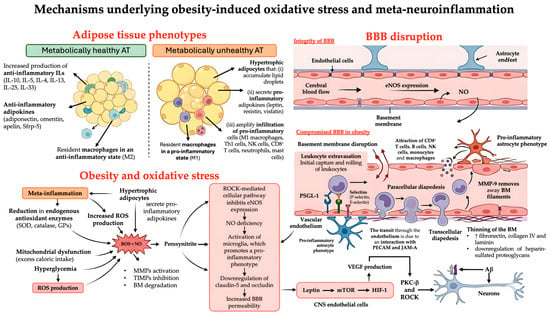
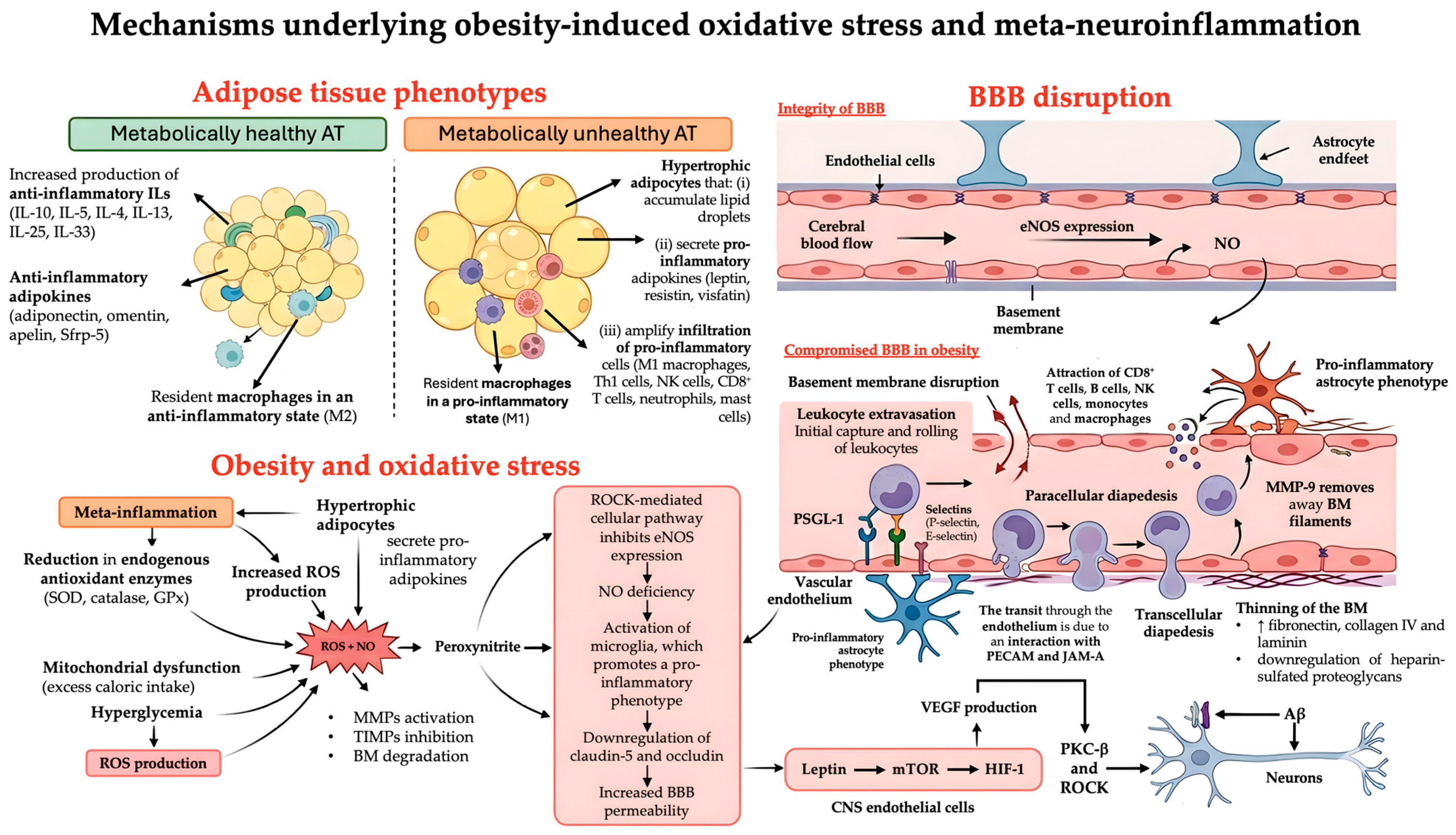
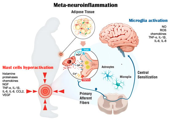
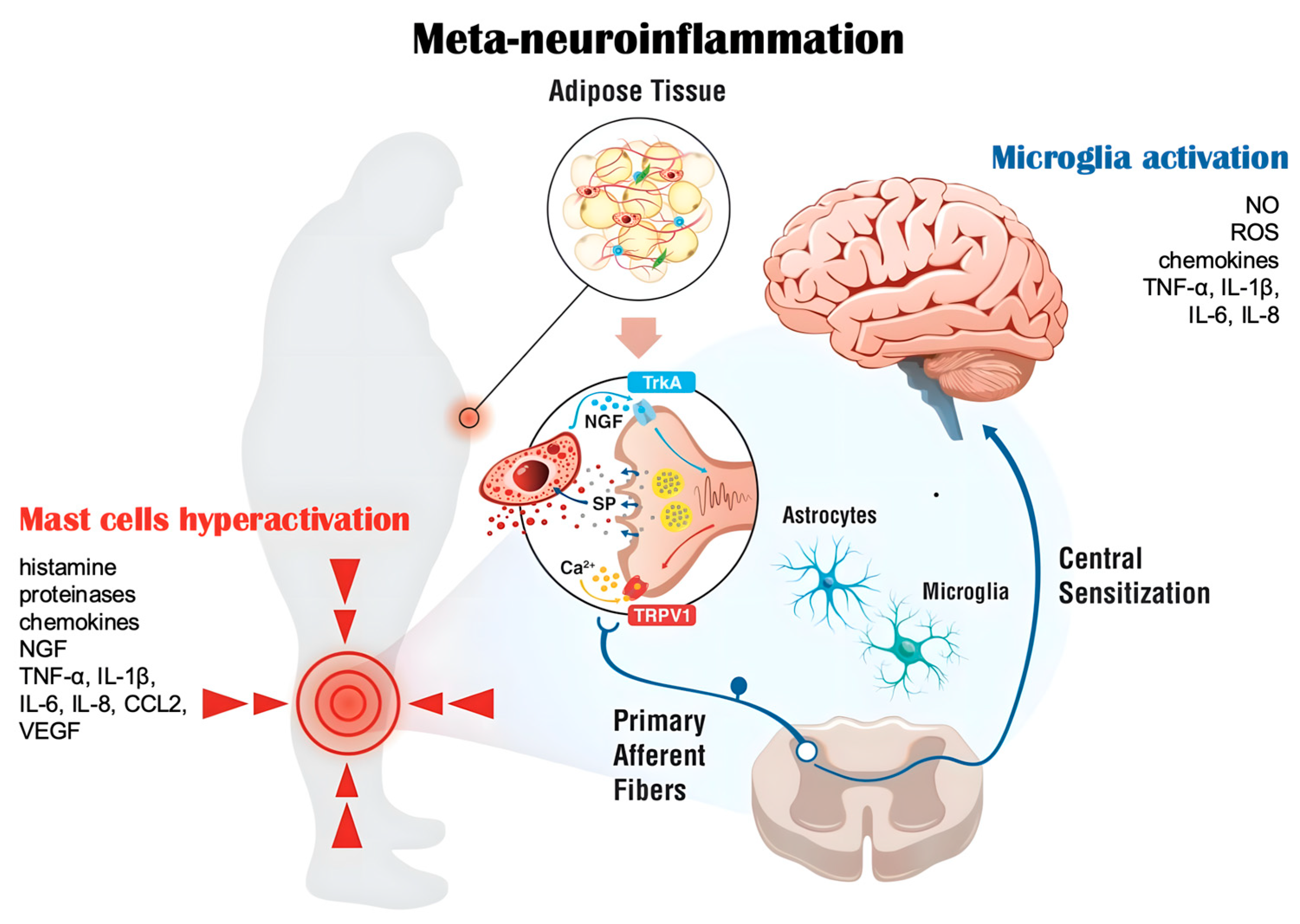
Disclaimer/Publisher’s Note: The statements, opinions and data contained in all publications are solely those of the individual author(s) and contributor(s) and not of MDPI and/or the editor(s). MDPI and/or the editor(s) disclaim responsibility for any injury to people or property resulting from any ideas, methods, instructions or products referred to in the content. |
© 2026 by the authors. Licensee MDPI, Basel, Switzerland. This article is an open access article distributed under the terms and conditions of the Creative Commons Attribution (CC BY) license.