Potential of Silver Nanoparticles in Imaging Diagnostics and Image-Guided Applications: A Narrative Review



Abstract
1. Introduction
2. Materials and Methods
3. Results and Discussion
3.1. AgNPs in Optical Imaging Diagnostics
3.2. CT Imaging
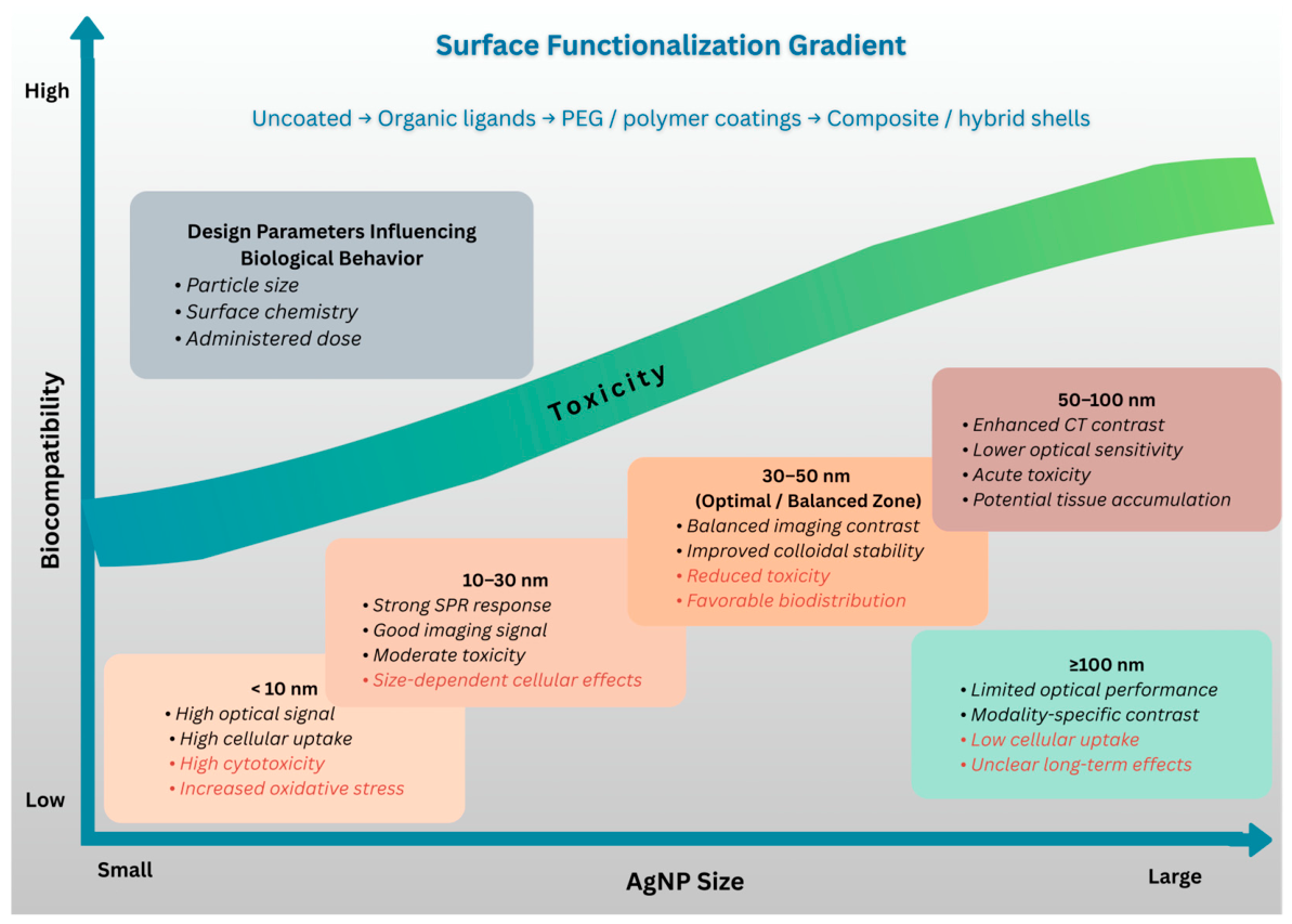
3.3. Biocompatibility and Toxicity
4. Conclusions
Author Contributions
Funding
Institutional Review Board Statement
Informed Consent Statement
Data Availability Statement
Conflicts of Interest
References
- Hussain, S. Modern Diagnostic Imaging Technique Applications and Risk Factors in the Medical Field: A Review. BioMed Res. Int. 2022, 2022, 5164970. [Google Scholar] [CrossRef]
- Ketineni, R.R.; Singh, B.; Chandralekha, A.R.; MS, I.; Soni, K.; Lodha, N.; Singh, B. Advances in Radiological Imaging Modalities and Their Expanding Role in the Early Diagnosis, Monitoring, and Prognosis of Internal Medicine Disorders: A Comprehensive Review. Cureus 2025, 17, e99513. [Google Scholar] [CrossRef]
- Butt, A.; Bach, H. Advancements in Nanotechnology for Diagnostics: A Literature Review, Part II: Advanced Techniques in Nuclear and Optical Imaging. Nanomedicine 2024, 20, 183–206. [Google Scholar] [CrossRef]
- Tripathi, D.; Hardaniya, M.; Pande, S.; Maity, D. Advances in Optical Contrast Agents for Medical Imaging: Fluorescent Probes and Molecular Imaging. J. Imaging 2025, 11, 87. [Google Scholar] [CrossRef]
- Mishra, V.; Kumari, N.; Vyas, M.; Aljabali, A.A.A.; Chattaraj, A.; Mishra, Y. Advances in Multimodal Imaging Techniques in Nanomedicine: Enhancing Drug Delivery Precision. RSC Adv. 2025, 15, 27187–27209. [Google Scholar] [CrossRef] [PubMed]
- Truong, T.T.; Mondal, S.; Doan, M.; Tak, S.; Choi, J.; Oh, H.; Nguyen, T.D.; Misra, M.; Lee, B.; Oh, J. Precision-Engineered Metal and Metal-Oxide Nanoparticles for Biomedical Imaging and Healthcare Applications. Adv. Colloid Interface Sci. 2024, 332, 103263. [Google Scholar] [CrossRef]
- Shahalaei, M.; Azad, A.K.; Mohd, W.; Derakhshani, A.; Mofakham, E.B.; Mallandrich, M.; Kumarasamy, V.; Subramaniyan, V. A Review of Metallic Nanoparticles: Present Issues and Prospects Focused on the Preparation Methods, Characterization Techniques, and Their Theranostic Applications. Front. Chem. 2024, 12, 1398979. [Google Scholar] [CrossRef] [PubMed]
- Ajib, R.; Shanmugaraj, K.; Yadav, R.M.; Brito, T.P.; Singh, D.P. Metallic Nanoparticles: From Biosynthesis to Biomedical Applications, Current Scenario and Prospects. Nanoscale Adv. 2026, 8, 2482–2511. [Google Scholar] [CrossRef]
- Georgeous, J.; AlSawaftah, N.; Abuwatfa, W.H.; Husseini, G.A. Review of Gold Nanoparticles: Synthesis, Properties, Shapes, Cellular Uptake, Targeting, Release Mechanisms and Applications in Drug Delivery and Therapy. Pharmaceutics 2024, 16, 1332. [Google Scholar] [CrossRef]
- Duman, H.; Akdaşçi, E.; Eker, F.; Bechelany, M.; Karav, S. Gold Nanoparticles: Multifunctional Properties, Synthesis, and Future Prospects. Nanomaterials 2024, 14, 1805. [Google Scholar] [CrossRef] [PubMed]
- Zhang, D.; Zhang, J.; Bian, X.; Zhang, P.; Wu, W.; Zuo, X. Iron Oxide Nanoparticle-Based T1 Contrast Agents for Magnetic Resonance Imaging: A Review. Nanomaterials 2024, 15, 33. [Google Scholar] [CrossRef]
- Aboushoushah, S.F. Iron Oxide Nanoparticles Enhancing Magnetic Resonance Imaging: A Review of the Latest Advancements. J. Sci. Adv. Mater. Devices 2025, 10, 100875. [Google Scholar] [CrossRef]
- Huang, X.; Zhang, Z.; Chen, J.; Mao, Z.; Zhu, H.; Liu, Y.; Zhu, Z.; Chen, H. One Dimensional Magneto-Optical Nanocomplex from Silver Nanoclusters and Magnetite Nanorods Containing Ordered Mesocages for Sensitive Detection of PD-L1. Biosens. Bioelectron. 2021, 189, 113385. [Google Scholar] [CrossRef]
- Lee, S.H.; Jun, B.-H. Silver Nanoparticles: Synthesis and Application for Nanomedicine. Int. J. Mol. Sci. 2019, 20, 865. [Google Scholar] [CrossRef]
- Sati, A.; Ranade, T.N.; Mali, S.N.; Yasin, A.; Pratap, A. Silver Nanoparticles (AgNPs): Comprehensive Insights into Bio/Synthesis, Key Influencing Factors, Multifaceted Applications, and Toxicity—A 2024 Update. ACS Omega 2025, 10, 7549–7582. [Google Scholar] [CrossRef]
- Alzahrani, R.A.; Alhaddad, F.G.; Alshammari, E.O.; Alsowaileh, F.S.; Alghamdi, M.D.; Modwi, A.; Goda, M.N.; Alqarni, L.S. Silver Nanoparticles in Gas Sensing: A Comprehensive Review of Synthesis, Mechanisms, Performance Metrics, and Emerging Applications. J. Sci. Adv. Mater. Devices 2025, 10, 100964. [Google Scholar] [CrossRef]
- Ahmad, M.Y.; Liu, S.; Tegafaw, T.; Al, A.; Zhao, D.; Liu, Y.; Nam, S.-W.; Chang, Y.; Lee, G.H. Heavy Metal-Based Nanoparticles as High-Performance X-Ray Computed Tomography Contrast Agents. Pharmaceuticals 2023, 16, 1463. [Google Scholar] [CrossRef] [PubMed]
- Jiang, Z.; Zhang, M.; Li, P.; Wang, Y.; Fu, Q. Nanomaterial-Based CT Contrast Agents and Their Applications in Image-Guided Therapy. Theranostics 2023, 13, 483–509. [Google Scholar] [CrossRef]
- Pivetta, T.P.; Botteon, C.E.A.; Ribeiro, P.A.; Marcato, P.D.; Raposo, M. Nanoparticle Systems for Cancer Phototherapy: An Overview. Nanomaterials 2021, 11, 3132. [Google Scholar] [CrossRef] [PubMed]
- Wei, G.; Yang, G.; Wang, Y.; Jiang, H.; Fu, Y.; Yue, G.; Ju, R. Phototherapy-Based Combination Strategies for Bacterial Infection Treatment. Theranostics 2020, 10, 12241–12262. [Google Scholar] [CrossRef]
- Li, J.; Wang, S.; Fontana, F.; Tapeinos, C.; Shahbazi, M.; Han, H.; Santos, H.A. Nanoparticles-Based Phototherapy Systems for Cancer Treatment: Current Status and Clinical Potential. Bioact. Mater. 2023, 23, 471–507. [Google Scholar] [CrossRef]
- Tavakkoli Yaraki, M.; Liu, B.; Tan, Y.N. Emerging Strategies in Enhancing Singlet Oxygen Generation of Nano-Photosensitizers toward Advanced Phototherapy. Nano-Micro Lett. 2022, 14, 123. [Google Scholar] [CrossRef]
- Cui, X.; Ruan, Q.; Zhuo, X.; Xia, X.; Hu, J.; Fu, R.; Li, Y.; Wang, J.; Xu, H. Photothermal Nanomaterials: A Powerful Light-To-Heat Converter. Chem. Rev. 2023, 123, 6891–6952. [Google Scholar] [CrossRef] [PubMed]
- Zhang, J.; Wang, J.; Fan, G.; Zhang, B.; Ma, G.; Xiao, H.; Wang, L. Daptomycin-Biomineralized Silver Nanoparticles for Enhanced Photothermal Therapy with Anti-Tumor Effect. Polymers 2022, 14, 2787. [Google Scholar] [CrossRef] [PubMed]
- Ren, Y.; Zhang, Y.; Li, X. Application of AgNPs in Biomedicine: An Overview and Current Trends. Nanotechnol. Rev. 2024, 13, 20240030. [Google Scholar] [CrossRef]
- Lee, J.S.; Kim, J.; Ye, Y.-S.; Kim, T.-I. Materials and Device Design for Advanced Phototherapy Systems. Adv. Drug Deliv. Rev. 2022, 186, 114339. [Google Scholar] [CrossRef]
- Mundekkad, D.; Cho, W.C. Nanoparticles in Clinical Translation for Cancer Therapy. Int. J. Mol. Sci. 2022, 23, 1685. [Google Scholar] [CrossRef]
- Kah, G.; Chandran, R.; Abrahamse, H. Biogenic Silver Nanoparticles for Targeted Cancer Therapy and Enhancing Photodynamic Therapy. Cells 2023, 12, 2012. [Google Scholar] [CrossRef]
- Yang, J.; Zhang, H.; Chen, B. Application of Nanoparticles to Reverse Multi-Drug Resistance in Cancer. Nanotechnol. Rev. 2016, 5, 489–496. [Google Scholar] [CrossRef]
- Liu, S.; Phillips, S.; Northrup, S.; Levi, N. The Impact of Silver Nanoparticle-Induced Photothermal Therapy and Its Augmentation of Hyperthermia on Breast Cancer Cells Harboring Intracellular Bacteria. Pharmaceutics 2023, 15, 2466. [Google Scholar] [CrossRef]
- Melo, R.M.; Albuquerque, G.M.; Monte, J.P.; Pereira, G.A.L.; Pereira, G. Recent Advances in the Application of Silver Nanoparticles for Enhancing Phototherapy Outcomes. Pharmaceuticals 2025, 18, 970. [Google Scholar] [CrossRef]
- Kurul, F.; Turkmen, H.; Cetin, A.E.; Topkaya, S.N. Nanomedicine: How Nanomaterials Are Transforming Drug Delivery, Bio-Imaging, and Diagnosis. Next Nanotechnol. 2025, 7, 100129. [Google Scholar] [CrossRef]
- Laib, I.; Gheraissa, N.; Benaissa, A.; Benkhira, L.; Azzi, M.; Benaissa, Y.; Abdelaziz, A.G.; Tian, F.; Walsh, M.; Bechelany, M.; et al. Tailoring Innovative Silver Nanoparticles for Modern Medicine: The Importance of Size and Shape Control and Functional Modifications. Mater. Today Bio 2025, 33, 102071. [Google Scholar] [CrossRef]
- Singh, P.; Pandit, S.; Balusamy, S.R.; Madhusudanan, M.; Singh, H.; Amsath, M.; Mijakovic, I. Advanced Nanomaterials for Cancer Therapy: Gold, Silver, and Iron Oxide Nanoparticles in Oncological Applications. Adv. Healthc. Mater. 2024, 14, 2403059. [Google Scholar] [CrossRef]
- Salgueiro, M.J.; Zubillaga, M. Theranostic Nanoplatforms in Nuclear Medicine: Current Advances, Emerging Trends, and Perspectives for Personalized Oncology. J. Nanotheranostics 2025, 6, 27. [Google Scholar] [CrossRef]
- Hosny, S.; Gaber, G.A.; Ragab, M.S.; Ragheb, M.A.; Anter, M.; Mohamed, L.Z. A Comprehensive Review of Silver Nanoparticles (AgNPs): Synthesis Strategies, Toxicity Concerns, Biomedical Applications, AI-Driven Advancements, Challenges, and Future Perspectives. Arab. J. Sci. Eng. 2025, 1–48. [Google Scholar] [CrossRef]
- Gupta, D.; Roy, I.; Gandhi, S. Metallic Nanoparticles for CT-Guided Imaging of Tumors and Their Therapeutic Applications. OpenNano 2023, 12, 100146. [Google Scholar] [CrossRef]
- Zhang, X.-F.; Shen, W.; Gurunathan, S. Silver Nanoparticle-Mediated Cellular Responses in Various Cell Lines: An in Vitro Model. Int. J. Mol. Sci. 2016, 17, 1603. [Google Scholar] [CrossRef] [PubMed]
- Serkova, N.J.; Glunde, K.; Haney, C.R.; Farhoud, M.; De Lille, A.; Redente, E.F.; Simberg, D.; Westerly, D.; Griffin, L.; Mason, R.P. Preclinical Applications of Multi-Platform Imaging in Animal Models of Cancer. Cancer Res. 2021, 81, 1189–1200. [Google Scholar] [CrossRef]
- Bruzgo-Grzybko, M.; Kalita, I.S.; Olichwier, A.J.; Bielicka, N.; Chabielska, E.; Gromotowicz-Poplawska, A. Preclinical PET and SPECT Imaging in Small Animals: Technologies, Challenges and Translational Impact. Cells 2025, 15, 73. [Google Scholar] [CrossRef]
- Zhang, R.; Dias, M.; Li, Y.; Rütten, S.; Kiessling, F.; Lammers, T.; Pallares, R.M. Structural Engineering of Silver Nanoparticles for Enhanced Photoacoustic Imaging. Nanoscale Adv. 2025, 7, 6110–6119. [Google Scholar] [CrossRef]
- Fong, K.E.; Yung, L.-Y.L. Localized Surface Plasmon Resonance: A Unique Property of Plasmonic Nanoparticles for Nucleic Acid Detection. Nanoscale 2013, 5, 12043–12071. [Google Scholar] [CrossRef]
- Pryshchepa, O.; Pomastowski, P.; Buszewski, B. Silver Nanoparticles: Synthesis, Investigation Techniques, and Properties. Adv. Colloid Interface Sci. 2020, 284, 102246. [Google Scholar] [CrossRef]
- Amirjani, A.; Koochak, N.N.; Haghshenas, D.F. Investigating the Shape and Size-Dependent Optical Properties of Silver Nanostructures Using UV–Vis Spectroscopy. J. Chem. Educ. 2019, 96, 2584–2589. [Google Scholar] [CrossRef]
- Khalid, A.; Tomljenovic-Hanic, S. Emerging Fluorescent Nanoparticles for Non-Invasive Bioimaging. Molecules 2024, 29, 5594. [Google Scholar] [CrossRef] [PubMed]
- Nadhiya, K.; Sivakumar, K.; Balamurugan, M.; Abhishek, A. The Development of a Fluorescence Sensor Using Rh6G@AgNPs to Detect Fe3+ Ions in Water Samples and for Cell Imaging. J. Mol. Struct. 2025, 1346, 143203. [Google Scholar] [CrossRef]
- Kenmotsu, S.; Hirasawa, M.; Tamadate, T.; Matsumoto, C.; Osone, S.; Inomata, Y.; Seto, T. Surface-Enhanced Raman Scattering on Size-Classified Silver Nanoparticles Generated by Laser Ablation. ACS Omega 2024, 9, 37716–37723. [Google Scholar] [CrossRef] [PubMed]
- Yu, P.; Shen, C.; Yin, X.; Cheng, J.; Liu, C.; Yu, Z. Au-Ag Bimetallic Nanoparticles for Surface-Enhanced Raman Scattering (SERS) Detection of Food Contaminants: A Review. Foods 2025, 14, 2109. [Google Scholar] [CrossRef]
- Mammari, N.; Duval, R.E. Photothermal/Photoacoustic Therapy Combined with Metal-Based Nanomaterials for the Treatment of Microbial Infections. Microorganisms 2023, 11, 2084. [Google Scholar] [CrossRef]
- Zhu, S.; Song, W.; He, Y.; Wang, Y.; Li, X.; Wu, Y.; Meng, X.; Lin, C.; Wang, W.; Wang, H.; et al. Biosynthetic Ag2S Nanoparticles for Photoacoustic Imaging-Guided Photothermal Therapy. ACS Appl. Mater. Interfaces 2025, 17, 37465–37478. [Google Scholar] [CrossRef]
- Nie, S. Probing Single Molecules and Single Nanoparticles by Surface-Enhanced Raman Scattering. Science 1997, 275, 1102–1106. [Google Scholar] [CrossRef]
- Kneipp, K.; Wang, Y.; Kneipp, H.; Perelman, L.T.; Itzkan, I.; Dasari, R.R.; Feld, M.S. Single Molecule Detection Using Surface-Enhanced Raman Scattering (SERS). Phys. Rev. Lett. 1997, 78, 1667–1670. [Google Scholar] [CrossRef]
- Ma, K.; Yuen, J.M.; Shah, N.C.; Walsh, J.T.; Glucksberg, M.R.; Van, R.P. In Vivo, Transcutaneous Glucose Sensing Using Surface-Enhanced Spatially Offset Raman Spectroscopy: Multiple Rats, Improved Hypoglycemic Accuracy, Low Incident Power, and Continuous Monitoring for Greater than 17 Days. Anal. Chem. 2011, 83, 9146–9152. [Google Scholar] [CrossRef]
- Guerrini, L.; Garcia-Ramos, J.V.; Domingo, C.; Sanchez-Cortes, S. Sensing Polycyclic Aromatic Hydrocarbons with Dithiocarbamate-Functionalized Ag Nanoparticles by Surface-Enhanced Raman Scattering. Anal. Chem. 2009, 81, 953–960. [Google Scholar] [CrossRef]
- Yue, Z.; Zhuang, F.; Kumar, R.; Wong, I.; Cronin, S.B.; Liu, Y.-H. Cell Kinase Activity Assay Based on Surface Enhanced Raman Spectroscopy. Spectrochim. Acta Part A Mol. Biomol. Spectrosc. 2009, 73, 226–230. [Google Scholar] [CrossRef][Green Version]
- Gessner, R.; Rösch, P.; Kiefer, W.; Popp, J. Raman Spectroscopy Investigation of Biological Materials by Use of Etched and Silver Coated Glass Fiber Tips. Biopolymers 2002, 67, 327–330. [Google Scholar] [CrossRef] [PubMed]
- Lucotti, A.; Pesapane, A.; Zerbi, G. Use of a Geometry Optimized Fiber-Optic Surface-Enhanced Raman Scattering Sensor in Trace Detection. Appl. Spectrosc. 2007, 61, 260–268. [Google Scholar] [CrossRef] [PubMed]
- Ji, X.; Xu, S.; Wang, L.; Liu, M.; Pan, K.; Yuan, H.; Ma, L.; Xu, W.; Li, J.; Bai, Y.; et al. Immunoassay Using the Probe-Labeled Au/Ag Core-Shell Nanoparticles Based on Surface-Enhanced Raman Scattering. Colloid Surf. A Physicochem. Eng. Asp. 2005, 257–258, 171–175. [Google Scholar] [CrossRef]
- Griaznova, O.Y.; Belyaev, I.B.; Sogomonyan, A.S.; Zelepukin, I.V.; Tikhonowski, G.V.; Popov, A.A.; Komlev, A.S.; Nikitin, P.I.; Gorin, D.A.; Kabashin, A.V.; et al. Laser Synthesized Core-Satellite Fe-Au Nanoparticles for Multimodal in Vivo Imaging and in Vitro Photothermal Therapy. Pharmaceutics 2022, 14, 994. [Google Scholar] [CrossRef]
- Tiwari, H.; Singh, S.; Kumar, R.; Mandal, A.; Pathak, A.; Verma, N.K.; Kumar, L.; Gautam, V. Novel Advancements in Nanomaterials-Based Contrast Agents across Multimodal Imaging and Theranostic Applications. Nanoscale Adv. 2025, 7, 6753–6773. [Google Scholar] [CrossRef]
- Birch, G.P.; Campbell, T.; Bradley, M.; Dhaliwal, K. Optical Molecular Imaging of Inflammatory Cells in Interventional Medicine—An Emerging Strategy. Front. Oncol. 2019, 9, 882. [Google Scholar] [CrossRef]
- Staderini, M.; Megia-Fernandez, A.; Dhaliwal, K.; Bradley, M. Peptides for Optical Medical Imaging and Steps towards Therapy. Bioorg. Med. Chem. 2018, 26, 2816–2826. [Google Scholar] [CrossRef]
- Ashton, J.R.; West, J.L.; Badea, C.T. In Vivo Small Animal Micro-CT Using Nanoparticle Contrast Agents. Front. Pharmacol. 2015, 6, 256. [Google Scholar] [CrossRef]
- Das, N.M.; Hatsell, S.; Nannuru, K.; Huang, L.; Wen, X.; Wang, L.; Wang, L.-H.; Idone, V.; Meganck, J.A.; Murphy, A.; et al. In Vivo Quantitative Microcomputed Tomographic Analysis of Vasculature and Organs in a Normal and Diseased Mouse Model. PLoS ONE 2016, 11, e0150085. [Google Scholar] [CrossRef]
- Luengo Morato, Y.; Ovejero Paredes, K.; Lozano Chamizo, L.; Marciello, M.; Filice, M. Recent Advances in Multimodal Molecular Imaging of Cancer Mediated by Hybrid Magnetic Nanoparticles. Polymers 2021, 13, 2989. [Google Scholar] [CrossRef]
- More, P.R.; Pandit, S.; Filippis, A.D.; Franci, G.; Mijakovic, I.; Galdiero, M. Silver Nanoparticles: Bactericidal and Mechanistic Approach against Drug Resistant Pathogens. Microorganisms 2023, 11, 369. [Google Scholar] [CrossRef]
- Gomes, M.; Ramalho, M.J.; Loureiro, J.A.; Pereira, M.C. Advancing Brain Targeting: Cost-Effective Surface-Modified Nanoparticles for Faster Market Entry. Pharmaceutics 2025, 17, 661. [Google Scholar] [CrossRef]
- Le, H.; Vu, G.T.T.; Abioye, A.; Adejare, A. Recent Advances in Nanoparticle-Based Drug Delivery Strategies to Cross the Blood–Brain Barrier in Targeted Treatment of Alzheimer’s Disease. Pharmaceutics 2026, 18, 192. [Google Scholar] [CrossRef]
- Qi, S.; Wang, X.; Chang, K.; Shen, W.; Yu, G.; Du, J. The Bright Future of Nanotechnology in Lymphatic System Imaging and Imaging-Guided Surgery. J. Nanobiotechnol. 2022, 20, 24. [Google Scholar] [CrossRef]
- Han, C.Y.; Choi, S.-H.; Chi, S.-H.; Hong, J.H.; Cho, Y.-E.; Kim, J. Nano-Fluorescence Imaging: Advancing Lymphatic Disease Diagnosis and Monitoring. Nano Converg. 2024, 11, 53. [Google Scholar] [CrossRef]
- Altinbasak, I.; Alp, Y.; Sanyal, R.; Sanyal, A. Theranostic Nanogels: Multifunctional Agents for Simultaneous Therapeutic Delivery and Diagnostic Imaging. Nanoscale 2024, 16, 14033–14056. [Google Scholar] [CrossRef] [PubMed]
- Adhipandito, C.F.; Cheung, S.-H.; Lin, Y.-H.; Wu, S.-H. Atypical Renal Clearance of Nanoparticles Larger than the Kidney Filtration Threshold. Int. J. Mol. Sci. 2021, 22, 11182. [Google Scholar] [CrossRef] [PubMed]
- Nieves, L.M.; Dong, Y.C.; Rosario-Berríos, D.N.; Mossburg, K.; Hsu, J.C.; Cramer, G.M.; Busch, T.M.; Maidment, A.D.A.; Cormode, D.P. Renally Excretable Silver Telluride Nanoparticles as Contrast Agents for X-Ray Imaging. ACS Appl. Mater. Interfaces 2022, 14, 34354–34364. [Google Scholar] [CrossRef] [PubMed]
- Che, X.; Yang, C.; Pan, L.; Gu, D.; Dai, G.; Shu, J.; Yang, L. Achieving Safe and High-Performance Gastrointestinal Tract Spectral CT Imaging with Small-Molecule Lanthanide Complex. Biomater. Res. 2023, 27, 119. [Google Scholar] [CrossRef]
- Qi, M.; Wang, X.; Chen, J.; Liu, Y.; Liu, Y.; Jia, J.; Li, L.; Yue, T.; Gao, L.; Yan, B.; et al. Transformation, Absorption and Toxicological Mechanisms of Silver Nanoparticles in the Gastrointestinal Tract Following Oral Exposure. ACS Nano 2023, 17, 8851–8865. [Google Scholar] [CrossRef]
- Kose, O.; Béal, D.; Motellier, S.; Pelissier, N.; Collin-Faure, V.; Blosi, M.; Bengalli, R.; Costa, A.; Furxhi, I.; Mantecca, P.; et al. Physicochemical Transformations of Silver Nanoparticles in the Oro-Gastrointestinal Tract Mildly Affect Their Toxicity to Intestinal Cells in Vitro: An AOP-Oriented Testing Approach. Toxics 2023, 11, 199. [Google Scholar] [CrossRef]
- Gao, Z.; Hao, Y.; Zheng, M.; Chen, Y. A Fluorescent Dye with Large Stokes Shift and High Stability: Synthesis and Application to Live Cell Imaging. RSC Adv. 2017, 7, 7604–7609. [Google Scholar] [CrossRef]
- Wagner, A.M.; Knipe, J.M.; Orive, G.; Peppas, N.A. Quantum Dots in Biomedical Applications. Acta Biomater. 2019, 94, 44–63. [Google Scholar] [CrossRef]
- Xue, C.; Xue, Y.; Dai, L.; Urbas, A.; Li, Q. Size- and Shape-Dependent Fluorescence Quenching of Gold Nanoparticles on Perylene Dye. Adv. Opt. Mater. 2013, 1, 581–587. [Google Scholar] [CrossRef]
- Wolfbeis, O.S. An Overview of Nanoparticles Commonly Used in Fluorescent Bioimaging. Chem. Soc. Rev. 2015, 44, 4743–4768. [Google Scholar] [CrossRef]
- Lyu, N.; Hassanzadeh-Barforoushi, A.; Rey, L.M.; Zhang, W.; Wang, Y. SERS Biosensors for Liquid Biopsy towards Cancer Diagnosis by Detection of Various Circulating Biomarkers: Current Progress and Perspectives. Nano Converg. 2024, 11, 22. [Google Scholar] [CrossRef] [PubMed]
- Luo, J.; Sun, J.; Chen, H.; Zhao, C.; Ye, M. A Review of High-Sensitivity SERS-Active Photonic Crystal Fiber Sensors for Chemical and Biological Detection. Sensors 2025, 25, 6982. [Google Scholar] [CrossRef] [PubMed]
- Fu, S.-Y.; Xu, S.; Li, H.; Guo, X.-M.; Lin, J.-S.; Guan, B.; Chen, B.; Wang, T.; Zhang, Y.-J.; Li, J.-F. A High Sensitivity Prostate-Specific Antigen SERS Detection Platform Based on Laser Resonance Nanoparticles. Nanoscale 2025, 17, 5171–5180. [Google Scholar] [CrossRef] [PubMed]
- Mondal, I.; Raj, S.; Roy, P.; Poddar, R. Silver Nanoparticles (AgNPs) as a Contrast Agent for Imaging of Animal Tissue Using Swept-Source Optical Coherence Tomography (SSOCT). Laser Phys. 2017, 28, 015601. [Google Scholar] [CrossRef]
- Xu, L.; Wang, Y.-Y.; Huang, J.; Chen, C.-Y.; Wang, Z.-X.; Xie, H. Silver Nanoparticles: Synthesis, Medical Applications and Biosafety. Theranostics 2020, 10, 8996–9031. [Google Scholar] [CrossRef]
- Al-Thani, A.N.; Jan, A.G.; Abbas, M.; Geetha, M.; Sadasivuni, K.K. Nanoparticles in Cancer Theragnostic and Drug Delivery: A Comprehensive Review. Life Sci. 2024, 352, 122899. [Google Scholar] [CrossRef]
- Tran, V.T.; Ju, H. Fluorescence Based on Surface Plasmon Coupled Emission for Ultrahigh Sensitivity Immunoassay of Cardiac Troponin I. Biomedicines 2021, 9, 448. [Google Scholar] [CrossRef]
- Dasgupta, S.; Ray, K. Plasmon-Enhanced Fluorescence for Biophotonics and Bio-Analytical Applications. Front. Chem. 2024, 12, 1407561. [Google Scholar] [CrossRef]
- Yang, Q.; Wu, Y.; Chen, J.; Lu, M.; Wang, X.; Zhang, Z.; Xiong, H.; Choo, J.; Chen, L. Plasmonic Nanomaterial-Enhanced Fluorescence and Raman Sensors: Multifunctional Platforms and Applications. Coord. Chem. Rev. 2024, 507, 215768. [Google Scholar] [CrossRef]
- Zhang, P.; Zhou, X.; Wang, S. Plasmonic Scattering Microscopy for Label-Free Imaging of Molecular Binding Kinetics: From Single Molecules to Single Cells. Chem. Methods 2023, 3, e202200066. [Google Scholar] [CrossRef]
- Xue, R.-S.; Dai, J.-Y.; Wang, X.-J.; Chen, M.-Y. Research Progress of Surface-Enhanced Raman Scattering (SERS) Technology in Food, Biomedical, and Environmental Monitoring. Photonics 2025, 12, 809. [Google Scholar] [CrossRef]
- Cialla-May, D.; Bonifacio, A.; Markin, A.; Markina, N.; Fornasaro, S.; Dwivedi, A.; Dib, T.; Farnesi, E.; Liu, C.; Ghosh, A.; et al. Recent Advances of Surface Enhanced Raman Spectroscopy (SERS) in Optical Biosensing. TrAC Trends Anal. Chem. 2024, 181, 117990. [Google Scholar] [CrossRef]
- Wang, S.; Lin, J.; Wang, T.; Chen, X.; Huang, P. Recent Advances in Photoacoustic Imaging for Deep-Tissue Biomedical Applications. Theranostics 2016, 6, 2394–2413. [Google Scholar] [CrossRef]
- Liu, Y.; Bhattarai, P.; Dai, Z.; Chen, X. Photothermal Therapy and Photoacoustic Imaging via Nanotheranostics in Fighting Cancer. Chem. Soc. Rev. 2019, 48, 2053–2108. [Google Scholar] [CrossRef]
- Wołąkiewicz, G.; Pietrzak, M.; Szabelski, M. Silver Nanoparticles Improve Fluorophore Photostability: Application to a Hypericin Study. Int. J. Mol. Sci. 2024, 25, 9963. [Google Scholar] [CrossRef]
- Maiti, P.; Sarkar, S.; Singha, T.; Roy, S.D.; Mahato, M.; Karmakar, P.; Paul, S.; Paul, P.K. Enhancement of Fluorescence Mediated by Silver Nanoparticles: Implications for Cell Imaging. Langmuir 2023, 39, 6713–6729. [Google Scholar] [CrossRef]
- Lee, D.; Song, J.; Song, G.; Pang, Y. Metal-Enhanced Fluorescence of Dyes with Quadrupole Surface Plasmon Resonance of Silver Nanoparticles. Nanoscale Adv. 2022, 4, 2794–2805. [Google Scholar] [CrossRef] [PubMed]
- Lee, S.; Kang, S.H. Wavelength-Dependent Metal-Enhanced Fluorescence Biosensors via Resonance Energy Transfer Modulation. Biosensors 2023, 13, 376. [Google Scholar] [CrossRef]
- Shi, Y.; Wang, L.; Li, L.; Feng, C.; Cao, Y. Innovative Progress of LSPR-Based Dark-Field Scattering Spectral Imaging in the Biomedical Assay at the Single-Particle Level. Chem. Open 2025, 14, e202400017. [Google Scholar] [CrossRef] [PubMed]
- Gao, P.F.; Lei, G.; Huang, C.Z. Dark-Field Microscopy: Recent Advances in Accurate Analysis and Emerging Applications. Anal. Chem. 2021, 93, 4707–4726. [Google Scholar] [CrossRef] [PubMed]
- Mehta, N.; Sahu, S.P.; Shaik, S.; Devireddy, R.; Gartia, M.R. Dark-Field Hyperspectral Imaging for Label Free Detection of Nano-Bio-Materials. WIREs Nanomed. Nanobiotechnol. 2020, 13, e1661. [Google Scholar] [CrossRef]
- Tran, M.H.; Ma, L.; Yuan, M.; Fei, B. Medical Hyperspectral Imaging: An Updated Review of Technology Advancements and Biomedical Applications. J. Biomed. Opt. 2026, 31, 030901. [Google Scholar] [CrossRef]
- Nadeem, A.; Rahmani, M.; Wang, Y.; Yari, S.; Lopez, R.M.; Kim, M.; Roxbury, D. Spectral Fingerprinting of Engineered Nanomaterials for Precision Biosensing. ACS Nano 2026, 20, 3921–3943. [Google Scholar] [CrossRef]
- Truong, H.; Hoang, T.; Chung, K.; Lee, L.P. Plasmonic Biosensors and Actuators for Integrated Point-of-Care Diagnostics. npj Biosens. 2025, 2, 45. [Google Scholar] [CrossRef]
- Datta, R.; Heaster, T.M.; Sharick, J.T.; Gillette, A.A.; Skala, M.C. Fluorescence Lifetime Imaging Microscopy: Fundamentals and Advances in Instrumentation, Analysis, and Applications. J. Biomed. Opt. 2020, 25, 1. [Google Scholar] [CrossRef] [PubMed]
- Nandhini, G.U.; Minakshi, M.; Sivasubramanian, R.; Dharmalingam, G. Metal Manipulated Fluorescence: Mechanisms, Materials, and Plasmonic Strategies for Enhanced Emission. Nanomaterials 2026, 16, 298. [Google Scholar] [CrossRef]
- Connor, T.; Weerasinghe, H.; Lathia, J.; Burda, C.; Yildirim, M. Advances in Deep Brain Imaging with Quantum Dots: Structural, Functional, and Disease-Specific Roles. Photonics 2024, 12, 3. [Google Scholar] [CrossRef]
- Adhikari, S.; Spaeth, P.; Kar, A.; Baaske, M.D.; Khatua, S.; Orrit, M. Photothermal Microscopy: Imaging the Optical Absorption of Single Nanoparticles and Single Molecules. ACS Nano 2020, 14, 16414–16445. [Google Scholar] [CrossRef] [PubMed]
- Miyazaki, J.; Kobayahsi, T. Photothermal Microscopy for High Sensitivity and High Resolution Absorption Contrast Imaging of Biological Tissues. Photonics 2017, 4, 32. [Google Scholar] [CrossRef]
- Kim, D.; Kim, H. Analysis of Temperature Behavior in Biological Tissue in Photothermal Therapy according to Laser Irradiation Angle. Bioengineered 2023, 14, 2252668. [Google Scholar] [CrossRef]
- Zhang, W.; Zang, Y.; Lu, Y.; Han, J.; Xiong, Q.; Xiong, J. Photothermal Effect and Multi-Modality Imaging of Up-Conversion Nanomaterial Doped with Gold Nanoparticles. Int. J. Mol. Sci. 2022, 23, 1382. [Google Scholar] [CrossRef]
- Zhang, Y.; Li, Z.; Du, Z.; Pan, J.; Huang, Y. Multifunctional Upconversion Nanoparticles Transforming Photoacoustic Imaging: A Review. Nanomaterials 2025, 15, 1074. [Google Scholar] [CrossRef]
- Varghese, N.; Das, R.; Nair, Y. Upconversion Nanoparticles for Detection of Small Biomolecules and Ions. In Upconversion Nanoparticles for Biomedical Applications; Elsevier: Amsterdam, The Netherlands, 2026; pp. 623–679. ISBN 9780443266614. [Google Scholar]
- Petrik, V.; Apok, V.; Britton, J.A.; Bell, B.A.; Papadopoulos, M.C. Godfrey Hounsfield and the Dawn of Computed Tomography. Neurosurgery 2006, 58, 780–787. [Google Scholar] [CrossRef] [PubMed]
- Arlazarov, V.L.; Nikolaev, D.P.; Arlazarov, V.V.; Chukalina, M.V. X-Ray Tomography: The Way from Layer-By-Layer Radiography to Computed Tomography. Comp. Opt. 2021, 45, 897–906. [Google Scholar] [CrossRef]
- Ramaekers, M.; Viviers, C.; Hellström, T.; Ewals, L.; Tasios, N.; Jacobs, I.; Nederend, J.; Sommen, F.; Luyer, M.; on behalf of the E/MTIC Oncology Collaborative Group. Improved Pancreatic Cancer Detection and Localization on CT Scans: A Computer-Aided Detection Model Utilizing Secondary Features. Cancers 2024, 16, 2403. [Google Scholar] [CrossRef]
- Bourdoncle, S.; Eche, T.; McGale, J.; Yiu, K.; Partouche, E.; Yeh, R.; Ammari, S.; Rousseau, H.; Dercle, L.; Mokrane, F.-Z. Investigating of the Role of CT Scan for Cancer Patients during the First Wave of COVID-19 Pandemic. Res. Diagn. Interv. Imaging 2022, 1, 100004. [Google Scholar] [CrossRef]
- Kuruc, R.; Szórádová, A.; Šikuta, J.; Mikuláš, Ľ.; Šidlo, J. A Comparative Study of Intravital CT and Autopsy Findings in Fatal Traumatic Injuries. Healthcare 2022, 10, 1465. [Google Scholar] [CrossRef] [PubMed]
- Shariaty, F.; Orooji, M.; Velichko, E.N.; Zavjalov, S.V. Texture Appearance Model, a New Model-Based Segmentation Paradigm, Application on the Segmentation of Lung Nodule in the CT Scan of the Chest. Comput. Biol. Med. 2022, 140, 105086. [Google Scholar] [CrossRef] [PubMed]
- Caschera, L.; Lazzara, A.; Piergallini, L.; Ricci, D.; Tuscano, B.; Vanzulli, A. Contrast Agents in Diagnostic Imaging: Present and Future. Pharmacol. Res. 2016, 110, 65–75. [Google Scholar] [CrossRef]
- Padovani, R.P.; Kasamatsu, T.S.; Nakabashi, C.C.D.; Camacho, C.P.; Andreoni, D.M.; Malouf, E.Z.; Marone, M.M.S.; Maciel, R.M.B.; Biscolla, R.P.M. One Month Is Sufficient for Urinary Iodine to Return to Its Baseline Value after the Use of Water-Soluble Iodinated Contrast Agents in Post-Thyroidectomy Patients Requiring Radioiodine Therapy. Thyroid 2012, 22, 926–930. [Google Scholar] [CrossRef]
- Khumaeni, A.; Setia Budi, W.; Avicenna, S.; Muniroh, M.; Kusumaningrum, N.; Damayanti, O.; Fitria, S. Gold and Silver Nanoparticles as Computed Tomography (Ct) Contrast Agents Produced by a Pulsed Laser Ablation Technique: Study In-Vitro and In-Vivo. Rasayan J. Chem. 2023, 16, 502–508. [Google Scholar] [CrossRef]
- Starosolski, Z.; Villamizar, C.; Rendon, D.A.; Paldino, M.J.; Milewicz, D.M.; Ghaghada, K.B.; Annapragada, A. Ultra High-Resolution in Vivo Computed Tomography Imaging of Mouse Cerebrovasculature Using a Long Circulating Blood Pool Contrast Agent. Sci. Rep. 2015, 5, 10178. [Google Scholar] [CrossRef]
- Dou, Y.; Guo, Y.; Li, X.; Li, X.; Wang, S.; Wang, L.; Lv, G.; Zhang, X.; Wang, H.; Gong, X.; et al. Size-Tuning Ionization to Optimize Gold Nanoparticles for Simultaneous Enhanced CT Imaging and Radiotherapy. ACS Nano 2016, 10, 2536–2548. [Google Scholar] [CrossRef]
- Gandhi, S.; Roy, I. Synthesis and Characterization of Manganese Ferrite Nanoparticles, and Its Interaction with Bovine Serum Albumin: A Spectroscopic and Molecular Docking Approach. J. Mol. Liq. 2019, 296, 111871. [Google Scholar] [CrossRef]
- Singh, A.; Amiji, M.M. Application of Nanotechnology in Medical Diagnosis and Imaging. Curr. Opin. Biotechnol. 2022, 74, 241–246. [Google Scholar] [CrossRef]
- Charmi, J.; Salehiabar, M.; Ghaffarlou, M.; Danafar, H.; Nosrati, H. Nanoparticle Based CT Contrast Agents. In Harnessing Materials for X-Ray Based Cancer Therapy and Imaging; Nanomedicine and Nanotoxicology; Sharma, S.K., Nosrati, H., Kavetskyy, T., Eds.; Springer: Cham, Switzerland, 2022; pp. 217–240. [Google Scholar] [CrossRef]
- Pereira, J.; Proaño, O.; Albán, A.; Zambonino, M.; Mouheb, L.; Desmau, M.; Rahman, A.; Agathos, S.N.; Dahoumane, S.A. Paradoxical Features Empower Biogenic Silver Nanoparticles. Molecules 2025, 30, 4152. [Google Scholar] [CrossRef]
- Yang, L.; Kuang, H.; Zhang, W.; Aguilar, Z.P.; Wei, H.; Xu, H. Comparisons of the Biodistribution and Toxicological Examinations after Repeated Intravenous Administration of Silver and Gold Nanoparticles in Mice. Sci. Rep. 2017, 7, 3303. [Google Scholar] [CrossRef]
- Mbanga, O.; Cukrowska, E.; Gulumian, M. A Comparative Study of the Biodurability and Persistence of Gold, Silver and Titanium Dioxide Nanoparticles Using the Continuous Flow through System. Nanomaterials 2023, 13, 1653. [Google Scholar] [CrossRef]
- Kamyab, H.; Khalili, E.; Yuzir, A.; Taheri, M.M.; Zambrano, A.K.; Rajendran, S. Emerging Nanoparticle-Based Strategies for Advanced Cancer Imaging and Diagnosis. Int. J. Pharm. 2025, 683, 126046. [Google Scholar] [CrossRef]
- Bartolomé, M.; Villaseñor, M.J.; Ríos, Á. Surface Plasmon Resonance Optical Sensors Involving Nanomaterials as Reliable Analytical Tools: A Critical View about Performance and Applications. Anal. Chim. Acta 2025, 1382, 344693. [Google Scholar] [CrossRef]
- Pilot, R.; Signorini, R.; Durante, C.; Orian, L.; Bhamidipati, M.; Fabris, L. A Review on Surface-Enhanced Raman Scattering. Biosensors 2019, 9, 57. [Google Scholar] [CrossRef]
- Chang, H.; Hur, W.; Kang, H.; Jun, B.-H. In Vivo Surface-Enhanced Raman Scattering Techniques: Nanoprobes, Instrumentation, and Applications. Light Sci. Appl. 2025, 14, 79. [Google Scholar] [CrossRef]
- Steinberg, I.; Huland, D.M.; Vermesh, O.; Frostig, H.E.; Tummers, W.S.; Gambhir, S.S. Photoacoustic Clinical Imaging. Photoacoustics 2019, 14, 77–98. [Google Scholar] [CrossRef]
- Wang, L.; Zeng, W.; Long, K.; Chen, H.; Lan, R.; Liu, L.; Siok, W.T.; Wang, N. Advances in Photoacoustic Imaging Reconstruction and Quantitative Analysis for Biomedical Applications. Vis. Comput. Ind. Biomed. Art 2026, 9, 3. [Google Scholar] [CrossRef]
- Mutreja, I.; Maalej, N.; Kaushik, A.; Kumar, D.; Raja, A. High Atomic Number Nanoparticles to Enhance Spectral CT Imaging Aspects. Mater. Adv. 2023, 4, 3967–3988. [Google Scholar] [CrossRef]
- Wang, M.; Wang, Y.; Fu, Q. Magneto-Optical Nanosystems for Tumor Multimodal Imaging and Therapy In-Vivo. Mater. Today Bio 2024, 26, 101027. [Google Scholar] [CrossRef]
- Bergin, I.L.; Wilding, L.; Morishita, M.; Walacavage, K.; Ault, A.P.; Axson, J.L.; Stark, D.I.; Hashway, S.A.; Capracotta, S.S.; Leroueil, P.R.; et al. Effects of Particle Size and Coating on Toxicologic Parameters, Fecal Elimination Kinetics and Tissue Distribution of Acutely Ingested Silver Nanoparticles in a Mouse Model. Nanotoxicology 2016, 10, 352–360. [Google Scholar] [CrossRef]
- Nallanthighal, S.; Chan, C.; Bharali, D.J.; Mousa, S.A.; Vásquez, E.; Reliene, R. Particle Coatings but Not Silver Ions Mediate Genotoxicity of Ingested Silver Nanoparticles in a Mouse Model. NanoImpact 2017, 5, 92–100. [Google Scholar] [CrossRef]
- Filippo, E.; Serra, A.; Manno, D. Self-Assembly and Branching of Sucrose Stabilized Silver Nanoparticles by Microwave Assisted Synthesis: From Nanoparticles to Branched Nanowires Structures. Colloids Surf. A Physicochem. Eng. Asp. 2009, 348, 205–211. [Google Scholar] [CrossRef]
- Filippo, E.; Serra, A.; Buccolieri, A.; Manno, D. Green Synthesis of Silver Nanoparticles with Sucrose and Maltose: Morphological and Structural Characterization. J. Non-Cryst. Solids 2010, 356, 344–350. [Google Scholar] [CrossRef]
- Gliga, A.R.; Skoglund, S.; Odnevall Wallinder, I.; Fadeel, B.; Karlsson, H.L. Size-Dependent Cytotoxicity of Silver Nanoparticles in Human Lung Cells: The Role of Cellular Uptake, Agglomeration and Ag Release. Part. Fibre Toxicol. 2014, 11, 11. [Google Scholar] [CrossRef]
- Kyriakides, T.R.; Raj, A.; Tseng, T.H.; Xiao, H.; Nguyen, R.; Mohammed, F.S.; Halder, S.; Xu, M.; Wu, M.J.; Bao, S.; et al. Biocompatibility of Nanomaterials and Their Immunological Properties. Biomed. Mater. 2021, 16, 042005. [Google Scholar] [CrossRef]
- Kaymaz, S.V.; Nobar, H.M.; Sarıgül, H.; Soylukan, C.; Akyüz, L.; Yüce, M. Nanomaterial Surface Modification Toolkit: Principles, Components, Recipes, and Applications. Adv. Colloid Interface Sci. 2023, 322, 103035. [Google Scholar] [CrossRef]
- Stoyanova, M.; Milusheva, M.; Georgieva, M.; Ivanov, P.; Miloshev, G.; Krasteva, N.; Hristova-Panusheva, K.; Feizi-Dehnayebi, M.; Ghodsi, M.Z.; Stojnova, K.; et al. Synthesis, Cytotoxic and Genotoxic Evaluation of Drug-Loaded Silver Nanoparticles with Mebeverine and Its Analog. Pharmaceuticals 2025, 18, 397. [Google Scholar] [CrossRef]
- Meher, A.; Tandi, A.; Moharana, S.; Chakroborty, S.; Mohapatra, S.S.; Mondal, A.; Dey, S.; Chandra, P. Silver Nanoparticle for Biomedical Applications: A Review. Hybrid Adv. 2024, 6, 100184. [Google Scholar] [CrossRef]
- Nieves Lira, C.; Carpenter, A.P.; Baio, J.E.; Harper, B.J.; Harper, S.L.; Mackiewicz, M.R. Size- and Shape-Dependent Interactions of Lipid-Coated Silver Nanoparticles: An Improved Mechanistic Understanding through Model Cell Membranes and in Vivo Toxicity. Chem. Res. Toxicol. 2024, 37, 968–980. [Google Scholar] [CrossRef] [PubMed]
- Nie, P.; Zhao, Y.; Xu, H. Synthesis, Applications, Toxicity and Toxicity Mechanisms of Silver Nanoparticles: A Review. Ecotoxicol. Environ. Saf. 2023, 253, 114636. [Google Scholar] [CrossRef] [PubMed]
- Jaswal, T.; Gupta, J. A Review on the Toxicity of Silver Nanoparticles on Human Health. Mater. Today Proc. 2021, 81, 859–863. [Google Scholar] [CrossRef]
- Rozalen, M.; Sánchez-Polo, M.; Fernández-Perales, M.; Widmann, T.J.; Rivera-Utrilla, J. Synthesis of Controlled-Size Silver Nanoparticles for the Administration of Methotrexate Drug and Its Activity in Colon and Lung Cancer Cells. RSC Adv. 2020, 10, 10646–10660. [Google Scholar] [CrossRef]
- Oćwieja, M.; Barbasz, A.; Wasilewska, M.; Smoleń, P.; Duraczyńska, D.; Napruszewska, B.D.; Kozak, M.; Węgrzynowicz, A. Surface Charge-Modulated Toxicity of Cysteine-Stabilized Silver Nanoparticles. Molecules 2024, 29, 3629. [Google Scholar] [CrossRef] [PubMed]
- Vuković, B.; Milić, M.; Dobrošević, B.; Milić, M.; Ilić, K.; Pavičić, I.; Šerić, V.; Vrček, I.V. Surface Stabilization Affects Toxicity of Silver Nanoparticles in Human Peripheral Blood Mononuclear Cells. Nanomaterials 2020, 10, 1390. [Google Scholar] [CrossRef]
- Recordati, C.; De Maglie, M.; Cella, C.; Argentiere, S.; Paltrinieri, S.; Bianchessi, S.; Losa, M.; Fiordaliso, F.; Corbelli, A.; Milite, G.; et al. Repeated Oral Administration of Low Doses of Silver in Mice: Tissue Distribution and Effects on Central Nervous System. Part. Fibre Toxicol. 2021, 18, 23. [Google Scholar] [CrossRef]
- Rosário, F.; Creylman, J.; Verheyen, G.; Miert, S.V.; Santos, C.; Hoet, P.; Oliveira, H. Impact of Particle Size on Toxicity, Tissue Distribution and Excretion Kinetics of Subchronic Intratracheal Instilled Silver Nanoparticles in Mice. Toxics 2022, 10, 260. [Google Scholar] [CrossRef] [PubMed]
- Bruna, T.; Maldonado-Bravo, F.; Jara, P.; Caro, N. Silver Nanoparticles and Their Antibacterial Applications. Int. J. Mol. Sci. 2021, 22, 7202. [Google Scholar] [CrossRef]
- Abbas, R.; Luo, J.; Qi, X.; Naz, A.; Khan, I.A.; Liu, H.; Yu, S.; Wei, J. Silver Nanoparticles: Synthesis, Structure, Properties and Applications. Nanomaterials 2024, 14, 1425. [Google Scholar] [CrossRef]
- Liu, J.; Li, Z. Control of Surface Plasmon Resonance in Silver Nanocubes by CEP-Locked Laser Pulse. Photonics 2022, 9, 53. [Google Scholar] [CrossRef]
- Chen, J.; Shi, S.; Su, R.; Qi, W.; Huang, R.; Wang, M.; Wang, L.; He, Z. Optimization and Application of Reflective LSPR Optical Fiber Biosensors Based on Silver Nanoparticles. Sensors 2015, 15, 12205–12217. [Google Scholar] [CrossRef]
- Loiseau, A.; Zhang, L.; Hu, D.; Salmain, M.; Mazouzi, Y.; Flack, R.; Liedberg, B.; Boujday, S. Core–Shell Gold/Silver Nanoparticles for Localized Surface Plasmon Resonance-Based Naked-Eye Toxin Biosensing. ACS Appl. Mater. Interfaces 2019, 11, 46462–46471. [Google Scholar] [CrossRef] [PubMed]
- Peng, S.; Zhang, Z.; Xin, M.; Liu, D. SERS-Based Ag NCs@PDMS Flexible Substrate Combined with Chemometrics for Rapid Detection of Foodborne Pathogens on Egg Surface. Microchim. Acta 2024, 191, 612. [Google Scholar] [CrossRef]
- Sotiriou, G.A.; Pratsinis, S.E. Engineering Nanosilver as an Antibacterial, Biosensor and Bioimaging Material. Curr. Opin. Chem. Eng. 2011, 1, 3–10. [Google Scholar] [CrossRef] [PubMed]
- Haque, S.; Norbert, C.C.; Acharyya, R.; Mukherjee, S.; Kathirvel, M.; Patra, C.R. Biosynthesized Silver Nanoparticles for Cancer Therapy and in Vivo Bioimaging. Cancers 2021, 13, 6114. [Google Scholar] [CrossRef]
- Liao, C.; Li, Y.; Tjong, S. Bactericidal and Cytotoxic Properties of Silver Nanoparticles. Int. J. Mol. Sci. 2019, 20, 449. [Google Scholar] [CrossRef]
- Gherasim, O.; Puiu, R.A.; Bîrcă, A.C.; Burdușel, A.-C.; Grumezescu, A.M. An Updated Review on Silver Nanoparticles in Biomedicine. Nanomaterials 2020, 10, 2318. [Google Scholar] [CrossRef]
- Long, Y.; Hu, L.; Yan, X.; Zhao, X.; Zhou, Q.; Cai, Y.; Jiang, G. Surface Ligand Controls Silver Ion Release of Nanosilver and Its Antibacterial Activity against Escherichia Coli. Int. J. Nanomed. 2017, 12, 3193–3206. [Google Scholar] [CrossRef]
- Mahmudin, L.; Wulandani, R.; Riswan, M.; Sari, E.K.; Jayanti, P.D.; Ulum, M.S.; Arifin, M.; Suharyadi, E. Silver Nanoparticles-Based Localized Surface Plasmon Resonance Biosensor for Escherichia Coli Detection. Spectrochim. Acta Part A Mol. Biomol. Spectrosc. 2024, 311, 123985. [Google Scholar] [CrossRef] [PubMed]
- Suk, J.S.; Xu, Q.; Kim, N.; Hanes, J.; Ensign, L.M. PEGylation as a Strategy for Improving Nanoparticle-Based Drug and Gene Delivery. Adv. Drug Deliv. Rev. 2016, 99, 28–51. [Google Scholar] [CrossRef]
- Kalatzis, A.; Vlachou, M.; Pispas, S.; Pippa, N. The Ubiquitous Use of Polyethylene Glycol in Pharmaceutical Design and Development: Technological Aspects and Future Perspectives. Nanomaterials 2025, 15, 1762. [Google Scholar] [CrossRef]
- Yu, M.; Huang, S.; Yu, K.J.; Clyne, A.M. Dextran and Polymer Polyethylene Glycol (PEG) Coating Reduce Both 5 and 30 Nm Iron Oxide Nanoparticle Cytotoxicity in 2D and 3D Cell Culture. Int. J. Mol. Sci. 2012, 13, 5554–5570. [Google Scholar] [CrossRef]
- Aiswarya Gowri, V.S.; Enoch, K.; Pati, S.S.; Somasundaram, A.A. Improved Stability and Hyperthermia Efficiency of PEG-Coated Magnetite Nanoparticles. Res. Surf. Interfaces 2025, 21, 100652. [Google Scholar] [CrossRef]
- Zhang, P.; Ma, X.; Guo, R.; Ye, Z.; Fu, H.; Fu, N.; Guo, Z.; Zhang, J.; Zhang, J. Organic Nanoplatforms for Iodinated Contrast Media in CT Imaging. Molecules 2021, 26, 7063. [Google Scholar] [CrossRef] [PubMed]
- Song, Y.; Tan, X.; Yu, K.; Wang, Y.; Wang, J. Tumor-Responsive PEGylated Mesoporous Nanoparticles Achieve Enhanced Chemotherapy and Reduced Toxicity in Prostate Cancer. Int. J. Pharm. X 2026, 11, 100492. [Google Scholar] [CrossRef] [PubMed]
- Sciortino, F.; Thivolle, M.; Kahn, M.L.; Gaillard, C.; Chevance, S.; Gauffre, F. Structure and Elasticity of Composite Nanoparticle/Polymer Nanoshells (Hybridosomes®). Soft Matter 2017, 13, 4393–4400. [Google Scholar] [CrossRef]
- Manuel, L.D.B.; Vincely, V.D.; Bayer, C.L.; McPeak, K.M. Monodisperse Sub-100 nm Au Nanoshells for Low-Fluence Deep-Tissue Photoacoustic Imaging. Nano Lett. 2023, 23, 7334–7340. [Google Scholar] [CrossRef]
- Zhang, D.; Chen, Y.; Hao, M.; Xia, Y. Putting Hybrid Nanomaterials to Work for Biomedical Applications. Angew. Chem. Int. Ed. 2024, 63, e202319567. [Google Scholar] [CrossRef] [PubMed]
- Jacob, S.; Varkey, N.R.; Boddu, S.H.S.; Gorain, B.; Rao, R.; Nair, A.B. Advances in Lipid-Polymer Hybrid Nanoparticles: Design Strategies, Functionalization, Oncological and Non-Oncological Clinical Prospects. Pharmaceuticals 2025, 18, 1772. [Google Scholar] [CrossRef] [PubMed]
- Feng, J.; He, D.; Chen, J.; Li, M.; Luo, J.; Han, Y.; Wei, X.; Ren, S.; Wang, Z.; Wu, Y.; et al. Cell Membrane Biomimetic Nanoplatforms: A New Strategy for Immune Escape and Precision Targeted Therapy. Mater. Today Bio 2025, 35, 102343. [Google Scholar] [CrossRef]



| Application Focus | Experimental Model | AgNP Size (nm) | Surface Modification | Imaging Modality | Key Outcome | References |
|---|---|---|---|---|---|---|
| Tumor imaging | In vitro | 20–40 | PEGylation | Optical imaging | Enhanced contrast and prolonged circulation | [59,60] |
| Inflammation imaging | In vivo | 10–30 | Peptide conjugation | Optical imaging | Targeted signal accumulation | [61,62] |
| Vascular imaging | In vivo | 30–60 | Polymer coating | CT imaging | Improved vessel contrast | [63,64] |
| Multimodal diagnostics | In vitro/in vivo | 40–80 | Hybrid shell | Optical + CT | Complementary imaging information | [5,65] |
| Bacterial infection imaging | In vitro/in vivo | 10–25 | Antibiotic conjugation | Optical imaging | Selective binding to bacterial cells and enhanced detection | [66] |
| Brain imaging (BBB targeting) | In vivo | 15–35 | Transferrin functionalization | Optical imaging | Improved blood–brain barrier penetration | [67,68] |
| Lymph node mapping | In vivo | 20–50 | PEG + dye labeling | Optical imaging | Efficient lymphatic drainage and node visualization | [69,70] |
| Theranostics | In vitro/in vivo | 30–70 | Drug-loaded polymer shell | Optical + photoacoustic | Simultaneous imaging and therapeutic delivery | [33,71] |
| Renal clearance studies | In vivo | 5–15 | Citrate coating | Optical imaging | Rapid clearance and kidney tracking | [72,73] |
| Gastrointestinal imaging | In vivo | 50–100 | Biocompatible polymer coating | CT imaging | Enhanced contrast in GI tract | [32,74,75,76] |
| Optical Imaging Technique | AgNP Feature Enabling Imaging | Functionalization Strategy | Diagnostic Advantage | Limitation | References |
|---|---|---|---|---|---|
| Fluorescence enhancement | Plasmon-enhanced emission | Antibody conjugation | Improved sensitivity and localization | Signal quenching at high concentrations | [87,88,89] |
| Light scattering imaging | Strong plasmonic scattering | Polymer stabilization | Real-time visualization | Limited depth penetration | [90] |
| SERS | Electromagnetic field amplification | Raman reporters; targeting ligands | Ultra-high sensitivity | Complex probe design | [91,92] |
| Photoacoustic imaging | Efficient photothermal conversion | PEGylation; hybrid structures | Improved tissue penetration | Thermal safety considerations | [93,94] |
| Metal-enhanced fluorescence | LSPR coupling | Dye conjugation; silica shell | Increased fluorescence intensity and photostability | Distance-dependent effects | [95,96,97,98] |
| Dark-field microscopy | Strong elastic scattering (LSPR) | Antibody or peptide targeting | High-contrast single-particle imaging | Limited tissue penetration | [99,100,101] |
| Hyperspectral imaging | Tunable plasmonic spectra | Surface coating with targeting ligands | Spectral fingerprint-based detection | Complex data analysis | [102,103,104] |
| Fluorescence lifetime imaging | Plasmon-modified decay rates | Fluorophore coupling | Quantitative imaging independent of intensity | Instrumentation complexity | [105,106] |
| Two-photon imaging | Nonlinear optical response enhancement | Polymer or ligand functionalization | Deeper tissue imaging and reduced photodamage | Lower efficiency compared to dyes | [107] |
| Photothermal imaging | Heat generation upon absorption of light | PEGylation; antibody targeting | High sensitivity for single particles | Thermal effects on tissue | [108,109,110,111] |
| Upconversion-assisted imaging | Energy transfer with upconversion nanoparticles | Hybrid AgNP–UCNP systems | Reduced background autofluorescence | Complex nanostructure synthesis | [112,113] |
| Imaging Modality | AgNP Properties | Functionalization | Diagnostic Advantages | Limitations | References |
|---|---|---|---|---|---|
| Optical imaging | Localized SPR, light scattering | Antibodies, peptides, polymers | High sensitivity; signal amplification; real-time imaging | Limited tissue penetration depth; photothermal effects | [132] |
| Surface-enhanced Raman scattering (SERS) | Strong electromagnetic field enhancement | Raman reporters; targeting ligands | Ultra-high sensitivity; molecular specificity | Complex synthesis; limited in vivo validation | [133,134] |
| Photoacoustic imaging | Efficient light absorption, heat generation | Polymer coating; hybrid nanostructures | Improved imaging, high contrast | Potential thermal effects; limited clinical studies | [135,136] |
| CT | High atomic number; X-ray attenuation | PEG; targeting ligands | Prolonged circulation time; enhanced contrast | Dose-dependent toxicity; accumulation concerns | [17,137] |
| optical/CT | Optical, X-ray contrast properties | Multifunctional surface coatings | Complementary diagnostics; image-guided therapies | Increased system complexity; regulatory challenges | [60,138] |
| AgNP Size Range | Surface Coating | Imaging Performance | Toxicity | References |
|---|---|---|---|---|
| <10 nm | Uncoated, not stabilized | High optical signal; cellular uptake | Higher cytotoxicity; increased oxidative stress | [164,165] |
| 10–30 nm | small organic ligands | Strong SPR response; good signal | Moderate toxicity; size-dependent cellular effects | [166,167] |
| 30–50 nm | PEG or polymer coatings | Balanced imaging contrast; improved stability | Reduced toxicity; improved biocompatibility | [168,169,170,171] |
| 50–100 nm | PEG, protein coatings | Lower optical sensitivity; enhanced CT contrast | acute toxicity; potential accumulation | [172,173] |
| 100 nm or >100 nm | Composite or hybrid shells | Limited optical performance; modality-specific | Low cellular uptake; unclear and long-term effects | [174,175,176,177,178] |
Disclaimer/Publisher’s Note: The statements, opinions and data contained in all publications are solely those of the individual author(s) and contributor(s) and not of MDPI and/or the editor(s). MDPI and/or the editor(s) disclaim responsibility for any injury to people or property resulting from any ideas, methods, instructions or products referred to in the content. |
© 2026 by the authors. Licensee MDPI, Basel, Switzerland. This article is an open access article distributed under the terms and conditions of the Creative Commons Attribution (CC BY) license.
Share and Cite
Gledacheva, V.; Nikolova, S. Potential of Silver Nanoparticles in Imaging Diagnostics and Image-Guided Applications: A Narrative Review. Pharmaceuticals 2026, 19, 722. https://doi.org/10.3390/ph19050722
Gledacheva V, Nikolova S. Potential of Silver Nanoparticles in Imaging Diagnostics and Image-Guided Applications: A Narrative Review. Pharmaceuticals. 2026; 19(5):722. https://doi.org/10.3390/ph19050722
Chicago/Turabian StyleGledacheva, Vera, and Stoyanka Nikolova. 2026. "Potential of Silver Nanoparticles in Imaging Diagnostics and Image-Guided Applications: A Narrative Review" Pharmaceuticals 19, no. 5: 722. https://doi.org/10.3390/ph19050722
APA StyleGledacheva, V., & Nikolova, S. (2026). Potential of Silver Nanoparticles in Imaging Diagnostics and Image-Guided Applications: A Narrative Review. Pharmaceuticals, 19(5), 722. https://doi.org/10.3390/ph19050722