Article

Evidence for Potentiation of M-Type Potassium Current by Flavonoid Corylin (3-(2,2-Dimethylchromen-6-yl)-7-hydroxychromen-4-one)

1
Department of Medical Research, An Nan Hospital, China Medical University, No. 66, Section 2, Changhe Road, An Nan District, Tainan 70965, Taiwan
2
Laboratory of Ophthalmology, Institute of Neuroscience, Lithuanian University of Health Sciences, Eivenių St. 4, LT-50161 Kaunas, Lithuania
3
Department of Plastic Surgery, An Nan Hospital, China Medical University, Tainan 70965, Taiwan
*
Authors to whom correspondence should be addressed.
Pharmaceuticals 2026, 19(5), 713; https://doi.org/10.3390/ph19050713
Submission received: 19 March 2026 / Revised: 16 April 2026 / Accepted: 27 April 2026 / Published: 30 April 2026

Abstract

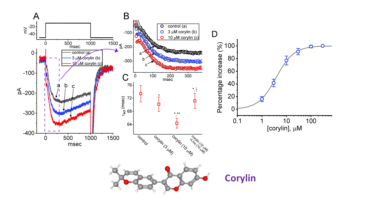
Background: Corylin (3-(2,2-dimethylchromen-6-yl)-7-hydroxychromen-4-one), a bioactive flavonoid, has been reported to exercise anti-inflammatory, antineoplastic, and antioxidant effects, and may also possess lifespan-extending properties. Objectives: Any modifications of transmembrane ionic currents produced by corylin remain largely unknown. Methods: The patch-clamp technique and docking prediction were used in this study. Results: In pituitary GH3 somatolactotrophs, corylin concentration-dependently increased the magnitude of the M-type K+ current (IK(M)), with an EC50 of 3.8 μM. Concurrently, the activation time constant of IK(M) was shortened. The addition of linopirdine (10 μM), an IK(M) inhibitor, suppressed the current amplitude. Corylin also induced a leftward shift in the steady-state activation curve and enhanced IK(M) during pulse-train stimulation. Moreover, corylin increases the hysteretic strength of IK(M) evoked by a long-lasting triangular ramp pulse; this effect was attenuated by linopirdine. The stimulatory effect of corylin on IK(M) was not altered by carvedilol or iberiotoxin but was reduced by dapagliflozin. In contrast, depolarization-activated IK(M) was not affected by 17β-estradiol alone. In cell-attached recordings, corylin increased M-type K+ (KM)-channel activity with minimal change in single-channel amplitude, while prolonging the mean open time. This stimulatory effect was reversed by linopirdine or dapagliflozin. Additionally, corylin slightly inhibited the erg-mediated current. Docking analysis further suggested that corylin potentially interacts with residues in KCNQ2 or KCNH2 channels via hydrogen bonding and hydrophobic interactions. Conclusions: These findings suggest that corylin modulates ionic currents, primarily through KM (KCNQ/KV7) channels, which may underlie its in vivo actions and those of related flavonoids. These effects may contribute to the regulation of functional activities of neuronal, neuroendocrine, and endocrine cells.

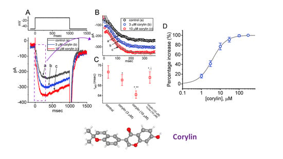
Graphical Abstract

1. Introduction

Corylin (3-(2,2-dimethylchromen-6-yl)-7-hydroxychromen-4-one) is a major bioactive flavonoid isolated from the fruit of Psoralea corylifolia Linne (family Fabaceae), also known as buguchi or Bo-Gol-Zhee. It has been increasingly demonstrated to exert a wide range of pharmacological actions, including direct free radical scavenging, inhibition of biomolecules, suppression of lipopolysaccharide-induced inflammation, and modulation of antioxidant defense [1,2,3,4,5,6,7,8,9,10,11,12,13,14,15,16,17,18,19,20]. This compound might also have potential in treating brain inflammation and attenuating the progression of neurodegeneration. For example, the extract from the seeds of Psoralea corylifolia has been demonstrated to be effective against palmitate-induced neuronal apoptosis in PC12 cells [21] and to exert neuroprotective and anti-neuroinflammatory effects in hippocampal cells, microglia and retinal ganglion cells [4,22]. Notably, in addition to stimulating L-type Ca2+ currents [23,24], quercetin—another flavonoid—has been reported to enhance the activity of M-type K+ channels [25,26,27]. Several botanical medicines have also been recently shown to activate KCNQ channels [26,27]. However, the extent to which corylin or other related compounds can alter the magnitude, gating properties, and voltage-dependent hysteresis (Hys(V)) of plasmalemmal ionic currents in excitable cells remains largely unresolved.
The KCNQ (KV7) family of K+ channels consists of five members, designated KCNQ1-KCNQ5 (KV7.1–KV7.5). Among these, KCNQ2, KCNQ3, and KCNQ5 encode the principal subunits of KV7.2, KV7.3, and KV7.5 channels, respectively, which are broadly expressed in both nervous and endocrine tissues. Notably, heteromeric KCNQ2/KCNQ3 channels closely replicate the native M-type K+ current (IK(M)), sharing its biophysical properties and sensitivity to pharmacological inhibitors such as linopirdine. The designation “M-type” reflects the current’s regulation by muscarinic acetylcholine receptors [27,28,29]. The magnitude of these currents can widely regulate membrane excitability in varying types of excitable cells that include endocrine cells [28,29,30]. Once activated by membrane depolarization, they exhibit slow activation and deactivation kinetics [31,32,33,34]. It has also been shown that by enhancing Na+-current recovery, the IK(M) amplitude induced by high frequency stimulation can expedite action potential (AP) firing with stable waveforms and reliable synaptic transmission [34,35]. Moreover, pharmacological targeting of IK(M) (or KCNQ/KV7 channel-mediated currents) has been recently recognized as a potential adjunctive strategy for a range of neurological disorders characterized by neuronal hyperexcitability, including cognitive dysfunction, epilepsy, major depression, and neuropathic pain [29,35,36,37].
Building on these considerations, the present study investigated whether and how corylin modulates the magnitude, gating kinetics, and Hys(V) behavior of IK(M) in pituitary tumor (GH3) cells. Importantly, our findings demonstrate that, beyond its previously described anti-inflammatory, antineoplastic, and antioxidative actions, corylin can interact with KM (KCNQ or KV7) channels to enhance IK(M) in a concentration-, time-, voltage-, and Hys(V)-dependent manner in excitable cells such as GH3 lactotrophs.

2. Results

2.1. Stimulatory Effect of Corylin on M-Type K+ Current (IK(M)) Recorded from Pituitary GH3 Cells

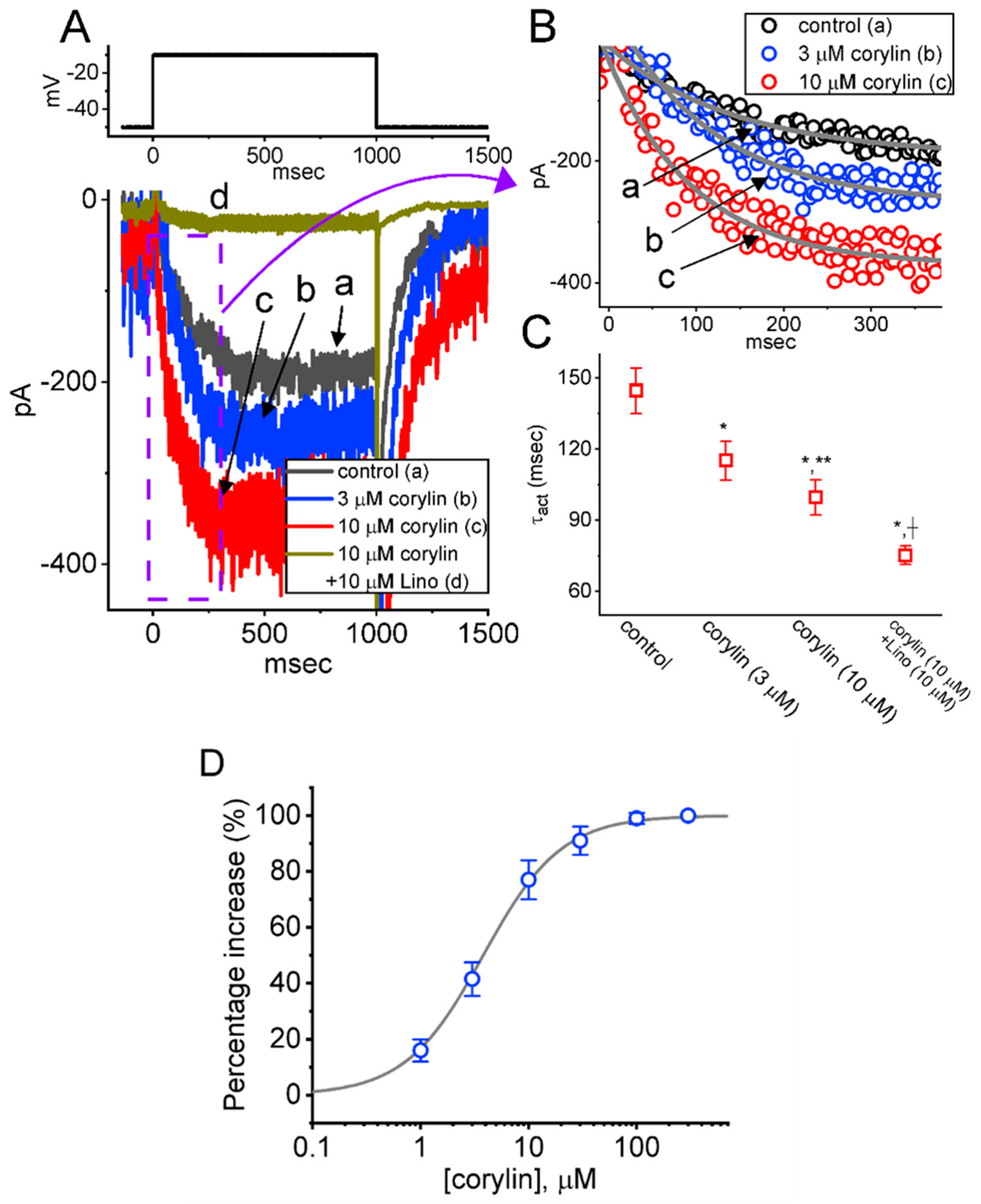
In the initial set of experiments, we examined whether IK(M) in GH3 cells can be modified by corylin. Cells were bathed in a high-K+, Ca2+-free solution containing 1 μM tetrodotoxin (TTX) and 0.5 mM CdCl2, while the recording pipette was filled with a K+-based internal solution. After establishing whole-cell current recordings, a 1 s depolarizing voltage pulse from −50 to −10 mV was applied to evoke inward IK(M), which displayed a slowly activating, non-inactivating time course, consistent with previous reports [26,31,32,34,38,39,40]. However, of additional interest, as cells were exposed to corylin, the amplitude of IK(M) activated by long-lasting step depolarization progressively increased (Figure 1A). For example, during exposure to 10 μM corylin, IK(M) increased from 194 ± 25 to 348 ± 31 pA (n = 8, paired t-test, p = 0.018). Following the removal of corylin, the current amplitude returned to 202 ± 26 pA (n = 8). Concurrently, the value of activation time constant (τact) of IK(M) became shortened, as evidenced by a significant reduction in τact from 144.6 ± 9.6 to 99.7 ± 7.4 ms (n = 8, paired t-test, p = 0.014) by adding 10 μM corylin (Figure 1B,C). Furthermore, as cells were continually exposed to 10 μM corylin, the subsequent addition of 10 μM linopirdine could attenuate current amplitude as well as decrease τact value effectively (Figure 1C). Linopirdine was reported to be an inhibitor of IK(M) [31,34,40,41]. These results suggest that corylin is capable of modulating the activation kinetics of IK(M).
We subsequently established the concentration-dependent relationship of corylin-induced stimulation of IK(M) and the findings are presented in Figure 1D. Notably, this compound can increase the amplitude of IK(M) in a concentration-dependent manner. Based on a least-squares fit to the modified equation (as indicated in Section 4), the data yielded a half-maximal effective concentration (EC50) for IK(M) stimulation of 3.8 μM and a Hill coefficient of 1.2. The results lead us to indicate that the addition of corylin would exert a stimulatory effect on IK(M) in these cells.

2.2. Effect of Corylin on the Steady-State Current Versus Voltage (I-V) Relation and Activation Curve of IK(M)

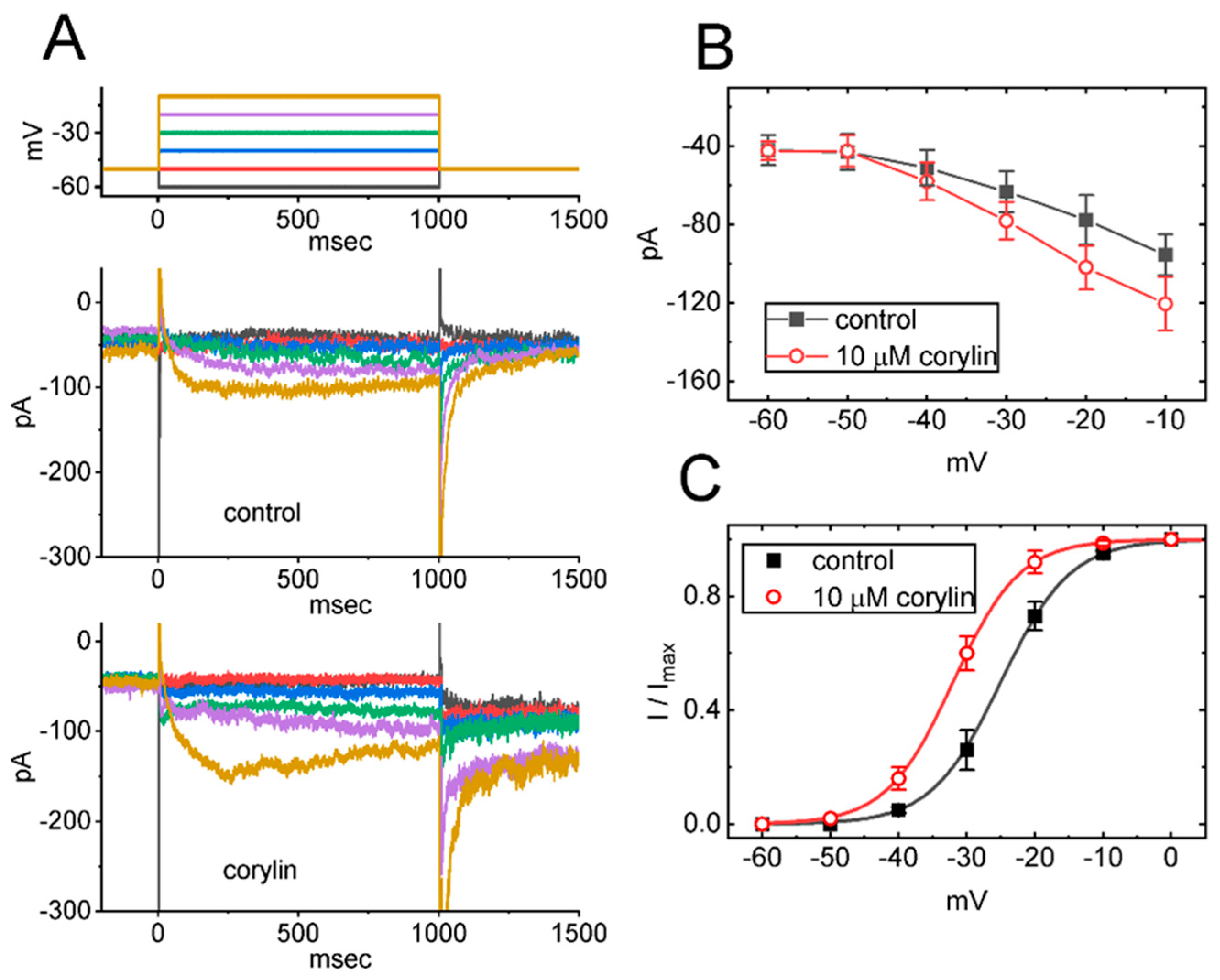
Next, we wanted to test if IK(M) activated by different levels of membrane potential can be altered by the existence of corylin. As illustrated in Figure 2A,B, when a series of voltage steps ranging from −60 to −10 mV was applied to the test cells from a holding potential of −50 mV, the absolute amplitude of IK(M) increased progressively, particularly at membrane potentials more depolarized to −40 mV. In addition, the quasi-steady-state activation curve of IK(M), in the absence and presence of corylin, was constructed, and the results are presented in Figure 2C. The relationship between membrane potential and normalized IK(M) amplitude was also fitted with a Boltzmann function (see Section 4), and the fit was evaluated using goodness-of-fit analysis. In the control (i.e., corylin was not present), V1/2 = −25.1 ± 1.5 mV and q = 4.9 ± 0.2 e (n = 8), while during cell exposure to 10 μM corylin, V1/2 = −31.8 ± 1.7 mV and q = 5.1 ± 0.2 e (n = 8). Moreover, corylin increased the absolute amplitude of the deactivating tail IK(M). For instance, during membrane depolarization from −50 to −10 mV, the tail IK(M) measured upon repolarization to −80 mV increased from 252 ± 14 to 321 ± 19 pA (n = 8, paired t-test, p = 0.021). The results indicated that, in the presence of corylin, the quasi-steady-state IK(M) activation curve was shifted leftward (toward more hyperpolarized potentials) by approximately 7 mV in the presence of corylin, without any noticeable change in the gating charge associated with channel activation.

2.3. Corylin’s Effect on IK(M) Amplitude Evoked During a Train of Depolarizing Command Voltages in GH3 Cells

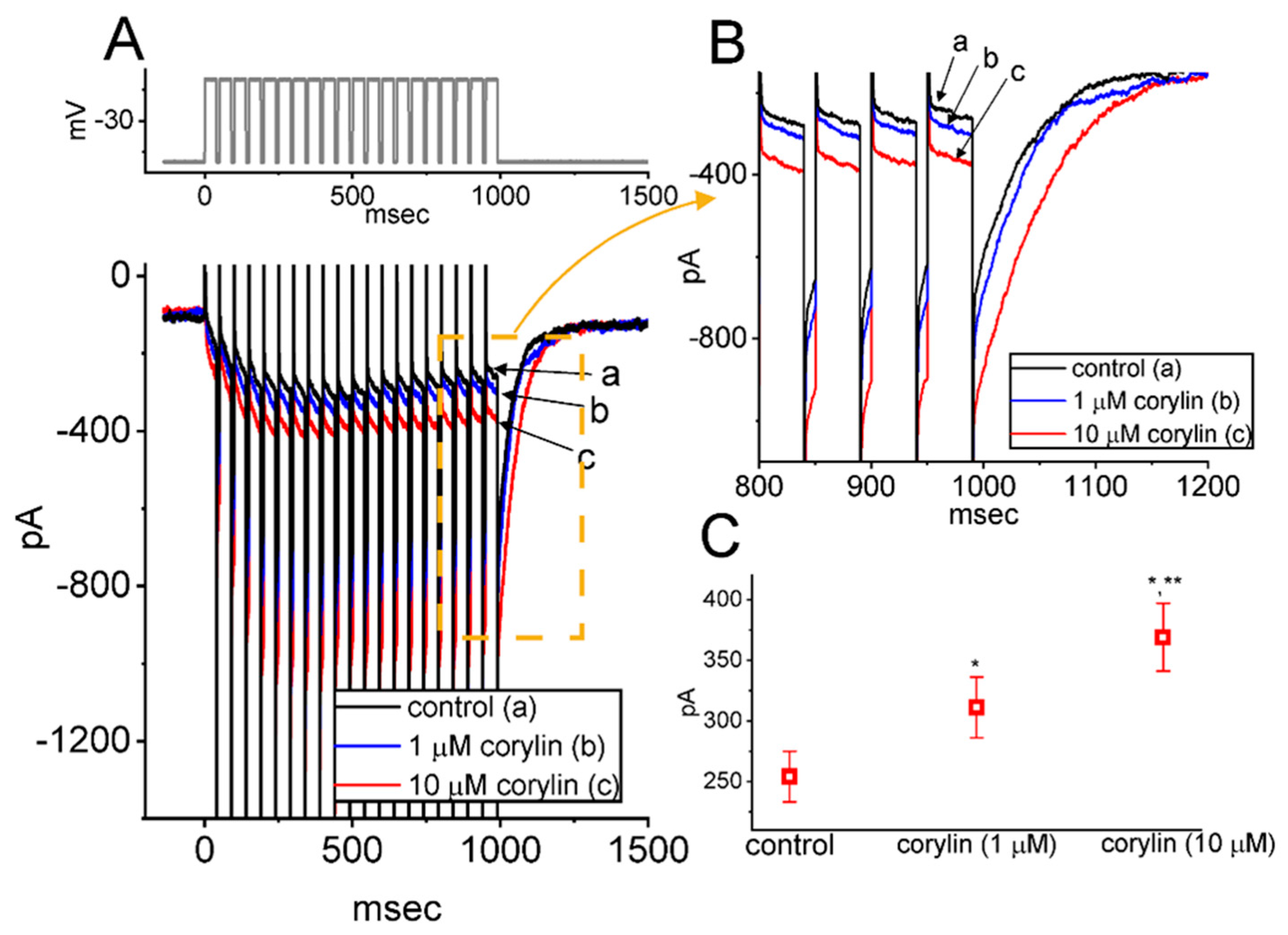
Recent work has demonstrated the effectiveness of the train of depolarizing pulses in modifying the IK(M) magnitude [26,34,35]. For this reason, we continued to explore whether the corylin-mediated stimulation of IK(M) in these cells can be modified during pulse-train stimulation. In this set of whole-cell current measurements, we applied a 20 Hz train of depolarizing pulses from −50 to −10 mV to the tested cell. As demonstrated in Figure 3A,B, the current activation and deactivation evoked by responding to such pulse-train stimulation were robustly observed. Furthermore, exposure of cells to corylin increased IK(M) during a train of depolarizing pulses (Figure 3C). Concomitantly, the time course of current activation became faster in the presence of corylin. For instance, exposure to 10 μM corylin significantly increased the absolute amplitude of IK(M) measured at the end of pulse-train stimulation, rising from 254 ± 21 to 369 ± 28 pA (n = 7, paired t-test, p = 0.018). In parallel, the amplitude of deactivating IK(M) was markedly elevated from 623 ± 43 to 983 pA (n = 7, paired t-test, p = 0.015). Additionally, the τact value for IK(M) during a train of command voltages was measurably shortened to 53 ± 9 ms (n = 7, paired t-test, p = 0.019) from a control value of 114 ± 14 ms (n = 7). Therefore, exposure to corylin produced a considerable increase in the amplitude of IK(M), along with a reduction in the τact value during a train of depolarizing pulses.

2.4. Augmentation of the Strength in Voltage-Dependent Hysteresis (Hys(V)) of IK(M) Caused by Corylin

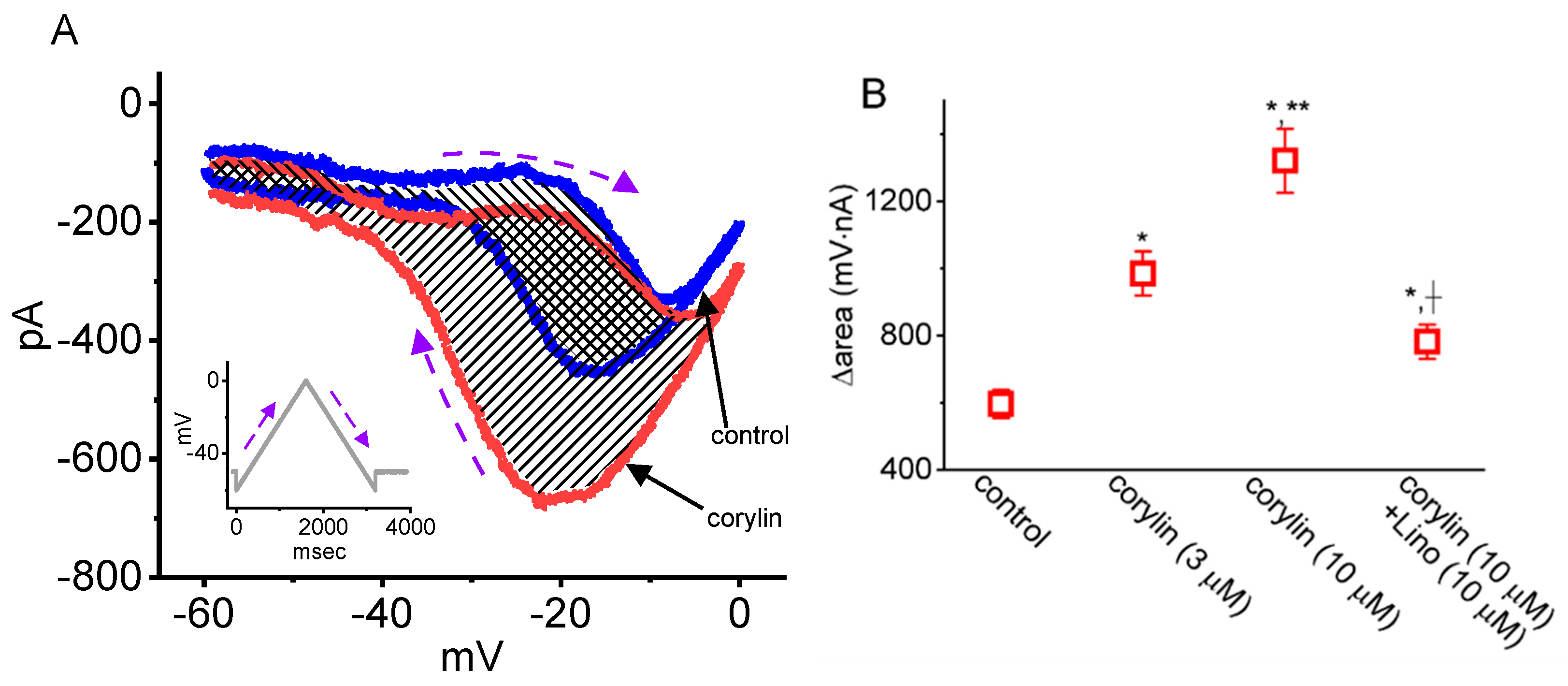
It has been demonstrated that Vramp-induced Hys(V) of IK(M) could contribute to AP firing in various types of excitable cells [26,32,34]. Hys(V) refers to a pronounced lag in current magnitude when the membrane potential is linearly ramped in the opposite direction. Accordingly, the experiments were designed to determine whether Vramp-induced Hys(V) was functionally active in GH3 cells and to evaluate how cell exposure to corylin may influence the Hys(V)’s strength in IK(M). In whole-cell voltage-clamp recordings, the cell was held at −50 mV, after which a series of double triangular Vramp pulses (ranging between −60 and 0 mV, 3.6 s in duration, delivered at 0.05 Hz) was applied via a digital-to-analog conversion. In accordance with previous observations [26,32,34], when a double Vramp was applied to the examined cells, the current amplitude at a given membrane potential evoked during the ascending (forward or upsloping) limb of Vramp was markedly smaller than that measured at the same voltage during the descending (backward or downsloping) limb of the voltage (Figure 4A). For example, in the control period (i.e., in the absence of corylin), the absolute amplitude of IK(M) at −20 mV differed significantly between the ascending (upsloping) and descending (downsloping) phases of Vramp, measuring 198 ± 22 and 418 ± 28 pA (n = 8, paired t-test, p = 0.011). These findings strongly indicate the presence of IK(M)’s Hys(V) in response to an upright isosceles-triangular Vramp in GH3 cells [34].
Of additional notice, as GH3 cells were continually exposed to corylin, the Hys(V) strength of IK(M) became accentuated. For example, in the presence of 10 μM corylin, IK(M) amplitude (in absolute value) at the level of −20 mV evoked during the ascending or descending end of Vramp became measurably raised to 218 ± 23 pA (n = 8, paired t-test, p = 0.014) or 682 ± 38 pA (n = 8, paired t-test, p = 0.013), respectively. It was therefore observed that the current magnitude at the descending end of Vramp increased more significantly than that at the ascending phase of Vramp. We then quantify the strength of the Hys(V) loop by estimating the total area (∆area, shaded region), which encircles the current amplitude between the ascending and descending ends of Vramp. The experimental data were compiled and are summarized in Figure 4B. These observations clearly demonstrate that corylin (3 or 10 μM) increased Hys(V)’s ∆area of IK(M) in GH3 cells. Additionally, during the continued presence of corylin (10 μM), the subsequent addition of linopirdine (10 μM) was able to attenuate the corylin-increased ∆area of IK(M) evoked during isosceles-triangular Vramp. Therefore, these findings suggest that the presence of corylin increases IK(M) in a concentration- and Hys(V)-dependent manner in these cells.

2.5. Comparison Among Effects of Corylin, Corylin Plus Carvedilol (Carv), Corylin Plus Iberiotoxin (Iber), 17β-Estradiol, and Corylin Plus Dapagliflozin (Dapa) on IK(M) Amplitude Observed in GH3 Cells

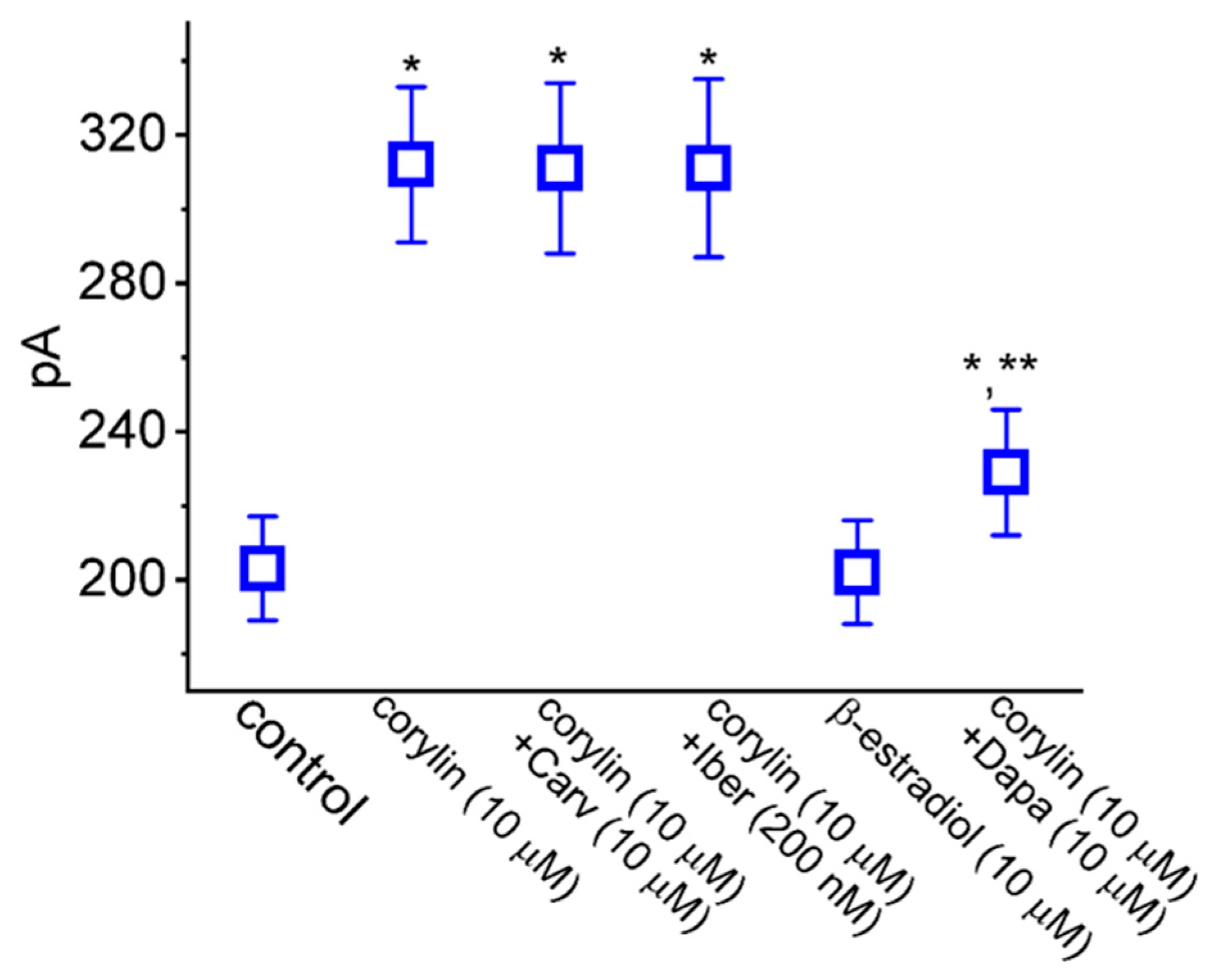
Recent reports have demonstrated the ability of corylin to bind to β3-adrenergic receptors in adipocytes [11,18,42]. The induction of osteoblastic differentiation caused by corylin was mediated through its binding to and interaction with estrogen receptors [18,42]. Pituitary lactotrophs have been previously reported to express the activity of estrogen receptors [43]. It would thus be important to examine whether corylin-stimulated IK(M) presented herein could be a result of its binding to β-adrenergic or estrogen receptors. Under our experimental conditions, as cells were continually exposed to 10 μM corylin, the addition of neither carvedilol (Carv, 10 μM) nor iberiotoxin (200 nM) was able to have any modifications on corylin-stimulated IK(M), as summarized in Figure 5. Carvedilol, a non-selective β-adrenergic blocker, was shown previously to antagonize the activity of β3-adrenergic receptors expressed in cardiac tissue [44]. It is thus thought that carvedilol might block the IK(M)-stimulating effects of corylin via β-adrenergic receptors. Iberiotoxin was an inhibitor of large-conductance Ca2+-activated K+ channels [45]. Moreover, the addition of 17β-estradiol (10 μM) was found to have no effect on IK(M). In the continued presence of 10 μM corylin, the subsequent addition of dapagliflozin (10 μM) could attenuate the corylin-induced stimulation of IK(M). Dapagliflozin (Dapa), an inhibitor of Na+-dependent glucose co-transporters, has been reported to suppress IK(M) directly [39]. Based on the present observations, it is therefore unlikely that the corylin-stimulated IK(M) observed in GH3 cells results primarily from its binding to β-adrenergic or estrogen receptors.

2.6. Effect of Corylin on M-Type K+ (KM) Channels Recorded from GH3 Cells

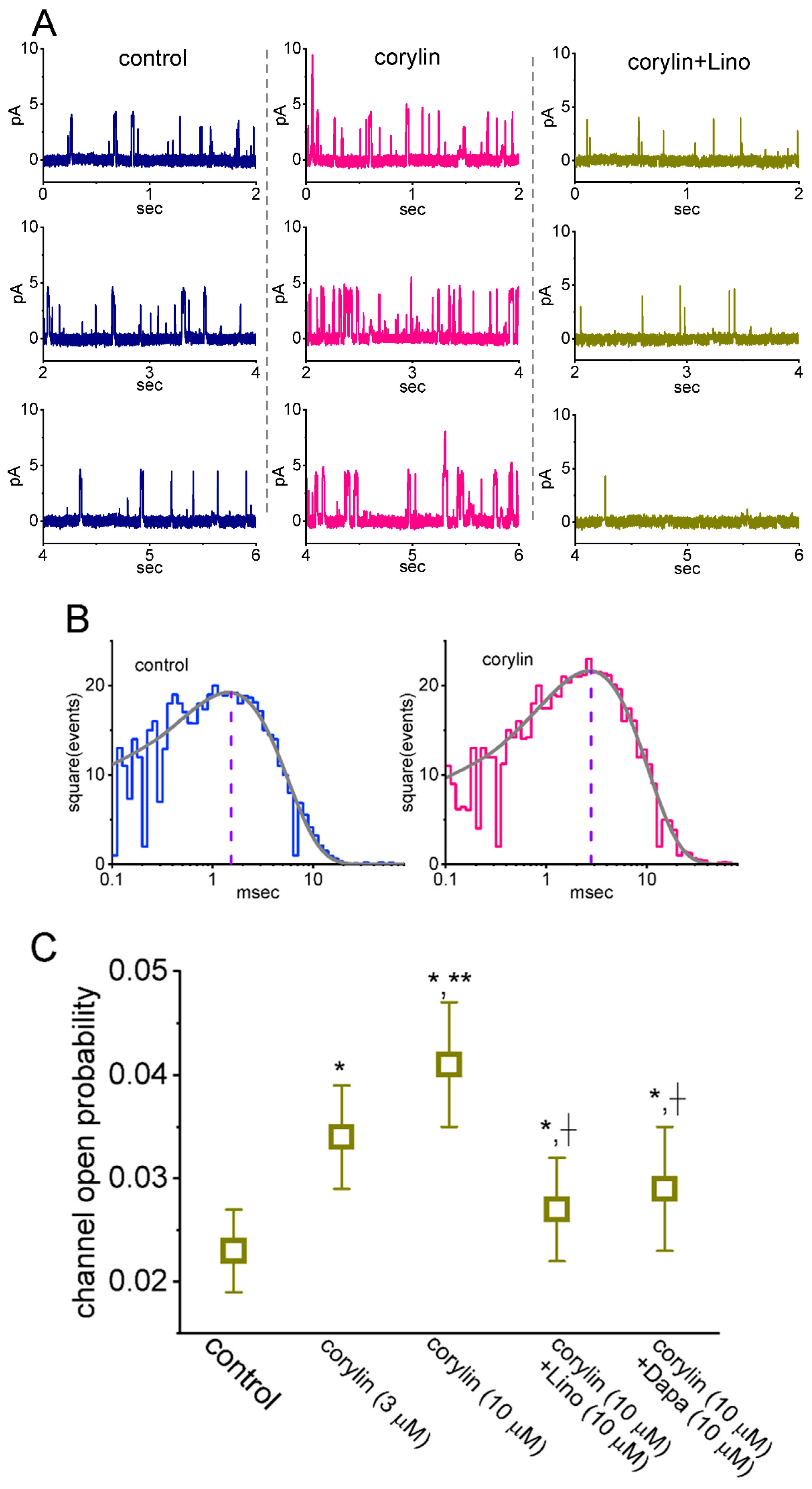
The corylin-induced enhancement of whole-cell IK(M) may result from several mechanisms. Specifically, alterations in channel open probability, single-channel conductance, gating kinetics (e.g., mean open time), or a combination of these factors could underlie the observed stimulation of IK(M). To further investigate single-channel activity in KM channels in the presence or absence of corylin, additional measurements were performed. In these cell-attached recordings, cells were maintained in a high-K+, Ca2+-free solution, while the recording electrode was filled with a low-K+ (5.4 mM) solution. As illustrated in Figure 6, when the tested cell was voltage-clamped at +20 mV relative to the bath, the activity of single KM channels was robustly observed [31,39,40]. When corylin was applied to the bath, the channel open probability was progressively increased. In the continued presence of corylin, further addition of linopirdine attenuated a corylin-induced increase in channel activity. For example, the presence of 10 μM corylin conceivably increased the probability of KM-channel openings from 0.023 ± 0.004 to 0.041 ± 0.006 (n = 8, paired t-test, p = 0.011); however, no modification in the single-channel amplitude was found (3.3 ± 0.2 pA [control] versus 3.4 ± 0.3 pA [in the presence of corylin]; n = 8, paired t-test, p = 0.091). After the washout of corylin, channel activity recovered to 0.024 ± 0.006 (n = 7, paired t-test, p = 0.021).
We further examined and analyzed the kinetic properties of KM channels obtained with or without the addition of corylin. As demonstrated in Figure 6B, the distribution of open durations was least-squares fitted by a single exponential. The mean open time of KM channels rose from 1.5 ± 0.2 to 2.8 ± 0.3 ms (n = 8, paired t-test, p = 0.018). The corylin-induced modulation of KM-channel activity likely reflects an increase in channel open duration rather than any change in single-channel amplitude.
Moreover, in the continued presence of 10 μM corylin, further addition of linopirdine (10 μM) or dapagliflozin (10 μM) was able to attenuate corylin-enhanced channel activity effectively (Figure 6C). Linopirdine can suppress the activity of KM channels, while dapagliflozin, known to be an inhibitor of Na+-dependent glucose co-transporter, was reported to inhibit IK(M) amplitude [34,39]. Therefore, corylin-stimulated IK(M) could be reasonably explained by the increased channel open probability as well as by its prolongation in the mean open time of KM channels.

2.7. Effect of Corylin on Erg-Mediated K+ Current (IK(erg)) Recorded from GH3 Cells

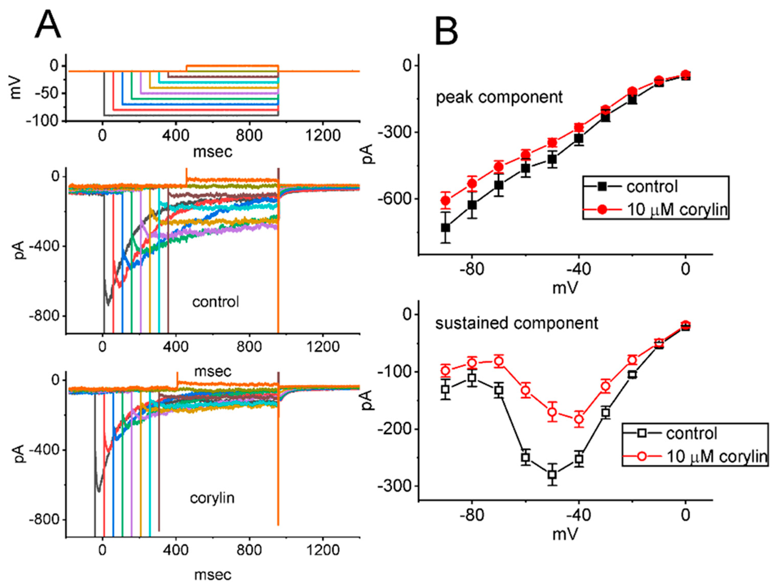
In another set of measurements, we attempted to examine if another type of K+ current (e.g., IK(erg)) would be sensitive to any perturbations by the presence of corylin. To measure IK(erg) [39,46], we put GH3 cells in a high-K+, Ca2+-free solution, and we then filled up the recording pipette with a K+-enriched solution. As the whole-cell configuration was established, we held the examined cell at −10 mV and a series of command voltage steps ranging between −90 and 0 mV with varying durations was imposed on it. As shown in Figure 7, under cell exposure to 10 μM corylin, the IK(erg) amplitudes measured throughout the entire voltage-clamp voltages imposed were conceivably reduced. For example, upon exposure to 10 μM corylin, the absolute peak amplitude of deactivating IK(M), elicited by membrane hyperpolarization from −10 to −90 mV, decreased from 729 ± 70 to 607 ± 38 pA (n = 8, paired t-test, p = 0.021). After washout of the compound, the current amplitude was returned to 724 ± 68 pA (n = 8). Therefore, unlike its stimulation of IK(M), the IK(erg) observed in GH3 cells [30,39,46] was susceptible to being mildly inhibited by adding corylin.

2.8. Docking Results of the Molecular Interactions Between Corylin and the KCNQ2 or KCNH2 Channel

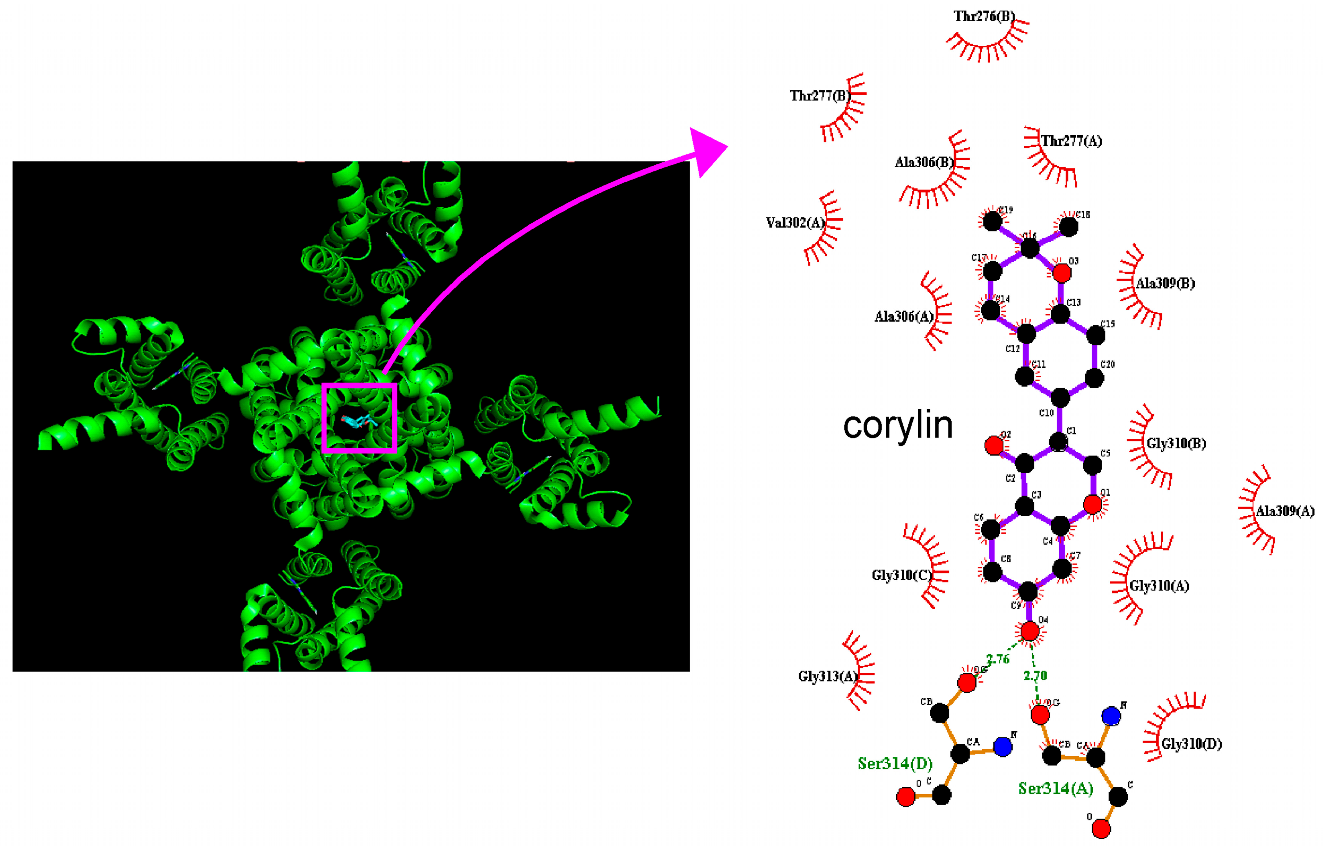
In this study, we examined how the protein of KCNQ2 could be auto-docked by corylin through PyRx 0.8 software (https://pyrx.sourceforge.io, accessed on 18 March 2026). The predicted binding sites of corylin are presented in Figure 8. It needs to be emphasized that corylin can form hydrophobic contact with several amino acid residues, including Thr276, Thr277, Val302, Ala306, Ala309, Gly310 and Gly313, while it forms hydrogen bonds with residue Ser 314 with distances of 2.70 and 2.76 Å, and that the estimated binding affinity among this interaction was −8.4 kcal/mol. Based on the experimental findings described above, the docking analysis suggests that corylin may interact with the intracellular domain located near the transmembrane S6 segment of the channel. Such binding implies that the functional activity of KM (KCNQ or KV7) channels in excitable cells could be subject to modulation by corylin or by its structurally related analogs [26,27].
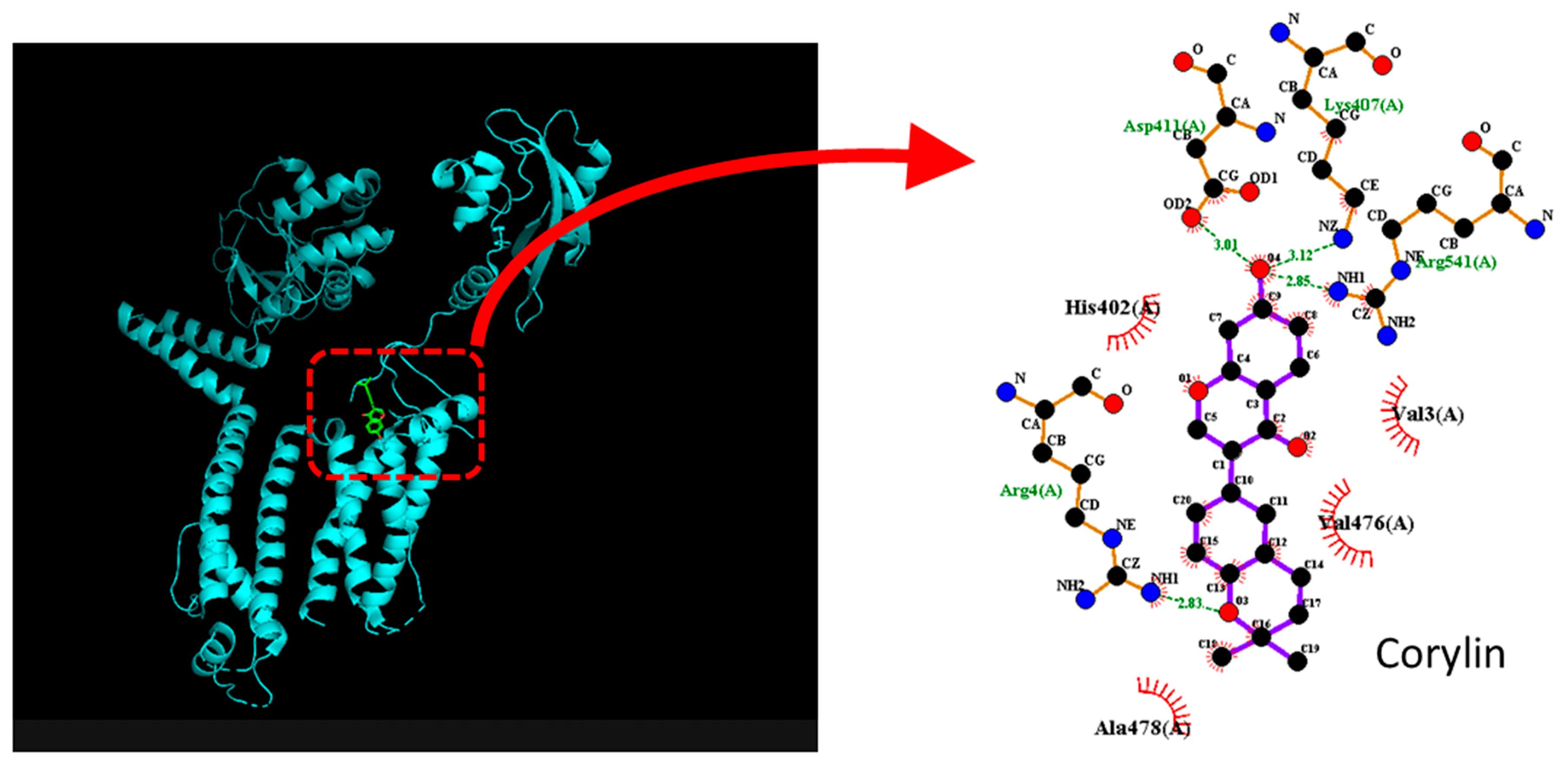
Because corylin can inhibit the amplitude of IK(erg), the KCNH2 (HERG, human ether-à-go-go-related gene) protein was further docked with corylin using PyRx software. The predicted binding sides of corylin on this channel protein are illustrated in Figure 9. This compound was observed to establish hydrophobic interactions with several residues, including Val3(A), His402(A), Val476(A), and Ala478(C). The corylin molecule can form hydrogen bonds with residues Arg4(A), Lys407(A), Asp411(A), and Arg541(A), with bond lengths of 2.81, 3.12, 3.01, and 2.85 Å, respectively. The results estimate a strong binding affinity of −7.8 kcal/mol. The predicted interaction thus suggests that corylin-mediated inhibition of IK(erg) in GH3 cells is due to its binding to KCNH2 channels. However, it needs to be mentioned that the proposed interaction with KCNQ2 and KCNH2 channels is hypothetical and based primarily on in silico docking results, rather than direct functional validation. Interactions with other subtypes, such as KCNQ3 and KCNQ5 channels, also cannot be excluded.

3. Discussion

The striking findings demonstrated in this work are that corylin, a bioactive flavonoid currently recognized as a potential life-prolonging agent [47], produces a stimulatory action on IK(M) in a concentration-, voltage-, and Hys(V)-dependent fashion in GH3 lactotrophs. A leftward shift in the steady-state activation curve of IK(M) was observed in the presence of this compound, with no change in the gating charge of the curve. Cell exposure to it can elevate the probability of KM channels that would be open, in combination with a measurable lengthening in the mean open time of the channel. However, the IK(erg) in GH3 cells was slightly suppressed by the presence of corylin. Docking analysis revealed specific atomic-level interactions between the corylin molecule and the KCNQ2 or KCNH2 channel structure.
The magnitude of Na+ currents can rapidly decline in an exponential manner during high-frequency stimulation, as previously demonstrated in GH3 cells [48]. However, it needs to be emphasized that because of its slow activation and deactivation kinetics, the IK(M) amplitude can progressively rise during the repetitive firing of APs. During high-frequency stimulation, the accumulation of IK(M) is allowed to hyperpolarize the afterpotential and hence speed the recovery of Na+ channels from inactivation. As a corollary, the augmentation of IK(M) magnitude caused by corylin during high-frequency activity is of particular significance and thus capable of facilitating the firing of neuronal APs with a stable waveform and high-fidelity synaptic signaling [26,34,35].
Like the Hys(V) behavior in solar cells [34,49], the current investigations clearly observed the appearance of the overall behavior in IK(M)’s Hys(V) evoked by a long-lasting upright isosceles-triangular Vramp [30,32,39]. That is, the magnitude of these instantaneous currents measured between ascending and descending ends of double Vramp turned out to be strikingly distinguishable. Alternatively, as the membrane potential becomes depolarized (i.e., upward ramp of triangular Vramp), the voltage dependence of KM channels may shift the mode of Hys(V) to one that occurs at less negative potentials with smaller current magnitude, leading to a minor effect on the rising phase of AP. However, as the membrane potential is hyperpolarized (i.e., during the downward limb of Vramp or repolarizing phase of AP), the voltage dependence of IK(M) activation would switch to more hyperpolarized voltages with a higher current magnitude, thereby having the tendency to increase membrane repolarization as well as to increase the recovery of Na+ currents. Furthermore, these findings revealed that the triangular Vramp-induced IK(M) exhibited a pronounced voltage-dependent Hys(V), and the magnitude of these Hys(V) loops was further enhanced by the addition of corylin. In other words, the ∆area (indicated in the shaded area of Figure 4A) of the IK(M) loop evoked in response to long-lasing triangular Vramp significantly increased following the application of corylin. As such, the existence of corylin may increase IK(M) magnitude in a concentration- and Hys(V)-dependent fashion. However, further work needs to be conducted to examine if corylin-perturbed modifications in Hys(V) behavior of IK(M) are tightly linked to conformational changes or docking interactions in the voltage sensors of the KM channel.
Earlier investigations have revealed the ability of corylin to bind to and then to activate β3-adrenergic receptors present in adipocytes [11,42]. It has also been demonstrated that corylin might interact with estrogen receptors to induce osteoblastic differentiation [18,50]. It has been noticed that estrogen receptors were expressed in pituitary lactotrophs [43]. However, in our study, during the continued exposure to corylin, further application of carvedilol, known to block β3 adrenoceptors in cardiac tissue, failed to have any effect on corylin-stimulated IK(M). Moreover, the presence of 17β-estradiol alone did not cause any perturbations in IK(M) observed in GH3 cells. In this scenario, it appears unlikely that under our experimental conditions, corylin-mediated stimulation of IK(M) or KM-channel activity is attributed to its high-affinity binding to either β-adrenergic or estrogen receptors.
The EC50 of corylin required to stimulate IK(M in GH3 cells was determined to be approximately 3.8 μM. This value aligns closely with the concentration ranges (1–300 μM) previously reported for its antioxidative, anti-inflammatory, and antineoplastic effects [4,7,8,9,11,12,15,16,47,51,52]. It should also be noted that the perturbations of corylin on membrane excitability may be strongly influenced by several confounding factors, including the concentration of corylin, the baseline resting potential, the firing patterns of APs, or a combination of these variables. It is, therefore, anticipated that the KM channel is an important target for the action of corylin. The concentrations used to affect the magnitude, gating kinetics and Hys(V) behaviors of IK(M) presented herein would be pharmacologically significant in body fluids and tissues. The corylin molecule may have the propensity to exercise a higher affinity to the open state than to the resting (closed) state in the KM channel, thereby destabilizing the open conformation, while the detailed ionic mechanism of corylin actions on ionic currents is not thoroughly understood.
The use of a high-K+ external solution in our whole-cell recordings was primarily intended to enhance the detectability and measurement accuracy of IK(M). Specifically, elevating the extracellular K+ concentration reduces the K+ equilibrium potential difference and increases the driving force for inward K+ currents at subthreshold membrane potentials. This manipulation allows IK(M) to be more readily resolved, particularly in GH3 cells, where the basal amplitude of this current can be relatively small under physiological K+ conditions. In addition, the adoption of symmetrical or near-symmetrical K+ conditions in this study is a widely used approach to enhance signal resolution and reduce interference from other ionic currents [38]. Increasing extracellular K+ concentration may influence channel gating kinetics, conductance, and pharmacological sensitivity. However, our primary objective was to examine the modulatory effect of corylin on IK(M) under controlled and consistent recording conditions. Therefore, all experimental comparisons (control vs. corylin-treated) were conducted under the same high-K+ environment, ensuring that the relative changes observed can be attributed to the action of corylin rather than differences in ionic conditions. Nonetheless, future studies under physiological K+ conditions will be valuable to further validate the findings.
The use of GH3 tumor cells in this study could present limitations in terms of translatability to neurons and in vivo systems. The electrophysiological properties of IK(M) and IK(erg) in these cells have been shown to be consistent with those in neurons as well as in neuroendocrine or endocrine cells. Therefore, our findings—particularly corylin-induced stimulation of IK(M)—may have broader relevance and could be applicable to other types of electrically excitable cells.
Although the concentrations of corylin applied in this study (up to 300 μM) exceed typical physiological levels, the compound demonstrated notable activity. The EC50 for corylin-induced enhancement of IK(M) in GH3 cells was 3.8 μM. Importantly, this stimulatory effect was influenced by membrane voltage, ongoing action potential firing, and the intrinsic strength of IK(M) Hys(V). Because our experiments were conducted at room temperature (approximately 25 °C), it remains essential to determine whether corylin produces comparable modulation of IK(M) under true physiological conditions. Taken together, these findings indicate that corylin’s ability to stimulate IK(M) may retain pharmacological relevance.
A previous paper [53] reported that the single-channel amplitude of KM channels was lower than that observed in the present study. The reason for this discrepancy remains unclear. One possible explanation is that the single-channel conductance of KM channels may vary among different tissue preparations. Our findings are consistent with those reported in earlier studies [31,38,54]. Because the single-channel conductance of KCNQ2 and KCNQ3 channels was reported to be higher than that of KCNQ4 and KCNQ5 channels, it remains to be determined whether corylin differentially regulates distinct populations of KM (KCNQ/K7) channels.
In this study, we did not perform functional validation of the predicted binding sites on KCNQ2 or KCNH2 channels. Moreover, binding affinity values alone are insufficient to confirm channel modulation under physiological conditions. Docking results suggest a potential binding interaction but do not establish causality or physiological relevance. Complementary studies—including gene knockdown or overexpression systems to evaluate channel-specific effects, electrophysiological recording to assess channel activity, and site-directed mutagenesis to verify predicted binding residues—will be necessary and are planned for future investigation. Moreover, the use of subtype-selective blockers or modulators of KCNQ2 and KCNH2 channels to assess functional involvement is important to further elucidate their functional involvement.

4. Materials and Methods

4.1. Chemicals, Drugs, Reagents and Solutions

Corylin (IUPAC name: 3-(2,2-dimethylchromen-6-yl)-7-hydroxychromen-4-one, 53947-92-5, SCHEMBL1096083, CHEMBL1271888, C20H16O4, CAS: 53947-92-5; PubChem CID: 5316097) was supplied by MedChemExpress (GeneChain, Kaohsiung, Taiwan). Iberiotoxin was purchased from Alomone Labs (Asia Bioscience, Taipei, Taiwan), dapagliflozin (Dapa) was from Cayman (Ann Arbo, MI, USA), and carvedilol (Carv) was from Tocris (Union Biomed, Taipei, Taiwan), while 17β-estradiol, linopirdine (Lino), and tetrodotoxin (TTX) were from Sigma-Aldrich (Merck, Taipei, Taiwan). Corylin, carvedilol, dapagliflozin, and linopirdine were dissolved in dimethyl sulfoxide (DMSO) as a 20 mM stock solution and were thereafter diluted in extracellular solution to the final concentration achieved, while iberiotoxin was dissolved in 0.9% NaCl. For cell preparations, all culture media, horse and fetal calf sera, L-glutamine, and trypsin/EDTA were acquired from HyCloneTM (Thermo Fisher, Logan, UT, USA), while other chemicals or reagents were of laboratory grade and taken from standard sources.
The extracellular solution (normal Tyrode’s solution buffered with HEPES) contained the following ionic composition (in mM): NaCl 136.5, KCl 5.4, MgCl2 0.53, CaCl2 1.8, glucose 5.5, and HEPES 5.5, adjusted to pH 7.4 with NaOH. For the recording of macroscopic K+ currents (IK(M) or IK(erg)), the pipette solution consisted of (in mM): KCl 140, MgCl2 1, Na2ATP 4, Na2GTP 0.1, EGTA 0.1, and HEPES, titrated to pH 7.2 with KOH. To measure IK(M), IK(erg), or KM-channel activity, the bath solution contained a high K+ solution (in mM): KCl 130, NaCl 10, MgCl2 3, glucose 6, and HEPES 10, adjusted to pH 7.4 with KOH. To record the activity of single KM channels, the pipette solution was composed of the following (in mM): NaCl 136.5, KCl 5.4, MgCl2 0.53, and HEPES-NaOH buffer 5 (pH 7.4).

4.2. Cell Preparations

GH3 pituitary tumor cells (BCRC-60015; Bioresources Collection and Research Center, Hsinchu, Taiwan) were cultured in Ham’s F-12 media supplemented with 15% horse serum (v/v), 2.5% fetal calf serum (v/v), and 2 mM L-glutamine [23,48,55]. To induce differentiation, cells were transferred to a serum-free, Ca2+-free medium. Under these experimental conditions, cell viability typically remained at 80–90% for up to two weeks. Cultures were maintained at 37 °C in a humidified incubator with a CO2/air mixture (1:19).

4.3. Electrophysiological Measurements

Before each experiment, GH3 cells were carefully dispersed with 1% trypsin/EDTA solution, and we thereafter quickly put an aliquot of cell suspension in a recording chamber mounted on the stage of a CKX-41 inverted microscope (Olympus; Yuan Yu, Taipei, Taiwan). Cells were immersed at room temperature (20–25 °C) in HEPES-buffered normal Tyrode’s solution that contained 1.8 mM CaCl2. When they were left to adhere to the chamber’s bottom for several minutes, the measurements were performed. Ionic currents were recorded with patch electrodes in the cell-attached or whole-cell configuration of a modified patch clamp technique, as described elsewhere [34,38,54,55]. GΩ-seals were typically formed in an all-or-none manner, leading to an improvement in signal-to-noise ratio. The recording pipette was connected to the input stage of an RK-400 (Bio-Logic, Claix, France) or an Axopatch-200B patch-clamp amplifier (Molecular Devices, Bestgen Biotech, New Taipei City, Taiwan). Patch electrodes (3–5 MΩ in bathing solution) were made from Kimax®-51 borosilicate capillary tubes (#34500; Merck, Taipei, Taiwan) using a two-step vertical puller (PB-7; Narishige, Taiwan Instrument, Tainan, Taiwan), and their tips were heat-polished in an MF-83 microforge (Narishige). All potentials were corrected for the liquid junction potential that would develop at the pipette tip in situations where the composition of the pipette internal solution was different from that in the bath medium. Tested compounds were applied by perfusion or added to the bath to obtain the final concentration indicated. In the experiments with corylin plus linopirdine, linopirdine was applied after the addition of corylin. When high-frequency stimuli were needed, we used an Astro-Med Grass S85X dual output pulse stimulator (Grass; Zhong Yan, Kaohsiung, Taiwan) [26,48,56].
The current and voltage signals were monitored in real time and recorded onto a laptop computer. The recorded data were low-pass filtered at 2 kHz using an FL-4 four-pole Bessel filter (Dagan, Minneapolis, MN, USA) and digitized at 10 kHz or more with a Digidata 1440A interface (Molecular Devices). The device was connected to either an RK-400 or Axopatch-200B patch-clamp amplifier, which was controlled via a universal serial bus (USB) connection using the pClamp 10.6 software (Molecular Devices). Ionic currents obtained from whole-cell and single-channel recordings were analyzed offline using pClamp 10.7, OriginPro® (OriginLab; Scientific Formosa, Kaohsiung, Taiwan) and custom-written macros in Excel® 2021 (Microsoft, Redmond, WA, USA) running on Windows 11. Capacitive transients following repolarization were commonly observed; therefore, the tail-deactivating K+ currents were measured after the capacitive currents had settled, typically between 10 and 20 ms after the end of the voltage pulse.

4.4. Data Analyses

To determine the concentration-dependent stimulatory effect of corylin on the amplitude of IK(M), GH3 cells were bathed in a high-K+, Ca2+-free solution, while the recording electrode was filled with a K+-containing solution. To measure IK(M) amplitude, we voltage-clamped each tested cell at −50 mV, and the depolarizing pulse up to 1 s in duration to −10 mV was imposed on it. The IK(M) amplitude measured at the end of depolarizing pulses in the presence of 300 μM corylin was defined as 1.0 (i.e., 100%). The corresponding amplitudes obtained during the control period (in the absence of corylin) and during exposure to different concentrations of corylin (1–300 μM) were measured and compared. The concentration required to stimulate 50% of the current amplitude was determined according to a modified Hill function. That is,
p e r c e n t a g e   i n c r e a s e = E m a x × [ C ] n H E C 50 n H + [ C ] n H
In this equation, EC50 = the concentration required for 50% stimulation; nH = the Hill coefficient; [C] = the corylin concentration applied; and Emax = maximal stimulation. This formula enables optimal convergence, providing the best fit line and accurate parameter estimates (e.g., EC50 and nH).
The activation time constants (τact) of IK(M) in response to prolonged membrane depolarization, obtained in the absence or presence of corylin, were determined by fitting the digitized current traces with a single-exponential function, as indicated in Figure 1B.
To characterize the stimulatory action of corylin on IK(M) amplitude, we constructed the quasi-steady-state activation curve of the current. The relationships between the membrane potentials and the normalized amplitudes of IK(M) with or without the existence of this compound were established and thereafter fitted with a Boltzmann function given by:
I I m a x = 1 1 + e ( V V 1 / 2 ) q F ( R T )
where Imax = the maximal activated current of IK(M); V = the membrane potential; V1/2 = the voltage for half-maximal stimulation; q = the apparent gating charge of the activation curve of IK(M); F = Faraday’s constant; R = the universal gas constant; and T = the absolute temperature.
Linear (e.g., single-channel conductance) and nonlinear (e.g., concentration-dependent relationships and voltage-dependent activation curves) fittings were performed on the datasets using an interactive least-squares approach. Data analysis was carried out with software tools including the Solver add-in in Excel® 2021 (Microsoft) and OriginPro® 2026 (OriginLab).

4.5. Single-Channel Analysis of the KM Channel

Single KM-channel currents in GH3 cells were recorded and analyzed by pClamp 10.7 (Molecular Devices). To evaluate single-channel opening events, amplitude distributions were fitted with multi-Gaussian adjustments. Channel open probabilities were determined through an iterative process to minimize the χ2 values across a sufficiently large set of independent observations. Open lifetime distributions of KM channels (i.e., mean open time), obtained with or without corylin’s presence, were fitted using least-squares analysis with logarithmically scaled bin widths.

4.6. Statistical Analyses

All data are presented as the mean ± standard error of the mean (SEM), where n denotes the number of cells sampled. The normality of data distribution was evaluated using appropriate statistical tests. Differences between the two groups were analyzed using either paired or unpaired Student’s t-tests, depending on the experimental design. For comparisons involving multiple groups, one-way or two-way analysis of variance (ANOVA) was applied, with repeated-measures designs used when appropriate. When significant effects were detected, Fisher’s least-significant difference (LSD) post hoc test was performed for pairwise comparisons. A probability of p < 0.05 was considered statistically significant, and significance levels are denoted in the figure by *, **, or †.

5. Conclusions

This study demonstrates that corylin, a bioactive flavonoid, enhances IK(M) in GH3 cells in a manner dependent on concentration, membrane voltage, and Hys(V). In the presence of corylin, the activation curve of IK(M) is shifted leftward, indicating facilitated channel activation. Additionally, corylin increases the open-state probability of KM channels, accompanied by a prolongation of the mean open time. Docking analysis suggests that the corylin-induced activation of KM channels is likely associated with its binding to KCNQ2 channels. Together, these effects may influence the regulation of electrical activity in various excitable cells under physiological conditions in vivo.

Author Contributions

Writing—review and editing, supervision, validation, investigation, data curation, conceptualization—S.-N.W.; investigation, data curation, funding acquisition, conceptualization, project administration—R.L.; investigation, data curation, funding acquisition, conceptualization, project administration—S.-C.L. All authors have read and agreed to the published version of the manuscript.

Funding

This work was supported in part by the National Science and Technology Council (NSTC), Taiwan (NSTC-112-2923-B-006-0016-028), and by An Nan Hospital-China Medical University, Taiwan (ANHRF114-06, ANHRF114-43, and ANHRF114-49). This research was also funded by the Lithuania-Latvia-Taiwan collaborative project. All conclusions, recommendations, and expressed opinions within this work are based on the authors’ independent research. They do not reflect the official policies or views of the funding organizations or institutions that supported this research.

Institutional Review Board Statement

Not applicable.

Informed Consent Statement

Not applicable.

Data Availability Statement

The original contributions presented in this study are included in the article. Further inquiries can be directed to the corresponding author(s).

Acknowledgments

The authors are grateful to Zi-Han Gao for her assistance during the early stage of the experiments.

Conflicts of Interest

All authors declare that they have no conflicts of interest relevant to this study. The content and writing of this paper are solely the responsibility of the authors.

Abbreviations

The following abbreviations are used in this manuscript:
ANOVAAnalysis of variance
APAction potential
CarvCarvedilol
Corylin3-(2,2-dimethylchromen-6-yl)-7-hydroxychromen-4-one
DapaDapagliflozin
EC50Concentration required for 50% stimulation
ErgEther-à-go-go-related gene
HERGHuman ether-à-go-go-related gene
Hys(V)Voltage-dependent hysteresis;
I-VCurrent versus voltage
IK(erg)Erg-mediated K+ current
IK(M)M-type K+ current
KM channelM-type K+ (KCNQ/K7) channel
LinoLinopirdine
LSD testLeast-significant difference test
SEMStandard error of mean
TTXTetrodotoxin
τactActivation time constant
VrampRamp voltage

References

  1. Khushboo, P.S.; Jadhav, V.M.; Kadam, V.J.; Sathe, N.S. Psoralea corylifolia Linn.—“Kushtanashini”. Pharmacogn. Rev. 2010, 4, 69–76. [Google Scholar] [CrossRef] [PubMed]
  2. Lee, S.W.; Yun, B.R.; Kim, M.H.; Park, C.S.; Lee, W.S.; Oh, H.M.; Rho, M.C. Phenolic compounds isolated from Psoralea corylifolia inhibit IL-6-induced STAT3 activation. Planta Med. 2012, 78, 903–906. [Google Scholar] [CrossRef] [PubMed]
  3. Chopra, B.; Dhingra, A.K.; Dhar, K.L. Psoralea corylifolia L. (Buguchi)—Folklore to modern evidence: Review. Fitoterapia 2013, 90, 44–56. [Google Scholar] [CrossRef]
  4. Kim, K.A.; Shim, S.H.; Ahn, H.R.; Jung, S.H. Protective effects of the compounds isolated from the seed of Psoralea corylifolia on oxidative stress-induced retinal damage. Toxicol. Appl. Pharmacol. 2013, 269, 109–120. [Google Scholar] [CrossRef]
  5. Wang, T.X.; Yin, Z.H.; Zhang, W.; Peng, T.; Kang, W.Y. [Chemical constituents from Psoralea corylifolia and their antioxidant alpha-glucosidase inhibitory and antimicrobial activities]. Zhongguo Zhong Yao Za Zhi 2013, 38, 2328–2333. (In Chinese) [Google Scholar]
  6. Alam, F.; Khan, G.N.; Asad, M.H.H.B. Psoralea corylifolia L: Ethnobotanical, biological, and chemical aspects: A review. Phytother. Res. 2018, 32, 597–615. [Google Scholar] [CrossRef]
  7. Hung, Y.L.; Fang, S.H.; Wang, S.C.; Cheng, W.C.; Liu, P.L.; Su, C.C.; Chen, C.S.; Huang, M.Y.; Hua, K.F.; Shen, K.H.; et al. Corylin protects LPS-induced sepsis and attenuates LPS-induced inflammatory response. Sci. Rep. 2017, 7, 46299. [Google Scholar] [CrossRef]
  8. Chen, C.C.; Chen, C.Y.; Ueng, S.H.; Hsueh, C.; Yeh, C.T.; Ho, J.Y.; Chou, L.F.; Wang, T.H. Corylin increases the sensitivity of hepatocellular carcinoma cells to chemotherapy through long noncoding RNA RAD51-AS1-mediated inhibition of DNA repair. Cell. Death Dis. 2018, 9, 543. [Google Scholar] [CrossRef] [PubMed]
  9. Chen, C.Y.; Chen, C.C.; Shieh, T.M.; Hsueh, C.; Wang, S.H.; Leu, Y.L.; Lian, J.H.; Wang, T.H. Corylin Suppresses Hepatocellular Carcinoma Progression via the Inhibition of Epithelial-Mesenchymal Transition, Mediated by Long Noncoding RNA GAS5. Int. J. Mol. Sci. 2018, 19, 380. [Google Scholar] [CrossRef] [PubMed]
  10. Chen, C.C.; Li, H.Y.; Leu, Y.L.; Chen, Y.J.; Wang, C.J.; Wang, S.H. Corylin Inhibits Vascular Cell Inflammation, Proliferation and Migration and Reduces Atherosclerosis in ApoE-Deficient Mice. Antioxidants 2020, 9, 275. [Google Scholar] [CrossRef] [PubMed]
  11. Chen, C.C.; Kuo, C.H.; Leu, Y.L.; Wang, S.H. Corylin reduces obesity and insulin resistance and promotes adipose tissue browning through SIRT-1 and β3-AR activation. Pharmacol. Res. 2021, 164, 105291. [Google Scholar] [CrossRef]
  12. Chen, I.C.; Wang, S.C.; Chen, Y.T.; Tseng, H.H.; Liu, P.L.; Lin, T.C.; Wu, H.E.; Chen, Y.R.; Tseng, Y.H.; Hsu, J.H.; et al. Corylin Ameliorates LPS-Induced Acute Lung Injury via Suppressing the MAPKs and IL-6/STAT3 Signaling Pathways. Pharmaceuticals 2021, 14, 1046. [Google Scholar] [CrossRef]
  13. Huang, M.Y.; Tu, C.E.; Wang, S.C.; Hung, Y.L.; Su, C.C.; Fang, S.H.; Chen, C.S.; Liu, P.L.; Cheng, W.C.; Huang, Y.W.; et al. Corylin inhibits LPS-induced inflammatory response and attenuates the activation of NLRP3 inflammasome in microglia. BMC Complement. Altern. Med. 2018, 18, 221. [Google Scholar] [CrossRef]
  14. Qin, Z.; Li, S.; Yao, Z.; Hong, X.; Xu, J.; Lin, P.; Zhao, G.; Gonzalez, F.J.; Yao, X. Metabolic profiling of corylin in vivo and in vitro. J. Pharm. Biomed. Anal. 2018, 155, 157–168. [Google Scholar] [CrossRef]
  15. Che, L.; Yang, H.; Wang, D.; Liu, S. Corylin sensitizes breast cancer cells to overcome tamoxifen resistance by regulating OAS1/miR-22-3p/SIRT1 axis. Acta Biochim. Pol. 2021, 68, 757–764. [Google Scholar] [CrossRef]
  16. Liu, S.; Wang, L.; Zhang, R. Corylin suppresses metastasis of breast cancer cells by modulating miR-34c/LINC00963 target. Libyan J. Med. 2021, 16, 1883224. [Google Scholar] [CrossRef] [PubMed]
  17. Zhao, Z.; Liu, T.; Zhu, S.; Yang, Y.; Wang, Z.; Ma, H.; Wang, X.; Liu, T.; Guo, P.; Pi, J.; et al. Development and evaluation studies of Corylin loaded nanostructured lipid carriers gel for topical treatment of UV-induced skin aging. Exp. Gerontol. 2021, 153, 111499. [Google Scholar] [CrossRef] [PubMed]
  18. Yu, A.X.; Xu, M.L.; Yao, P.; Kwan, K.K.; Liu, Y.X.; Duan, R.; Dong, T.T.; Ko, R.K.; Tsim, K.W. Corylin, a flavonoid derived from Psoralea Fructus, induces osteoblastic differentiation via estrogen and Wnt/β-catenin signaling pathways. FASEB J. 2020, 34, 4311–4328. [Google Scholar] [CrossRef] [PubMed]
  19. Yu, A.X.; Xiao, J.; Zhao, S.Z.; Kong, X.P.; Kwan, K.K.; Zheng, B.Z.; Wu, K.Q.; Dong, T.T.; Tsim, K.W. Biological Evaluation and Transcriptomic Analysis of Corylin as an Inhibitor of Osteoclast Differentiation. Int. J. Mol. Sci. 2021, 22, 3540. [Google Scholar] [CrossRef]
  20. Chang, Z.Y.; Liu, H.M.; Leu, Y.L.; Hsu, C.H.; Lee, T.Y. Modulation of Gut Microbiota Combined with Upregulation of Intestinal Tight Junction Explains Anti-Inflammatory Effect of Corylin on Colitis-Associated Cancer in Mice. Int. J. Mol. Sci. 2022, 23, 2667. [Google Scholar] [CrossRef]
  21. Lee, Y.; Jun, H.S.; Oh, Y.S. Protective effect of Psoralea corylifolia L. seed extract against palmitate-induced neuronal apoptosis in PC12 cells. Evid.-Based Complement. Altern. Med. 2016, 2016, 5410419. [Google Scholar] [CrossRef]
  22. Kim, Y.J.; Lim, H.S.; Lee, J.; Jeong, S.J. Quantitative Analysis of Psoralea corylifolia Linne and its Neuroprotective and Anti-Neuroinflammatory Effects in HT22 Hippocampal Cells and BV-2 Microglia. Molecules 2016, 21, 1076. [Google Scholar] [CrossRef]
  23. Wu, S.N.; Chiang, H.T.; Shen, A.Y.; Lo, Y.K. Differential effects of quercetin, a natural polyphenolic flavonoid, on L-type calcium current in pituitary tumor (GH3) cells and neuronal NG108-15 cells. J. Cell. Physiol. 2003, 195, 298–308. [Google Scholar] [CrossRef]
  24. Fusi, F.; Spiga, O.; Trezza, A.; Sgaragli, G.; Saponara, S. The surge of flavonoids as novel, fine regulators of cardiovascular Cav channels. Eur. J. Pharmacol. 2017, 796, 158–174. [Google Scholar] [CrossRef]
  25. Redford, K.E.; Abbott, G.W. KCNQ Potassium Channels as Targets of Botanical Folk Medicines. Annu. Rev. Pharmacol. Toxicol. 2022, 62, 447–464. [Google Scholar] [CrossRef]
  26. Liu, Y.C.; So, E.C.; Wu, S.N. Cannabidiol Modulates M-Type K+ and Hyperpolarization-Activated Cation Currents. Biomedicines 2023, 11, 2651. [Google Scholar] [CrossRef]
  27. Abbott, G.W.; Redford, K.E.; Yoshimura, R.F.; Manville, R.W.; Moreira, L.; Tran, K.; Arena, G.; Kookootsedes, A.; Lasky, E.; Gunnison, E. KCNQ and KCNE Isoform-Dependent Pharmacology Rationalizes Native American Dual Use of Specific Plants as Both Analgesics and Gastrointestinal Therapeutics. Front. Physiol. 2021, 12, 777057. [Google Scholar] [CrossRef] [PubMed]
  28. Brown, D.A.; Adams, P.R. Muscarinic suppression of a novel voltage-sensitive K+ current in a vertebrate neurone. Nature 1980, 283, 673–676. [Google Scholar] [CrossRef] [PubMed]
  29. Springer, K.; Soh, H.; Paz Zavala, R.; Varghese, N.; Lutz, C.; Zuberi, A.R.; Jackson, A.C.; Tzingounis, A.V. Mislocalization of KCNQ2 Channels as a Pathogenic Mechanism in KCNQ2 Developmental and Epileptic Encephalopathy. J. Neurosci. 2026, 46, e0947252025. [Google Scholar] [CrossRef] [PubMed]
  30. Wu, S.N.; Huang, C.W. Editorial to the special issue “Electrophysiology”. Int. J. Mol. Sci. 2021, 22, 2956. [Google Scholar] [CrossRef]
  31. Selyanko, A.A.; Hadley, J.K.; Wood, I.C.; Abogadie, F.C.; Delmas, P.; Buckley, N.J.; London, B.; Brown, D.A. Two types of K+ channel subunit, Erg1 and KCNQ2/3, contribute to the M-like current in a mammalian neuronal cell. J. Neurosci. 1999, 19, 7742–7756. [Google Scholar] [CrossRef]
  32. Chang, W.T.; Liu, P.Y.; Gao, Z.H.; Lee, S.W.; Lee, W.K.; Wu, S.N. Evidence for the Effectiveness of Remdesivir (GS-5734), a Nucleoside-Analog Antiviral Drug in the Inhibition of IK(M) or IK(DR) and in the Stimulation of IMEP. Front. Pharmacol. 2020, 11, 1091. [Google Scholar] [CrossRef]
  33. Lu, T.L.; Gao, Z.H.; Li, S.W.; Wu, S.N. High Efficacy by GAL-021: A Known Intravenous Peripheral Chemoreceptor Modulator that Suppresses BKCa-Channel Activity and Inhibits IK(M) or Ih. Biomolecules 2020, 10, 188. [Google Scholar] [CrossRef] [PubMed]
  34. Lu, T.L.; Liutkevičienė, R.; Rovite, V.; Gao, Z.H.; Wu, S.N. Evaluation of Small-Molecule Candidates as Modulators of M-Type K+ Currents: Impacts on Current Amplitude, Gating, and Voltage-Dependent Hysteresis. Int. J. Mol. Sci. 2025, 26, 1504. [Google Scholar] [CrossRef]
  35. Zhang, Y.; Li, D.; Darwish, Y.; Fu, X.; Trussell, L.O.; Huang, H. KCNQ channels enable reliable presynaptic spiking and synaptic transmission at high frequency. J. Neurosci. 2022, 42, 3305–3315. [Google Scholar] [CrossRef]
  36. Costi, S.; Han, M.H.; Murrough, J.W. The potential of KCNQ potassium channel openers as novel antidepressants. CNS Drugs 2022, 36, 207–216. [Google Scholar] [CrossRef]
  37. Li, S.B.; Damonte, V.M.; Chen, C.; Wang, G.X.; Kebschull, J.M.; Yamaguchi, H.; Bian, W.J.; Purmann, C.; Pattni, R.; Urban, A.E.; et al. Hyperexcitable arousal circuits drive sleep instability during aging. Science 2022, 375, eabh3021. [Google Scholar] [CrossRef]
  38. Sankaranarayanan, S.; Simasko, S.M. Characterization of an M-like current modulated by thyrotropin-releasing hormone in normal rat lactotrophs. J. Neurosci. 1996, 16, 1668–1678. [Google Scholar] [CrossRef]
  39. So, E.C.; Liu, P.Y.; Wu, S.N. Effectiveness in the inhibition of dapagliflozin and canagliflozin on M-type K+ current and α-methylglucoside-induced current in pituitary tumor (GH3) and pheochromocytoma PC12 cells. Eur. J. Pharmacol. 2020, 879, 173141. [Google Scholar] [CrossRef] [PubMed]
  40. Lo, Y.C.; Lin, C.L.; Fang, W.Y.; Lőrinczi, B.; Szatmári, I.; Chang, W.H.; Fülöp, F.; Wu, S.N. Effective activation by kynurenic acid and its aminoalkylated derivatives on M-type K+ current. Int. J. Mol. Sci. 2021, 22, 1300. [Google Scholar] [CrossRef] [PubMed]
  41. Passmore, G.M.; Selyanko, A.A.; Mistry, M.; Al-Qatari, M.; Marsh, S.J.; Matthews, E.A.; Dickenson, A.H.; Brown, T.A.; Burbidge, S.A.; Main, M.; et al. KCNQ/M currents in sensory neurons: Significance for pain therapy. J. Neurosci. 2003, 23, 7227–7236. [Google Scholar] [CrossRef]
  42. Qiu, Z.C.; Tang, X.Y.; Wu, Q.C.; Tang, Z.L.; Wong, M.S.; Chen, J.X.; Yao, X.S.; Dai, Y. A new strategy for discovering effective substances and mechanisms of traditional Chinese medicine based on standardized drug containing plasma and the absorbed ingredients composition, a case study of Xian-Ling-Gu-Bao capsules. J. Ethnopharmacol. 2021, 279, 114396. [Google Scholar] [CrossRef]
  43. Zárate, S.; Seilicovich, A. Estrogen receptors and signaling pathways in lactotropes and somatotropes. Neuroendocrinology 2010, 92, 215–223. [Google Scholar] [CrossRef] [PubMed]
  44. Zhang, R.; Kang, X.; Wang, Y.; Wang, F.; Yu, P.; Shen, J.; Fu, L. Effects of carvedilol on ventricular remodeling and the expression of β3-adrenergic receptor in a diabetic rat model subjected myocardial infarction. Int. J. Cardiol. 2016, 222, 178–184. [Google Scholar] [CrossRef] [PubMed]
  45. Meera, P.; Wallner, M.; Toro, L. A neuronal beta subunit (KCNMB4) makes the large conductance, voltage- and Ca2+-activated K+ channel resistant to charybdotoxin and iberiotoxin. Proc. Natl. Acad. Sci. USA 2000, 97, 5562–5567. [Google Scholar] [CrossRef]
  46. Wu, S.N.; Yang, W.H.; Yeh, C.C.; Huang, H.C. The inhibition by di(2-ethylhexyl)-phthalate of erg-mediated K+ current in pituitary tumor (GH3) cells. Arch. Toxicol. 2012, 86, 713–723. [Google Scholar] [CrossRef] [PubMed]
  47. Wang, T.H.; Tseng, W.C.; Leu, Y.L.; Chen, C.Y.; Lee, W.C.; Chi, Y.C.; Cheng, S.F.; Lai, C.Y.; Kuo, C.H.; Yang, S.L.; et al. The flavonoid corylin exhibits lifespan extension properties in mouse. Nat. Commun. 2022, 13, 1238. [Google Scholar] [CrossRef]
  48. Wu, C.L.; Chuang, C.W.; Cho, H.Y.; Chuang, T.H.; Wu, S.N. The Evidence for Effective Inhibition of INa Produced by Mirogabalin ((1R,5S,6S)-6-(aminomethyl)-3-ethyl-bicyclo [3.2.0] hept-3-ene-6-acetic acid), a Known Blocker of CaV Channels. Int. J. Mol. Sci. 2022, 23, 3845. [Google Scholar] [CrossRef]
  49. Meng, G.; Shi, Y.; Wang, Y.; Shi, Y. Modulation of SpiroOMeTAD Hole-Transport Layers for Carbon-Based Perovskite Solar Cells. Small 2026, 22, e12519. [Google Scholar] [CrossRef]
  50. Zhang, T.; Zhong, S.; Meng, Y.; Deng, W.; Hou, L.; Wang, Y.; Xing, X.; Guan, T.; Zhang, J.; Li, T. Quantitative structure-activity relationship for estrogenic flavonoids from Psoralea corylifolia. J. Pharm. Biomed. Anal. 2018, 161, 129–135. [Google Scholar] [CrossRef]
  51. Men, W.J.; Cheng, L.Y.; Chen, M.Y.; Zhang, X.Y.; Zhang, Y.; Zhou, K. Study on pharmacokinetics of eight active compounds from Bufei-Huoxue Capsule based on UHPLC-MS/MS. J. Chromatogr. B Anal. Technol. Biomed. Life Sci. 2021, 1184, 122974. [Google Scholar] [CrossRef] [PubMed]
  52. Lin, Z.R.; Yin, H.; Wan, T.F.; Ruan, Z.; Mao, J.Y.; Pan, Z.Y.; Huang, Y.; Zeng, S.Y.; Ran, L.; Liu, J.H.; et al. Corylin ameliorates diabetic osteoporosis by inhibiting inflammation via targeting RAG1. Phytomedicine 2026, 150, 157671. [Google Scholar] [CrossRef]
  53. Li, Y.; Gamper, N.; Shapiro, M.S. Single-channel analysis of KCNQ K+ channels reveals the mechanism of augmentation by a cysteine-modifying reagent. J. Neurosci. 2004, 24, 5079–5090. [Google Scholar] [CrossRef]
  54. Selyanko, A.A.; Brown, D.A. Regulation of M-type potassium channels in mammalian sympathetic neurons: Action of intracellular calcium on single channel currents. Neuropharmacology 1996, 35, 933–947. [Google Scholar] [CrossRef]
  55. Wu, S.N.; Wang, Y.J.; Gao, Z.H.; Liutkevičienė, R.; Rovite, V. Recent Advances in Ionic Mechanisms in Pituitary Cells: Implications for Electrophysiological and Electropharmacological Research. J. Clin. Med. 2025, 14, 3117. [Google Scholar] [CrossRef] [PubMed]
  56. Lin, C.Y.; Gao, Z.H.; Cheung, C.W.; So, E.C.; Wu, S.N. Modulation of Voltage-Gated Na+ Channel Currents by Small Molecules: Effects on Amplitude and Gating During High-Frequency Stimulation. Sci. Pharm. 2025, 93, 33. [Google Scholar] [CrossRef]
Figure 1. Effect of corylin on M-type K+ current (IK(M)) recorded from pituitary tumor (GH3) cells. In these experiments, we used a high-K+, Ca2+-free solution as a bathing solution, and the recording electrode was filled with a K+-containing solution. (A) Superimposed current traces acquired in the control period (i.e., corylin was not present, a, black color) and during cell exposure to 3 μM corylin (b, blue color), 10 μM corylin (c, red color), or 10 μM corylin plus 10 μM linopirdine (Lino) (d, brown color). The top part indicates the voltage-clamp protocol applied (i.e., 1-s depolarizing step from −50 to −10 mV). (B) Enhancing effect of corylin on the activation time course of IK(M). The time course of IK(M) activation in the absence (a, open black circles) and in the presence of 3 μM corylin (b, open blue circles) or 10 μM corylin (c, open red circles) was fitted using a single-exponential function (gray lines). The current trajectory labeled “d” in (A) is not shown. The current traces in (B) were an expanded record from the purple dashed box of (A). (C) Scatter graph summarizing effects of corylin (3 or 10 μM) and corylin (10 μM) plus linopirdine (10 μM, Lino) on the value of activation time constant (τact) of IK(M) in GH3 cells (mean ± SEM; n = 8 for each point). The IK(M) was evoked by the depolarizing command voltage pulse to −10 mV for a duration of 1 s from a holding potential of −50 mV. The statistical analyses were performed using ANOVA-1, p < 0.05, followed by a post hoc Fisher’s LSD test, p < 0.05. * Significantly different from control (p < 0.05), ** significantly different from corylin (3 μM)-alone group (p < 0.05), and † significantly different from corylin (10 μM)-alone group (p < 0.05). (D) Concentration-dependent effect of corylin (1–300 μM) on the percentage increase in IK(M) amplitude (mean ± SEM; n = 8 for each point). The smooth gray line represents the best fit to the data points with a modified Hill equation, as mentioned in Section 4. The EC50 and Hill coefficient for corylin-stimulated IK(M) were 3.8 μM and 1.2, respectively.
Pharmaceuticals 19 00713 g001
Figure 2. Effect of corylin on the steady-state current versus voltage (I-V) relationship (A) and activation curve (B) of IK(M) present in GH3 cells. (A) Representative current traces acquired in the control period (i.e., corylin was not present, upper) and during cell exposure to 10 μM corylin (lower). The top part indicates the voltage-clamp protocol applied, and the voltage shown in different colors corresponds to the current trace evoked by the voltage at the same color. (B) Mean I-V relationship of IK(M) acquired in the control (filled black squares) and during exposure to 10 μM corylin (open red circles) (mean ± SEM; n = 8 for each point). (C) Mean relationship of the relative current amplitude (I/Imax) versus membrane potential of IK(M) (i.e., the steady-state activation curve of the current) (mean ± SEM; n = 8 for each point). ■: control; : corylin (10 μM).
Pharmaceuticals 19 00713 g002
Figure 3. Stimulatory effect of corylin on IK(M) amplitude induced during a 20 Hz train of depolarizing pulses in GH3 cells. The train was specifically designed to consist of 20 40 ms pulses separated by 10 ms intervals at −50 mV for a duration of 1 s, and through digital-to-analog conversion, it was imposed over the tested cell. (A) Representative current traces acquired in the control period (a, black color) and during cell exposure to 1 μM corylin (b, blue color) or 10 μM corylin (c, red color). The voltage-clamp protocol is indicated in the top part. To provide high resolution, current traces in (B) denote an expanded record from the dashed yellow box in (A). (C) Summary graph demonstrating the effect of corylin (1 and 10 μM) on the IK(M) amplitude in response to a train of depolarizing command voltage from −50 to −10 mV (mean ± SEM; n = 7 for each point). Current amplitude was measured at the end of each train of depolarizing pulses. Of note, cell exposure to corylin produces an increase in IK(M) amplitude activated by a train of pulses. The statistical analyses were performed using ANOVA-1, p < 0.05, followed by a post hoc Fisher’s LSD test, p < 0.05. * Significantly different from control (p < 0.05), and ** significantly different from the corylin (1 μM)-alone group (p < 0.05).
Pharmaceuticals 19 00713 g003
Figure 4. Stimulatory effect of corylin on the strength of voltage-dependent hysteresis (Hys(V)) activated by upright isosceles-triangular ramp pulse (Vramp). In this series of whole-cell current recordings, the tested cell was voltage-clamped at −50 mV, and a triangular Vramp with a duration of 3.6 s (ramp speed ± 16.7 mV/sec) was applied to elicit instantaneous IK(M). Under these conditions, whole-cell IK(M) was robustly evoked during the forward (ascending from −60 to 0 mV) and backward (descending from 0 to −60 mV) limbs of Vramp commands. (A) Relationship between IK(M) and membrane potential (i.e., Hys(V) behavior) obtained under the control conditions (blue trace) and during exposure to 10 μM corylin (red trace). The inset illustrates the voltage-clamp pulse protocol, and the dashed purple arrows in both the inset and panel (A) indicate the direction of the current trajectory over time. A clockwise Hys(V) loop evoked by the double Vramp protocol (duration 3.2 s; ramp speed ±16.7 mV/s) was clearly observed. The application of corylin (10 μM) increased the strength of the Vramp-induced Hys(V), as indicated by the shaded region. The left hatched region (outlined by the blue lines) was obtained under control conditions. The right hatched region (outlined by the red lines) was acquired in the presence of corylin. The cross-hatched area represents the overlap between the two regions. (B) Scatter plot summarizing changes in the hysteresis area (∆area) of the Vramp-induced Hys(V) loop measured during exposure to 3 or 10 μM corylin, as well as 10 μM corylin in the presence of 10 μM linopirdine (Lino) (mean ± SEM; n = 8 for each point). The ∆area of the Hys(V) loop was calculated as the area enclosed by the current traces generated during the forward (upsloping) and backward (downsloping) limbs of the triangular Vramp. The statistical analyses were performed using ANOVA-1, p < 0.05, followed by a post hoc Fisher’s LSD test, p < 0.05. * Significantly different from control (p < 0.05), ** significantly different from corylin (3 μM)-alone group (p < 0.05), and † significantly different from corylin (10 μM)-alone group (p < 0.05).
Pharmaceuticals 19 00713 g004
Figure 5. Summary scatter graph demonstrating effects of corylin, corylin plus carvedilol (Carv), corylin plus iberiotoxin (Iber), 17β-estradiol (β-estradiol), and corylin plus dapagliflozin (Dapa) on IK(M) amplitude in GH3 cells. In these experiments, we placed cells in a high-K+, Ca2+-free solution and the recording pipette was filled with a K+-containing solution. In absolute value, current amplitudes during cell exposure to different tested compounds were measured at the end of a 1 s depolarizing pulse from −50 to −10 mV. Each data point represents the mean ± SEM (n = 8). The statistical analyses were performed using ANOVA-1, p < 0.05, followed by a post hoc Fisher’s LSD test, p < 0.05. * Significantly different from control (p < 0.05) and ** significantly different from corylin (10 mM) alone (p < 0.05).
Pharmaceuticals 19 00713 g005
Figure 6. Effect of corylin on the activity of M-type K+ (KM) channels recorded from GH3 cells. This set of cell-attached recordings was made in cells maintained in a high-K+, Ca2+-free solution, and the recording pipette was filled with a low-K+ (5.4 mM) solution. (A) Representative channel activity acquired in the control period (left, blue color), after the addition of 10 μM corylin (middle, red color), and after the addition of 10 μM corylin plus 10 μM linopirdine (Lino) (right, brown color). The single-channel events were measured as the tested cell was voltage-clamped at +20 mV relative to the bath. The upward deflection indicates the opening event of the KM channel, which occurs with rapid open–closed transitions. (B) Effect of corylin on the mean open time of KM channels. In the control (left), data were obtained from the measurements of 243 channel openings, with a total recording time of 2 min, whereas in the presence of 10 μM corylin (right), data were from 278 channel openings, with a total recording time of 1 min. Of note, the x- and y-axes indicate the square root of the event number and the logarithm of open time (ms), respectively. In each lifetime distribution, the continuous line represents the optimal fit to a single-exponential function, while the vertical dashed line marks the corresponding constant, indicating the mean open time. (C) Summary scatter graph demonstrating the effects of corylin (3 or 10 μM), corylin plus linopirdine (Lino), and corylin plus dapagliflozin (Dapa) on channel open probability (mean ± SEM; n = 8 for each point). Channel activity was measured at +20 mV relative to the bath. The statistical analyses were performed using ANOVA-1, p < 0.05, followed by a post hoc Fisher’s LSD test, p < 0.05. * Significantly different from control (paired t-test, p < 0.05), ** significantly different from corylin (3 μM)-alone group (p < 0.05), and † significantly different from corylin (10 μM)-alone group (p < 0.05).
Pharmaceuticals 19 00713 g006
Figure 7. Inhibitory effect of corylin on erg-mediated K+ current (IK(erg)) residing in GH3 cells. In these experiments, cells were suspended in a high-K+, Ca2+-free solution containing 1 μM TTX and 0.5 mM CdCl2, and the recording pipette used was filled with K+-enriched solution. After establishing the whole-cell configuration, the tested cell was held at −10 mV, and a series of rectangular voltage pulses ranging from −90 to 0 mV in 10-mV increments was applied. (A) Superimposed current traces acquired in the absence (upper part) and presence (lower part) of 10 μM corylin. The uppermost graph in (A) denotes the voltage-clamp protocol applied to the examined cell. The voltage traces shown in different colors correspond with current ones evoked by the same levels of membrane potential. (B) Average I-V relationship of peak (upper, filled symbols) and sustained (lower, open symbols) components of deactivating IK(erg) obtained in the absence (black squares) or presence (red circles) of 10 μM corylin (mean ± SEM; n = 8 for each point). Current amplitudes were measured at the start (peak component) and end pulse (sustained component) of various command voltage steps. Current amplitudes measured between −50 and −80 mV exhibit an inwardly rectifying property of the absolute IK(erg). Of note, the IK(erg) in GH3 cells was subjected to mild inhibition by the existence of corylin (10 μM).
Pharmaceuticals 19 00713 g007
Figure 8. Docking results of the KCNQ2 channel and corylin. Protein structure of the KCNQ2 channel was acquired from PDB (PDB ID: 7CR1), while the three-dimensional structure of corylin is from PubChem (compound CID: 5316097). The structure of KCNQ2 auto-docked with the corylin molecule was made through PyRx (http://pyrx.sourceforge.io/, accessed on 18 March 2026). The diagram of interaction between KCNQ2 and the corylin molecule was generated by LigPlot+ v.2.3 (https://www.ebi.ac.uk/thornton-srv/software/LigPlus/download.html, accessed on 18 March 2026). The (right) panel shows an enlarged view of the region highlighted by the pink box with the curved arrow in the (left) panel. Notably, in this and the subsequent figures, red arcs with spokes directed toward the ligand (corylin) denote hydrophobic interactions between the protein and the corylin molecule, whereas green dotted lines indicate hydrogen bonds. In the central part of the right panel of this and the next figures, the chemical structure of corylin is shown. The parentheses following the amino acid indicate the chain identifier.
Pharmaceuticals 19 00713 g008
Figure 9. Docking results of HERG (KCNH2) and corylin. The protein structure of KCNH2 was acquired from PDB (PDB ID:5VA1) and the three-dimensional structure of corylin is from PubChem (Compound CID: 5316097). The structure of KCNH2 was optimally docked with corylin using PyRx, as highlighted in the red dashed box on the left and indicated by the red curved arrow.
Pharmaceuticals 19 00713 g009
Disclaimer/Publisher’s Note: The statements, opinions and data contained in all publications are solely those of the individual author(s) and contributor(s) and not of MDPI and/or the editor(s). MDPI and/or the editor(s) disclaim responsibility for any injury to people or property resulting from any ideas, methods, instructions or products referred to in the content.