1. Introduction
In terms of global disease burden, Parkinson disease (PD) occupies the position of the second most frequently encountered neurodegenerative disorder, exceeded in prevalence only by Alzheimer disease, whereas epidemiological statistics reveal a continuous annual rise in documented cases [
1]. From a clinical perspective, affected individuals typically present with cardinal motor deficits encompassing tremor at rest, bradykinesia, muscular hypertonia, and impairments in postural stability and gait, accompanied by nonmotor sequelae including hyposmia, disrupted sleep architecture, autonomic failure, and progressive neurocognitive decline [
2,
3]. Histopathologically, the defining alterations consist of selective attrition among dopaminergic neuronal populations situated in the substantia nigra of the ventral midbrain. This is accompanied by the intracellular accumulation of fibrillar proteinaceous deposits designated as Lewy bodies, the primary biochemical constituent of which is aggregated alpha-synuclein [
4]. The pathogenesis of this disease involves multiple mechanisms, including mitochondrial dysfunction, oxidative stress, inflammation, and apoptosis [
5,
6,
7]. Currently, the treatment of Parkinson’s disease primarily relies on pharmacological and surgical approaches. Pharmacologically, dopaminergic medications are used as replacement therapy to supplement the deficiency of dopamine in the brain. However, these methods are still unable to fundamentally halt the progression of the disease [
8,
9]. The high diagnostic and treatment costs severely impact patients’ quality of life and have significant socioeconomic consequences, making the development of new drugs an urgent medical challenge that needs to be addressed [
10,
11].
The medicine and food homology (MFH) substances have garnered significant attention in both the food industry and medical research due to their dual nutritional and therapeutic value [
12]. Not only do they provide essential daily nutrients, but they also exhibit various biological activities, including anti-fatigue effects, immune regulation, and neuroprotection, demonstrating considerable potential in the prevention and management of chronic diseases.
Rosa roxburghii Tratt (RRT), a species of the Rosaceae family, is a distinctive plant with both edible and medicinal properties, offering unique nutritional and therapeutic value [
13]. As a wild plant unique to the mountainous regions of western China, its fruit is highly sought after by consumers for its high nutritional value and health benefits [
14]. The fruit of RRT is rich in functional bioactive components such as polysaccharides, terpenoids, multiple vitamins, flavonoids, superoxide dismutase (SOD), polyphenols, trace elements, and organic acids. It exhibits various biological activities, including anticancer, antioxidant, anti-aging, anti-atherosclerotic, hypoglycemic, immunomodulatory, radioprotective, digestive regulatory, anti-inflammatory, cardioprotective, antibacterial, and gut microbiota modulating effects. As such, it represents an important natural resource with significant nutritional and medicinal value [
13,
14,
15]. Studies have found that in a type 1 diabetic mouse model, RRT juice can improve insulin signaling, promote hepatic glycogen synthesis and glucose transport by activating the PI3K/AKT signaling pathway, thereby effectively lowering blood glucose, regulating blood lipids, and reducing oxidative damage. In a reproductive system injury model, activation of the PI3K/AKT signaling pathway can inhibit testicular cell apoptosis, alleviate oxidative stress and inflammation, and facilitate DNA repair, thereby improving sperm quality and testicular function [
16,
17]. These two studies collectively reveal the pleiotropic regulatory mechanism of RRT juice based on the PI3K/AKT pathway, providing key mechanistic evidence for its application as a MFH substance in the fields of metabolic and reproductive health. Given that PI3K/AKT signaling is crucial for dopaminergic neuron survival and its dysfunction contributes to PD pathogenesis, RRT juice’s activation of this pathway suggests potential relevance to PD neuroprotection. Furthermore, a specific triterpenoid saponin compound from the fruit of RRT, Kaji-ichigoside F1, has been shown to significantly alleviate NMDA-induced neurotoxicity by modulating the BDNF/Akt/mTOR signaling pathway, demonstrating clear neuroprotective effects in vitro [
18]. Therefore, RRT juice may hold potential for protecting dopaminergic neurons and alleviating symptoms in PD. However, its specific mechanisms remain unclear.
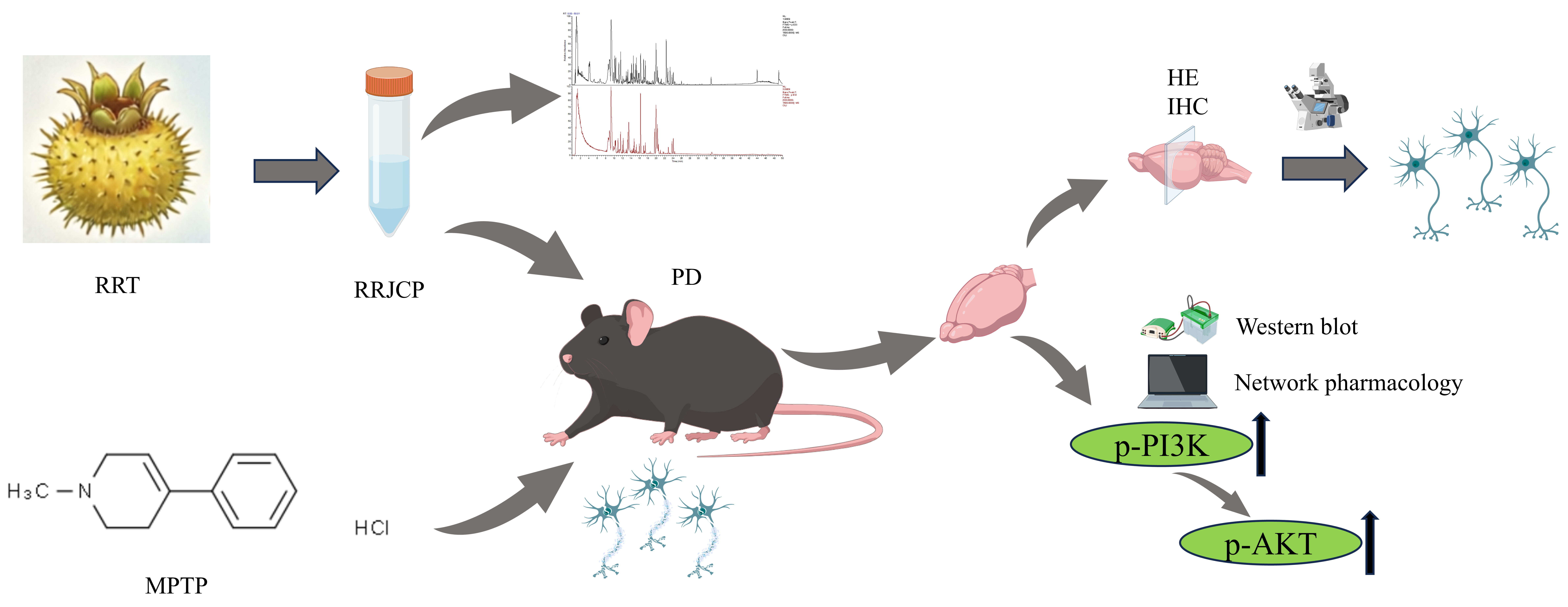
This study aimed to investigate the preventive, interventional, and therapeutic effects of RRJCP in an MPTP-induced chronic Parkinson’s disease mouse model, and to elucidate its underlying molecular mechanisms by integrating network pharmacology prediction with experimental validation. The study evaluated the intervention effects of RRJCP across three stages—before, during, and after PD onset—thereby transcending the limitations of previous studies that focused on a single administration time point. The findings revealed that RRJCP exerts multi-target neuroprotective effects by activating the PI3K/AKT signaling pathway and inhibiting oxidative stress and apoptosis. This work provides an experimental basis for the development of safe, orally administrable RRJCP-based health foods or novel herbal medicines as adjunctive interventions for PD, offering the potential to reduce long-term medication costs and improve patients’ quality of life, while also providing a modern scientific interpretation of the Traditional Chinese Medicine principle of “preventive treatment of disease”.
3. Discussion
Neurodegenerative diseases are becoming an increasingly pressing global health challenge, particularly in the context of an aging population. Among them, PD is one of the most common and severe [
25]. Although advances have been achieved in elucidating the underlying pathophysiology and molecular pathways of the disease, existing therapeutic approaches are predominantly palliative in nature and do not halt or reverse the core degenerative process. This gap in disease-modifying interventions highlights the pressing demand for innovative and efficacious treatment modalities. Bioactive constituents obtained from natural origins, particularly those sourced from edible plants, are increasingly regarded as attractive therapeutic avenues for combating neurodegenerative conditions [
26]. Studies have demonstrated that RRJCP is rich in various natural products and nutrients, including organic acids, phenolic compounds, flavonoids, and terpenoids, and exhibits strong free radical scavenging and anti-inflammatory effects [
27]. This investigation systematically assessed the neuroprotective capacity of RRJCP across three distinct temporal windows—disease prevention, progression intervention, and post-onset treatment—utilizing a chronic MPTP-induced mouse model of Parkinson’s disease. The findings demonstrate that RRJCP administration markedly alleviated motor behavioral deficits, substantially attenuated the degeneration and depletion of dopaminergic neurons within the substantia nigra, and concurrently suppressed oxidative stress responses while impeding apoptotic cascades. Further mechanistic investigation revealed that the observed neuroprotection was closely associated with the activation of the PI3K/AKT signal transduction axis. Collectively, this work not only provides experimental evidence for the multi-target neuroprotective attributes of RRJCP but also offers novel mechanistic insights into how medicinal-edible homologous compounds regulate the prevention and management of neurodegenerative disorders.
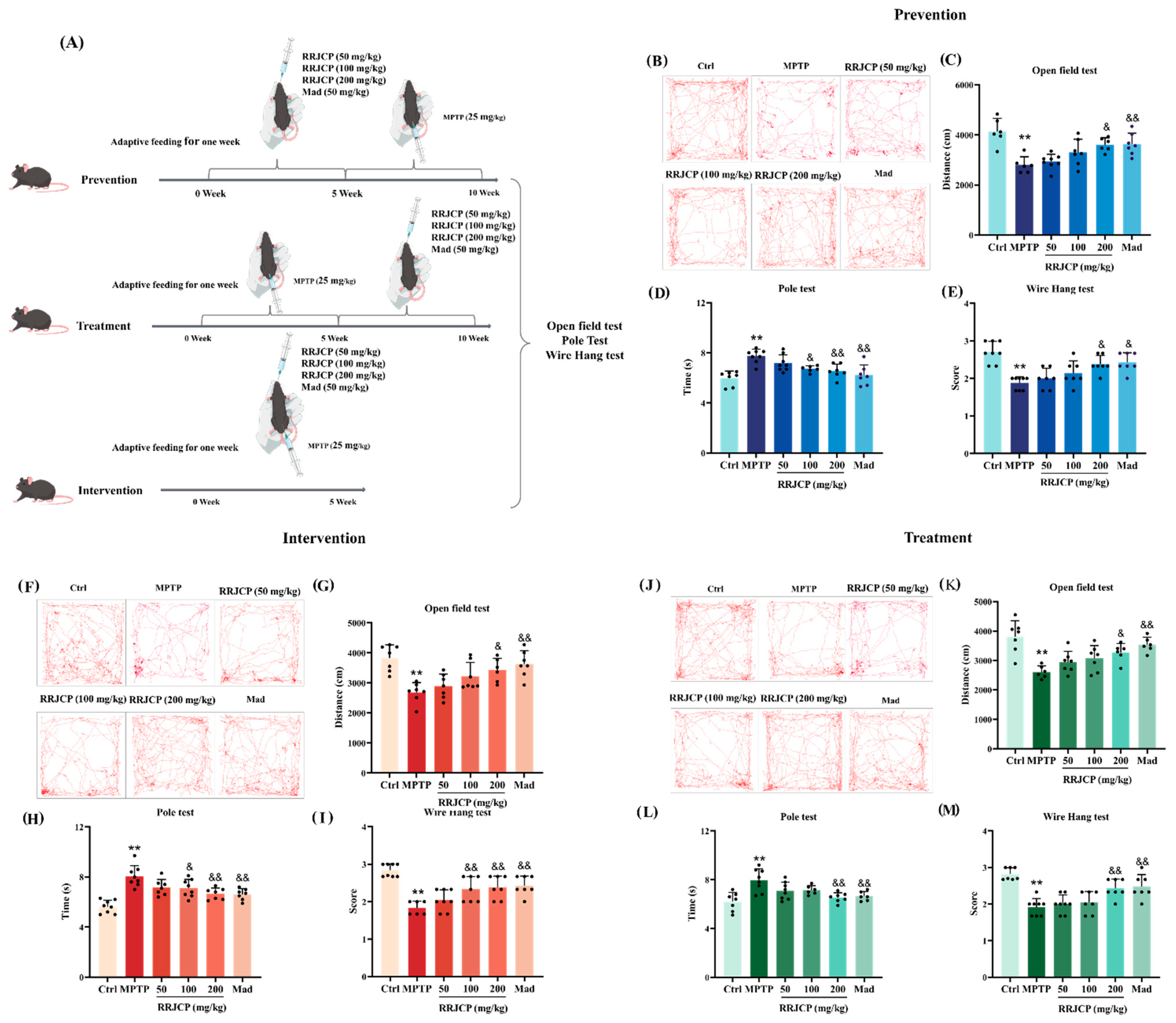
Behavioral improvement is a direct reflection of functional recovery. The open field test, pole test, and Wire Hang test evaluated the motor functions of the mice in terms of autonomous exploration, motor coordination, and muscle strength/endurance, respectively [
28,
29]. In this study, the open field test, pole test, and wire hang test were employed to evaluate motor function from the perspectives of spontaneous locomotor activity, motor coordination, and muscle strength/endurance, respectively. The results demonstrated that RRJCP resulted in comprehensive improvement across all behavioral assessments: the increased total distance traveled in the open field reflected enhanced exploratory behavior, whereas improvements in the pole and wire hang tests more specifically indicated the normalization of coordination within the cortical-basal ganglia-spinal motor pathway and muscle tone. Notably, high-dose RRJCP (200 mg/kg) significantly reversed MPTP-induced behavioral deficits irrespective of whether administration occurred during the preventive, interventional, or therapeutic phases, suggesting that its neuroprotective efficacy is not confined to a specific intervention time window. Nevertheless, cross-sectional comparison of the effects elicited by the medium dose (100 mg/kg) revealed that the preventive and interventional groups exhibited markedly greater improvements in the pole and wire hang tests compared with the therapeutic group, with the interventional group demonstrating the most pronounced enhancement. This difference implies that RRJCP administration prior to the establishment of irreversible neural circuit damage induced by MPTP toxicity (i.e., during the preventive or interventional stages) may more efficiently preserve motor coordination by maintaining synaptic plasticity and the functional integrity of the neuromuscular junction. In contrast, once significant behavioral deficits have manifested during the therapeutic stage, a higher dose is required to achieve an equivalent degree of recovery. Collectively, the behavioral improvements not only provide intuitive functional evidence for the neuroprotective effects of RRJCP but also preliminarily underscore the temporal advantage conferred by early intervention.
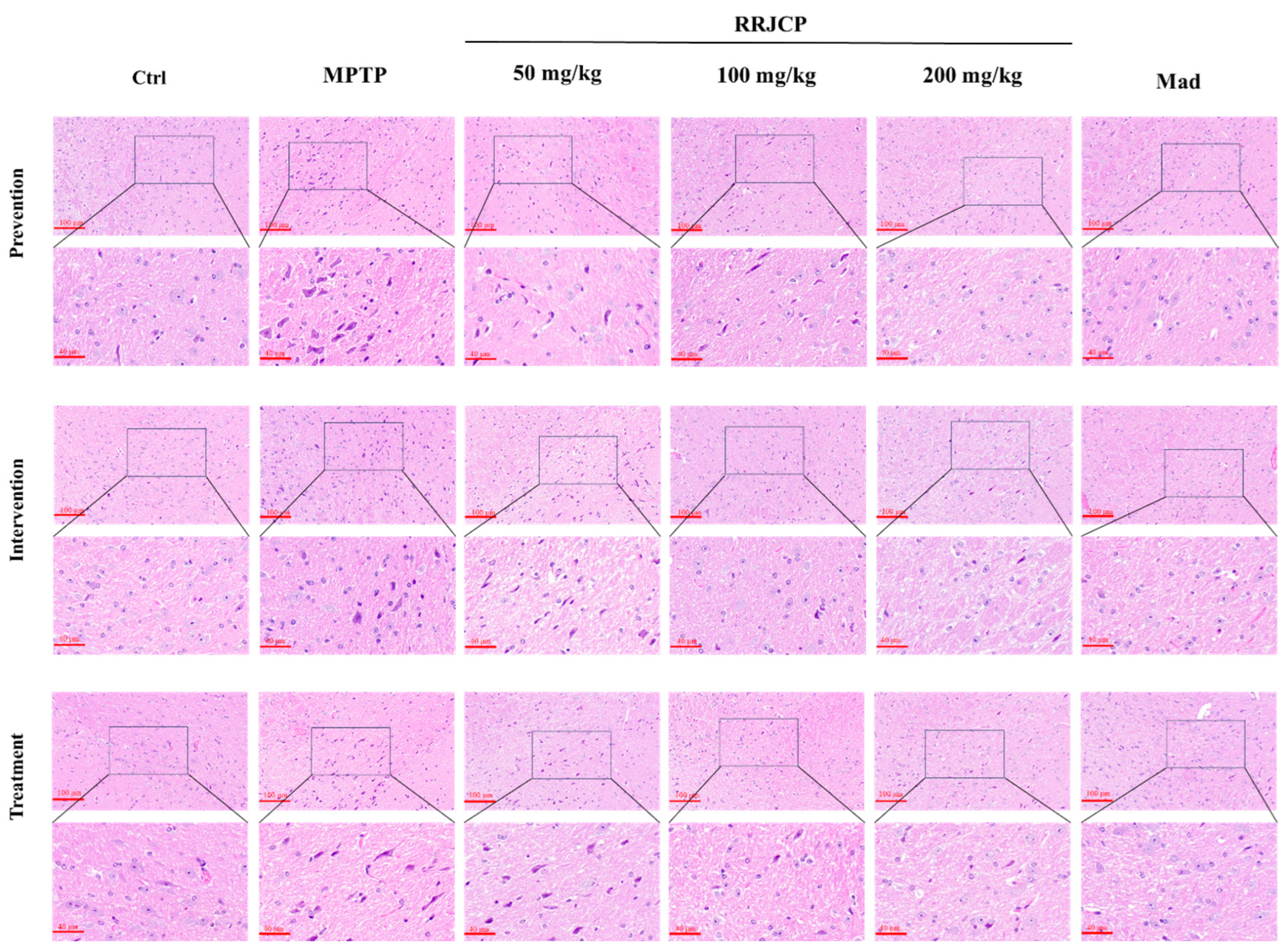
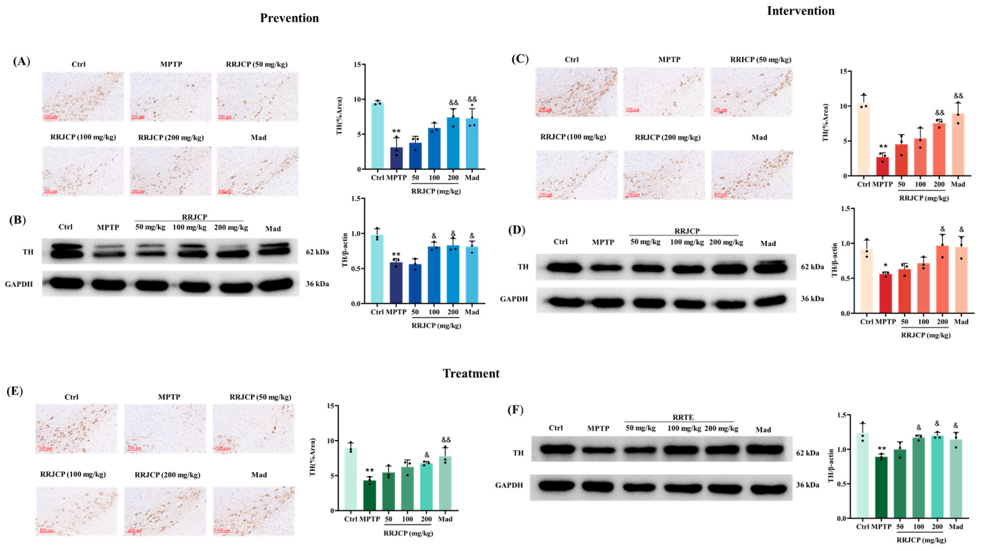
Histopathological results revealed the morphological basis of protection. HE staining showed that MPTP caused disorganized arrangement, reduced numbers, and pyknotic necrosis of neurons in the substantia nigra pars compacta. This finding is consistent with previous studies. After RRJCP treatment, the neuronal morphology was largely restored to normal, with more orderly arrangement and reduced nuclear pyknosis. This result visually confirmed that RRJCP could mitigate the direct toxic damage of MPTP to substantia nigra neurons, protecting neuronal survival and structural integrity. More importantly, TH immunohistochemistry and Western blot results further localized this protective effect to dopaminergic neurons. TH is the rate-limiting enzyme for dopamine synthesis, and its expression levels directly reflect the quantity and functional state of dopaminergic neurons [
30]. In this study, MPTP induced the loss of dopaminergic neurons in the midbrain substantia nigra. RRJCP treatment significantly increased the area of TH-positive neurons in the substantia nigra and the expression levels of TH protein, clearly demonstrating its antagonistic effect on the core pathological aspect of PD-the loss of midbrain substantia nigra dopaminergic neurons. This provides a solid cellular foundation for the observed behavioral improvements. Notably, irrespective of whether administration occurred during the preventive, interventional, or therapeutic phases, high-dose RRJCP consistently reversed MPTP-induced downregulation of TH expression, suggesting that its mechanism of action may encompass a dual attribute: providing reparative support for already compromised neurons while concurrently exerting preemptive defensive effects on intact neurons. Further analysis of the medium-dose (100 mg/kg) effects revealed that both the preventive and therapeutic groups exhibited superior restoration of TH protein expression compared with the interventional group. This discrepancy implies that preventive pre-administration may confer a state of “preconditioning” tolerance upon dopaminergic neurons against subsequent neurotoxin exposure, thereby enabling effective neuroprotection at a relatively lower dose.
Oxidative stress is considered an early and persistent event in the pathological cascade of PD [
31]. The neurotoxin MPTP is a classic model for studying PD mechanisms, as it can selectively induce pathological and behavioral manifestations in primates that closely resemble those of human PD [
32,
33]. MPTP itself is a protoxin, converted in the brain by monoamine oxidase B (MAO-B) in astrocytes into the toxic cation MPP
+. MPP
+ is then specifically taken up into DA neurons via the high-affinity dopamine transporter (DAT), where it inhibits complex I activity in mitochondria, blocking oxidative phosphorylation and ultimately leading to energy depletion, exacerbated oxidative stress, and apoptosis [
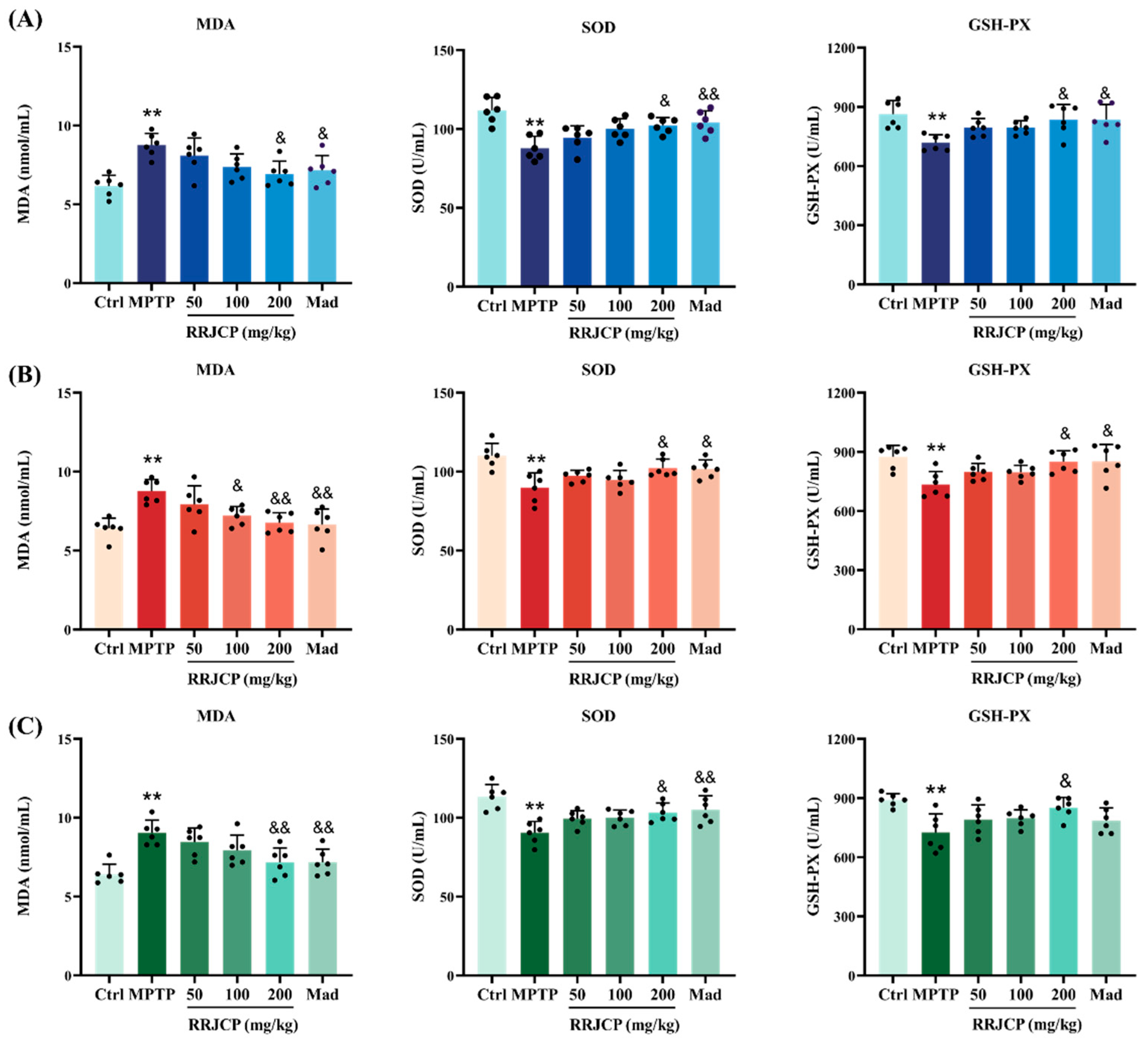
34,
35]. This study measured serum levels of MDA, SOD, and GSH-PX. After MPTP modeling, the oxidative damage marker MDA content increased significantly, while the activities of the antioxidant defense markers SOD and GSH-PX decreased significantly, consistent with previous studies. RRJCP treatment effectively reversed this trend, reducing MDA levels and increasing SOD and GSH-PX activities. This finding not only confirms the potent direct or indirect antioxidant capacity of RRJCP but also directly links its protective effects to a key pathogenic mechanism of PD-redox imbalance. Reducing oxidative stress helps maintain mitochondrial function, reduce protein misfolding, and inhibit activation of apoptotic signaling pathways. Notably, high-dose RRJCP consistently ameliorated oxidative stress markers irrespective of the timing of administration, indicating that its antioxidant efficacy demonstrates robust efficacy regardless of timing. Further analysis of the medium-dose (100 mg/kg) effects revealed that the interventional group exhibited superior reduction in MDA levels compared with both the preventive and therapeutic groups. This discrepancy implies that concurrent administration of RRJCP during MPTP exposure enables its active constituents to exert immediate and efficient blockade of oxidative damage at the peak window of oxidative insult, potentially through direct scavenging of MPP
+-induced free radicals or chelation of pro-oxidant metal ions. In contrast, pre-activation of antioxidant enzyme systems during the preventive phase and oxidative damage repair during the therapeutic phase may require higher doses to achieve comparable suppression of lipid peroxidation.
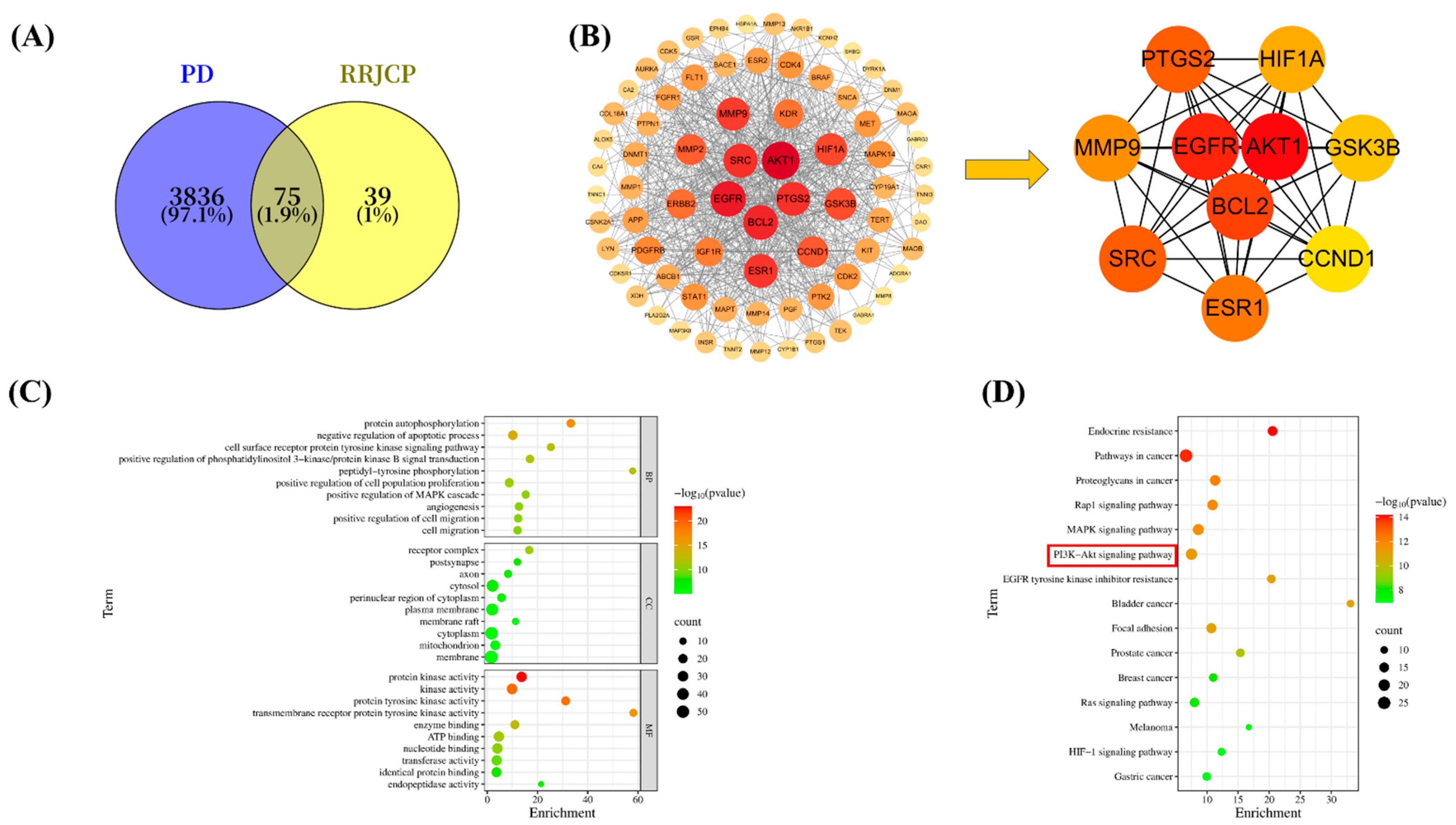
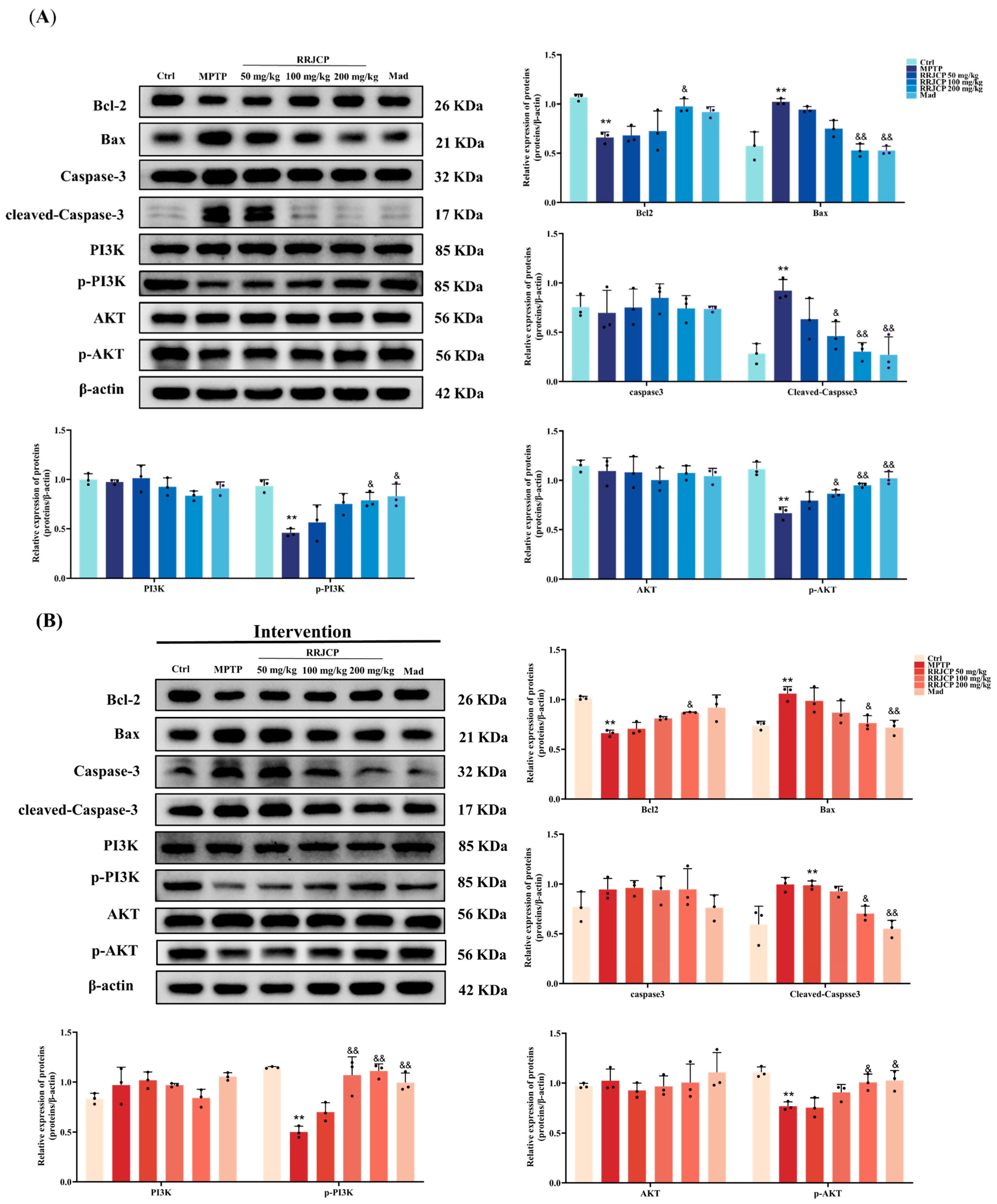
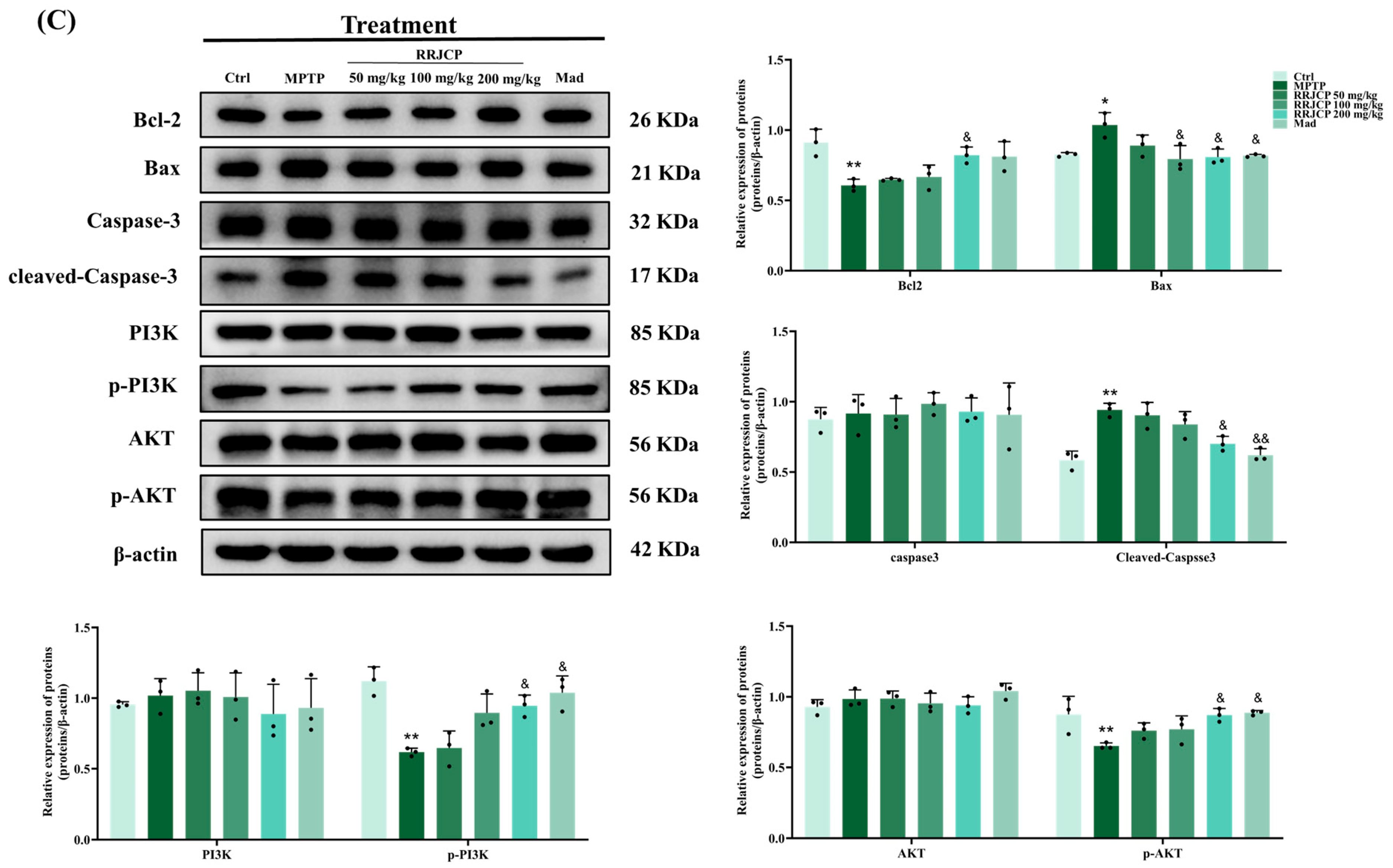
The pathological mechanisms of Parkinson’s disease are complex, involving mitochondrial dysfunction, oxidative stress, neuroinflammation, and apoptosis, among other processes. This study combined traditional in vivo phenotypic analysis with modern network pharmacology prediction, providing a clear roadmap for elucidating the multi-target mechanisms of RRJCP. Core targets (such as AKT1, EGFR, BCL2) and significantly enriched pathways (such as the PI3K-AKT signaling pathway) identified through network analysis guided the direction of subsequent Western blot validation. The PI3K/AKT pathway is a key signaling pathway regulating cell survival, proliferation, and apoptosis, and its function is often inhibited in neurodegenerative diseases [
36]. Activated AKT can inhibit multiple pro-apoptotic factors such as Bad and GSK-3β through phosphorylation while promoting pro-survival pathways such as mTOR, ultimately stabilizing the mitochondrial membrane and inhibiting Caspase activation [
37,
38]. At the mechanistic level, there is a close causal link between the inhibition of oxidative stress and apoptosis. The observed reduction in MDA and elevation of SOD and GSH-PX in this study indicated a decrease in intracellular reactive oxygen species levels and an enhancement of the antioxidant defense system. This improved redox state is a key prerequisite for inhibiting mitochondrial pathway apoptosis. Studies have shown that oxidative stress can directly lead to a decrease in mitochondrial membrane potential, promoting cytochrome C release and subsequently activating the Caspase cascade, leading to apoptosis [
39,
40]. In this study, while reducing oxidative stress, RRJCP significantly increased the expression levels of p-PI3K, p-AKT, and Bcl-2, and decreased the levels of Bax and Cleaved Caspase-3 in MPTP-damaged substantia nigra tissue. Therefore, our data support a mechanism of action: RRJCP may directly or indirectly activate the PI3K/AKT pathway through its active components. Activation of this pathway, on the one hand, enhances cellular antioxidant responses by regulating downstream factors (such as Nrf2) (manifested as increased SOD and GSH-PX, decreased MDA), and on the other hand, inhibits the mitochondrial apoptotic pathway by modulating the balance of Bcl-2 family proteins. Ultimately, this collectively protects substantia nigra dopaminergic neurons from MPTP-induced toxic damage, achieving comprehensive improvements in behavioral, pathological, and biochemical indicators. Notably, the molecular effects elicited by the medium dose (100 mg/kg) exhibited phase-specific divergence: the preventive group displayed more pronounced activation of p-AKT and suppression of Cleaved Caspase-3; the interventional group showed greater efficacy in upregulating p-PI3K; whereas the therapeutic group achieved significant improvement solely in TH expression. This differential molecular response profile suggests that preventive pre-administration may enhance the threshold of neuronal resistance to subsequent apoptotic stimuli through pre-activation of downstream AKT survival signaling. During the interventional phase, concurrent administration of RRJCP and MPTP may preferentially trigger PI3K-mediated membrane-proximal signaling events to counteract acute toxicity. In the therapeutic phase, however, the efficiency of reactivating key signaling cascades is limited, necessitating a higher dose to reconstitute a fully integrated survival signaling network. Collectively, these findings underscore the unique value of preventive strategies at the molecular level: by pre-emptively initiating the phosphorylation cascade of the PI3K/AKT pathway, a robust endogenous defense barrier can be established prior to the onset of neurotoxic insult, thereby fundamentally reducing the probability of apoptotic execution pathway activation. This provides mechanistic rationale supporting early intervention with RRJCP in populations at elevated risk for PD.
In the present study, the neuroprotective effects of RRJCP were evaluated in an MPTP-induced chronic PD mouse model across three administration paradigms—preventive, interventional, and therapeutic—revealing a potential advantage of early intervention, particularly preventive administration. By integrating network pharmacology predictions with in vivo experimental validation, the involvement of the PI3K/AKT signaling pathway in RRJCP-mediated antioxidant and anti-apoptotic effects was examined, providing experimental reference for the application of medicine–food homologous resources in neurodegenerative diseases. Regarding phytochemical characterization, LC-MS analysis with stringent spectral filtering tentatively identified 25 compounds in RRJCP, spanning flavonoids, phenolic acids, triterpenoids, organic acids, and amino acids, among which 15 were consistent with previous reports, whereas the remaining 10 compounds, although tentatively assigned by spectral library matching, have not been previously documented with authentic standard verification in Rosa roxburghii fruit or juice. Nevertheless, RRJCP is a multi-component extract, and it remains unclear whether its neuroprotective effects derive from a single active constituent or a synergistic interplay among multiple components. Although prior studies have reported that certain compound classes present in Rosa roxburghii exhibit antioxidant or neuroprotective potential, the present study did not perform activity validation on isolated monomers; thus, the observed pharmacodynamic effects cannot be attributed to specific chemical entities. These issues require further elucidation through subsequent activity-guided fractionation and monomer validation studies. Furthermore, the mechanistic investigation herein was primarily focused on the PI3K/AKT pathway, and whether RRJCP engages additional signaling networks implicated in PD pathogenesis warrants further exploration. Finally, notable discrepancies persist between animal models and human PD regarding pathological progression and clinical manifestations, and the translational potential of RRJCP requires further validation in preclinical and clinical settings. Future studies incorporating organoid models or patient-derived cells, along with comprehensive dose–effect and long-term toxicity assessments, will be essential to advance the application of RRJCP as a neuroprotective functional food or adjunctive therapeutic agent. Furthermore, the genus Rosa encompasses a wide variety of species with distinct phytochemical profiles. Future studies comparing the neuroprotective efficacy of RRJCP with extracts from closely related species, such as Rosa rugosa or Rosa canina, would be valuable for elucidating the structure-activity relationships and species-specificity of the observed effects.
In summary, this investigation integrated behavioral assessments, biochemical assays, histopathological evaluations, molecular biology techniques, and bioinformatics analyses to comprehensively characterize the neuroprotective profile of RRJCP in an MPTP-induced murine model of PD. The underlying mechanistic basis appears to involve, at least in part, activation of the PI3K/AKT signaling cascade, attenuation of oxidative damage, and suppression of apoptotic cell death. These observations extend the prospective utility of RRJCP in the field of neuroprotection and provide additional experimental substantiation and conceptual foundations for advancing natural product-derived interventions against PD. Owing to its status as a medicinal-edible resource, RRJCP exhibits favorable safety profiles and broad accessibility, highlighting its potential as both a prophylactic and an adjunctive therapeutic regimen for Parkinson’s disease.
4. Materials and Methods
4.1. Animals
A total of 144 male C57BL/6 mice, 8 weeks old, weighing 18–20 g, and of specific pathogen-free (SPF) grade, were obtained from the Experimental Animal Center of Guizhou Medical University. The mice were housed in an SPF-classified animal facility affiliated with the Natural Products Research Center of Guizhou Province, featuring a barrier-sustained environment and individually ventilated cages. This experimental site holds license SYXK (Qian) 2018-0001. In accordance with the ethical standards set forth in the Chinese Guidelines for the Care and Use of Laboratory Animals, the present study received institutional approval (No. 2400109) from the Animal Ethics Committee at Guizhou Medical University, ensuring full procedural compliance.
4.2. RRJCP Sample Preparation
Guizhou Peihua Company (Guiyang, China) supplied the RRT juice utilized in this investigation. The RRT juice was evaporated and concentrated through rotating evaporation and vacuum freeze-drying. Finally, the RRT juice concentrate powder was stored at 4 °C in sterilized glass bottles [
27].
4.3. LC-MS/MS Analysis of RRJCP
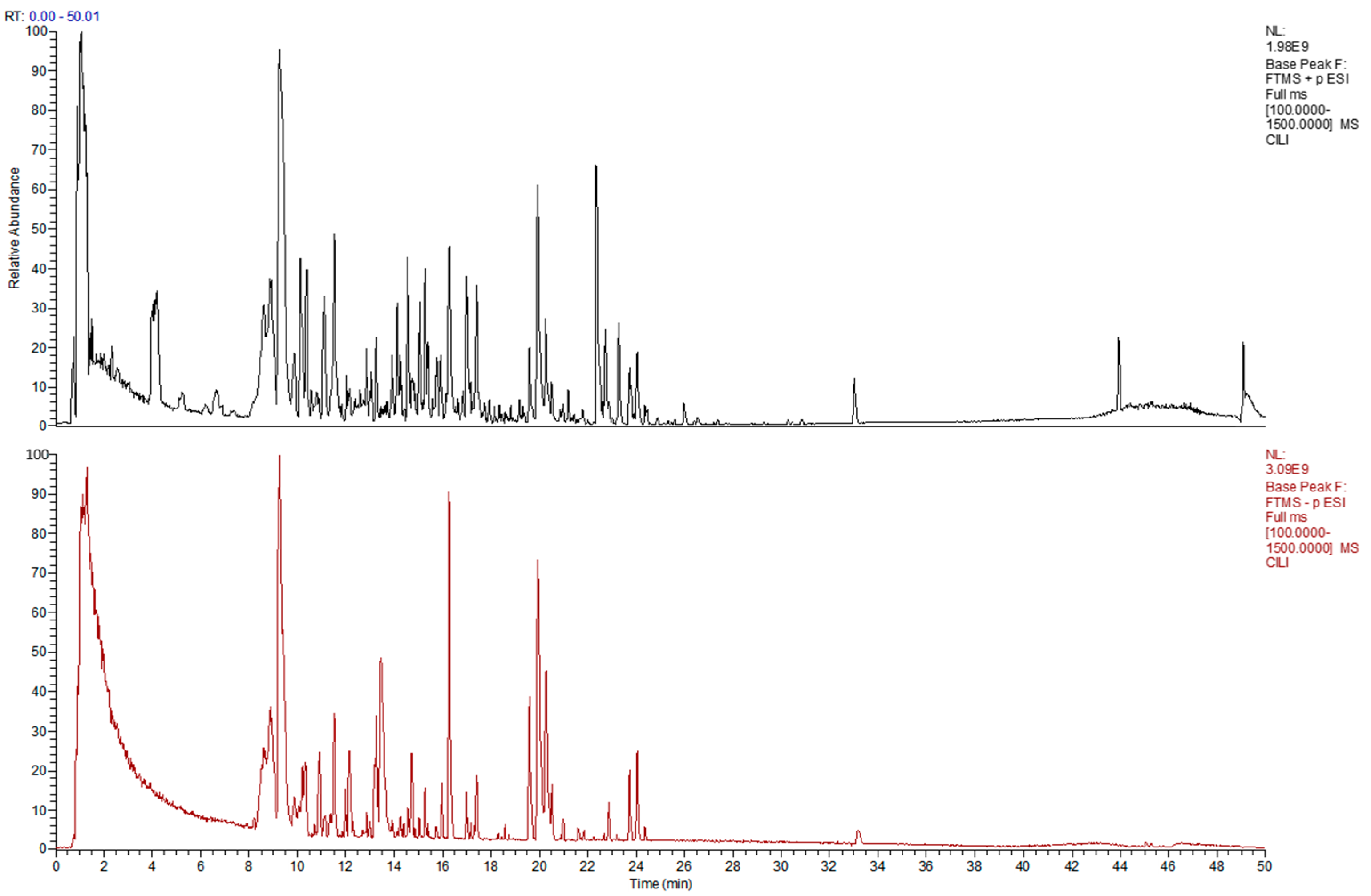
RRJCP was dissolved in 80% methanol to a final concentration of 10 mg/mL. After vortexing and ultrasonication, the mixture was centrifuged, and the supernatant was filtered through a 0.22 μm membrane. Analysis was performed using a Dionex Ultimate 3000 RSLC liquid chromatography system (Thermo Fisher Scientific, Sunnyvale, CA, USA) coupled with a Thermo Scientific Q Exactive Focus high-resolution mass spectrometer (Thermo Fisher Scientific, Waltham, MA, USA). Chromatographic separation was achieved on a Waters Acquity UPLC HSS T3 column (100 mm × 2.1 mm, 1.8 μm) (Waters Corporation, Milford, MA, USA) maintained at 40 °C. The mobile phase consisted of 0.1% formic acid in water (A) and 0.1% formic acid in acetonitrile (B), delivered at a flow rate of 0.3 mL/min. The gradient elution program was as follows: 0–5 min, 5% B; 5–42 min, 5% → 95% B; 42–47 min, 95% B; 47.1–50 min, 5% B. Mass spectrometric detection was carried out using a HESI-II source operated in positive/negative ion switching mode. Source parameters were set as follows: spray voltage, 3.0 kV (+)/2.5 kV (−); capillary temperature, 320 °C; sheath gas, 35 arb; auxiliary gas, 10 arb; probe heater temperature, 350 °C; and S-Lens RF level, 60. Data were acquired in Full MS-ddMS2 mode. Full MS scans were performed over an m/z range of 100–1500 at a resolution of 70,000, with an AGC target of 1 × 10⁶ and a maximum injection time of 100 ms. MS/MS scans were acquired at a resolution of 17,500, with an AGC target of 2 × 10⁵ and a maximum injection time of 50 ms. The isolation window was set to 1.5 m/z, and stepped normalized collision energies of 20, 40, and 60 were applied. Dynamic exclusion was set to 5 s. Raw data were processed using Compound Discoverer software (version 3.3), and compound identification was performed by matching accurate mass, retention time, and fragmentation patterns against spectral libraries.
4.4. Mouse Model
Comprising 144 male C57BL/6 mice, the experimental population underwent random division into three main groups. (prevention, intervention, treatment; n = 48), each containing six subgroups (n = 8). A chronic MPTP (Shanghai yuanye Bio-Technology Co., Ltd., Shanghai, China) model was induced by 10 injections over 5 weeks (25 mg/kg MPTP, preceded by 250 mg/kg probenecid (MedChemExpress, Monmouth Junction, NJ, USA) 30 min earlier). RRJCP (50, 100, 200 mg/kg) or Madopar (Mad) (Shandong Xinhua Pharmaceutical Co., Ltd., Zibo, China) (50 mg/kg) was administered by gavage. Ctrl groups received equivalent volumes of saline and/or DMSO. Prevention Group: Different doses of RRJCP, Mad, or saline were administered by gavage for 5 weeks, followed by 5 weeks of probenecid/MPTP injections The normal control subgroup received DMSO + saline injections. Intervention Group: Different doses of RRJCP, Mad, or saline were administered by gavage for 5 weeks, concurrent with 5 weeks of probenecid/MPTP injections The normal control subgroup received concurrent DMSO + saline injections. Treatment Group: Probenecid/MPTP injections were administered for 5 weeks to establish the model first, followed by 5 weeks of gavage administration with different doses of RRJCP, Mad, or saline. The normal control subgroup received saline by gavage for 5 weeks first, followed by DMSO + saline injections for 5 weeks (
Figure 1A).
Figure 1A created with BioGDP.com [
41].
4.5. Behavioral Tests
Open field test: After drug administration was completed, the open field test was conducted on all mice. Individual subjects were positioned within an open field apparatus measuring 60 cm × 60 cm × 60 cm, and their locomotor activity was videotaped for a 10 min interval via the Test Video 4 system. Between successive trials, the arena was thoroughly cleaned with 75% ethanol to remove olfactory traces deposited by prior occupants. Post hoc quantification of aggregate travel distance for each experimental group was performed using EthoVision XT 17 software.
Pole test: After drug administration, the pole test was performed. Construction of the apparatus involved a wooden pole measuring 50 cm in height and 10 mm in diameter, covered with gauze to improve grip, with a small sphere of 2.5 cm in diameter mounted at the summit. Animals were situated with forepaws contacting the sphere and the head directed superiorly. Chronometric recording captured the temporal interval for descent from the spherical apex to the basal platform. Triplicate trials were conducted for each mouse, and the arithmetic mean of the latencies on the pole was recorded as the final value.
Wire Hang test: In the Wire Hang test, a 5 mm diameter steel wire was fixed horizontally. Each mouse was allowed to grip the wire with its two forepaws, and whether it could also grasp the wire with its hind paws was observed. Grasping behavior was quantified via a tiered scoring system: bilateral hind paw contact yielded three points, unilateral hind paw contact yielded two points, and absence of grip yielded one point. Assessment occurred in triplicate for each animal, with inter-trial intervals maintained at or exceeding 10 min. Aggregation of individual scores and calculation of mean values preceded statistical analysis.
4.6. Antioxidant Indexes Measurements
Upon termination of behavioral procedures, all mice entered a 12 h fast with continuous water provision. Anesthesia was achieved using pentobarbital sodium (4%, 50 mg/kg) administered intraperitoneally, facilitating blood withdrawal from the orbital sinus. Specimens were centrifuged at 2000× g for 20 min to separate the serum. The serum levels of MDA, SOD, and GSH-PX (Nanjing Jiancheng Biotechnology Research Institute Co., Ltd., Nanjing, China) were measured according to the respective commercial assay kits’ instructions.
4.7. Hematoxylin and Eosin Staining
Harvested cerebral tissues underwent primary fixation in 4% paraformaldehyde. Ethanol gradient ascent (70%, 80%, 90%, 100%) achieved dehydration enroute to paraffin processing and microtome sectioning. Sections were dewaxed with xylene and rehydrated through a descending ethanol gradient (100%, 90%, 80%, 70%) before immersion in distilled water. Chromogenic visualization employed hematoxylin, bluing, eosin counterstain, dehydration, and neutral balsam preservation. Terminal pathological assessment and photomicrography utilized optical microscopy.
4.8. Immunohistochemical Staining
Paraffin sections underwent antigen retrieval via heat induction in citrate buffer (pH 6.0, 20 min), with natural cooling to room temperature. Endogenous peroxidase activity was quenched using 3% hydrogen peroxide (10 min, room temperature), prior to goat serum blocking (20 min). Overnight incubation with primary TH antibody (1:50, 4 °C) (Servicebio, Wuhan, China)was followed by PBS washing and secondary antibody application (37 °C, 30 min). DAB substrate enabled colorimetric detection, and hematoxylin counterstaining (3 min). Dehydration through ascending ethanol grades (75%, 85%, 95%, 100%) and xylene clearing (10 min each) preceded air drying and neutral resin mounting. Quantitative assessment of TH positive area employed ImageJ (Fiji) software (version 1.54f).
4.9. Identification of Active Constituents and Target Prediction in RRJCP
Based on prior investigations conducted by our research group, a total of 25 distinct chemical constituents within RRJCP were characterized utilizing liquid chromatography-mass spectrometry (LC-MS) [
41]. To identify bioactive candidates, these compounds were subsequently filtered through the Traditional Chinese Medicine Systems Pharmacology database and analysis platform (TCMSP,
https://www.tcmsp-e.com/, accessed on 27 January 2025). The screening parameters were established with a threshold of oral bioavailability (OB) ≥ 30% combined with a drug-likeness (DL) index ≥ 0.18. For those ingredients meeting the specified criteria, their corresponding Simplified Molecular Input Line Entry System (SMILES) notations were retrieved from the PubChem repository (
https://pubchem.ncbi.nlm.nih.gov/, accessed on 27 January 2025). The acquired SMILES strings were then submitted to the Swiss Target Prediction web server (
http://www.swisstargetprediction.ch/, accessed on 27 January 2025) to perform reverse docking-based target inference. Following the consolidation and removal of redundant entries from the predicted targets, a comprehensive spectrum of potential therapeutic targets associated with the active components of RRJCP was established. Concurrently, a collection of targets implicated in the pathology of Parkinson’s disease was sourced by querying the GeneCards (
https://www.genecards.org/, accessed on 27 January 2025) and OMIM databases (
https://www.omim.org/, accessed on 27 January 2025) using “Parkinson disease” as the keyword. The search outputs derived from both platforms were merged, and overlapping entries were eliminated, thereby yielding a definitive set of potential therapeutic targets relevant to Parkinson’s disease.
4.10. Construction of Protein–Protein Interaction Network
The overlapping targets shared between the bioactive constituents of RRJCP and the pathological targets implicated in Parkinson’s disease were determined using the VENNY 2.1.0 online tool (
https://bioinfogp.cnb.csic.es/tools/venny/, accessed on 27 January 2025). A Venn diagram was subsequently plotted to visualize the intersection, thereby delineating the prospective therapeutic targets through which RRJCP may exert its anti-PD effects. The set of intersecting genes was then submitted to the STRING database (
https://cn.string-db.org/, accessed on 27 January 2025) to construct a protein–protein interaction (PPI) network. During this process, isolated nodes lacking any interactions were removed from the analysis. Finally, the resulting network data were exported and imported into Cytoscape version 3.10.3 for graphical rendering and subsequent topological interrogation.
4.11. Gene Ontology (GO) Analysis and Kyoto Encyclopedia of Genes and Genomes (KEGG) Pathway
To elucidate the biological significance of the overlapping gene set shared between RRJCP active components and Parkinson’s disease pathology, Gene Ontology (GO) annotation and Kyoto Encyclopedia of Genes and Genomes (KEGG) pathway enrichment assessments were conducted using the DAVID functional annotation suite (
https://davidbioinformatics.nih.gov/summary.jsp, accessed on 27 January 2025). Specifically, GO analysis was employed to characterize the involvement of these intersecting genes across three distinct categories: biological processes (BP), cellular components (CC), and molecular functions (MF). Subsequent interpretation of the KEGG enrichment data facilitated the identification of pivotal molecular pathways governing essential physiological activities. Ultimately, the resulting GO terms and KEGG enrichment outputs were transferred to the online bioinformatics visualization platform (
http://www.bioinformatics.com.cn/, accessed on 27 January 2025) for graphical representation and further in-depth examination.
4.12. Western Blotting
Homogenization of substantia nigra specimens in RIPA lysis buffer containing 1% PMSF (Beyotime, Shanghai, China) utilized a tissue grinder to generate approximately 10% homogenate. Post homogenate ice incubation (30 min) preceded centrifugation at 12,000 rpm, 4 °C, 10 min in a high speed refrigerated centrifuge, yielding supernatant for EP tube transfer and total protein extraction. BCA (Servicebio, Wuhan, China) quantification determined loading buffer volume. Heat denaturation (100 °C, 10 min, metal bath) was followed by ambient cooling and −80 °C storage. Prepared SDS PAGE gel placement in electrophoresis chamber preceded sample boiling (100 °C, 10 min) and mixing. Electrophoretic separation occurred initially at 80 V (30 min), then 120 V (60 min). Transfer onto PVDF membrane utilized 220 mA constant current for 1.5 h. Subsequent processing comprised 5% BSA blocking, TBST washing, and primary antibody overnight incubation (4 °C). (TH, 1:2000, Proteintech; Bax, 1:2000, Proteintech; Bcl-2, 1:2000, Proteintech; Caspase-3, 1:2000, Proteintech; Cleaved-Caspase-3, 1:2000, Proteintech; PI3K, 1:2000, Proteintech p-PI3K, 1:2000, Proteintech; AKT, 1:2000, Proteintech; p-AKT, 1:2000, Proteintech, Wuhan, China; GAPDH, 1:10,000, HUABIO; β-actin, 1:10,000, HUABIO, Hangzhou, China). The next day, the membrane was washed with TBST, incubated with a diluted secondary antibody on a horizontal shaker at room temperature for 120 min, and washed again with TBST. Finally, chemiluminescent detection was performed in a darkroom. The exposed protein bands were imported into ImageJ (Fiji) software for grayscale value analysis.
4.13. Statistical Analysis
All data underwent statistical analysis via Microsoft Excel and GraphPad Prism 10.1.2. Results are reported as arithmetic mean ± SD. One-way ANOVA served for group comparisons, and p values less than 0.05 indicated statistical significance.