

Amorphous Solid Dispersions of Polyphenols: Current State of the Art (Part I)
 ,
,  , ,
, , 
Abstract
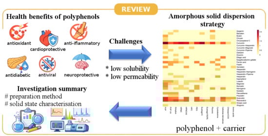
1. Introduction
2. Scope and Methodology of the Review
2.1. Time Frame
2.2. Search Strategy and Keywords
2.3. Inclusion Criteria
- The investigated compound belonged to the group of polyphenols, including flavonoids, stilbenes, phenolic acids, or their derivatives;
- The formulation involved an amorphous solid dispersion, co-amorphous system, or amorphous multicomponent system;
- The amorphous nature of the system was experimentally confirmed using solid-state characterization techniques (e.g., DSC, XRPD);
- The study reported physicochemical, biopharmaceutical, or biological performance metrics of the amorphous system, such as solubility, dissolution behavior, stability, permeability, or bioavailability;
- The article or patent was published in a peer-reviewed scientific journal within the defined time frame (2004–December 2025).
2.4. Exclusion Criteria
- The formulation did not involve an amorphous solid dispersion or co-amorphous system;
- The investigated compound was not a polyphenol;
- The amorphous character of the system was not experimentally verified
- The study focused exclusively on crystalline forms, emulsions, inclusion complexes, liposomes, or nanocrystals without an amorphous phase;
- The publication was a conference abstract, editorial, or commentary without original experimental data;
- Duplicate records;
- Studies with insufficient methodological detail.
3. Polyphenols Investigated in Amorphous Solid Dispersions
3.1. Literature Overview
- Apigenin (APG)
- Baicalein (BAC)
- Butein (BUT)
- Chrysin
- Chrysosplenol C (CRSP)
- Curcumin (CUR)
- Curcumin–Hesperetin (CUR-HSP)
- Curcumin–Magnolol
- Curcumin-Piperine (CUR-PIP)
- Daidzein
- Diosmin
- Ellagic acid
- Epigallocatechin gallate
- Ferulic acid (FA)
- Fisetin (FIS)
- Genistein (GEN)
- Hesperetin (HES)
- Hesperetin–Naringenin (HES-NAR)
- Hesperetin–Piperine (HES-PIP)
- Hesperidin (HED)
- Kaempferol (KMP)
- Luteolin (LUT)
- Magnolol (MAG)
- Myricetin (MYR)
3.2. Patent Landscape
4. Carriers Used in ASDs of Polyphenols
4.1. Cyclodextrins and Their Derivatives
4.2. Amino Acids and Low-Molecular-Weight Compounds
4.3. Polyvinylpyrrolidone and Its Derivatives
4.4. HPMC and Its Derivatives
4.5. Acrylic Polymers (Eudragit and Derivatives)
4.6. Surfactants and Poloxamers
4.7. Polyethylene Glycol (PEG)
5. Preparation Methods of Amorphous Solid Dispersions
5.1. Solvent Evaporation
5.2. Spray Drying
5.3. Hot-Melt Extrusion (HME)
5.4. Freeze Drying/Lyophilization
5.5. Milling
5.6. Cryo-Grinding/Cryo-Milling
5.7. Supercritical Carbon Dioxide (scCO2)
5.8. Quench Cooling
5.9. Electrospinning
6. Solid-State Characterization Techniques Used to Confirm Amorphization
7. Challenges and Future Perspectives
8. Summary and Scope of Part II
Author Contributions
Funding
Institutional Review Board Statement
Informed Consent Statement
Data Availability Statement
Conflicts of Interest
Abbreviations
| BCS | Biopharmaceutical classification system |
| ASDs | Amorphous solid dispersions |
| HME | Hot-melt extrusion |
| DSC | Differential scanning calorimetry |
| XRPD | X-ray powder diffraction |
| FT-IR | Fourier-transform infrared spectroscopy |
| HPMC | Hydroxypropyl methylcellulose |
| HPC | Hydroxypropyl cellulose |
| SDS | Sodium dodecyl sulfate |
| TG | Thermogravimetry |
| SEM | Scanning electron microscopy |
| PLM | Polarized light microscopy |
| DLS | Dynamic light scattering |
| PVP | Polyvinylopirolidon |
| COS | Amorphous chitosan oligosaccharide |
| IR | Infrared spectroscopy |
| HPMCAS | Hydroxypropylmethylcellulose acetate succinate |
| PEG | Polyethylene glycol |
| PVP VA | Copolymer of vinylpyrrolidone with vinyl acetate |
| CCAB | 6-carboxycellulose acetate butyrate |
| CASub | Cellulose acetate suberate |
| CMCAB | Carboxymethyl cellulose acetate butyrate |
| CAAdP | Cellulose acetate adipate propionate |
| GC-MS | Gas chromatography–mass spectrometry |
| NMR | Nuclear magnetic resonance |
| UV-Vis | Ultraviolet–visible spectroscopy |
| HPMCAS | Hydroxypropyl methylcellulose phthalate |
| CMCS | Carboxymethyl chitosan |
| MDSC | Modulated differential scanning calorimetry |
| AFM | Atomic force microscopy |
| TEM | Transmission electron microscopy |
| PAA | Polyacrylic acid |
| HP-β-CD | Hydroxypropyl-β-cyclodextrin |
| EDS | Energy-dispersive spectroscopy |
| BET analysis | Brunauer–Emmett–Teller surface-area analysis |
| PXRD | Powder X-ray diffraction |
| NIR | Near-infrared spectroscopy |
| TPS | Terahertz pulsed spectroscopy |
| ssNMR | Solid-state nuclear magnetic resonance |
| DVS | Dynamic vapor sorption |
| IGC | Inverse gas chromatography |
| XPS | X-ray photoelectron spectroscopy |
References
- Stagos, D. Antioxidant activity of polyphenolic plant extracts. Antioxidants 2019, 9, 19. [Google Scholar] [CrossRef]
- Lv, Q.; Long, J.; Gong, Z.; Nong, K.; Liang, X.; Qin, T.; Huang, W.; Yang, L. Current state of knowledge on the antioxidant effects and mechanisms of action of polyphenolic compounds. Nat. Prod. Commun. 2021, 16, 1934578X211027745. [Google Scholar] [CrossRef]
- Gündeşli, M.A.; Korkmaz, N.; Okatan, V. Polyphenol content and antioxidant capacity of berries: A review. Int. J. Agric. For. Life Sci. 2019, 3, 350–361. [Google Scholar]
- Yan, Z.; Zhong, Y.; Duan, Y.; Chen, Q.; Li, F. Antioxidant mechanism of tea polyphenols and its impact on health benefits. Anim. Nutr. 2020, 6, 115–123. [Google Scholar] [CrossRef]
- Omodanisi, E.; Aboua, Y.; Oguntibeju, O. Assessment of the Anti-Hyperglycaemic, Anti-Inflammatory and Antioxidant Activities of the Methanol Extract of Moringa Oleifera in Diabetes-Induced Nephrotoxic Male Wistar Rats. Molecules 2017, 22, 439. [Google Scholar] [CrossRef] [PubMed]
- Nagulapalli Venkata, K.C.; Swaroop, A.; Bagchi, D.; Bishayee, A. A small plant with big benefits: Fenugreek (Trigonella foenum-graecum Linn.) for disease prevention and health promotion. Mol. Nutr. Food Res. 2017, 61, 1600950. [Google Scholar] [CrossRef] [PubMed]
- Sajid, M.; Khan, M.R.; Shah, S.A.; Majid, M.; Ismail, H.; Maryam, S.; Batool, R.; Younis, T. Investigations on anti-inflammatory and analgesic activities of Alnus nitida Spach (Endl). stem bark in Sprague Dawley rats. J. Ethnopharmacol. 2017, 198, 407–416. [Google Scholar] [CrossRef] [PubMed]
- González-Sarrías, A.; Núñez-Sánchez, M.Á.; Tomás-Barberán, F.A.; Espín, J.C. Neuroprotective Effects of Bioavailable Polyphenol-Derived Metabolites against Oxidative Stress-Induced Cytotoxicity in Human Neuroblastoma SH-SY5Y Cells. J. Agric. Food Chem. 2017, 65, 752–758. [Google Scholar] [CrossRef]
- Luo, J.; Wei, Z.; Zhang, S.; Peng, X.; Huang, Y.; Zhang, Y.; Lu, J. Phenolic fractions from muscadine grape “noble” pomace can inhibit breast cancer cell MDA-MB-231 better than those from European grape “cabernet sauvignon” and induce s-phase arrest and apoptosis. J. Food Sci. 2017, 82, 1254–1263. [Google Scholar] [CrossRef]
- Odongo, G.A.; Schlotz, N.; Herz, C.; Hanschen, F.S.; Baldermann, S.; Neugart, S.; Trierweiler, B.; Frommherz, L.; Franz, C.M.A.P.; Ngwene, B. The role of plant processing for the cancer preventive potential of Ethiopian kale (Brassica carinata). Food Nutr. Res. 2017, 61, 1271527. [Google Scholar] [CrossRef]
- Reboredo-Rodríguez, P.; Figueiredo-González, M.; González-Barreiro, C.; Simal-Gándara, J.; Salvador, M.D.; Cancho-Grande, B.; Fregapane, G. State of the art on functional virgin olive oils enriched with bioactive compounds and their properties. Int. J. Mol. Sci. 2017, 18, 668. [Google Scholar] [CrossRef]
- Ben Mansour, R.; Wided, M.K.; Cluzet, S.; Krisa, S.; Richard, T.; Ksouri, R. LC-MS identification and preparative HPLC isolation of Frankenia pulverulenta phenolics with antioxidant and neuroprotective capacities in PC12 cell line. Pharm. Biol. 2017, 55, 880–887. [Google Scholar] [CrossRef] [PubMed]
- Mężyńska, M.; Brzóska, M.M. Związki polifenolowe w leczeniu i profilaktyce wybranych chorób cywilizacyjnych–dowody z badań epidemiologicznych. Pol. Prz. Nauk Zdr. 2016, 3, 269–278. [Google Scholar]
- Szajdek, A.; Borowska, J. Właściwości przeciwutleniające żywności pochodzenia roślinnego. Żywność Nauk. Technol. Jakość 2004, 11. [Google Scholar]
- D’ Archivio, M.; Filesi, C.; Di Benedetto, R.; Gargiulo, R.; Giovannini, C.; Masella, R. Polyphenols, dietary sources and bioavailability. Ann.-Ist. Super. Di Sanita 2007, 43, 348. [Google Scholar]
- Truzzi, F.; Tibaldi, C.; Zhang, Y.; Dinelli, G.; D’ Amen, E. An overview on dietary polyphenols and their biopharmaceutical classification system (BCS). Int. J. Mol. Sci. 2021, 22, 5514. [Google Scholar] [CrossRef] [PubMed]
- Lin, T.-C.; Yang, C.-Y.; Wu, T.-H.; Tseng, C.-H.; Yen, F.-L. Myricetin Nanofibers Enhanced Water Solubility and Skin Penetration for Increasing Antioxidant and Photoprotective Activities. Pharmaceutics 2023, 15, 906. [Google Scholar] [CrossRef]
- Rosiak, N.; Tykarska, E.; Cielecka-Piontek, J. Amorphous Pterostilbene Delivery Systems Preparation—Innovative Approach to Preparation Optimization. Pharmaceutics 2023, 15, 1231. [Google Scholar] [CrossRef]
- Rosiak, N.; Wdowiak, K.; Tykarska, E.; Cielecka-Piontek, J. Amorphous Solid Dispersion of Hesperidin with Polymer Excipients for Enhanced Apparent Solubility as a More Effective Approach to the Treatment of Civilization Diseases. Int. J. Mol. Sci. 2022, 23, 15198. [Google Scholar] [CrossRef]
- Han, J.; Wei, Y.; Lu, Y.; Wang, R.; Zhang, J.; Gao, Y.; Qian, S. Co-amorphous systems for the delivery of poorly water-soluble drugs: Recent advances and an update. Expert Opin. Drug Deliv. 2020, 17, 1411–1435. [Google Scholar] [CrossRef] [PubMed]
- Yarlagadda, D.L.; Das, S.; Anand Vullendula, S.K.; Manandhar, S.; Dengale, S.J.; Ranganath Pai, K.S.; Bhat, K. Computational-Based Polyphenol Therapy for Nonsmall Cell Lung Cancer: Naringin Coamorphous Systems for Solubility and Bioavailability Enhancement. Mol. Pharm. 2024, 21, 3951–3966. [Google Scholar] [CrossRef]
- Kapoor, D.U.; Singh, S.; Sharma, P.; Prajapati, B.G. Amorphization of Low Soluble Drug with Amino Acids to Improve Its Therapeutic Efficacy: A State-of-Art-Review. AAPS PharmSciTech 2023, 24, 253. [Google Scholar] [CrossRef] [PubMed]
- Aher, A.A.; Shaikh, K.S.; Chaudhari, P.D. Stability of co-Amorphous Solid Dispersions: Physical and Chemical Aspects. J. Struct. Chem. 2023, 64, 686–738. [Google Scholar] [CrossRef]
- Löbmann, K.; Jensen, K.T.; Laitinen, R.; Rades, T.; Strachan, C.J.; Grohganz, H. Stabilized Amorphous Solid Dispersions with Small Molecule Excipients. In Amorphous Solid Dispersions: Theory and Practice; Shah, N., Sandhu, H., Choi, D.S., Chokshi, H., Malick, A.W., Eds.; Springer: New York, NY, USA, 2014; pp. 613–636. [Google Scholar]
- Karagianni, A.; Kachrimanis, K.; Nikolakakis, I. Co-amorphous solid dispersions for solubility and absorption improvement of drugs: Composition, preparation, characterization and formulations for oral delivery. Pharmaceutics 2018, 10, 98. [Google Scholar] [CrossRef]
- Löbmann, K.; Grohganz, H.; Laitinen, R.; Strachan, C.; Rades, T. Amino acids as co-amorphous stabilizers for poorly water soluble drugs—Part 1: Preparation, stability and dissolution enhancement. Eur. J. Pharm. Biopharm. 2013, 85, 873–881. [Google Scholar] [CrossRef]
- Tomar, D.; Singh, P.K.; Hoque, S.; Modani, S.; Sriram, A.; Kumar, R.; Madan, J.; Khatri, D.; Dua, K. Amorphous systems for delivery of nutraceuticals: Challenges opportunities. Crit. Rev. Food Sci. Nutr. 2022, 62, 1204–1221. [Google Scholar] [CrossRef]
- Rode, K.; Maji, I.; Mahajan, S.; Singh, P.K. Unlocking the potential of flavonoid-based co-crystal and co-amorphous systems. Drug Discov. Today 2024, 29, 104050. [Google Scholar] [CrossRef]
- Ciurzyńska, A.; Lenart, A. Freeze-drying-application in food processing and biotechnology—A review. Pol. J. Food Nutr. Sci. 2011, 61, 165–171. [Google Scholar] [CrossRef]
- Chen, X.D.; Mujumdar, A.S. Drying Technologies in Food Processing; John Wiley & Sons: Hoboken, NJ, USA, 2009. [Google Scholar]
- Wdowiak, K.; Pietrzak, R.; Tykarska, E.; Cielecka-Piontek, J. Hot-Melt Extrusion as an Effective Technique for Obtaining an Amorphous System of Curcumin and Piperine with Improved Properties Essential for Their Better Biological Activities. Molecules 2023, 28, 3848. [Google Scholar] [CrossRef]
- Rosiak, N.; Tykarska, E.; Cielecka-Piontek, J. The Study of Amorphous Kaempferol Dispersions Involving FT-IR Spectroscopy. Int. J. Mol. Sci. 2023, 24, 17155. [Google Scholar] [CrossRef]
- Garbiec, E.; Rosiak, N.; Tykarska, E.; Zalewski, P.; Cielecka-Piontek, J. Sinapic Acid Co-Amorphous Systems with Amino Acids for Improved Solubility and Antioxidant Activity. Int. J. Mol. Sci. 2023, 24, 5533. [Google Scholar] [CrossRef]
- Garbiec, E.; Rosiak, N.; Zalewski, P.; Tajber, L.; Cielecka-Piontek, J. Genistein Co-Amorphous Systems with Amino Acids: An Investigation into Enhanced Solubility and Biological Activity. Pharmaceutics 2023, 15, 2653. [Google Scholar] [CrossRef]
- Roos, Y.H. Glass transition temperature and its relevance in food processing. Annu. Rev. Food Sci. Technol. 2010, 1, 469–496. [Google Scholar] [CrossRef]
- Stasiłowicz-Krzemień, A.; Rosiak, N.; Racaniello, G.F.; Denora, N.; Cielecka-Piontek, J. Effective and Stable Senomorphic Apigenin Delivery System Obtained by Supercritical Carbon Dioxide Processing. Int. J. Mol. Sci. 2025, 26, 8126. [Google Scholar] [CrossRef]
- Rosiak, N.; Tykarska, E.; Miklaszewski, A.; Pietrzak, R.; Cielecka-Piontek, J. Enhancing the Solubility and Dissolution of Apigenin: Solid Dispersions Approach. Int. J. Mol. Sci. 2025, 26, 566. [Google Scholar] [CrossRef]
- Altamimi, M.A.; Elzayat, E.M.; Alshehri, S.M.; Mohsin, K.; Ibrahim, M.A.; Al Meanazel, O.T.; Shakeel, F.; Alanazi, F.K.; Alsarra, I.A. Utilizing spray drying technique to improve oral bioavailability of apigenin. Adv. Powder Technol. 2018, 29, 1676–1684. [Google Scholar] [CrossRef]
- Ding, F.; Cao, W.; Wang, R.; Wang, N.; Li, A.; Wei, Y.; Qian, S.; Zhang, J.; Gao, Y.; Pang, Z. Mechanistic Study on Transformation of Coamorphous Baicalein-Nicotinamide to Its Cocrystal Form. J. Pharm. Sci. 2023, 112, 513–524. [Google Scholar] [CrossRef] [PubMed]
- Jangid, A.K.; Jain, P.; Medicherla, K.; Pooja, D.; Kulhari, H. Solid-state properties, solubility, stability and dissolution behaviour of co-amorphous solid dispersions of baicalin. CrystEngComm 2020, 22, 6128–6136. [Google Scholar] [CrossRef]
- Zhang, Y.; Luo, R.; Chen, Y.; Ke, X.; Hu, D.; Han, M. Application of carrier and plasticizer to improve the dissolution and bioavailability of poorly water-soluble baicalein by hot melt extrusion. AAPS PharmSciTech 2014, 15, 560–568. [Google Scholar] [CrossRef] [PubMed]
- Kim, N.A.; Oh, H.K.; Lee, J.C.; Choi, Y.H.; Jeong, S.H. Comparison of solubility enhancement by solid dispersion and micronized butein and its correlation with in vivo study. J. Pharm. Investig. 2021, 51, 53–60. [Google Scholar] [CrossRef]
- Wang, C.; Liu, X.; Zhao, R.; Yang, M.; Liu, W.; Dai, Q.; Bao, X.; Chen, Y.; Ma, J. The Amorphous Solid Dispersion of Chrysin in Plasdone® S630 Demonstrates Improved Oral Bioavailability and Antihyperlipidemic Performance in Rats. Pharmaceutics 2023, 15, 2378. [Google Scholar] [CrossRef] [PubMed]
- Lee, S.H.; Lee, Y.; Song, J.G.; Han, H.-K. Improved In vivo Effect of Chrysin as an Absorption Enhancer Via the Preparation of Ternary Solid Dispersion with Brij®L4 and Aminoclay. Curr. Drug Deliv. 2018, 16, 86–92. [Google Scholar] [CrossRef] [PubMed]
- Fan, W.; Zhang, X.; Zhu, W.; Zhang, X.; Di, L. Preparation of Curcumin-Eudragit® E PO Solid Dispersions with Gradient Temperature through Hot-Melt Extrusion. Molecules 2021, 26, 4964. [Google Scholar] [CrossRef] [PubMed]
- Fan, N.; Lu, T.; Li, J. Surface Tracking of Curcumin Amorphous Solid Dispersions Formulated by Binary Polymers. J. Pharm. Sci. 2020, 109, 1068–1078. [Google Scholar] [CrossRef]
- Mai, N.N.S.; Otsuka, Y.; Kawano, Y.; Hanawa, T. Preparation and Characterization of Solid Dispersions Composed of Curcumin, Hydroxypropyl Cellulose and/or Sodium Dodecyl Sulfate by Grinding with Vibrational Ball Milling. Pharmaceuticals 2020, 13, 383. [Google Scholar] [CrossRef]
- Fan, W.; Zhang, X.; Zhu, W.; Di, L. The Preparation of Curcumin Sustained-Release Solid Dispersion by Hot-Melt Extrusion-II. Optimization of Preparation Process and Evaluation In Vitro and In Vivo. J. Pharm. Sci. 2020, 109, 1253–1260. [Google Scholar] [CrossRef]
- Huang, R.; Han, J.; Wang, R.; Zhao, X.; Qiao, H.; Chen, L.; Li, W.; Di, L.; Zhang, W.; Li, J. Surfactant-free solid dispersion of BCS class IV drug in an amorphous chitosan oligosaccharide matrix for concomitant dissolution in vitro—Permeability increase. Eur. J. Pharm. Sci. 2019, 130, 147–155. [Google Scholar] [CrossRef]
- He, Y.; Liu, H.; Bian, W.; Liu, Y.; Liu, X.; Ma, S.; Zheng, X.; Du, Z.; Zhang, K.; Ouyang, D. Molecular Interactions for the Curcumin-Polymer Complex with Enhanced Anti-Inflammatory Effects. Pharmaceutics 2019, 11, 442. [Google Scholar] [CrossRef]
- Shin, M.-S.; Yu, J.S.; Lee, J.; Ji, Y.S.; Joung, H.J.; Han, Y.-M.; Yoo, H.H.; Kang, K.S. A Hydroxypropyl Methylcellulose-Based Solid Dispersion of Curcumin with Enhanced Bioavailability and its Hepatoprotective Activity. Biomolecules 2019, 9, 281. [Google Scholar] [CrossRef]
- Fan, N.; Ma, P.; Wang, X.; Li, C.; Zhang, X.; Zhang, K.; Li, J.; He, Z. Storage stability and solubilization ability of HPMC in curcumin amorphous solid dispersions formulated by Eudragit E100. Carbohydr. Polym. 2018, 199, 492–498. [Google Scholar] [CrossRef]
- Li, J.; Wang, X.; Li, C.; Fan, N.; Wang, J.; He, Z.; Sun, J. Viewing Molecular and Interface Interactions of Curcumin Amorphous Solid Dispersions for Comprehending Dissolution Mechanisms. Mol. Pharm. 2017, 14, 2781–2792. [Google Scholar] [CrossRef] [PubMed]
- Kadota, K.; Otsu, S.; Fujimori, M.; Sato, H.; Tozuka, Y. Soluble hydrolysis-resistant composite formulation of curcumin containing α-glucosyl hesperidin and polyvinylpyrrolidone. Adv. Powder Technol. 2016, 27, 442–447. [Google Scholar] [CrossRef]
- Kadota, K.; Okamoto, D.; Sato, H.; Onoue, S.; Otsu, S.; Tozuka, Y. Hybridization of polyvinylpyrrolidone to a binary composite of curcumin/α-glucosyl stevia improves both oral absorption and photochemical stability of curcumin. Food Chem. 2016, 213, 668–674. [Google Scholar] [CrossRef]
- Kerdsakundee, N.; Mahattanadul, S.; Wiwattanapatapee, R. Development and evaluation of gastroretentive raft forming systems incorporating curcumin-Eudragit® EPO solid dispersions for gastric ulcer treatment. Eur. J. Pharm. Biopharm. 2015, 94, 513–520. [Google Scholar] [CrossRef]
- Li, J.; Lee, I.W.; Shin, G.H.; Chen, X.; Park, H.J. Curcumin-Eudragit® E PO solid dispersion: A simple and potent method to solve the problems of curcumin. Eur. J. Pharm. Biopharm. 2015, 94, 322–332. [Google Scholar] [CrossRef]
- Gangurde, A.B.; Kundaikar, H.S.; Javeer, S.D.; Jaiswar, D.R.; Degani, M.S.; Amin, P.D. Enhanced solubility and dissolution of curcumin by a hydrophilic polymer solid dispersion and its insilico molecular modeling studies. J. Drug Deliv. Sci. Technol. 2015, 29, 226–237. [Google Scholar] [CrossRef]
- Chuah, A.M.; Jacob, B.; Jie, Z.; Ramesh, S.; Mandal, S.; Puthan, J.K.; Deshpande, P.; Vaidyanathan, V.V.; Gelling, R.W.; Patel, G. Enhanced bioavailability and bioefficacy of an amorphous solid dispersion of curcumin. Food Chem. 2014, 156, 227–233. [Google Scholar] [CrossRef]
- Wegiel, L.A.; Zhao, Y.; Mauer, L.J.; Edgar, K.J.; Taylor, L.S. Curcumin amorphous solid dispersions: The influence of intra and intermolecular bonding on physical stability. Pharm. Dev. Technol. 2014, 19, 976–986. [Google Scholar] [CrossRef]
- Li, B.; Konecke, S.; Wegiel, L.A.; Taylor, L.S.; Edgar, K.J. Both solubility and chemical stability of curcumin are enhanced by solid dispersion in cellulose derivative matrices. Carbohydr. Polym. 2013, 98, 1108–1116. [Google Scholar] [CrossRef]
- Onoue, S.; Takahashi, H.; Kawabata, Y.; Seto, Y.; Hatanaka, J.; Timmermann, B.; Yamada, S. Formulation design and photochemical studies on nanocrystal solid dispersion of curcumin with improved oral bioavailability. J. Pharm. Sci. 2010, 99, 1871–1881. [Google Scholar] [CrossRef] [PubMed]
- Paradkar, A.; Ambike, A.A.; Jadhav, B.K.; Mahadik, K.R. Characterization of curcumin–PVP solid dispersion obtained by spray drying. Int. J. Pharm. 2004, 271, 281–286. [Google Scholar] [CrossRef]
- Wdowiak, K.; Tajber, L.; Miklaszewski, A.; Cielecka-Piontek, J. Application of the Box–Behnken Design in the Development of Amorphous PVP K30–Phosphatidylcholine Dispersions for the Co-Delivery of Curcumin and Hesperetin Prepared by Hot-Melt Extrusion. Pharmaceutics 2025, 17, 26. [Google Scholar] [CrossRef] [PubMed]
- Han, J.; Li, L.; Su, M.; Heng, W.; Wei, Y.; Gao, Y.; Qian, S. Deaggregation and crystallization inhibition by small amount of polymer addition for a co-amorphous curcumin-magnolol system. Pharmaceutics 2021, 13, 1725. [Google Scholar] [CrossRef] [PubMed]
- Panizzon, G.P.; Giacomini Bueno, F.; Ueda-Nakamura, T.; Nakamura, C.V.; Dias Filho, B.P. Manufacturing Different Types of Solid Dispersions of BCS Class IV Polyphenol (Daidzein) by Spray Drying: Formulation and Bioavailability. Pharmaceutics 2019, 11, 492. [Google Scholar] [CrossRef] [PubMed]
- Anwer, M.K.; Ahmed, M.M.; Alshetaili, A.; Almutairy, B.K.; Alalaiwe, A.; Fatima, F.; Ansari, M.N.; Iqbal, M. Preparation of spray dried amorphous solid dispersion of diosmin in soluplus with improved hepato-renoprotective activity: In vitro anti-oxidant and in-vivo safety studies. J. Drug Deliv. Sci. Technol. 2020, 60, 102101. [Google Scholar] [CrossRef]
- Li, B.; Harich, K.; Wegiel, L.; Taylor, L.S.; Edgar, K.J. Stability and solubility enhancement of ellagic acid in cellulose ester solid dispersions. Carbohydr. Polym. 2013, 92, 1443–1450. [Google Scholar] [CrossRef]
- Cao, Y.; Teng, J.; Selbo, J. Amorphous Solid Dispersion of Epigallocatechin Gallate for Enhanced Physical Stability and Controlled Release. Pharmaceuticals 2017, 10, 88. [Google Scholar] [CrossRef]
- Albuquerque, I.d.S. Selection of Suitable Polymer and Obtaining New Amorphous Solid Dispersions with Ferulic Acid and Soluplus® 2020. Bachelor’s Thesis, Universidade Federal do Rio Grande do Norte, Natal, Brazil, 2020. [Google Scholar]
- Huang, W.; Yang, Y.; Zhao, B.; Liang, G.; Liu, S.; Liu, X.-L.; Yu, D.-G. Fast dissolving of ferulic acid via electrospun ternary amorphous composites produced by a coaxial process. Pharmaceutics 2018, 10, 115. [Google Scholar] [CrossRef]
- Nadal, J.M.; Gomes, M.L.S.; Borsato, D.M.; Almeida, M.A.; Barboza, F.M.; Zawadzki, S.F.; Farago, P.V.; Zanin, S.M.W. Spray-dried solid dispersions containing ferulic acid: Comparative analysis of three carriers, in vitro dissolution, antioxidant potential and in vivo anti-platelet effect. Drug Dev. Ind. Pharm. 2016, 42, 1813–1824. [Google Scholar] [CrossRef]
- Rosiak, N.; Tykarska, E.; Cielecka-Piontek, J. Mechanochemical Approach to Obtaining a Multicomponent Fisetin Delivery System Improving Its Solubility and Biological Activity. Int. J. Mol. Sci. 2024, 25, 3648. [Google Scholar] [CrossRef]
- Sip, S.; Rosiak, N.; Sip, A.; Żarowski, M.; Hojan, K.; Cielecka-Piontek, J. A Fisetin Delivery System for Neuroprotection: A Co-Amorphous Dispersion Prepared in Supercritical Carbon Dioxide. Antioxidants 2023, 13, 24. [Google Scholar] [CrossRef] [PubMed]
- Skiba, M.; Gasmi, H.; Milon, N.; Bounoure, F.; Lahiani-Skiba, M. Water Solubility and Dissolution Enhancement of Fisetin by Spherical Amorphous Solid Dispersion in Polymer of Cyclodextrin. Austin J. Biotechnol. Bioeng. 2021, 8, 1106. [Google Scholar] [CrossRef]
- Novo, D.C.; Benson, E.G.; Petrova, S.P.; Thompsonc, J.E.; Mosquera-Giraldo, L.I.; Taylor, L.S.; Edgar, K.J. Capturing flavanol benefits using cellulose-based amorphous solid dispersions: Enhanced genistein and quercetin solution concentrations in vitro. Des. Synth. Complex Fluoresc. Labeled Cellul. Deriv. Orally Adm. Drug Deliv. Syst. 2023, 131–176. [Google Scholar]
- Zaini, E.; Putri, V.Z.; Octavia, M.D.; Ismed, F. Peningkatan Laju Disolusi Dispersi Padat Amorf Genistein dengan PVP K-30. J. Sains Farm. Klin. 2017, 4, 67. [Google Scholar] [CrossRef]
- Wdowiak, K.; Rosiak, N.; Tykarska, E.; Żarowski, M.; Płazińska, A.; Płaziński, W.; Cielecka-Piontek, J. Amorphous Inclusion Complexes: Molecular Interactions of Hesperidin and Hesperetin with HP-Β-CD and Their Biological Effects. Int. J. Mol. Sci. 2022, 23, 4000. [Google Scholar] [CrossRef]
- Wdowiak, K.; Miklaszewski, A.; Pietrzak, R.; Cielecka-Piontek, J. Amorphous System of Hesperetin and Piperine—Improvement of Apparent Solubility, Permeability, and Biological Activities. Int. J. Mol. Sci. 2023, 24, 4859. [Google Scholar] [CrossRef]
- Koromili, M.; Kapourani, A.; Barmpalexis, P. Preparation and Evaluation of Amorphous Solid Dispersions for Enhancing Luteolin’s Solubility in Simulated Saliva. Polymers 2022, 15, 169. [Google Scholar] [CrossRef]
- Alshehri, S.; Imam, S.S.; Altamimi, M.A.; Hussain, A.; Shakeel, F.; Elzayat, E.; Mohsin, K.; Ibrahim, M.; Alanazi, F. Enhanced Dissolution of Luteolin by Solid Dispersion Prepared by Different Methods: Physicochemical Characterization and Antioxidant Activity. ACS Omega 2020, 5, 6461–6471. [Google Scholar] [CrossRef]
- Cao, J.; Zhang, S.; Hao, Y.; Fan, K.; Wang, L.; Zhao, X.; He, X. Amorphous solid dispersion preparation via coprecipitation improves the dissolution, oral bioavailability, and intestinal health enhancement properties of magnolol. Poult. Sci. 2023, 102, 102676. [Google Scholar] [CrossRef] [PubMed]
- Zhao, J.; Gao, P.; Mu, C.; Ning, J.; Deng, W.; Ji, D.; Sun, H.; Zhang, X.; Yang, X. Preparation and Evaluation of Novel Supersaturated Solid Dispersion of Magnolol: Theme: Advancements in Amorphous Solid Dispersions to Improve Bioavailability. AAPS PharmSciTech 2022, 23, 97. [Google Scholar] [CrossRef]
- Liu, H.-Z. Different preparations of magnolol: Preparation, characterization and pharmacokinetics comparative study in SD rats. Chin. Tradit. Herb. Drugs 2020, 51, 4442–4448. [Google Scholar]
- Rosiak, N.; Tykarska, E.; Cielecka-Piontek, J. Myricetin Amorphous Solid Dispersions—Antineurodegenerative Potential. Molecules 2024, 29, 1287. [Google Scholar] [CrossRef] [PubMed]
- Zhang, S.; Zhang, X.; Meng, J.; Lu, L.; Du, S.; Xu, H.; Wu, S. Study on the Effect of Polymer Excipients on the Dispersibility, Interaction, Solubility, and Scavenging Reactive Oxygen Species of Myricetin Solid Dispersion: Experiment and Molecular Simulation. ACS Omega 2022, 7, 1514–1526. [Google Scholar] [CrossRef]
- Mureşan-Pop, M.; Pop, M.M.; Borodi, G.; Todea, M.; Nagy-Simon, T.; Simon, S. Solid dispersions of Myricetin with enhanced solubility: Formulation, characterization and crystal structure of stability-impeding Myricetin monohydrate crystals. J. Mol. Struct. 2017, 1141, 607–614. [Google Scholar] [CrossRef]
- Ishimoto, K.; Shimada, Y.; Ohno, A.; Otani, S.; Ago, Y.; Maeda, S.; Lin, B.; Nunomura, K.; Hino, N.; Suzuki, M.; et al. Physicochemical and Biochemical Evaluation of Amorphous Solid Dispersion of Naringenin Prepared Using Hot-Melt Extrusion. Front. Nutr. 2022, 9, 850103. [Google Scholar] [CrossRef]
- Jha, D.K.; Shah, D.S.; Amin, P.D. Thermodynamic Aspects of the Preparation of Amorphous Solid Dispersions of Naringenin with Enhanced Dissolution Rate. Int. J. Pharm. 2020, 583, 119363. [Google Scholar] [CrossRef]
- Khan, A.W.; Kotta, S.; Ansari, S.H.; Sharma, R.K.; Ali, J. Enhanced dissolution and bioavailability of grapefruit flavonoid Naringenin by solid dispersion utilizing fourth generation carrier. Drug Dev. Ind. Pharm. 2015, 41, 772–779. [Google Scholar] [CrossRef]
- Wegiel, L.A.; Mauer, L.J.; Edgar, K.J.; Taylor, L.S. Mid-infrared spectroscopy as a polymer selection tool for formulating amorphous solid dispersions. J. Pharm. Pharmacol. 2014, 66, 244–255. [Google Scholar] [CrossRef]
- Li, B.; Liu, H.; Amin, M.; Wegiel, L.A.; Taylor, L.S.; Edgar, K.J. Enhancement of naringenin solution concentration by solid dispersion in cellulose derivative matrices. Cellulose 2013, 20, 2137–2149. [Google Scholar] [CrossRef]
- Kanaze, F.I.; Kokkalou, E.; Niopas, I.; Georgarakis, M.; Stergiou, A.; Bikiaris, D. Dissolution enhancement of flavonoids by solid dispersion in PVP and PEG matrixes: A comparative study. J. Appl. Polym. Sci. 2006, 102, 460–471. [Google Scholar] [CrossRef]
- Onoue, S.; Uchida, A.; Takahashi, H.; Seto, Y.; Kawabata, Y.; Ogawa, K.; Yuminoki, K.; Hashimoto, N.; Yamada, S. Development of high-energy amorphous solid dispersion of nanosized nobiletin, a citrus polymethoxylated flavone, with improved oral bioavailability. J. Pharm. Sci. 2011, 100, 3793–3801. [Google Scholar] [CrossRef]
- Onoue, S.; Nakamura, T.; Uchida, A.; Ogawa, K.; Yuminoki, K.; Hashimoto, N.; Hiza, A.; Tsukaguchi, Y.; Asakawa, T.; Kan, T. Physicochemical and biopharmaceutical characterization of amorphous solid dispersion of nobiletin, a citrus polymethoxylated flavone, with improved hepatoprotective effects. Eur. J. Pharm. Sci. 2013, 49, 453–460. [Google Scholar] [CrossRef]
- Rosiak, N.; Tykarska, E.; Cielecka-Piontek, J. Enhanced Antioxidant and Neuroprotective Properties of Pterostilbene (Resveratrol Derivative) in Amorphous Solid Dispersions. Int. J. Mol. Sci. 2024, 25, 2774. [Google Scholar] [CrossRef]
- Paczkowska-Walendowska, M.; Tajber, L.; Miklaszewski, A.; Cielecka-Piontek, J. Hot Melt Extrusion for Improving the Physicochemical Properties of Polydatin Derived from Polygoni cuspidati Extract; A Solution Recommended for Buccal Applications. Pharmaceuticals 2023, 16, 1226. [Google Scholar] [CrossRef]
- Van Hecke, E.; Benali, M. Solid dispersions of quercetin-PEG matrices: Miscibility prediction, preparation and characterization. Food Biosci. 2022, 49, 101868. [Google Scholar] [CrossRef]
- Wang, Y.; Qin, W.; Liang, Q.; Zhou, F.; Yan, C.; Deng, Y. The combination of hydroxypropylmethylcellulose acetate succinate and L-lysine into ternary amorphous solid dispersions of quercetin to enhance its dissolution. Carbohydr. Polym. Technol. Appl. 2021, 2, 100053. [Google Scholar] [CrossRef]
- Hatwar, P.; Pathan, I.B.; Chishti, N.A.H.; Ambekar, W. Pellets containing quercetin amino acid co-amorphous mixture for the treatment of pain: Formulation, optimization, in-vitro and in-vivo study. J. Drug Deliv. Sci. Technol. 2021, 62, 102350. [Google Scholar] [CrossRef]
- Wang, Y.; Fang, Y.; Zhou, F.; Liang, Q.; Deng, Y. The Amorphous Quercetin/Hydroxypropylmethylcellulose Acetate Succinate Solid Dispersions Prepared by Co-Precipitation Method to Enhance Quercetin Dissolution. J. Pharm. Sci. 2021, 110, 3230–3237. [Google Scholar] [CrossRef] [PubMed]
- Shi, X.; Fan, N.; Zhang, G.; Sun, J.; He, Z.; Li, J. Quercetin amorphous solid dispersions prepared by hot melt extrusion with enhanced solubility and intestinal absorption. Pharm. Dev. Technol. 2020, 25, 472–481. [Google Scholar] [CrossRef]
- Febriyenti, F.; Indra, P.; Zaini, E.; Ismed, F.; Lucida, H. Preparation and characterization of quercetin-polyvinylpyrrolidone K-30 spray dried solid dispersion. J. Pharm. Pharmacogn. Res. 2020, 8, 127–134. [Google Scholar] [CrossRef]
- Uchiyama, H.; Wada, Y.; Takamatsu, M.; Kadota, K.; Tozuka, Y. Improved solubility of quercetin by preparing amorphous solid with transglycosylated rutin and isoquercitrin. Environ. Control Biol. 2018, 56, 161–165. [Google Scholar] [CrossRef]
- Gilley, A.D.; Arca, H.C.; Nichols, B.L.B.; Mosquera-Giraldo, L.I.; Taylor, L.S.; Edgar, K.J.; Neilson, A.P. Novel cellulose-based amorphous solid dispersions enhance quercetin solution concentrations in vitro. Carbohydr. Polym. 2017, 157, 86–93. [Google Scholar] [CrossRef] [PubMed]
- Li, B.; Konecke, S.; Harich, K.; Wegiel, L.; Taylor, L.S.; Edgar, K.J. Solid dispersion of quercetin in cellulose derivative matrices influences both solubility and stability. Carbohydr. Polym. 2013, 92, 2033–2040. [Google Scholar] [CrossRef]
- Kakran, M.; Sahoo, N.G.; Li, L. Dissolution enhancement of quercetin through nanofabrication, complexation, and solid dispersion. Colloids Surf. B Biointerfaces 2011, 88, 121–130. [Google Scholar] [CrossRef]
- Almeida, H.; Ferreira, B.; Fernandes-Lopes, C.; Araújo, F.; Bonifácio, M.J.; Vasconcelos, T.; Sarmento, B. Third-Generation Solid Dispersion Through Lyophilization Enhanced Oral Bioavailability of Resveratrol. ACS Pharmacol. Transl. Sci. 2024, 7, 888–898. [Google Scholar] [CrossRef]
- Almeida, H.; Teixeira, N.; Sarmento, B.; Vasconcelos, T. Freeze-drying cycle optimization of an amorphous solid dispersion of resveratrol. Eur. J. Pharm. Sci. 2024, 200, 106855. [Google Scholar] [CrossRef] [PubMed]
- Jia, X.; Chen, J.; Cheng, H.; Pan, X.; Ke, Y.; Fu, T.; Qiao, H.; Cui, X.; Li, W.; Zou, L. Use of surfactant-based amorphous solid dispersions for BDDCS class II drugs to enhance oral bioavailability: A case report of resveratrol. Int. J. Pharm. 2023, 641, 123059. [Google Scholar] [CrossRef]
- Fan, W.; Wu, J.; Gao, M.; Zhang, X.; Zhu, W. Preparation of Solid Dispersion of Polygonum Cuspidatum Extract by Hot Melt Extrusion to Enhance Oral Bioavailability of Resveratrol. Molecules 2023, 28, 737. [Google Scholar] [CrossRef]
- Yu, C.; Zhang, C.; Guan, X.; Yuan, D. The solid dispersion of resveratrol with enhanced dissolution and good system physical stability. J. Drug Deliv. Sci. Technol. 2023, 84, 104507. [Google Scholar] [CrossRef]
- Han, G.; Wang, B.; Jia, M.; Ding, S.; Qiu, W.; Mi, Y.; Mi, Z.; Qin, Y.; Zhu, W.; Liu, X. Optimization and evaluation of resveratrol amorphous solid dispersions with a novel polymeric system. Math. Biosci. Eng. 2022, 19, 8019–8034. [Google Scholar] [CrossRef]
- Riccio, B.V.F.; Nascimento, A.L.C.S.d.; Meneguin, A.B.; Rodero, C.F.; Santos, K.P.; Sábio, R.M.; Annunzio, S.R.d.; Fontana, C.R.; Barud, H.d.S.; Ferrari, P.C.; et al. Solid Dispersions Incorporated into PVP Films for the Controlled Release of Trans-Resveratrol: Development, Physicochemical and In Vitro Characterizations and In Vivo Cutaneous Anti-Inflammatory Evaluation. Pharmaceutics 2022, 14, 1149. [Google Scholar] [CrossRef]
- Vasconcelos, T.; Prezotti, F.; Araújo, F.; Lopes, C.; Loureiro, A.; Marques, S.; Sarmento, B. Third-generation solid dispersion combining Soluplus and poloxamer 407 enhances the oral bioavailability of resveratrol. Int. J. Pharm. 2021, 595, 120245. [Google Scholar] [CrossRef]
- Zhu, W.; Fan, W.; Zhang, X.; Gao, M. Sustained-Release Solid Dispersion of High-Melting-Point and Insoluble Resveratrol Prepared through Hot Melt Extrusion to Improve Its Solubility and Bioavailability. Molecules 2021, 26, 4982. [Google Scholar] [CrossRef] [PubMed]
- Ha, E.-S.; Choi, D.H.; Baek, I.; Park, H.; Kim, M.-S. Enhanced Oral Bioavailability of Resveratrol by Using Neutralized Eudragit E Solid Dispersion Prepared via Spray Drying. Antioxidants 2021, 10, 90. [Google Scholar] [CrossRef] [PubMed]
- Wang, L.; Tan, A.; Zhao, S.; Zhang, J.; Dong, D.; Chen, Y.; Liang, H.; Ma, X.; Lv, G. Preparation and optimization of a resveratrol solid dispersion to improve the physicochemical properties and oral bioavailability of resveratrol. J. Dispers. Sci. Technol. 2021, 42, 605–613. [Google Scholar] [CrossRef]
- Rajakumari, R.; Volova, T.; Oluwafemi, O.S.; Rajesh Kumar, S.; Thomas, S.; Kalarikkal, N. Grape seed extract-soluplus dispersion and its antioxidant activity. Drug Dev. Ind. Pharm. 2020, 46, 1219–1229. [Google Scholar] [CrossRef] [PubMed]
- Fan, W.; Zhu, W.; Zhang, X.; Xu, Y.; Di, L. Application of the combination of ball-milling and hot-melt extrusion in the development of an amorphous solid dispersion of a poorly water-soluble drug with high melting point. RSC Adv. 2019, 9, 22263–22273. [Google Scholar] [CrossRef]
- Wegiel, L.A.; Mauer, L.J.; Edgar, K.J.; Taylor, L.S. Crystallization of Amorphous Solid Dispersions of Resveratrol during Preparation and Storage—Impact of Different Polymers. J. Pharm. Sci. 2013, 102, 171–184. [Google Scholar] [CrossRef]
- Wegiel, L.A.; Mosquera-Giraldo, L.I.; Mauer, L.J.; Edgar, K.J.; Taylor, L.S. Phase behavior of resveratrol solid dispersions upon addition to aqueous media. Pharm. Res. 2015, 32, 3324–3337. [Google Scholar] [CrossRef]
- Wei, Q.; Keck, C.M.; Müller, R.H. Preparation and tableting of long-term stable amorphous rutin using porous silica. Eur. J. Pharm. Biopharm. 2017, 113, 97–107. [Google Scholar] [CrossRef]
- Zhu, N.; Li, J.; Zhu, J.; Wang, X.; Zhang, J. Characterization and bioavailability of wogonin by different administration routes in beagles. Med. Sci. Monit. Int. Med. J. Exp. Clin. Res. 2016, 22, 3737. [Google Scholar] [CrossRef]
- Shatalova, O.V.; Krivandin, A.V.; Aksenova, N.A.; Solov’eva, A.B. Structure of pluronic F-127 and its tetraphenylporphyrin complexes: X-ray diffraction study. Polym. Sci. Ser. A 2008, 50, 417–421. [Google Scholar] [CrossRef]
- Ng, C.L.; Lee, S.-E.; Lee, J.-K.; Kim, T.-H.; Jang, W.S.; Choi, J.-S.; Kim, Y.-H.; Kim, J.-K.; Park, J.-S. Solubilization and formulation of chrysosplenol C in solid dispersion with hydrophilic carriers. Int. J. Pharm. 2016, 512, 314–321. [Google Scholar] [CrossRef]
- Fan, N.; He, Z.; Ma, P.; Wang, X.; Li, C.; Sun, J.; Sun, Y.; Li, J. Impact of HPMC on inhibiting crystallization and improving permeability of curcumin amorphous solid dispersions. Carbohydr. Polym. 2018, 181, 543–550. [Google Scholar] [CrossRef]
- Colombo, M.; de Lima Melchiades, G.; Michels, L.R.; Figueiró, F.; Bassani, V.L.; Teixeira, H.F.; Koester, L.S. Solid Dispersion of Kaempferol: Formulation Development, Characterization, and Oral Bioavailability Assessment. AAPS PharmSciTech 2019, 20, 106. [Google Scholar] [CrossRef]
- Cao, Y.; Teng, J.; Selbo, G.A. Amorphous Dispersions of Epigallocatechin Gallate. WO 2019/018774 A1, 24 January 2019. Available online: https://patents.google.com/patent/WO2019018774A1/es (accessed on 4 February 2026).
- Yuji, M.; Tsukasa, T. Oral Ingestion Composition. U.S. Patent 11,672,770 B2, 13 June 2023. Available online: https://patents.google.com/patent/US11672770B2/fr (accessed on 4 February 2026).
- Breitenbach, J.; Kessler, T.K.; Schneider, K.; Das, T.; Sathya, S.; Chuah, A.M.; Patel, G.C. Curcuminoid Solid Dispersion Formulation. WO 2012/049253 A1, 19 April 2012. Available online: https://patents.google.com/patent/WO2012049253A1/en (accessed on 4 February 2026).
- Breitenbach, J.; Kessler, T.K.; Schneider, K.; Das, T.; Sathya, S.; Chuah, A.M.; Patel, G.C. Curcuminoid Solid Dispersion Formulation. EP2627195B1, 20 August 2014. Available online: https://patents.google.com/patent/EP2627195B1/en (accessed on 4 February 2026).
- Makitruk, V.; Satanovskyy, Y. Water-Soluble Solid Dispersion of Quercetin, Forms Thereof, a Method of Obtaining Thereof, Use of Alkaline Agent, and a Kit. EP4106725B1, 13 August 2025. Available online: https://patents.google.com/patent/EP4106725B1/en (accessed on 4 February 2026).
- Butreddy, A. Hydroxypropyl methylcellulose acetate succinate as an exceptional polymer for amorphous solid dispersion formulations: A review from bench to clinic. Eur. J. Pharm. Biopharm. 2022, 177, 289–307. [Google Scholar] [CrossRef]
- Craig, D.Q.M. The mechanisms of drug release from solid dispersions in water-soluble polymers. Int. J. Pharm. 2002, 231, 131–144. [Google Scholar] [CrossRef] [PubMed]
- Zhang, J.; Guo, M.; Luo, M.; Cai, T. Advances in the development of amorphous solid dispersions: The role of polymeric carriers. Asian J. Pharm. Sci. 2023, 18, 100834. [Google Scholar] [CrossRef]
- Stasiłowicz-Krzemień, A.; Rosiak, N.; Płazińska, A.; Płaziński, W.; Miklaszewski, A.; Tykarska, E.; Cielecka-Piontek, J. Cyclodextrin Derivatives as Promising Solubilizers to Enhance the Biological Activity of Rosmarinic Acid. Pharmaceutics 2022, 14, 2098. [Google Scholar] [CrossRef] [PubMed]
- Stasiłowicz, A.; Tykarska, E.; Lewandowska, K.; Kozak, M.; Miklaszewski, A.; Kobus-Cisowska, J.; Szymanowska, D.; Plech, T.; Jenczyk, J.; Cielecka-Piontek, J. Hydroxypropyl-β-cyclodextrin as an effective carrier of curcumin–piperine nutraceutical system with improved enzyme inhibition properties. J. Enzyme Inhib. Med. Chem. 2020, 35, 1811–1821. [Google Scholar] [CrossRef]
- Stasiłowicz, A.; Rosiak, N.; Tykarska, E.; Kozak, M.; Jenczyk, J.; Szulc, P.; Kobus-Cisowska, J.; Lewandowska, K.; Płazińska, A.; Płaziński, W.; et al. Combinations of piperine with hydroxypropyl-β-cyclodextrin as a multifunctional system. Int. J. Mol. Sci. 2021, 22, 4195. [Google Scholar] [CrossRef] [PubMed]
- Ntuli, S.; Leuschner, M.; Bester, M.J.; Serem, J.C. Stability, Morphology, and Effects of In Vitro Digestion on the Antioxidant Properties of Polyphenol Inclusion Complexes with β-Cyclodextrin. Molecules 2022, 27, 3808. [Google Scholar] [CrossRef]
- Aree, T. How cyclodextrin encapsulation improves molecular stability of apple polyphenols phloretin, phlorizin, and ferulic acid: Atomistic insights through structural chemistry. Food Chem. 2023, 409, 135326. [Google Scholar] [CrossRef]
- Paczkowska-Walendowska, M.; Rosiak, N.; Plech, T.; Karpiński, T.M.; Miklaszewski, A.; Witkowska, K.; Jaskólski, M.; Erdem, C.; Cielecka-Piontek, J. Electrospun Nanofibers Loaded with Marigold Extract Based on PVP/HPβCD and PCL/PVP Scaffolds for Wound Healing Applications. Materials 2024, 17, 1736. [Google Scholar] [CrossRef]
- Jaskólski, M.; Paczkowska-Walendowska, M.; Rybarczyk, Z.; Rosiak, N.; Miklaszewski, A.; Cielecka-Piontek, J. Cyclodextrin/PVP-Based Nanofibers with Rhadiola rosea Extract as a New System for Increasing Bioavailability of Active Components. Molecules 2025, 30, 3359. [Google Scholar] [CrossRef]
- Garbiec, E.; Rosiak, N.; Sip, S.; Zalewski, P.; Cielecka-Piontek, J. Curcumin solubility and bioactivity enhancement through amorphization with tryptophan via supercritical fluid technology. Int. J. Mol. Sci. 2025, 26, 855. [Google Scholar] [CrossRef]
- Wdowiak, K.; Tajber, L.; Miklaszewski, A.; Cielecka-Piontek, J. Sweeteners Show a Plasticizing Effect on PVP K30—A Solution for the Hot-Melt Extrusion of Fixed-Dose Amorphous Curcumin-Hesperetin Solid Dispersions. Pharmaceutics 2024, 16, 659. [Google Scholar] [CrossRef] [PubMed]
- Kulawik, A.; Kulawik, M.; Rosiak, N.; Lu, W.; Cielecka-Piontek, J.; Zalewski, P. Amorphous Lycopene-PVP K30 Dispersions Prepared by Ball Milling: Improved Solubility and Antioxidant Activity. Polymers 2025, 17, 2916. [Google Scholar] [CrossRef] [PubMed]
- Nikghalb, L.A.; Singh, G.; Singh, G.; Kahkeshan, K.F. Solid dispersion: Methods and polymers to increase the solubility of poorly soluble drugs. J. Appl. Pharm. Sci. 2012, 2, 170–175. [Google Scholar]
- Ibrahim, N.B.E.; Halim, N.S.E.S.; Mohd-Noor, F.; Aziz, S.M. Structure of Intermolecular Drug-Polymer Interactions: Ascorbic Acid− PVP K30 Complex. Malaysian J. Fundam. Appl. Sci. 2025, 21, 2738–2746. [Google Scholar] [CrossRef]
- Franco, P.; De Marco, I. The Use of Poly(N-vinyl pyrrolidone) in the Delivery of Drugs: A Review. Polymers 2020, 12, 1114. [Google Scholar] [CrossRef]
- Kulawik, A.; Wdowiak, K.; Kulawik, M.; Rosiak, N.; Paczkowska-Walendowska, M.; Cielecka-Piontek, J.; Zalewski, P. Application of Hot-Melt Extrusion in Modifying the Solubility of Lycopene. Appl. Sci. 2025, 16, 17. [Google Scholar] [CrossRef]
- Kulawik, A.; Kulawik, M.; Rosiak, N.; Lu, W.; Kryszak, A.; Cielecka-Piontek, J.; Zalewski, P. Improving the Pharmaceutical Potential of Lycopene Using Hot-Melt Extrusion. Appl. Sci. 2025, 15, 12311. [Google Scholar] [CrossRef]
- Ahuja, D.; Shah, A. Eudragit literature: A revolutionary overview. Bull. Pure Appl. Sci. 2024, 43, 432. [Google Scholar]
- Patra, C.N.; Priya, R.; Swain, S.; Jena, G.K.; Panigrahi, K.C.; Ghose, D. Pharmaceutical significance of Eudragit: A review. Futur. J. Pharm. Sci. 2017, 3, 33–45. [Google Scholar] [CrossRef]
- Nikam, A.; Sahoo, P.R.; Musale, S.; Pagar, R.R.; Paiva-Santos, A.C.; Giram, P.S. A Systematic Overview of Eudragit® Based Copolymer for Smart Healthcare. Pharmaceutics 2023, 15, 587. [Google Scholar] [CrossRef] [PubMed]
- Correa-Soto, C.E.; Gao, Y.; Indulkar, A.S.; Zhang, G.G.Z.; Taylor, L.S. Role of surfactants in improving release from higher drug loading amorphous solid dispersions. Int. J. Pharm. 2022, 625, 122120. [Google Scholar] [CrossRef] [PubMed]
- Yang, R.; Zhang, G.G.Z.; Zemlyanov, D.Y.; Purohit, H.S.; Taylor, L.S. Drug release from surfactant-containing amorphous solid dispersions: Mechanism and role of surfactant in release enhancement. Pharm. Res. 2023, 40, 2817–2845. [Google Scholar] [CrossRef]
- Qutub, M.; Tatode, A.; Premchandani, T.; Taksande, J.; Mane, D.; Umekar, M. Blending induced variations in Poloxamer’s/Pluronic’s® gelation: Thermodynamic and rheological perspectives. JCIS Open 2024, 16, 100126. [Google Scholar] [CrossRef]
- Santos, S. Associative Polymer-Polymer and Polymer-Surfactant Systems: Phase Behaviour and the Influence of Chemical Reactions. Doctoral Dissertation, Lund University, Lund, Sweden, 2010. [Google Scholar]
- Vyas, S.P.; Agrawal, U.; Sharma, R. Smart Polymeric Nano-Constructs in Drug Delivery: Concept, Design and Therapeutic Applications; Academic Press: New York, NY, USA, 2022. [Google Scholar]
- Nair, A.R.; Lakshman, Y.D.; Anand, V.S.K.; Sree, K.S.N.; Bhat, K.; Dengale, S.J. Overview of extensively employed polymeric carriers in solid dispersion technology. AAPS PharmSciTech 2020, 21, 309. [Google Scholar] [CrossRef]
- Vasconcelos, T.; Sarmento, B.; Costa, P. Solid dispersions as strategy to improve oral bioavailability of poor water soluble drugs. Drug Discov. Today 2007, 12, 1068–1075. [Google Scholar] [CrossRef]
- Vasconcelos, T.; Marques, S.; das Neves, J.; Sarmento, B. Amorphous solid dispersions: Rational selection of a manufacturing process. Adv. Drug Deliv. Rev. 2016, 100, 85–101. [Google Scholar] [CrossRef]
- Singh, A.; Van den Mooter, G. Spray drying formulation of amorphous solid dispersions. Adv. Drug Deliv. Rev. 2016, 100, 27–50. [Google Scholar] [CrossRef]
- Pandi, P.; Bulusu, R.; Kommineni, N.; Khan, W.; Singh, M. Amorphous solid dispersions: An update for preparation, characterization, mechanism on bioavailability, stability, regulatory considerations and marketed products. Int. J. Pharm. 2020, 586, 119560. [Google Scholar] [CrossRef] [PubMed]
- Bordoloi, R.; Ganguly, S. Extrusion technique in food processing and a review on its various technological parameters. Indian J. Sci. Res. Technol. 2014, 2, 1–3. [Google Scholar]
- Sarode, A.L.; Sandhu, H.; Shah, N.; Malick, W.; Zia, H. Hot melt extrusion (HME) for amorphous solid dispersions: Predictive tools for processing and impact of drug–polymer interactions on supersaturation. Eur. J. Pharm. Sci. 2013, 48, 371–384. [Google Scholar] [CrossRef]
- Mishra, D.K.; Dhote, V.; Bhargava, A.; Jain, D.K.; Mishra, P.K. Amorphous solid dispersion technique for improved drug delivery: Basics to clinical applications. Drug Deliv. Transl. Res. 2015, 5, 552–565. [Google Scholar] [CrossRef] [PubMed]
- Ghule, P.; Gilhotra, R.; Jithan, A.; Bairagi, S.; Aher, A. Amorphous solid dispersion: A promising technique for improving oral bioavailability of poorly water-soluble drugs. SA Pharm. J. 2018, 85, 50–56. [Google Scholar]
- Rey, L. Freeze-Drying/Lyophilization of Pharmaceutical and Biological Products; CRC Press: Boca Raton, FL, USA, 2016. [Google Scholar]
- Jayant, V.; Kumar, R.; Tyagi, P.; Yusuf, M. Grinding and milling (mechanochemical) methods: An excellent greener approach to develop sustainable materials. In Green Chemistry; Elsevier: Amsterdam, The Netherlands, 2025; pp. 133–168. [Google Scholar]
- Arfelis, S.; Martín-Perales, A.I.; Nguyen, R.; Pérez, A.; Cherubin, I.; Len, C.; Malpartida, I.; Bala, A.; Fullana-i-Palmer, P. Linking mechanochemistry with the green chemistry principles. Heliyon 2024, 10, e34655. [Google Scholar] [CrossRef] [PubMed]
- Price, R.; Young, P.M. On the physical transformations of processed pharmaceutical solids. Micron 2005, 36, 519–524. [Google Scholar] [CrossRef]
- Thanatuksorn, P.; Kawai, K.; Kajiwara, K.; Suzuki, T. Effects of ball-milling on the glass transition of wheat flour constituents. J. Sci. Food Agric. 2009, 89, 430–435. [Google Scholar] [CrossRef]
- Willart, J.F.; Descamps, M. Solid state amorphization of pharmaceuticals. Mol. Pharm. 2008, 5, 905–920. [Google Scholar] [CrossRef]
- Lim, R.T.Y.; Ng, W.K.; Tan, R.B.H. Dissolution enhancement of indomethacin via amorphization using co-milling and supercritical co-precipitation processing. Powder Technol. 2013, 240, 79–87. [Google Scholar] [CrossRef]
- Einfalt, T.; Planinšek, O.; Hrovat, K. Methods of amorphization and investigation of the amorphous state. Acta Pharm. 2013, 63, 305–334. [Google Scholar] [CrossRef]
- Caron, V.; Hu, Y.; Tajber, L.; Erxleben, A.; Corrigan, O.I.; McArdle, P.; Healy, A.M. Amorphous Solid Dispersions of Sulfonamide/Soluplus® and Sulfonamide/PVP Prepared by Ball Milling. AAPS PharmSciTech 2013, 14, 464–474. [Google Scholar] [CrossRef]
- Loh, Z.H.; Samanta, A.K.; Heng, P.W.S. Overview of milling techniques for improving the solubility of poorly water-soluble drugs. Asian J. Pharm. Sci. 2015, 10, 255–274. [Google Scholar] [CrossRef]
- Fitriani, L.; Ramadhani, S.; Zaini, E. Preparation and characterization of solid dispersion famotidine–mannitol by co-grinding method. Asian J Pharm. Clin. Res. 2017, 10, 249–253. [Google Scholar] [CrossRef]
- Guo, J.-Q.; Li, M.-J.; He, Y.-L.; Jiang, T.; Ma, T.; Xu, J.-L.; Cao, F. A systematic review of supercritical carbon dioxide (S-CO2) power cycle for energy industries: Technologies, key issues, and potential prospects. Energy Convers. Manag. 2022, 258, 115437. [Google Scholar] [CrossRef]
- Milovanović, S.; Lukić, I. An overview on the application of supercritical carbon dioxide for the processing of pharmaceuticals. Arh. Za Farm. 2022, 72, 566–590. [Google Scholar] [CrossRef]
- Mucha, I.; Karolewicz, B.; Górniak, A. Stability Studies of Amorphous Ibrutinib Prepared Using the Quench-Cooling Method and Its Dispersions with Soluplus®. Polymers 2024, 16, 1961. [Google Scholar] [CrossRef]
- Han, J.; Tong, M.; Li, S.; Yu, X.; Hu, Z.; Zhang, Q.; Xu, R.; Wang, J. Surfactant-free amorphous solid dispersion with high dissolution for bioavailability enhancement of hydrophobic drugs: A case of quercetin. Drug Dev. Ind. Pharm. 2021, 47, 153–162. [Google Scholar] [CrossRef]
- Yu, D.-G.; Li, J.-J.; Williams, G.R.; Zhao, M. Electrospun amorphous solid dispersions of poorly water-soluble drugs: A review. J. Control. Release 2018, 292, 91–110. [Google Scholar] [CrossRef]
- Hao, Q.; Schossig, J.; Towolawi, A.; Xu, K.; Bayiha, E.; Mohanakanthan, M.; Savastano, D.; Jayaraman, D.; Zhang, C.; Lu, P. High-speed electrospinning of ethyl cellulose nanofibers via Taylor cone optimization. ACS Appl. Eng. Mater. 2024, 2, 2454–2467. [Google Scholar] [CrossRef]
- Qavi, I.; Tan, G.Z. Deep learning based computer vision for predicting electrospinning stability via Taylor cone analysis. J. Intell. Manuf. 2025, 1–22. [Google Scholar] [CrossRef]
- Becelaere, J.; Van Den Broeck, E.; Schoolaert, E.; Vanhoorne, V.; Van Guyse, J.F.R.; Vergaelen, M.; Borgmans, S.; Creemers, K.; Van Speybroeck, V.; Vervaet, C. Stable amorphous solid dispersion of flubendazole with high loading via electrospinning. J. Control. Release 2022, 351, 123–136. [Google Scholar] [CrossRef]
- Łyszczarz, E.; Sosna, O.; Srebro, J.; Rezka, A.; Majda, D.; Mendyk, A. Electrospun amorphous solid dispersions with lopinavir and ritonavir for improved solubility and dissolution rate. Nanomaterials 2024, 14, 1569. [Google Scholar] [CrossRef]
- Sip, S.; Paczkowska-Walendowska, M.; Rosiak, N.; Miklaszewski, A.; Grabańska-Martyńska, K.; Samarzewska, K.; Cielecka-Piontek, J. Chitosan as Valuable Excipient for Oral and Topical Carvedilol Delivery Systems. Pharmaceuticals 2021, 14, 712. [Google Scholar] [CrossRef]
- Thomas, W.Y.L.; Nathan, A.B.; Hui, H.W.; Chow, S.F.; Wan, K.Y.; Albert, H.L.C. Delivery of poorly soluble compounds by amorphous solid dispersions. Curr. Pharm. Des. 2014, 20, 303–324. [Google Scholar] [CrossRef]
- Liu, X.; Feng, X.; Williams, R.O.; Zhang, F. Characterization of amorphous solid dispersions. J. Pharm. Investig. 2018, 48, 19–41. [Google Scholar] [CrossRef]
- Skrdla, P.J.; Floyd, P.D.; Dell’Orco, P.C. Modeling Recrystallization Kinetics Following the Dissolution of Amorphous Drugs. Mol. Pharm. 2020, 17, 219–228. [Google Scholar] [CrossRef]
- Knapik-Kowalczuk, J.; Chmiel, K.; Jurkiewicz, K.; Correia, N.T.; Sawicki, W.; Paluch, M. Physical Stability and Viscoelastic Properties of Co-Amorphous Ezetimibe/Simvastatin System. Pharmaceuticals 2019, 12, 40. [Google Scholar] [CrossRef]
- Descamps, M.; Willart, J.-F. Scaling laws and size effects for amorphous crystallization kinetics: Constraints imposed by nucleation and growth specificities. Int. J. Pharm. 2018, 542, 186–195. [Google Scholar] [CrossRef]
- Swain, R.P.; Subudhi, B.B. Effect of semicrystalline copolymers in solid dispersions of pioglitazone hydrochloride: In vitro-in vivo correlation. Drug Dev. Ind. Pharm. 2019, 45, 775–786. [Google Scholar] [CrossRef] [PubMed]
- Lee, J.-H.; Park, C.; Weon, K.-Y.; Kang, C.-Y.; Lee, B.-J.; Park, J.-B. Improved Bioavailability of Poorly Water-Soluble Drug by Targeting Increased Absorption through Solubility Enhancement and Precipitation Inhibition. Pharmaceuticals 2021, 14, 1255. [Google Scholar] [CrossRef]
- Budiman, A.; Nurani, N.V.; Laelasari, E.; Muchtaridi, M.; Sriwidodo, S.; Aulifa, D.L. Effect of Drug–Polymer Interaction in Amorphous Solid Dispersion on the Physical Stability and Dissolution of Drugs: The Case of Alpha-Mangostin. Polymers 2023, 15, 3034. [Google Scholar] [CrossRef]
- Ignacyk, M.; Krasiński, Z.; Michniak-Kohn, B.; Cielecka-Piontek, J. Innovative Wound Healing Strategy Through Amorphization of Active Pharmaceutical Ingredients as an Effective Approach for Hydrogel Formulation. Pharmaceuticals 2025, 18, 1427. [Google Scholar] [CrossRef]
- Zhong, H.; Lu, T.; Wang, R.; Ouyang, D. Quantitative Analysis of Physical Stability Mechanisms of Amorphous Solid Dispersions by Molecular Dynamic Simulation. AAPS J. 2024, 27, 9. [Google Scholar] [CrossRef]
- Shen, P.; Zhang, C.; Hu, E.; Gao, Y.; Qian, S.; Zhang, J.; Wei, Y.; Heng, W. A prediction system: Regulating effect of small-molecule additives on properties of amorphous solid dispersions prepared by hot-melt extrusion technology. Eur. J. Pharm. Biopharm. 2023, 189, 56–67. [Google Scholar] [CrossRef] [PubMed]
- Chatterjee, B.; Reddy, A.; Santra, M.; Khamanga, S. Amorphization of Drugs for Transdermal Delivery—A Recent Update. Pharmaceutics 2022, 14, 983. [Google Scholar] [CrossRef] [PubMed]


| Carrier | Method | Identification | References |
|---|---|---|---|
| APIGENIN | |||
| Soluplus | scCO2 | XRPD FT-IR | [36] |
| Pluronic F-68 Pluronic F-127 | Ball milling | XRPD TG DSC SEM FT-IR | [37] |
| Pluronic F-127 | Spray drying | XRPD DSC SEM FT-IR | [38] |
| BAICALEIN | |||
| Nicotinamide | Solvent evaporation | XRPD DSC SEM FT-IR PLM | [39] |
| Histidine | Solvent evaporation | XRPD DSC FT-IR | [40] |
| PVP VA 64 Eudragit E PO | HME | XRPD DSC FT-IR | [41] |
| BUTEIN | |||
| PVP K30 PVP K30/poloxamer 407 | Solvent evaporation | DLS XRPD DSC | [42] |
| CHRYSIN | |||
| Plasdone® S630 | Solvent evaporation | XRPD | [43] |
| Brij®L4 | Solvent evaporation | XRPD DSC SEM | [44] |
| CURCUMIN | |||
| Eudragit E PO | HME | XRPD DSC FT-IR | [45] |
| Eudragit/PVP, Eudragit/HPMC | Solvent evaporation | XRPD FT-IR | [46] |
| HPC SDS | Vibrational ball milling | XRPD DSC FT-IR | [47] |
Eudragit RSPO Eudragit RLPO | HME | DSC XRPD | [48] |
| COS | Ball milling | DSC XRPD | [49] |
| PVP K30 | Solvent evaporation | DSC IR XRPD Raman SEM NMR | [50] |
| HPMC | Solution mixing method | DSC SEM | [51] |
| HPMC Eudragit E100 | Solvent evaporation | PLM | [52] |
| HPMC E5 Eudragit E100 | Solvent evaporation | Raman Raman imaging IR Fluorescence DLS | |
| PEG 6000 PVP K30 Eudragit E PO PVP K30/Eudragit E PO Eudragit E PO + HPMC E50 | Solvent evaporation | Raman Raman imaging IR | [53] |
| PVP K30 | Solvent evaporation | DSC XRPD | [54] |
| α-glucosyl stevia PVP K30 | Freeze drying | DSC XRPD | [55] |
Eudragit E PO | Solvent evaporation | DSC XRPD | [56] |
Eudragit E PO | Solution mixing | DSC XRPD FT-IR 1H NMR | [57] |
| Eudragit E PO | Spray drying Rotary evaporation | DSC XRPD | [58] |
| HPMC/lecithin/isomalt | Hot-melt extrusion | XRPD DSC | [59] |
| Eudragit E100 HPMC E5 | Solvent evaporation Cryo-milling | XRPD DSC UV-Vis IR | [60] |
| HPMCAS CMCAB CAAdP | Spray drying | XRPD DSC FT-IR | [61] |
| HPMC-AS | Freeze drying | XRPD DSC SEM PLM TEM | [62] |
| PVP | Spray drying | DSC XRPD | [63] |
| CURCUMIN-HESPERETIN | |||
| PVP K30/phosphatidylcholine | HME | DSC XRPD FT-IR | [64] |
| CURCUMIN-MAGNOLOL | |||
| HPMC HPC PVP K30 | Quench cooling | XRPD DSC Raman FT-IR NMR | [65] |
| CURCUMIN-PIPERINE | |||
| PVP VA64 | HME | DSC XRPD FT-IR | [31] |
| DAIDZEIN | |||
| PVP K90 | Spray drying | XRPD DSC SEM | [66] |
| DIOSMIN | |||
| Soluplus | Spray drying | XRPD DSC SEM FT-IR | [67] |
| ELLAGIC ACID | |||
| CMCAB CAAdP HPMCAS PVP CAAdP/PVP | Spray drying Co-precipitation Solvent evaporation | XRPD MDSC IR NMR | [68] |
EPIGALLOCATECHIN GALLATE | |||
| PMCAS HPMCP Soluplus Cellulose acetate | Freeze drying | XRPD SEM PLM TG DSC | [69] |
| FERULIC ACID * (complete amorphization was not obtained) | |||
| HPMC Soluplus PVP VA64 PVP K30 PEG 6000 | Kneading Solvent evaporation | XRPD FT-IR SEM DSC | [70] |
| PVA PVA/PVP K10 | Electrospinning | XRPD | [71] |
| PVP K30 PEG 6000 Poloxamer 188 | Spray drying | SEM XRPD TGA IR | [72] |
| FISETIN | |||
| Eudragit EL100 Eudragit EPO Eudragit EL100/HPβCD Eudragit EPO/HPβCD | Ball milling | DSC XRPD FT-IR | [73] |
| P(VP-co-VAc) | Supercritical carbon dioxide | XRPD FT-IR | [74] |
| Polymers based on cyclodextrins Poly-aβ-CD Poly- aγ-CD Poly-aβγ-CD Poly-methyl-β-CD | Spray drying | TG DSC SEM NMR FT-IR | [75] |
| GENISTEIN | |||
| CAG CP-HPC HPMCAS | Solvent evaporation | XRPD DSC FT-IR | [76] |
| Lysine Arginine | Ball milling | XRPD TG DSC FT-IR | [34] |
| PVP K-30 | Solvent evaporation | XRPD DSC SEM FT-IR | [77] |
| HESPERETIN | |||
| HP-β-CD | Solvent evaporation | XRPD DSC FT-IR | [78] |
| HESPERETIN-PIPERINE | |||
| PVP VA64 | Ball milling | XRPD DSC FT-IR | [79] |
| HESPERIDIN | |||
| Alginate sodium HPMC Soluplus | Ball milling | XRPD DSC FT-IR | [19] |
| HP-β-CD | Solvent evaporation | XRPD DSC FT-IR | [78] |
| KAEMPFEROL | |||
| Eudragit E PO Eudragit EL100 Eudragit EL100-55 | Ball milling | XRPD DSC FT-IR | [32] |
| LUTEOLIN | |||
| PVP K90 PVP VA64 Soluplus HPC-SL HPMCAS Eudragit E PO | Film casting | PLM XRPD DSC FT-IR | [80] |
| PEG 4000 | Fusion Solvent evaporation Microwave irradiation | XRPD DSC SEM NMR FT-IR | [81] |
MAGNOLOL | |||
| HPMCAS | Antisolvent coprecipitation | XRPD DSC SEM FT-IR | [82] |
| PlasdoneS-630 PVP VA64 PVP K30 Soluplus | Solvent volatilization | XRPD FT-IR | [83] |
| PVP K30 | Solvent evaporation | XRPD | [84] |
| MYRICETIN | |||
| PVP K30 | Solvent evaporation + freeze drying | DSC XRPD FT-IR | [85] |
| HPMC PEG PVP | Combination of rotary evaporation and freeze drying | DSC XRPD FT-IR | [86] |
| PVP K30 | Spray drying | DSC XRPD FT-IR | [87] |
NARINGENIN | |||
| PVP K25 PGFE | HME | DSC XRPD | [88] |
| Poloxamer 188/Neusilin US2 | HME | DSC XRPD HSM IR | [89] |
| Soluplus | Solvent evaporation Kneading | DSC XRPD FT-IR | [90] |
| Eudragit EL100 PVP K29/32 HPMCAS HPMC CMCABP PAA | XRPD IR | [91] | |
| CAAdP CMCAB HPMCAS PVP K29/32 | Spray drying | XRPD DSC TG FT-IR NMR | [92] |
| PVP K30 PEG 4000 | Solvent evaporation | XRPD SEM TEM IR | [93] |
| NOBILETIN | |||
| HPC-SSL | Wet milling Freeze drying | XRPD DSC SEM PLM | [94,95] |
| PTEROSTILBENE | |||
| PVP K30 PVP VA64 | Ball milling | XRPD DSC FT-IR | [96] |
| Soluplus | Ball milling | XRPD DSC SEM FT-IR | [18] |
| POLYDATIN | |||
| PVP VA64 Soluplus Kollicoat IR | HME | XRPD DSC FT-IR | [97] |
QUERCETIN | |||
| CAG CP-HPC HPMCAS (reference polymer) | Spray drying | XRPD DSC PLM | [76] |
| PEG 1000 PEG 4000 PEG 6000 | Melt mixing | XRPD DSC FT-IR DVS | [98] |
| HPMCAS PVP K30 Lysine | Solvent evaporation | DSC | [99] |
| COS | Ball milling | XRPD DSC FT-IR SEM PLM | [49] |
| Arginine Glutamic acid Aspartic acid Tryptophan Glycin | Ball milling | XRPD IR | [100] |
| PVP K30 HPMCAS-HF HPMCAS-MF HPMCAS-LF | Solvent evaporation Co-precipitation | XRPD DSC IR | [101] |
| PEG 6000 PVP VA64 HPMC E5 Poloxamer 188 (Pluronic F68) Soluplus | HME | XRPD DLS Fluorescence IR Raman | [102] |
| PVP K30 | Spray drying | XRPD IR SEM | [103] |
| Isoquercitrin α-glucosyl rutin | Solvent evaporation | XRPD DSC SEM | [104] |
| CCAB HPMCAS CASub/PVP CCAB/PVP | Spray drying | XRD DSC FT-IR | [105] |
| CMCAB HPMCAS CAAdP PVP K29/32 | Spray drying | XRPD DSC GC-MS NMR UV-Vis IR | [106] |
| β-cyclodextrin PVP (MW 40,000) Pluronic F127 | Evaporative precipitation of nanosuspension | SEM | [107] |
RESVERATROL | |||
| PVP HPMC HPMCAS | Freeze drying | XRPD DSC PLM | [108] |
| Eudragit E PO/Gelucire 44/14 | Freeze drying | SEM | [109] |
| Soluplus PVPVA HPMCAS HPMCP HPMC Surfactant | Spray drying | DSC | [110] |
| Eudragit E PO | HME | XRPD DSC SEM PLM | [111] |
| Eudragit E PO PEG 6000 PVP K30 Soluplus | Solvent method | SEM PLM DSC XRPD NMR FT-IR | [112] |
| CMCS PVP K29/32 | Solvent evaporation | XRPD MDSC SEM FT-IR | [113] |
| Low-molecular-mass chitosan (poly-D-glucosamine) | Spray drying | XRPD DSC SEM FT-IR | [114] |
| Soluplus/poloxamer 407 | Solvent-based methods | XRPD FT-IR | [115] |
| Eudragit RS PEG 6000 | HME | XRPD DSC SEM FT-IR | [116] |
| Eudragit E | Spray drying | XRPD SEM | [117] |
| PEG 6000 Poloxamer F68 | Melting | XRPD DSC FT-IR | [118] |
| Soluplus | Freeze drying | XRPD SEM AFM TEM FT-IR | [119] |
| Eudragit E PO | Ball milling + HME | XRPD DSC SEM FT-IR | [120] |
| PVP 29/32 PAA HPMC HPMCAS CMCAB Eudragit EL100 | Solvent evaporation | XRPD DSC FT-IR | [121] |
| PVP 29/32 PAA HPMC HPMCAS CMCAB Eudragit EL100 | Solvent evaporation | XRPD PLM UV–Vis Raman | [122] |
| RUTIN | |||
| AEROPERL® 300 Pharma | Solvent evaporation | XRPD DSC SEM LM | [123] |
| SINAPIC ACID | |||
| Arginine Histidine Lysine Tryptophan Proline | Ball milling Solvent evaporation Freeze drying | XRPD TG DSC SEM FT-IR | [33] |
| WOGONIN | |||
| PVP K30 | Solvent evaporation | XRPD DSC | [124] |
| Technique | Information Generated | Property Determined | ||
|---|---|---|---|---|
| Main impact on formulation | physical stability | DSC (MDSC) | Tg, heat capacity, and excess properties | Phase miscibility, crystallinity, and impurity |
| TGA | Weight loss against temperature | Water content | ||
| PXRD | Diffraction pattern | Phase miscibility and crystallinity | ||
| FTIR/NIR/Raman | IR spectrum and Raman spectrum | Intermolecular interactions (e.g., hydrogen bonding), crystalline and amorphous identification, and phase separation | ||
| Fluorescence spectroscopy | Fluorescence spectrum | Drug–polymer miscibility, phase separation, and drug dissolution behavior in ASDs | ||
| TPS | Electromagnetic spectrum | long-range crystalline lattice vibrations, low-energy torsion, and hydrogen-bonding vibrations | ||
| ssNMR | Spin-lattice relaxation time | Phase miscibility, intermolecular interaction, and molecular mobility | ||
| NQR | Nuclear quadrupole resonance spectrum | Crystalline phase identification, polymorphism, molecular environment of quadrupolar nuclei, and detection of crystallinity and phase transitions | ||
| DVS | Water sorption isotherm | Hygroscopicity | ||
| IGC | Retention volume and dispersive surface free energy | Kinetics of surface relaxation (tendency of surface crystallization) | ||
| PLM | Amorphous crystallinity (birefringence) | Crystalline morphology and size, polymorphic transitions, and crystallization route | ||
| XPS | Surface chemical composition and drug–polymer interaction | Surface elemental composition, chemical state of elements, drug–polymer interactions, and surface composition of amorphous dispersions | ||
| Main impact on formulation | dissolution rate | AFM | Surface topography, phase separation, and drug–polymer miscibility | Particle size and miscibility |
| SEM (EDS) | Particle morphology and size, rapid measurement of surface crystal, and chemical distribution map (EDS) | Particle size | ||
| Laser diffraction | Particle size distribution | Particle size | ||
| Dissolution | Drug release profile | Solubility and supersaturation level | ||
| BET analysis | BET adsorption profile | Specific surface area | ||
| Densitometer | Density | Porosity and crystallinity | ||
| Viscometer | Viscosity | Viscosity | ||
| bioavailability | Dissolution | Drug release profile | Solubility and supersaturation level | |
| HPLC | Sample concentration | Drug loading and encapsulation efficiency | ||
Disclaimer/Publisher’s Note: The statements, opinions and data contained in all publications are solely those of the individual author(s) and contributor(s) and not of MDPI and/or the editor(s). MDPI and/or the editor(s) disclaim responsibility for any injury to people or property resulting from any ideas, methods, instructions or products referred to in the content. |
© 2026 by the authors. Licensee MDPI, Basel, Switzerland. This article is an open access article distributed under the terms and conditions of the Creative Commons Attribution (CC BY) license.
Share and Cite
Rosiak, N.; Ignacyk, M.; Kryszak, A.; Piontek, J.; Cielecka-Piontek, J. Amorphous Solid Dispersions of Polyphenols: Current State of the Art (Part I). Pharmaceuticals 2026, 19, 598. https://doi.org/10.3390/ph19040598
Rosiak N, Ignacyk M, Kryszak A, Piontek J, Cielecka-Piontek J. Amorphous Solid Dispersions of Polyphenols: Current State of the Art (Part I). Pharmaceuticals. 2026; 19(4):598. https://doi.org/10.3390/ph19040598
Chicago/Turabian StyleRosiak, Natalia, Miłosz Ignacyk, Aleksandra Kryszak, Jakub Piontek, and Judyta Cielecka-Piontek. 2026. "Amorphous Solid Dispersions of Polyphenols: Current State of the Art (Part I)" Pharmaceuticals 19, no. 4: 598. https://doi.org/10.3390/ph19040598
APA StyleRosiak, N., Ignacyk, M., Kryszak, A., Piontek, J., & Cielecka-Piontek, J. (2026). Amorphous Solid Dispersions of Polyphenols: Current State of the Art (Part I). Pharmaceuticals, 19(4), 598. https://doi.org/10.3390/ph19040598