Article

Psoralen and Isopsoralen from Psoralea corylifolia Suppress NSCLC by Dual Mechanisms: STAT3 Inhibition and ROS Modulation

1
Department of Pharmacy, Xiamen Medical College, Xiamen 361023, China
2
Fujian Provincial University Engineering Research Centre for Marine Biomedical Resources, Xiamen Medical College, Xiamen 361023, China
3
Xiamen Key Laboratory of Natural Products Resources of Marine Medicine, Xiamen Medical College, Xiamen 361023, China
4
Department of Food Science, National Taiwan Ocean University, Keelung 20224, Taiwan
5
Department of Home Economics, Western Philippines University, San Juan, Aborlan 5302, Philippines
6
Department of Basic Medicine, Xiamen Medical College, Xiamen 361023, China
*
Author to whom correspondence should be addressed.
Pharmaceuticals 2026, 19(2), 257; https://doi.org/10.3390/ph19020257
Submission received: 18 December 2025 / Revised: 24 January 2026 / Accepted: 26 January 2026 / Published: 1 February 2026
(This article belongs to the Special Issue Natural Products with Anticancer Activity)

Abstract

Background: Non-small cell lung carcinoma (NSCLC) is the most prevalent form of lung cancer, and its progression is closely associated with constitutive activation of signal transducer and activator of transcription 3 (STAT3). This study used surface plasmon resonance (SPR) technology to develop a STAT3-targeting recognition system and identify natural STAT3-targeting compounds from the traditional Chinese medicine Psoralea corylifolia and to evaluate their anti-NSCLC activities, with particular attention to reactive oxygen species (ROS) regulation. Methods: The SPR biosensor immobilized with STAT3 was used to screen and enrich STAT3-binding constituents of Psoralea corylifolia, and to determine ligand-STAT3 affinities. Molecular docking was performed to characterize interactions within the STAT3 SH2 domain. Functional effects were assessed in A549 cells using proliferation and scratch migration assays. Antioxidant capacity was evaluated via hydroxyl radical and superoxide anion scavenging assays, and intracellular ROS levels were measured in hydrogen peroxide (H2O2)-induced oxidative stress models in human umbilical vein endothelial cells (HUVECs) and A549 cells. Results: SPR analysis showed that psoralen and isopsoralen bind to STAT3, with equilibrium dissociation constants (KD) of 80.92 µM and 28.11 µM, respectively. Molecular docking further confirmed their interaction with the STAT3 SH2 domain. Both compounds inhibited A549 proliferation and reduced migration. Beyond direct STAT3 inhibition, both compounds demonstrated notable free radical scavenging activity. In a H2O2-induced oxidative stress model, pretreatment with psoralen or isopsoralen significantly reduced ROS levels in HUVECs, while increasing ROS accumulation in A549 lung cancer cells. Conclusions: This work identifies psoralen and isopsoralen as novel dual-function STAT3 inhibitors that exert anti-NSCLC effects through combined STAT3 suppression and context-dependent ROS modulation, and demonstrates the utility of SPR for screening bioactive natural products.

1. Introduction

Lung cancer stands as the primary driver of global cancer mortality. According to global statistics, approximately 1.8 million individuals succumbed to lung cancer in 2022 [1]. Non-small cell lung cancer (NSCLC) constitutes roughly 85% of all lung carcinoma diagnoses [2]. Notably, nearly two-thirds of NSCLC cases are identified at advanced stages, resulting in a 5-year net survival rate of only 15–30% [3]. Targeted therapies, especially epidermal growth factor receptor tyrosine kinase inhibitors (EGFR-TKIs), have revolutionized outcomes for patients harboring specific driver mutations. However, their long-term effectiveness is limited by several key challenges: restricted indications, treatment-related toxicities and adverse effects, and the inevitable emergence of acquired resistance [4,5]. Platinum-based chemotherapy remains the standard of care for patients lacking actionable driver mutations (approximately 50% of NSCLC cases), underscoring the urgent need to develop novel antitumor agents with high efficacy and low toxicity [6].
Signal transducer and activator of transcription 3 (STAT3) regulates genes involved in diverse physiological and pathological processes. STAT3 is constitutively activated in more than 50% of NSCLC patients [7]. As a critical driver of multiple cancers, aberrantly activated STAT3 plays a pivotal role in promoting tumorigenesis, drug resistance, and metastasis in NSCLC, with multidimensional regulatory mechanisms underlying its oncogenic activity [3] (Figure 1). STAT3 can be phosphorylated and activated by the Janus kinase family to upregulate anti-apoptotic proteins (e.g., Bcl-xL and Mcl-1), cell-cycle regulators (e.g., cyclin D1 and c-Myc), and vascular endothelial growth factor (VEGF) expression, thereby promoting tumor proliferation, survival, and angiogenesis [8]. Additionally, STAT3 encourages the expansion of myeloid-derived suppressor cells (MDSCs), inducing immunosuppression in lung cancer [9]. High STAT3 activation in the tumor microenvironment promotes the expression of epithelial-to-mesenchymal transition (EMT)-related transcription factors, including Snail, Twist, and ZEB1, which drive cancer metastasis by upregulating genes involved in metastasis [10]. Cisplatin remains the primary chemotherapeutic agent for advanced NSCLC treatment; STAT3 expression is closely associated with tumor cell sensitivity to chemotherapeutic agents. Inhibiting STAT3 activity or expression can enhance tumor cell sensitivity to chemotherapeutic agents such as cisplatin, thereby improving treatment outcomes [11]. Consequently, discovering new drugs with STAT3-inhibitory activity is crucial to improving NSCLC therapy. No direct STAT3 inhibitors have been approved for marketing, although several are currently under preclinical and clinical studies. Most small molecules (e.g., the imidazopyridine analogue W2014-S) can inhibit STAT3 dimerization or phosphorylation, but they suffer from insufficient selectivity and off-target effects [12,13]. Natural products have become a revolutionary trend in the development of anti-cancer drugs due to their multi-purpose regulatory properties and low toxicity. However, existing studies (e.g., on resveratrol and berberine) lack direct evidence of target binding [14].
Surface Plasmon Resonance (SPR) biosensors facilitate real-time, label-free monitoring of biomolecular interactions, especially in characterizing small molecule-protein binding events (e.g., determining affinity and dissociation constants) [15]. This technology has demonstrated advantages, including label-free detection, real-time monitoring capability, and high specificity. Currently, SPR technology finds essential applications in research on active ingredients derived from traditional Chinese medicine (TCM) [16]. Notably, this technique eliminates the need to pre-separate components in herbal extracts, allowing direct screening of active ingredients that interact with specific target proteins. When combined with UPLC-Q-TOF-MS/MS, it enables accurate structural characterization of binding components. Compared to conventional SPR screening methods, the SPR-automated injection and retrieval system (SPR-AIRS) offers significant advantages, including higher detection throughput, enhanced enrichment capacity, and more definitive structural information [17].
Psoralea corylifolia Linn., also known as “Fructus Psoraleae” or “Bu Gu Zhi”, refers to the dried fruits of this leguminous medicinal plant. As a traditional Chinese medicine (TCM), its earliest recorded mention appears in the ancient text “Lei Gong Pao Zhi Lun” (Eastern Han Dynasty) and was formally included in the “Tang Ben Cao” (Tang Dynasty) [18]. Its nature is pungent and bitter, warm in property, and it targets the kidney and spleen meridians. It is primarily used to treat conditions such as kidney yang deficiency-induced lumbago and knee cold pain, spermatorrhea, nocturia, frequent urination, the five fatigues and seven injuries, wind-cold disorders, and bone marrow injuries [19]. Additionally, it is widely used to manage vitiligo (leukoderma), psoriasis, and other skin conditions [20,21]. The primary chemical constituents of Psoralea corylifolia include coumarins, monoterpene phenols, flavonoids, and benzofuranes. Contemporary pharmacological studies have revealed that extracts and monomer compounds from Psoralea corylifolia possess unique therapeutic effects against infectious diseases, inflammatory disorders, tumors, and depression [22,23]. Recent findings indicate that the active compounds of Psoralea corylifolia demonstrate significant inhibitory and therapeutic effects on malignant tumor cells, including hepatocellular carcinoma [23], breast cancer [24], prostate cancer [24], colorectal cancer [25], gastric cancer [26], and lung cancer [27]. This echoes the aberrant STAT3 activation observed in malignant tumors. Current research has also primarily focused on the regulation of classical pathways, such as epidermal growth factor receptor (EGFR), by Psoralea corylifolia components. In contrast, the direct mechanistic intervention on the STAT3 signaling pathway remains unclear [28]. Investigating whether small molecules from Psoralea corylifolia exert antitumor effects by targeting STAT3 could not only deepen research on TCM modernization but also provide novel strategies for developing natural-origin STAT3 inhibitors [29].
Reactive oxygen species (ROS) are critically involved in the progression of multiple cancer types, across various malignancies, particularly by driving tumorigenesis through oxidative damage to intracellular lipids, proteins, and DNA, which can lead to genomic instability [30]. Within the tumor microenvironment, elevated ROS levels function as signaling molecules to activate oncogenic pathways (e.g., the ERK signaling pathway), thereby promoting cancer cell proliferation, angiogenesis, and metastasis [31]. Additionally, ROS exhibit a complex regulatory relationship with STAT3, acting as upstream activators that modulate STAT3 signaling [32]. These observations suggest that compounds capable of simultaneously targeting STAT3 and modulating ROS homeostasis may exert enhanced and selective anticancer effects.
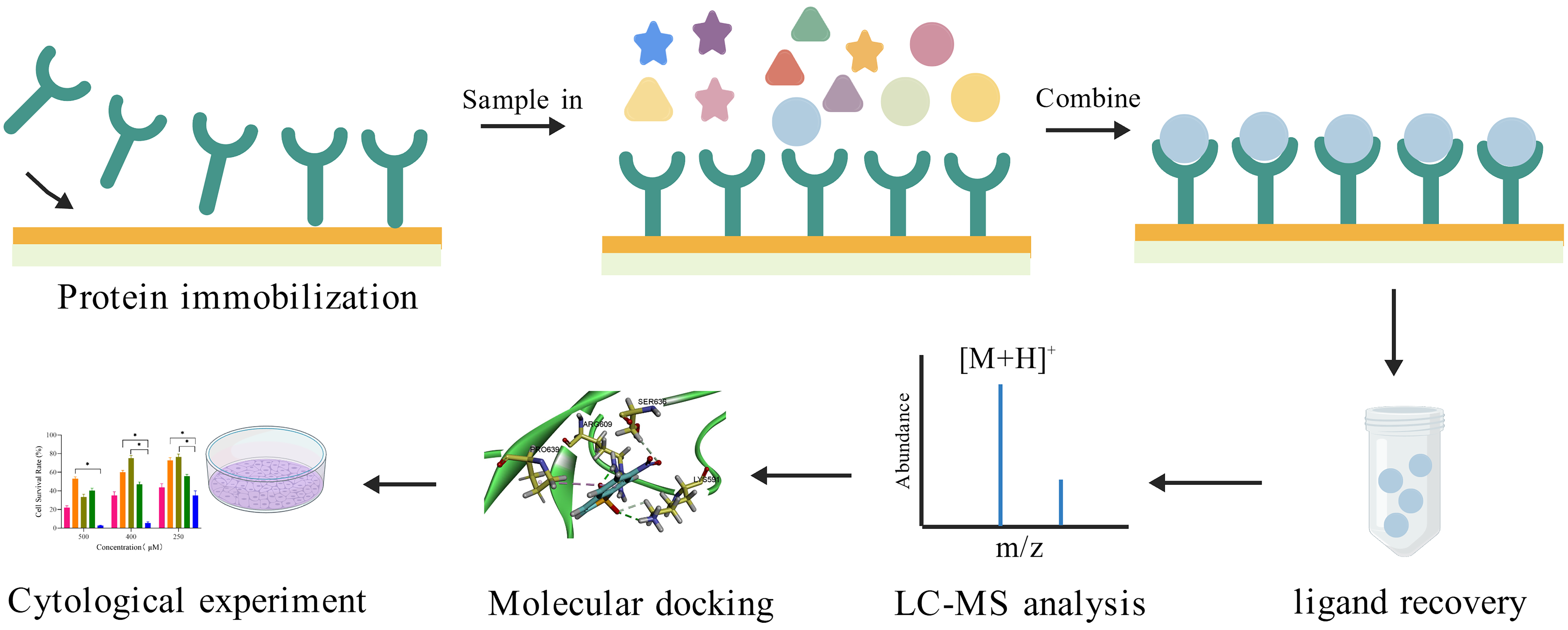
Given the critical role of STAT3 in NSCLC, this study used the SPR-AIRS system to identify small-molecule ligands targeting STAT3 proteins from Psoralea corylifolia. Molecular docking was employed to characterize their binding modes within the STAT3 SH2 domain, followed by in vitro assays to evaluate their effects on NSCLC cell proliferation, migration, and redox regulation. By integrating target-based screening with functional validation. This study aims to establish a systematic strategy for the discovery of natural STAT3 inhibitors and to elucidate the mechanisms underlying their anti-NSCLC activity. The experimental flow is depicted in Figure 2.

2. Results

2.1. Coupling Results of STAT3 Protein

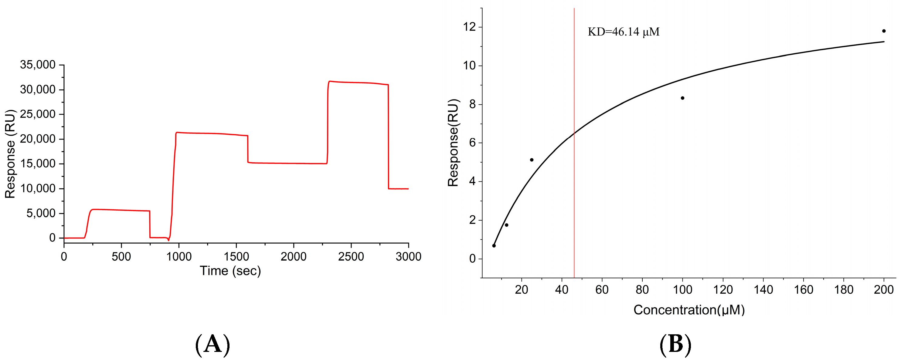
To establish a reliable STAT3-based SPR screening platform, optimal immobilization conditions for STAT3 immobilization on the CM5 chip were identified as 10 mM sodium acetate at pH 4.0 and a STAT3 concentration of 50 μg/mL. Under these conditions, the ligand coupling channel yielded a robust RU value of 12,000 RU (Figure 3A). This coupling level is adequate for subsequent experiments.

2.2. Validation of STAT3 Protein Chip Specificity

To evaluate the specificity and performance of the STAT3-immobilized sensor chip, binding affinity analysis was conducted using Stattic, a well-characterized small-molecule STAT3 inhibitor. Affinity analysis revealed a KD of 46.14 μM for Stattic with the STAT3 protein (Figure 3B). KD values are inversely correlated with binding affinity; lower KD values indicate stronger binding between a small molecule and its target protein. Typically, KD values for drug-grade small molecules binding to target proteins range from nM to μM. The KD value of 46.14 μM for Stattic in this study falls within this range. Therefore, Stattic can serve as a positive control for screening herbal compounds.

2.3. SPR Screening of STAT3-Binding Components from Psoralea corylifolia

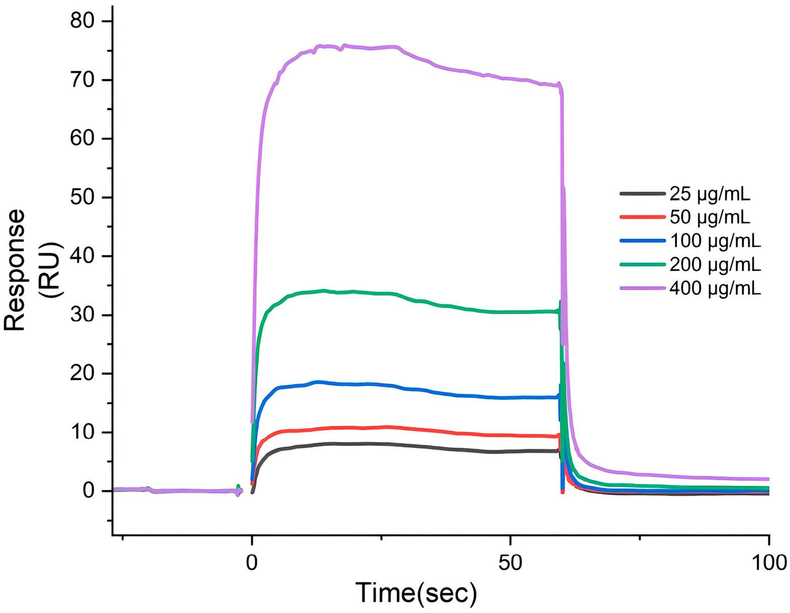
SPR screening was performed to assess the binding activity of Psoralea corylifolia extracts toward STAT3. Both 60% and 90% ethanol extracts of Psoralea corylifolia at 100 μg/mL showed binding activity to the STAT3 protein, with the 90% extract exhibiting stronger interaction (response values of 20.7 RU and 24.1 RU, respectively). It is generally accepted that a response value exceeding 20 RU, after subtracting the reference channel [31], indicates specific binding. Further concentration-dependent experiments confirmed that the response values of the psoralen 90% ethanol extract increased with concentration on the protein chip (Figure 4). This behavior clearly indicates that the extract contains bioactive components that specifically bind to STAT3.

2.4. UPLC-Q-TOF-MS/MS Analysis and Identification

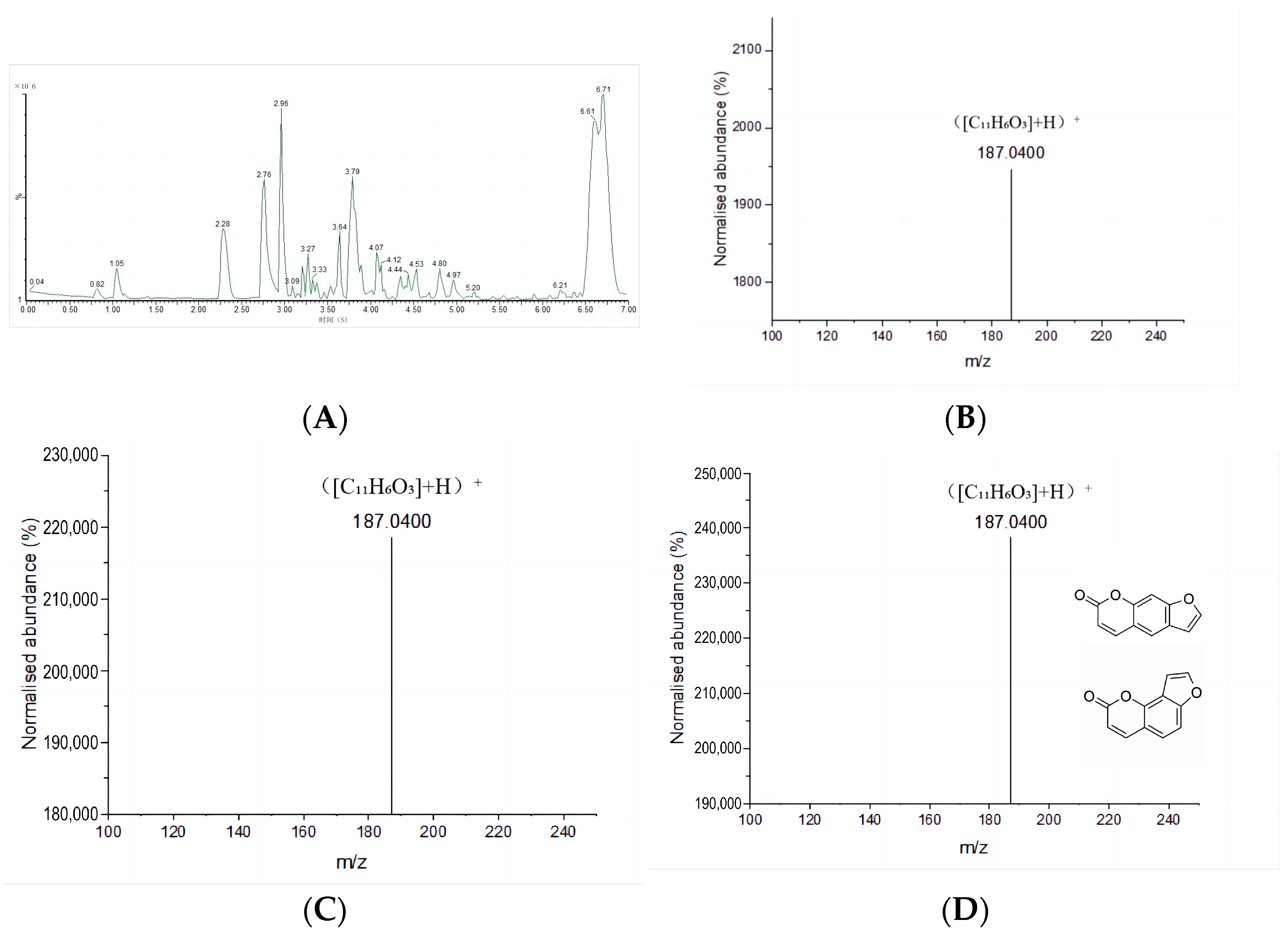
The 90% ethanol extract of Psoralea corylifolia was injected into the SPR system using high-affinity coupling to STAT3 protein, aiming to recover the components enriched on the chip. UPLC-Q-TOF-MS/MS was used to analyze and identify both the extract and the recovered samples. The Base peak index/chromatogram (BPI) of the recovered sample is shown in Figure 5A. The same ion signal peaks were identified in both the Psoralea corylifolia extract and its recovered sample, as shown in Figure 5B,C (Rt = 2.94 min, m/z = 187.0400 ([M + H]+)). The compound was queried as psoralen, and its tautomer, isopsoralen, was retrieved from the ChemSpider Data Sources database. The ion signal peaks of psoralen and isopsoralen standards under the same conditions are shown in Figure 5D (Rt = 2.94 min, m/z = 187.0400 ([M + H]+)). The detailed identification of the compounds is presented in Table 1. (See Figures S1–S3 and Table S1 for additional TIC/BPI data and compounds information)

2.5. Validation of Affinity Between STAT3 Protein and Candidate Compounds

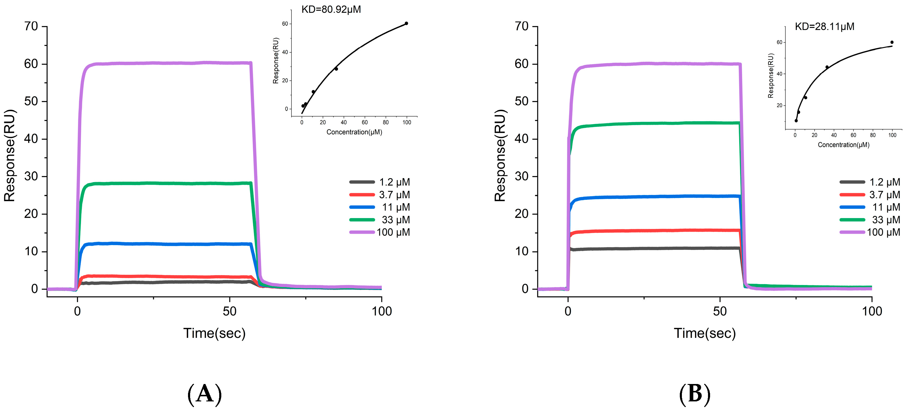
To verify whether psoralen and isopsoralen were directly bound to STAT3 protein, the affinity between both compounds and STAT3 was measured using a SPR assay. The results demonstrated stable, concentration-dependent binding signals between psoralen, isopsoralen, and STAT3 protein. The sensing signal graph and fitting curve for psoralen and STAT3 protein are depicted in Figure 6A, with a KD value of 80.92 µM, and those for isopsoralen and STAT3 protein are shown in Figure 6B, with a KD value of 28.11 µM. These results indicate that psoralen and isopsoralen bind specifically to STAT3.

2.6. Molecular Docking

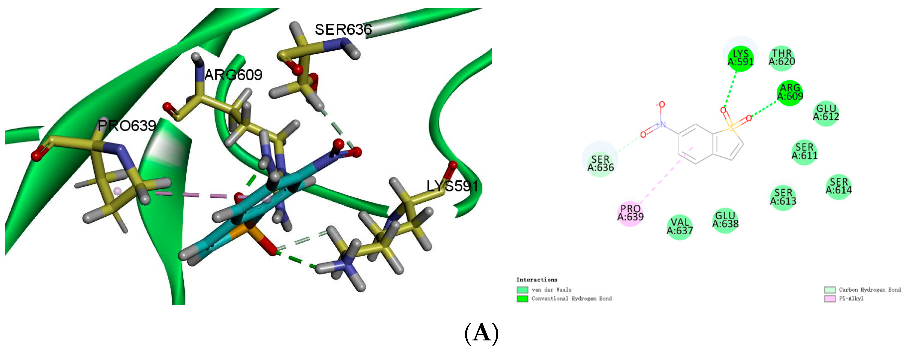
The SH2 domain, a critical binding site for numerous non-peptide STAT3 inhibitors, contains three sub-pockets on its surface: the crucial phosphorylated Tyr705-specific binding pocket (pY pocket, residues Lys591, Arg609-620), the Leu706 subpocket (pY + 1 pocket, residues 626–639), and the hydrophobic side pocket (pY-X pocket, residues 592–605), where the pY pocket exhibits the highest affinity [32]. Thus, the STAT3-SH2 domain protein (amino acid positions 586–685) was selected for docking with the candidate compounds and the positive control, Stattic.
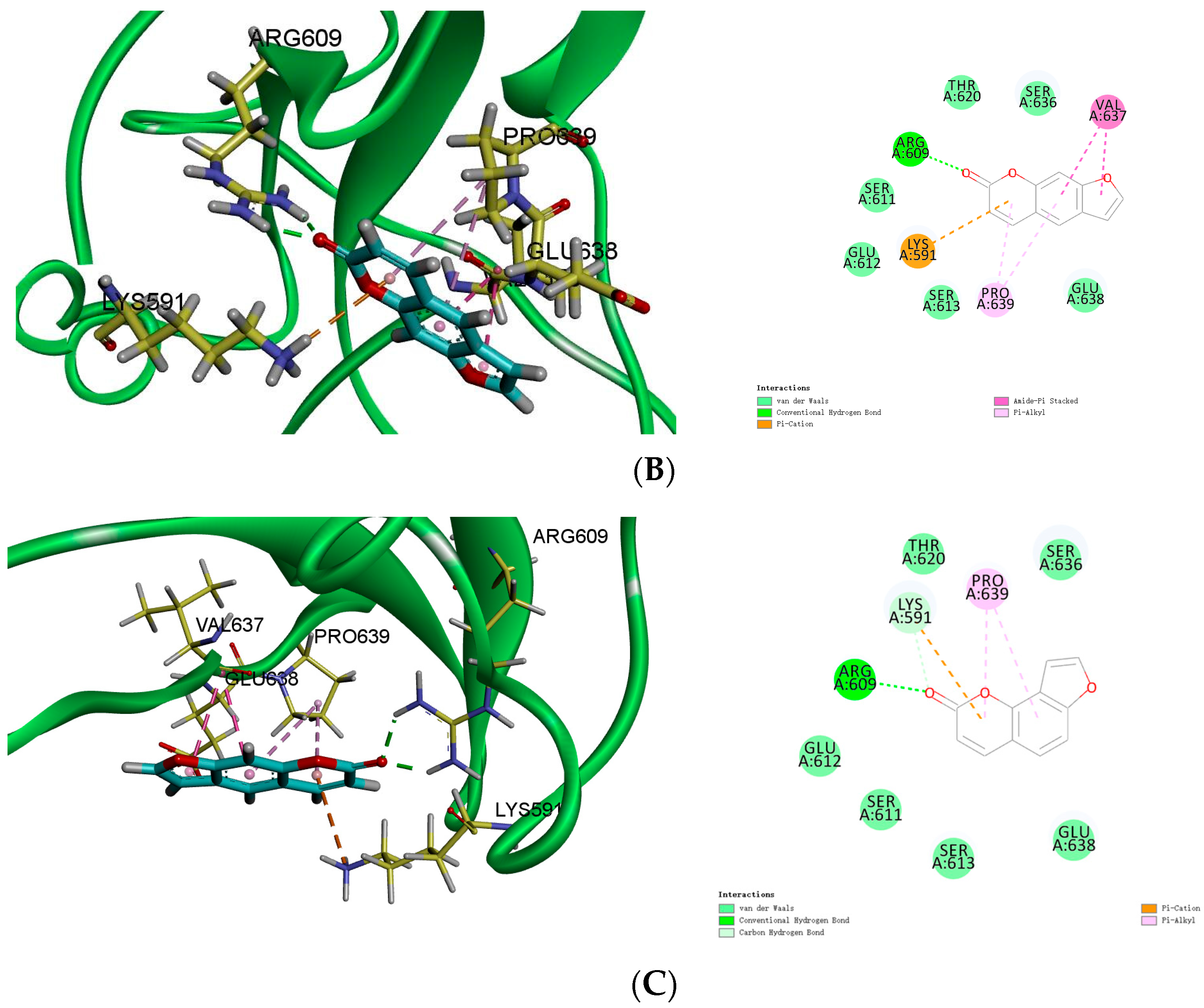
The docking results of Stattic with the SH2 domain of STAT3 protein are presented in Figure 7A. Stattic forms hydrogen bonds with residues Lys591, Arg609, and Ser636, and alkyl hydrophobic interactions with Pro639, yielding a CDOCKER binding affinity of −23.494 kJ/mol. As shown in Figure 7B, psoralen forms cation-π interactions with Lys591, hydrogen bonds with Arg609, π-hydrophobic interactions with Val637, and alkyl hydrophobic interactions with Pro639, with a CDOCKER binding affinity of −18.6669 kJ/mol. Figure 7C shows the results for isopsoralen, which forms a cation-π interaction with Lys591, a hydrogen bond with Arg609, and an alkyl hydrophobic interaction with Pro639, resulting in a CDOCKER binding affinity of −20.9622 kJ/mol. Molecular docking reveals that both compounds establish strong hydrogen bonds within the pY pocket, indicating beneficial affinity for the SH2 domain of the STAT3 protein.

2.7. Proliferation Inhibitory Effects of Psoralen and Isopsoralen on A549 Cells

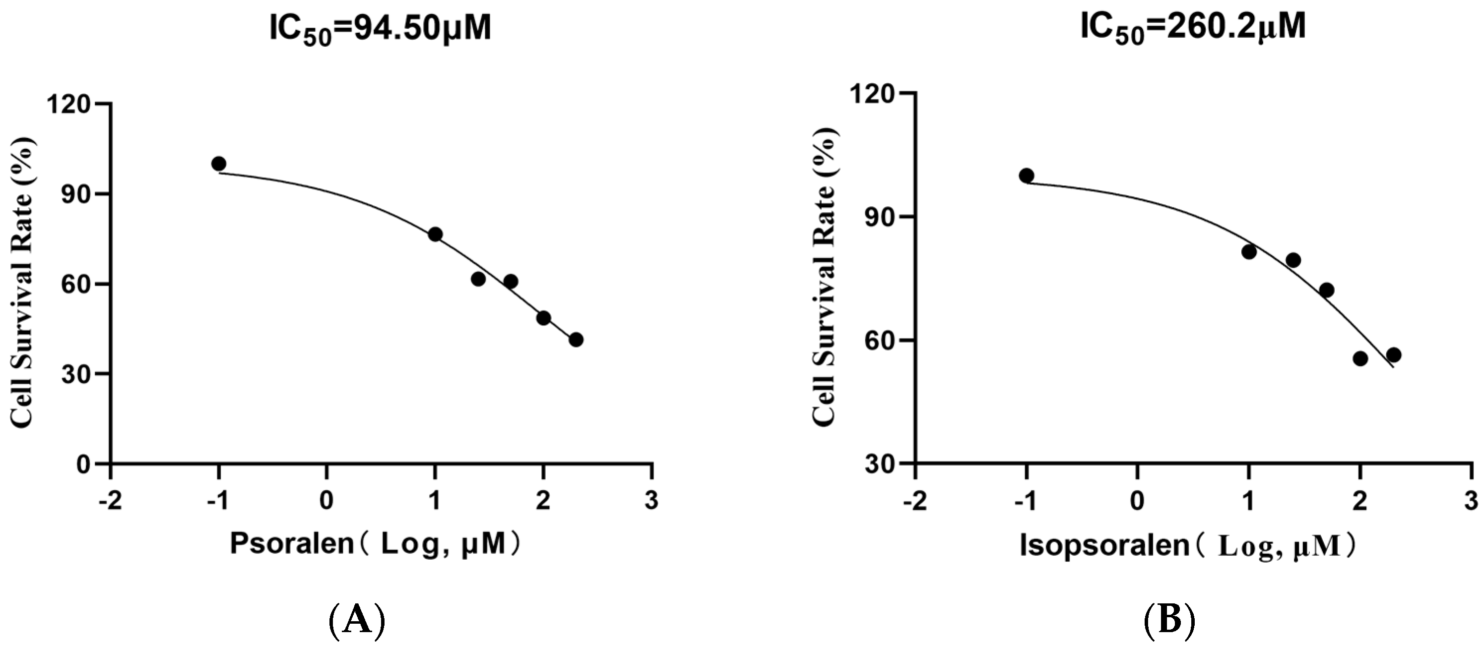
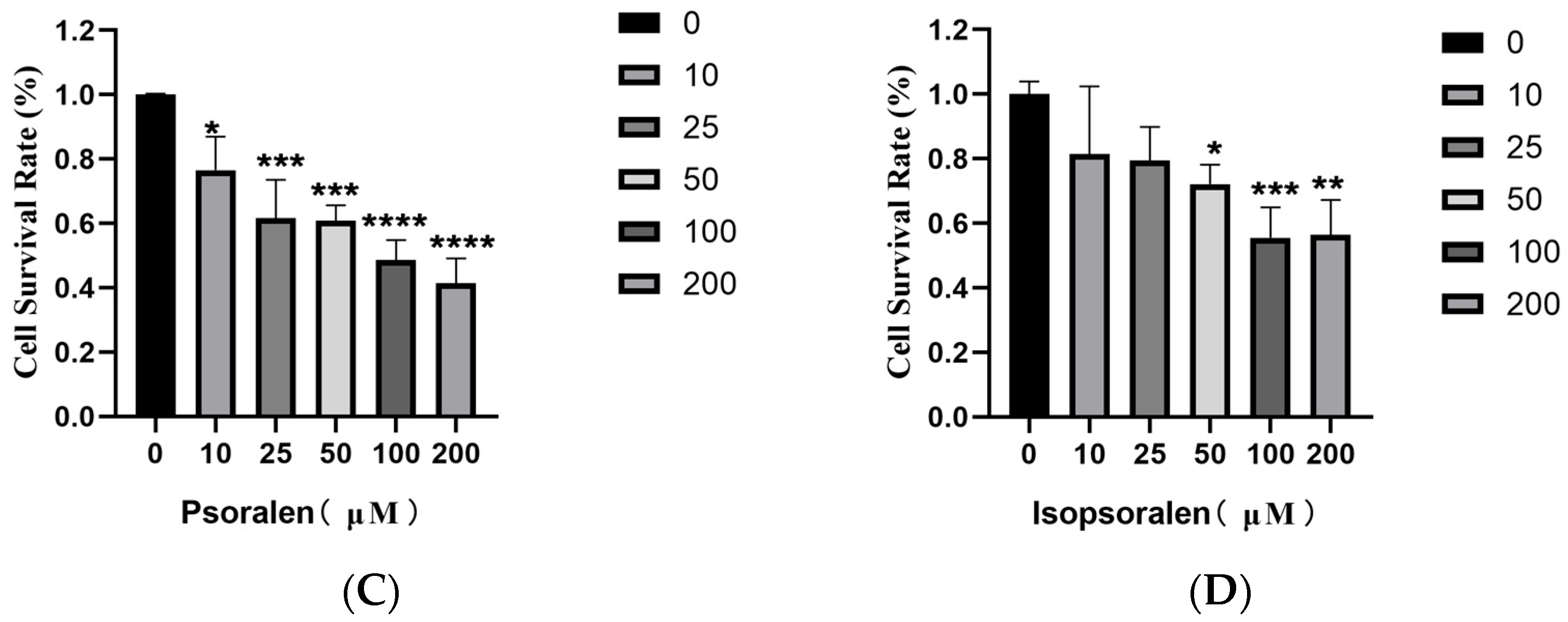
The effects of psoralen and isopsoralen on the proliferation of A549 NSCLC cells were evaluated. Different concentrations of these compounds were applied to A549 cells. Absorbance values were measured to calculate cell survival rates, and IC50 values were 94.50 μM for psoralen and 260.2 μM for isopsoralen (Figure 8A–D). Results demonstrated that psoralen exhibited the most potent inhibitory effect among the compounds. Significant differences were also observed between treatment groups and the blank control group (p < 0.01), with cell survival rates decreasing in a concentration-dependent manner.

2.8. The Inhibitory Effect of Psoralen and Isopsoralen on A549 Cell Migration Using Scratch Assay

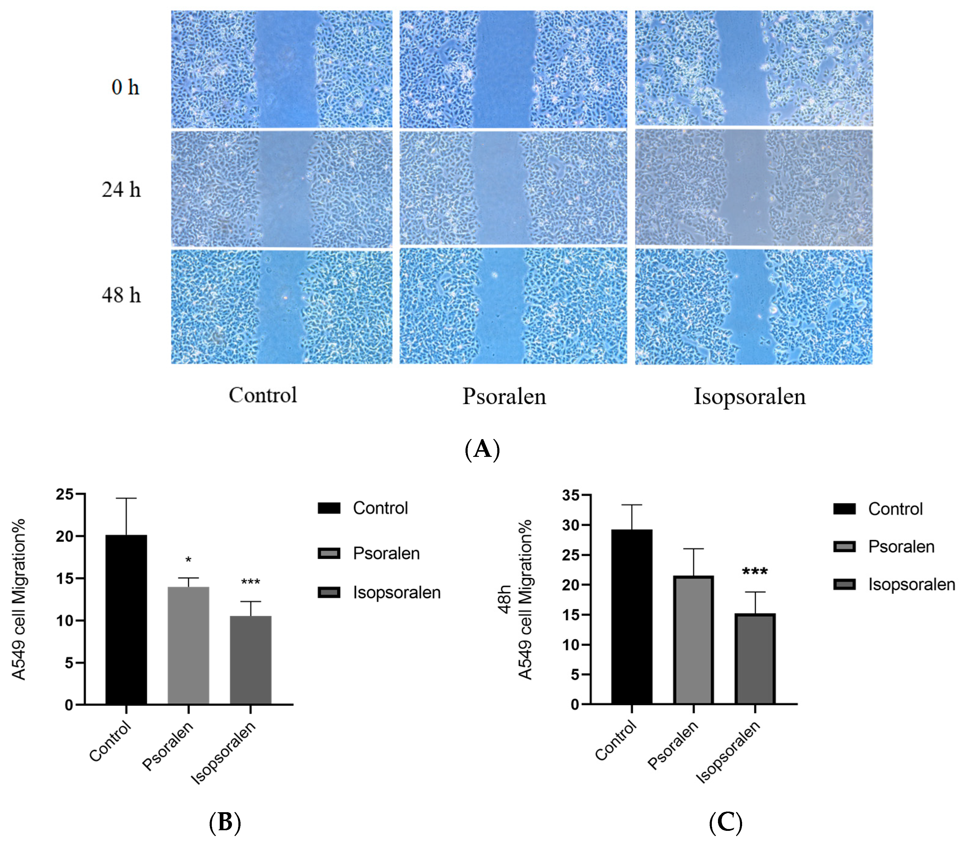
The impact of psoralen and isopsoralen on A549 cell migration was assessed using a scratch wound-healing assay. Based on proliferation assay results, concentrations of 100 μM for psoralen and 200 μM for isopsoralen were selected. The wound-healing assay was performed using concentrations close to or below the IC50 values to minimize nonspecific cytotoxic effects, allowing assessment of cell migratory capacity rather than cell viability. Migration areas were compared at 0, 24, and 48 h between the blank group and the treated groups. Images were captured at these time points, and migration rates were calculated to determine significant differences. Results indicated that both compounds inhibited A549 cell migration (Figure 9A). At 24 h and 48 h, 200 μM isopsoralen showed the most significant inhibition (p < 0.001), while psoralen at 100 μM also significantly reduced migration rates (Figure 9B,C).

2.9. Extracellular Antioxidant Activity of Psoralen and Isopsoralen

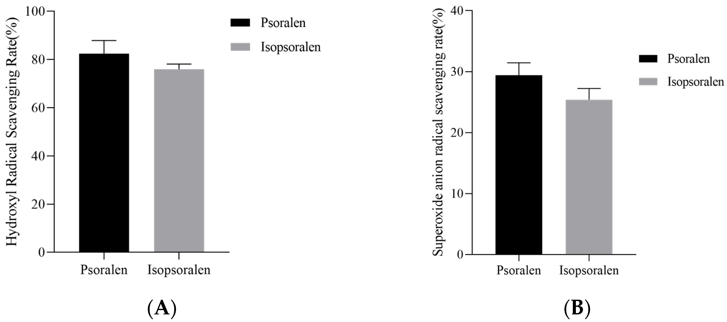
The antioxidant capacities of psoralen and isopsoralen were first evaluated using hydroxyl radical scavenging and superoxide anion scavenging assays. As shown in Figure 10A, the hydroxyl radical scavenging assay using the salicylic acid method showed 82.44% and 76.04% scavenging rates for psoralen and isopsoralen, respectively, indicating potent hydroxyl radical scavenging activity for both compounds. Subsequently, in the superoxide anion scavenging assay (Figure 10B), psoralen demonstrated a scavenging rate of 29.43%, while isopsoralen showed a rate of 25.38%, highlighting their ability to eliminate superoxide anions effectively. These experimental findings confirm significant antioxidant properties in psoralen and isopsoralen.

2.10. Effects of Psoralen and Isopsoralen on Intracellular ROS Levels

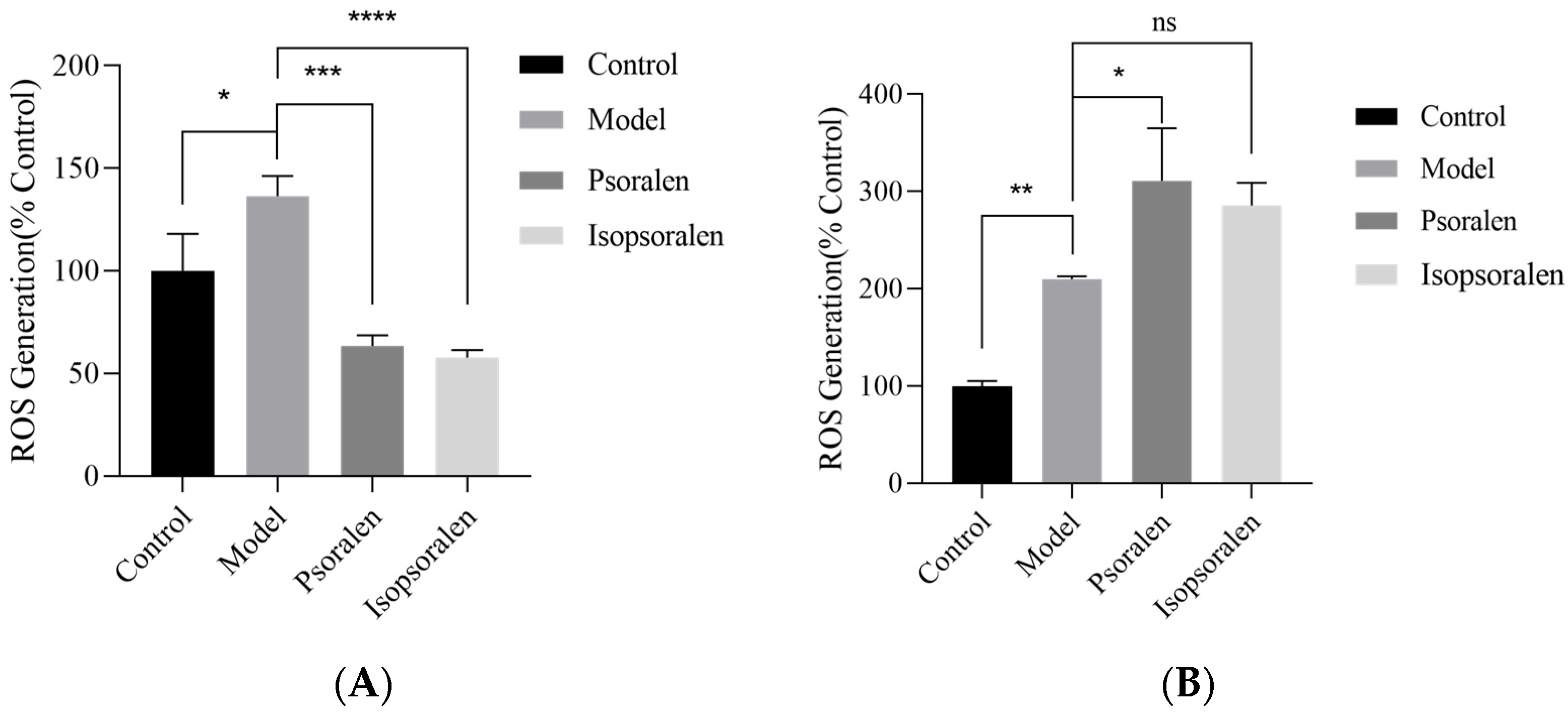
The effects of psoralen and isopsoralen on ROS modulation were investigated in normal HUVECs (Figure 11A) and lung cancer A549 cells (Figure 11B). In a hydrogen peroxide-induced oxidative stress model, pretreatment with psoralen and isopsoralen significantly reduced ROS levels in HUVECs (p < 0.01), indicating a protective effect against oxidative stress. Specifically, psoralen and isopsoralen decreased ROS levels in HUVECs by 73.00% and 78.63%, respectively, compared with the H2O2-treated group. In contrast, pretreatment with psoralen and isopsoralen increased intracellular ROS levels in A549 cells (p < 0.05). ROS levels were elevated by 101.10% and 75.86%, respectively, compared with the H2O2-treated group, suggesting enhanced oxidative stress that may contribute to ROS-mediated cellular damage.

2.11. Effects of Psoralen and Isopsoralen on HUVECs Proliferation

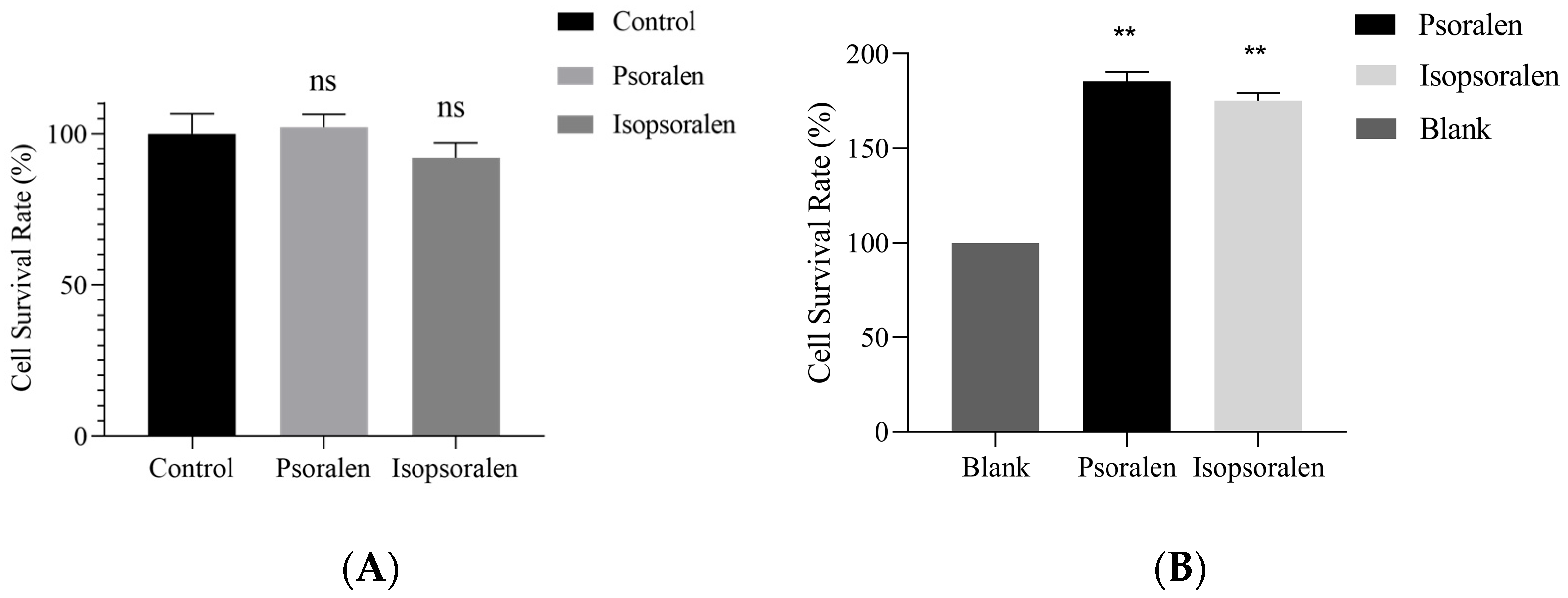
HUVECs are primary cells isolated from umbilical cord veins and are sensitive to drugs, making them a standard tool for cytotoxicity testing. In this study, psoralen and isopsoralen were applied to HUVECs at 500 μM to investigate their effects on normal cell proliferation (Figure 12A). Compared with the blank control group, neither psoralen nor isopsoralen at 500 μM significantly affected HUVECs, indicating low cytotoxicity toward endothelial cells at the tested concentration.

2.12. Effects of Psoralen and Isopsoralen on RAW264.7 Cell Proliferation

RAW264.7 cells, commonly used to model human immune responses, were treated with 500 μM of psoralen or isopsoralen. Compared to the control group, both compounds significantly increased the survival rate of RAW264.7 cells, with increases of 85.33% and 75.00%, respectively (p < 0.01). These results indicate that they do not exert cytotoxic effects on immune cells under the experimental conditions. (Figure 12B).

3. Discussion

In this study, for the first time, the STAT3-specific small-molecule inhibitors psoralen and isopsoralen from Psoralea corylifolia were identified, using SPR-AIRS technology. These compounds effectively inhibited the proliferation and migration of NSCLC A549 cells, exhibiting a selective anticancer profile and unique regulation of cellular redox status.
STAT3, as a key transcription factor, plays a central role in the onset, progression, metastasis, drug resistance, and immune escape of NSCLC [33,34]. Its sustained activation (constitutive or aberrant) is an essential feature of NSCLC and is closely associated with poor prognosis and chemoresistance [10]. Therefore, targeted inhibition of the STAT3 signaling pathway is considered a promising therapeutic strategy for NSCLC. However, the development of efficient and specific direct STAT3 inhibitors remains a significant challenge, largely due to the absence of well-defined druggable pockets and concerns regarding off-target effects [35].
SPR-AIRS technology provides an efficient and reliable screening platform for STAT3-targeted compounds [36]. It offers higher target specificity than traditional cellular phenotypic screening, enabling direct identification of active ingredients from complex herbal extracts and real-time verification of binding affinity. We first screened two specific STAT3 small-molecule inhibitors, psoralen and isopsoralen, from Psoralea corylifolia. SPR results demonstrated that both compounds bound STAT3 directly and efficiently. Molecular docking analysis further revealed that psoralen and isopsoralen explicitly bound to the SH2 domain of STAT3, a functional domain critical for dimerization and activation. This mechanism is similar to that of known STAT3 inhibitors (e.g., Stattic) [37], suggesting that psoralen and isopsoralen may inhibit STAT3 transcriptional activity by blocking its phosphorylation. These findings provide mechanistic evidence supporting STAT3 as a molecular target of psoralen and isopsoralen.
Previous studies have reported the anticancer potential of Psoralea corylifolia extracts and its constituent compounds in a variety of malignancies, including lung cancer [38]. However, most of these studies primarily focused on indirect regulatory pathways or phenotypic outcomes without demonstrating direct target engagement. By contrast, the present work establishes a clear molecular link between psoralen/isopsoralen and STAT3, and both compounds significantly inhibited the cell growth and migratory capacity of A549 lung carcinoma cells.
Notably, psoralen exhibited a more potent anti-proliferative effect despite its lower STAT3 binding affinity compared to isopsoralen. This apparent discrepancy suggests cellular responses are not solely determined by binding affinity, but rather by the integration of multiple mechanisms, including intracellular signaling modulation and redox regulation. Such divergence between biochemical affinity and cellular efficacy is commonly observed for small-molecule inhibitors and underscores the importance of functional validation beyond target-binding studies.
ROS are increasingly recognized as key regulators of cancer cell fate, functioning as both tumor-promoting signaling mediators and inducers of oxidative damage [39]. In the present study, psoralen and isopsoralen exhibited notable free radical scavenging activity in cell-free systems and effectively reduced ROS accumulation in normal HUVECs under oxidative stress conditions, suggesting a cytoprotective antioxidant effect. Conversely, in A549 cancer cells, they triggered significant ROS accumulation, exacerbating oxidative stress and suppressing proliferation. Accumulating evidence suggests that STAT3 plays a critical role in maintaining redox homeostasis in cancer cells. A reciprocal regulatory loop exists between STAT3 signaling and cellular redox homeostasis. Inhibition of STAT3 may impair antioxidant defenses, thereby promoting ROS accumulation in cancer cells, while elevated ROS can further suppress STAT3 activity [40]. In A549 lung cancer cells, STAT3 promotes cell survival and enhances antioxidant defense mechanisms [41]. Therefore, we hypothesize that suppression of STAT3 activation by psoralen and isopsoralen disrupted cellular redox balance, resulting in elevated intracellular ROS levels. In addition, modulation of ROS levels within the tumor microenvironment has been reported to reshape immunosuppressive conditions and promote antitumor immune responses [39]. Thus, the ability of psoralen and isopsoralen to regulate ROS may not only contribute to their direct cytotoxic effects on tumor cells but also potentially influence tumor-immune interactions, further enhancing their anticancer efficacy.
It should be acknowledged that the present study has certain limitations. The biological activities of the crude Psoralea corylifolia extract were not directly compared with those of the isolated compounds. Herbal extracts may exhibit synergistic effects arising from multiple constituents, and future studies will aim to systematically compare extract-level and monomer-level activities. In addition, future work should focus on in vivo validation and further elucidation of the STAT3-ROS-associated signaling cascade, including phosphorylation status, nuclear translocation of STAT3, and regulation of downstream targets such as Bcl-xL and cyclin D1, to strengthen the mechanistic understanding of these compounds.

4. Materials and Methods

4.1. Major Materials

STAT3 protein was ordered from WuXiAppTec (Shanghai, China). CM5 microarray, amino-coupling kit, 50 mM sodium hydroxide solution, glycine hydrochloride (pH 3.0), and sodium acetate (pH 4.0) were obtained from GE Healthcare (Chicago, IL, USA). Psoralea corylifolia, a traditional Chinese medicine, was provided by Associate Prof. Wang Qing from the Xiamen Key Laboratory of Traditional Chinese Medicine Bioengineering. Psoralen and isopsoralen standards were ordered from Orileaf (Shanghai, China). Ammonium bicarbonate was purchased from Merck Sigma-Aldrich (Burlington, MA, USA). NSCLC cells (A549), human umbilical vein endothelial cells (HUVECs), and RAW264.7 cell lines were obtained from the Chinese Academy of Sciences (CAS) Cell Bank (Shanghai, China). The Cell Proliferation-Toxicity Assay Kit (CCK-8) was purchased from Biosharp (Hefei, China). Stattic was ordered from TargetMol (Shanghai, China). The ROS assay kit was ordered from Beyotime (Shanghai, China).

4.2. Immobilization of STAT3 Protein on SPR Sensor Chip

SPR experiments were performed on a Biacore T200 system (GE Healthcare, USA) to immobilize STAT3 onto a CM5 sensing chip surface using the standard amine coupling protocol, with channels 2 or 4 serving as the detection pathway for STAT3 protein coupling. Initially, a 1:1 mixture of 1-Ethyl-3-(3-dimethylaminopropyl) carbodiimide (EDC) and N-hydroxysuccinimide (NHS) was introduced at a flow rate of 10 μL/min for 10 min to activate the carboxyl groups on the dextran matrix surface. Subsequently, the STAT3 protein prepared at 50 μg/mL in 10 mM Acetate buffer (pH 4.0) was delivered at 10 μL/min for 7 min to facilitate covalent conjugation. Finally, residual active groups were blocked by injecting ethanolamine. The immobilization level was quantified by measuring the bound Response Units (RU).

4.3. Examination of STAT3 Protein Chip Specificity

To validate the reliability of the experimental system, Stattic—a well-characterized small-molecule inhibitor of STAT3 was employed as a positive control [30]. At the same time, phosphate-buffered saline (PBS) served as the negative control. A serial dilution of Stattic was prepared in running buffer, and the kinetics/affinity experimental template was selected for affinity measurements. The Stattic concentration gradient was then analyzed at 25 °C with the following parameters: 60 s injection time, 30 μL/min flow rate, and 60 s dissociation time. The binding signals were specifically analyzed using reference-channel subtraction and blank subtraction. Data were fitted to a 1:1 steady-state affinity model, from which the equilibrium dissociation constants (KD) were directly derived to characterize the binding affinity between Stattic and STAT3 protein.

4.4. Preparation and Screening of Psoralea corylifolia Extract

Psoralea corylifolia was crushed and passed through a 40-mesh sieve. One gram of the powder was weighed and separately extracted with 10 mL of either 60% or 90% ethanol using ultrasonic extraction for 30 min. The extracts were then filtered through 0.22 μm syringe filters. Each filtered extract was diluted with running buffer and centrifuged to obtain the supernatant. The Biacore T200 Control Software (version 3.2.2) was launched, and the manual mode was selected. Then, Psoralea corylifolia extract was injected over the sensor chip surface for an association phase of 60 s, followed by a dissociation phase of 60 s, and then regenerated for 60 s. PBS buffer was injected between sample injections as a negative control to determine specific binding of the extract to immobilized STAT3 protein.

4.5. Recovery of Active Ingredients Bound to STAT3 Protein

To recover STAT3-bound components, all four channels of a CM5 chip were immobilized with STAT3 protein. The flow rate was then set to 10 μL/min and the binding time to 420 s. Sample angling experiments were performed using the “inject and recover” method with 10 recovery cycles, employing 0.5% trifluoroacetic acid (TFA) as the regeneration solution and 50 mM NH4HCO3 as the deposition solution. After which, all recovered solutions were combined, dried under nitrogen, redissolved in methanol, and centrifuged. The supernatant was filtered and transferred to a liquid chromatography vial.

4.6. UPLC-Q-TOF-MS/MS Analysis and Affinity Validation

The recovered samples from Section 4.5 and the corresponding Psoralea corylifolia extracts (dried and adjusted to 100 mg/mL in methanol) were analyzed using Acquity UPLC H-Class Plus Xevo Tof (Waters, Milford, MA, USA). Chromatographic conditions were as follows: Mobile phase A was 0.1% formic acid in water; mobile phase B was 0.1% formic acid in acetonitrile; gradient elution: 0–0.5 min, 10% B; 0.5–1.0 min, 10–70% B; 1.0–4.5 min, 70–100% B; 4.5–5.5 min, 100% B; 5.5–6.5 min, 100–10% B; 6.5–7.0 min, 10% B. Column temperature was maintained at 30 °C, flow rate adjusted to 0.3 mL/min, and injection volume set to 1 µL. Following the analysis, compounds in Psoralea corylifolia extracts that potentially bind to STAT3 protein were identified using Progenesis QI version 2.1 software by querying the ChemSpider Data Sources database for chemical information.
Psoralen and isopsoralen were separately diluted using the running buffer to generate multiple concentration gradients. These gradient samples were then employed to assess the binding affinity for STAT3 proteins, as described in Section 4.3.

4.7. Molecular Docking Analysis

To investigate the potential specific binding of candidate small molecules to the active site of the STAT3 protein, molecular docking was employed to assess the interactions of psoralen and isopsoralen with the SH2 domain of STAT3. The SH2 domain serves as a dimerization module and key binding site for numerous non-peptidyl STAT3 inhibitors. It functions as a critical adaptor protein, facilitating STAT3 dimerization upon activation, mediating interactions between activated receptors and downstream signaling molecules, thereby promoting pathway activation. This signaling cascade plays a critical role in the pathogenesis of NSCLC. Therefore, the SH2 domain of STAT3 was selected as the target for molecular docking with the candidate ligands (psoralen and isopsoralen) and the established STAT3 SH2 inhibitor, Stattic (used as a positive control).
The crystal structure of STAT3 was obtained from the Protein Data Bank under accession ID 1BG1. Optimization was performed using BIOVIA Discovery Studio 2019 (DS) software (v19.1.0) to remove DNA strands and water molecules, and the STAT3-SH2 domain (residues 586–685) was selected as the docking site, with a 10 Å radius. The molecular structures of the two candidate compounds, stattic and isostatticin, were obtained from the Traditional Chinese Medicine Systematic Pharmacology Database and Analysis Platform (TCMSP). Protein-receptor docking was carried out using the CDOCKER module in DS software. Stattic was used as a positive control alongside the candidate compounds, and the optimal conformation was determined by CDOCKER binding energy.

4.8. A549 Cell Proliferation Assay

A549 cells in log-phase growth were plated into 96-well plates at a density targeted to reach approximately 70–80% confluence overnight. Candidate compounds at concentrations of 0, 10, 25, 50, 100, and 200 μM were added to the A549 cells. 10 μL of the corresponding drug solution was added per well, and the plates were incubated for 24 h. After incubation, the medium was discarded, and 90 μL of fresh medium, along with 10 μL of 3-(4,5-Dimethylthiazol-2-yl)-2,5-diphenyltetrazolium bromide (MTT) solution (5 mg/mL), was added to each well. The plate was incubated for four hours. The medium was subsequently discarded, and 100 μL of crystal violet dissolving solution was added to each well. The plates were shaken for 15 min. and absorbance was measured at 570 nm using a microplate reader (Biotek, Winooski, VT, USA).

4.9. A549 Cell Scratch Experiment

The cell scratch assay was used to evaluate the effect of candidate compounds on A549 cell migration [42]. Log-phase A549 cells were seeded in 6-well plates to attain 90–100% confluence by the following day. Using a marker pen and a straightedge, lines were scribed on the plate surface. Wounds were created by scratching with a P200 pipette tip to ensure uniformity. The wells were then washed twice with PBS. Subsequently, the drug solution was added at a concentration near the IC50 (as determined from the MTT experiment). The cells were treated with STAT3 small-molecule inhibitors and incubated. Images were captured at 0, 24, and 48 h of culture using a fluorescence inverted microscope (Leica, Düsseldorf, Germany). The scratch wound areas were quantified using ImageJ version 1.48 software (version 1.48). The cell migration rate was calculated based on wound closure as follows:
Cell migration rate (%) = [(Initial scratch area − Scratch area at time t)/Initial scratch area] × 100%

4.10. Hydroxyl Radical Scavenging Capacity Assay

The salicylic acid method was used to determine the hydroxyl radical scavenging capacity of candidate compounds [43]. The reaction system consisted of 100 µM psoralen or isopsoralen, 6 mM FeSO4, and 6 mM H2O2, with each component added at a volume of 50 µL in a 96-well plate. The mixture was incubated for 10 min at room temperature. Subsequently, 50 μL of 6 mM salicylic acid prepared in ethanol was added, followed by incubation at room temperature for 30 min, avoiding light. The absorbance was measured at 510 nm, and the value was recorded as A1. For the controls, the assay was performed identically; however, the sample was replaced with water to determine the absorbance value (recorded as A0), and the H2O2 solution was replaced with water to determine the absorbance value (recorded as A2). The clearance rate was calculated following the formula:
H y d r o x y l   r a d i c a l   s c a v e n g i n g   r a t e % = [ 1 ( A 1 A 2 ) / A 0 ] × 100

4.11. Superoxide Anion Scavenging Capacity Assay

The superoxide anion scavenging capacity of psoralen and isopsoralen was quantified using a modified pyrogallol autoxidation assay [44]. One hundred μL of 100 μM psoralen or isopsoralen solution was thoroughly mixed with 450 μL of Tris-HCl buffer (0.1 M, pH 8.2) and maintained at room temperature for 20 min. Then, 50 μL of 2.5 M pyrogallol was added to initiate the reaction. After 5 min of reaction, 100 μL of 0.8 M HCl was added immediately to terminate the process. Absorbance was subsequently measured at 325 nm and recorded as A1. For control experiments, a blank control (A0) was prepared by replacing the sample solution with an equal volume of water, and a background control (A2) was prepared by replacing the pyrogallol solution with an equal volume of water. The clearance rate was calculated following the formula:
S u p e r o x i d e   a n i o n   s c a v e n g i n g   r a t e % = [ 1 ( A 1 A 2 ) / A 0 ] × 100

4.12. Intracellular ROS Level Assay

Intracellular ROS levels were measured following treatment of human umbilical vein endothelial cells (HUVECs) and A549 cells with the candidate compounds to assess their effects and potential selectivity against H2O2 stress. HUVECs and A549 cells were plated separately in 96-well black plates. Following overnight culture, cells in the experimental groups were treated with serum-free medium containing 100 μM of either psoralen or isopsoralen. After 24 h of incubation, cells were washed with PBS. Then, cells from both experimental and control groups were exposed to serum-free medium supplemented with 500 μM H2O2 for four hours to induce oxidative stress. Intracellular ROS were then labeled with DCFH-DA, as instructed in the manufacturer’s ROS assay kit. Fluorescence was subsequently measured using a fluorescence microplate reader.

4.13. HUVECs Cytotoxicity Assay

HUVECs in log-phase growth were plated in 96-well plates at 1.5 × 10⁴ cells/well, with six replicate wells designated for each experimental group. After culturing for 24 h, the culture medium was carefully aspirated and replaced with 100 μL of fresh medium containing candidate compounds at 500 μM, and the culture was incubated for an additional 24 h. Subsequently, 10 μL CCK-8 solution was added to each well and incubated for 30 min before the optical density was measured at 450 nm.

4.14. RAW264.7 Cytotoxicity Assay

The murine macrophage cell line RAW264.7 in log phase was plated at 5 × 10⁵ cells/well in 96-well culture plates. After a 24-h incubation, the culture medium was aspirated and replaced with 100 μL of fresh medium supplemented with test compounds (500 μM final concentration). After an additional 24-h treatment period, 10 μL CCK-8 solution was added to each well and incubated for 30 min before optical density was measured at 450 nm.

4.15. Statistical Analysis

All experiments were performed in triplicate (n = 3) unless otherwise stated. Data are presented as mean ± SD. Statistical analyses were explicitly conducted with GraphPad Prism 8.0 software. One-way ANOVA was used to compare groups, with p < 0.05 as the threshold for statistical significance; figures were created with Origin version 2025b software.

5. Conclusions

This study provides the first direct identification of psoralen and isopsoralen derived from Psoralea corylifolia as STAT3-binding small molecules, supported by SPR-based target engagement data rather than indirect phenotypic inference. The integration of SPR-AIRS “inject-and-recover” screening with UPLC-Q-TOF-MS/MS enables direct target-oriented identification of bioactive compounds from a complex herbal matrix. Together with STAT3 binding, ROS modulation, proliferation inhibition, and the migration-suppressive effects suggest that these compounds exert multifaceted regulatory effects on NSCLC cell behavior. Their activity stems from a combination of direct STAT3 interaction and context-dependent, bidirectional modulation of ROS. The differential redox regulation, acting as an antioxidant in normal cells but a pro-oxidant in cancer cells, underscores its selective anticancer mechanism. Collectively, these findings identify psoralen and isopsoralen as early-stage STAT3-targeting bioactive compounds and underscore the utility of SPR-based target-oriented screening as an effective approach for discovering functional natural products from complex botanical sources.

Supplementary Materials

The following supporting information can be downloaded at: https://www.mdpi.com/article/10.3390/ph19020257/s1, Figure S1: Base peak index/chromatogram (BPI) of the extract from Psoralea corylifolia; Figure S2: The total ion chromatogram (TIC) of the recovered sample; Figure S3: The total ion chromatogram (TIC) of the extract from Psoralea corylifolia; Table S1: Identification of chemical components from anging samples.

Author Contributions

Methodology and data curation, L.B. and G.C.; validation, W.L. and J.F.; resources, Z.Y.; data curation, S.C.; composition of the original manuscript, L.B., Y.L. and A.T.C.III; evaluation and revision, Y.-W.C. and Y.-H.C. All authors have read and agreed to the published version of the manuscript.

Funding

This research was funded by the Fujian Provincial Natural Science Foundation (No.2023J011658), the Natural Science Foundation of Xiamen (No.3502Z202374041), and the Natural Science program of Xiamen Medical College, grant number (K2021-01).

Institutional Review Board Statement

Not applicable.

Informed Consent Statement

Not applicable.

Data Availability Statement

The original contributions presented in this study are included in the article/Supplementary Materials. Further inquiries can be directed to the corresponding authors.

Acknowledgments

The authors gratefully acknowledge Qiliang Zuo for his technical guidance, Qing Wang for providing traditional medicine Psoralea corylifolia, Shanshan Zhu and Fei Qin for providing equipment and resources, Jingna Wu for providing HUVECs, and Cheng He of Bailaibobio Ltd. for the data visualization.

Conflicts of Interest

The authors declare no conflict of interest.

References

  1. Bray, F.; Laversanne, M.; Sung, H.; Ferlay, J.; Siegel, R.L.; Soerjomataram, I.; Jemal, A. Global Cancer Statistics 2022: GLOBOCAN Estimates of Incidence and Mortality Worldwide for 36 Cancers in 185 Countries. CA Cancer J. Clin. 2024, 74, 229–263. [Google Scholar] [CrossRef]
  2. Mohrherr, J.; Uras, I.Z.; Moll, H.P.; Casanova, E. STAT3: Versatile Functions in Non-Small Cell Lung Cancer. Cancers 2020, 12, 1107. [Google Scholar] [CrossRef] [PubMed]
  3. Zhu, H.; Xu, Y.; Gao, H.; Fan, X.; Fan, M.; Zhao, K.; Yang, H.; Zhu, Z.; Wu, K. Long-term Outcome of Definitive Radiotherapy for Locally Advanced Non-small Cell Lung Cancer: A Real-world Single-center Study in the Pre-durvalumab Era. Cancer Med. 2024, 13, e70051. [Google Scholar] [CrossRef] [PubMed]
  4. Tang, Y.; Wang, Y.; Wang, X.; Zhao, Z.; Cai, H.; Xie, M.; Jiang, X.; Zhang, L.; Cheng, J.; Yang, L.; et al. Acetylshikonin Exerts Anti-Tumor Effects on Non-Small Cell Lung Cancer through Dual Inhibition of STAT3 and EGFR. Phytomedicine 2022, 101, 154109. [Google Scholar] [CrossRef] [PubMed]
  5. Sui, H.; Xiao, S.; Jiang, S.; Wu, S.; Lin, H.; Cheng, L.; Ye, L.; Zhao, Q.; Yu, Y.; Tao, L.; et al. Regorafenib Induces NOX5-Mediated Endoplasmic Reticulum Stress and Potentiates the Anti-Tumor Activity of Cisplatin in Non-Small Cell Lung Cancer Cells. Neoplasia 2023, 39, 100897. [Google Scholar] [CrossRef]
  6. Ni, Y.; Wu, S.; Wang, X.; Zhu, G.; Chen, X.; Ding, Y.; Jiang, W. Cucurbitacin I Induces Pro-death Autophagy in A549 Cells via the ERK-mTOR-STAT3 Signaling Pathway. J. Cell. Biochem. 2018, 119, 6104–6112. [Google Scholar] [CrossRef]
  7. Lai, I.-C.; Lai, G.-M.; Chow, J.-M.; Lee, H.-L.; Yeh, C.-F.; Li, C.-H.; Yan, J.-L.; Chuang, S.-E.; Whang-Peng, J.; Bai, K.-J.; et al. Active Fraction (HS7) from Taiwanofungus Camphoratus Inhibits AKT-mTOR, ERK and STAT3 Pathways and Induces CDK Inhibitors in CL1-0 Human Lung Cancer Cells. Chin. Med. 2017, 12, 33. [Google Scholar] [CrossRef]
  8. Lin, C.-C.; Yeh, H.-H.; Huang, W.-L.; Yan, J.-J.; Lai, W.-W.; Su, W.-P.; Chen, H.H.W.; Su, W.-C. Metformin Enhances Cisplatin Cytotoxicity by Suppressing Signal Transducer and Activator of Transcription–3 Activity Independently of the Liver Kinase B1–AMP-Activated Protein Kinase Pathway. Am. J. Respir. Cell Mol. Biol. 2013, 49, 241–250. [Google Scholar] [CrossRef]
  9. Gharibi, T.; Babaloo, Z.; Hosseini, A.; Abdollahpour-Alitappeh, M.; Hashemi, V.; Marofi, F.; Nejati, K.; Baradaran, B. Targeting STAT3 in Cancer and Autoimmune Diseases. Eur. J. Pharmacol. 2020, 878, 173107. [Google Scholar] [CrossRef]
  10. Morelli, A.P.; Tortelli, T.C.; Mancini, M.C.S.; Pavan, I.C.B.; Silva, L.G.S.; Severino, M.B.; Granato, D.C.; Pestana, N.F.; Ponte, L.G.S.; Peruca, G.F.; et al. STAT3 Contributes to Cisplatin Resistance, Modulating EMT Markers, and the mTOR Signaling in Lung Adenocarcinoma. Neoplasia 2021, 23, 1048–1058. [Google Scholar] [CrossRef]
  11. Jin, H.-O.; Lee, Y.-H.; Park, J.-A.; Kim, J.-H.; Hong, S.-E.; Kim, H.-A.; Kim, E.-K.; Noh, W.C.; Kim, B.-H.; Ye, S.-K.; et al. Blockage of Stat3 Enhances the Sensitivity of NSCLC Cells to PI3K/mTOR Inhibition. Biochem. Biophys. Res. Commun. 2014, 444, 502–508. [Google Scholar] [CrossRef] [PubMed]
  12. Zhou, Y.; Peng, X.; Fang, C.; Peng, X.; Tang, J.; Wang, Z.; Long, Y.; Chen, J.; Peng, Y.; Zhang, Z.; et al. Histones Methyltransferase NSD3 Inhibits Lung Adenocarcinoma Glycolysis Through Interacting with PPP1CB to Decrease STAT3 Signaling Pathway. Adv. Sci. 2024, 11, 2400381. [Google Scholar] [CrossRef] [PubMed]
  13. Zhao, Y.; Zhang, X.; Li, Y.; Li, Y.; Zhang, H.; Song, Z.; Xu, J.; Guo, Y. A Natural Xanthone Suppresses Lung Cancer Growth and Metastasis by Targeting STAT3 and FAK Signaling Pathways. Phytomedicine 2022, 102, 154118. [Google Scholar] [CrossRef] [PubMed]
  14. Dong, J.; Cheng, X.-D.; Zhang, W.-D.; Qin, J.-J. Recent Update on Development of Small-Molecule STAT3 Inhibitors for Cancer Therapy: From Phosphorylation Inhibition to Protein Degradation. J. Med. Chem. 2021, 64, 8884–8915. [Google Scholar] [CrossRef]
  15. Dinakar, Y.H.; Kumar, H.; Mudavath, S.L.; Jain, R.; Ajmeer, R.; Jain, V. Role of STAT3 in the Initiation, Progression, Proliferation and Metastasis of Breast Cancer and Strategies to Deliver JAK and STAT3 Inhibitors. Life Sci. 2022, 309, 120996. [Google Scholar] [CrossRef]
  16. Xu, M.H.; Meng, Y.L.; Wang, X.X.; Xu, H.X.; Wang, B.; Wang, W.M. Effect of baicalin on expression of fibrogenic factors TGF-β1, mmp2 and timp2 in tissue of mice with pulmonary fibrosis. China J. Chin. Mater. Medica 2020, 45, 5738–5744. [Google Scholar] [CrossRef]
  17. Chen, L.; Lv, D.; Chen, X.; Liu, M.; Wang, D.; Liu, Y.; Hong, Z.; Zhu, Z.; Hu, X.; Cao, Y.; et al. Biosensor-Based Active Ingredients Recognition System for Screening STAT3 Ligands from Medical Herbs. Anal. Chem. 2018, 90, 8936–8945. [Google Scholar] [CrossRef]
  18. Chen, L.; Chen, S.; Sun, P.; Liu, X.; Zhan, Z.; Wang, J. Psoralea corylifolia L.: A Comprehensive Review of Its Botany, Traditional Uses, Phytochemistry, Pharmacology, Toxicology, Quality Control and Pharmacokinetics. Chin. Med. 2023, 18, 4. [Google Scholar] [CrossRef]
  19. Shi, P.; Wang, L.; Qiu, X.; Yu, X.; Hayakawa, Y.; Han, N.; Yin, J. The Flavonoids from the Fruits of Psoralea corylifolia and Their Potential in Inhibiting Metastasis of Human Non-Small Cell Lung Cancers. Bioorganic Chem. 2024, 150, 107604. [Google Scholar] [CrossRef]
  20. Fan, Y.; Yin, L.; Zhong, X.; He, Z.; Meng, X.; Chai, F.; Kong, M.; Zhang, Q.; Xia, C.; Tong, Y.; et al. An Integrated Network Pharmacology, Molecular Docking and Experiment Validation Study to Investigate the Potential Mechanism of Isobavachalcone in the Treatment of Osteoarthritis. J. Ethnopharmacol. 2024, 326, 117827. [Google Scholar] [CrossRef]
  21. Shi, Z.; Gao, J.; Pan, J.; Zhang, Z.; Zhang, G.; Wang, Y.; Gao, Y. A Systematic Review on the Safety of Psoraleae Fructus: Potential Risks, Toxic Characteristics, Underlying Mechanisms and Detoxification Methods. Chin. J. Nat. Med. 2022, 20, 805–813. [Google Scholar] [CrossRef]
  22. Chopra, B.; Dhingra, A.K.; Dhar, K.L. Psoralea corylifolia L. (Buguchi)—Folklore to Modern Evidence: Review. Fitoterapia 2013, 90, 44–56. [Google Scholar] [CrossRef] [PubMed]
  23. Wang, Y.; Hong, C.; Zhou, C.; Xu, D.; Qu, H. Screening Antitumor Compounds Psoralen and Isopsoralen from Psoralea corylifolia L. Seeds. Evid. Based Complement. Alternat. Med. 2011, 2011, 363052. [Google Scholar] [CrossRef]
  24. Wang, X.; Xu, C.; Hua, Y.; Cheng, K.; Zhang, Y.; Liu, J.; Han, Y.; Liu, S.; Zhang, G.; Xu, S.; et al. Psoralen Induced Cell Cycle Arrest by Modulating Wnt/β-Catenin Pathway in Breast Cancer Cells. Sci. Rep. 2018, 8, 14001. [Google Scholar] [CrossRef] [PubMed]
  25. Wang, X.; Peng, P.; Pan, Z.; Fang, Z.; Lu, W.; Liu, X. Psoralen Inhibits Malignant Proliferation and Induces Apoptosis through Triggering Endoplasmic Reticulum Stress in Human SMMC7721 Hepatoma Cells. Biol. Res. 2019, 52, 34. [Google Scholar] [CrossRef] [PubMed]
  26. Li, K.; Zheng, Q.; Chen, X.; Wang, Y.; Wang, D.; Wang, J. Isobavachalcone Induces ROS-Mediated Apoptosis via Targeting Thioredoxin Reductase 1 in Human Prostate Cancer PC-3 Cells. Oxid. Med. Cell. Longev. 2018, 2018, 1915828. [Google Scholar] [CrossRef]
  27. Sun, C.; Zhao, L.; Wang, X.; Hou, Y.; Guo, X.; Lu, J.; Chen, X. Psoralidin, a Natural Compound from Psoralea corylifolia, Induces Oxidative Damage Mediated Apoptosis in Colon Cancer Cells. J. Biochem. Mol. Toxicol. 2022, 36, e23051. [Google Scholar] [CrossRef]
  28. Lv, L.; Liu, B. Anti-Tumor Effects of Bakuchiol on Human Gastric Carcinoma Cell Lines Are Mediated through PI3K/AKT and MAPK Signaling Pathways. Mol. Med. Rep. 2017, 16, 8977–8982. [Google Scholar] [CrossRef]
  29. Hao, W.; Zhang, X.; Zhao, W.; Chen, X. Psoralidin Induces Autophagy through ROS Generation Which Inhibits the Proliferation of Human Lung Cancer A549 Cells. PeerJ 2014, 2, e555. [Google Scholar] [CrossRef]
  30. Kumar, S.; Clair, D.S. Radioresistance in Prostate Cancer: Focus on the Interplay between NF-κB and SOD. Antioxidants 2021, 10, 1925. [Google Scholar] [CrossRef]
  31. Ranbhise, J.S.; Singh, M.K.; Ju, S.; Han, S.; Yun, H.R.; Kim, S.S.; Kang, I. The Redox Paradox: Cancer’s Double-Edged Sword for Malignancy and Therapy. Antioxidants 2025, 14, 1187. [Google Scholar] [CrossRef] [PubMed]
  32. Kulkarni, V.V.; Wang, Y.; Pantaleon Garcia, J.; Evans, S.E. Redox-Dependent Activation of Lung Epithelial STAT3 Is Required for Inducible Protection against Bacterial Pneumonia. Am. J. Respir. Cell Mol. Biol. 2023, 68, 679–688. [Google Scholar] [CrossRef] [PubMed]
  33. Hu, Y.; Dong, Z.; Liu, K. Unraveling the Complexity of STAT3 in Cancer: Molecular Understanding and Drug Discovery. J. Exp. Clin. Cancer Res. 2024, 43, 23. [Google Scholar] [CrossRef] [PubMed]
  34. Shiah, J.V.; Grandis, J.R.; Johnson, D.E. Targeting STAT3 with Proteolysis Targeting Chimeras and Next-Generation Antisense Oligonucleotides. Mol. Cancer Ther. 2021, 20, 219–228. [Google Scholar] [CrossRef]
  35. Li, S.; Wang, X.; Huang, J.; Cao, X.; Liu, Y.; Bai, S.; Zeng, T.; Chen, Q.; Li, C.; Lu, C.; et al. Decoy-PROTAC for Specific Degradation of “Undruggable” STAT3 Transcription Factor. Cell Death Dis. 2025, 16, 197. [Google Scholar] [CrossRef]
  36. Lv, D.; Xu, J.; Qi, M.; Wang, D.; Xu, W.; Qiu, L.; Li, Y.; Cao, Y. A Strategy of Screening and Binding Analysis of Bioactive Components from Traditional Chinese Medicine Based on Surface Plasmon Resonance Biosensor. J. Pharm. Anal. 2022, 12, 500–508. [Google Scholar] [CrossRef]
  37. Kasembeli, M.M.; Kaparos, E.; Bharadwaj, U.; Allaw, A.; Khouri, A.; Acot, B.; Tweardy, D.J. Aberrant Function of Pathogenic STAT3 Mutant Proteins Is Linked to Altered Stability of Monomers and Homodimers. Blood 2023, 141, 1411–1424. [Google Scholar] [CrossRef]
  38. Liu, G.T.; Gou, W.F.; Xu, F.F.; Guo, J.H.; Wang, Y.F.; Li, W.L.; Hou, W.B. Research progress on antitumor mechanism of Psoraleae Fructus. Chin. Tradit. Herb. Drugs 2024, 55, 3529–3538. [Google Scholar]
  39. Glorieux, C.; Liu, S.; Trachootham, D.; Huang, P. Targeting ROS in Cancer: Rationale and Strategies. Nat. Rev. Drug Discov. 2024, 23, 583–606. [Google Scholar] [CrossRef]
  40. Ferrer, B.; Suresh, H.; Santamaria, A.; Rocha, J.B.; Bowman, A.B.; Aschner, M. The antioxidant role of STAT3 in methylmercury-induced toxicity in mouse hypothalamic neuronal GT1-7 cell line. Free Radic. Biol. Med. 2021, 171, 245–259. [Google Scholar] [CrossRef]
  41. Qin, Y.G.; Mao, Z.C.; Chen, L.; Ma, X.L.; Wei, J.H.; Huang, R.Z.; Zhang, Y. Discovery of a novel diphenyl piperidinone-naphthoquinone STAT3 inhibitor as chemoimmunotherapeutic agent. Bioorg. Chem. 2025, 167, 109274. [Google Scholar] [CrossRef]
  42. Ghauri, M.A.; Su, Q.; Ullah, A.; Wang, J.; Sarwar, A.; Wu, Q.; Zhang, D.; Zhang, Y. Sanguinarine impedes metastasis and causes inversion of epithelial to mesenchymal transition in breast cancer. Phytomedicine 2021, 84, 153500. [Google Scholar] [CrossRef]
  43. Tian, S.; Peng, Z.; Zhang, J.; Yan, D.; Liang, J.; Zhao, G.; Zhong, P.; Li, H.; Yang, D.; Zhao, Z. Structural analysis and biological activity of cell wall polysaccharides and enzyme-extracted polysaccharides from pomelo (Citrus maxima (Burm.) Merr.). Int. J. Biol. Macromol. 2024, 279, 135249. [Google Scholar] [CrossRef]
  44. Lv, J.H.; Yao, L.; Zhang, J.X.; Wang, L.A.; Zhang, J.; Wang, Y.P.; Xiao, S.Y.; Li, C.T.; Li, Y. Novel 2,5-Diarylcyclopentenone Derivatives from the Wild Edible Mushroom and Their Antioxidant Activities. J. Agric. Food Chem. 2021, 69, 5040–5048. [Google Scholar] [CrossRef]
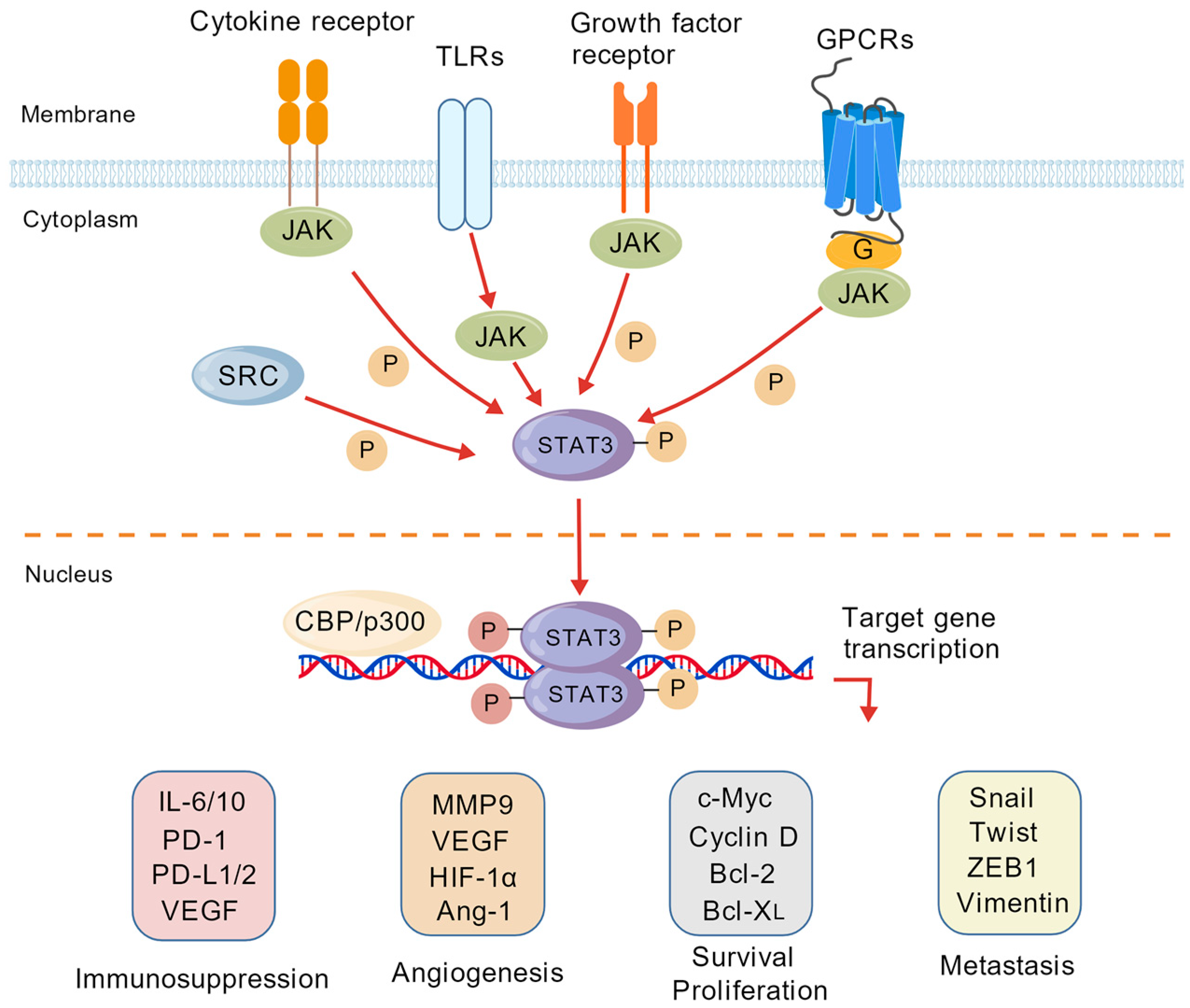
Figure 1. STAT3 signaling pathway in tumorigenesis and progression.
Pharmaceuticals 19 00257 g001
Figure 2. The screening process for targeted inhibitors derived from natural products. The figure shows a pipeline from target-based binding selection to chemical identification, mechanism prediction, and cellular validation.
Pharmaceuticals 19 00257 g002
Figure 3. Sensor diagram of STAT3 protein immobilization onto the CM5 chip (A). The affinity constant between Stattic and STAT3 was 46.14 µM (B).
Pharmaceuticals 19 00257 g003
Figure 4. Binding response of 90% ethanol extract from Psoralea corylifolia to STAT3 protein.
Pharmaceuticals 19 00257 g004
Figure 5. Base peak index/chromatogram (BPI) of the recovered sample from Psoralea corylifolia (A), Mass spectra of Psoralea corylifolia (B), Mass spectra of recovered samples from Psoralea corylifolia (C), Mass spectra of psoralen and isopsoralen standards (D).
Pharmaceuticals 19 00257 g005
Figure 6. The affinity constant between psoralen and STAT3 (A), and between isopsoralen and STAT3 (B).
Pharmaceuticals 19 00257 g006
Figure 7. The interplay between the SH2 domain of STAT3 and Stattic (A) or psoralen (B), or isopsoralen (C).
Pharmaceuticals 19 00257 g007aPharmaceuticals 19 00257 g007b
Figure 8. IC50 of psoralen (A) and isopsoralen (B) on A549 cell proliferation. Effects of varying concentrations of psoralen (C) and isopsoralen (D) on A549 cell proliferation. * p < 0.05, ** p < 0.01, *** p < 0.001, **** p < 0.0001.
Pharmaceuticals 19 00257 g008aPharmaceuticals 19 00257 g008b
Figure 9. Inhibition of A549 cell migration by psoralen and isopsoralen (A), the effect of psoralen and isopsoralen treatment for 24 h (B) or 48 h (C) on the migration rate of A549 cells. * p < 0.05, *** p < 0.001.
Pharmaceuticals 19 00257 g009
Figure 10. Scavenging rate of hydroxyl radicals (A) or superoxide anion radicals (B) by psoralen and isopsoralen.
Pharmaceuticals 19 00257 g010
Figure 11. ROS levels in HUVECs (A) or A549 cells (B) in the oxidative stress model pretreated with psoralen and isopsoralen. * p < 0.05, ** p < 0.01, *** p < 0.001, **** p < 0.0001, ns: p > 0.05.
Pharmaceuticals 19 00257 g011
Figure 12. Effects of 500 μM psoralen and isopsoralen on the proliferation of HUVECs (A) or RAW264.7 cells (B). ** p < 0.01, ns: p > 0.05.
Pharmaceuticals 19 00257 g012
Table 1. Identification of chemical components from Psoralea corylifolia.
Serial NumbertR/minMolecular FormulaMass-to-Charge Ratio
(m/z)
Ion ModeError/ppmFragment ScoreCompound
12.94C11H6O3187.0400M + H5.729216.3psoralen
22.94C11H6O3187.0400M + H5.729216.3isopsoralen
Disclaimer/Publisher’s Note: The statements, opinions and data contained in all publications are solely those of the individual author(s) and contributor(s) and not of MDPI and/or the editor(s). MDPI and/or the editor(s) disclaim responsibility for any injury to people or property resulting from any ideas, methods, instructions or products referred to in the content.