Review

Regulation of L-Lactate in Glutamate Excitotoxicity Under Cerebral Ischemia: Pathophysiology and Preventive Strategy

1
Department of Medical Genetics, College of Basic Medical Science, Army Medical University, 30 Gaotanyan Main Street, Shapingba District, Chongqing 400038, China
2
Department of Neurology, Xinqiao Hospital, Army Medical University, 83 Xinqiao Main Street, Shapingba District, Chongqing 400038, China
*
Author to whom correspondence should be addressed.
Pharmaceuticals 2025, 18(7), 935; https://doi.org/10.3390/ph18070935
Submission received: 24 April 2025 / Revised: 10 June 2025 / Accepted: 18 June 2025 / Published: 20 June 2025
(This article belongs to the Section Biopharmaceuticals)

Abstract

:
Glutamate is an excitatory neurotransmitter in the central nervous system (CNS) that mediates synaptic transmission. However, glutamate homeostasis among neural cells is broken in cerebral ischemia. Excessive glutamate triggers N-methyl-d-aspartate receptors (NMDARs) in postsynaptic neurons, leading to intracellular calcium (Ca2+) overload and excitoneurotoxicity. At this moment, L-lactate may affect NMDARs and play a protective role in cerebral ischemia. This work proposes that L-lactate regulates glutamate signaling among neural cells. But, dysregulation of L-lactate in glutamate signaling cascades contributes to glutamate excitotoxicity in cerebral ischemia. In detail, L-lactate regulates the glutamine(Gln)-glutamate cycle between astrocytes and presynaptic neurons, which triggers the astroglial L-lactate-sensitive receptor (LLR)-cyclic adenosine monophosphate (cAMP)/protein kinase A (PKA) pathway, coordinating astroglial glutamate uptake and neuronal glutamate transmission. L-lactate mediates glutamate signaling and synaptic transmission among neural cells. In addition, L-lactate promotes the function of mitochondrial calcium uniporter complex (MCUC), which quickly depletes intracellular Ca2+ in postsynaptic neurons. In addition, L-lactate can promote the conversion of microglia from the pro-inflammatory (M1) to anti-inflammatory (M2) phenotype. Therefore, regulation of L-lactate in glutamate signaling in the CNS might become a preventive target for cerebral ischemia.

Graphical Abstract

1. Introduction

In cerebral ischemia, the rapid release of glutamate and insufficient glutamate uptake result in accumulation of extracellular glutamate, which over-activates glutamate receptors [1]. Metabotropic glutamate receptors (mGluRs) and ionotropic glutamate receptors (iGluRs) are glutamate receptors, whose activity is changed following cerebral ischemia [2]. N-Methyl-d-aspartate receptors (NMDARs) are one of the iGluRs, which are ligand-gated and calcium (Ca2+)-permeable ion channels, localizing to the pre- and postsynaptic membranes [3]. Overactivation of NMDARs evokes neuronal excitotoxicity. Also, prolonged activation of extrasynaptic NMDARs causes Ca2+ overload and neuronal apoptosis [4]. This work primarily discusses glutamate excitotoxicity resulting from NMDAR hyperactivity, and then explores the possibility of regulating glutamate signaling among neural cells in cerebral ischemia.
A clinical trial revealed that glutamate and L-lactate are biomarkers that predict patient death within 3 days after severe head trauma [5,6]. Glutamate excitotoxicity has been extensively discussed, but L-lactate signaling between neural cells and its role in cerebral ischemia have not been discussed. Glutamate is the main excitatory transmitter in the brain. L-lactate not only serves as a source of pyruvate but also as a precursor of glutamate. Importantly, L-lactate significantly reduces the release of glutamate from astrocytes to neuronal synapses [7]. It has been proved that L-lactate is released from astrocytes and oligodendrocytes (OLs) and is used as an energetic substrate of neurons [8]. Even astrocytic L-lactate reduction contributes to neurodegeneration under stroke, suggesting that L-lactate from astrocytes plays a preventive role in stroke [9]. L-lactate is an energetic substrate of neurons and also functions as a signal in the modulation of neuronal excitability, synaptic plasticity, and memory formation through monocarboxylate transporters (MCTs) and G protein-coupled receptors (GPCRs) [10,11]. Specifically, L-lactate produced by glial cells is transported to neurons where lactate stimulates gene expression related to long-term memory formation [11]. Until recently, histone lactylation was observed to activate transcription, which increases glycolytic activity [12]. Hence, L-lactate may play a role as a regulator in glutamate excitotoxicity under cerebral ischemia. This work extensively discusses the regulation of L-lactate in glutamate signaling among neural cells. Importantly, it hypothesizes a mechanism by which L-lactate produced by glial cells (astrocytes, microglia, and oligodendrocytes) alleviates intracellular Ca2+ overload induced by NMDAR hyperactivity. Firstly, L-lactate regulates the Glutamine (Gln)-glutamate cycle between astrocytes and neurons. Secondly, L-lactate directly regulates glutamate signaling from presynaptic to postsynaptic neurons. Thirdly, L-lactate promotes mitochondrial calcium uniporter complex (MCUC) in the function of intracellular Ca2+ transported into mitochondria in the postsynaptic region. Finally, L-lactate regulates the phenotype of microglia in the response to inflammation.

2. Regulation of L-Lactate in Glutamate Excitotoxicity Under Cerebral Ischemia

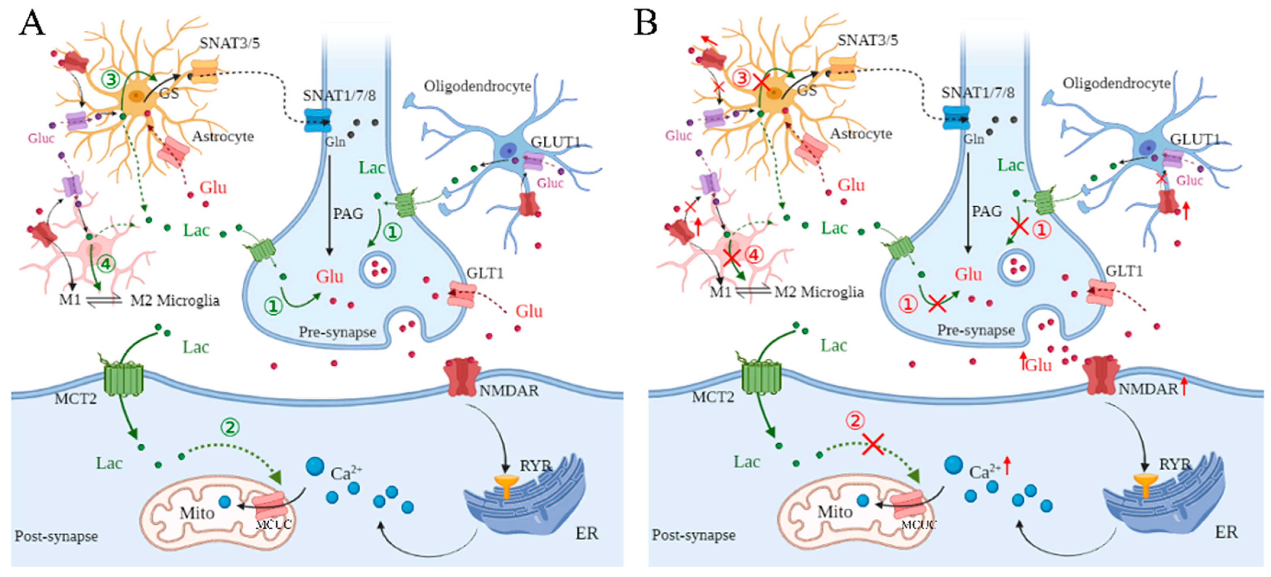
2.1. L-Lactate Regulates Glutamate Metabolism Among Neural Cells

In the brain, glutamate is an important excitatory neurotransmitter that plays an important role in synaptic plasticity [13]. In human brains, there is a high level of glutamatergic activity of synapses and energy demand [14]. Glutamate is released from presynaptic membrane via exocytosis and then triggers glutamate receptors (GluRs) at postsynaptic membrane [15]. Synaptic transmission can be mediated by the activation of NMDARs, which are one of the iGluRs. They are ligand-gated and Ca2+-permeable ion channels [16,17]. However, the disturbance of release and reuptake, such as in cerebral ischemia, leads to excessive glutamate accumulation in the synaptic cleft [18]. As a result, NMDARs are over-activated, which induces excitatory neurotoxicity, promoting neuronal death [19]. Evidence suggests that neurons collaborate with astrocytes in the regulation of energy metabolism and neurotransmission [20].
In neurons, which do not contain pyruvate carboxylase to synthesize glutamate, astrocytes must transfer a recyclable substrate via the Gln-glutamate cycle. Glutamate is transported by high-affinity transporters on neurons when it is received by synapses. Gln is absorbed by neurons via high-affinity transporters when it reaches synapses. Upon entering the brain, it is converted to glutamate by phosphate-activated glutaminase [21]. Normally, astrocytes prevent the accumulation of glutamate in extracellular spaces through the absorption of glutamate [22]. In astrocytes, the released glutamate is taken up by glutamate transporter 1 (GLT1) and glutamate-aspartate transporter (GLAST), then converted to Gln through glutamine synthetase (GS). This process not only reduces the excitatory toxicity of neurons, but also promotes glutamate recycling between neurons and astrocytes [23,24]. In addition, glutamate triggers astrocytic mGluRs and the downstream phospholipase C (PLC) cascade, leading to intracellular Ca2+ increases. The increased Ca2+ promotes the release of gamma-aminobutyric acid (GABA), D-serine, adenosine triphosphate (ATP), and glutamate. Then, these gliotransmitters regulate glutamate release and transmission in presynaptic and postsynaptic neurons [25].
NMDARs have a positive effect on the migration and differentiation of oligodendrocyte precursor cells (OPCs). It is recognized that AMPARs can mediate neuron-oligodendrocyte precursor cell (OPC) synapses [26]. AMPAR-mediated signaling at axon-OPC synapses in the mouse corpus callosum is important for balancing the response of OPCs to proliferation and differentiation cues [27]. Moreover, glutamate is released from neurons, and then stimulates oligodendrocytic NMDARs, which mobilize glucose transporter 1 (GLUT1) into the myelin membrane, which subsequently promotes glucose uptake from the extracellular space [28]. Further, the glucose is converted to L-lactate in OLs, and then the L-lactate is transported into neuronal axons, where it is fasted metabolized to sustain axonal transmission. In addition, L-lactate transport from myelin to axons is mediated by MCTs [29].
Physiologically, microglia are in a “resting” state (M0), which is mediated by “find-me” signals released from neurons [30]. Additionally, glutamate released from the presynaptic compartment triggers microglial GluRs, leading to the release of inflammatory factors [31]. Also, inflammatory factors activate the release of microglial glutamate, stimulating synaptic receptors and altering neurotransmission processes [32] Under pathological conditions, microglia are driven from M0 to the “classically activated” pro-inflammatory (M1) phenotype and the “alternatively activated” anti-inflammatory (M2) phenotype [33]. Microglia in the M2 phenotype promote axonal regeneration, neurogenesis, remyelination, and angiogenesis. However, microglia in the M1 phenotype exacerbate neuronal damage and impede neurogenesis [34]. Hence, a shift of microglia from the M1 to M2 phenotype is beneficial for recovery after stroke [35].
It has been proved that astrocyte-derived L-lactate regulates the glutamate signaling pathway, which is protective against ischemic stroke-induced neurodegeneration [9]. In astrocytes, glutamate activates aerobic glycolysis, which enhances glucose uptake and L-lactate production [36]. Also, L-lactate can regulate glutamate transport between astrocytes and neurons [37]. At post-synapse, glutamate activates NMDARs, which trigger ryanodine receptors (RyRs), leading to Ca2+ release from endoplasmic reticulum (ER) [38]. The depletion of the ER Ca2+ content is mainly mediated by the mitochondrial matrix through the mitochondrial calcium uniporter complex (MCUC) [39]. MCUC is an evolutionarily conserved calcium channel. MCUC consists of the inner membrane-spanning subunit mitochondrial calcium uniporter protein (MCU), MCU regulatory subunit b (MCUb) and essential MCU regulator (EMRE), and the intermembrane subunits mitochondrial calcium uptake protein 1 (MICU1) and 2 (MICU2). Its function is relevant to bioenergetics, cell death signaling, and the immune system [36]. However, there is no evidence to support the regulation of lactate in MCUC. But, it is recognized that L-lactate upregulates expressions of some reparative genes through lactylation [40,41]. Here, it is hypothesized that L-lactate upregulates MCUC expression through lactylation, which promotes Ca2+ uptake in mitochondria.
Although L-lactate has not been used as a drug in the clinical setting, it has been shown that L-lactate has extensive pharmacological effects. Firstly, L-lactate drives cellular deoxyribonucleic acid (DNA) repair capacity, which may promote undesirable alterations in cancer physiology and mitigate retroviral infections [42]. Secondly, L-lactate is a metabolite necessary for multiple functions in the brain and is an alternative energy source during excitotoxic brain injury. Moreover, L-lactate mediates neuroprotection against glutamate-induced excitotoxicity [43]. Importantly, L-lactate modulates astrocytic and microglial inflammation, promotes plasticity-related protein expression, and reduces neurological deficits by potentiating GPR81 signaling in cerebral ischemia [44,45,46]. Hence, L-lactate might become a potential biological drug in brain injury and stroke.
Conclusively, glutamate is released from pre-synapse, and then activates NMDARs at post-synapse, promising excitatory transmission between neurons. NMDAR activation triggers RYRs of the ER, then leads to the release of Ca2+, which is mainly depleted by MCUC in mitochondria. Besides, NMDAR activation promotes glucose transport and L-lactate production in astrocytes, microglia, and OLs. Importantly, the released L-lactate plays a vital role in glutamate signaling among neural cells. At pre-synapse, L-lactate regulates glutamate release. At post-synapse, L-lactate contributes to Ca2+ uptake of mitochondria through the regulation of MCUC. In addition, L-lactate regulates the Gln-glutamate cycle in astrocytes and promotes the transformation of microglia from the M1 to M2 phenotype (Figure 1A).

2.2. Dysregulation of L-Lactate in Glutamate Metabolism Among Neural Cells Under Cerebral Ischemia

Cerebral ischemia often leads to brain dysfunction and mortality [47]. In cerebral ischemia, glutamate is rapidly released from presynaptic membranes in combination with a deficiency in glutamate uptake, causing accumulation of extracellular glutamate [1], which leads to overactivation of glutamate receptors, including iGluRs and mGluRs [48]. Glutamate accumulation occurs because of increased neuronal release and interrupted astrocyte uptake [49]. As a result, increased excitotoxicity may result from the increase in GS activity. Furthermore, other studies have shown that GS-mediated glutamine synthesis normalizes extracellular glutamate in astrocytes and protects neurons from damage caused by cerebral ischemia [50]. Excess glutamate accumulates quickly at synapses, stimulating NMDARs, which eventually cause neurotoxicity. Thus, the inability of astrocytes in removing excessive glutamate from cellular spaces indirectly aggravates glutamate neuroexcitotoxicity [51]. Moreover, in cerebral ischemia, OPCs migrate and differentiate to OLs in the demyelinated axons, and this process is mediated by glutamate and its receptor NMDARs [52]. Especially, glutamate released from cultured cortical neurons promotes OPC migration via NMDARs by coupling to and activating the T-cell lymphoma invasion and metastasis 1 (Tiam1)/Rac1 pathway [53].
Following stroke, microglia are driven from M0 to the M1 or M2 phenotype [41]. Proinflammatory cytokines secreted by the M1 phenotype of microglia promote glutamate excitotoxicity via hyperexcitation of NMDARs and inhibit synaptic plasticity, leading to neuronal death [54]. Inositol 1,4,5-trisphosphate receptors (IP3Rs) on ER are activated by redox imbalance under stroke, which mobilizes Ca2+ into the cytosol. The increased Ca2+ signaling in microglia triggers Src at the plasma membrane, which leads to the increased permeability of microglial gap junctions. As a result, glutamate release is promoted during hypoxia [30]. Glutamate can act on both mGluRs and iGluRs of microglia and activate different signaling pathways. Activation of microglial mGluRs suppresses the production of nitric oxide (NO) and reactive oxygen species (ROS) [55,56]. In microglia, mGluRs promote the release of proinflammatory cytokines and trigger the inflammatory process [57,58].
Therefore, in cerebral ischemia, excessive glutamate is released from presynaptic neurons, and then activates NMDARs of postsynaptic neurons, overly promoting synaptic transmission. Additionally, the decreased uptake of glutamate in astrocytes aggravates intercellular glutamate accumulation. Also, NMDAR and AMPAR activation regulates the morphological development of OPCs, but overly triggered NMDARs and AMPARs fail to regulate the morphology, development, and myelination of OPCs [59]. Following excessive glutamate release, microglia are driven from the M0 to M1 phenotype, secreting inflammatory cytokines. Then, these cytokines lead to inflammatory injury of neurons and continuous glutamate release from microglia. Unfortunately, the regulation of L-lactate in glutamate homeostasis among neural cells is impeded, presenting the exacerbation of neuronal damage in cerebral ischemia (Figure 1B).

3. Dysregulation of L-Lactate in Postsynaptic Ca2+ Concentration Under Cerebral Ischemia

NMDARs are directly coupled to Ca2+ channels, the activation of which determines cytosolic Ca2+ influx. IP3Rs regulate Ca2+ release from the ER to the intracellular compartment [60,61]. Further, IP3Rs can be activated by NMDARs through the triggering of large-conductance cation channels [62]. In addition, RyRs play a role similar to that of IP3Rs in the regulation of Ca2+ release from the ER [63]. Normally, the intracellular Ca2+ concentration can be regulated by channels, pumps, and buffering systems. Sarcoendoplasmic reticulum Ca2+-ATPase (SERCA) pumps Ca2+ into the ER, and then Ca2+ is transported into the Golgi apparatus via secretory protein calcium ATPase (SPCA) [64]. Furthermore, Ca2+ can be pumped across the plasma membrane with the assistance of plasma membrane Ca2+ transport ATPase (PMCA) [65], and Ca2+ is transported without energy consumption through the Na+/Ca2+ exchanger (NCX) [66]. MCUC mediates Ca2+ entry into the mitochondria, which regulate the signaling pattern of intracellular Ca2+ [36]. Significantly, L-lactate-derived lactylation of histone lysine residues serves as an epigenetic modification that directly stimulates gene transcription from chromatin [67]. Here, it is suggested that L-lactate may upregulate MCUC, which promotes Ca2+ entry into the mitochondria, lowering the intracellular Ca2+ concentration (Figure 2A).
The ER lumen is the major Ca2+ storage compartment, and depletion of the ER Ca2+ content is followed by rapid accumulation inside the mitochondrial matrix through MCUC. The close proximity between the ER and mitochondria comprises mitochondrial reticular and ER networks, which are termed mitochondria-associated membranes (MAMs) [68]. In cerebral ischemia, NMDAR overactivation induces ER stress, and the quantity of ER-mitochondria connections is significantly increased, which promises Ca2+ uptake in mitochondria and ATP production [69]. In ER stress, IP3Rs or RyRs are triggered by NMDAR overactivation, facilitating the release of Ca2+ from the ER. In MAMs, voltage-dependent anion channels (VDACs) form large voltage-gated pores in the outer mitochondrial membrane at the ER–mitochondria contacts, which transfer the released Ca2+ to the mitochondrial intermembrane space [70]. To reach the mitochondrial matrix, the Ca2+ located inside the intermembrane space must pass through the MCUC, which rapidly decreases the intracellular Ca2+ concentration [71].
NMDARs are over-activated, which leads to cytosolic Ca2+ influx and activation of IP3Rs and RyRs, resulting in an increase in the intracellular Ca2+ concentration [72]. This overload of intracellular Ca2+ leads to necrosis or apoptosis, resulting in fragmentation of deoxyribonucleic acid (DNA), degraded cross-linking and cytoskeletal proteins, apoptosis, and phagocytosis [60,73]. Additionally, the decreased L-lactate supplement from glial cells leads to MCUC dysfunction, which accentuates cytosolic Ca2+ overload in cerebral ischemia (Figure 2B).

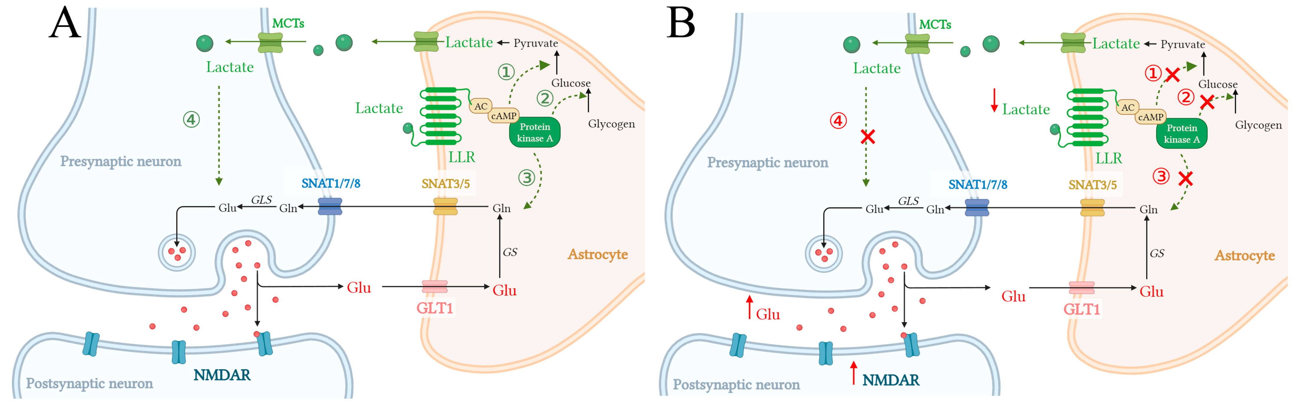
4. L-Lactate Triggers Astroglial LLR-cAMP/PKA Pathway, Promoting Glycolysis and Glutamate Uptake in Astrocytes

Reduction of astrocytic L-lactate contributes to neurodegeneration under stroke. Meanwhile, L-lactate plays a preventive role in cerebral ischemia [9]. Blood-derived glucose is transported into astrocytes, then decomposed into L-lactate. L-lactate is supplied to neurons, where it serves as an energy source [74]. Further, it is suggested that astrocyte-derived L-lactate is required for numerous neuronal cell functions [10]. L-lactate functions as a signaling molecule by serving as an agonist for GPCRs [75]. Specific GPCRs of lactate include G-protein-coupled receptor 81 (GPR81), neuronal L-lactate-sensitive receptor (LLR), astroglial unidentified LLR, L-lactate-sensitive olfactory receptor 78 (Olfr78), and proton-sensitive G-protein coupled receptor 4 (GPR4) [9,76]. GPR81 couples to Gi/0-proteins, leading to cAMP downregulation; LLR and Olfr78 couple to Gs-proteins and increase cAMP production; and GPR4 takes part in presumable allosteric modulation. However, L-lactate at physiological levels can hardly activate GPR81, which is normally excited during exercise or pathology [77]. Astroglial LLR and its downstream cAMP signaling pathway are triggered at a physiologic L-lactate concentration, which might contribute to neurodevelopmental disorders or ischemic stroke-induced neurodegeneration [9,78].
Glutamate uptake of astrocytes activates astrocytic aerobic glycolysis, leading to glucose uptake and lactate release [36]. Astrocytes generate glycolytically derived L-lactate and L-serine, which are transported into neurons, sustaining neuronal energy needs and modulating neurotransmitter-receptor activity [79]. Disruption of this metabolic coupling may contribute to the progression of neurological diseases, including cerebral ischemia [44]. L-lactate can be covalently modified to lysine residues of histones, a process known as lactylation [67]. This histone lactylation provides a molecular mechanism for lactate in physiological or pathological processes, such as metabolic disability or ischemic injury [80,81]. In cerebral ischemia, enriched lactate induces histone lactylation in high mobility group box 1 (HMGB1) and promotes HMGB1 expression. HMGB1 leads to pyroptosis in cerebral ischemia [81]. Pyroptosis is a pro-inflammatory death mode. It has been suggested that the pyroptosis pathway might be a novel treatment target for cerebral ischemia, but rigorous experimental evidence is lacking [82].
However, malignant astrocyte swelling and impaired glutamate clearance are found in cerebral ischemia [83]. In physiology, the Gln-glutamate cycle controls glutamate metabolism. In this cycle, astrocytes uptake extracellular glutamate through GLT1, and then glutamate is converted to Gln via GS in astrocytes. Gln is transferred to the extracellular milieu via sodium-coupled neutral amino acid transporter (SNAT)3/5, and then into presynaptic neurons via SNAT1/7/8 [84]. In presynaptic neurons, Gln is converted into glutamate by glutaminase (GLS) [85]. Rapid uptake and glutamate conversion in astrocytes prevent prolonged activation of glutamatergic receptors in postsynaptic neurons. Once this process is inhibited or unchecked, it will trigger excitotoxicity in neurons. Hence, the extracellular glutamate must be promptly removed by astrocytes in cerebral ischemia (Figure 3).
Astroglial LLR couples to Gs-proteins and triggers the cAMP/PKA pathway. Meanwhile, cAMP/PKA signaling facilitates glutamate uptake into cells [86]. In the CNS, L-lactate secreted by astrocytes is released through MCTs and binds to astroglial LLR, stimulating adenylate cyclase (AC) and cAMP production. This activation of the cAMP/PKA pathway promotes glycogen degradation, glycolysis, and increased L-lactate production [75]. In addition, this L-lactate-positive feedback in astrocytes ensures an L-lactate concentration gradient between astrocytes and neighboring cells, maintaining cellular activity, such as the regulation of L-lactate in glutamate excitotoxicity. Here, it is suggested that L-lactate triggers the astroglial LLR-cAMP/PKA signaling pathway, which promotes extracellular astroglial glutamate uptake and alleviates neuronal glutamate excitotoxicity (Figure 4A). However, this L-lactate-glutamate coupling loop might be inhibited in cerebral ischemia. In that scenario, L-lactate can hardly activate the astroglial LLR-cAMP/PKA signaling pathway, which leads to reduced glutamate uptake by astrocytes. As a result, the extracellular lactate concentration is increased and over-activates NMDAR in postsynaptic neurons, leading to neuronal excitotoxicity (Figure 4B).

5. Discussion

Glucose is the obligatory fuel for brain cells; it is metabolized mostly by aerobic glycolysis and rarely by anaerobic glycolysis [37]. In the CNS, L-lactate is mainly derived from astrocytes and OLs, and is further utilized by neurons [87]. Astrocyte–neuron L-lactate flow was primarily discussed in a scheme of glutamate-induced glycolysis in astrocytes during physiological activation [88]. In detail, glutamate depolarizes neurons through its receptors, which can be terminated by an efficient glutamate uptake system in astrocytes [89]. Especially, glutamate-cotransported Na+ can activate Na+/K+ ATPase, which fuels glycolytic enzymes and stimulates glycolysis, producing L-lactate in astrocytes. L-lactate, once released from astrocytes, can be recycled by neurons [90]. L-lactate is recognized as an energetic metabolite that is transferred from astrocytes, microglia, and OLs to neurons, supporting neuronal transmission [91]. Until recently, it was not known that L-lactate regulates gene expression through histone lactylation [65]. Here, this work reveals that L-lactate regulates glutamate signaling among neural cells. But, under cerebral ischemia, the role of L-lactate in glutamate signaling is suppressed, which exacerbates glutamate excitotoxicity.
In cerebral ischemia, the concentration of extracellular glutamate is rapidly increased [19]. Accumulated glutamate in the synaptic cleft triggers neuronal death, causing mental or physical handicap [92]. Glutamate accumulation in the synaptic cleft induces excessive NMDAR activation at the postsynaptic region [93]. Ca2+ signals induced by synaptic NMDAR regulate neuronal plasticity, and Ca2+ is released from ER following the activation of IP3Rs or RYRs [94]. Meanwhile, excessive glutamate-induced NMDAR hyperactivity promotes Ca2+ release from the ER, leading to intracellular Ca2+ overload [95]. Intracellular Ca2+ accumulation leads to excitotoxicity, apoptosis, and cell death [96]. Meanwhile, L-lactate plays a role as a positive modulator of NMDAR-mediated signaling in plasticity gene expression and memory consolidation [97].
This study postulates that L-lactate is protective against glutamate excitotoxicity under physiological conditions. L-lactate functions as a signal molecule, transported from glial cells to neurons. However, this process is suppressed under cerebral ischemia, leading to dysregulation of L-lactate in the glutamate signaling pathway among neural cells. As a result, glutamate transmission in synapses is abnormal, and then NMDARs are hyperactivated. NMDAR overactivation at synapses triggers RYRs, which promote Ca2+ release from the ER. Physiologically, MCUC largely depletes most of the released Ca2+. But, in cerebral ischemia, MCUC function is inhibited following L-lactate dysregulation, and intracellular Ca2+ overload leads to apoptosis. Additionally, L-lactate regulates glutamate transmission from presynaptic neurons to postsynaptic neurons in an astrocyte–neuron lactate shuttle (ANLS) shuttle. However, this L-lactate-glutamate coupling mode is disabled in cerebral ischemia. As a result, glutamate is accumulated in neurons, which over-activates NMDARs and causes an overload of intracellular Ca2+, inducing neuronal excitotoxicity. Therefore, targeting L-lactate metabolism and its regulation in glutamate signaling in neural cells could become a promising preventive strategy for cerebral ischemia.

Author Contributions

Conceptualization, M.Z. and H.G.; methodology, Z.G.; software, G.G.; validation, Z.G., W.J. and G.G.; formal analysis, M.Z.; investigation, M.Z., Y.W.; writing—original draft preparation, M.Z.; writing—review and editing, M.Z.; visualization, Y.W.; supervision, H.G.; project administration, M.Z.; funding acquisition, M.Z. All authors have read and agreed to the published version of the manuscript.

Funding

This work was supported by grants from the Natural Science Foundation of Chongqing (Grant No. CSTB2024NSCQ-MSX0558) and Science and Technology Research Project of Chongqing Education Commission (Grant No. KJQN202412808).

Institutional Review Board Statement

Not applicable.

Informed Consent Statement

Not applicable.

Data Availability Statement

Not applicable.

Conflicts of Interest

The authors declare no conflicts of interest.

Abbreviations

The following abbreviations are used in this manuscript:
CNSCentral nervous system
NMDARsN-Methyl-D-aspartate receptors
Ca2+Calcium
GluGlutamate
GlnGlutamine
MitoMitochondria
MCUCMitochondrial calcium uniporter complex
LLRL-lactate-sensitive receptor
cAMPCyclic adenosine monophosphate
PKAProtein kinase A
iGluRs Ionotropic glutamate receptors
mGluRsMetabotropic glutamate receptors
OLs Oligodendrocytes
MCTsMonocarboxylate transporters
GPCRs G protein-coupled receptors
GluRsGlutamate receptors
AMPARsA-amino-3-hydroxy-5-methyl-4-isox-azolepropionic acid receptors
GLT1Glutamate transporter 1
GLASTGlutamate-aspartate transporter
GSGlutamine synthetase
PLCPhospholipase C
ATPAdenosine triphosphate
GABAGamma-aminobutyric acid
OPCsOligodendrocyte precursor cells
OPCOligodendrocyte precursor cell
GLUT1Glucose transporter 1
M0A “resting” state
M1“Classically activated” pro-inflammatory
M2“Alternatively activated” anti-inflammatory
RyRsRyanodine receptors
EREndoplasmic reticulum
Tiam1T lymphoma invasion and metastasis 1
IP3RsInositol 1,4,5-trisphosphate receptor
NONitric oxide
ROSReactive oxygen species
SERCASarcoendoplasmic reticulum Ca2+-ATPase
SPCASecretory protein calcium ATPase
PMCAPlasma membrane Ca2+ transport ATPase
NCXNa+/Ca2+ exchanger
MAMsMitochondria-associated membranes
VDACsVoltage-dependent anion channels
DNADeoxyribonucleic acid
Olfr78L-lactate-sensitive olfactory receptor 78
GPR4G-protein coupled receptor 4
HMGB1 High mobility group box 1
SNATSodium-coupled neutral amino acid transporter
GLSGlutaminase
ACAdenylate cyclase
ANLS Astrocyte–neuron lactate shuttle

References

  1. Sun, Y.; Feng, X.; Ding, Y.; Li, M.; Yao, J.; Wang, L.; Gao, Z. Phased treatment strategies for cerebral ischemia based on glutamate receptors. Front. Cell. Neurosci. 2019, 13, 168. [Google Scholar] [CrossRef] [PubMed]
  2. Giza, C.C.; Maria, N.S.; Hovda, D.A. N-Methyl-D-aspartate receptor subunit changes after traumatic injury to the developing brain. J. Neurotrauma 2006, 23, 950–961. [Google Scholar] [CrossRef] [PubMed]
  3. Holmes, A.; Zhou, N.; Donahue, D.L.; Balsara, R.; Castellino, F.J. A deficiency of the GluN2C subunit of the N-methyl-D-aspartate receptor is neuroprotective in a mouse model of ischemic stroke. Biochem. Biophys. Res. Commun. 2018, 495, 136–144. [Google Scholar] [CrossRef]
  4. Engin, A.; Engin, A.B. N-Methyl-D-Aspartate receptor signaling-protein kinases crosstalk in cerebral ischemia. Adv. Exp. Med. Biol. 2021, 1275, 259–283. [Google Scholar]
  5. Stefani, M.A.; Modkovski, R.; Hansel, G.; Zimmer, E.R.; Kopczynski, A.; Muller, A.P.; Strogulski, N.R.; Rodolphi, M.S.; Carteri, R.K.; Schmidt, A.P.; et al. Elevated glutamate and lactate predict brain death after severe head trauma. Ann. Clin. Transl. Neurol. 2017, 4, 392–402. [Google Scholar] [CrossRef]
  6. Quintard, H.; Patet, C.; Zerlauth, J.-B.; Suys, T.; Bouzat, P.; Pellerin, L.; Meuli, R.; Magistretti, P.J.; Oddo, M. Improvement of neuroenergetics by hypertonic lactate therapy in patients with traumatic brain injury is dependent on baseline cerebral lactate/pyruvate ratio. J. Neurotrauma 2016, 33, 681–687. [Google Scholar] [CrossRef] [PubMed]
  7. Montana, V.; Flint, D.; Waagepetersen, H.S.; Schousboe, A.; Parpura, V. Two metabolic fuels, glucose and lactate, differentially modulate exocytotic glutamate release from cultured astrocytes. Neurochem. Res. 2021, 46, 2551–2579. [Google Scholar] [CrossRef]
  8. Proia, P.; Di Liegro, C.M.; Schiera, G.; Fricano, A.; Di Liegro, I. Lactate as a metabolite and a regulator in the central nervous system. Int. J. Mol. Sci. 2016, 17, 1450. [Google Scholar] [CrossRef]
  9. Yamagata, K. Lactate supply from astrocytes to neurons and its role in ischemic stroke-induced neurodegeneration. Neuroscience 2022, 481, 219–231. [Google Scholar] [CrossRef]
  10. Magistretti, P.J.; Allaman, I. Lactate in the brain: From metabolic end-product to signalling molecule. Nat. Rev. Neurosci. 2018, 19, 235–249. [Google Scholar] [CrossRef] [PubMed]
  11. Sun, S.; Li, H.; Chen, J.; Qian, Q. Lactic acid: No longer an inert and end-product of glycolysis. Physiology 2017, 32, 453–463. [Google Scholar] [CrossRef] [PubMed]
  12. Pan, R.-Y.; He, L.; Zhang, J.; Liu, X.; Liao, Y.; Gao, J.; Liao, Y.; Yan, Y.; Li, Q.; Zhou, X.; et al. Positive feedback regulation of microglial glucose metabolism by histone H4 lysine 12 lactylation in Alzheimer’s disease. Cell Metab. 2022, 34, 634–648. [Google Scholar] [CrossRef]
  13. Ribeiro, F.M.; Vieira, L.B.; Pires, R.G.; Olmo, R.P.; Ferguson, S.S. Metabotropic glutamate receptors and neurodegenerative diseases. Pharmacol. Res. 2017, 115, 179–191. [Google Scholar] [CrossRef] [PubMed]
  14. Plaitakis, A.; Sidiropoulou, K.; Kotzamani, D.; Litso, I.; Zaganas, I.; Spanaki, C. Evolution of glutamate metabolism via GLUD2 enhances lactate-dependent synaptic plasticity and complex cognition. Int. J. Mol. Sci. 2024, 25, 5297. [Google Scholar] [CrossRef]
  15. Andersen, J.V.; Markussen, K.H.; Jakobsen, E.; Schousboe, A.; Waagepetersen, H.S.; Rosenberg, P.A.; Aldana, B.I. Glutamate metabolism and recycling at the excitatory synapse in health and neurodegeneration. Neuropharmacology 2021, 196, 108719. [Google Scholar] [CrossRef]
  16. Rodriguez Chavez, V.; Moran, J.; Molina Salinas, G.; Zepeda Ruiz, W.A.; Rodriguez, M.C.; Picazo, O.; Cerbon, M. Participation of Glutamatergic Ionotropic Receptors in Excitotoxicity: The Neuroprotective Role of Prolactin. Neuroscience 2021, 461, 180–193. [Google Scholar] [CrossRef] [PubMed]
  17. Reiner, A.; Levitz, J. Glutamatergic signaling in the central nervous system: Ionotropic and metabotropic receptors in concert. Neuron 2018, 98, 1080–1098. [Google Scholar] [CrossRef]
  18. Shen, Z.; Xiang, M.; Chen, C.; Ding, F.; Wang, Y.; Shang, C.; Xin, L.; Zhang, Y.; Cui, X. Glutamate excitotoxicity: Potential therapeutic target for ischemic stroke. Biomed. Pharmacother. 2022, 151, 113125. [Google Scholar] [CrossRef]
  19. Lai, T.W.; Zhang, S.; Wang, Y.T. Excitotoxicity and stroke: Identifying novel targets for neuroprotection. Prog. Neurobiol. 2014, 115, 157–188. [Google Scholar] [CrossRef]
  20. Andersen, J.V.; Schousboe, A.; Verkhratsky, A. Astrocyte energy and neurotransmitter metabolism in Alzheimer’s disease: Integration of the glutamate/GABA-glutamine cycle. Prog. Neurobiol. 2022, 217, 102331. [Google Scholar] [CrossRef]
  21. Albrecht, J.; Zielińska, M.; Norenberg, M.D. Glutamine as a mediator of ammonia neurotoxicity: A critical appraisal. Biochem. Pharmacol. 2010, 80, 1303–1308. [Google Scholar] [CrossRef]
  22. Perrillat Mercerot, A.; Bourmeyster, N.; Guillevin, C.; Miranville, A.; Guillevin, R. Analysis of a mathematical model for the glutamate/glutamine cycle in the brain. Bull. Math. Biol. 2019, 81, 4251–4270. [Google Scholar] [CrossRef]
  23. Sonnewald, U.; Schousboe, A. Introduction to the glutamate-glutamine cycle. Adv. Neurobiol. 2016, 13, 1–7. [Google Scholar] [PubMed]
  24. Limón, I.D.; Angulo Cruz, I.; Sánchez Abdon, L.; Patricio Martínez, A. Disturbance of the glutamate-glutamine cycle, secondary to hepatic damage, compromises memory function. Front. Neurosci. 2021, 15, 578922. [Google Scholar] [CrossRef] [PubMed]
  25. Kofuji, P.; Araque, A. G-protein-coupled receptors in astrocyte-neuron communication. Neuroscience 2021, 456, 71–84. [Google Scholar] [CrossRef]
  26. Habermacher, C.; Angulo, M.C.; Benamer, N. Glutamate versus GABA in neuron-oligodendroglia communication. Glia 2019, 67, 2092–2106. [Google Scholar] [CrossRef] [PubMed]
  27. Chen, T.-J.; Kula, B.; Nagy, B.; Barzan, R.; Gall, A.; Ehrlich, I.; Kukley, M. In Vivo Regulation of Oligodendrocyte Precursor Cell Proliferation and Differentiation by the AMPA-Receptor Subunit GluA2. Cell Rep. 2018, 25, 852–861.e7. [Google Scholar] [CrossRef]
  28. Saab, A.S.; Tzvetavona, I.D.; Trevisiol, A.; Baltan, S.; Dibaj, P.; Kusch, K.; Möbius, W.; Goetze, B.; Jahn, H.M.; Huang, W.; et al. Oligodendroglial NMDA receptors regulate glucose import and axonal energy metabolism. Neuron 2016, 91, 119–132. [Google Scholar] [CrossRef]
  29. Jha, M.K.; Morrison, B.M. Lactate transporters mediate glia-neuron metabolic crosstalk in homeostasis and disease. Front. Cell. Neurosci. 2020, 14, 589582. [Google Scholar] [CrossRef]
  30. Li, Y.; Du, X.F.; Du, J.L. Resting microglia respond to and regulate neuronal activity in vivo. Commun. Integr. Biol. 2013, 6, e24493. [Google Scholar] [CrossRef]
  31. Zhang, X.; Wang, D.; Zhang, B.; Zhu, J.; Zhou, Z.; Cui, L. Regulation of microglia by glutamate and its signal pathway in neurodegenerative diseases. Drug Discov. 2020, 25, 1074–1085. [Google Scholar] [CrossRef] [PubMed]
  32. Illes, P.; Rubini, P.; Ulrich, H.; Zhao, Y.; Tang, Y. Regulation of microglial functions by purinergic mechanisms in the healthy and diseased CNS. Cells 2020, 9, 1108. [Google Scholar] [CrossRef] [PubMed]
  33. Xiong, X.Y.; Liu, L.; Yang, Q.W. Functions and mechanisms of microglia/ macrophages in neuroinflammation and neurogenesis after stroke. Prog. Neurobiol. 2016, 142, 23–44. [Google Scholar] [CrossRef]
  34. Hu, X.; Leak, R.K.; Shi, Y.; Suenaga, J.; Gao, Y.; Zheng, P.; Chen, J. Microglial and macrophage polarization-new prospects for brain repair. Nat. Rev. Neurol. 2015, 11, 56–64. [Google Scholar] [CrossRef]
  35. Liu, X.; Wen, S.; Yan, F.; Liu, K.; Liu, L.; Wang, L.; Zhao, S.; Ji, X. Salidroside provides neuroprotection by modulating microglial polarization after cerebral ischemia. J. Neuroinflamm. 2018, 15, 39. [Google Scholar] [CrossRef] [PubMed]
  36. Beard, E.; Lengacher, S.; Dias, S.; Magistretti, P.J.; Finsterwald, C. Astrocytes as key regulators of brain energy metabolism: New therapeutic perspectives. Front. Physiol. 2022, 12, 825816. [Google Scholar] [CrossRef]
  37. Dienel, G.A. Brain glucose metabolism: Integration of energetics with function. Physiol. Rev. 2019, 99, 949–1045. [Google Scholar] [CrossRef]
  38. Ramírez, O.A.; Córdova, A.; Cerda, M.; Lobos, P.; Härtel, S.; Couve, A.; Hidalgo, C. Ryanodine receptor-mediated Ca2+ release and atlastin-2 GTPase activity contribute to IP3-induced dendritic Ca2+ signals in primary hippocampal neurons. Cell Calcium 2021, 96, 102399. [Google Scholar] [CrossRef]
  39. Kamer, K.J.; Mootha, V.K. The molecular era of the mitochondrial calcium uniporter. Nat Rev Mol. Cell Biol. 2015, 16, 545–553. [Google Scholar]
  40. Wang, N.; Wang, W.; Wang, X.; Mang, G.; Chen, J.; Yan, X.; Tong, Z.; Yang, Q.; Wang, M.; Chen, L.; et al. Histone lactylation boosts reparative gene activation post-myocardial infarction. Circ. Res. 2022, 131, 893–908. [Google Scholar] [CrossRef]
  41. Zhou, Y.; Yang, L.; Liu, X.; Wang, H. Lactylation may be a novel posttranslational modification in inflammation in neonatal hypoxic-ischemic encephalopathy. Front. Pharmacol. 2022, 13, 926802. [Google Scholar] [CrossRef] [PubMed]
  42. Ciszewski, W.M.; Sobierajska, K.; Stasiak, A.; Wagner, W. Lactate drives cellular DNA repair capacity: Role of lactate and related short-chain fatty acids in cervical cancer chemoresistance and viral infection. Front. Cell Dev. Biol. 2022, 10, 1012254. [Google Scholar] [CrossRef]
  43. Llorente-Folch, I.; Rueda, C.B.; Pérez-Liébana, I.; Satrústegui, J.; Pardo, B. L-Lactate-Mediated Neuroprotection against Glutamate-Induced Excitotoxicity Requires ARALAR/AGC1. J. Neurosci. 2016, 36, 4443–4456. [Google Scholar] [CrossRef] [PubMed]
  44. Xu, J.; Ji, T.; Li, G.; Zhang, H.; Zheng, Y.; Li, M.; Ma, J.; Li, Y.; Chi, G. Lactate attenuates astrocytic inflammation by inhibiting ubiquitination and degradation of NDRG2 under oxygen-glucose deprivation conditions. J. Neuroinflamm. 2022, 19, 314. [Google Scholar] [CrossRef]
  45. Zhang, Y.; Zhang, S.; Yang, L.; Zhang, Y.; Cheng, Y.; Jia, P.; Lv, Y.; Wang, K.; Fan, P.; Zhang, P.; et al. Lactate modulates microglial inflammatory responses through HIF-α-mediated CCL7 signaling after cerebral ischemia in mice. Int. Immunopharmacol. 2024, 146, 113801. [Google Scholar] [CrossRef] [PubMed]
  46. Zhai, X.; Li, J.; Li, L.; Sun, Y.; Zhang, X.; Xue, Y.; Lv, J.; Gao, Y.; Li, S.; Yan, W.; et al. L-lactate preconditioning promotes plasticity-related proteins expression and reduces neurological deficits by potentiating GPR81 signaling in rat traumatic brain injury model. Brain Res. 2020, 1746, 146945. [Google Scholar] [CrossRef]
  47. Hu, X.; De Silva, T.M.; Chen, J.; Faraci, F.M. Cerebral vascular disease and neurovascular injury in ischemic stroke. Circ. Res. 2017, 120, 449–471. [Google Scholar] [CrossRef]
  48. Amantea, D.; Bagetta, G. Excitatory and inhibitory amino acid neurotransmitters in stroke: From neurotoxicity to ischemic tolerance. Curr. Opin. Pharmacol. 2017, 35, 111–119. [Google Scholar] [CrossRef]
  49. Yuan, Q.; Yuan, Y.; Zheng, Y.; Sheng, R.; Liu, L.; Xie, F.; Tan, J. Anti-cerebral ischemia reperfusion injury of polysaccharides: A review of the mechanisms. Biomed. Pharmacother. 2021, 137, 111303. [Google Scholar] [CrossRef]
  50. Kumagai, A.; Sasaki, T.; Matsuoka, K.; Abe, M.; Tabata, T.; Itoh, Y.; Fuchino, H.; Wugangerile, S.; Suga, M.; Yamaguchi, T.; et al. Monitoring of glutamate-induced excitotoxicity by mitochondrial oxygen consumption. Synapse 2019, 73, e22067. [Google Scholar] [CrossRef]
  51. Zaric, M.; Drakulic, D.; Stojanovic, I.G.; Mitrovic, N.; Grkovic, I.; Martinovic, J. Regional-specific effects of cerebral ischemia/reperfusion and dehydroepiandrosterone on synaptic NMDAR/PSD-95 complex in male Wistar rats. Brain Res. 2018, 1688, 73–80. [Google Scholar] [CrossRef] [PubMed]
  52. Ohta, K.; Iwai, M.; Sato, K.; Omori, N.; Nagano, I.; Shoji, M.; Abe, K. Dissociative increase of oligodendrocyte progenitor cells between young and aged rats after transient cerebral ischemia. Neurosci. Lett. 2003, 335, 159–162. [Google Scholar] [CrossRef]
  53. Xiao, L.; Hu, C.; Yang, W.; Guo, D.; Li, C.; Shen, W.; Liu, X.; Aijun, H.; Dan, W.; He, C. NMDA receptor couples Rac1-GEF Tiam1 to direct oligodendrocyte precursor cell migration. Glia 2013, 61, 2078–2099. [Google Scholar] [CrossRef]
  54. Nagy, E.E.; Frigy, A.; Szász, J.A.; Horváth, E. Neuroinflammation and microglia/macrophage phenotype modulate the molecular background of post-stroke depression: A literature review. Exp. Ther. Med. 2020, 20, 2510–2523. [Google Scholar] [CrossRef] [PubMed]
  55. Byrnes, K.R.; Stoica, B.; Loane, D.J.; Riccio, A.; Davis, M.I.; Faden, A.I. Metabotropic glutamate receptor 5 activation inhibits microglial associated inflammation and neurotoxicity. Glia 2009, 57, 550–560. [Google Scholar] [CrossRef] [PubMed]
  56. Byrnes, K.R.; Loane, D.J.; Faden, A.I. Metabotropic glutamate receptors as targets for multipotential treatment of neurological disorders. Neurotherapeutics 2009, 6, 94–107. [Google Scholar] [CrossRef]
  57. Durand, D.; Carniglia, L.; Turati, J.; Ramírez, D.; Saba, J.; Caruso, C.; Lasaga, M. Amyloid-beta neurotoxicity and clearance are both regulated by glial group II metabotropic glutamate receptors. Neuropharmacology 2017, 123, 274–286. [Google Scholar] [CrossRef]
  58. Noda, M. Dysfunction of glutamate receptors in microglia may cause neurodegeneration. Curr. Alzheimer Res. 2016, 13, 381–386. [Google Scholar] [CrossRef]
  59. Fannon, J.; Tarmier, W.; Fulton, D. Neuronal activity and AMPA-type glutamate receptor activation regulates the morphological development of oligodendrocyte precursor cells. Glia 2015, 63, 1021–1035. [Google Scholar] [CrossRef]
  60. Radak, D.; Katsiki, N.; Resanovic, I.; Jovanovic, A.; Sudar-Milovanovic, E.; Zafirovic, S.; Mousad, S.A.; Isenovic, E.R. Apoptosis and acute brain ischemia in ischemic stroke. Curr. Vasc. Pharmacol. 2017, 15, 115–122. [Google Scholar] [CrossRef]
  61. Vallese, F.; Barazzuol, L.; Maso, L.; Brini, M.; Cali, T. ER-mitochondria calcium transfer, organelle contacts and neurodegenerative diseases. Adv. Exp. Med. Biol. 2020, 1131, 719–746. [Google Scholar] [PubMed]
  62. Gaspers, L.D.; Bartlett, P.J.; Politi, A.; Burnett, P.; Metzger, W.; Johnston, J.; Joseph, S.K.; Hofer, T.; Thomas, A.P. Hormone-induced calcium oscillations depend on cross-coupling with inositol 1,4,5-trisphosphate oscillations. Cell Rep. 2014, 9, 1209–1218. [Google Scholar] [CrossRef]
  63. Zalk, R.; Clarke, O.B.; des Georges, A.; Grassucci, R.A.; Reiken, S.; Mancia, F.; Hendrickson, W.A.; Frank, J.; Marks, A.R. Structure of a mammalian ryanodine receptor. Nature 2015, 517, 44–49. [Google Scholar] [CrossRef]
  64. Vandecaetsbeek, I.; Vangheluwe, P.; Raeymaekers, L.; Wuytack, F.; Vanoevelen, J. The Ca2+ pumps of the endoplasmic reticulum and Golgi apparatus. Cold Spring Harb. Perspect. Biol. 2011, 3, a004184. [Google Scholar] [CrossRef]
  65. Bruce, J.I.E. Metabolic regulation of the PMCA: Role in cell death and survival. Cell Calcium 2018, 69, 28–36. [Google Scholar] [CrossRef] [PubMed]
  66. Verkhratsky, A.; Trebak, M.; Perocchi, F.; Khananshvili, D.; Sekler, I. Crosslink between calcium and sodium signalling. Exp. Physiol. 2018, 103, 157–169. [Google Scholar] [CrossRef]
  67. Zhang, D.; Tang, Z.; Huang, H.; Zhou, G.; Cui, C.; Weng, Y.; Liu, W.; Kim, S.; Lee, S.; Perez-Neut, M.; et al. Metabolic regulation of gene expression by histone lactylation. Nature 2019, 574, 575–580. [Google Scholar] [CrossRef] [PubMed]
  68. Giorgi, C.; Missiroli, S.; Patergnani, S.; Duszynski, J.; Wieckowski, M.R.; Pinton, P. Mitochondria-associated membranes: Composition, molecular mechanisms, and physiopathological implications. Antioxid. Redox Signal. 2015, 22, 995–1019. [Google Scholar] [CrossRef]
  69. Williams, G.S.; Boyman, L.; Lederer, W.J. Mitochondrial calcium and the regulation of metabolism in the heart. J. Mol. Cell Cardiol. 2015, 78, 35–45. [Google Scholar] [CrossRef]
  70. Varughese, J.T.; Buchanan, S.K.; Pitt, A.S. The role of voltage-dependent anion channel in mitochondrial dysfunction and human disease. Cells 2012, 10, 1737. [Google Scholar] [CrossRef]
  71. Oxenoid, K.; Dong, Y.; Cao, C.; Cui, T.; Sancak, Y.; Markhard, A.L.; Grabarek, Z.; Kong, L.; Liu, Z.; Ouyang, B.; et al. Architecture of the mitochondrial calcium uniporter. Nature 2016, 533, 269–273. [Google Scholar] [CrossRef] [PubMed]
  72. Skowrońska, K.; Kozłowska, H.; Albrecht, J. Neuron-derived factors negatively modulate ryanodine receptor-mediated calcium release in cultured mouse astrocytes. Cell Calcium 2020, 92, 102304. [Google Scholar] [CrossRef]
  73. Zhao, Q.; Wang, S.; Li, Y.; Wang, P.; Li, S.; Guo, Y.; Yao, R. The role of the mitochondrial calcium uniporter in cerebral ischemia/reperfusion injury in rats involves regulation of mitochondrial energy metabolism. Mol. Med. Rep. 2013, 7, 1073–1080. [Google Scholar] [CrossRef] [PubMed]
  74. Brooks, G.A. Cell-cell and intracellular lactate shuttles. J. Physiol. 2019, 587, 5591–5600. [Google Scholar] [CrossRef] [PubMed]
  75. Horvat, A.; Zorec, R.; Vardjan, N. Lactate as an astroglial signal augmenting aerobic glycolysis and lipid metabolism. Front. Physiol. 2021, 12, 735532. [Google Scholar] [CrossRef] [PubMed]
  76. Orecchioni, M.; Matsunami, H.; Ley, K. Olfactory receptors in macrophages and inflammation. Front. Immunol. 2022, 13, 1029244. [Google Scholar] [CrossRef]
  77. Mosienko, V.; Teschemacher, A.G.; Kasparov, S. Is L-lactate a novel signaling molecule in the brain? J. Cereb. Blood Flow Metab. 2015, 35, 1069–1075. [Google Scholar] [CrossRef]
  78. Abi-Saab, W.M.; Maggs, D.G.; Jones, T.; Jacob, R.; Srihari, V.; Thompson, J.; Kerr, D.; Leone, P.; Krystal, J.H.; Spencer, D.D.; et al. Striking differences in glucose and lactate levels between brain extracellular fluid and plasma in conscious human subjects: Effects of hyperglycemia and hypoglycemia. J. Cereb. Blood Flow Metab. 2022, 22, 271–279. [Google Scholar] [CrossRef]
  79. Bonvento, G.; Bolaños, J.P. Astrocyte-neuron metabolic cooperation shapes brain activity. Cell Metab. 2021, 33, 1546–1564. [Google Scholar] [CrossRef]
  80. Yang, K.; Fan, M.; Wang, X.; Xu, J.; Wang, Y.; Tu, F.; Gill, P.S.; Ha, T.; Liu, L.; Williams, D.L.; et al. Lactate promotes macrophage HMGB1 lactylation, acetylation, and exosomal release in polymicrobial sepsis. Cell Death Differ. 2022, 29, 133–146. [Google Scholar] [CrossRef]
  81. Yao, X.; Li, C. Lactate dehydrogenase A mediated histone lactylation induced the pyroptosis through targeting HMGB1. Metab. Brain Dis. 2023, 38, 1543–1553. [Google Scholar] [CrossRef]
  82. Zhang, Q.; Jia, M.; Wang, Y.; Wang, Q.; Wu, J. Cell death mechanisms in cerebral ischemia-reperfusion injury. Neurochem. Res. 2022, 47, 3525–3542. [Google Scholar] [CrossRef] [PubMed]
  83. Menyhárt, Á.; Frank, R.; Farkas, A.E.; Süle, Z.; Varga, V.É.; Nyúl-Tóth, Á.; Meiller, A.; Ivánkovits-Kiss, O.; Lemale, C.L.; Szabó, Í.; et al. Malignant astrocyte swelling and impaired glutamate clearance drive the expansion of injurious spreading depolarization foci. J. Cereb. Blood Flow Metab. 2022, 42, 584–599. [Google Scholar] [CrossRef]
  84. Dejakaisaya, H.; Kwan, P.; Jones, N.C. Astrocyte and glutamate involvement in the pathogenesis of epilepsy in Alzheimer’s disease. Epilepsia 2021, 62, 1485–1493. [Google Scholar] [CrossRef]
  85. McKenna, M.C. The glutamate-glutamine cycle is not stoichiometric: Fates of glutamate in brain. J. Neurosci. Res. 2007, 85, 3347–3358. [Google Scholar] [CrossRef] [PubMed]
  86. Gheni, G.; Ogura, M.; Iwasaki, M.; Yokoi, N.; Minami, K.; Nakayama, Y.; Harada, K.; Hastoy, B.; Wu, X.; Takahashi, H.; et al. Glutamate acts as a key signal linking glucose metabolism to incretin/cAMP action to amplify insulin secretion. Cell Rep. 2014, 9, 661–673. [Google Scholar] [CrossRef] [PubMed]
  87. Li, F.; Sami, A.; Noristani, H.N.; Slattery, K.; Qiu, J.; Groves, T.; Wang, S.; Veerasammy, K.; Chen, Y.X.; Morales, J.; et al. Glial metabolic rewiring promotes axon regeneration and functional recovery in the central nervous system. Cell Metab. 2020, 32, 767–785. [Google Scholar] [CrossRef]
  88. Díaz García, C.M.; Mongeon, R.; Lahmann, C.; Koveal, D.; Zucker, H.; Yellen, G. Neuronal stimulation triggers neuronal glycolysis and not lactate uptake. Cell Metab. 2017, 26, 361–374. [Google Scholar] [CrossRef]
  89. Kirdajova, D.B.; Kriska, J.; Tureckova, J.; Anderova, M. Ischemia-triggered glutamate excitotoxicity from the perspective of glial cells. Front. Cell. Neurosci. 2020, 14, 51. [Google Scholar] [CrossRef]
  90. Moraga-Amaro, R.; Jerez-Baraona, J.; Simon, F.; Stehberg, J. Role of astrocytes in memory and psychiatric disorders. J. Physiol. Paris 2014, 108, 240–251. [Google Scholar] [CrossRef]
  91. Hirrlinger, J.; Nave, K.A. Adapting brain metabolism to myelination and long-range signal transduction. Glia 2014, 62, 1749–1761. [Google Scholar] [CrossRef] [PubMed]
  92. Zhang, L.N.; Hao, L.; Guo, Y.S.; Wang, H.Y.; Li, L.L.; Liu, L.Z.; Li, W.B. Are glutamate transporters neuroprotective or neurodegenerative during cerebral ischemia? J. Mol. Med. 2019, 97, 281–289. [Google Scholar] [CrossRef] [PubMed]
  93. Gascón, S.; Sobrado, M.; Roda, J.M.; Rodríguez-Peña, A.; Díaz-Guerra, M. Excitotoxicity and focal cerebral ischemia induce truncation of the NR2A and NR2B subunits of the NMDA receptor and cleavage of the scaffolding protein PSD-95. Mol. Psychiatry 2008, 13, 99–114. [Google Scholar] [CrossRef] [PubMed]
  94. Bading, H. Nuclear calcium signalling in the regulation of brain function. Nat. Rev. Neurosci. 2013, 14, 593–608. [Google Scholar] [CrossRef]
  95. Zhou, J.J.; Shao, J.Y.; Chen, S.R.; Li, D.P.; Pan, H.L. α2δ-1-dependent NMDA receptor activity in the hypothalamus is an effector of genetic-environment interactions that drive persistent hypertension. J. Neurosci. 2021, 41, 6551–6563. [Google Scholar] [CrossRef]
  96. Calvo, M.; Villalobos, C.; Núñez, L. Calcium imaging in neuron cell death. Methods Mol. Biol. 2015, 1254, 73–85. [Google Scholar]
  97. Jourdain, P.; Rothenfusser, K.; Ben-Adiba, C.; Allaman, I.; Marquet, P.; Magistretti, P.J. Dual action of L-Lactate on the activity of NR2B-containing NMDA receptors: From potentiation to neuroprotection. Sci. Rep. 2018, 8, 13472. [Google Scholar] [CrossRef]
Figure 1. Dysregulation of L-lactate in glutamate metabolism under cerebral ischemia. (A) Glutamate (Glu) is an excitatory neurotransmitter, which is released from pre-synapse, and activates N-methyl-d-aspartate receptors (NMDARs) at post-synapse. The activation of NMDAR triggers ryanodine receptors (RyRs), leading to calcium (Ca2+) release from endoplasmic reticulum (ER). Also, Glu activates NMDARs in astrocytes, microglia, and OLs, promoting the activity of glucose transporter type 1 (GLUT1). As a result, glucose (Gluc) is transported into astrocytes, microglia, and OLs, and then converted to L-lactate. In addition, Glu triggers NMDAR in microglia, leading “resting” state (M0) microglia to convert to the “classically activated” pro-inflammatory (M1) phenotype and the “alternatively activated” anti-inflammatory (M2) phenotype. Normally, the released Glu is taken up by glutamate transporter 1 (GLT1) by pre-synapse and astrocytes. In astrocytes, Glu is converted into glutamine (Gln) through glutamine synthetase (GS), and then transported to neurons by sodium-coupled neutral amino acid transporter (SNAT)3/5 and SNAT1/7/8. In pre-synapse, Gln is converted to Glu by phosphate-activated enzyme glutaminase (PAG). Under physiological conditions, L-lactate mediates glutamate metabolism among neural cells. Firstly, ① L-lactate regulates Glu release from pre-synapse. ② At post-synapse, L-lactate decreases intracellular Ca2+ concentration through regulating mitochondrial calcium uniporter complex (MCUC) in inter-membrane of mitochondria (Mito). ③ In astrocytes, L-lactate regulates GS activity, affecting the Glu-Gln cycle. ④ Finally, L-lactate promotes the conversion of microglia from the M1 to M2 phenotype. (B) In cerebral ischemia, excessive Glu is released from pre-synapse, which over-activates NMDARs in postsynaptic neurons, leading to intracellular Ca2+ overload. Further, overactivation of NMDARs in astrocytes, microglia, and OLs leads to incapability in the regulation of GLUT1, decreasing L-lactate production in astrocytes, microglia, and oligodendrocytes. As a result, ① the release of Glu is dysregulated in presynaptic neurons; ② intracellular Ca2+ concentration is overloaded, inducing synaptic injury; ③ the Glu-Gln cycle is inhibited, which may further aggravate glutamate accumulation; ④ conversion of microglia from the M1 to M2 phenotype is suppressed, leading to inflammation.
Pharmaceuticals 18 00935 g001
Figure 2. Dysregulation of L-lactate in postsynaptic Ca2+ concentration in cerebral ischemia. (A) Glutamate (Glu) activates N-methyl-d-aspartate receptors (NMDARs) in postsynaptic neurons, which further triggers ryanodine receptors (RyRs) in the endoplasmic reticulum (ER). Then, calcium (Ca2+) is released from internal stores in the ER. Normally, the released Ca2+ can be regulated by channels and pumps, and the intracellular Ca2+ concentration stays at a stable level. ① Most Ca2+ is pumped into mitochondria (Mito) through the mitochondrial calcium uniporter complex (MCUC). ② Also, Ca2+ is recycled by the ER through sarcoendoplasmic reticulum Ca2+-ATPase (SERCA), and then transported into the Golgi apparatus by secretory protein calcium ATPase (SPCA). ③ Na+/Ca2+ exchanger (NCX) permits Ca2+ extrusion from the plasma membrane. ④ Finally, Ca2+ is pumped across the plasma membrane by plasma membrane Ca2+ transport ATPase (PMCA). Importantly, depletion of intracellular Ca2+ is mainly mediated by the mitochondrial matrix through MCUC. (B) In cerebral ischemia, ① Glu is excessively released from presynaptic neurons and over-activates NMDARs in postsynaptic neurons. As a result, RyRs are triggered, and excessive Ca2+ is released from the ER, increasing the intracellular Ca2+ concentration. Particularly, ② the L-lactate released from glial cells is reduced, which may suppress the function of MCUC in the depletion of intracellular Ca2+. Thus, ③ intracellular Ca2+ is overloaded, leading to apoptosis.
Pharmaceuticals 18 00935 g002
Figure 3. L-lactate regulates the glutamate shuttle between astrocytes and neurons. Glucose is transported into astrocytes and neurons from capillary through glucose transporter 1 (GLUT1) and glucose transporter 1 (GLUT3), respectively. In neurons, glucose is mainly metabolized through aerobic oxidation. In astrocytes, glucose is catalyzed to L-lactate via glycolysis, and then transported into neurons via monocarboxylate transporters (MCTs) and used as an energetic substrate. Further, L-lactate regulates the glutamine (Gln)-glutamate cycle between astrocytes and neurons. In this cycle, astrocytes uptake extracellular glutamate via glutamate transporter 1 (GLT1), which is then converted to Gln via glutamine synthetase (GS). Then, Gln is transferred to presynaptic neurons with the assistance of sodium-coupled neutral amino acid transporter (SNAT)3/5 and SNAT1/7/8. Further, Gln is metabolized to glutamate by glutaminase (GLS), is then released from presynaptic neurons, and binds to N-methyl-d-aspartate receptors (NMDARs) at postsynaptic neurons, promising excitatory neurotransmission.
Pharmaceuticals 18 00935 g003
Figure 4. L-lactate regulates astroglial glutamate uptake and glycolysis through the L-lactate-sensitive receptor (LLR)-cyclic adenosine monophosphate (cAMP)/protein kinase A (PKA) pathway. (A) Astroglial L-lactate is transported into the intercellular space via monocarboxylate transporters (MCTs). L-lactate triggers LLR, which couples Gs-proteins and activates the adenylate cyclase (AC)/cAMP/PKA pathway. This signaling ① promotes glycogen degradation, ② glycolysis, and increased L-lactate production. Importantly, L-lactate regulates ③ glutamate uptake of astrocytes and ④ neuronal glutamate transmission by mediating the glutamine (Gln)-glutamate (Glu) cycle between astrocytes and neurons. (B) In cerebral ischemia, the regulation of L-lactate and its downstream LLR-cAMP/PKA signaling pathway is impeded, which aggravates neuronal glutamate excitotoxicity.
Pharmaceuticals 18 00935 g004
Disclaimer/Publisher’s Note: The statements, opinions and data contained in all publications are solely those of the individual author(s) and contributor(s) and not of MDPI and/or the editor(s). MDPI and/or the editor(s) disclaim responsibility for any injury to people or property resulting from any ideas, methods, instructions or products referred to in the content.

