Article

Combination Treatment with Free Doxorubicin and Inductive Moderate Hyperthermia for Sarcoma Saos-2 Cells

by
Valerii E. Orel
1,2,*,
Anatolii G. Diedkov
1,
Vasyl V. Ostafiichuk
1,
Sergii A. Lyalkin
1,
Igor O. Tkachenko
1,
Denys L. Kolesnyk
3,
Valerii B. Orel
1,2,
Olga Yo. Dasyukevich
1,
Oleksandr Yu. Rykhalskyi
1,
Oleksii V. Movchan
1,
Alexander Yu. Galkin
2 and
Anna B. Prosvietova
2
1
National Cancer Institute, 33/43 Zdanovska Str., 03022 Kyiv, Ukraine
2
National Technical University of Ukraine “Igor Sikorsky Kyiv Polytechnic Institute”, 16/2 Yangel Str., 03056 Kyiv, Ukraine
3
R.E. Kavetsky Institute of Experimental Pathology, Oncology and Radiobiology, 45 Vasylkivska Str., 03022 Kyiv, Ukraine
*
Author to whom correspondence should be addressed.
Pharmaceuticals 2025, 18(6), 852; https://doi.org/10.3390/ph18060852
Submission received: 10 April 2025 / Revised: 11 May 2025 / Accepted: 3 June 2025 / Published: 6 June 2025
(This article belongs to the Special Issue Osteosarcomas: Treatment Strategies, 2nd Edition)

Abstract

:
Background: Osteosarcoma (OS) is the most common primary malignant bone tumor. Doxorubicin (DOX) is extensively used in OS chemotherapy, yet improving patient outcomes remains challenging. This study investigated the effect of free DOX combined with inductive moderate hyperthermia (IMH) on Saos-2 human OS cells. Methods: Cell viability was assessed by trypan blue exclusion. Flow cytometry analyzed apoptosis, necrosis, and reactive oxygen species (ROS) in cells exposed to control (no treatment), IMH (42 MHz frequency, 500 μT magnetic field induction, 564 V/m electric field strength, 15 W output power, and 30 min duration) alone, DOX (0.06 μg/mL) alone, or DOX combined with IMH. The expression of p14ARF tumor suppressor and epidermal growth factor receptor (EGFR) was evaluated by immunocytochemistry. Spatial autocorrelation analysis quantified the heterogeneity of p14ARF and EGFR distributions in acquired images. Results: The half maximal inhibitory concentration (IC50) of DOX in Saos-2 cells had minimal variation between 48 h (0.060 ± 0.01 μg/mL) and 72 h (0.055 ± 0.003 μg/mL). DOX + IMH resulted in a 15% increase in early apoptosis and a 20% elevation in ROS levels compared with DOX alone. Immunocytochemical analysis revealed a 37% increase in p14ARF and a 32% reduction in EGFR expression following combined treatment in comparison to DOX alone. Image analysis showed that DOX + IMH treatment caused the highest Moran’s index values for p14ARF and EGFR, reflecting less heterogeneous spatial distributions (p < 0.05). Conclusions: IMH enhanced DOX-induced cytotoxicity in Saos-2 cells by initiating ROS-mediated apoptosis and reducing heterogeneity of cellular responses.

Graphical Abstract

1. Introduction

Osteosarcoma (OS) is a rare but aggressive form of bone cancer, accounting for approximately 40–50% of bone sarcomas. OS primarily affects adolescents and young adults, with a peak incidence from the ages of 10 to 25. While most cases occur sporadically, a small percentage may be associated with inherited genetic disorders. The development of chromosomal abnormalities in cells from sarcoma specimens has been found to contain oncogenes, e.g., epidermal growth factor receptor (EGFR), and tumor suppressor genes, e.g., p14ARF, which contribute to uncontrolled cell growth. Treatment for localized OS, diagnosed in as many as 80% of cases, typically involves a combination of neoadjuvant and adjuvant chemotherapy with surgery. This approach has improved treatment outcomes in patients, with observed cure rates ranging from 60% to 70%. The prognosis is less favorable, however, for patients with metastatic disease or tumors located in the axial skeleton, where cure rates may be as low as 30%. Nevertheless, possible risks and anticipated long-term side effects of given therapy, such as cardiovascular and neurodegenerative disorders and secondary cancers, should be considered since they substantially impact the quality of life for OS survivors [1,2,3].
Doxorubicin (DOX), an anthracycline antibiotic, is widely used as a first-line treatment for bone sarcomas. There are several mechanisms by which DOX acts on cancer cells: DNA intercalation, topoisomerase II inhibition, and free radical formation. It is important to note that DOX-induced free radical generation depends on drug metabolism to semiquinone intermediate [4]. Further involvement of semiquinone DOX in redox cycling gives rise to superoxide and hydrogen peroxide. Anthracyclines also play a role in redox cycling between ferrous and ferric iron ions, disrupting iron regulation in cancer cells and producing reactive oxygen species (ROS) through the Fenton and Haber–Weiss reactions [5,6]. An imbalance between ROS generation and antioxidant capacity leads to oxidative stress. ROS cause substantial damage to DNA, proteins, and lipids in the cells. They are also integrated into redox signaling pathways that regulate cell differentiation, proliferation, and death [7]. DOX can be given as a free drug or nanoparticle formulation encapsulated in liposomes. A number of clinical factors, including patient comorbidities and toxicity from previous chemotherapy courses, guide the appropriate choice of drug form [8]. Compared with liposomal DOX, free DOX is eliminated faster from blood circulation, as it has a shorter half-life and higher total body clearance [9].
Non-ionizing electromagnetic fields have previously been shown to modulate the levels of ROS [10]. Moreover, electromagnetic fields applied within the radiofrequency range can enhance the antitumor efficacy of chemotherapeutic drugs that produce free radicals to inflict damage on cancer cells [11]. The mechanism by which electromagnetic fields modify the levels of free radicals in the cells is based on the conversion between the singlet and the triplet spin states of unpaired electrons in the radical pair, wherein a shift towards the singlet state is more likely to promote ROS generation and oxidative stress [12,13,14,15]. In addition, spin state conversions of ion–radical pairs induced by Zeeman interactions and hyperfine coupling under the influence of an applied field contribute to biochemical processes, such as the enzymatic synthesis of ATP [16]. Exposure of cancer cells to electromagnetic fields also affects gene transcription and protein expression. Yet, the parameters of applied fields, such as frequency, amplitude and waveform, can be tailored to target specific protein interactions [17].
One approach to translating the biological effects of non-ionizing electromagnetic radiation into clinical practice is inductive moderate hyperthermia (IMH). In this treatment modality, the source of heating comes from eddy currents induced in tumor tissue in response to an applied electromagnetic field. Radiofrequency fields within the megahertz range manipulate ionic movement, giving rise to conduction current flows, and, to a lesser extent, cause dipolar molecules to oscillate [18]. The main advantages of IMH over other hyperthermia methods include deeper tissue penetration, localized heating, and compatibility with a range of tumor locations. Unlike high-temperature ablation techniques, IMH is intended to activate apoptotic signaling cascades through ROS generation rather than to cause necrotic cell death triggered by heat stress [19]. This minimizes collateral damage from the inflammatory response to the surrounding tissues, which in turn increases the proportion of patients undergoing organ-conserving surgery and improves 5-year overall survival [11].
IMH exploits both the thermal (moderate heating ≤ 42 °C) and the non-thermal (ROS and ion transport modulation) effects of radiofrequency electromagnetic fields, thereby providing a rationale for combining IMH with chemotherapy to produce a synergistic effect via enhanced oxidative stress [20,21,22,23]. Prior work reported more favorable outcomes of chemotherapy combinations with regional hyperthermia for patients with soft-tissue sarcoma than chemotherapy alone [24,25]. Synergistic effects of IMH and DOX are largely attributed to enhanced tumor perfusion, improved drug delivery, and ROS-mediated sensitization of cancer cells [26,27]. Nevertheless, the role of IMH in increasing the antitumor efficacy of free DOX in OS cells has not been well studied. Herein, we aim to evaluate the effects of free DOX in combination with IMH on Saos-2 OS cells. The data obtained are compared with our previous in vitro study focused on liposomal DOX [28].

2. Results

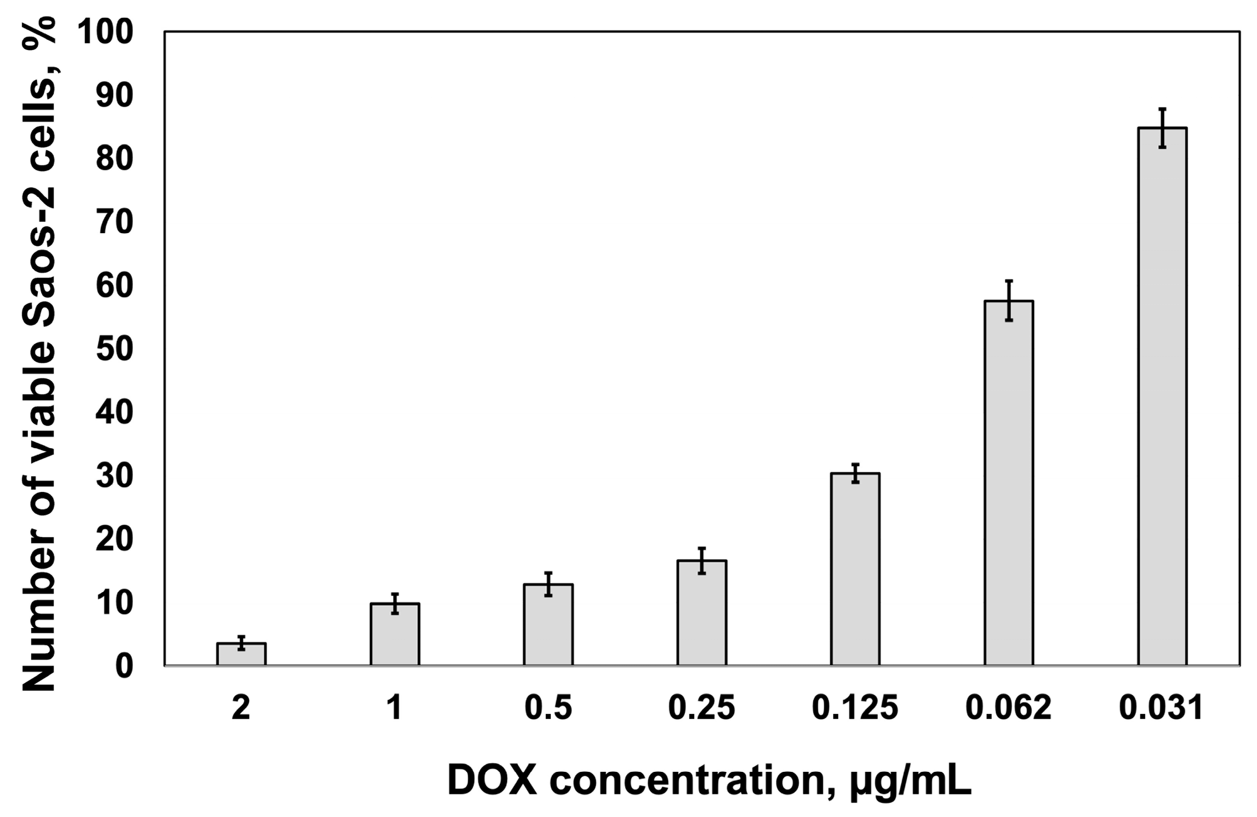
2.1. Cytotoxic Response

The half maximal inhibitory concentration (IC50) values for DOX treatment in the Saos-2 cell line at 48 h of incubation are shown in Figure 1. There was a small difference in IC50 between 48 h (0.060 ± 0.01 μg/mL) and 72 h (0.055 ± 0.003 μg/mL) of incubation with the drug, which is consistent with the results of previous work in Ref. [29]. This observation indicates that Saos-2 cell responses to DOX were relatively stable over the given duration of drug exposure.

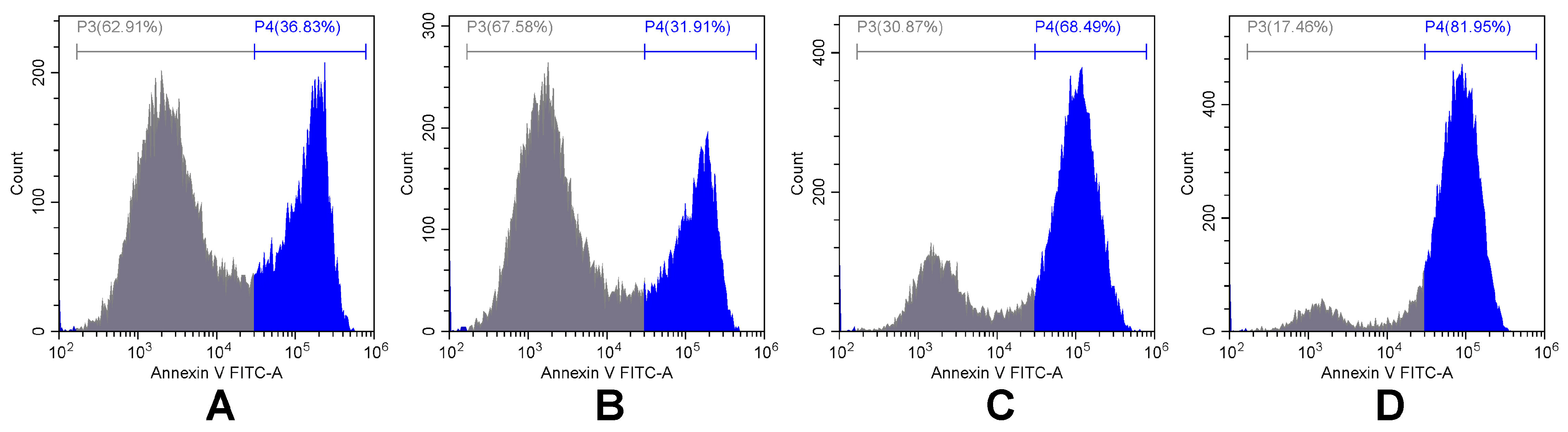
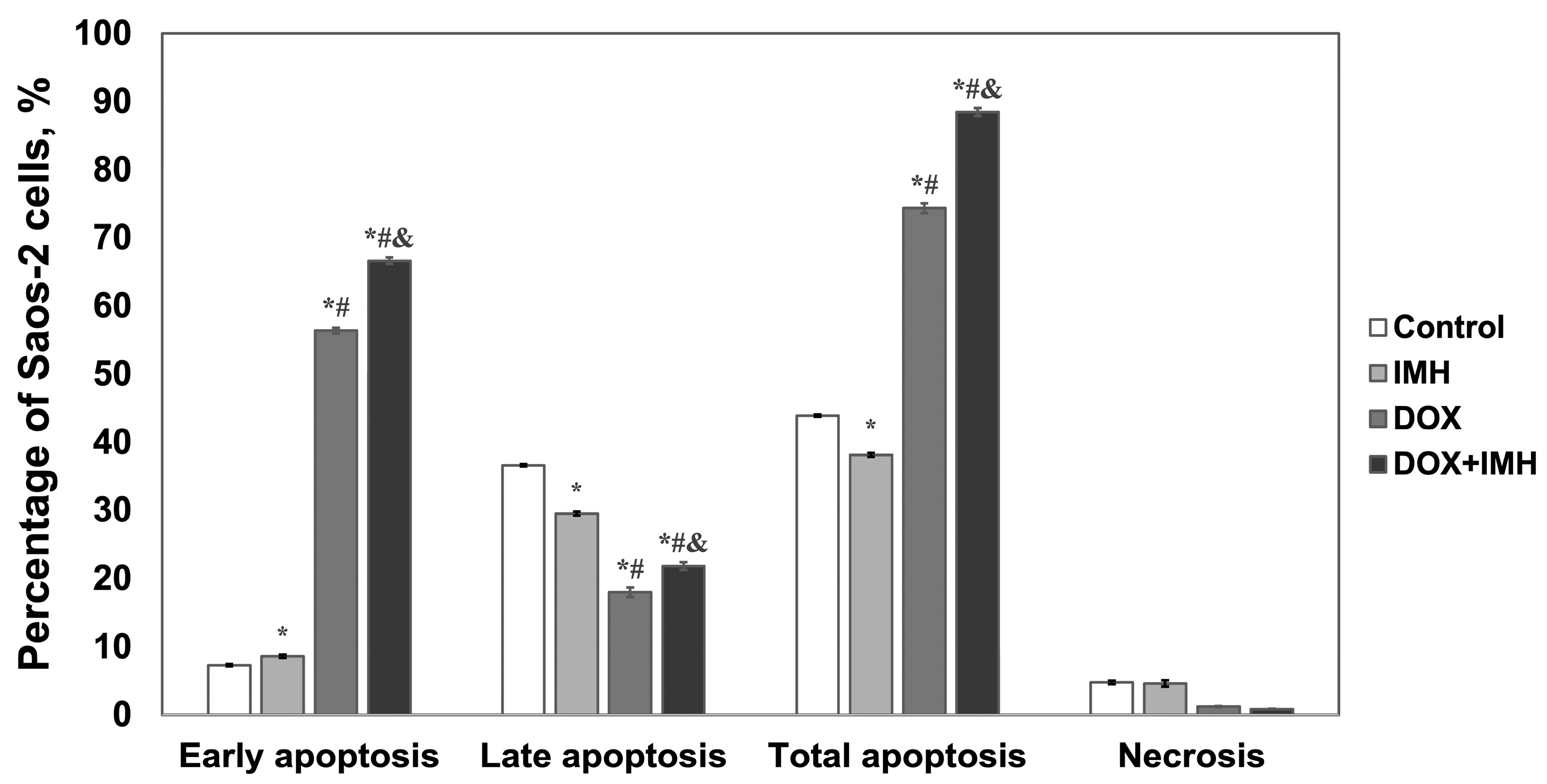
2.2. Apoptosis and Necrosis Detection

In our analysis, we discriminated between early apoptotic, late apoptotic, and necrotic cell populations by gating them in four regions on propidium iodide conjugated with energy-coupled dye (PI-ECD) versus annexin V conjugated with fluorescein (annexin V-FITC) dot plots, as described in our previous work [28]. Cell debris and doublets were excluded from the analysis. Treatment-induced changes in Saos-2 cell death profiles are illustrated in representative histograms comparing the distribution of annexin V-FITC (Figure 2) and quantified as the percentages of cells undergoing apoptosis and necrosis (Figure 3) at 48 h after exposure to no treatment, IMH, DOX, or DOX + IMH.
Exposure to DOX alone and DOX + IMH resulted in a 7.7- and 9.1-fold increase in the percentage of early apoptotic Saos-2 cells compared with control cells given no treatment. The percentage of cells undergoing early apoptosis was 15% higher in response to IMH than in the control group. By comparing the effects of DOX and DOX + IMH, we found that the exposure of cells to IMH induced an additional 15% increase in the early apoptotic fraction compared to DOX treatment alone. A lower percentage of Saos-2 cells in the late apoptosis stage was likely observed due to competition between DNA intercalation of DOX and propidium iodide. Nonetheless, the combined action of DOX with IMH led to the highest fraction of cells undergoing apoptosis in total (p < 0.05). There was no significant difference in the percentage of necrotic Saos-2 cells between the groups.
We next sought to study the effects of DOX and IMH on ROS generation involved in apoptosis and necrosis [30].

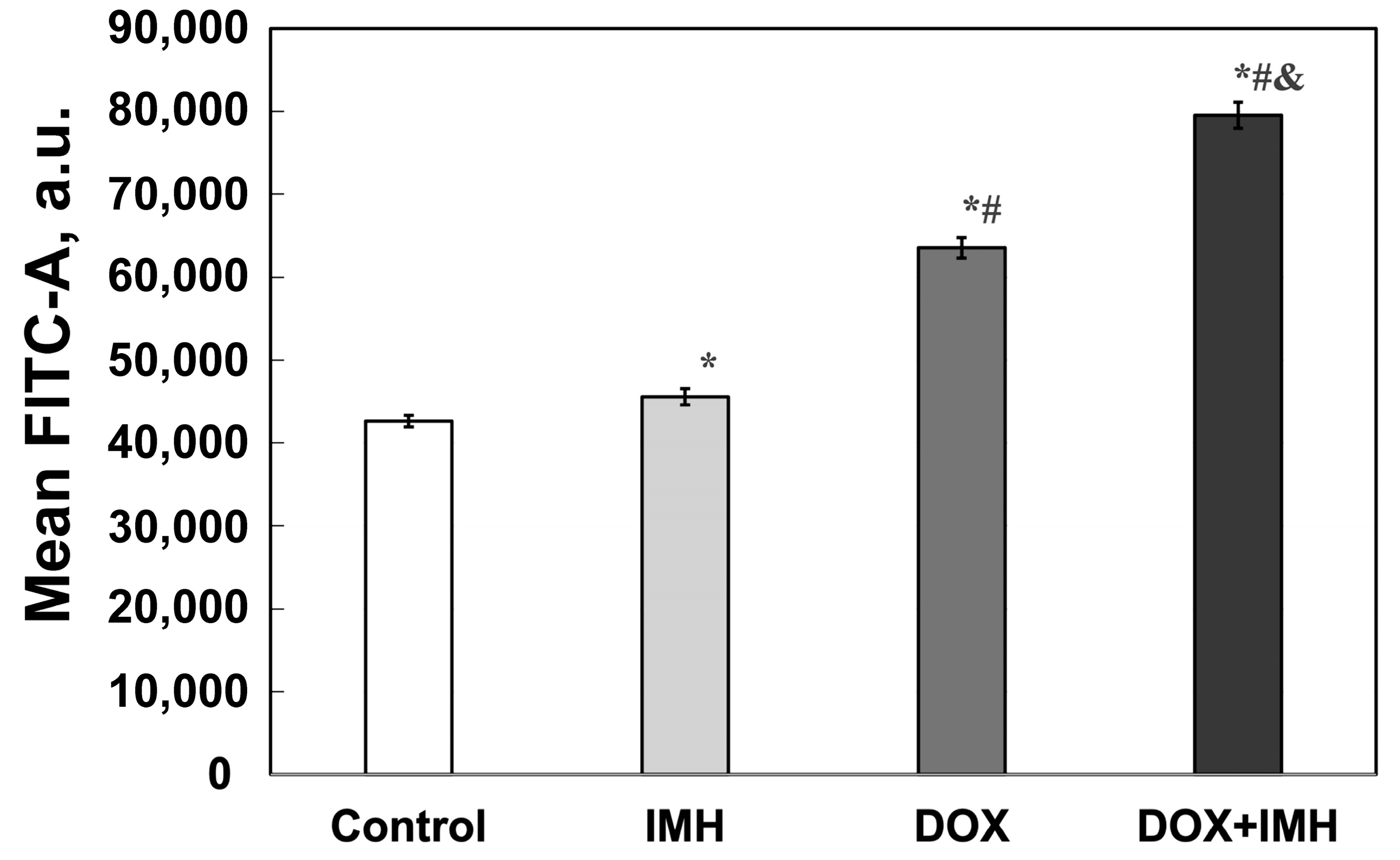
2.3. Reactive Oxygen Species Measurements

As shown in Figure 4, either IMH or DOX action alone increased the level of ROS produced in Saos-2 cells by 7% and 33% compared with the control group. The combination treatment with DOX and IMH, however, led to a 47% elevation in ROS level in comparison with untreated cells, a 20% rise compared with DOX alone and a 43% increase compared with IMH exposure (p < 0.05). It is known that ROS are involved in numerous signal transduction pathways in cancer cells, including p14ARF tumor suppressor and EGFR [31,32].

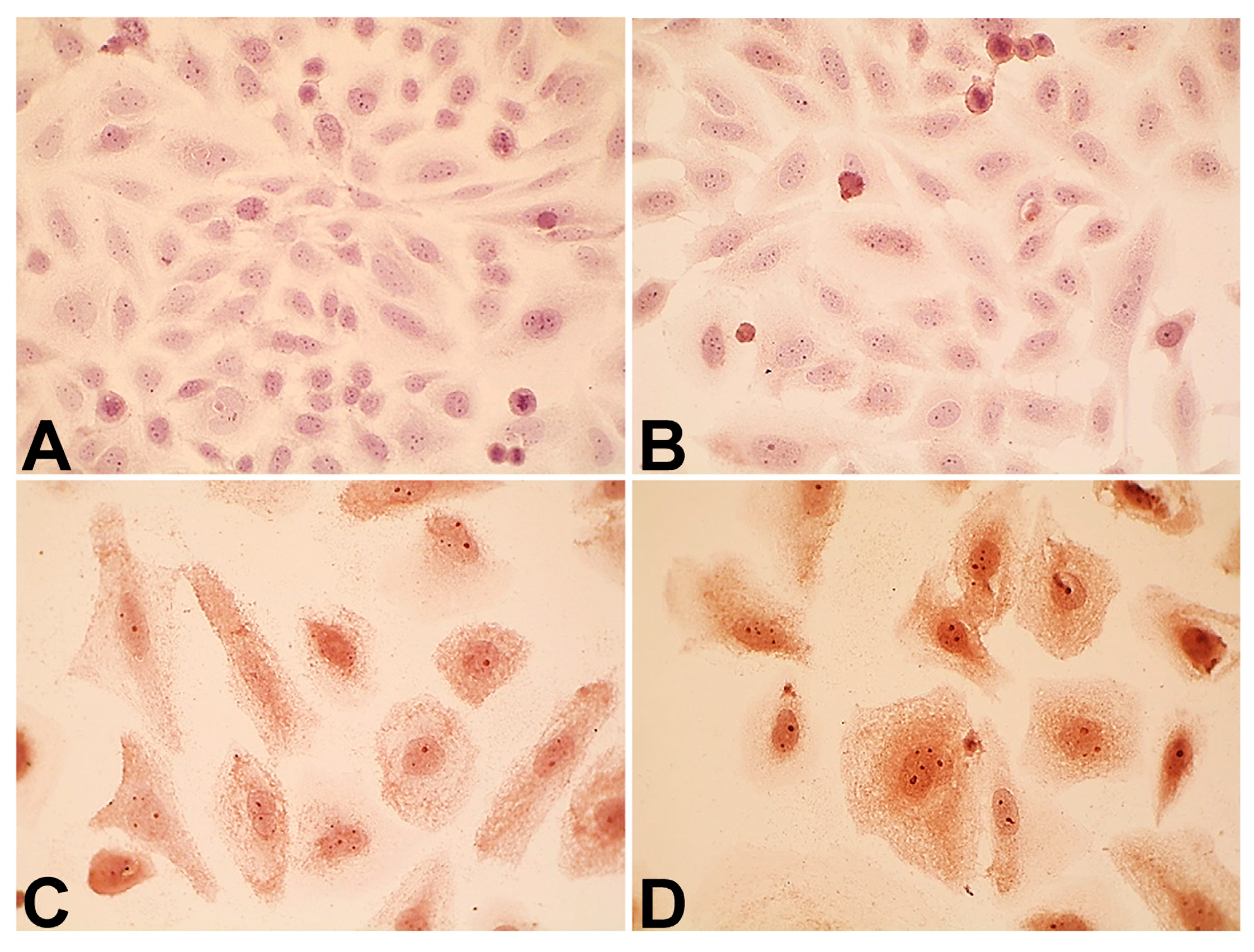
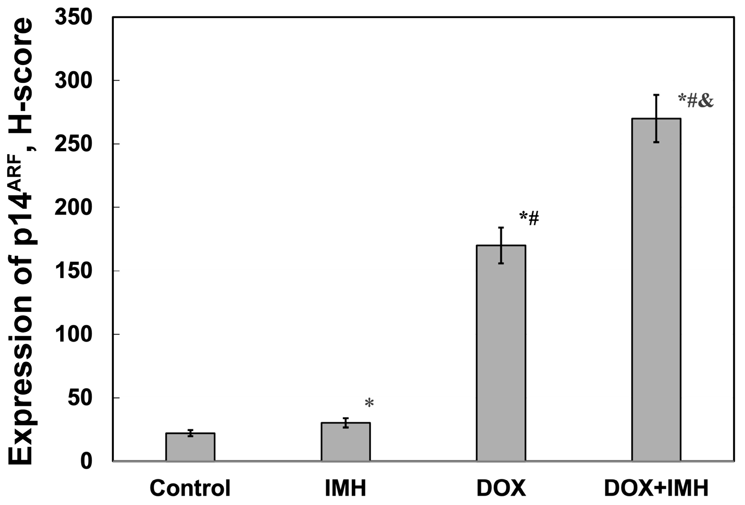
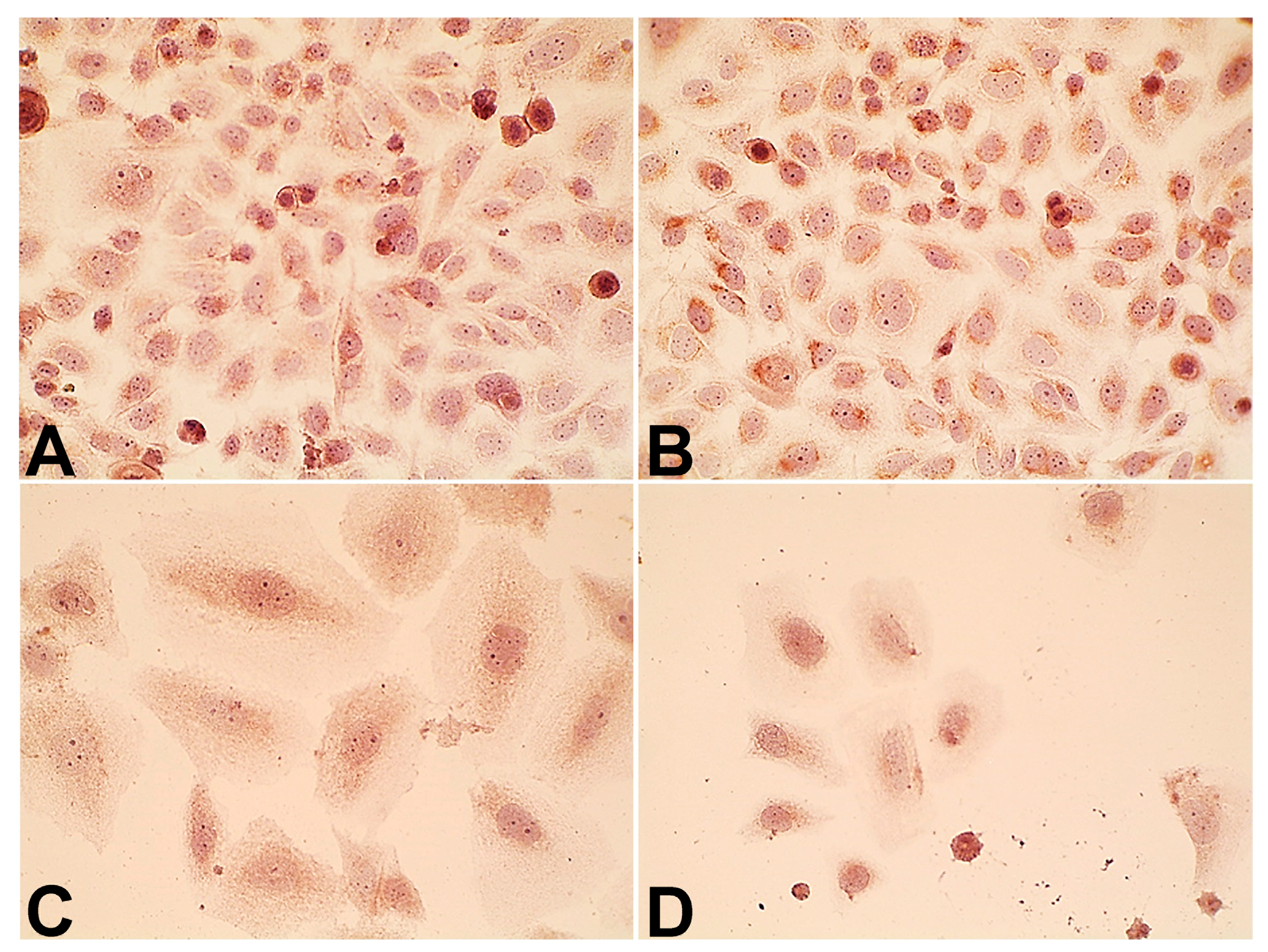
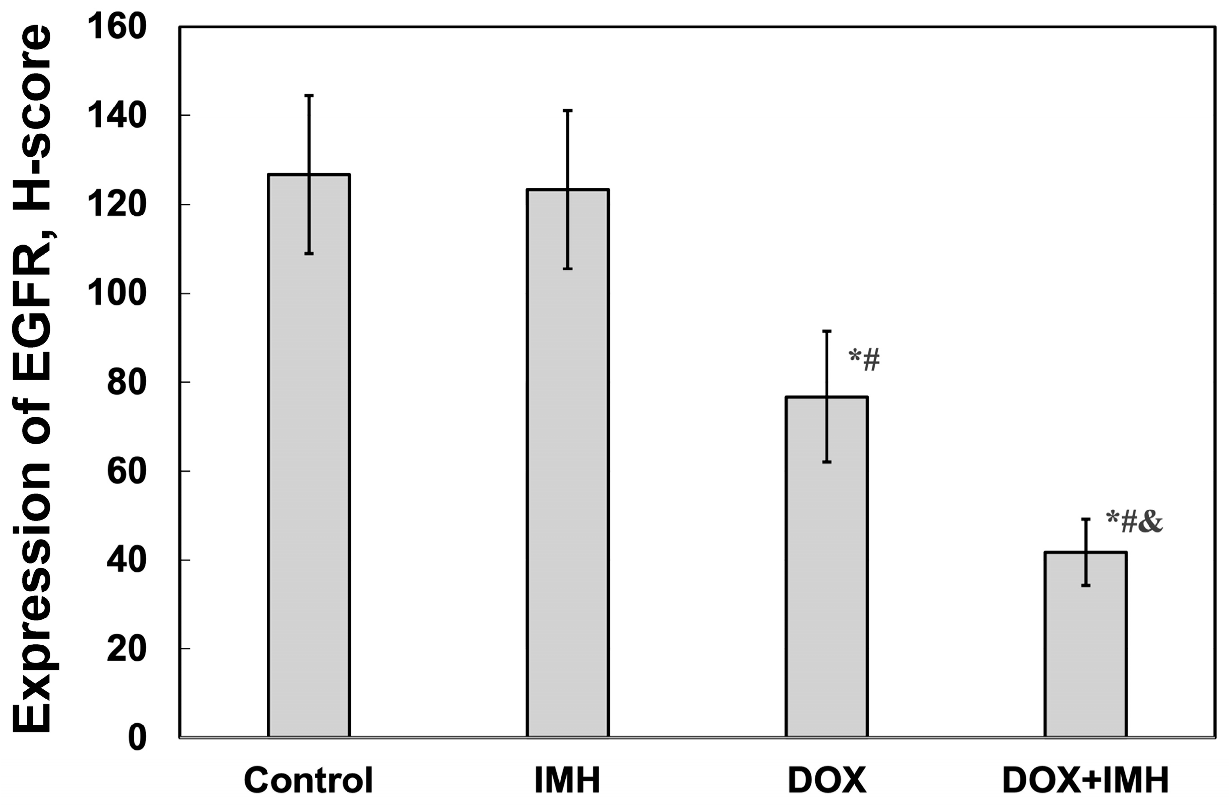
2.4. Expression of p14ARF and Epidermal Growth Factor Receptor

The effects of either DOX or IMH alone and DOX combination with IMH on p14ARF and EGFR expression were compared
Saos-2 cells in the control group (Figure 5 and Figure 6). DOX combination with IMH further increased the level of p14ARF expression by 92% and 37% compared with the untreated cells to study the molecular mechanisms by which electromagnetic fields may predispose Saos-2 cells to apoptosis and necrosis. Prior studies have provided evidence that the tumor suppressor protein p14ARF mediates apoptosis in OS cell lines lacking functional p53, namely, Saos-2 cells [33]. Despite their mesenchymal origin, Saos-2 cells express EGFR on the cytoplasmic membrane surface, where the receptor serves as a signal transducer to activate molecular pathways, leading to DOX resistance and cell proliferation [34,35].
IMH caused a 27% increase in the level of p14ARF, whereas DOX displayed an 87% higher level of tumor suppressor than and DOX alone, respectively (p < 0.05).
We did not observe a significant difference in the level of EGFR between Saos-2 cells subjected to no treatment and IMH. However, DOX exposure decreased the EGFR level by 40% compared with the control group (Figure 7 and Figure 8). The exposure of Saos-2 cells to DOX and IMH combination resulted in a 67% and 32% decrease in the EGFR level compared with control and DOX-treated cells, respectively (p < 0.05). These findings suggest that DOX and IMH have a synergistic effect on p14ARF and EGFR expression in Saos-2 cells.
Figure 9 shows the results of spatial autocorrelation analysis based on Moran’s index (Moran’s I) calculated for p14ARF- and EGFR-stained Saos-2 cell immunocytochemistry images. DOX and IMH combination resulted in the highest value of Moran’s I, indicating the lowest degree of spatial heterogeneity for p14ARF and EGFR distributions compared with no treatment, IMH, and DOX alone. In addition, exposure to IMH also led to a statistically significant increase in Moran’s I compared with the control group (p < 0.05).

3. Discussion

Improving cure rates in OS patients still represents a significant challenge. DOX is one of the most useful chemotherapeutic drugs in neoadjuvant and adjuvant treatment regimens [36]. Tissue hypoxia is a major source of chemo- and radiotherapy resistance of bone tumors. While bone is considered a highly vascularized connective tissue, with increased oxygen demand during growth and repair, its perfusion shows a heterogeneous pattern, with partial oxygen pressure ranging from 50 mm Hg (normoxic region) in the periosteum to 9.9 mm Hg (hypoxic region) in the extravascular bone marrow compartment [37]. The bone microenvironment is hypoxic given the low oxygen tension in the sinusoids, high oxygen consumption by haemopoietic cells, and increased resistance to oxygen diffusion in the bone matrix [38,39]. ROS can influence the relationship between osteoblasts and osteoclasts, leading to decreased mineralization, increased osteoblast apoptosis, and increased bone resorption under hypoxic conditions [40]. Among other osteoblast-like features, Saos-2 cells are capable of secreting extracellular matrix, which makes it possible to use them as an in vitro model of OS. Saos-2 cells were found to reach a stage similar to mature human osteocytes in response to different oxygen concentrations [41]. Furthermore, prior work has already established that non-ionizing electromagnetic fields enhance the antitumor activity of DOX in cancer cells by inducing changes in the respiratory chain of mitochondria and subsequent ROS-mediated cell damage [42]. In the present study, we exposed Saos-2 cells to radiofrequency electromagnetic fields in order to examine the effects of IMH and free DOX on cell viability, ROS generation, and p14ARF and EGFR expression.
There are two primary mechanisms by which DOX combined with IMH could initiate a more pronounced antitumor effect in Saos-2 cells than DOX alone: (1) local temperature increase, and (2) a change in electron transfer processes and spin dynamics of radical pairs. While DOX undergoes redox cycling and acts as a pro-oxidant, IMH facilitates DOX-induced ROS generation, contributing to enhanced oxidative stress in cancer cells. This synergistic action increases mitochondrial membrane permeability, leading to the release of cytochrome c and activation of intrinsic apoptotic pathways. At the same time, elevated ROS levels can irreversibly damage lipids, proteins, and nucleic acids, disrupting redox-dependent signaling pathways in the cells [43,44,45]. In the following paragraphs, we discuss the relevance of our results to IMH effects on DOX-induced ROS generation and p14ARF and EGFR expression, as well as Saos-2 cell apoptosis. We also compare the obtained results with our previous investigation focused on the application of IMH to potentiate the action of liposomal DOX.
Encapsulating DOX in liposomes increased the IC50 in Saos-2 cells from 0.06 ± 0.01 μg/mL (free DOX) to 1.8 ± 0.2 μg/mL (liposomal DOX) at 48 h incubation. From 48 to 72 h, the IC50 values for liposomal DOX showed a 4.7-fold decrease (p < 0.05) [28], while there was no significant difference between cells subjected to free DOX. Likewise, previous studies noted a more pronounced cytotoxic effect of free DOX than its liposomal formulation [46]. The DOX + IMH combination resulted in a significantly larger fraction of apoptotic cells (Figure 3) and a higher level of ROS (Figure 4) produced in Saos-2 cells than either DOX or IMH alone, as determined by using flow cytometry (p < 0.05). IMH and DOX initiated apoptosis as a prevalent form of cell death rather than necrosis. These findings are consistent with those reported for liposomal DOX and IMH. Together with the work described above, our results demonstrate that free DOX combined with IMH led to a 19% increase in Saos-2 cells undergoing early apoptosis and a 29% elevation in ROS level compared with the combination treatment based on liposomal DOX (p < 0.05).
As is well known, DOX causes mitochondrial iron overload through redox reactions between Fe²⁺ and Fe³⁺ ions, wherein ROS arise as byproducts of the Fenton and Haber–Weiss reactions. Iron homeostasis appears to play an important role in tumor cells, contributing to redox signaling, ROS generation, oxidative stress, cell damage, and death [47]. This is one of the mechanisms through which DOX inhibits cancer cell proliferation in addition to DNA intercalation and topoisomerase-2A inhibition [48]. Since iron ions have unpaired electrons in their outer shell, they exhibit magnetic properties, allowing external electromagnetic fields to interact with them and induce both thermal and non-thermal effects. The absorption of electromagnetic energy in biological objects is associated with local temperature rise—for instance, due to viscous resistance to rotating magnetic particles under the influence of an applied field (Brownian relaxation). Note that temperatures above 42 °C are generally avoided during IMH to minimize the risk of side effects associated with chemotherapy resistance mediated through heat-shock protein expression, tumor vascular shutdown, and heat pain thresholds. On the other hand, non-thermal effects of electromagnetic fields, such as magnetochemical and magneto-mechanical effects, are based on transitions between different electronic spin states of the radical pair and the magnetic force exerted on magnetic particles in response to the applied field, respectively. The conversion of the triplet state to the singlet state increases the probability of free radical recombination and may also promote ROS formation [21,22,47,49,50].
In contrast to free DOX, liposomal formulations are composed of an aqueous core with DOX enclosed within a phospholipid bilayer containing unsaturated lipids [51]. It becomes immediately apparent that unsaturated linkages in these lipids are susceptible to peroxidation initiated by ROS. More importantly, lipid peroxidation can alter the anticancer activity of DOX released in the tumor and its microenvironment. The proposed mechanism for free and liposomal DOX involvement in free radical reactions is presented in Equations (1)–(8) based on Refs. [28,42,46,47,48,51,52,53]. As shown in Equation (1), DOX engages in redox cycling between Fe2+ and Fe3+, leading to hydroxyl radical (OH) formation. DOX can catalyze the transfer of electrons from Fe2+ to molecular oxygen, giving rise to superoxide anion (O2•−) radical in Equation (2). The conversion of superoxide radical to hydrogen peroxide (H2O2) by the enzyme superoxide dismutase in Equation (3) further contributes to the Fenton reaction. Hydroxyl radical in Equation (4) initiates lipid peroxidation chain reactions in the cell membranes, including Equation (5). Unsaturated fatty acids within the phospholipid (LH) bilayer of DOX-containing liposomes are also susceptible to ROS oxidation, resulting in lipid hydroperoxide (LOOH) formation, shown in Equation (6), which in turn destabilizes the liposome and promotes DOX release. The application of electromagnetic fields to induce moderate heating of Saos-2 cells also influences electron transfer processes in the cells and medium through magnetochemical effects, favoring an increase in DOX semiquinone (DOX•−) radical in Equation (7) and Fe2+ concentration in Equation (8). The latter facilitates the Fenton and Haber–Weiss reactions involved in oxidative stress.
Free DOX:
Fe2+ + H2O2 → Fe3+ + HO + OH
O2 + e → O2•−
2O2•− + 2H+ → H2O2 + O2
RH + OH → R + H2O
R + O2 → RO2
Liposomal DOX:
RO2 + LH → LOOH + R
IMH:
DOX + e   e l e c t r o m a g n e t i c   f i e l d   DOX
Fe 3 + + e   e l e c t r o m a g n e t i c   f i e l d   Fe 2 +
Equations (1)–(8) DOX and IMH effects on ROS formation in cancer cells.
A growing body of evidence suggests that ROS serve a critical role in cell signaling pathways, wherein the signal is transduced across a group of proteins via electron transfer processes between individual protein members, forming tightly regulated redox chains [54]. Clearly, local temperature changes also affect electron transport and redox homeostasis in biological objects. It was previously shown that the Fenton and Haber–Weiss reactions are temperature-dependent [55]. For this reason, we applied only a moderate temperature increase (<42 °C) during IMH to prevent hydrogen peroxide from degrading into oxygen and water. Otherwise, higher temperatures would have limited these reactions.
In particular, we found that DOX and IMH increased p14ARF expression in Saos-2 cells (Figure 5 and Figure 6). The upregulation of p14ARF initiates cell cycle arrest and apoptosis following p53-dependent and independent pathways, both of which enhance the efficacy of DOX [56]. Tumor cell responses to oxidative and heat stresses are also mediated by p14ARF through interactions with heat-shock proteins, namely, Hsp70, leading to β-catenin degradation and subsequent apoptosis [57]. Redox reactions regulate various cellular processes, including activation of membrane receptors and ion transport channels. EGFR is an example of a transmembrane receptor whose activation is also influenced by ROS. OS cells express EGFR, functioning as a prevention of self-destructive mechanisms. Interestingly, it was demonstrated that immunotherapeutic agents targeting EGFR were not useful for generating antitumor effects in OS cells [58]. Another study established that the exposure of tumor cells to radiofrequency electromagnetic radiation decreased EGFR levels and reduced cell viability, possibly through changes in electrostatic interactions and non-covalent binding [59]. As shown in Figure 7 and Figure 8, free DOX and IMH action resulted in the lowest level of EGFR expression in Saos-2 cells.
However, the combination of liposomal DOX and IMH caused the highest level of pro-apoptotic Bax protein among the cells subjected to no treatment, liposomal DOX alone or IMH alone. Based on experiments measuring ROS, we propose that different ROS levels in Saos-2 cells initiate different cell signaling pathways. It should be noted that the exposure of Saos-2 cells to free or liposomal DOX with IMH had the highest Moran’s I values calculated for Bax, p14ARF, and EGFR spatial distributions on immunocytochemistry images (p < 0.05). The higher the Moran’s I value, the lower the heterogeneity of pixel distributions for biomarkers in acquired immunocytochemistry images [60,61,62].
Altogether, enclosing the drug in a phospholipid bilayer alters the uptake and distribution of DOX within cells compared with its free form and initiates a distinct cascade of signaling events. DOX delivery depends on ionization state and membrane binding [63,64]. Combining free DOX or its liposomal formulation with IMH produces changes in the kinetics of free radical chain reactions (Equations (1)–(8)) and ROS diffusion in cells compared with their separate effects. In our earlier study, liposomal DOX induced apoptosis in Saos-2 cells mediated by Bax upregulation, which reflected a response characterized by a lower ROS level. In the current study, free DOX triggered apoptosis through p14ARF upregulation and EGFR downregulation associated with a higher ROS level. These observations are in agreement with the fact that free DOX results in a more pronounced nuclear stress than its liposomal formulation [65]. In addition, we found differences in the spatial heterogeneity of apoptosis-related biomarkers between the two forms of DOX and their combinations with IMH, indicating distinct structural and functional alterations in Saos-2 cell populations. Considering the inherent heterogeneity of cell populations within OS, it might be interesting to evaluate the effects of DOX combined with IMH in a panel of DOX-resistant OS cell lines, such as 143-B-DX-R and Saos-2-DX-R.
Although a wide range of experimental evidence now offers comparisons between free and liposomal DOX combinations with other moderate hyperthermia techniques, far less research has focused on the role of IMH in potentiating DOX cytotoxic activity, particularly in OS models, wherein both the magnetic and the electric components of the applied electromagnetic field contribute to the antitumor effect. A comparative examination of our experimental data, therefore, shows that IMH not only enhances cytotoxic effects initiated by both forms of DOX but also alters the expression and heterogeneity of different cellular responses across Saos-2 cell populations, providing a novel scientific perspective on the inherent heterogeneity of cell populations within osteosarcoma. Moreover, future research is needed to validate our findings in animal models of OS, which will provide a deeper understanding of thermal and nonthermal effects produced by IMH in vivo and potentially accelerate its translation into clinical practice [66,67].

4. Materials and Methods

To ensure consistency in comparisons, this paper follows a similar structure and methodology as our earlier work [28]. In the current study, we focus on the cytotoxic effects and underlying mechanisms of free DOX with IMH. Furthermore, we provide a comparative analysis of free and liposomal DOX under identical conditions.

4.1. Cell Culture

The Saos-2 cell line is an extensively studied culture model of primary bone tumors derived from the bone of an 11-year-old female patient with OS, which displays epithelial morphology and osteoblastic features, including the ability to synthesize alkaline phosphatase and high resistance to DOX [68]. Saos-2 cells (ATCC HTB-85) were obtained from the cell line bank of R.E. Kavetsky Institute of Experimental Pathology, Oncology and Radiobiology, and cultured in Dulbecco’s modified Eagle’s medium (DMEM/F12, Sigma-Aldrich, Taufkirchen, Germany) supplemented with 10% fetal bovine serum (Sigma-Aldrich, Taufkirchen, Germany) and antibiotic-antimycotic (Sigma-Aldrich, St. Louis, MO, USA) under standard conditions of 37 °C in 5% CO2 [69].

4.2. Inductive Moderate Hyperthermia

Saos-2 cells were exposed to non-ionizing electromagnetic radiation using a MagTherm device (Radmir, Kharkiv, Ukraine) operated at a 42 MHz frequency and 15 W of output power for 30 min. Exposure plans were prepared using COMSOL Multiphysics v. 5.6 software (COMSOL AB, Stockholm, Sweden) to ensure optimal distribution of electromagnetic fields and temperature. The software provided an interface to couple the magnetic fields and bioheat transfer modules. As the applied field had two components, we created a treatment plan for the magnetic (Figure 10A) and the electric (Figure 10B) field distributions in response to IMH. COMSOL simulations predicted the maximum values of magnetic induction of 500 µT, electric field strength of 564 V/m, and specific absorption rate of 10.6 W/kg. Additionally, we recorded temperature measurements in the center of the Petri dish using a fiber-optic sensor connected to a digital thermometer TM-4 (Radmir, Ukraine) throughout IMH. Saos-2 cells were gradually heated, reaching a temperature of 41.23 °C. A detailed description of IMH planning and methodology can be found in our earlier work [28].

4.3. Cell Viability

When Saos-2 cells were 90% confluent, they were harvested using trypsin in saline disodium ethylenediaminetetraacetic acid (S-EDTA) and added to each well of a 96-well plate (TPP, Trasadingen, Switzerland) in DMEM/F12 medium supplemented with 10% fetal bovine serum and antibiotic–antimycotic at a concentration of 104 cells per well. Cells were treated with different concentrations of DOX (Ebewe Pharma, Unterach, Austria, Ministry of Health of Ukraine approval registry: LH8929). The drug was initially supplied in a liquid form as a concentrate (2 mg/mL) for the solution for infusion, then diluted in culture medium and incubated under standard conditions (37 °C, 5% CO2) for 48 or 72 h. After incubation with DOX, Saos-2 cells were fixed by adding 10% trichloroacetic acid (Sigma-Aldrich, Taufkirchen, Germany) and stained with sulforhodamine B (Sigma-Aldrich, St. Louis, MO, USA). Excess dye was removed by washing the cells with 1% acetic acid solution (Sigma-Aldrich, Taufkirchen, Germany), and the remaining dye was resolubilized in 10 mM Tris base solution (Sigma-Aldrich, Taufkirchen, Germany) [70]. Sample absorbance at 510 nm was measured by a multi-well spectrophotometer (ThermoLabsystems Multiskan EX, Waltham, MA, USA). IC50 values were calculated by linear regression analysis.
The viable Saos-2 cell number was assessed by trypan blue exclusion assay (Sigma-Aldrich, Taufkirchen, Germany) in response to no treatment (control group), IMH exposure, DOX, and DOX combined with IMH [71]. Cells were seeded in Petri dishes with DMEM/F12, 10% fetal bovine serum, and antibiotic–antimycotic at a concentration of 1.5 × 10⁵ per dish. DOX was added at a concentration of 0.06 μg/mL and then the cells were exposed to IMH for 30 min. Following IMH treatment, the Saos-2 cells were incubated under standard conditions for 48–72 h and counted using a hemocytometer (Micromed, Kyiv, Ukraine).

4.4. Flow Cytometry

After 48 h of incubation, Saos-2 cells were labeled fluorescently to detect apoptotic and necrotic cells by adding annexin V-FITC and PI-ECD (Dojindo, Munich, Germany) [72,73]. A DxFlex flow cytometer (Beckman Coulter, Brea, CA, USA) was used to examine the resulting samples, analyzing at least 21,000 events for each sample. Data collection and analysis were conducted with the CytExpert v. 2.5 software supplied with the cytometer. The levels of ROS were measured by a 2′,7′-dichlorodihydrofluorescein diacetate (DCFH-DA, Dojindo, Germany) assay according to the methodology described in Ref. [74].

4.5. Immunocytochemical Assay

After 72 h of incubation, Saos-2 cells were fixed in methanol (Sigma-Aldrich, USA) and acetone (Chimreserv, Kyiv, Ukraine). Next, primary antibodies recognizing EGFR (clone 111.6, Thermo Scientific, Waltham, MA, USA) and tumor suppressor p14ARF (clone 14P02, Thermo Scientific) were added for 1 h at room temperature [75,76]. Immunodetection was performed using a Super Picture polymer detection kit (Thermo Fisher Scientific) with hematoxylin counterstain. Immunocytochemistry results for these two biomarkers were assessed using the H-score [77].

4.6. Image Analysis

Saos-2 cells were segmented in collected immunocytochemistry images by adopting the k-means clustering approach using ImageJ v. 1.53k software (NIH, Bethesda, MD, USA). We then calculated Moran’s I to quantify changes in the spatial heterogeneity of intracellular p14ARF and EGFR distributions using Autocorrelation v. 1.0 software (NCI, Kyiv, Ukraine) [78,79].

4.7. Statistical Analysis

Statistical differences were determined using Statistica v. 6.0 (Statsoft Inc., Tulsa, OK, USA) and IBM SPSS Statistics v. 25.0 (IBM Inc., Chicago, IL, USA) software. Two groups were compared using the Student’s t-test or the Mann–Whitney U-test. Experimental data between three or more groups were compared using a one-way analysis of variance (ANOVA) followed by the Games–Howell post hoc test or the Kruskal–Wallis test. A p-value < 0.05 was taken as statistically significant.

5. Conclusions

The objective of this study was to examine the antitumor effect of free DOX combined with IMH on Saos-2 human OS cells, with a particular focus on the interaction of a non-ionizing electromagnetic field with a chemotherapeutic agent acting through the free radical mechanism. In addition, we compared the obtained results with our previously published data on the use of liposomal DOX with IMH.
Here, we show that free DOX combination with IMH caused a 19% increase in Saos-2 cells undergoing early apoptosis and a 29% elevation in ROS compared with the results observed for liposomal DOX with IMH. Using DOX in combination with IMH also resulted in the highest expression of p14ARF tumor suppressor and the lowest expression of EGFR. Immunocytochemistry image analysis revealed the lowest values of Moran’s I for both p14ARF and EGFR, indicating a less heterogeneous pattern of spatial distribution (p < 0.05).
In summary, our findings demonstrate that IMH can enhance the antitumor activity of DOX in Saos-2 OS cells, providing experimental data on the more potent effects of IMH combination with free DOX rather than its liposomal formulation. Future preclinical studies are required to compare the efficacy of IMH with free and liposomal DOX in vivo to further support the application of non-ionizing electromagnetic radiation benefits to OS treatment outcomes.

Author Contributions

Conceptualization, V.E.O. and V.B.O.; methodology, V.E.O., A.G.D., V.V.O., S.A.L., I.O.T., O.V.M., D.L.K., V.B.O. and A.Y.G.; software, O.Y.R., V.B.O. and A.B.P.; validation, A.G.D., V.V.O., S.A.L., I.O.T., O.V.M., A.Y.G. and O.Y.D.; formal analysis, V.E.O. and V.B.O.; investigation, V.E.O., D.L.K., O.Y.D. and V.B.O.; resources, V.E.O., A.G.D., V.V.O., S.A.L., I.O.T. and O.V.M.; data curation, O.Y.D. and V.B.O.; writing—original draft preparation, V.E.O. and V.B.O.; writing—review and editing, V.B.O., V.E.O. and A.B.P.; visualization, V.B.O., O.Y.R. and A.B.P.; supervision, V.E.O. and A.G.D.; project administration, V.E.O. and A.G.D.; funding acquisition, V.E.O., A.G.D., V.V.O., S.A.L., I.O.T. and O.V.M. All authors have read and agreed to the published version of the manuscript.

Funding

This research was partially funded by the Ministry of Health of Ukraine to develop a method of antitumor therapy of primary malignant bone tumors based on magnetochemical technology using nanocomplexes (code BH.14.01.07.204-23, registration number 0123U100711).

Institutional Review Board Statement

Not applicable.

Informed Consent Statement

Not applicable.

Data Availability Statement

Data is contained in the paper.

Conflicts of Interest

The authors declare no conflicts of interest.

Abbreviations

The following abbreviations are used in this manuscript:
Annexin V-FITCAnnexin V conjugated with fluorescein
DOXDoxorubicin
EGFREpidermal growth factor receptor
FSC-A/SSC-AForward/side scatter area
IC50Half maximal inhibitory concentration
IMHInductive moderate hyperthermia
OSOsteosarcoma
PI-ECDPropidium iodide conjugated with energy-coupled dye
ROSReactive oxygen species

References

  1. Beird, H.C.; Bielack, S.S.; Flanagan, A.M.; Gill, J.; Heymann, D.; Janeway, K.A.; Livingston, J.A.; Roberts, R.D.; Strauss, S.J.; Gorlick, R. Osteosarcoma. Nat. Rev. Dis. Primers 2022, 8, 77. [Google Scholar] [CrossRef] [PubMed]
  2. Misaghi, A.; Goldin, A.; Awad, M.; Kulidjian, A.A. Osteosarcoma: A Comprehensive Review. SICOT J. 2018, 4, 12. [Google Scholar] [CrossRef]
  3. Picci, P. Osteosarcoma (Osteogenic Sarcoma). Orphanet J. Rare Dis. 2007, 2, 6. [Google Scholar] [CrossRef] [PubMed]
  4. Thorn, C.F.; Oshiro, C.; Marsh, S.; Hernandez-Boussard, T.; McLeod, H.; Klein, T.E.; Altman, R.B. Doxorubicin Pathways: Pharmacodynamics and Adverse Effects. Pharmacogenet. Genom. 2011, 21, 440–446. [Google Scholar] [CrossRef] [PubMed]
  5. Kwok, J.C.; Richardson, D.R. Unexpected Anthracycline-Mediated Alterations in Iron-Regulatory Protein-RNA-Binding Activity: The Iron and Copper Complexes of Anthracyclines Decrease RNA-Binding Activity. Mol. Pharmacol. 2002, 62, 888–900. [Google Scholar] [CrossRef]
  6. Minotti, G.; Ronchi, R.; Salvatorelli, E.; Menna, P.; Cairo, G. Doxorubicin Irreversibly Inactivates Iron Regulatory Proteins 1 and 2 in Cardiomyocytes: Evidence for Distinct Metabolic Pathways and Implications for Iron-Mediated Cardiotoxicity of Antitumor Therapy. Cancer Res. 2001, 61, 8422–8428. [Google Scholar]
  7. de Almeida, A.J.P.O.; de Oliveira, J.C.P.L.; da Silva Pontes, L.V.; de Souza Júnior, J.F.; Gonçalves, T.A.F.; Dantas, S.H.; de Almeida Feitosa, M.S.; Silva, A.O.; de Medeiros, I.A. ROS: Basic Concepts, Sources, Cellular Signaling, and Its Implications in Aging Pathways. Oxid. Med. Cell. Longev. 2022, 2022, 1225578. [Google Scholar] [CrossRef]
  8. Wallrabenstein, T.; Daetwyler, E.; Oseledchyk, A.; Rochlitz, C.; Vetter, M. Pegylated Liposomal Doxorubicin (PLD) in Daily Practice-A Single Center Experience of Treatment with PLD in Patients with Comorbidities and Older Patients with Metastatic Breast Cancer. Cancer Med. 2023, 12, 13388–13396. [Google Scholar] [CrossRef]
  9. Waterhouse, D.N.; Tardi, P.G.; Mayer, L.D.; Bally, M.B. A Comparison of Liposomal Formulations of Doxorubicin with Drug Administered in Free Form: Changing Toxicity Profiles. Drug Saf. 2001, 24, 903–920. [Google Scholar] [CrossRef]
  10. Barnes, F.; Greenebaum, B. Role of Radical Pairs and Feedback in Weak Radio Frequency Field Effects on Biological Systems. Environ. Res. 2018, 163, 165–170. [Google Scholar] [CrossRef]
  11. Loboda, A.; Smolanka Sr, I.; Orel, V.E.; Syvak, L.; Golovko, T.; Dosenko, I.; Lyashenko, A.; Smolanka, I.; Dasyukevich, O.; Tarasenko, T.; et al. Efficacy of Combination Neoadjuvant Chemotherapy and Regional Inductive Moderate Hyperthermia in the Treatment of Patients With Locally Advanced Breast Cancer. Technol. Cancer Res. Treat. 2020, 19, 1533033820963599. [Google Scholar] [CrossRef] [PubMed]
  12. Consales, C.; Merla, C.; Marino, C.; Benassi, B. Electromagnetic Fields, Oxidative Stress, and Neurodegeneration. Int. J. Cell Biol. 2012, 2012, 683897. [Google Scholar] [CrossRef] [PubMed]
  13. Orel, V.; Shevchenko, A.; Romanov, A.; Tselepi, M.; Mitrelias, T.; Barnes, C.H.W.; Burlaka, A.; Lukin, S.; Shchepotin, I. Magnetic Properties and Antitumor Effect of Nanocomplexes of Iron Oxide and Doxorubicin. Nanomed. Nanotechnol. Biol. Med. 2015, 11, 47–55. [Google Scholar] [CrossRef]
  14. Wong, S.Y.; Benjamin, P.; Hore, P.J. Magnetic Field Effects on Radical Pair Reactions: Estimation of B1/2 for Flavin-Tryptophan Radical Pairs in Cryptochromes. Phys. Chem. Chem. Phys. 2023, 25, 975–982. [Google Scholar] [CrossRef]
  15. Zadeh-Haghighi, H.; Simon, C. Magnetic Field Effects in Biology from the Perspective of the Radical Pair Mechanism. J. R. Soc. Interface 2022, 19, 20220325. [Google Scholar] [CrossRef]
  16. Buchachenko, A.L.; Kuznetsov, D.A. Magnetic Field Affects Enzymatic ATP Synthesis. J. Am. Chem. Soc. 2008, 130, 12868–12869. [Google Scholar] [CrossRef] [PubMed]
  17. Piszczek, P.; Wójcik-Piotrowicz, K.; Guzdek, P.; Gil, K.; Kaszuba-Zwoińska, J. Protein Expression Changes during Phagocytosis Influenced by Low-Frequency Electromagnetic Field Exposure. Int. J. Biol. Macromol. 2022, 217, 481–491. [Google Scholar] [CrossRef]
  18. Ferdous, M.S.; Koupaie, E.H.; Eskicioglu, C.; Johnson, T. An Experimental 13.56 MHz Radio Frequency Heating System for Efficient Thermal Pretreatment of Wastewater Sludge. Prog. Electromagn. Res. B 2017, 79, 83–101. [Google Scholar] [CrossRef]
  19. Li, Z.; Deng, J.; Sun, J.; Ma, Y. Hyperthermia Targeting the Tumor Microenvironment Facilitates Immune Checkpoint Inhibitors. Front. Immunol. 2020, 11, 595207. [Google Scholar] [CrossRef]
  20. Brummelhuis, I.S.G.; Crezee, J.; Witjes, J.A. Evaluation of Thermal Dose Effect in Radiofrequency-Induced Hyperthermia with Intravesical Chemotherapy for Nonmuscle Invasive Bladder Cancer. Int. J. Hyperth. 2023, 40, 2157498. [Google Scholar] [CrossRef]
  21. Wust, P.; Veltsista, P.D.; Oberacker, E.; Yavvari, P.; Walther, W.; Bengtsson, O.; Sterner-Kock, A.; Weinhart, M.; Heyd, F.; Grabowski, P.; et al. Radiofrequency Electromagnetic Fields Cause Non-Temperature-Induced Physical and Biological Effects in Cancer Cells. Cancers 2022, 14, 5349. [Google Scholar] [CrossRef] [PubMed]
  22. Orel, V.B.; Papazoglou, A.S.; Tsagkaris, C.; Moysidis, D.V.; Papadakos, S.; Galkin, O.Y.; Orel, V.E.; Syvak, L.A. Nanotherapy Based on Magneto-Mechanochemical Modulation of Tumor Redox State. WIREs Nanomed. Nanobiotechnol. 2023, 15, e1868. [Google Scholar] [CrossRef]
  23. Nizhelska, O.I.; Marynchenko, L.V.; Piasetskyi, V.I. Biological risks of using non-thermal non-ionizing electromagnetic fields. Innov. Biosyst. Bioeng. 2020, 4, 95–109. [Google Scholar] [CrossRef]
  24. Issels, R.D.; Lindner, L.H.; Verweij, J.; Wessalowski, R.; Reichardt, P.; Wust, P.; Ghadjar, P.; Hohenberger, P.; Angele, M.; Salat, C.; et al. Effect of Neoadjuvant Chemotherapy Plus Regional Hyperthermia on Long-Term Outcomes Among Patients With Localized High-Risk Soft Tissue Sarcoma: The EORTC 62961-ESHO 95 Randomized Clinical Trial. JAMA Oncol. 2018, 4, 483–492. [Google Scholar] [CrossRef]
  25. Roussakow, S. Neo-Adjuvant Chemotherapy Alone or with Regional Hyperthermia for Soft-Tissue Sarcoma. Lancet Oncol. 2017, 18, e629. [Google Scholar] [CrossRef] [PubMed]
  26. Gao, J.; Wang, J.; Jin, Y.; Zhang, F.; Yang, X. Intratumoral Radiofrequency Hyperthermia-Enhanced Chemotherapy of Liposomal Doxorubicin on Hepatocellular Carcinoma. Am. J. Transl. Res. 2018, 10, 3619–3627. [Google Scholar]
  27. Salvador, D.; Bastos, V.; Oliveira, H. Hyperthermia Enhances Doxorubicin Therapeutic Efficacy against A375 and MNT-1 Melanoma Cells. Int. J. Mol. Sci. 2022, 23, 35. [Google Scholar] [CrossRef]
  28. Orel, V.E.; Diedkov, A.G.; Ostafiichuk, V.V.; Lykhova, O.O.; Kolesnyk, D.L.; Orel, V.B.; Dasyukevich, O.Y.; Rykhalskyi, O.Y.; Diedkov, S.A.; Prosvietova, A.B. Combination Treatment with Liposomal Doxorubicin and Inductive Moderate Hyperthermia for Sarcoma Saos-2 Cells. Pharmaceuticals 2024, 17, 133. [Google Scholar] [CrossRef]
  29. Kullenberg, F.; Degerstedt, O.; Calitz, C.; Pavlović, N.; Balgoma, D.; Gråsjö, J.; Sjögren, E.; Hedeland, M.; Heindryckx, F.; Lennernäs, H. In Vitro Cell Toxicity and Intracellular Uptake of Doxorubicin Exposed as a Solution or Liposomes: Implications for Treatment of Hepatocellular Carcinoma. Cells 2021, 10, 1717. [Google Scholar] [CrossRef]
  30. Wang, S.; Konorev, E.A.; Kotamraju, S.; Joseph, J.; Kalivendi, S.; Kalyanaraman, B. Doxorubicin Induces Apoptosis in Normal and Tumor Cells via Distinctly Different Mechanisms. Intermediacy of H(2)O(2)- and P53-Dependent Pathways. J. Biol. Chem. 2004, 279, 25535–25543. [Google Scholar] [CrossRef]
  31. Saito, K.; Iioka, H.; Kojima, C.; Ogawa, M.; Kondo, E. Peptide-based Tumor Inhibitor Encoding Mitochondrial P14ARF Is Highly Efficacious to Diverse Tumors. Cancer Sci. 2016, 107, 1290–1301. [Google Scholar] [CrossRef] [PubMed]
  32. Weng, M.-S.; Chang, J.-H.; Hung, W.-Y.; Yang, Y.-C.; Chien, M.-H. The Interplay of Reactive Oxygen Species and the Epidermal Growth Factor Receptor in Tumor Progression and Drug Resistance. J. Exp. Clin. Cancer Res. 2018, 37, 61. [Google Scholar] [CrossRef] [PubMed]
  33. Mason, S.L.; Loughran, Ö.; La Thangue, N.B. p14ARF Regulates E2F Activity. Oncogene 2002, 21, 4220–4230. [Google Scholar] [CrossRef] [PubMed]
  34. Hughes, D.P.M.; Thomas, D.G.; Giordano, T.J.; Baker, L.H.; McDonagh, K.T. Cell Surface Expression of Epidermal Growth Factor Receptor and Her-2 with Nuclear Expression of Her-4 in Primary Osteosarcoma. Cancer Res. 2004, 64, 2047–2053. [Google Scholar] [CrossRef]
  35. Sevelda, F.; Mayr, L.; Kubista, B.; Lötsch, D.; van Schoonhoven, S.; Windhager, R.; Pirker, C.; Micksche, M.; Berger, W. EGFR Is Not a Major Driver for Osteosarcoma Cell Growth in Vitro but Contributes to Starvation and Chemotherapy Resistance. J. Exp. Clin. Cancer Res. 2015, 34, 134. [Google Scholar] [CrossRef] [PubMed]
  36. Damerell, V.; Pepper, M.S.; Prince, S. Molecular Mechanisms Underpinning Sarcomas and Implications for Current and Future Therapy. Sig. Transduct. Target. Ther. 2021, 6, 246. [Google Scholar] [CrossRef]
  37. Lafage-Proust, M.-H.; Roche, B.; Langer, M.; Cleret, D.; Vanden Bossche, A.; Olivier, T.; Vico, L. Assessment of Bone Vascularization and Its Role in Bone Remodeling. Bonekey Rep. 2015, 4, 662. [Google Scholar] [CrossRef] [PubMed]
  38. Wu, C.; Rankin, E.B.; Castellini, L.; Fernandez-Alcudia, J.; Lagory, E.L.; Andersen, R.; Rhodes, S.D.; Wilson, T.L.S.; Mohammad, K.S.; Castillo, A.B.; et al. Oxygen-Sensing PHDs Regulate Bone Homeostasis through the Modulation of Osteoprotegerin. Genes. Dev. 2015, 29, 817–831. [Google Scholar] [CrossRef]
  39. Zahm, A.M.; Bucaro, M.A.; Ayyaswamy, P.S.; Srinivas, V.; Shapiro, I.M.; Adams, C.S.; Mukundakrishnan, K. Numerical Modeling of Oxygen Distributions in Cortical and Cancellous Bone: Oxygen Availability Governs Osteonal and Trabecular Dimensions. Am. J. Physiol.-Cell Physiol. 2010, 299, C922–C929. [Google Scholar] [CrossRef]
  40. Hannah, S.S.; McFadden, S.; McNeilly, A.; McClean, C. “Take My Bone Away?” Hypoxia and Bone: A Narrative Review. J. Cell. Physiol. 2021, 236, 721–740. [Google Scholar] [CrossRef]
  41. Zelmer, A.R.; Starczak, Y.; Solomon, L.B.; Richter, K.; Yang, D.; Atkins, G.J. Saos-2 Cells Cultured under Hypoxia Rapidly Differentiate to an Osteocyte-like Stage and Support Intracellular Infection by Staphylococcus Aureus. Physiol. Rep. 2023, 11, e15851. [Google Scholar] [CrossRef] [PubMed]
  42. Ramazi, S.; Salimian, M.; Allahverdi, A.; Kianamiri, S.; Abdolmaleki, P. Synergistic Cytotoxic Effects of an Extremely Low-Frequency Electromagnetic Field with Doxorubicin on MCF-7 Cell Line. Sci. Rep. 2023, 13, 8844. [Google Scholar] [CrossRef]
  43. Hou, C.-H.; Lin, F.-L.; Hou, S.-M.; Liu, J.-F. Hyperthermia Induces Apoptosis through Endoplasmic Reticulum and Reactive Oxygen Species in Human Osteosarcoma Cells. Int. J. Mol. Sci. 2014, 15, 17380–17395. [Google Scholar] [CrossRef] [PubMed]
  44. Wang, Q.; Zhang, H.; Ren, Q.Q.; Ye, T.H.; Liu, Y.M.; Zheng, C.S.; Zhou, G.F.; Xia, X.W. Sublethal hyperthermia enhances anticancer activity of doxorubicin in chronically hypoxic HepG2 cells through ROS-dependent mechanism. Biosci. Rep. 2021, 41, BSR20210442. [Google Scholar] [CrossRef] [PubMed]
  45. Yi, G.Y.; Kim, M.J.; Kim, H.I.; Park, J.; Baek, S.H. Hyperthermia Treatment as a Promising Anti-Cancer Strategy: Therapeutic Targets, Perspective Mechanisms and Synergistic Combinations in Experimental Approaches. Antioxidants 2022, 11, 625. [Google Scholar] [CrossRef]
  46. Ibrahim, M.; Abuwatfa, W.H.; Awad, N.S.; Sabouni, R.; Husseini, G.A. Encapsulation, Release, and Cytotoxicity of Doxorubicin Loaded in Liposomes, Micelles, and Metal-Organic Frameworks: A Review. Pharmaceutics 2022, 14, 254. [Google Scholar] [CrossRef]
  47. Santini, S.J.; Cordone, V.; Falone, S.; Mijit, M.; Tatone, C.; Amicarelli, F.; Di Emidio, G. Role of Mitochondria in the Oxidative Stress Induced by Electromagnetic Fields: Focus on Reproductive Systems. Oxidative Med. Cell. Longev. 2018, 2018, 5076271. [Google Scholar] [CrossRef]
  48. Ichikawa, Y.; Ghanefar, M.; Bayeva, M.; Wu, R.; Khechaduri, A.; Naga Prasad, S.V.; Mutharasan, R.K.; Naik, T.J.; Ardehali, H. Cardiotoxicity of Doxorubicin Is Mediated through Mitochondrial Iron Accumulation. J. Clin. Investig. 2014, 124, 617–630. [Google Scholar] [CrossRef]
  49. Stanley, S.A.; Friedman, J.M. Electromagnetic Regulation of Cell Activity. Cold Spring Harb. Perspect. Med. 2019, 9, a034322. [Google Scholar] [CrossRef]
  50. Jiang, H.; Zuo, J.; Li, B.; Chen, R.; Luo, K.; Xiang, X.; Lu, S.; Huang, C.; Liu, L.; Tang, J.; et al. Drug-Induced Oxidative Stress in Cancer Treatments: Angel or Devil? Redox Biol. 2023, 63, 102754. [Google Scholar] [CrossRef]
  51. Rideau, E.; Dimova, R.; Schwille, P.; Wurm, F.R.; Landfester, K. Liposomes and Polymersomes: A Comparative Review towards Cell Mimicking. Chem. Soc. Rev. 2018, 47, 8572–8610. [Google Scholar] [CrossRef] [PubMed]
  52. Zhen, C.; Zhang, G.; Wang, S.; Wang, J.; Fang, Y.; Shang, P. Electromagnetic Fields Regulate Iron Metabolism in Living Organisms: A Review of Effects and Mechanism. Progress. Biophys. Mol. Biol. 2024, 188, 43–54. [Google Scholar] [CrossRef]
  53. Wang, Y.; Branicky, R.; Noë, A.; Hekimi, S. Superoxide Dismutases: Dual Roles in Controlling ROS Damage and Regulating ROS Signaling. J. Cell Biol. 2018, 217, 1915–1928. [Google Scholar] [CrossRef] [PubMed]
  54. Martinovich, G.G.; Martinovich, I.V.; Voinarouski, V.V.; Grigorieva, D.V.; Gorudko, I.V.; Panasenko, O.M. Free Radicals and Signal Transduction in Cells. Biophysics 2023, 68, 537–551. [Google Scholar] [CrossRef]
  55. Wydra, R.J.; Oliver, C.E.; Anderson, K.W.; Dziubla, T.D.; Hilt, J.Z. Accelerated Generation of Free Radicals by Iron Oxide Nanoparticles in the Presence of an Alternating Magnetic Field. RSC Adv. 2015, 5, 18888–18893. [Google Scholar] [CrossRef]
  56. Gallagher, S.; Kefford, R.F.; Rizos, H. Enforced Expression of p14ARF Induces P53-Dependent Cell Cycle Arrest but Not Apoptosis. Cell Cycle 2005, 4, 465–472. [Google Scholar] [CrossRef]
  57. Damalas, A.; Velimezi, G.; Kalaitzakis, A.; Liontos, M.; Papavassiliou, A.G.; Gorgoulis, V.; Angelidis, C. Loss of p14ARF Confers Resistance to Heat Shock- and Oxidative Stress-Mediated Cell Death by Upregulating β-Catenin. Int. J. Cancer 2011, 128, 1989–1995. [Google Scholar] [CrossRef]
  58. Lee, J.A.; Ko, Y.; Kim, D.H.; Lim, J.S.; Kong, C.-B.; Cho, W.H.; Jeon, D.-G.; Lee, S.-Y.; Koh, J.-S. Epidermal Growth Factor Receptor: Is It a Feasible Target for the Treatment of Osteosarcoma? Cancer Res. Treat. 2012, 44, 202–209. [Google Scholar] [CrossRef]
  59. Ulasov, I.V.; Foster, H.; Butters, M.; Yoon, J.-G.; Ozawa, T.; Nicolaides, T.; Figueroa, X.; Hothi, P.; Prados, M.; Butters, J.; et al. Precision Knockdown of EGFR Gene Expression Using Radio Frequency Electromagnetic Energy. J. Neurooncol. 2017, 133, 257–264. [Google Scholar] [CrossRef]
  60. Lindner, A.U.; Salvucci, M.; McDonough, E.; Cho, S.; Stachtea, X.; O’Connell, E.P.; Corwin, A.D.; Santamaria-Pang, A.; Carberry, S.; Fichtner, M.; et al. An Atlas of Inter- and Intra-Tumor Heterogeneity of Apoptosis Competency in Colorectal Cancer Tissue at Single-Cell Resolution. Cell Death Differ. 2022, 29, 806–817. [Google Scholar] [CrossRef]
  61. Xu, M.; Kumar, A.; LeBeau, J.M. Correlating Local Chemical and Structural Order Using Geographic Information Systems-Based Spatial Statistics. Ultramicroscopy 2023, 243, 113642. [Google Scholar] [CrossRef] [PubMed]
  62. Yu, Y.; Gorman, B.P.; Hering, A.S. Objective Identification of Local Spatial Structure for Material Characterization. Stat. Anal. Data Min. ASA Data Sci. J. 2020, 13, 377–393. [Google Scholar] [CrossRef]
  63. Fugit, K.D.; Xiang, T.X.; Choi du, H.; Kangarlou, S.; Csuhai, E.; Bummer, P.M.; Anderson, B.D. Mechanistic model and analysis of doxorubicin release from liposomal formulations. J. Control. Release 2015, 217, 82–91. [Google Scholar] [CrossRef] [PubMed]
  64. Ning, S.; Macleod, K.; Abra, R.M.; Huang, A.H.; Hahn, G.M. Hyperthermia induces doxorubicin release from long-circulating liposomes and enhances their anti-tumor efficacy. Int. J. Radiat. Oncol. Biol. Phys. 1994, 29, 827–834. [Google Scholar] [CrossRef] [PubMed]
  65. Rahman, A.; More, N.; Schein, P.S. Doxorubicin-induced chronic cardiotoxicity and its protection by liposomal administration. Cancer Res. 1982, 42, 1817–1825. [Google Scholar]
  66. Gallego, B.; Murillo, D.; Rey, V.; Huergo, C.; Estupiñán, Ó.; Rodríguez, A.; Tornín, J.; Rodríguez, R. Addressing Doxorubicin Resistance in Bone Sarcomas Using Novel Drug-Resistant Models. Int. J. Mol. Sci. 2022, 23, 6425. [Google Scholar] [CrossRef]
  67. Post, S.M. Mouse Models of Sarcomas: Critical Tools in Our Understanding of the Pathobiology. Clin. Sarcoma Res. 2012, 2, 20. [Google Scholar] [CrossRef]
  68. Fogh, J.; Trempe, G. New Human Tumor Cell Lines. In Human Tumor Cells In Vitro; Fogh, J., Ed.; Springer: Boston, MA, USA, 1975; ISBN 978-1-4757-1649-8. [Google Scholar]
  69. Capes-Davis, A.; Freshney, R.I. Freshney’s Culture of Animal Cells: A Manual of Basic Technique and Specialized Applications, 8th ed.; Wiley-Blackwell: Hoboken, NJ, USA, 2021; pp. 1–832. [Google Scholar]
  70. Vichai, V.; Kirtikara, K. Sulforhodamine B Colorimetric Assay for Cytotoxicity Screening. Nat. Protoc. 2006, 1, 1112–1116. [Google Scholar] [CrossRef]
  71. Strober, W. Trypan Blue Exclusion Test of Cell Viability. Curr. Protoc. Immunol. 2015, 111, A3.B.1–A3.B.3. [Google Scholar] [CrossRef]
  72. Logue, S.E.; Elgendy, M.; Martin, S.J. Expression, Purification and Use of Recombinant Annexin V for the Detection of Apoptotic Cells. Nat. Protoc. 2009, 4, 1383–1395. [Google Scholar] [CrossRef]
  73. Vincenzi, F.; Rotondo, J.C.; Pasquini, S.; Di Virgilio, F.; Varani, K.; Tognon, M. A3 Adenosine and P2X7 Purinergic Receptors as New Targets for an Innovative Pharmacological Therapy of Malignant Pleural Mesothelioma. Front. Oncol. 2021, 11, 679285. [Google Scholar] [CrossRef] [PubMed]
  74. Eruslanov, E.; Kusmartsev, S. Identification of ROS Using Oxidized DCFDA and Flow-Cytometry. In Advanced Protocols in Oxidative Stress II; Armstrong, D., Ed.; Humana Press: Totowa, NJ, USA, 2010; pp. 57–72. ISBN 978-1-60761-411-1. [Google Scholar]
  75. Freeman, S.S.; Allen, S.W.; Ganti, R.; Wu, J.; Ma, J.; Su, X.; Neale, G.; Dome, J.S.; Daw, N.C.; Khoury, J.D. Copy Number Gains in EGFR and Copy Number Losses in PTEN Are Common Events in Osteosarcoma Tumors. Cancer 2008, 113, 1453–1461. [Google Scholar] [CrossRef] [PubMed]
  76. Müer, A.; Overkamp, T.; Gillissen, B.; Richter, A.; Pretzsch, T.; Milojkovic, A.; Dörken, B.; Daniel, P.T.; Hemmati, P. p14ARF-Induced Apoptosis in P53 Protein-Deficient Cells Is Mediated by BH3-Only Protein-Independent Derepression of Bak Protein through Down-Regulation of Mcl-1 and Bcl-xL Proteins. J. Biol. Chem. 2012, 287, 17343–17352. [Google Scholar] [CrossRef] [PubMed]
  77. Detre, S.; Jotti, G.S.; Dowsett, M. A “Quickscore” Method for Immunohistochemical Semiquantitation: Validation for Oestrogen Receptor in Breast Carcinomas. J. Clin. Pathol. 1995, 48, 876–878. [Google Scholar] [CrossRef]
  78. González-García, I.; Solé, R.V.; Costa, J. Metapopulation Dynamics and Spatial Heterogeneity in Cancer. Proc. Natl. Acad. Sci. USA 2002, 99, 13085–13089. [Google Scholar] [CrossRef]
  79. Orel, V.E.; Ashykhmin, A.; Golovko, T.; Rykhalskyi, O.; Orel, V.B. Texture Analysis of Tumor and Peritumoral Tissues Based on 18F-Fluorodeoxyglucose Positron Emission Tomography/Computed Tomography Hybrid Imaging in Patients with Rectal Cancer. J. Comput. Assist. Tomogr. 2021, 45, 820–828. [Google Scholar] [CrossRef]
Figure 1. Cytotoxicity IC50 of Saos-2 cell line exposed to DOX for 48 h.
Pharmaceuticals 18 00852 g001
Figure 2. Representative flow cytometry analysis data from annexin V assay in Saos-2 cells: control (A), IMH (B), DOX (C), and DOX + IMH (D). P3 includes cells stained negatively for annexin V; P4 includes cells stained positively for annexin V.
Pharmaceuticals 18 00852 g002
Figure 3. Flow cytometry analysis of apoptosis and necrosis in Saos-2 cells. * Significant difference from control; # significant difference from IMH; & significant difference from DOX (p < 0.05).
Pharmaceuticals 18 00852 g003
Figure 4. Flow cytometry measurement of ROS produced in Saos-2 cells. * Significant difference from control; # significant difference from IMH; & significant difference from DOX (p < 0.05).
Pharmaceuticals 18 00852 g004
Figure 5. Immunocytochemical staining of p14ARF in Saos-2 cells (magnification 1000×): control (A), IMH (B), DOX (C), and DOX + IMH (D).
Pharmaceuticals 18 00852 g005
Figure 6. Expression of p14ARF protein in Saos-2 cells. * Significant difference from control; # significant difference from IMH; & significant difference from DOX (p < 0.05).
Pharmaceuticals 18 00852 g006
Figure 7. Immunocytochemical staining of EGFR in Saos-2 cells (magnification 1000×): control (A), IMH (B), DOX (C), and DOX + IMH (D).
Pharmaceuticals 18 00852 g007
Figure 8. Expression of EGFR in Saos-2 cells. * Significant difference from control; # significant difference from IMH; & significant difference from DOX (p < 0.05).
Pharmaceuticals 18 00852 g008
Figure 9. Spatial autocorrelation analysis of p14ARF (A) and EGFR (B) in Saos-2 cells. * Significant difference from control; # significant difference from IMH; & significant difference from DOX (p < 0.05).
Pharmaceuticals 18 00852 g009
Figure 10. COMSOL simulations of IMH: distribution of the magnetic (A) and electric (B) fields. 1—Applicator; 2—Petri dish with Saos-2 cells and the medium.
Pharmaceuticals 18 00852 g010
Disclaimer/Publisher’s Note: The statements, opinions and data contained in all publications are solely those of the individual author(s) and contributor(s) and not of MDPI and/or the editor(s). MDPI and/or the editor(s) disclaim responsibility for any injury to people or property resulting from any ideas, methods, instructions or products referred to in the content.

