Article

Modulation of SIRT-1, NF-κB/TNF-α/IL-6, and ERK/Caspase-3 by Lutein Mitigates Methotrexate-Induced Hepatotoxicity

by
Areej M. Abdelwahab
1,
Heba A. Habib
2,*,
Mostafa A. Darwish
1,
Yousef A. Bin Jardan
3 and
Gehan H. Heeba
2,*
1
Department of Pharmacology and Toxicology, Faculty of Pharmacy, Sphinx University, New Assuit City 71515, Egypt
2
Department of Pharmacology and Toxicology, Faculty of Pharmacy, Minia University, Minia 61519, Egypt
3
Department of Pharmaceutics, College of Pharmacy, King Saud University, Riyadh 11451, Saudi Arabia
*
Authors to whom correspondence should be addressed.
Pharmaceuticals 2025, 18(12), 1787; https://doi.org/10.3390/ph18121787
Submission received: 15 October 2025 / Revised: 11 November 2025 / Accepted: 19 November 2025 / Published: 24 November 2025

Abstract

Background/Objectives: Methotrexate (MTX) is an effective immunosuppressant and chemotherapeutic agent used to manage various cancers and inflammatory illnesses, but its hepatotoxic hazards pose a major challenge to its clinical application. The preventive influence of lutein against MTX-elicited liver damage was assessed in the present study, focusing on its impact on the SIRT-1, NF-κB -TNF-α-IL-6, and ERK-caspase-3 signaling pathways. Methods: Forty male Wistar rats were randomly assigned into control, Lutein 100 mg, MTX, MTX + Lutein 50 mg, and MTX + Lutein 100 mg groups. MTX groups were intraperitoneally injected with MTX (40 mg/kg) on day 7, while lutein was daily administered by oral route for 10 days. Results: MTX-induced liver damage was evident by significant structural damage and elevation in hepatic biochemical markers. MTX intoxication induced disturbance in the oxidant/antioxidant equilibrium, downregulated SIRT-1, inflammation reflected by upregulation of pro-inflammatory cytokines, NF-κB, eventual TNF-α and IL-6 levels, and apoptosis evident by elevated ERK-caspase-3 in hepatic tissue. Notably, pre-conditioning with lutein mitigated these harmful effects. Lutein’s therapeutic impact is mechanistically explained by its antioxidant potential, in addition to its ability to enhance SIRT-1 expression and abrogate the inflammatory NF-κB -TNF-α-IL-6 pathway and apoptotic ERK-caspase-3 response. Conclusions: Overall, these outcomes suggest that lutein could be applied as a promising therapeutic tool to be combined with MTX to attenuate the risk of its hepatotoxicity.

Graphical Abstract

1. Introduction

Methotrexate (MTX) is a widely used chemotherapeutic agent that is applied for managing multiple types of malignancies [1]. It is a folate derivative that principally inhibits dihydrofolate reductase (DHFR), eventually inhibiting synthesis and repair of DNA as well as cellular division, specifically in cells with high proliferation rates [2]. Its uses vary depending on doses. At low doses, it is commonly applied for managing conditions of autoimmune and inflammatory insult due to its immunomodulatory and anti-inflammatory properties [3,4]. At high doses, it is used for controlling various malignancies [5,6]. Despite its clinical efficacy, MTX is associated with several adverse effects harming the gastrointestinal tract, liver, lung, and kidney, which can limit its long-term use [7,8]. MTX’s hepatotoxicity, such as cirrhosis as well as fibrosis, occurs in approximately twenty-six and fifty percent of MTX administers [9,10].
Although MTX is known to induce toxicity in the liver, the precise mechanism underlying this toxicity remains not fully understood [11,12]. Folylpolyglutamate synthetase (FPGS) in hepatocytes transforms MTX into MTX polyglutamates (MTX-PGs), resulting in oxidative stress within the liver [13]. The hepatic folate level is restricted by MTX-PGs, which, in turn, block the synthesis of proteins, particular amino acids, and nucleic acids, causing structural damage, impairing liver function, enzyme leakage, and ultimately cellular apoptosis [14,15].
Sirtuin-1 (SIRT-1), a NAD+ dependent deacetylase, plays a critical role in regulating hepatic cellular homeostasis and protecting against hepatotoxicity. The inverse relation between SIRT-1 and oxidative stress, inflammation, and apoptosis pathways has been confirmed [16,17] due to its ability to deacetylate and inhibit inflammatory markers, thereby reducing inflammatory responses, while also enhancing antioxidant defenses by activating pathways like nuclear factor erythroid 2-related factor 2 (Nrf2). SIRT-1 downregulation, leading to oxidative damage and eventual inflammatory and apoptotic cascades, has been extensively reported in the context of liver impairment [18,19,20,21]. Thus, SIRT-1 modulation is considered a promising therapeutic strategy to mitigate hepatotoxicity induced by various toxins and chemotherapeutic agents [22].
Lutein, a naturally occurring carotenoid, is widely found in green leafy vegetables, egg yolk, and certain fruits [23]. It is vital for preserving eye health by accumulating in the macula and retina, filtering harmful blue light, and neutralizing reactive oxygen species (ROS) [24,25]. Beyond its ophthalmoprotective effects, lutein has recently shown potential benefits in cognitive health [26] and cardiovascular protection [27]. Of interest is that the hepatoprotective effect of lutein has also been demonstrated in various experimental models of liver dysfunction triggered by carbon tetrachloride (CCl4), paracetamol, ethanol [28], diclofenac [29], chronic alcohol intake [30], non-alcoholic fatty liver disease (NAFLD) [31], and paraquat [32], discussing its antioxidant and anti-inflammatory impact. Nevertheless, its role in the prevention and management of MTX-produced liver illness is not reported.
In light of the published reports, this study aimed to evaluate, among first attempts, the ameliorative effects of lutein against MTX-induced hepatotoxicity at different doses. Additionally, the possible signaling pathways involved in these protective impacts were investigated in terms of SIRT-1, nuclear factor kappa-B (NF-κB), tumor necrosis factor-α (TNF-α), interleukin-6 (IL-6), and extracellular signal-regulated kinases (ERK)-caspase-3.

2. Results

2.1. Effect of MTX and/or Lutein on Markers of Hepatic Performance

Following the MTX intoxication, the MTX group tended to increase relative hepatic weight, while the two MTX groups pretreated with lutein tended to decrease this parameter. However, no significant difference can be observed among the five groups involved in the study, as shown in Table 1.
Meanwhile, MTX treatment led to a significant (p < 0.05) deterioration in liver function as indicated by high levels of serum alanine aminotransferase (ALT) and aspartate aminotransferase (AST) compared to the control group. However, these markers were significantly (p < 0.05) lowered in groups treated with low and high doses of lutein, as illustrated in Table 1.
Remarkably, MTX-intoxicated rats treated with a high dose of lutein showed a more pronounced decline in serum ALT and AST levels relative to the group subjected to a low dose of lutein, as shown in Table 1.

2.2. Effect of MTX and/or Lutein on Hepatic Histopathological Features

Figure 1(A1,A2,B1,B2) demonstrate sections of liver tissue from the control and lutein 100 mg groups. The tissue sections revealed well-preserved hepatic lobules with healthy hepatocytes organized in radiating plates around the central vein. The hepatocytes displayed prominent round nuclei and eosinophilic cytoplasm, preserving normal cellular morphology. The sinusoids were lined by fenestrated endothelial cells, along with a normal distribution of Kupffer cells. The portal area, including the bile duct, portal vein, and hepatic artery, presented a normal histological structure without any evidence of inflammation or damage.
In contrast, sections of hepatic tissue from the MTX group showed disrupted hepatic structure with disorganized hepatic cords and degenerative changes in hepatocytes. These changes included focal necrosis, hydropic degeneration, and a few microvesicular steatosis and apoptotic bodies. There was also a significant rise in portal lymphocytes, reflecting marked inflammation (Figure 1(C1,C2)).
Meanwhile, MTX + Lutein 50 mg revealed hepatocellular swelling and a limited number of apoptotic cells. Hepatic sinusoids appeared narrowed, accompanied by Kupffer cell hyperplasia. A discreet increase in the number of portal lymphocytes was noticed (Figure 1(D1,D2)).
Notably, the hepatic tissue sections from the MTX + Lutein 100 mg showed normal organization of hepatic cords with hyperplasia of Kupffer cells. The hepatocytes in the peripheral zone showed mild swelling and narrowing of hepatic sinusoids. The number of portal lymphocytes remained normal (Figure 1(E1,E2)).
Figure 1F,G showed a bar chart of histopathological scoring of inflammation and necrosis in all studied groups, revealing that the intoxication of MTX induced significant necrosis and inflammation. However, pretreatment with either a low dose or a high dose of lutein showed obvious improvements in MTX-induced hepatic damage with more pronounced improvement achieved by high dose.

2.3. Effect of MTX and/or Lutein on Hepatic Oxidative Status

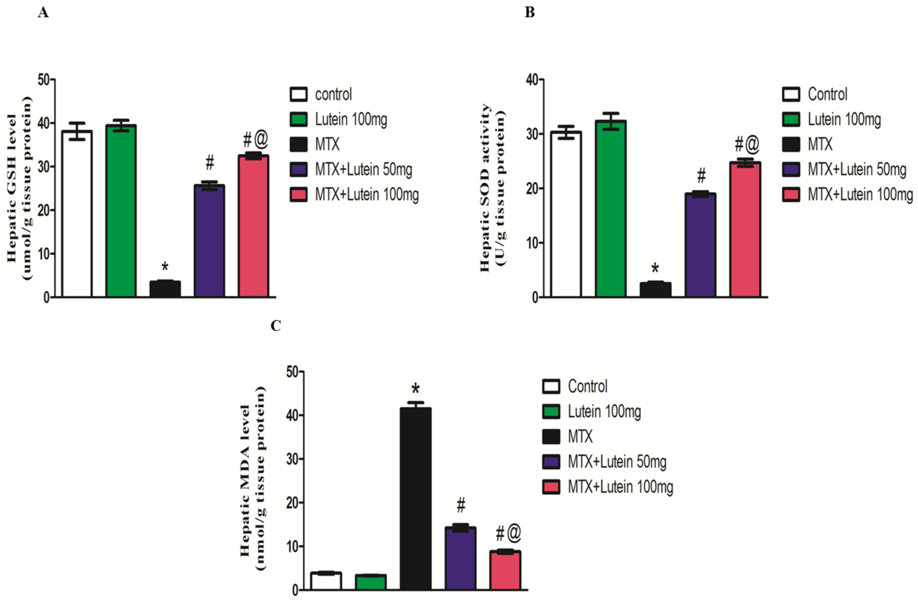
When compared with the control group, a single dose of MTX (40 mg/kg, I.P.) led to a significant (p < 0.05) increase in malondialdehyde (MDA) level in hepatic homogenate. Additionally, administration of MTX led to a marked decrease in reduced glutathione (GSH) level and superoxide dismutase (SOD) activity relative to the control group.
On the other hand, concomitant treatment with lutein at different doses (50 and 100 mg/kg, given I.G.) significantly (p < 0.05) alleviated the MTX-elicited increase in MDA level. Both doses also restored the GSH content and SOD activity compared to the MTX-treated group, as illustrated in Figure 2. It is worth noticing that the higher dose of lutein demonstrated greater antioxidant potential, represented by a more pronounced reduction in MDA level and elevation in antioxidant defensive tools, GSH and SOD, in comparison to the lower dose.

2.4. Effect of MTX and/or Lutein on Expression of SIRT-1 in Hepatic Tissue

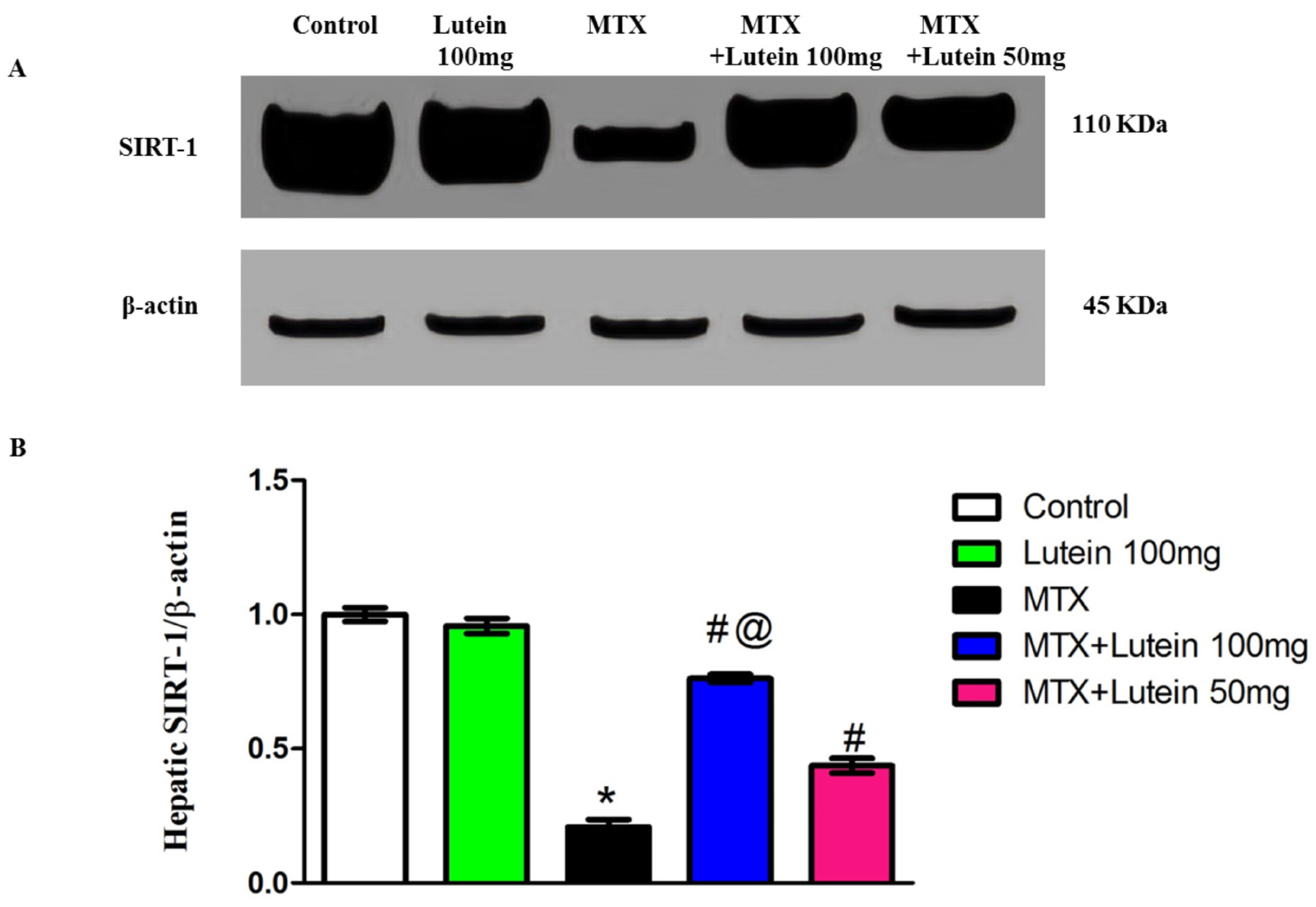
In comparison with the control group, MTX (40 mg/kg, I.P.) challenge showed a significant suppression of SIRT-1 expression in the investigated tissues. Interestingly, prior treatment with lutein, either at low or high dose, significantly (p < 0.05) mitigated MTX-induced downregulation in SIRT-1 expression when compared to rats that were treated with MTX alone.
Moreover, enhancement of SIRT-1 expression in hepatic tissues was significantly (p < 0.05) more observable in the MTX rats subjected to preconditioning with the higher dose (100 mg/kg) compared to those treated with the lower dose (50 mg/kg) (Figure 3).

2.5. Effect of MTX and/or Lutein on Expression of NF-κB in Hepatic Tissue

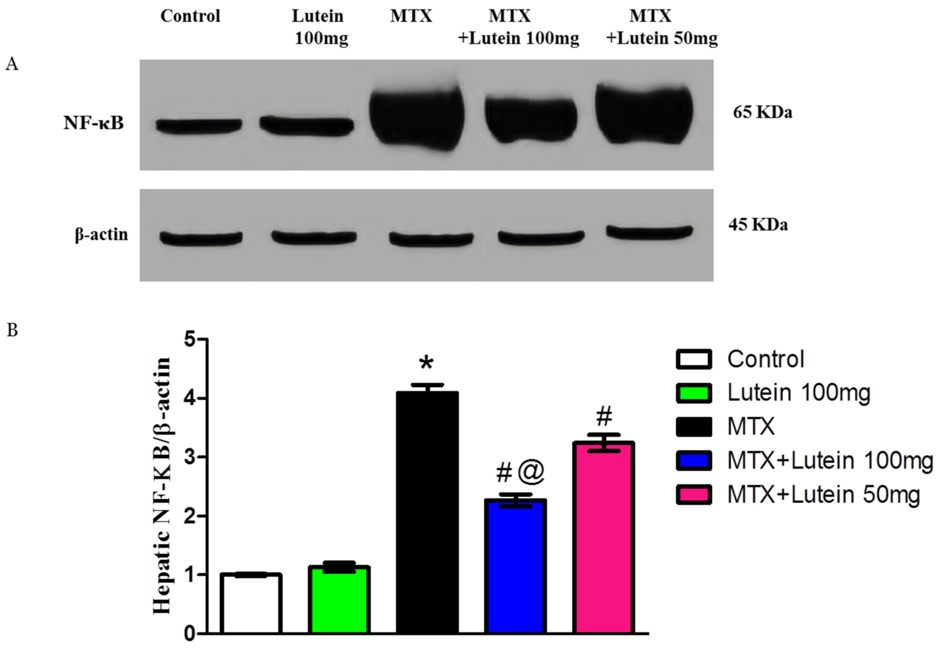
As illustrated in Figure 4, protein expression of NF-κB in the liver was significantly (p < 0.05) increased in MTX group compared to control animals. Moreover, administration of lutein at low and high doses I.G. significantly (p < 0.05) suppressed this elevation induced by MTX. In addition, lutein co-treatment with MTX resulted in a dose-related reduction in NF-κB expression.
The group that received 100 mg/kg lutein demonstrated a more substantial reduction in NF-κB expression than the group that received 50 mg/kg lutein.

2.6. Effect of MTX and/or Lutein on TNF-α and IL-6 in Hepatic Tissue

Methotrexate (MTX) induces inflammation, which is one of the most important pathways that contribute to its toxicity. Therefore, levels of TNF-α and IL-6 proteins were tested as inflammatory biomarkers. Similar to the NF-κB pattern, a significant (p < 0.05) increase in TNF-α and IL-6 protein levels was observed in the MTX-intoxicated animals compared to the control group. Intriguingly, lutein administration at both examined doses significantly (p < 0.05) attenuated MTX-provoked robust elevation in TNF-α and IL-6 levels as compared to the group challenged with MTX alone.
The reduction in TNF-α and IL-6 was dose-related; the higher dose of lutein provided a further attenuation in the aforementioned inflammatory markers as shown in Figure 5.

2.7. Effect of MTX and/or Lutein on Gene Expression and Levels of ERK and Caspase-3 in Hepatic Tissue

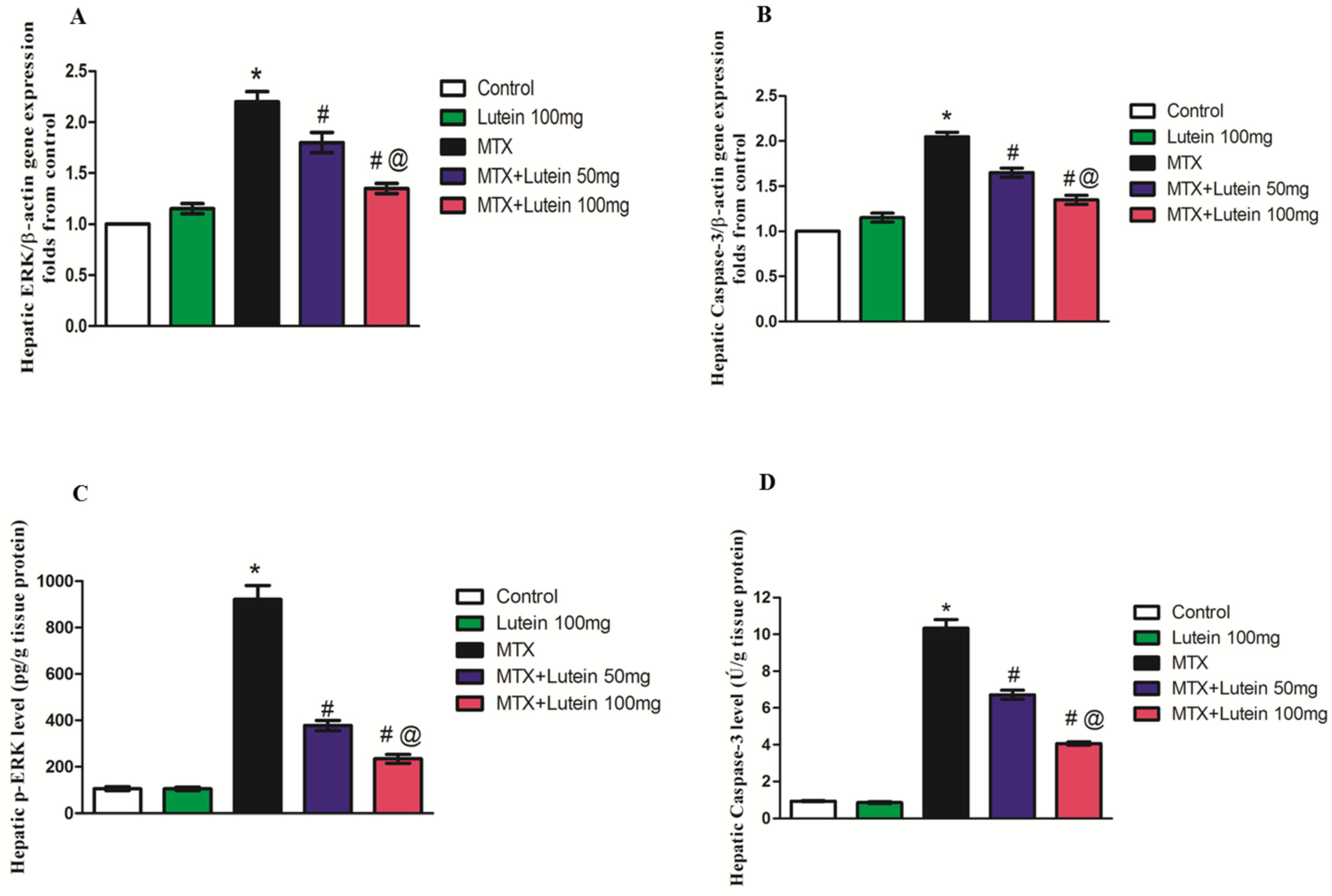
A significant (p < 0.05) increase in ERK and caspase-3 mRNA expression and their levels was found in the hepatic tissues of MTX-injected animals in comparison with normal rats, as shown by quantitative real-time polymerase chain reaction (qRT-PCR) analyses and the ELISA technique. Treatment with the two different investigated doses of lutein before MTX injection caused a remarkable decline in the mRNA expression and level of ERK and caspase-3 when compared with the MTX-injected rats. Furthermore, the decline in hepatic ERK and caspase-3 was more pronounced in MTX rats treated with 100 mg/kg of lutein than in the MTX + lutein 50 mg group, as presented in Figure 6.

3. Discussion

Methotrexate (MTX) is a commonly used antineoplastic and immunosuppressant drug; however, its application is often clinically limited due to the associated hazards, particularly hepatic injury [8]. This study reports the inhibitory effect of lutein on MTX-induced hepatotoxicity in rats, as presented here by restoration of liver performance and preservation of intact liver structure. This protective effect might be mediated by SIRT-1 modulation and antagonizing oxidative damage and inflammatory and apoptotic responses.
Liver injury occurred here after MTX intoxication presented in the form of elevated ALT and AST values, which are indicators of liver dysfunction [14] and a remarkable destruction of hepatic tissue, observed upon histological examination: disorganization of hepatic cords, focal necrosis, and hydropic degeneration of hepatocytes with hepatic apoptosis. These observations are aligned with prior studies [33,34,35,36]. Degenerated hepatocytes and loss of the integrity of hepatocyte structure with subsequent leakage of its intracellular contents underlie the noticed increase in the liver enzymes [37,38].
It is worth mentioning that lutein pretreatment at both used doses provided hepatoprotection, verified by a significant attenuation in MTX-induced elevated ALT and AST concentrations and noticeable preservation of hepatic architecture; reduced hepatocellular degeneration and minimized inflammatory cell infiltration are consistent with earlier studies reporting the lutein hepatoprotective influence [28,29,30,32]. Of interest, disturbance in liver enzymes and histological alteration were significantly attenuated in the MTX + Lutein 100 mg group compared to MTX-challenged rats treated with a lower dose, as seen in the experiment carried out by Edward, Ajibade [32]. These outcomes spotlight the possible effect of lutein in hindering liver impairment induced by MTX. In the context of these observations, we dug deeper to explore the implicated molecular mechanisms that can explain the hepatoprotection of lutein in the MTX setting.
Although the exact mechanisms of MTX-induced liver dysfunction remain unclear, increased hepatic MTX-PGs are embroiled in MTX-elicited hepatic injury due to eventual reduction in folic acid, which, in turn, leads to multiple harmful events; impaired equilibrium between oxidant/antioxidant factors besides inflammatory and apoptotic responses [14].
Oxidative damage is remarkably engaged with MTX hepatic impairment [9,14,39]. MTX disrupts mitochondrial DNA, resulting in mitochondrial dysfunction, which consequently induces excessive ROS generation [40,41]. The robust increase in ROS damages protein and DNA and peroxidizes membrane lipid. Lipid peroxidation disarranges the membrane, receptors, and enzymes, which alters membrane permeability, illustrating the observed increase in ALT and AST [14,42].
These reported findings align with the results observed in this study, where MTX administration led to an obvious increase in MDA content, a lipid peroxidation indicator, and a remarkable decline in the protective antioxidant tools, GSH as well as SOD activity, in hepatic tissue [43]. MTX inhibits NADPH, which is important for maintaining glutathione reductase, which explains the noted attenuation in GSH [44].
N-acetylcysteine and silymarin are known hepatoprotective therapeutic approaches through battling oxidative stress [45,46,47]. Protection exerted by N-acetylcysteine is mediated by its rapid deacetylation to cysteine, the rate-limiting substrate for hepatic GSH synthesis, thereby restoring the liver’s intrinsic detoxification capacity [48]. Through free radical scavenging, enhanced antioxidant enzyme activity, stabilization of cell membrane, and prevention of lipid peroxidation, silymarin provided hepatoprotection in various liver disorders [49].
Here, lutein could significantly restore the balance of the oxidative condition, indicated by decreased MDA, in addition to increased GSH and SOD. This protective antioxidant activity was afforded by the two examined lutein doses, but 100 mg produced a more pronounced antioxidant effect. The lutein antioxidant impact was reported in various experimental investigations, including age-related macular degeneration [50], some cardiovascular and neurological disorders like Alzheimer’s, Parkinson’s, and age-related cognitive impairment [51,52]. Additionally, in vitro experiments confirmed that lutein possesses free radical scavenging activity by inhibiting the production of ROS [53].
Regarding liver injury, lutein could protect against cisplatin [54]- and NAFLD [31]-induced hepatotoxicity by significantly lowering hepatic MDA levels and enhancing total antioxidant capacity. Zeaxanthin, another carotenoid, showed a potential preservative effect in an experimental rodent model of NAFLD mediated by antagonizing liver oxidative stress [55].
Furthermore, there is a strong interplay between MTX hepatotoxicity and SIRT-1 downregulation [18,19], as observed in this study. SIRT-1 produces hepatoprotection via various protective mechanisms, antagonizing inflammatory pathways, oxidative cellular damage and apoptosis [16,17]. It is worth mentioning that SIRT-1-mediated Nrf2, a master organizer of antioxidant response, deacetylation boosts the antioxidant response, increasing hepatocellular resilience against oxidative stress [56]. Of interest, Wang, Wang [57] demonstrated that lutein activates the SIRT-1 pathway. Huang, Duan [58] reported the connection between amelioration of Parkinson’s disease and SIRT-1 activation. Here, the inverse relation between SIRT-1 and oxidative stress was mirrored by reduced SIRT-1 expression and distorted oxidative balance in the MTX group. However, the reduction in hepatic SIRT-1 expression was alleviated by lutein treatment at doses of either 50 mg/kg or 100 mg/kg, with more pronounced alleviation observed in rats pretreated with a high dose. Therefore, we propose that MTX-induced oxidative stress may be mediated by the suppression of SIRT-1 expression and targeting SIRT-1 may be a prospective tool to dampen MTX-related hepatic illnesses.
Additionally, the contribution of inflammation in MTX-induced development of liver injury was documented [59,60]. MTX intoxication leads to an increase in NF-κB and TNF-α in liver tissues, suggesting a connection between these factors and MTX-induced functional impairment [59,60]. NF-κB signaling activity is heightened by a variety of pro-inflammatory cytokines, such as TNF-α, and its activation triggers the production of numerous genes that code for pro-inflammatory mediators such as TNF-α, IL-6, and IL-1β. This interaction between NF-κB and TNF-α has been previously documented [60].
Furthermore, there is a notable link between NF-κB, SIRT-1, and inflammation. SIRT-1 exerts anti-inflammatory effects by either preventing NF-κB translocation and activation or directly downregulating pro-inflammatory cytokine genes [56,61]. Parallel to reports discussing this link, the liver tissues of rats intoxicated by MTX showed low expression of SIRT-1, along with NF-κB/TNF-α/IL-6 upregulation.
Besides lutein’s antioxidant activity, it has been documented to possess anti-inflammatory properties, indicating its role in counteracting inflammation and oxidative stress associated with various disease states [62]. The current study examines how co-treatment with lutein provides hepatoprotection through reducing the elevated expression of NF-κB/TNF-α/IL-6 due to MTX challenge. These findings are consistent with previous studies that have reported lutein’s anti-inflammatory impacts in diverse contexts, including neurodegenerative disorders and cardiovascular disease [27,58,63]. Moreover, lutein effectively counteracts MTX-induced lung damage by decreasing oxidative stress, inflammation, and apoptosis [64]. Aktas, Gur [54] also reported the inhibitory effect of lutein against cisplatin hepatic impairment by downregulation of TNF-α. Astaxanthin, zeaxanthin’s metabolite, exhibited an organ-protective effect in experimental model of a high fructose diet through modulation of the SIRT-1/NF-κB pathway alongside mitigation of oxidative stress [65].
The observed protective effect of lutein on MTX-induced liver damage in this study appears to be mediated by its ability to modulate the SIRT-1/NF-κB/TNF-α/IL-6 signaling pathway, depending on the dosage used.
To gain a more comprehensive understanding of lutein’s protective effects against MTX-induced hepatic apoptosis, we examined its effects on the expression and levels of key markers of programmed cell death, specifically ERK and caspase-3. Our findings were in agreement with earlier studies [12,14] reporting that MTX-PGs triggered caspase-3 overexpression in liver tissue. This overexpression may be linked to exaggerated ROS generation and increased levels of pro-inflammatory cytokines. The intoxicated group exhibited a significant elevation in these markers mediating the apoptotic pathway in hepatic tissues [66].
The anti-apoptotic influence of lutein has been previously documented against apoptosis induced by glutamate in an in vitro study [67]. Moreover, Chucair, Rotstein [68] highlighted lutein’s ability to hinder apoptosis, protecting photoreceptors. The influence of lutein on ERK and caspase-3 has been investigated in the setting of MTX-induced liver injury. Our findings notably show that lutein treatment significantly reduces the levels of both ERK and caspase-3 when compared to MTX treatment alone. These findings emphasize the anti-apoptotic and cytoprotective properties of lutein, which are facilitated by its significant impact on essential signaling pathways.
Moreover, the ERK/caspase-3, NF-κB and downregulated SIRT-1 pathways are known to interact closely, forming a complex network that controls inflammation, apoptosis, and cell survival [69]. These pathological cascades are reported in hepatic impairment produced by MTX [14,18,19]. These findings suggest that therapeutically manipulating one might indirectly influence the others and mitigate the harmful effects on the liver [21,70]. Therefore, lutein reduces NF-κB/TNF-α/IL-6 and promotes SIRT-1 and mitigates apoptosis, as evidenced by attenuated ERK/caspase-3.
Importantly, clinical studies investigating the anticancer properties of lutein have suggested its possible safeguarding role, especially in the prevention and slowing of the progression of certain cancers. Epidemiological studies suggest that increased dietary intake or higher blood concentrations of lutein are linked to a lower risk of developing cancers, including breast, colon, and lung cancer [71,72]. In cell culture studies, lutein induced cell cycle arrest and promoted programmed cell death in cancer cells without significantly affecting normal cells, exhibiting a selective cytotoxic effect. It also lessened oxidative DNA damage and modulated the expression of crucial regulatory proteins such as Bcl-2-associated X protein (Bax), B-cell lymphoma 2 (Bcl-2), and caspases [73].
In animal models, lutein supplementation led to shrinkage of tumors, as well as reduction in angiogenesis and metastasis, likely by the inhibition of pathways such as NF-κB and mitogen-activated protein kinase (MAPK) that are playing a role in inflammation and injury [74].
Overall, this study provides evidence of lutein’s hepatoprotective effect against MTX-related toxicity, acting through its antioxidant, anti-inflammatory, and anti-apoptotic properties. Lutein enhanced SIRT-1 expression, lowered MDA levels, increased SOD activity and GSH content, inhibited NF-κB/TNF-α/IL-6/signaling, and reduced caspase-3 and ERK expression in liver tissue, resulting in improved liver function markers and histological structure. Lutein, as a natural compound, may be a hopeful tool to lessen the prevalence and seriousness of MTX-related side effects, ultimately improving patient outcomes. However, further clinical research is mandatory to validate these findings in humans and to identify the effectiveness and safety of lutein to mitigate the toxic effects of anticancer drugs.
However, a comparison with N-acetylcysteine as a positive control hepatoprotective agent to benchmark lutein’s efficacy can be conducted. Mechanistic studies to further substantiate the causality of lutein’s protective effect in MTX hepatotoxicity are needed; the need to investigate direct SIRT-1 activation, mitochondrial function, and using pharmacological inhibitors of SIRT-1, such as EX-527, or genetic knockdown, besides pharmacokinetic investigations to precisely correlate lutein tissue levels with the observed therapeutic outcomes are considered limitations of this work that should be focused on in future studies. Moreover, inclusion of both female and male sexes can be performed in the future research to elucidate sex-based differences in MTX toxicity and antioxidant metabolism.

4. Materials and Methods

4.1. Drugs

Methotrexate (MTX) and lutein were bought from Hikma Pharma (Cairo, Egypt) and from NOW FOODS (Bloomingdale, DuPage County, IL, USA), respectively.

4.2. Animals

From Animal Care Unit at Faculty of Pharmacy, Nahda University, 40 adult male Wistar rats with weights between 180 and 220 g were utilized. Animals were placed in polypropylene cages for a two-week acclimation period, during which they were kept under standard conditions, including a 12-h light/dark cycle and an adjusted temperature of 25 ± 2 °C. Throughout the study, they had unrestricted access to a standard chow diet and fresh water.

4.3. Experimental Design and Induction of Hepatotoxicity

An independent investigator randomly divided the rats into five groups, with each group comprising 8 rats. Eight animals per group were selected based on previous studies [13,75,76,77] conducted in comparable experimental models.
1-Control group: The rats were given corn oil (the vehicle for lutein) by I.G. route for 10 successive days.
2-Lutein 100 mg group: The rats were administered lutein at a dosage of 100 mg/kg/day via I.G. route for 10 days.
3-MTX group: The rats received corn oil intragastrically for 10 successive days and were given a single I.P. MTX injection (40 mg/kg) on day 7.
4-MTX + Lutein 50 mg group: These rats received a low dose (50 mg/kg/day, I.G.) of lutein for 10 successive days and were also administered a single I.P. dose of MTX (40 mg/kg) on the 7th day of the experimentation.
5-MTX + Lutein 100 mg group: Rats were, daily, given a high dose (100 mg/kg, I.G.) of lutein for 10 consecutive days. On day 7, rats of this group were exposed to a single I.P. dose of MTX (40 mg/kg).
The MTX dosing strategy was adopted from methods used in previous studies [60,75,78] reporting the incidence of hepatotoxicity, while that of lutein is based on studies showing its ability to provide organ protection [32,54,79] and also on the preliminary trials conducted in this study.

4.4. Blood and Tissue Sampling

A day after the final dose was administered, the rats were anesthetized using pentobarbital sodium (50 mg/kg, I.P.) following the measurement of their body weight. Blood samples were then collected by cardiac puncture into clean centrifuge tubes and centrifuged for 10 min at 3500 rpm to separate the serum. Fresh serum samples were then immediately used for evaluating the status of liver function, including ALT and AST concentration. Parts from the liver were preserved in 10% formalin for histopathological examination, while the rest of the liver tissues were immediately frozen in liquid nitrogen and kept at −80 °C. For biochemical analysis, the liver samples were homogenized in ice-cold phosphate-buffered solution (PBS), centrifuged at 10,000 rpm for 10 min, and the supernatant was subsequently collected for further analysis.

4.5. Determination of Liver Function Markers

Levels of serum ALT and AST were used to evaluate liver function. Spectrophotometric determination of ALT and AST levels was performed using commercially available BioMed kits, Cairo, Egypt, based on the method outlined by Henry [80].

4.6. Liver Histopathology

Liver specimens were fixed in 10% neutral buffered formalin after being trimmed, rinsed, and dehydrated using graded alcohols, cleared in xylene, and then embedded in paraffin wax. The blocks were then sectioned into 4–6 µm thick slices and stained with H and E stain, adhering to the protocol outlined in reference [81]. The slides were then examined and visualized by an independent examiner who was blinded to experimental findings under a light microscope at 200× and 400× magnification. The histopathological evaluation included scoring of inflammatory changes according to Ramos, Sá [82] and that of necrosis in hepatocytes according to the METAVIR scoring system [83].

4.7. Assessment of Oxidative Status

A hallmark of lipid peroxidation, MDA, serves as a marker of oxidative stress. Its concentration was quantified using the thiobarbituric acid reactive species method outlined by Uchiyama and Mihara [84]. Thiobarbituric acid, in an acidic medium, reacts with MDA, forming a pink product spectrophotometrically measured at 534 nm. With the use of 1,1,3,3-tetramethoxypropane standard curve, the recorded absorbance represents the corresponding MDA concentration.
In addition, antioxidant defenses, specifically GSH and SOD, were also analyzed. GSH levels were measured following the protocol proposed by Ellman [85]. The sulfhydryl group present in GSH reducing Ellman’s reagent forms a product having a yellow color which can be detected at 412 nm.
Meanwhile, SOD activity was evaluated using the approach developed by Marklund [86]. Marklund’s [86] method is based on the ability of SOD to prevent autoxidation of pyrogallol. The SOD activity is directly correlated to the occurred inhibition in the investigated samples.

4.8. Determination of SIRT-1 and Nf-ķB Using Western Blot Analysis

The hepatic tissues were subjected to homogenization in 1 mL TriFast buffer (Peqlab, VWR company, Darmstadt, Germany) at 4 °C for 5–15 min, followed by centrifugation at a maximum of 12,000× g max for 10 min. The Bradford method was used to determine the concentration of the protein fraction. Equal protein content (30 μg) was loaded into each lane to be separated using sodium dodecyl sulphate-polyacrylamide gel electrophoresis. HybondTM nylon membrane (GE Healthcare, Chicago, IL, USA) served as the solid support for the proteins after transfer. A Tris buffer solution containing 5% non-fat dry milk was used for blocking. The membranes were treated with it for 60 min at ambient temperature. Then, the membrane was incubated overnight at 4 °C in antibody solution targeting NF-κB (catalog# 8242, abcam, Cambridge, MA, USA) and SIRT-1 (catalog# 110304, abcam, Cambridge, MA, USA). Afterwards, the membranes were incubated with the horseradish peroxidase (HRP)-conjugated secondary antibody for 1 h at room temperature. Visualization of protein bands was carried out by the chemiluminescence method, and the appeared bands were analyzed using ImageJ® software, version 1.54r (National Institutes of Health, Bethesda, MD, USA) regarding the bands of the loaded control β-actin and calibrated as fold-change values from the control.

4.9. Determination of TNF-α, IL-6, pERK, and Caspase-3 Using ELISA Technique

A commercially available IL-6 (catalog# E-EL-R0015, Elabscience Biotechnology, Inc., Houston, TX, USA), TNF-α (catalog# E-EL-R0019, Elabscience Biotechnology, Inc., Houston, TX, USA), p-ERK (catalog# ELK9246, ELK biotechnology, Sugar Land, TX, USA), and caspase-3 (SL 1366Ra, Sunlong Biotech Co., Hangzhou, China) ELISA kits for rats were used, which operate on the Sandwich ELISA principle. The kit included a microtiter plate that had been pre-coated with a biotin-conjugated antibody designed to specifically bind to TNF-α, IL-6, p-ERK, or caspase-3. After incubation with an avidin-HRP conjugate and a biotinylated antibody, a tetramethylbenzidine substrate solution was added. A color change occurred in wells containing TNF-α, IL-6, p-ERK, or caspase-3, along with the biotin-conjugated antibody and enzyme-conjugated avidin. The reaction was terminated by the addition of sulfuric acid, after which the absorbance was then measured using a spectrophotometer at 450 nm. The concentrations of TNF-α, IL-6, p-ERK, and caspase-3 were quantified by comparing the optical density of the samples to a standard curve, which directly correlated with their concentrations.

4.10. Determination of ERK and Caspase-3 Using qRT-PCR

Real-time PCR was conducted to assess the mRNA expression of target genes. TRIzol™ reagent (Life Technologies, Carlsbad, CA, USA) was used to extract the total RNA from the tissue samples, with the procedure being completed in an hour. A total of 1 μg of extracted RNA was reverse transcribed into cDNA utilizing the validated QuantiTect Reverse Transcription Kit (Qiagen, Germantown, MD, USA) with a random hexamer primer, in accordance with the manufacturer’s protocol, guaranteeing high efficiency and minimal inhibitor interference. The cDNA samples were then analyzed for ERK and caspase-3. β-actin, a housekeeping gene, served as an internal control, and a non-template control (water) was included to verify the absence of DNA contamination in the reaction mixture. RT-PCR data were analyzed using the 2−ΔΔCt method for relative gene expression quantification. The sequences of used primers are shown in Table 2.
All data collection and normalization were performed using the Rotor-Gene Q system (Qiagen, Germantown, MD, USA), with Ct values normalized to the average of the housekeeping gene β-actin. The stability of β-actin across all treatment groups was experimentally verified to ensure reliable normalization. In addition, PCR efficiency for all primer pairs, including β-actin, was confirmed by standard curve analysis to fall within the acceptable range of 90–110%, ensuring the overall accuracy and validity of the gene expression data.

4.11. Statistical Analysis

Data are expressed as mean ± SEM. Statistical analysis was performed using one-way ANOVA, followed by Tukey–Kramer post hoc testing to compare groups. Calculations were carried out with GraphPad Prism software (version 5.0.2; San Diego, CA, USA), and a p < 0.05 was considered statistically significant.

5. Conclusions

In conclusion, this research highlights the substantial protective role of lutein in dampening MTX-induced hepatic damage. Modulation of SIRT1, antagonizing oxidative damage and suppressing both inflammatory (NF-κB/TNF-α/IL-6) and apoptotic (ERK/caspase-3) pathways, justifies the lutein-produced improvement in hepatic function and preservation of liver structure. For lutein validation clinically, there is a demand for further clinical studies.

Author Contributions

A.M.A.: conception and design of the study, acquisition of data, analysis and interpretation of data, writing—original draft, editing, and final approval. H.A.H. and M.A.D.: conception and design of the study, acquisition of data, analysis and interpretation of data, supervision, writing—original draft, revising, final approval. Y.A.B.J.: conception and design of the study, acquisition of data, analysis and interpretation of data, revising, final approval, funding acquisition. G.H.H.: conception and design of the study, acquisition of data, analysis and interpretation of data, supervision, writing—original draft, revising, final approval. All authors have read and agreed to the published version of the manuscript.

Funding

The authors extend their sincere appreciation to the Ongoing Researcher Funding Program, King Saud University, Riyadh, Saudi Arabia, for funding this work through a Large Research Project under grant number (ORF-2025-457).

Institutional Review Board Statement

Approval for all experimental procedures was granted by the Research Ethics Committee for the laboratory animal’s care and use, affiliated with the Faculty of Pharmacy, Minia University. The project’s approval number is MPEC (240604) (the date of ethical approval 4 June 2024). All methods were carried out in strict compliance with the international regulations detailed in the U.S. National Institutes of Health’s ‘Guide for the Care and Use of Laboratory Animals’ (NIH Publication No. 85-23, revised 1996).” and complied with ARRIVE (Animal Research: Reporting of In Vivo Experiments) guidelines.

Data Availability Statement

The original contributions presented in this study are included in the article. Further inquiries can be directed to the corresponding authors.

Conflicts of Interest

The authors declare no conflicts of interest.

Abbreviations

The following abbreviations are used in this manuscript:
MTXMethotrexate
DHFRDihydrofolate reductase
FPGSFolylpolyglutamate synthetase
MTX-PGsMTX polyglutamates
SIRT-1Sirtuin-1
TNF-αTumor necrosis factor-α
NF-κB Nuclear factor kappa-B
IL-6Interleukin-6
Nrf2Nuclear factor erythroid 2-related factor 2
NAFLDNon-alcoholic fatty liver disease
ROSReactive oxygen species
ALTAlanine aminotransferase
ASTAspartate aminotransferase
H and EHematoxylin and eosin
MDAMalondialdehyde
GSHReduced glutathione
SODSuperoxide dismutase
I.G.Intragastric
ERKExtracellular signal-regulated kinases
qRT-PCRQuantitative real time polymerase chain reaction
BaxBcl-2 associated X protein
Bcl-2B-cell lymphoma 2
MAPKMitogen-activated protein kinase
I.P.Intraperitoneal

References

  1. de Medeiros, P.d.C.; Nascimento, C.C.; Perobelli, J.E. Antineoplastic drugs in environmentally relevant concentrations cause endocrine disruption and testicular dysfunction in experimental conditions. Environ. Toxicol. Pharmacol. 2023, 100, 104122. [Google Scholar] [CrossRef] [PubMed]
  2. Katturajan, R.; Vijayalakshmi, S.; Rasool, M.; Prince, S.E. Molecular toxicity of methotrexate in rheumatoid arthritis treatment: A novel perspective and therapeutic implications. Toxicology 2021, 461, 152909. [Google Scholar] [CrossRef] [PubMed]
  3. Muanda, F.T.; Blake, P.G.; Weir, M.A.; Ahmadi, F.; McArthur, E.; Sontrop, J.M.; Urquhart, B.L.; Kim, R.B.; Garg, A.X. Low-dose methotrexate and serious adverse events among older adults with chronic kidney disease. JAMA Netw. Open 2023, 6, e2345132. [Google Scholar] [CrossRef] [PubMed]
  4. Ouellette, S.; Shah, R.; Razi, S.; Ashforth, G.; Wassef, C. Fatal low-dose methotrexate toxicity: A case report and literature review. Dermatol. Ther. 2022, 35, e15945. [Google Scholar] [CrossRef]
  5. Chin, A.C. Methotrexate for gestational choriocarcinoma: A paradigm shift in oncology. Nat. Rev. Endocrinol. 2023, 19, 501. [Google Scholar] [CrossRef]
  6. Hamed, K.M.; Dighriri, I.M.; Baomar, A.F.; Alharthy, B.T.; Alenazi, F.E.; Alali, G.H.; Alenazy, R.H.; Alhumaidi, N.T.; Alhulayfi, D.H.; Alotaibi, Y.B. Overview of methotrexate toxicity: A comprehensive literature review. Cureus 2022, 14, e29518. [Google Scholar] [CrossRef]
  7. Bath, R.K.; Brar, N.K.; Forouhar, F.A.; Wu, G.Y. A review of methotrexate-associated hepatotoxicity. J. Dig. Dis. 2014, 15, 517–524. [Google Scholar] [CrossRef]
  8. Onda, K.; Honma, T.; Masuyama, K. Methotrexate-related adverse events and impact of concomitant treatment with folic acid and tumor necrosis factor-alpha inhibitors: An assessment using the FDA adverse event reporting system. Front. Pharmacol. 2023, 14, 1030832. [Google Scholar] [CrossRef]
  9. Roghani, M.; Kalantari, H.; Khodayar, M.J.; Khorsandi, L.; Kalantar, M.; Goudarzi, M.; Kalantar, H. Alleviation of Liver Dysfunction, Oxidative Stress and Inflammation Underlies the Protective Effect of Ferulic Acid in Methotrexate-Induced Hepatotoxicity. Drug Des. Dev. Ther. 2020, 14, 1933–1941. [Google Scholar] [CrossRef]
  10. Azzam, A.; Jiyad, Z.; O’Beirne, J. Is methotrexate hepatotoxicity associated with cumulative dose? A systematic review and meta-analysis. Australas. J. Dermatol. 2021, 62, 130–140. [Google Scholar] [CrossRef]
  11. Di Martino, V.; Verhoeven, D.W.; Verhoeven, F.; Aubin, F.; Avouac, J.; Vuitton, L.; Lioté, F.; Thévenot, T.; Wendling, D. Busting the myth of methotrexate chronic hepatotoxicity. Nat. Rev. Rheumatol. 2023, 19, 96–110. [Google Scholar] [CrossRef]
  12. El-Kashef, D.H.; Sewilam, H.M. Empagliflozin mitigates methotrexate-induced hepatotoxicity: Targeting ASK-1/JNK/Caspase-3 pathway. Int. Immunopharmacol. 2023, 114, 109494. [Google Scholar] [CrossRef] [PubMed]
  13. Goudarzi, M.; Kalantar, M.; Sadeghi, E.; Karamallah, M.H.; Kalantar, H. Protective effects of apigenin on altered lipid peroxidation, inflammation, and antioxidant factors in methotrexate-induced hepatotoxicity. Naunyn-Schmiedeberg’s Arch. Pharmacol. 2021, 394, 523–531. [Google Scholar] [CrossRef] [PubMed]
  14. Ezhilarasan, D. Hepatotoxic potentials of methotrexate: Understanding the possible toxicological molecular mechanisms. Toxicology 2021, 458, 152840. [Google Scholar] [CrossRef] [PubMed]
  15. Alfwuaires, M.A. Galangin mitigates oxidative stress, inflammation, and apoptosis in a rat model of methotrexate hepatotoxicity. Environ. Sci. Pollut. Res. 2022, 29, 20279–20288. [Google Scholar] [CrossRef]
  16. Fang, C.; Xu, H.; Yuan, L.; Zhu, Z.; Wang, X.; Liu, Y.; Zhang, A.; Shao, A.; Lou, M. Natural compounds for SIRT1-mediated oxidative stress and neuroinflammation in stroke: A potential therapeutic target in the future. Oxidative Med. Cell. Longev. 2022, 2022, 1949718. [Google Scholar] [CrossRef]
  17. Li, Y.; Luo, W.W.; Cheng, X.; Xiang, H.R.; He, B.; Zhang, Q.Z.; Peng, W.X. Curcumin attenuates isoniazid-induced hepatotoxicity by upregulating the SIRT1/PGC-1α/NRF1 pathway. J. Appl. Toxicol. 2022, 42, 1192–1204. [Google Scholar] [CrossRef]
  18. Ilhan, I.; Asci, H.; Candan, I.A.; Savran, M.; Imeci, O.B.; Sevuk, M.A. Cannabidiol mitigates methotrexate-induced hepatic injury via SIRT-1/p53 signaling and mitochondrial pathways: Reduces oxidative stress and inflammation. Drug Chem. Toxicol. 2025, 48, 210–218. [Google Scholar] [CrossRef]
  19. Ulusan, M.; Erdogan, M.A.; Simsek, O.; Ustundag, H.; Dogan, Z.; Ciftci, B.B.; Kocamuftuoglu, M.; Orhan, I.; Erbas, O. Pyridostigmine Mitigates Methotrexate-Induced Liver Fibrosis in Rats: Association with Changes in BMP-9, SIRT1, and Endoglin Expression. Biomedicines 2025, 13, 1502. [Google Scholar] [CrossRef]
  20. Ding, R.-B.; Bao, J.; Deng, C.-X. Emerging roles of SIRT1 in fatty liver diseases. Int. J. Biol. Sci. 2017, 13, 852. [Google Scholar] [CrossRef]
  21. Wang, M.; Zhao, J.; Chen, J.; Long, T.; Xu, M.; Luo, T.; Che, Q.; He, Y.; Xu, D. The role of sirtuin1 in liver injury: Molecular mechanisms and novel therapeutic target. PeerJ 2024, 12, e17094. [Google Scholar] [CrossRef] [PubMed]
  22. Kim, H.; Hong, J.; Lee, J.; Jeon, W.; Yeo, C.; Lee, Y.; Baek, S.; Ha, I. Curcuma aromatica Salisb. Protects from acetaminophen-induced hepatotoxicity by regulating the Sirt1/Ho-1 signaling pathway. Nutrients 2023, 15, 808. [Google Scholar] [CrossRef] [PubMed]
  23. Iwamoto, H.; Soccol, C.R.; Molina-Aulestia, D.T.; Cardoso, J.; de Melo Pereira, G.V.; de Souza Vandenberghe, L.P.; Manzoki, M.C.; Ambati, R.R.; Ravishankar, G.A.; De Carvalho, J.C. Lutein from microalgae: An industrial perspective of its production, downstream processing, and market. Fermentation 2024, 10, 106. [Google Scholar] [CrossRef]
  24. Akkewar, A.S.; Mishra, K.A.; Kamble, M.G.; Kumar, S.; Dey, J.; Sethi, K.K. A mechanistic review on growing multiple therapeutic applications of lutein and its global market research. Phytother. Res. 2024, 38, 3190–3217. [Google Scholar] [CrossRef]
  25. Elkholy, N.S.; Hariri, M.L.M.; Mohammed, H.S.; Shafaa, M.W. Lutein and β-carotene characterization in free and nanodispersion forms in terms of antioxidant activity and cytotoxicity. J. Pharm. Innov. 2023, 18, 1727–1744. [Google Scholar] [CrossRef]
  26. Mesbahzadeh, B.; Hassanzadeh-Taheri, M.; Aliparast, M.-s.; Baniasadi, P.; Hosseini, M. The protective effect of crocin on cisplatin-induced testicular impairment in rats. BMC Urol. 2021, 21, 117. [Google Scholar] [CrossRef]
  27. Abdelmonem, M.; Ibrahim, S.M.; Essam, R.M.; Amin, H.A.A.; Abd-Elmawla, M.A. Lutein exerts its cardioprotective effect against the experimental model of isoprenaline-induced myocardial infarction via MIAT/miR-200a/Nrf2/TXINP pathway. J. Biochem. Mol. Toxicol. 2021, 35, e22899. [Google Scholar] [CrossRef]
  28. Sindhu, E.R.; Firdous, A.P.; Preethi, K.C.; Kuttan, R. Carotenoid lutein protects rats from paracetamol-, carbon tetrachloride-and ethanol-induced hepatic damage. J. Pharm. Pharmacol. 2010, 62, 1054–1060. [Google Scholar] [CrossRef]
  29. Adikwu, E.; Bokolo, B.; Nnanna, T.B.; Yaakor, F. Preclinical Protective Activity of Lutein on Diclofenac-induced Hepatotoxicity. Arch. Pharmacol. Ther. 2025, 7, 52–58. [Google Scholar] [CrossRef]
  30. Zhao, S.; Zhang, Y.; Ding, H.; Hu, S.; Wu, X.; Ma, A.; Ma, Y. Lutein prevents liver injury and intestinal barrier dysfunction in rats subjected to chronic alcohol intake. Nutrients 2023, 15, 1229. [Google Scholar] [CrossRef]
  31. Qiu, X.; Gao, D.-H.; Xiang, X.; Xiong, Y.-F.; Zhu, T.-S.; Liu, L.-G.; Sun, X.-F.; Hao, L.-P. Ameliorative effects of lutein on non-alcoholic fatty liver disease in rats. World J. Gastroenterol. WJG 2015, 21, 8061. [Google Scholar] [CrossRef]
  32. Edward, T.; Ajibade, A.; Edward, S.; Umeaku, U. Histological and biochemical effects of Lutein on the liver of adult Wistar rats following Paraquat-induced toxicity. J. Exp. Clin. Anat. 2024, 21, 317–323. [Google Scholar] [CrossRef]
  33. Karlsson Sundbaum, J.; Eriksson, N.; Hallberg, P.; Lehto, N.; Wadelius, M.; Baecklund, E. Methotrexate treatment in rheumatoid arthritis and elevated liver enzymes: A long-term follow-up of predictors, surveillance, and outcome in clinical practice. Int. J. Rheum. Dis. 2019, 22, 1226–1232. [Google Scholar] [CrossRef] [PubMed]
  34. Mori, S.; Arima, N.; Ito, M.; Fujiyama, S.; Kamo, Y.; Ueki, Y. Non-alcoholic steatohepatitis-like pattern in liver biopsy of rheumatoid arthritis patients with persistent transaminitis during low-dose methotrexate treatment. PLoS ONE 2018, 13, e0203084. [Google Scholar] [CrossRef] [PubMed]
  35. Kamel, W.H.; Ali, M.F.; Afifi, S.H. Histopathological and Biochemical Studies of Methotrexate Hepatotoxicity on Albino Rats. Assiut Vet. Med. J. 2023, 69, 60–68. [Google Scholar]
  36. AbdelKader, G.; Abdelaziz, E.Z.; Hassan, R.; Greish, S.M.; Abogresha, N.M.; Sultan, B.O.; Yousef, E.M.; Morsi, S.; Abdelaziz, E.; Greish, S. Protective effects of crocin against methotrexate-induced hepatotoxicity in adult male albino rats: Histological, immunohistochemical, and biochemical study. Cureus 2023, 15, e34468. [Google Scholar] [CrossRef]
  37. McGill, M.R. The past and present of serum aminotransferases and the future of liver injury biomarkers. EXCLI J. 2016, 15, 817–828. [Google Scholar] [CrossRef]
  38. Rizk, F.H.; Saadany, A.A.E.; Dawood, L.; Elkaliny, H.H.; Sarhan, N.I.; Badawi, R.; Abd-Elsalam, S. Metformin ameliorated methotrexate-induced hepatorenal toxicity in rats in addition to its antitumor activity: Two birds with one stone. J. Inflamm. Res. 2018, 11, 421–429. [Google Scholar] [CrossRef]
  39. Pınar, N.; Kaplan, M.; Özgür, T.; Özcan, O. Ameliorating effects of tempol on methotrexate-induced liver injury in rats. Biomed. Pharmacother. 2018, 102, 758–764. [Google Scholar] [CrossRef]
  40. Hawwa, A.F.; AlBawab, A.; Rooney, M.; Wedderburn, L.R.; Beresford, M.W.; McElnay, J.C. Methotrexate polyglutamates as a potential marker of adherence to long-term therapy in children with juvenile idiopathic arthritis and juvenile dermatomyositis: An observational, cross-sectional study. Arthritis Res. Ther. 2015, 17, 295. [Google Scholar] [CrossRef]
  41. Yamamoto, T.; Shikano, K.; Nanki, T.; Kawai, S. Folylpolyglutamate synthase is a major determinant of intracellular methotrexate polyglutamates in patients with rheumatoid arthritis. Sci. Rep. 2016, 6, 35615. [Google Scholar] [CrossRef]
  42. Dalaklioglu, S.; Genc, G.E.; Aksoy, N.H.; Akcit, F.; Gumuslu, S. Resveratrol ameliorates methotrexate-induced hepatotoxicity in rats via inhibition of lipid peroxidation. Hum. Exp. Toxicol. 2013, 32, 662–671. [Google Scholar] [CrossRef]
  43. Al Kury, L.T.; Dayyan, F.; Ali Shah, F.; Malik, Z.; Khalil, A.A.K.; Alattar, A.; Alshaman, R.; Ali, A.; Khan, Z. Ginkgo biloba extract protects against methotrexate-induced hepatotoxicity: A computational and pharmacological approach. Molecules 2020, 25, 2540. [Google Scholar] [CrossRef] [PubMed]
  44. Vardi, N.; Parlakpinar, H.; Cetin, A.; Erdogan, A.; Cetin Ozturk, I. Protective effect of beta-carotene on methotrexate-induced oxidative liver damage. Toxicol. Pathol. 2010, 38, 592–597. [Google Scholar] [CrossRef] [PubMed]
  45. Demirci, T.; Gedikli, S.; Ozturk, N.; Aydemir Celep, N. The protective effect of N-acetylcysteine against methotrexate-induced hepatotoxicity in rat. Eurasian J. Med. Investig. 2019, 3, 219–226. [Google Scholar] [CrossRef]
  46. Said, H.; Salah, A.; Elshatory, A.; Elkholy, O.; Nazih, G.; Awad, W.; Shehab, A. N-acytel cysteine ameliorates methotrexate induced hepatic toxicity and genotoxicity in male albino rats. Egypt. J. Forensic Sci. Appl. Toxicol. 2016, 16, 37–47. [Google Scholar] [CrossRef]
  47. Ahmed, W.A.; Qatea, F.K.; Qasim, B.J.; Nashtar, S.B.; Al-Radeef, M.Y. Effects of Silymarin against Hepatic and Renal Toxicity Induced by Methotrexate in Rats. Tikrit J. Pharm. Sci. 2023, 17, 28–38. [Google Scholar] [CrossRef]
  48. Galicia-Moreno, M.; Monroy-Ramirez, H.C.; Caloca-Camarena, F.; Arceo-Orozco, S.; Muriel, P.; Sandoval-Rodriguez, A.; García-Bañuelos, J.; García-González, A.; Navarro-Partida, J.; Armendariz-Borunda, J. A new opportunity for N-acetylcysteine. An outline of its classic antioxidant effects and its pharmacological potential as an epigenetic modulator in liver diseases treatment. Naunyn-Schmiedeberg’s Arch. Pharmacol. 2025, 398, 2365–2386. [Google Scholar] [CrossRef]
  49. Shahsavari, K.; Ardekani, S.S.; Ardekani, M.R.S.; Esfahani, M.M.; Kazemizadeh, H.; Jamialahmadi, T.; Iranshahi, M.; Khanavi, M.; Hasanpour, M. Are alterations needed in Silybum marianum (Silymarin) administration practices? A novel outlook and meta-analysis on randomized trials targeting liver injury. BMC Complement. Med. Ther. 2025, 25, 134. [Google Scholar] [CrossRef]
  50. Park, H.-A.; Hayden, M.M.; Bannerman, S.; Jansen, J.; Crowe-White, K.M. Anti-apoptotic effects of carotenoids in neurodegeneration. Molecules 2020, 25, 3453. [Google Scholar] [CrossRef]
  51. Gür, F.M.; Bilgiç, S.; Aktaş, İ. Lutein, a non-provitamin A carotenoid, reduces cisplatin-induced cardiotoxicity. Prostaglandins Other Lipid Mediat. 2025, 177, 106965. [Google Scholar] [CrossRef]
  52. Min, J.-Y.; Min, K.-B. Serum lycopene, lutein and zeaxanthin, and the risk of Alzheimer’s disease mortality in older adults. Dement. Geriatr. Cogn. Disord. 2014, 37, 246–256. [Google Scholar] [CrossRef]
  53. Chang, C.-J.; Lin, J.-F.; Chang, H.-H.; Lee, G.-A.; Hung, C.-F. Lutein protects against methotrexate-induced and reactive oxygen species-mediated apoptotic cell injury of IEC-6 cells. PLoS ONE 2013, 8, e72553. [Google Scholar] [CrossRef] [PubMed]
  54. Aktas, I.; Gur, F.M.; Bilgiç, S. Protection of Lutein Against the Toxic Effect of Cisplatin on Liver in Male Rat. Prostaglandins Other Lipid Mediat. 2025, 178, 106995. [Google Scholar] [CrossRef] [PubMed]
  55. Chamberlain, S.M.; Hall, J.D.; Patel, J.; Lee, J.R.; Marcus, D.M.; Sridhar, S.; Romero, M.J.; Labazi, M.; Caldwell, R.W.; Bartoli, M. Protective effects of the carotenoid zeaxanthin in experimental nonalcoholic steatohepatitis. Dig. Dis. Sci. 2009, 54, 1460–1464. [Google Scholar] [CrossRef] [PubMed]
  56. Yan, T.; Huang, J.; Nisar, M.F.; Wan, C.; Huang, W. The beneficial roles of SIRT1 in drug-induced liver injury. Oxidative Med. Cell. Longev. 2019, 2019, 8506195. [Google Scholar] [CrossRef]
  57. Wang, N.; Wang, D.; Luo, G.; Zhou, J.; Tan, Z.; Du, Y.; Xie, H.; Liu, L.; Yang, X.; Hao, L. Lutein attenuates excessive lipid accumulation in differentiated 3T3-L1 cells and abdominal adipose tissue of rats by the SIRT1-mediated pathway. Int. J. Biochem. Cell Biol. 2021, 133, 105932. [Google Scholar] [CrossRef]
  58. Huang, H.; Duan, J.; Duan, W.; Pu, X.; Ma, C.; Xu, Z. Lutein ameliorates Parkinson’s disease by regulating miR-135b-5p/SIRT1 to inhibit microglial M1 polarization and inflammation. Brain Res. Bull. 2025, 230, 111487. [Google Scholar] [CrossRef]
  59. Manna, K.; Khan, Z.S.; Saha, M.; Mishra, S.; Gaikwad, N.; Bhakta, J.N.; Banerjee, K.; Das Saha, K. Manjari Medika Grape Seed Extract Protects Methotrexate-Induced Hepatic Inflammation: Involvement of NF-κB/NLRP3 and Nrf2/HO-1 Signaling System. J. Inflamm. Res. 2023, 16, 467–492. [Google Scholar] [CrossRef]
  60. Matouk, A.I.; Awad, E.M.; El-Tahawy, N.F.; El-Sheikh, A.A.; Waz, S. Dihydromyricetin alleviates methotrexate-induced hepatotoxicity via suppressing the TLR4/NF-κB pathway and NLRP3 inflammasome/caspase 1 axis. Biomed. Pharmacother. 2022, 155, 113752. [Google Scholar] [CrossRef]
  61. Liu, F.J.; Gu, T.J.; Wei, D.Y. Emodin alleviates sepsis-mediated lung injury via inhibition and reduction of NF-kB and HMGB1 pathways mediated by SIRT1. Kaohsiung J. Med. Sci. 2022, 38, 253–260. [Google Scholar] [CrossRef]
  62. Lin, Z.-X.; Zhang, M.; Yang, R.; Min, Y.; Guo, P.-T.; Zhang, J.; Wang, C.-K.; Jin, L.; Gao, Y.-Y. The anti-inflammatory effect of lutein in broilers is mediated by regulating Toll-like receptor 4/myeloid-differentiation-factor 88 signaling pathway. Poult. Sci. 2023, 102, 102622. [Google Scholar] [CrossRef]
  63. Firozjae, A.A.; Shiran, M.R.; Rashidi, M. The neuropharmacological and clinical effects of lutein: A systematic review. Horm. Mol. Biol. Clin. Investig. 2025, 46, 27–38. [Google Scholar] [CrossRef] [PubMed]
  64. Mammadov, R.; Suleyman, B.; Akturan, S.; Cimen, F.K.; Kurt, N.; Suleyman, Z.; Malkoc, İ. Effect of lutein on methotrexate-induced oxidative lung damage in rats: A biochemical and histopathological assessment. Korean J. Intern. Med. 2019, 34, 1279. [Google Scholar] [CrossRef] [PubMed]
  65. Iskender, H.; Yenice, G.; Dokumacioglu, E.; Hayirli, A.; Sevim, C.; Dokumacioglu, A.; Terim Kapakin, K.A. Astaxanthin alleviates renal damage of rats on high fructose diet through modulating NFκB/SIRT1 pathway and mitigating oxidative stress. Arch. Physiol. Biochem. 2020, 126, 89–93. [Google Scholar] [CrossRef] [PubMed]
  66. El-Dessouki, A.M.; Kaml, M.E.; El-Yamany, M.F. Modulation of AMPK by esomeprazole and canagliflozin mitigates methotrexate-induced hepatotoxicity: Involvement of MAPK/JNK/ERK, JAK1/STAT3, and PI3K/Akt signaling pathways. Naunyn-Schmiedeberg’s Arch. Pharmacol. 2025, 398, 10901–10919. [Google Scholar] [CrossRef]
  67. Li, Z.; Cao, Z.; Chen, F.; Li, B.; Jin, H. Lutein inhibits glutamate-induced apoptosis in HT22 cells via the Nrf2/HO-1 signaling pathway. Front. Neurosci. 2024, 18, 1432969. [Google Scholar] [CrossRef]
  68. Chucair, A.J.; Rotstein, N.P.; SanGiovanni, J.P.; During, A.; Chew, E.Y.; Politi, L.E. Lutein and zeaxanthin protect photoreceptors from apoptosis induced by oxidative stress: Relation with docosahexaenoic acid. Investig. Ophthalmol. Vis. Sci. 2007, 48, 5168–5177. [Google Scholar] [CrossRef]
  69. Kauppinen, A.; Suuronen, T.; Ojala, J.; Kaarniranta, K.; Salminen, A. Antagonistic crosstalk between NF-κB and SIRT1 in the regulation of inflammation and metabolic disorders. Cell. Signal. 2013, 25, 1939–1948. [Google Scholar] [CrossRef]
  70. Abdelzaher, W.Y.; Geddawy, A.; Attya, M.E.; Ali, A.H.S.A.; Elroby Ali, D.M.; Waggas, D.S.; Alshaeri, H.K.; Ibrahim, Y.F. Sirt1/Nrf2/TNFα; TLR4/Myd88/NF-κB signaling pathways are involved in mediating hepatoprotective effect of bupropion in rat model of myocardial infarction. Immunopharmacol. Immunotoxicol. 2024, 46, 872–883. [Google Scholar] [CrossRef]
  71. Gong, X.; Smith, J.R.; Swanson, H.M.; Rubin, L.P. Carotenoid lutein selectively inhibits breast cancer cell growth and potentiates the effect of chemotherapeutic agents through ROS-mediated mechanisms. Molecules 2018, 23, 905. [Google Scholar] [CrossRef]
  72. Kavalappa, Y.P.; Gopal, S.S.; Ponesakki, G. Lutein inhibits breast cancer cell growth by suppressing antioxidant and cell survival signals and induces apoptosis. J. Cell. Physiol. 2021, 236, 1798–1809. [Google Scholar] [CrossRef]
  73. Kim, E.-A.; Kang, N.; Heo, S.-Y.; Oh, J.-Y.; Lee, S.-H.; Cha, S.-H.; Kim, W.-K.; Heo, S.-J. Antioxidant, antiviral, and anti-inflammatory activities of lutein-enriched extract of Tetraselmis species. Mar. Drugs 2023, 21, 369. [Google Scholar] [CrossRef]
  74. Shokrzadeh, M.; Mortazavi, P.; Moghadami, A.; Khayambashi, B.; Motafeghi, F. Synergistic antiproliferative and anticancer activity of carotenoid lutein or coenzyme Q10 in combination with doxorubicin on the MCF7 cell line. Appl. Vitr. Toxicol. 2021, 7, 167–174. [Google Scholar] [CrossRef]
  75. Ismail, R.; Habib, H.A.; Anter, A.F.; Amin, A.; Heeba, G.H. Modified citrus pectin ameliorates methotrexate-induced hepatic and pulmonary toxicity: Role of Nrf2, galectin-3/TLR-4/NF-κB/TNF-α and TGF-β signaling pathways. Front. Pharmacol. 2025, 16, 1528978. [Google Scholar] [CrossRef] [PubMed]
  76. Özgöçmen, M.; Aşcı, H.; Doğan, H.K.; İlhan, İ.; Pekgöz, Ş.; Mustafaoğlu, A. A study on Wistar Albino rats: Investigating protective role of ramelteon on liver damage caused by methotrexate. Drug Chem. Toxicol. 2022, 45, 2678–2685. [Google Scholar] [CrossRef] [PubMed]
  77. Abuzaid, S.M.M.; Ali Abd El-Meguid, E.; El-Tahawy, N.F.; Seleem, H.S.; Khalifa Abd-Elhady, H.; Abdel-kareem, M.A. Antioxidant, anti-inflammatory, and anti-apoptotic impact of Moringa leave extract supplementation in livers of methotrexate treated rat. Minia J. Med. Res. 2025, 36, 12–28. [Google Scholar] [CrossRef]
  78. Dogra, A.; Gupta, D.; Bag, S.; Ahmed, I.; Bhatt, S.; Nehra, E.; Dhiman, S.; Kumar, A.; Singh, G.; Abdullah, S.T. Glabridin ameliorates methotrexate-induced liver injury via attenuation of oxidative stress, inflammation, and apoptosis. Life Sci. 2021, 278, 119583. [Google Scholar] [CrossRef]
  79. Bilgiç, S.; Gür, F.M.; Aktaş, İ. Biochemical and histopathological investigation of the protective effect of lutein in rat kidney exposed to cisplatin. Med. Rec. 2022, 4, 433–438. [Google Scholar] [CrossRef]
  80. Henry, R.J. Clinical Chemistry: Principles and Technics; Hoeber Medical Division, Harper and Row: New York, NY, USA, 1974. [Google Scholar]
  81. Bancroft, J.D.; Gamble, M. Theory and Practice of Histological Techniques; Elsevier Health Sciences: Amsterdam, The Netherlands, 2008. [Google Scholar]
  82. Ramos, C.A.F.; Sá, R.d.C.d.S.; Alves, M.F.; Benedito, R.B.; de Sousa, D.P.; Diniz, M.d.F.F.; Araújo, M.S.T.; de Almeida, R.N. Histopathological and biochemical assessment of d-limonene-induced liver injury in rats. Toxicol. Rep. 2015, 2, 482–488. [Google Scholar] [CrossRef]
  83. Abdullazade, S.; Kav, T.; Gököz, Ö.; Önder, S.; Sökmensüer, C. The relationship between treatment response and histological scoring systems applied in chronic hepatitis B. Kafkas J. Med. Sci. 2017, 7, 19–25. [Google Scholar] [CrossRef]
  84. Uchiyama, M.; Mihara, M. Determination of malonaldehyde precursor in tissues by thiobarbituric acid test. Anal. Biochem. 1978, 86, 271–278. [Google Scholar] [CrossRef]
  85. Ellman, G.L. Tissue sulfhydryl groups. Arch. Biochem. Biophys. 1959, 82, 70–77. [Google Scholar] [CrossRef]
  86. Marklund, S.L. Superoxide dismutase isoenzymes in tissues and plasma from New Zealand black mice, nude mice and normal BALB/c mice. Mutat. Res./Fundam. Mol. Mech. Mutagen. 1985, 148, 129–134. [Google Scholar] [CrossRef]
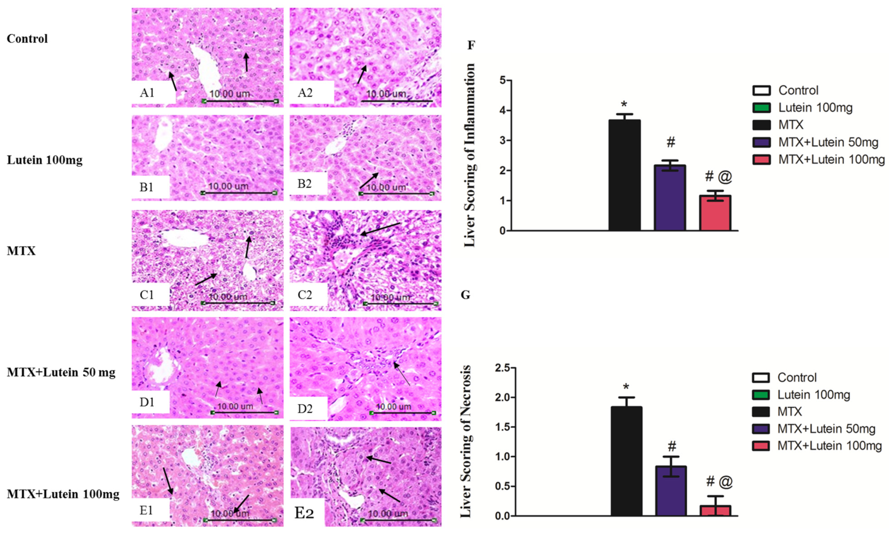
Figure 1. Effect of MTX and/or lutein at two different doses on hematoxylin and eosin (H and E)-stained hepatic sections. Photomicrographs of the liver tissue sections from control group ((A1), X200 and (A2), X400) and Lutein 100 mg group ((B1), X200 and (B2), X400)) exhibited a histological structure that appeared normal. The MTX group shows focal necrosis and hydropic degeneration of hepatocytes (C1, X200, arrow) and a notable increase in the portal lymphocyte number (C2, X400, arrow). The MTX + Lutein 50 mg group shows swelling of hepatocytes and few numbers of apoptotic cells (D1, X200, arrow) and portal lymphocytes (D2, X200, arrow). The MTX + Lutein 100 mg group shows normal organization of hepatic cords with hyperplasia of Kupffer cells (E1, X200, arrow) and swelling of hepatocytes with narrowing of sinusoids in the peripheral zone (E2, X200, arrow). (F,G) show the scoring of inflammation and necrosis, respectively, in hepatocyte of each group. Data are represented as mean ± SEM. * Significant different compared to the control group, # significant different compared to the MTX group, and @ significant different compared to the MTX + Lutein 50 mg group at p < 0.05. MTX: Methotrexate.
Pharmaceuticals 18 01787 g001
Figure 2. Effect of MTX and/or lutein at two different doses on hepatic GSH (A), SOD (B), and MDA (C). All data points are shown as the means ± SEM (n = 6–8). * Significant different compared to the control group, # significant different compared to the MTX group, and @ significant different compared to the MTX + Lutein 50 mg group at p < 0.05. MTX: Methotrexate, GSH: Reduced glutathione, SOD: Superoxide dismutase, and MDA: Malondialdehyde.
Pharmaceuticals 18 01787 g002
Figure 3. Effect of MTX and/or lutein at two different doses on hepatic SIRT-1 protein expression. (A) Representative Western blots showing target, SIRT-1, protein bands from each experimental group. (B) Bar graphs represent the quantified densitometric analysis of hepatic SIRT-1 protein expression. The value for each bar is represented as mean ± SEM. * Significant different compared to the control group, # significant different compared to the MTX group, and @ significant different compared to the MTX + Lutein 50 mg group at p < 0.05. MTX: Methotrexate, SIRT-1: Sirtuin-1.
Pharmaceuticals 18 01787 g003
Figure 4. Effect of MTX and/or lutein at two different doses on hepatic NF-κB protein expression. (A) Western blot representations illustrating NF-κB protein bands for each group. (B) Bar graphs represent the quantified densitometric analysis of hepatic expression of NF-κB protein. The value for each bar is represented as mean ± SEM. * Significant different compared to the control group, # significant different compared to the MTX group, and @ significant different compared to the MTX + Lutein 50 mg group at p < 0.05.MTX: Methotrexate, NF-κB: Nuclear factor kappa-B.
Pharmaceuticals 18 01787 g004
Figure 5. Effect of MTX and/or lutein at two different doses on hepatic TNF-α (A) and IL-6 levels (B). All data points are shown as the means ± SEM (n = 6). * Significant different compared to the control group, # significant different compared to the MTX group, and @ significant different compared to the MTX + Lutein 50 mg group at p < 0.05. MTX: Methotrexate, TNF-α: Tumor necrosis factors, IL-6: Interleukin-6.
Pharmaceuticals 18 01787 g005
Figure 6. Effects of lutein (50 and 100 mg/kg, I.G.) on hepatic ERK (A) and caspase-3 (B) gene expression and hepatic level of p-ERK (C) and caspase-3 (D) in a rodent model of MTX-induced hepatotoxicity. All data points are shown as the means ± SEM. * Significant different compared to the control group, # significant different compared to the MTX group, and @ significant different compared to the MTX + Lutein 50 mg group at p < 0.05. MTX: Methotrexate, ERK: Extracellular signal-regulated kinases.
Pharmaceuticals 18 01787 g006
Table 1. Effect of lutein (50 and 100 mg/kg/day, I.G.) on relative hepatic weight, serum ALT, and AST in a rodent model of MTX-induced hepatotoxicity.
GroupsRelative Hepatic WeightSerum ALT (U/L)Serum AST (U/L)
Control2.92 ± 0.1633.27 ± 1.0839.53 ± 0.77
Lutein 100 mg3.00 ± 0.1633.50 ± 1.0148.95 ± 2.14
MTX3.12 ± 0.1077.88 ± 2.29 *156.1 ± 10.34 *
MTX + Lutein 50 mg2.97 ± 0.1250.55 ± 2.17 #88.25 ± 7.63 #
MTX + Lutein 100 mg2.90 ± 0.1337.41 ± 3.64 #@43.90 ± 2.47 #@
All data points are shown as the means ± SEM (n = 6–8). * Significant different compared to the control group, # significant different compared to the MTX group, and @ significant different compared to the MTX + Lutein 50 mg group at p < 0.05. MTX: Methotrexate, ALT: Alanine aminotransferase, AST: Aspartate aminotransferase.
Table 2. List of primer sequences.
GeneSequences (5′-3′)
ForwardedReverse
ERKCCCTCTAAAACCAA GGTGGCTCATCCAATCACCCACACAC AG
Caspase-3TGAGCATTGA CACAATACACAAGCCGAA ACTCTTCATC
β-actinACCCACACTGTGCCC ATCTATGAGAGTACTTGCGCTCAGGAG GA
Disclaimer/Publisher’s Note: The statements, opinions and data contained in all publications are solely those of the individual author(s) and contributor(s) and not of MDPI and/or the editor(s). MDPI and/or the editor(s) disclaim responsibility for any injury to people or property resulting from any ideas, methods, instructions or products referred to in the content.