


Efficacy of Perampanel in Refractory and Super-Refractory Status Epilepticus with Suspected Inflammatory Etiology: A Case Series

 , , ,
, , ,
Abstract
:
1. Introduction
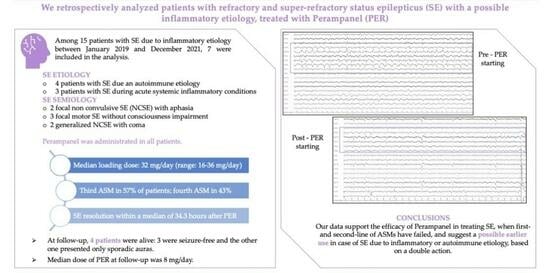
2. Results
3. Discussion
4. Materials and Methods
- -
- Patients with known autoimmune disease and symptomatic epilepsy (in the absence of other causes of SE);
- -
- -
- Patients with a systemic inflammatory state based on laboratory and clinical parameters;
- -
- Patients with NORSE, when other causes of SE have been excluded.
- -
- Age ≥ 18 years;
- -
- Diagnosis of RSE or SRSE with definite or possible inflammatory etiology;
- -
- Availability of continuous electroencephalographic (c-EEG) monitoring from the diagnosis to the resolution of SE;
- -
- Use of PER as treatment during SE;
- -
- Presence of complete clinical, laboratory, and instrumental data;
- -
- Clinical follow-up of at least 6 months in SE survivors.
4.1. Study Outcomes
4.2. Statistical Analysis
5. Limitations
6. Conclusions
Author Contributions
Funding
Institutional Review Board Statement
Informed Consent Statement
Data Availability Statement
Conflicts of Interest
References
- Trinka, E.; Cock, H.; Hesdorffer, D.; Rossetti, A.O.; Scheffer, I.E.; Shinnar, S.; Shorvon, S.; Lowenstein, D.H. A definition and classification of status epilepticus: Report of the ILAE Task Force on Classification of Status Epilepticus. Epilepsia 2015, 56, 1515–1523. [Google Scholar] [CrossRef] [PubMed]
- Logroscino, G.; Hesdorffer, D.C.; Cascino, G.; Hauser, W.A.; Coeytaux, A.; Galobardes, B.; Morabia, A.; Jallon, P. Mortality after a first episode of status epilepticus in the United States and Europe. Epilepsia 2005, 46, 46–48. [Google Scholar] [CrossRef] [PubMed]
- Leitinger, M.; Trinka, E.; Giovannini, G.; Zimmermann, G.; Florea, C.; Rohracher, A.; Kalss, G.; Neuray, C.; Kreidenhuber, R.; Höfler, J.; et al. Epidemiology of status epilepticus in adults: A population-based study on incidence, causes and outcomes. Epilepsia 2019, 60, 53–62. [Google Scholar] [CrossRef] [PubMed]
- Shorvon, S.; Ferlisi, M. The treatment of super-refractory status epilepticus: A critical review of available therapies and a clinical treatment protocol. Brain 2011, 134, 2802–2818. [Google Scholar] [CrossRef] [PubMed]
- Ceprian, M.; Fulton, D. Glial Cell AMPA Receptors in Nervous System Health, Injury and Disease. Int. J. Mol. Sci. 2019, 20, 2450. [Google Scholar] [CrossRef] [PubMed]
- Wasterlain, C.G.; Liu, H.; Naylor, D.E.; Thompson, K.W.; Suchomelova, L.; Niquet, J.; Mazarati, A.M.; Baldwin, R.A. Molecular basis of self-sustaining seizures and pharmacoresistance during status epilepticus: The receptor trafficking hypothesis revisited. Epilepsia 2009, 50 (Suppl. 12), 16–18. [Google Scholar] [CrossRef] [PubMed]
- Hanada, T. Ionotropic Glutamate Receptors in Epilepsy: A Review Focusing on AMPA and NMDA Receptors. Biomolecules 2020, 10, 464. [Google Scholar] [CrossRef] [PubMed]
- De Biase, S.; Gigli, G.L.; Nilo, A.; Romano, G.; Valente, M. Pharmacokinetic and pharmacodynamic considerations for the clinical efficacy of perampanel in focal onset seizures. Expert Opin. Drug Metab. Toxicol. 2019, 15, 93–102. [Google Scholar] [CrossRef]
- US FDA. Fycompa (Perampanel): Highlights of Prescribing Information. Available online: https://www.fycompa.com/media/media/Files/Fycompa/Fycompa_Prescribing_Information.pdf (accessed on 18 December 2023).
- European Medicines Agency. Perampanel Summary of Product Characteristics. Available online: https://www.ema.europa.eu/en/medicines/human/EPAR/fycompa (accessed on 18 December 2023).
- Lattanzi, S.; Cagnetti, C.; Foschi, N.; Ciuffini, R.; Osanni, E.; Chiesa, V.; Dainese, F.; Dono, F.; Canevini, M.P.; Evangelista, G.; et al. Adjunctive Perampanel in Older Patients with Epilepsy: A Multicenter Study of Clinical Practice. Drugs Aging 2021, 38, 603–610. [Google Scholar] [CrossRef]
- Maschio, M.; Pauletto, G.; Zarabla, A.; Maialetti, A.; Lus, T.; Villani, V.; Fabi, A.; Koudriavtseva, T.; Giannarelli, D. Perampanel in patients with brain tumor-related epilepsy in real-life clinical practice: A retrospective analysis. Int. J. Neurosci. 2019, 129, 593–597. [Google Scholar] [CrossRef]
- Nilo, A.; Pauletto, G.; Gigli, G.L.; Vogrig, A.; Dolso, P.; Valente, M. Perampanel as add-on therapy in epilepsies with known etiology: A single center experience with long-term follow-up. Epilepsy Behav. Rep. 2020, 15, 100393. [Google Scholar] [CrossRef] [PubMed]
- Wu, T.; Ido, K.; Osada, Y.; Kotani, S.; Tamaoka, A.; Hanada, T. The neuroprotective effect of perampanel in lithium-pilocarpine rat seizure model. Epilepsy Res. 2017, 137, 152–158. [Google Scholar] [CrossRef] [PubMed]
- Niu, H.X.; Wang, J.Z.; Wang, D.L.; Miao, J.J.; Li, H.; Liu, Z.G.; Yuan, X.; Liu, W.; Zhou, J.R. The Orally Active Noncompetitive AMPAR Antagonist Perampanel Attenuates Focal Cerebral Ischemia Injury in Rats. Cell. Mol. Neurobiol. 2018, 38, 459–466. [Google Scholar] [CrossRef] [PubMed]
- Beretta, S.; Padovano, G.; Stabile, A.; Coppo, A.; Bogliun, G.; Avalli, L.; Ferrarese, C. Efficacy and safety of perampanel oral loading in postanoxic super-refractory status epilepticus: A pilot study. Epilepsia 2018, 59, 243–248. [Google Scholar] [CrossRef] [PubMed]
- Redecker, J.; Wittstock, M.; Benecke, R.; Rösche, J. Efficacy of perampanel in refractory nonconvulsive status epilepticus and simple partial status epilepticus. Epilepsy Behav. 2015, 45, 176–179. [Google Scholar] [CrossRef] [PubMed]
- Rohracher, A.; Kalss, G.; Neuray, C.; Höfler, J.; Dobesberger, J.; Kuchukhidze, G.; Kreidenhuber, R.; Florea, C.; Thomschewski, A.; Novak, H.F.; et al. Perampanel in patients with refractory and super-refractory status epilepticus in a neurological intensive care unit: A single-center audit of 30 patients. Epilepsia 2018, 59, 234–242. [Google Scholar] [CrossRef] [PubMed]
- Perez, D.Q.; Espiritu, A.I.; Jamora, R.D.G. Perampanel in achieving status epilepticus cessation: A systematic review. Epilepsy Behav. 2022, 128, 108583. [Google Scholar] [CrossRef]
- Brigo, F.; Lattanzi, S.; Rohracher, A.; Russo, E.; Meletti, S.; Grillo, E.; Trinka, E. Perampanel in the treatment of status epilepticus: A systematic review of the literature. Epilepsy Behav. 2018, 86, 179–186. [Google Scholar] [CrossRef]
- Strzelczyk, A.; Knake, S.; Kalviainen, R.; Santamarina, E.; Toledo, M.; Willing, S.; Rohracher, A.; Trinka, E.; Rosenow, F. Perampanel for treatment of status epilepticus in Austria, Finland, Germany and Spain. Acta Neurol. Scand. 2019, 139, 369–376. [Google Scholar] [CrossRef]
- Alsherbini, K.; Abhi Pandhi, F.; Goyanes, J.; Deep, A.; Jones, G.M. A retrospective, observational study of perampanel in refractory and super-refractory status epilepticus. J. Neurol. Sci. 2020, 419, 117214. [Google Scholar] [CrossRef]
- Lim, S.N.; Wu, T.; Tseng, W.E.J.; Chiang, H.I.; Cheng, M.Y.; Lin, W.R.L.; Lin, C.N. Efficacy and safety of perampanel in refractory and super-refractory status epilepticus: Cohort study of 81 patients and literature review. J. Neurol. 2021, 268, 3744–3757. [Google Scholar] [CrossRef] [PubMed]
- Ho, C.J.; Lin, C.H.; Lu, Y.T.; Shih, F.Y.; Hsu, C.W.; Tsai, W.C.; Tsai, M.H. Perampanel treatment for refractory status epilepticus in a Neurological Intensive Care Unit. Neurocrit. Care 2019, 31, 24–29. [Google Scholar] [CrossRef] [PubMed]
- Trinka, E.; Höfler, J.; Leitinger, M.; Brigo, F. Pharmacotherapy for Status Epilepticus. Drugs 2015, 75, 1499–1521. [Google Scholar] [CrossRef] [PubMed]
- Rossetti, A.O.; Alvarez, V.; Januel, J.M.; Burnand, B. Treatment deviating from guidelines does not influence status epilepticus prognosis. J. Neurol. 2013, 260, 421–428. [Google Scholar] [CrossRef] [PubMed]
- Hocker, S.E.; Shorvon, S. Anesthetic drugs in status epilepticus: Risk or rescue? A 6-year cohort study. Neurology 2014, 83, 866. [Google Scholar] [CrossRef] [PubMed]
- Huang, T.H.; Lai, M.C.; Chen, Y.S.; Huang, C.W. The Roles of Glutamate Receptors and Their Antagonists in Status Epilepticus, Refractory Status Epilepticus, and Super-Refractory Status Epilepticus. Biomedicines 2023, 11, 686. [Google Scholar] [CrossRef]
- Leo, A.; Giovannini, G.; Russo, E.; Meletti, S. The role of AMPA receptors and their antagonists in status epilepticus. Epilepsia 2018, 59, 1098–1108. [Google Scholar] [CrossRef]
- Vogrig, A.; Gigli, G.L.; Nilo, A.; Pauletto, G.; Valente, M. Seizures, Epilepsy, and NORSE Secondary to Autoimmune Encephalitis: A Practical Guide for Clinicians. Biomedicines 2022, 11, 44. [Google Scholar] [CrossRef]
- Wang, T.; Wen, B.; Chi, Z.; Zhao, X. The well responsiveness of drug-resistant focal seizures in anti-AMPA2 receptor encephalitis to perampanel treatment. Neurol. Sci. 2022, 43, 525–532. [Google Scholar] [CrossRef]
- Akiyama, H.; Sasaki, R.; Hasegawa, Y. Efficacy of perampanel for anti-N-methyl-D-aspartate receptor encephalitis: A case report. Medicine 2019, 98, e14033. [Google Scholar] [CrossRef]
- Hanada, T.; Ido, K.; Kosasa, T. Effect of perampanel, a novel AMPA antagonist, on benzodiazepine-resistant status epilepticus in a lithium-pilocarpine rat model. Pharmacol. Res. Perspect. 2014, 2, e00063. [Google Scholar] [CrossRef] [PubMed]
- Chen, T.; Dai, S.H.; Jiang, Z.Q.; Luo, P.; Jiang, X.F.; Fei, Z.; Gui, S.-B.; Qi, Y.-L. The AMPAR antagonist perampanel attenuates traumatic brain injury through anti-oxidative and anti-inflammatory activity. Cell. Mol. Neurobiol. 2017, 37, 43–52. [Google Scholar] [CrossRef] [PubMed]
- Rohracher, A.; Höfler, J.; Kalss, G.; Leitinger, M.; Kuchukhidze, G.; Deak, I.; Dobesberger, J.; Novak, H.; Pilz, G.; Zerbs, A.; et al. Perampanel in patients with refractory and super-refractory status epilepticus in a neurological intensive care unit. Epilepsy Behav. 2015, 49, 354–358. [Google Scholar] [CrossRef] [PubMed]
- Newey, C.R.; Mullaguri, N.; Hantus, S.; Punia, V.; George, P. Super-Refractory Status Epilepticus Treated with High Dose Perampanel: Case Series and Review of the Literature. Case Rep. Crit. Care 2019, 2019, 3218231. [Google Scholar] [CrossRef] [PubMed]
- Holzer, F.J.; Seeck, M.; Korff, C.M. Autoimmunity and inflammation in status epilepticus: From concepts to therapies. Expert Rev. Neurother. 2014, 14, 1181–1202. [Google Scholar] [CrossRef] [PubMed]
- Vezzani, A.; Dingledine, R.; Rossetti, A.O. Immunity and inflammation in status epilepticus and its sequelae: Possibilities for therapeutic application. Expert Rev. Neurother. 2015, 15, 1081–1092. [Google Scholar] [CrossRef] [PubMed]
- Vezzani, A.; French, J.; Bartfai, T.; Baram, T.Z. The role of inflammation in epilepsy. Nat. Rev. Neurol. 2011, 7, 31–40. [Google Scholar] [CrossRef]
- Aronica, E.; Ravizza, T.; Zurolo, E.; Vezzani, A. Astrocyte immune response in epilepsy. Glia 2012, 60, 1258–1268. [Google Scholar] [CrossRef]
- Choi, J.; Nordli, D.R., Jr.; Alden, T.D.; DiPatri, A., Jr.; Laux, L.; Kelley, K.; Rosenow, J.; Schuele, S.U.; Rajaram, V.; Koh, S. Cellular injury and neuroinflammation in children with chronic intractable epilepsy. J. Neuroinflamm. 2009, 6, 38. [Google Scholar] [CrossRef]
- De Simoni, M.G.; Perego, C.; Ravizza, T.; Moneta, D.; Conti, M.; Marchesi, F.; De Luigi, A.; Garattini, S.; Vezzani, A. Inflammatory cytokines and related genes are induced in the rat hippocampus by limbic status epilepticus. Eur. J. Neurosci. 2000, 12, 2623–2633. [Google Scholar] [CrossRef]
- Williams, A.J.; Berti, R.; Yao, C.; Price, R.A.; Velarde, L.C.; Koplovitz, I.; Schultz, S.M.; Tortella, F.C.; Dave, J.R. Central neuroinflammatory gene response following soman exposure in the rat. Neurosci. Lett. 2003, 349, 147–150. [Google Scholar] [CrossRef] [PubMed]
- Jiang, J.; Yang, M.S.; Quan, Y.; Gueorguieva, P.; Ganesh, T.; Dingledine, R. Therapeutic window for cyclooxygenase-2 related anti-inflammatory therapy after status epilepticus. Neurobiol. Dis. 2015, 76, 126–136. [Google Scholar] [CrossRef] [PubMed]
- Borges, K.; Gearing, M.; McDermott, D.L.; Smith, A.B.; Almonte, A.G.; Wainer, B.H.; Dingledine, R. Neuronal and glial pathological changes during epileptogenesis in the mouse pilocarpine model. Exp. Neurol. 2003, 182, 21–34. [Google Scholar] [CrossRef] [PubMed]
- Glass, C.K.; Saijo, K.; Winner, B.; Marchetto, M.C.; Gage, F.H. Mechanisms underlying inflammation in neurodegeneration. Cell 2010, 140, 918–934. [Google Scholar] [CrossRef] [PubMed]
- Craft, J.M.; Watterson, D.M.; Van Eldik, L.J. Neuroinflammation: A potential therapeutic target. Expert Opin. Ther. Targets 2005, 9, 887–900. [Google Scholar] [CrossRef] [PubMed]
- Vogrig, A.; Joubert, B.; André-Obadia, N.; Gigli, G.L.; Rheims, S.; Honnorat, J. Seizure specifities in patients with antibody-mediates autoimmune encephalitis. Epilepsia 2019, 60, 1508–1525. [Google Scholar] [CrossRef] [PubMed]
- Zattoni, M.; Mura, M.L.; Deprez, F.; Schwendener, R.A.; Engelhardt, B.; Frei, K.; Fritschy, J.M. Brain infiltration of leukocytes contributes to the pathophysiology of the temporal lobe epilepsy. J. Neurosci. 2011, 31, 4037–4050. [Google Scholar] [CrossRef]
- Hanada, T. The discovery and development of perampanel for the treatment of epilepsy. Expert Opin. Drug Discov. 2014, 9, 449–458. [Google Scholar] [CrossRef]
- Graus, F.; Titulaer, M.; Balu, R.; Benseler, S.; Bien, C.G.; Cellucci, T.; Cortese, I.; Dale, R.C.; Gelfand, J.M.; Geschwind, M.; et al. A clinical approach to diagnosis of autoimmune encephalitis. Lancet Neurol. 2016, 15, 391–404. [Google Scholar] [CrossRef]
- Graus, F.; Vogrig, A.; Muniz-Castrillo, S.; Antoine, J.C.G.; Desestret, V.; Dubey, D.; Giometto, B.; Irani, S.R.; Joubert, B.; Leypoldt, F.; et al. Updated diagnostic criteria for paraneoplastic neurologic syndromes. Neurol. Neuroimmunol. Neuroinflamm. 2021, 8, e1014. [Google Scholar] [CrossRef]
- Rossetti, A.O.; Logroscino, G.; Milligan, T.A.; Michaelides, C.; Ruffieux, C.; Bromfield, E.B. Status Epilepticus Severity Score (STESS): A tool to orient early treatment strategy. J. Neurol. 2008, 255, 1561–1566. [Google Scholar] [CrossRef] [PubMed]
- Minicucci, F.; Ferlisi, M.; Brigo, F.; Mecarelli, O.; Meletti, S.; Aguglia, A.; Michelucci, R.; Mastrangelo, M.; Specchio, N.; Sartori, S.; et al. Management of status epilepticus in adults. Position paper of the Italian League against Epilepsy. Epilepsy Behav. 2020, 102, 106675. [Google Scholar] [CrossRef] [PubMed]
- Leitinger, M.; Trinka, E.; Gardella, E.; Rohracher, A.; Kalss, G.; Qerama, E.; Höfler, J.; Hess, A.; Zimmermann, G.; Kuchukhidze, G.; et al. Diagnostic accuracy of the Salzburg EEG criteria for non-convulsive status epilepticus: A retrospective study. Lancet Neurol. 2016, 15, 1054–1062. [Google Scholar] [CrossRef] [PubMed]
- Orlandi, N.; Giovannini, G.; Rossi, J.; Cioclu, M.C.; Meletti, S. Clinical outcomes and treatments effectiveness in status epilepticus resolved by antiepileptic drugs: A five-year observational study. Epilepsia Open 2020, 5, 166–175. [Google Scholar] [CrossRef]
- Redecker, J.; Wittstock, M.; Rosche, J. The efficacy of different kinds of intravenously applied antiepileptic drugs in the treatment of status epilepticus. How can it be determined? Epilepsy Behav. 2017, 71, 35–38. [Google Scholar] [CrossRef]


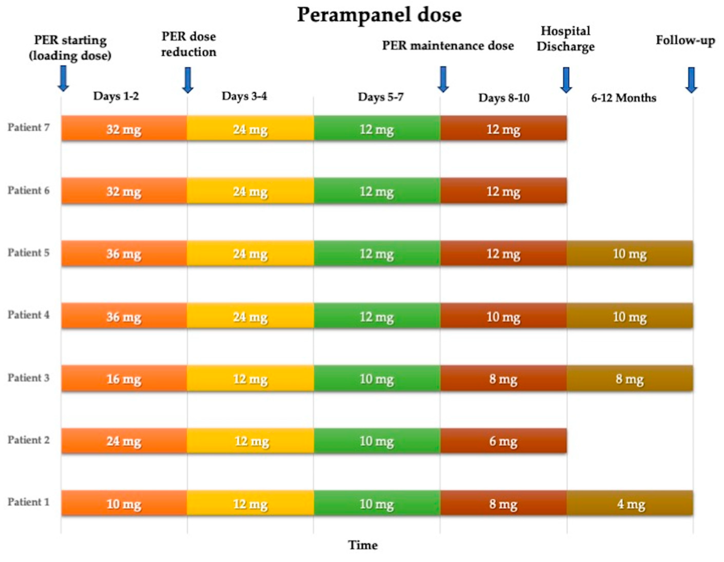
| Patient | Age/Gender | Epilepsy History | SE Type | SE Etiology | STESS | Previous ASMs | Other Treatments | ICUs Access | Time to SE Onset and PER Administration | Total SE Duration | Time between PER Administration and SE Resolution | Death | Epilepsy’s Outcome at 6–12 Months |
|---|---|---|---|---|---|---|---|---|---|---|---|---|---|
| Nr. 1 | 49 yrs/F | Yes | Focal NCSE with aphasia | Autoimmune SLE | 1 | 2 (LEV, LCM) | No | No | 48 h | 72 h | 24 h | No | Seizure-free |
| Nr. 2 | 91 yrs/F | Yes | Focal inhibitory SE | Systemic infection due to SARS-CoV-2 | 2 | 3 (LEV, LCM, VPA) | No | No | 48 h | 72 h | 24 h | Yes (pulmonary embolism) | - |
| Nr. 3 | 42 yrs/F | Yes | Focal tonic SE | Systemic bacterial sepsis | 0 | 2 (LEV, LCM) | No | No | 24 h | 48 h | 24 h | No | Seizure-free |
| Nr. 4 | 61 yrs/F | Yes | Focal NCSE with aphasia | Autoimmune: multiple sclerosis reactivation | 0 | 2 (LEV, VPA) | Steroids | No | 48 h | 72 h | 24 h | No | Only some auras |
| Nr. 5 | 70 yrs/M | No | Focal clonic motor SE | NORSE of presumed autoimmune etiology | 3 | 2 (LEV, LCM) | Steroids, intravenous immunoglobulin | Yes | 48 h | 96 h | 48 h | No | Seizure-free |
| Nr. 6 | 70 yrs/M | No | Generalized NCSE with coma | NORSE of presumed autoimmune etiology | 5 | 2 (LEV, LCM) | Intravenous immunoglobulin | Yes | 48 h | 96 h | 48 h | Yes (acute respiratory failure) | - |
| Nr. 7 | 62 yrs/F | No | Generalized NCSE with coma | Systemic infection due to SARS-CoV-2 | 4 | 3 (LEV, LCM, VPA) | No | Yes | 24 h | 72 h | 48 h | Yes (pulmonary embolism) | - |
Disclaimer/Publisher’s Note: The statements, opinions and data contained in all publications are solely those of the individual author(s) and contributor(s) and not of MDPI and/or the editor(s). MDPI and/or the editor(s) disclaim responsibility for any injury to people or property resulting from any ideas, methods, instructions or products referred to in the content. |
© 2023 by the authors. Licensee MDPI, Basel, Switzerland. This article is an open access article distributed under the terms and conditions of the Creative Commons Attribution (CC BY) license (https://creativecommons.org/licenses/by/4.0/).
Share and Cite
Nilo, A.; Vogrig, A.; Belluzzo, M.; Lettieri, C.; Verriello, L.; Valente, M.; Pauletto, G. Efficacy of Perampanel in Refractory and Super-Refractory Status Epilepticus with Suspected Inflammatory Etiology: A Case Series. Pharmaceuticals 2024, 17, 28. https://doi.org/10.3390/ph17010028
Nilo A, Vogrig A, Belluzzo M, Lettieri C, Verriello L, Valente M, Pauletto G. Efficacy of Perampanel in Refractory and Super-Refractory Status Epilepticus with Suspected Inflammatory Etiology: A Case Series. Pharmaceuticals. 2024; 17(1):28. https://doi.org/10.3390/ph17010028
Chicago/Turabian StyleNilo, Annacarmen, Alberto Vogrig, Marco Belluzzo, Christian Lettieri, Lorenzo Verriello, Mariarosaria Valente, and Giada Pauletto. 2024. "Efficacy of Perampanel in Refractory and Super-Refractory Status Epilepticus with Suspected Inflammatory Etiology: A Case Series" Pharmaceuticals 17, no. 1: 28. https://doi.org/10.3390/ph17010028
APA StyleNilo, A., Vogrig, A., Belluzzo, M., Lettieri, C., Verriello, L., Valente, M., & Pauletto, G. (2024). Efficacy of Perampanel in Refractory and Super-Refractory Status Epilepticus with Suspected Inflammatory Etiology: A Case Series. Pharmaceuticals, 17(1), 28. https://doi.org/10.3390/ph17010028