Article

Fabrication of Celecoxib PVP Microparticles Stabilized by Gelucire 48/16 via Electrospraying for Enhanced Anti-Inflammatory Action

by
Samar Zuhair Alshawwa
1,
Thanaa A. El-Masry
2,
Engy Elekhnawy
3,
Hadil Faris Alotaibi
1,
Al-Sayed Sallam
4 and
Dalia H. Abdelkader
5,*
1
Department of Pharmaceutical Sciences, College of Pharmacy, Princess Nourah bint Abdulrahman University, P.O. Box 84428, Riyadh 11671, Saudi Arabia
2
Pharmacology and Toxicology Department, Faculty of Pharmacy, Tanta University, Tanta 31527, Egypt
3
Pharmaceutical Microbiology Department, Faculty of Pharmacy, Tanta University, Tanta 31527, Egypt
4
Al-Taqaddom Pharmaceutical Industries, Amman 11947, Jordan
5
Pharmaceutical Technology Department, Faculty of Pharmacy, Tanta University, Tanta 31527, Egypt
*
Author to whom correspondence should be addressed.
Pharmaceuticals 2023, 16(2), 258; https://doi.org/10.3390/ph16020258
Submission received: 28 December 2022 / Revised: 29 January 2023 / Accepted: 2 February 2023 / Published: 8 February 2023

Abstract

:
Electrospraying (ES) technology is considered an efficient micro/nanoparticle fabrication technique with controlled dimensions and diverse morphology. Gelurice® 48/16 (GLR) has been employed to stabilize the aqueous dispersion of Celecoxib (CXB) for enhancing its solubility and oral bioavailability. Our formula is composed of CXB loaded in polyvinylpyllodine (PVP) stabilized with GLR to formulate microparticles (MPs) (CXB-GLR-PVP MPs). CXB-GLR-PVP MPs display excellent in vitro properties regarding particle size (548 ± 10.23 nm), zeta potential (−20.21 ± 2.45 mV), and drug loading (DL, 1.98 ± 0.059 mg per 10 mg MPs). CXB-GLR-PVP MPs showed a significant (p < 0.05) higher % cumulative release after ten minutes (50.31 ± 4.36) compared to free CXB (10.63 ± 2.89). CXB exhibited good dispersibility, proved by X-ray diffractometry (XRD), adequate compatibility of all components, confirmed by Fourier-Transform Infrared Spectroscopy (FTIR), and spherical geometry as revealed in scanning electron microscopy (SEM). Concerning our anti-inflammatory study, there was a significant decrease in the scores of the inflammatory markers’ immunostaining in the CXB-GLR-PVP MPs treated group. Also, the amounts of the oxidative stress biomarkers, as well as mRNA expression of interleukins (IL-1β and IL-6), considerably declined (p < 0.05) in CXB-GLR-PVP MPs treated group alongside an enhancement in the histological features was revealed. CXB-GLR-PVP MPs is an up-and-coming delivery system that could be elucidated in future clinical investigations.

1. Introduction

Celecoxib (CXB) is a Class II candidate that belongs to nonsteroidal anti-inflammatory drugs. It has superior anti-inflammatory, antipyretic, and analgesic potential [1]. Its pharmacological properties are attributed to hampering the activity of the COX-2 enzyme and decreasing the synthesis of prostaglandin (PG) [2]. Although it has several magic pharmacological features mentioned above, it possesses several formulation obstacles hindering its optimal delivery after oral administration. Poor aqueous solubility of CXB leads to a low dissolution rate and hence, low oral bioavailability [3]. So, improving the aqueous solubility of CXB in an urgent formulation is a necessity enabling it to exert an immediate anti-inflammatory action. The process of inflammation is a protective response of the human cells toward various inducers, such as infections, tissue injury, and certain chemical compounds. During this process, the induction of some reactions occurs via certain mediators, which leads to the occurrence of inflammation [4]. These mediators, like interleukin-6 (IL-6), interleukin 1 beta (IL-1β), and tumor necrosis factor-alpha (TNF-α), start and provoke the inflammatory process [5]. The enzymes that are involved in the inflammatory reaction, like cyclooxygenase 2 (COX-2), have the ability to increase the release of prostaglandins (PGs) during the inflammatory process [6].
Diverse formulations like micro-/nano-emulsions, liposomes, mixed micelles, and solid dispersion (SD) systems were previously utilized to enhance the solubility of poorly soluble agents [7,8,9,10,11]. Recently, efficient size reduction techniques like the fabrication of nano/microparticles via electrospraying (ES) are also included [12]. ES is a potential technology for the creation of microparticles (MPs) ideal for various therapeutic applications because it is a facile, uncomplicated, and direct method. Furthermore, it is crucial for the biomedical industry because of its adaptability to encapsulate pharmaceuticals in MPs and manage the processing of biodegradable polymeric MPs after tight adjustment of process variables, including feed rate, operating voltage, the distance separating the needle tip and collecting drum, etc. [12].
In our study, we utilized Gelurice® 48/16 (GLR) as a micellar solubilizer enhancing the aqueous solubility of CXB. In contrast to typical gelucires, Gelucire® 44/14 and Gelucire® 50/13, which are synthesized from lauroyl polyoxyl-32glycerides NF and stearoyl polyoxyl-32glycerides NF, respectively, Gelucire® 48/16 is a unique type composed from PEG-32 stearate [13]. The hydrophobic interface between drug particles and the micelle core created by different Gelucire grades may vary depending on their chemical characteristics. In addition, it has been suggested that Gelucire® 50/13 can be found in various crystalline forms that may affect its solubilizing abilities. Gelucire® 44/14 has surface-active properties and a great capacity to self-emulsify during dissolving in a water-based medium to produce fine emulsions. Similar to Gelucire® 48/16, which could effectively formulate micelle encapsulating hydrophobic drug when it comes into contact with an aqueous fluid [13].
The versatile behavior of different Gelucire® grades could be attributed to differences in HLB values leading to the changeable solubilizing power of hydrophobic drugs. For example, the improvement in indomethacin solubility with Gelucire® 48/16 aqueous solutions was more noticeable than with Gelucire® 44/14 or Gelucire® 50/13 aqueous solutions. This could be the result of Gelucire® 48/16 having greater HLB values, which could improve the drug’s miscibility and dispersibility inside the carrier and increase its solubility [14].
Polyvinylpyllodine (PVP) has also been included in our study for many purposes. PVP is a highly safe, water-soluble polymer, biocompatible. In addition, it is very soluble in a variety of organic solvents, making it a common ingredient in many pharmaceutical formulation processes [15]. It is widely employed in electrospinning/electrospraying to formulate nano-delivery systems due to its ideal properties of spinnability. PVP has previously been used to encapsulate natural plant extracts such as green tea [16]. Additionally, PVP could be utilized as a solubilizer, promoting higher hydrophilicity in pharmaceutical formulations. PVP’s impact on improved bioavailability makes it an attractive polymer in newly developed controlled or targeted drug delivery systems [17].
In our study, we formulated CXB into GLR-PVP MPs to enhance its poor aqueous solubility to increase CXB’s oral bioavailability, which could be confirmed by in vivo anti-inflammatory in experimental rats.

2. Results

2.1. Solubility Study of CXB in Gelurice® 48/16

Increasing GLR 48/16 concentration could significantly (p < 0.05) increase CXB’s solubility in 6% PVP aqueous solution, as displayed in Table 1. Starting with 6% PVP, the solubility of CXB was approximately 7 µg/mL and proportionally enhanced with increasing GLR concentration up to 25% w/v till the saturation point with no significant increase at 30% w/v. GLR concentration of 25% w/v was chosen to prepare the electrospraying solution. Also, values of Gibbs free energy change after the addition of Gelurice® 48/16 have been calculated using the following equation [18].
Δ G o tr = 2.303   RT   log   S s S 0
where Ss/S0 is the ratio of molar solubility of CXB in 6% PVP aqueous solution with different concentrations of GLR 48/16 related to 6%PVP aqueous solution with no GLR. ΔGotr significantly (p < 0.05) increased with increased GLR 48/16 till the concentration of 25% w/v.

2.2. Characterization of GLR-PVP MPs Loaded with CXB

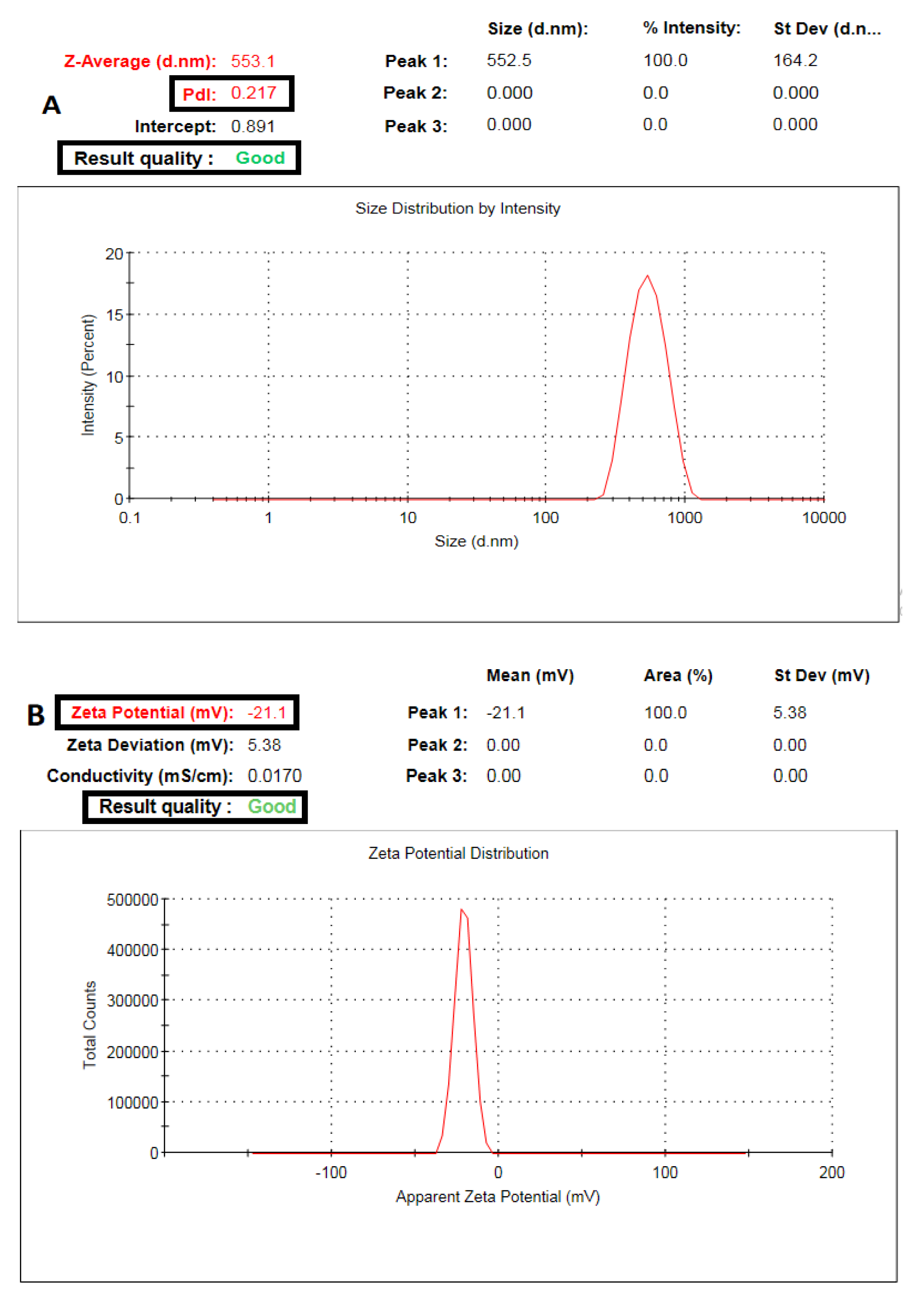
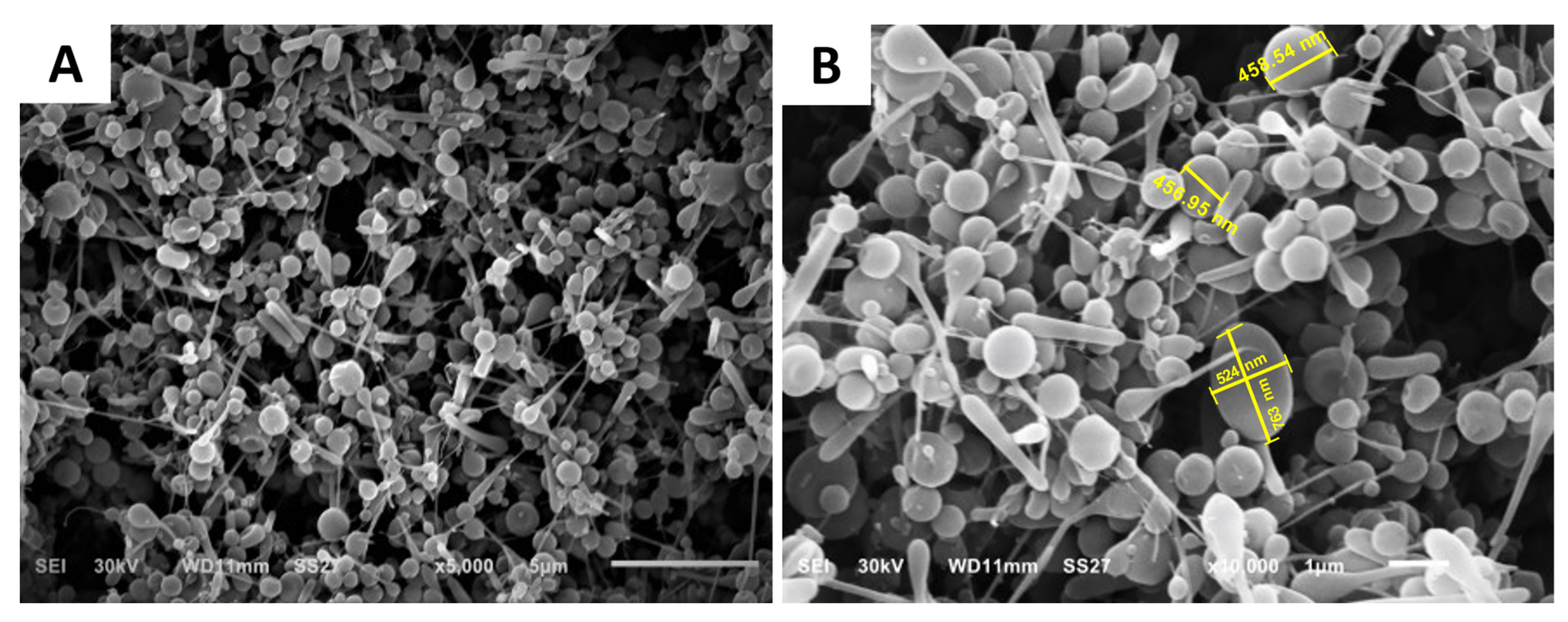
2.2.1. Particle Size, PDI, Zeta Potential, Drug Loading (DL), and Surface Morphology

CXB-GLR-PVP MPs particle size average was 553.1 nm, and the PDI value was 0.217 (Figure 1A). The determined zeta potential was equal to −21.1 mV (Figure 1B). Drug loading results showed that each 10 mg of CXB-GLR-PVP MPs entraps approximately 1.98 ± 0.1 mg of CXB. Morphological examination using SEM (Figure 2) showed spherical CXB-GLR-PVP MPs with highly matched measurements of their diameters compared with the readings of dynamic light scattering (DLS).

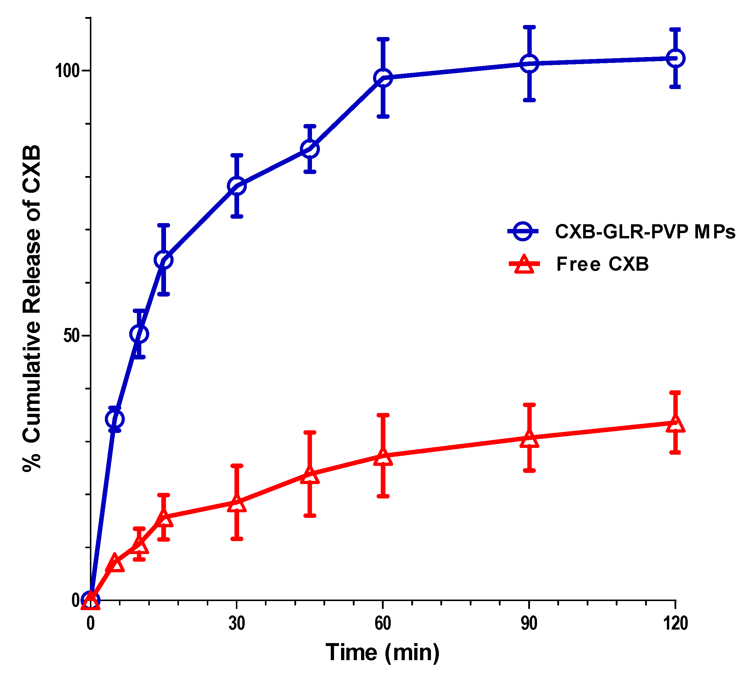
2.2.2. In Vitro Release Pattern

Regarding in vitro release behavior of CXB-GLR-PVP MPs, they showed a biphasic release pattern (Figure 3) starting with an initial burst at the first 10 min (% cumulative release equal to 50.31 ± 4.36) followed by a gradual increase of CXB mass released from microparticles till two hours. A significantly enhanced release (p < 0.01) of CXB from GLR-PVP polymeric matrix compared to free CXB. After 10 min, 50.3 ± 4.4% and 10.6 ± 2.8% have been cumulatively released from CXB-GLR-PVP MPs and free CXB, respectively.
The amount of released CXB increased significantly (p < 0.01) till reaching approximately 100% at 120 min, whereas free CXB exhibited a maximum cumulative % release of 33.6 ± 5.6 at the end of the experiment.

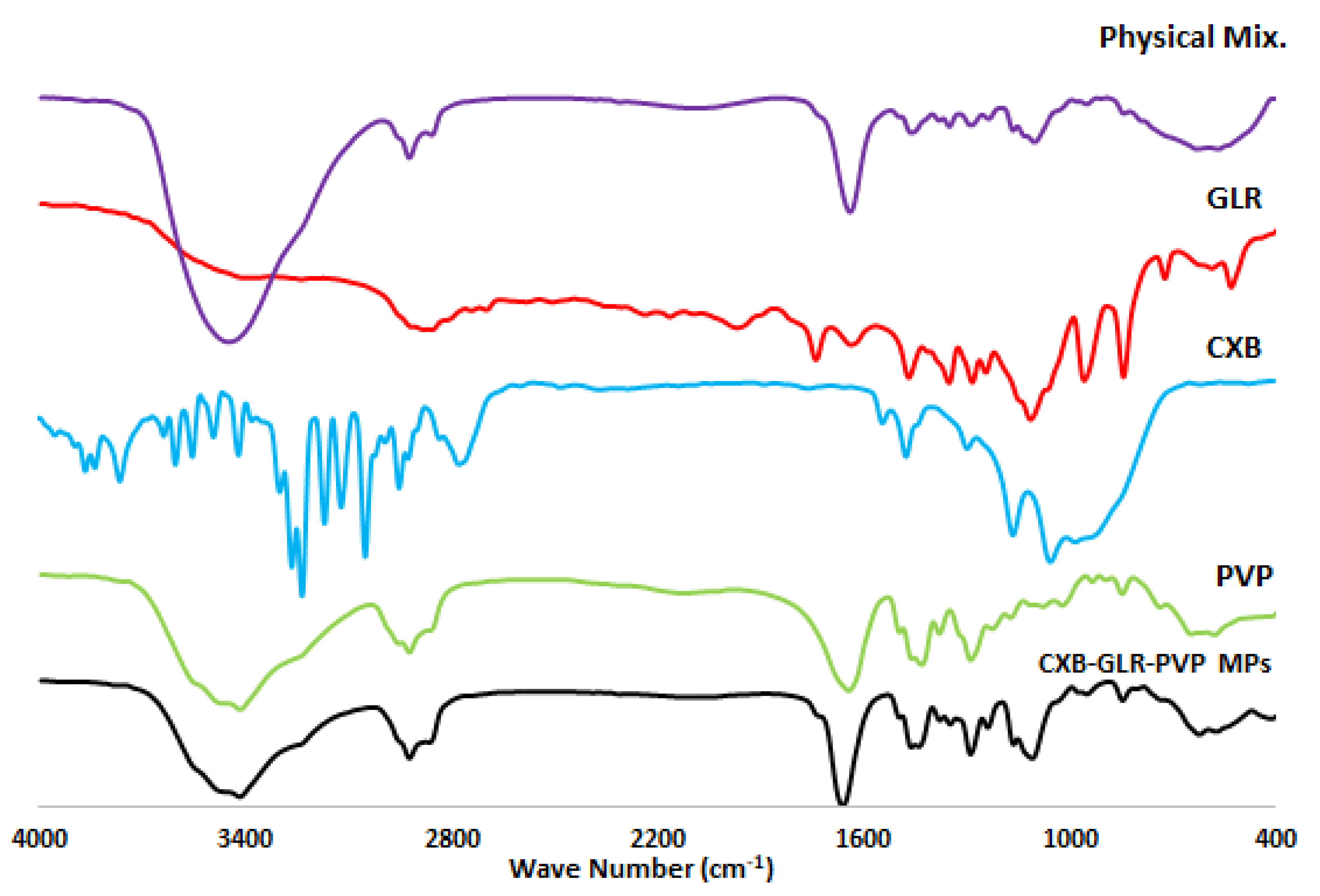
2.2.3. FTIR Spectroscopy

IR peaks of CXB-GLR-PVP MPs, PVP K30, Gelurice® 48/16, and free unprocessed CXB are shown in Figure 4. Table 2 shows the individual peaks of free CXB, PVP K30, and Gelurice® 48/16. The spectrum of CXB-GLR-PVP MPs shows no extra bands with slight broadness observed at 3417 cm−1 (O-H stretching). Mostly, all the characteristic peaks of PVP K30 and Gelurice® 48/16 are located at the same position in the spectrum of CXB-GLR-PVP MPs compared with their individual IR spectrums. Also, less intense peaks of CXB at 1162.60, 1347 cm−1 have been displayed in the IR spectrum of CXB-GLR-PVP MPs. The physical mixture prepared for IR analysis contained the amount of CXB, PVP K30, and Gelurice® 48/16 mixed with their ratio equivalent to their amounts in CXB-GLR-PVP MPs. FT-IR spectrum of the physical mixture displayed no observed interaction between the components of our microparticle system. Additionally, the main characteristic peaks of CXB (S=O, 1348, 1164 cm−1) were shown at the same position compared with the individual spectrum of free CXB.

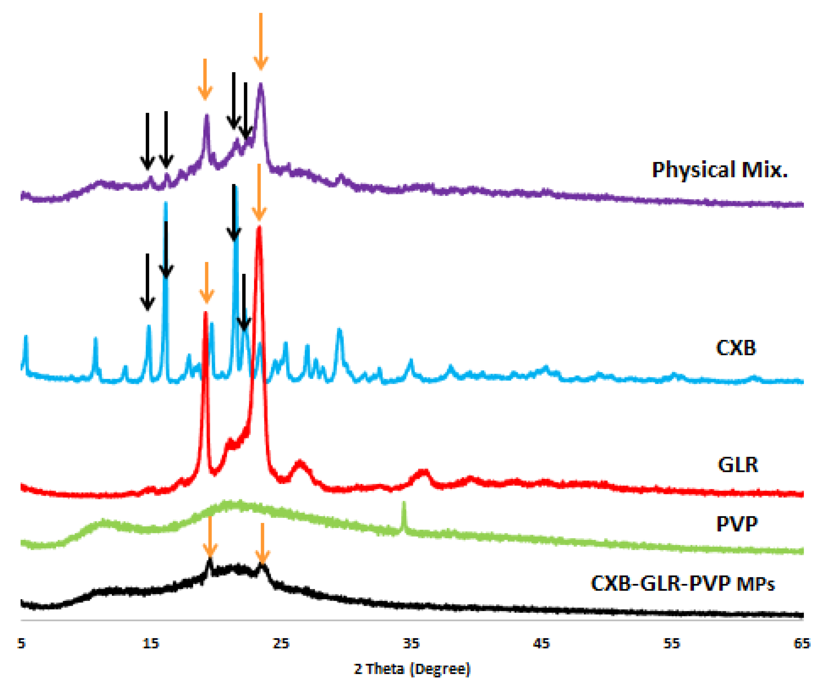
2.2.4. X-ray Diffractometry (XRD)

XRD chart (Figure 5) demonstrates the peaks of pure unprocessed CXB, GLR, PVP, and CXB-GLR-PVP MPs to investigate the crystallinity of CXB after the electrospraying process. As shown in Figure 5, all the characteristic peaks of CXB completely disappeared after loading in GLR PVP aqueous solution. Also, the pattern of GLR-PVP MPs is highly similar to that of PVP plus two additional peaks at 2Ꝋ values of 19.52 and 23.7 (the same position of two distinct peaks showed on the GLR pattern). Regarding the XRD chart of the physical mixture, the same four distinguishing peaks of CXB are still observed with less intensity at 2Ꝋ values of 14.8, 16.1, 19.2, and 21.5.

2.3. In Vivo Anti-Inflammatory Study

2.3.1. Edema Weight

As exposed in Figure 6, a remarkable decline (p < 0.05) in the edema weight was noticed in group V when compared with groups II, III, and IV. Group V revealed a marked decline with a percentage of 81.58% in relation to group II. In addition, it caused a marked diminish in relation to groups III and IV, with percentages of 65% and 63.61%, respectively.

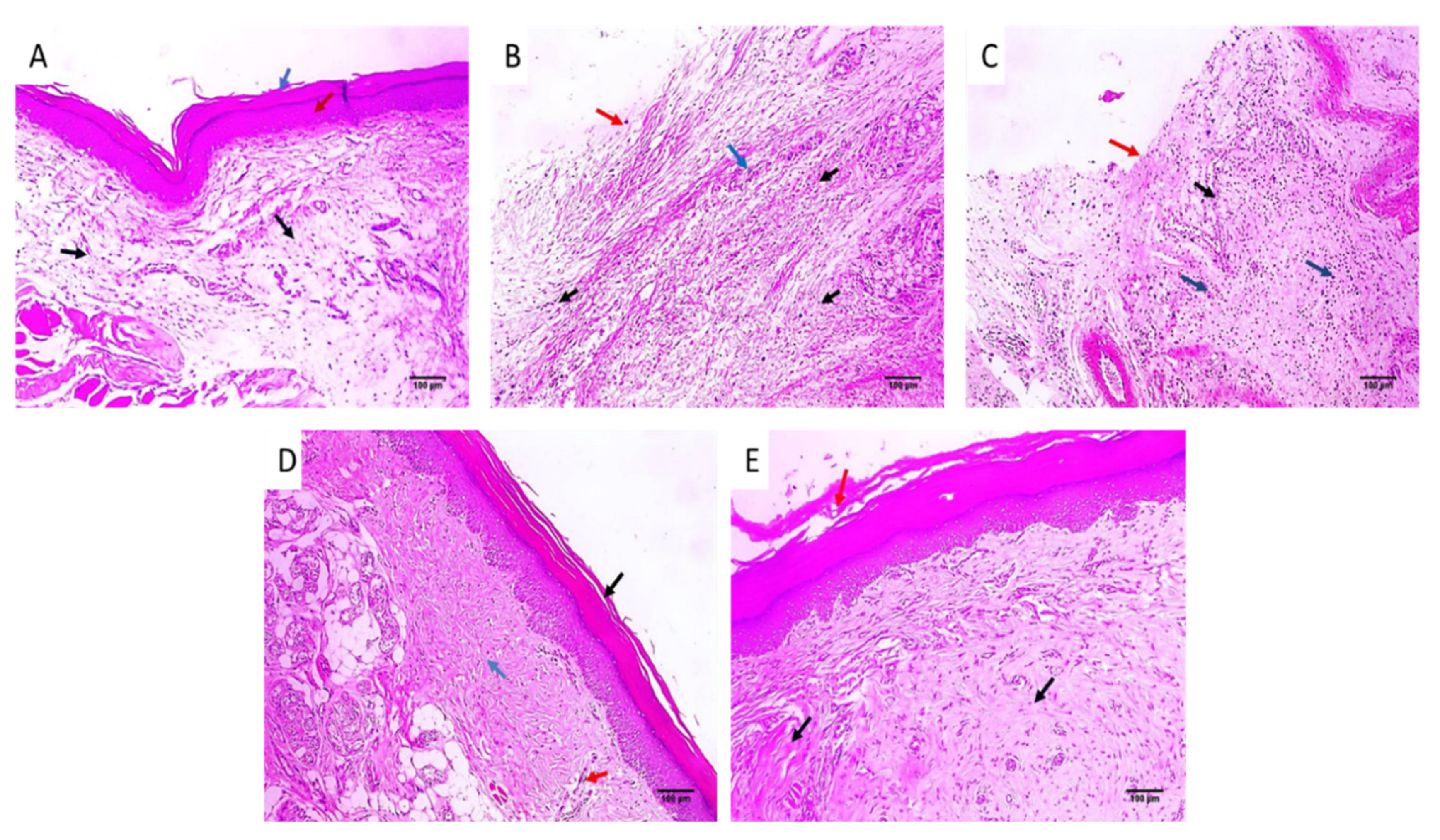
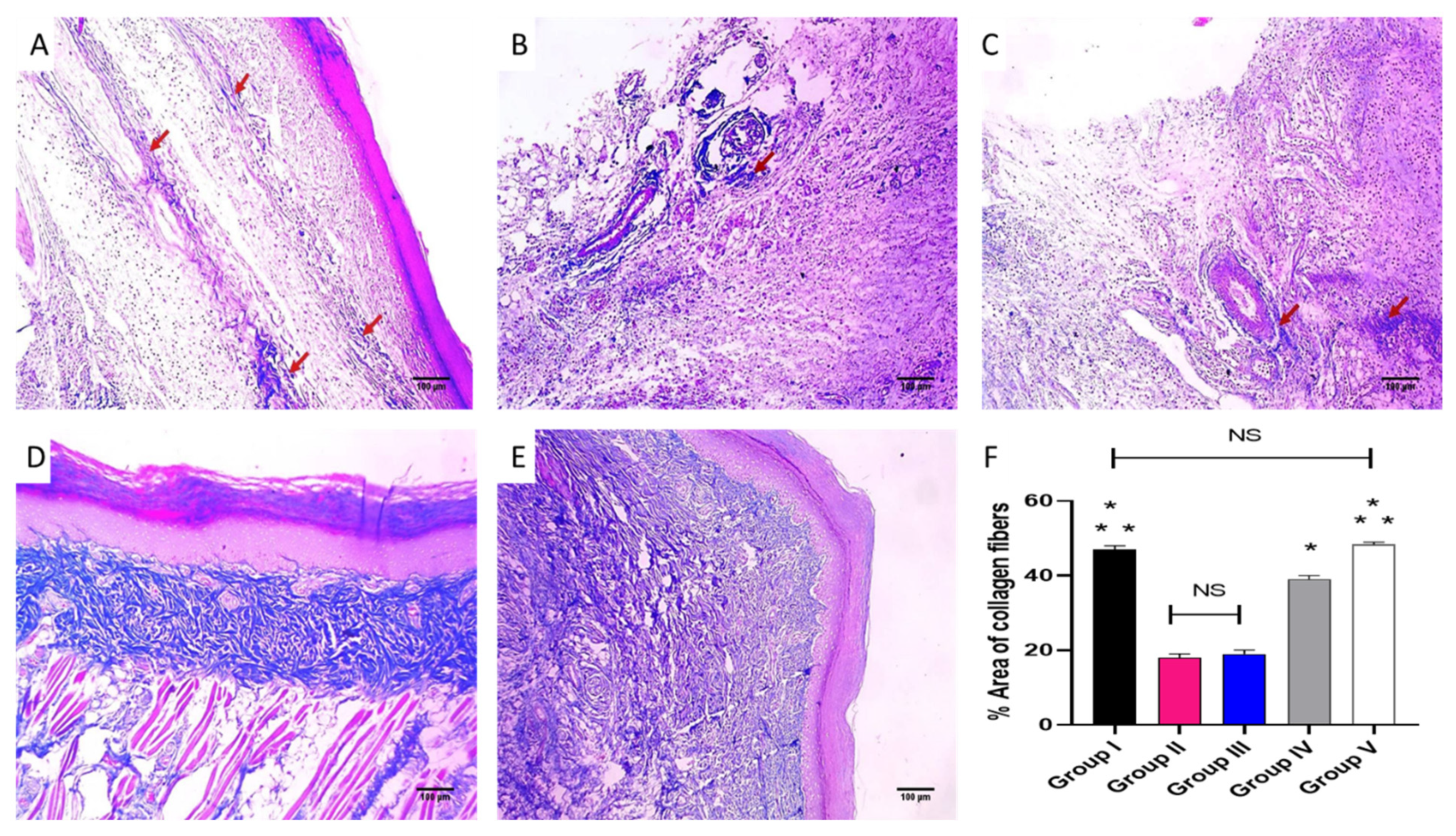
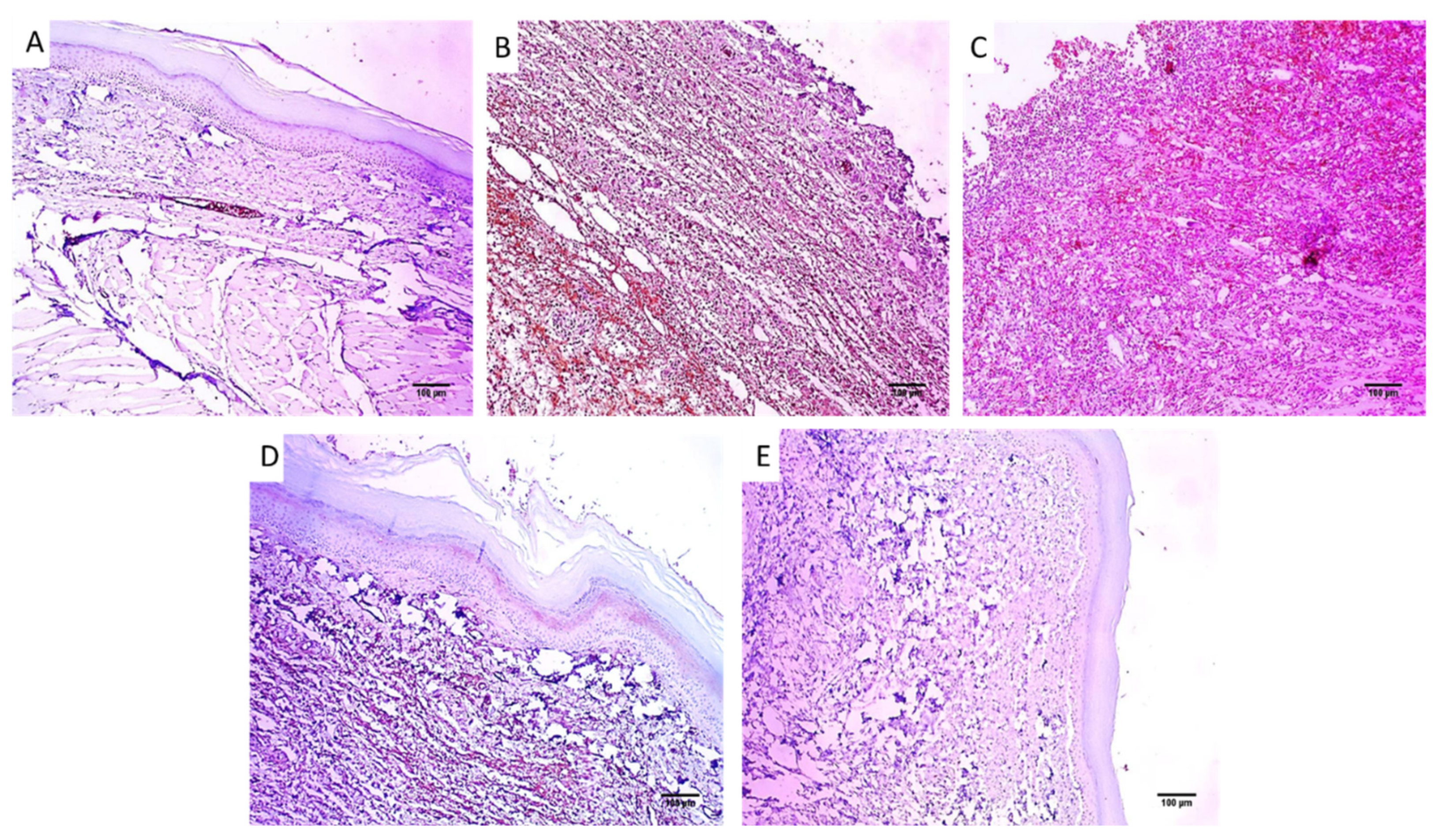
2.3.2. Histological Investigations

Paw sections of the studied experimental groups are revealed in Figure 7 and Figure 8 after staining with H&E and Masson’s trichrome stain, respectively.

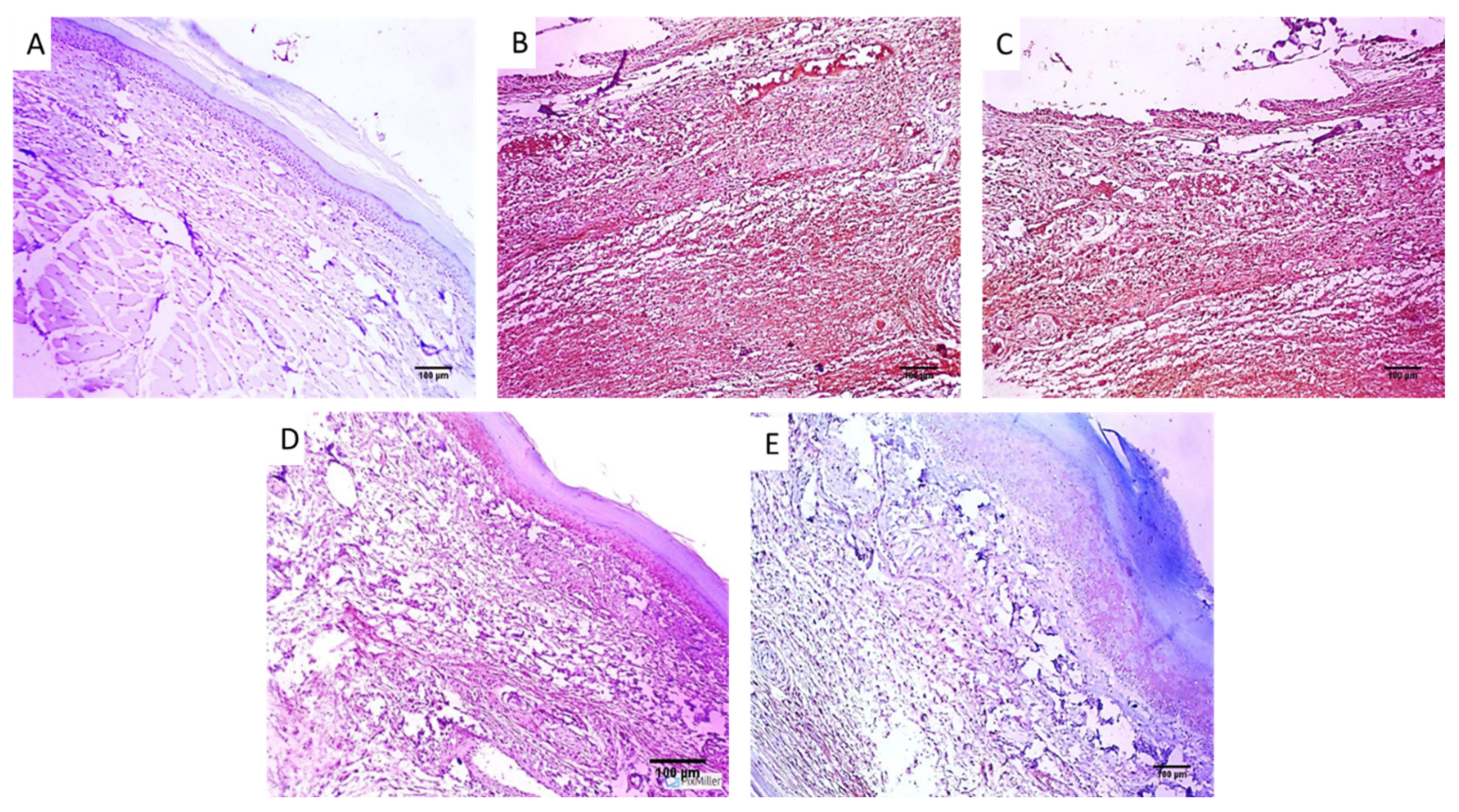
2.3.3. Immunohistochemical Investigations

The COX-2 and TNF-α immunostained paw tissue sections of the studied groups are revealed in Figure 9 and Figure 10.

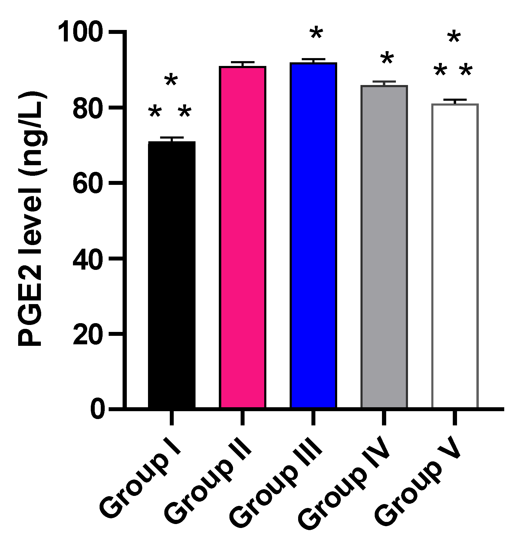
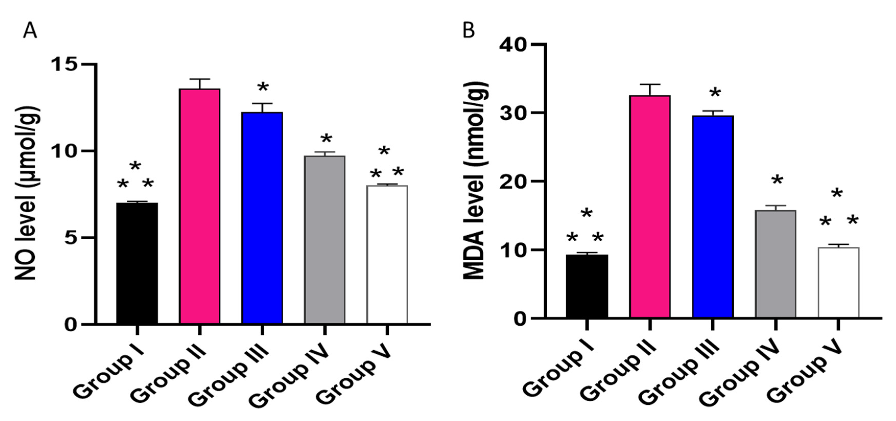
2.3.4. Pro-Inflammatory and Oxidative Stress Indicators

The influence of the placebo GLR PVP MPs, free CXB, and CXB-GLR-PVP MPs was investigated on PGE2 by ELISA (Figure 11) and NO as well as MDA (Figure 12) by colorimetric assay.
Group V revealed a considerable decline in PGE2 (11.11%) in relation to group II. Besides, it caused a marked decline in the level of PGE2 in relation to groups III and IV (13.04% and 5.88%, respectively).
Concerning the level of NO, in relation to group I, group II showed a noticeable rise with a percentage of 85.71%. Group V exposed a substantial reduction with a percentage of 37.69% in relation to group II.
Regarding the MDA level, in relation to group I, group II showed a noticeable rise (244.44%). Group V showed a marked lessening in MDA with a percentage of 67.74% in relation to group II.

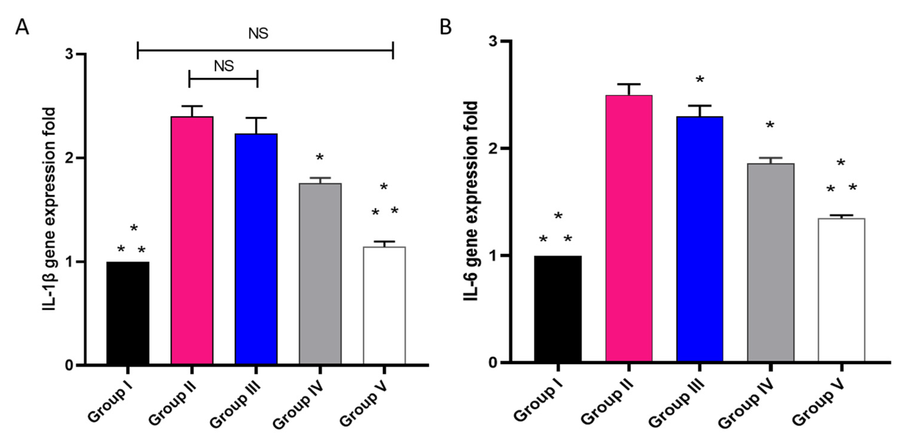
2.3.5. The mRNA Expression of IL-1β as Well as IL-6

The influence of placebo GLR PVP MPs, free CXB, and CXB-GLR-PVP MPs on IL-1β and IL-6 was elucidated by qRT-PCR, as revealed in Figure 13. In relation to group I, group II showed a significant rise in the expression of the IL-1β gene with a percentage of 130%. Group V showed a substantial decline, with a percentage of 52.17% in relation to group II.
Regarding IL-6 gene expression, in relation to group I, group II manifested a noticeable enhancement with a percentage of 140%. Group V manifested a marked decline with a percentage of 44.17% in relation to group II.

3. Discussion

Oral bioavailability is highly controlled by drug dissolution through GIT. So, aqueous solubility is a significant factor that should be fully considered, especially for Class II drugs. Celecoxib is a poorly soluble drug providing good stability at different pHs in GIT segments (pKa 11.1) [3]. The enhancement of aqueous solubility has been studied in several manuscripts using different formulation techniques such as solid dispersion [3,20], microemulsion [19], β-cyclodextrins’ complexation [21,22], physical manipulation of CXB’s solid state [23], etc. The reported aqueous solubility of CXB has been recorded to be 1.15 µg/mL. Additionally, PVP K30 has a crucial role in exaggerating CXB’s solubility and stability. It could efficiently entrap CXB into its matrix via hydrogen bonding formation producing a thermodynamically stable architecture of CXB in its amorphous state [24,25].
Gelurice® 48/16 grade is a typical amphiphilic surfactant efficiently used for micellar solubilization. The enhancement of GXB’s solubility by Gelurice® 48/16 could be attributed to additional wettability plus the micellar solubilization [13]. Values of Gibbs free energy change are an indication of the process of CXB molecules transfer into micelles offered by Gelurice® 48/16. Increasing the negative value of ΔGotr means spontaneous solubilization of CXB that is enhanced with a higher concentration of GLR 48/16 till the saturation (Table 1) [18].
The electrospraying technique produces spherical microparticles (Figure 2) with a uniform size distribution and small PDI value (Figure 1A) [12]. Additionally, a perfect match was observed between the measurements of microparticles displayed on scanning electron microscopy and the results of dynamic light scattering. Also, the low value of PDI has been confirmed by SEM visualization. The particle size of CXB-GLR-PVP MPs mostly lies in the same ranges indicating the reproducibility of the electrospraying technique as the optimum method of preparation.
Regarding surface charge (Figure 1B), CXB-GLR-PVP MPs showed a well-established negative value equal to −20.21 ± 2.45 mV controlling optimum stability with little liability to flocculation or aggregation. The negative value might be attributed to the combinatory effect of PVP and GLR 48/18 [7,12,26]. The loading of CXB into the PVP matrix concurrently with micellar solubilization using GLR 48/16 could be an effective polymeric combination that efficiently entraps CXB leading to a higher drug loading capacity (0.198 mg of CXB per 10 mg of electrosprayed microparticles) [9,15].
The increased solubility of CXB in the presence of Gelurice® 48/16 might be due to the drug’s increased wettability and micellar solubilization, which result in a high release profile of CXB-GLR-PVP MPs. The presence of Gelurice® 48/16 in aqueous solutions could reduce the angle of contact between drug particles and water, improving drug particle wettability and, consequently, accelerating drug release. Additionally, the coexistence of hydrophilic carriers like Gelurice® 48/16 may decrease the hydrophobicity of the drug particle surfaces, which will subsequently increase interaction with the aqueous medium and boost the rate of CXB’s dissolution [7]. Several previous studies showed the enhanced release performance exhibited after the addition of Gelurice® 48/16 [27,28,29].
In our manuscript, the exaggerated release profile of CXB-GLR-PVP MPs could be attributed to more than one promoter. Firstly Gelurice® 48/16 enhances CXB’s wettability and dispersibility via its surfactant properties. Secondly, the solubilizing effect offered by PVP, then ES, which has a stabilizing effect due to efficient drying keeping CXB in submicron level coated with a matrix composed of PVP and GLR [8].
Generally, the IR spectrum of CXB-GLR-PVP MPs indicates that no chemical interaction occurs during micellar solubilization and formulation of electrosprayed CXB-GLR-PVP MPs [9,30]. The broadness of OH peaks could be attributed to possible H- bonding formation between CXB and PVP background [15]. Additionally, less intense peaks of CXB might indicate the enclosed entrapment of CXB into the PVP matrix [15]. The peaks of CXB shown in the IR spectrum of the physical mixture indicate no possible interaction occurred with the active functional groups of PVP K30 and Gelurice® 48/16. Generally, the overall fingerprint of the spectra of the physical mixture and CXB-GLR-PVP MPs are quite different, especially in the range between 2000 to 400 cm−1 [15]. Regarding the XRD chart, the disappearance of CXB’s peaks in the XRD chat ensures the presence of CXB in its amorphous soluble form [31]. A combined background of PVP and GLR has been displayed in the pattern of CXB-GLR-PVP MPs gathering the peaks of both PVP and GLR (2Ꝋ values of 19.52 and 23.7), indicating that PVP and GLR have contributed to the formation of polymeric microparticles encapsulating CXB [32]. The diffractogram of the physical mixture showed less intense peaks of CXB due to the low loading of weight per unit mass of the physical mixture compared with pure free CXB. The presence of the main peaks of CXB in the chart of the physical mixture indicates that CXB still exists in its crystalline form [32].
The inflammation process leads to a progression of various pathophysiological proceedings that induce the development of diseases [33]. Different pharmacological models performed on animals are used to elucidate the anti-inflammatory action of various compounds. A model usually utilized to estimate the anti-inflammatory activity of different drugs is the paw edema induced by carrageenan. Carrageenan is an agent that has the capacity to provoke the discharge of many pro-inflammatory in addition to inflammatory mediators [34].
Herein, group V or the treated group with CXB-GLR-PVP MPs exhibited a marked decline (p < 0.05) in the weight of the paw edema in relation to groups III or the treated group with free CXB and group IV or the treated group with placebo GLR PVP MPs. Edema is one of the important characteristics of the local inflammatory processes, which involves the increase of the interstitial fluids in the affected tissues [35]. Edema has a negative effect on tissue functions because these fluids make the diffusion of nutrients and oxygen more difficult. This could adversely affect the cellular metabolism in the affected tissue [36].
During the inflammation process, many cells, like neutrophils and macrophages, are stimulated to travel toward the site of the inflammation, generating huge amounts of mediators. Such substances are able to provoke an inflammatory response [37]. It is known that CXB has antipyretic, analgesic, and anti-inflammatory potential. Certain studies have been conducted to formulate CXB to improve its bioavailability and/or lessen its side effects [38,39,40]. The stimulation of the COX-2 pathway and the resulting enhanced discharge of PGE2 are major consequences of the acute inflammation of the carrageenan model [41]. In the current study, group II, or the positive control (carrageenan group), exhibited an enhancement of the COX-2 and TNF-α immunostaining (score 3). Interestingly, this finding was significantly lessened in the group treated with CXB-GLR-PVP MPs (group V). Besides, the histopathological analysis of the paw skin tissues showed that the CXB-GLR-PVP MPs treated group did not have any inflammation or edema, with a noticeable rise in the density of collagen fibers.
Regarding the inflammatory and pro-inflammatory biomarkers, CXB-GLR-PVP MPs revealed a marked decline (p < 0.05) in the PGE2, MDA, NO, IL-1β, and IL-6 (using ELISA and qRT-PCR) when compared to the free CXB. Comparable results were stated by El-Medany et al. [42] and Bazan et al. [39] in acetic acid-induced colon injury.

4. Material and Methods

4.1. Chemicals and Material

All the utilized reagents, chemicals, and solvents are of high-grade purity within the acceptable standard obtained from Merck, USA.

4.2. Solubility Study of CXB in Gelurice® 48/16

A solubility study of CXB has been conducted according to Zhang et al. [43] with slight adaptation to determine the optimum concentration of GLR 48/16 in 6% PVP aqueous solution (the same composition of the electrosprayed polymeric solution for MPs fabrication). Variable concentrations of GLR 48/16 were prepared in a 6% PVP aqueous solution. An extra amount of CXB was added to GLR concentrations, followed by stirring overnight for 24 h at room temperature. After equilibrium, undissolved CXB was discarded after centrifugation. The samples were filtered by a membrane filter (0.45 μm). After proper dilution, CXB samples were assayed using a UV-Vis spectrophotometer (Evolution 300 spectrophotometer, Waltham, MA, USA) at 254 nm [44]. GLR 48/16 concentration which provides the highest CXB’s solubility, was selected to prepare CXB-GLR-PVP aqueous solution injected via electrospraying.

4.3. Electrospraying Process

Firstly, the polymeric solution composed of 6% PVP and 25% GLR loaded with CXB (50 mg) has been sprayed using NANON-01B, MECCO, LTD, Japan through a stainless steel needle provided with high voltage supply (28 KV). The spraying feed rate was adjusted to 0.1 mL/hr. The electrosprayed nozzles have been collected on a metallic drum coated with an aluminum sheet. The space separating the needle tip and the drum has been set at 10 cm [15]. After ten hours of electrospraying, CXB-GLR-PVP MPs were collected and kept in a desiccator for further in vitro and in vivo experimentation.

4.4. Characterization of GLR-PVP MPs Loaded with CXB

In our study, CXB-GLR-PVP MPs formulated via electrospraying have been characterized as particle size, uniformity of size distribution, zeta potential, drug loading, in vitro release platform, scanning electron microscopy, spectroscopic analysis by Fourier-Transform Infrared Spectroscopy (FTIR), and X-ray diffractometry (XRD) to ensure their suitability to be employed for in vivo anti-inflammatory study in experimental rats.

4.4.1. Particle Diameter, Polydispersity Index (PDI), and Surface Charge

The average size of the particles (nm), uniformity of size distribution, and Zeta potential (mV) have been determined using Malvern Zetasizer Nano-zs90 (Malvern Instruments Ltd., Malvern, UK). A sample of CXB-GLR-PVP MPs (5 mg) was properly diluted with deionized water for precise analysis. All measurements have been performed in triplicate, expressed as mean ± SD [45].

4.4.2. Drug Loading (DL)

An accurately weighted mass of CXB-GLR-PVP MPs was dissolved in dimethylformamide (DMF): ethanol with a ratio of 1:1 to study the amount of CXB loaded in the matrix of PVP-GLR. The sample was filtered, then assayed using a UV-Vis spectrophotometer, as previously mentioned in Section 4.2. DL was calculated as follows [45].
DL = M a s s   o f   C X B   i n   M P S   ( m g ) M a s s   o f   M P s   ( m g )

4.4.3. In Vitro Release

The accurate weight of CXB-GLR-PVP MPs (20 mg) has been immersed in a beaker of release medium (phosphate buffered saline, PBS, 100 mL, pH = 6.8) [8,15]. The release medium was agitated using magnetic stirring at 100 rpm and 37 °C. At different time periods till 2 h, samples of 2 mL were withdrawn for UV analysis. An equivalent fresh media was replenished, maintaining the total volume of the release medium at 100 mL. The samples were filtered prior to UV assay, as revealed in Section 4.2. Also, the release of free unprocessed CXB has been studied using the same previous procedures. All the release experiments have been implemented in triplicates, and the data presented as a mean of % cumulative release versus the time intervals till two hours.

4.4.4. Scanning Electron Microscopy (SEM)

Scanning electron microphotographs of CXB-GLR-PVP MPs were visualized using JSM- 6510LV microscope (JEOL, Tokyo, Japan). The instrument was operated with an accelerated voltage equal to 30 kV. The sample was fixed on an aluminum carrier, and a sputter coater SPI-MODULE TM Sputter Coater (USA) was used to coat the sample with a gold film for 10 min. Measurements shown on SEM images were displayed using image J software [15].

4.4.5. Spectroscopic Analysis by FTIR

It was employed to determine the possible interaction that could occur during micellar formation and the electrospraying process to prepare electrosprayed CXB-GLR-PVP MPs. FTIR spectra of unprocessed free CXB, GLR 48/16, PVPK30, and CXB-GLR-PVP MPs were documented in the 4000–400 cm−1 range by FTIR spectrophotometer (Bruker Tensor 27, Germany). Approximately three milligrams of each sample were blended with 200 mg of KBr (IR grade), followed by intense compaction into a spherical disc [46]. KBr-FTIR spectra have been developed at an optimum resolution of 4 cm−1 by a number of scans equal to 64. The acquisition parameters have been set at a smoothing of 17 and a base correction of 80.

4.4.6. XRD

It is a significant investigation showing the presence of CXB in its crystalline or amorphous state after loading into PVP MPs. XRD was performed for unprocessed free CXB, GLR 48/16, PVP, and CXB-GLR-PVP MPs by APD 2000 PRO X-ray Diffractometer (GNR Instrumental Group, Italy). The X-ray diffractor was set at a current and source power equal to 25 mA and 35 kV, respectively, with 2Ꝋ angles ranging from 5 to 65 and a step size of 0.02 [47].

4.5. In Vivo Anti-Inflammatory Study

4.5.1. Rats

Fifty Wistar albino male rats (their average weight 183.24 ± 8.23 g) were obtained. Their weights had a range of 180 to 210 g, and they were supplied with water (filtered) in addition to a diet of standard pellets. Our experimental protocol was credited by the Research Ethical Committee at the Faculty of Pharmacy, Tanta University, Egypt (TP/RE/10/22p-0053).

4.5.2. Inflammation-Induced by Carrageenan

It was performed as previously described [30] (Supplementary Material).

4.5.3. Protocol of the Experiment

Rats were grouped into five categories (n = 10). Normal control (group I) was administered orally with 0.9% saline. Positive control (group II) was inflamed by carrageenan injection and administered orally with 0.9% saline. Groups III, IV, and V were inflamed by carrageenan injection and administered orally placebo GLR-PVP-MPs, unprocessed free CXB, and CXB-GLR-PVP MPs, respectively (amount of unprocessed free CXB and loaded into CXB-GLR-PVP MPs was kept equivalent regarding rats’ weight). After six hours, all animals were anesthetized and euthanized. Then, we cut the left and right paws at nearly the same position, and their weights were identified. The paw edema average weight was determined by calculating the difference between the right and left paw weights [48].

4.5.4. Histological Investigations

Tissues of the paws were conserved in a formalin solution, retained in paraffin wax, spliced, and stained using hematoxylin and eosin (H&E) and Masson’s trichrome stain for staining of collagen fibers [49].

4.5.5. Immunohistochemical Studies

Using ABclonal Technology kits (Woburn, MA, USA), the COX-2 and TNF-α immune expression in the paw tissues was elucidated. The obtained pictures were analyzed, and they were given scores based on the percentage of the staining. The scores ranged from 0 to 3 regarding the positive staining percentages as previously described [50].

4.5.6. ELISA and Colorimetric Assay

The quantity of prostaglandin E2 (PGE2) that are present in the tissues of paws in the different groups was determined by the ELISA kit obtained from Creative-Biolabs, New York, NY, USA. The absorbance was identified at 450 nm using a microtitration plate reader.
The amounts of nitric oxide (NO) and malondialdehyde (MDA) were determined by Biodiagnostic kits (Biodiagnostics, Egypt) at an absorbance of 540 nm.

4.5.7. qRT-PCR

The inflammatory cytokines mRNA expression of IL-1β and IL-6 was studied in the paw tissues [51]. We used the beta-actin gene [52] as a housekeeping gene, and the utilized primers are presented in Table S1. After extraction of the RNA using the Purelink™ RNA Mini Kit (Thermo Scientific, Waltham, MA, USA), RNA was turned into cDNA using the power™ cDNA synthesis kit. qRT-PCR was employed using Rotor-Gene Q 5plex (Qiagen, Hilden, Germany).

4.6. Statistics

Using Graph-Pad Software (prism 8), we performed ANOVA and post-hoc tests. Experiments were performed three times, and the obtained numbers are displayed as the mean ± standard deviation (SD). The significance was at p < 0.05.

5. Conclusions

Poorly soluble drugs could be efficiently entrapped into dry MPs after the utilization of carefully selected surfactant and hydrophilic polymer followed by ES. Gelurice® 48/16 has stabilized the system by improving the wettability concurrently with controlling the crystallinity of CXB. The presence of PVP potentiates the hydrophilicity of the aqueous dispersion and perfect electrospraying properties. Finally, the dried system with a submicron particle diameter of CXB completes the suitability of the micro delivery system to produce a significantly higher in vitro % cumulative release compared with unprocessed CXB. Hence, greater oral bioavailability could be expected. CXB-GLR-PVP MPs showed an improved anti-inflammatory capacity by causing a marked decline (p < 0.05) in the pro-inflammatory and inflammatory biomarkers in addition to decreasing the NO and MDA levels. These consequences occurred in substantial suppression of the COX-2 pathway.

Supplementary Materials

The following supporting information can be downloaded at: https://www.mdpi.com/article/10.3390/ph16020258/s1, Figure S1: Malvern′s Zetasizer graphs showing multiple measurements of particle size of CXB-GLR-PVP; Figure S2: Malvern′s Zetasizer graphs showing multiple measurements of ζ-potential of CXB-GLR-PVP; Table S1: The sequences of the utilized primers.

Author Contributions

D.H.A.: Conceptualization, Methodology, Investigation, Data curation, Software, Writing—original draft. E.E.: Investigation, Visualization, Methodology, Data curation, Writing—original draft. T.A.E.-M.: Supervision, Funding acquisition. S.Z.A.: Funding acquisition, Resources, Writing-Review. H.F.A.: Funding acquisition, Resources. A.-S.S.: Resources. All authors have read and agreed to the published version of the manuscript.

Funding

This research was funded by the Deanship of Scientific Research at Princess Nourah bint Abdulrahman University through the Research Funding Program (Grant No# FRP-1441-14).

Institutional Review Board Statement

Our experimental protocol was credited by the Research Ethical Committee at the Faculty of Pharmacy, Tanta University, Egypt (TP/RE/10/22p-0053).

Informed Consent Statement

Not applicable.

Data Availability Statement

Data is contained within the article and supplementary material.

Acknowledgments

This research was funded by the Deanship of Scientific Research at Princess Nourah bint Abdulrahman University through the Research Funding Program (Grant No# FRP-1441-14). The authors would like to thank Waad Hazazi for her laboratory work assistance.

Conflicts of Interest

The authors declare no conflict of interest.

References

  1. Gong, L.; Thorn, C.F.; Bertagnolli, M.M.; Grosser, T.; Altman, R.B.; Klein, T.E. Celecoxib Pathways: Pharmacokinetics and Pharmacodynamics. Pharmacogenet. Genom. 2012, 22, 310–318. [Google Scholar]
  2. Alexanian, A.; Sorokin, A. Cyclooxygenase 2: Protein-Protein Interactions and Posttranslational Modifications. Physiol. Genom. 2017, 49, 667–681. [Google Scholar]
  3. Jo, K.; Cho, J.M.; Lee, H.; Kim, E.K.; Kim, H.C.; Kim, H.; Lee, J. Enhancement of Aqueous Solubility and Dissolution of Celecoxib through Phosphatidylcholine-Based Dispersion Systems Solidified with Adsorbent Carriers. Pharmaceutics 2018, 11, 1. [Google Scholar] [PubMed]
  4. Sychrová, A.; Koláriková, I.; Žemlička, M.; Šmejkal, K. Natural Compounds with Dual Antimicrobial and Anti-Inflammatory Effects. Phytochem. Rev. 2020, 19, 1471–1502. [Google Scholar] [CrossRef]
  5. Kaneko, N.; Kurata, M.; Yamamoto, T.; Morikawa, S.; Masumoto, J. The Role of Interleukin-1 in General Pathology. Inflamm. Regen. 2019, 39, 12. [Google Scholar] [CrossRef] [PubMed]
  6. Rea, I.M.; Gibson, D.S.; McGilligan, V.; McNerlan, S.E.; Alexander, H.D.; Ross, O.A. Age and Age-Related Diseases: Role of Inflammation Triggers and Cytokines. Front. Immunol. 2018, 9, 586. [Google Scholar] [CrossRef] [PubMed]
  7. Bertoni, S.; Albertini, B.; Passerini, N. Different BCS Class II Drug-Gelucire Solid Dispersions Prepared by Spray Congealing: Evaluation of Solid State Properties and in Vitro Performances. Pharmaceutics 2020, 12, 548. [Google Scholar]
  8. Dolenc, A.; Kristl, J.; Baumgartner, S.; Planinšek, O. Advantages of Celecoxib Nanosuspension Formulation and Transformation into Tablets. Int. J. Pharm. 2009, 376, 204–212. [Google Scholar] [CrossRef]
  9. Shinde, U.K.; Suryawanshi, D.G.; Amin, P.D. Development of Gelucire® 48/16 and TPGS Mixed Micelles and Its Pellet Formulation by Extrusion Spheronization Technique for Dissolution Rate Enhancement of Curcumin. AAPS PharmSciTech 2021, 22, 182. [Google Scholar]
  10. Febriyenti, F.; Rahmi, S.; Halim, A. Study of Gliclazide Solid Dispersion Systems Using PVP K-30 and PEG 6000 by Solvent Method. J. Pharm. Bioallied Sci. 2019, 11, 262–267. [Google Scholar] [CrossRef]
  11. Pandya, V.M.; Patel, D.J.; Patel, J.K.; Patel, R.P. Formulation, Characterization, and Optimization of Fast-Dissolve Tablets Containing Celecoxib Solid Dispersion. Dissolut. Technol. 2009, 16, 22–27. [Google Scholar] [CrossRef]
  12. Morais, A.Í.S.; Vieira, E.G.; Afewerki, S.; Sousa, R.B.; Honorio, L.M.C.; Cambrussi, A.N.C.O.; Santos, J.A.; Bezerra, R.D.S.; Furtini, J.A.O.; Silva-Filho, E.C.; et al. Fabrication of Polymeric Microparticles by Electrospray: The Impact of Experimental Parameters. J. Funct. Biomater. 2020, 11, 4. [Google Scholar]
  13. Panigrahi, K.C.; Patra, C.N.; Jena, G.K.; Ghose, D.; Jena, J.; Panda, S.K.; Sahu, M. Gelucire: A Versatile Polymer for Modified Release Drug Delivery System. Futur. J. Pharm. Sci. 2018, 4, 102–108. [Google Scholar] [CrossRef]
  14. Sistla, R.; Shastri, N.R. Modulating Drug Release Profiles by Lipid Semi Solid Matrix Formulations for BCS Class II Drug—An In Vitro and an In Vivo Study. Drug Deliv. 2014, 22, 418–426. [Google Scholar]
  15. Almukainzi, M.; El-Masry, T.A.; Negm, W.A.; Elekhnawy, E.; Saleh, A.; Sayed, A.E.; Ahmed, H.M.; Abdelkader, D.H. Co-Delivery of Gentiopicroside and Thymoquinone Using Electrospun m-PEG/PVP Nanofibers: In-Vitro and In Vivo Studies for Antibacterial Wound Dressing in Diabetic Rats. Int. J. Pharm. 2022, 625, 122106. [Google Scholar] [CrossRef]
  16. Sriyanti, I.; Sriyanti, I.; Edikresnha, D.; Munir, M.M.; Khairurrijal, K. Electrosprayed Polyvinylpyrrolidone (PVP) Submicron Particles Loaded by Green Tea Extracts. In Proceedings of the 5th International Conference on Advanced Materials Sciences and Technology (ICAMST 2017), Makassar, Indonesia, 19–20 September 2017; Volume 367. [Google Scholar]
  17. Luo, Y.; Hong, Y.; Shen, L.; Wu, F.; Lin, X. Multifunctional Role of Polyvinylpyrrolidone in Pharmaceutical Formulations. AAPS PharmSciTech 2021, 22, 34. [Google Scholar] [CrossRef]
  18. Bergström, C.A.S.; Larsson, P. Computational Prediction of Drug Solubility in Water-Based Systems: Qualitative and Quantitative Approaches Used in the Current Drug Discovery and Development Setting. Int. J. Pharm. 2018, 540, 185–193. [Google Scholar]
  19. Seok, S.H.; Lee, S.-A.; Park, E.-S. Formulation of a Microemulsion-Based Hydrogel Containing Celecoxib. J. Drug Deliv. Sci. Technol. 2018, 43, 409–414. [Google Scholar]
  20. Mukesh, S.; Joshi, P.; Bansal, A.K.; Kashyap, M.C.; Mandal, S.K.; Sathe, V.; Sangamwar, A.T. Amorphous Salts Solid Dispersions of Celecoxib: Enhanced Biopharmaceutical Performance and Physical Stability. Mol. Pharm. 2021, 18, 2334–2348. [Google Scholar]
  21. Jansook, P.; Kulsirachote, P.; Loftsson, T. Cyclodextrin Solubilization of Celecoxib: Solid and Solution State Characterization. J. Incl. Phenom. Macrocycl. Chem. 2018, 90, 75–88. [Google Scholar]
  22. Jain, S.K.; Gupta, Y.; Jain, A.; Bhola, M. Multivesicular Liposomes Bearing Celecoxib-β-Cyclodextrin Complex for Transdermal Delivery. Drug Deliv. 2007, 14, 327–335. [Google Scholar] [PubMed]
  23. Pöstges, F.; Kayser, K.; Stoyanov, E.; Wagner, K.G. Boost of Solubility and Supersaturation of Celecoxib via Synergistic Interactions of Methacrylic Acid-Ethyl Acrylate Copolymer (1: 1) and Hydroxypropyl Cellulose in Ternary Amorphous Solid Dispersions. Int. J. Pharm. X 2022, 4, 100115. [Google Scholar] [PubMed]
  24. Ghanavati, R.; Taheri, A.; Homayouni, A. Anomalous Dissolution Behavior of Celecoxib in PVP/Isomalt Solid Dispersions Prepared Using Spray Drier. Mater. Sci. Eng. C 2017, 72, 501–511. [Google Scholar] [CrossRef]
  25. Nazem, N.M.; Shokri, J.; Nourani, N.; Zangi, A.R.; Lam, M.; Nokhodchi, A.; Javadzadeh, Y. Combining Liquisolid and Co-Grinding Techniques to Enhance the Dissolution Rate of Celecoxib. J. Pharm. Innov. 2022, 1–10. [Google Scholar] [CrossRef]
  26. Zingale, E.; Rizzo, S.; Bonaccorso, A.; Consoli, V.; Vanella, L.; Musumeci, T.; Spadaro, A.; Pignatello, R. Optimization of Lipid Nanoparticles by Response Surface Methodology to Improve the Ocular Delivery of Diosmin: Characterization and In-Vitro Anti-Inflammatory Assessment. Pharmaceutics 2022, 14, 1961. [Google Scholar] [CrossRef]
  27. Patnaik, S.; Kurdekar, A.D.; Chunduri, L.A.A.; Prathibha, C.; Venkataramaniah, K. Naproxen-Gelucire Nanoformulations for Improved Solubility and Dissolution Rate of Poorly Water-Soluble Drug Naproxen. J. Drug Des. Med. Chem. 2017, 3, 77–85. [Google Scholar] [CrossRef]
  28. Shin, D.J.; Chae, B.R.; Goo, Y.T.; Yoon, H.Y.; Kim, C.H.; Sohn, S.I.; Oh, D.; Lee, A.; Song, S.H.; Choi, Y.W. Improved Dissolution and Oral Bioavailability of Valsartan Using a Solidified Supersaturable Self-Microemulsifying Drug Delivery System Containing Gelucire® 44/14. Pharmaceutics 2019, 11, 58. [Google Scholar] [PubMed]
  29. Radke, R.; Jain, N.K. Enhancement of Solubility and Bioavailability of BCS Class-II Ambrisentan: In Vitro, In Vivo And Ex Vivo Analysis. Int. J. App. Pharm. 2022, 14, 67–74. [Google Scholar]
  30. Abdelkader, D.H.; Abosalha, A.K.; Khattab, M.A.; Aldosari, B.N.; Almurshedi, A.S. A Novel Sustained Anti-Inflammatory Effect of Atorvastatin—Calcium PLGA Nanoparticles: In Vitro Optimization and In Vivo Evaluation. Pharmaceutics 2021, 13, 1658. [Google Scholar] [CrossRef]
  31. Salgado, C.; Guenee, L.; Černý, R.; Allemann, E.; Jordan, O. Nano Wet Milled Celecoxib Extended Release Microparticles for Local Management of Chronic Inflammation. Int. J. Pharm. 2020, 589, 119783. [Google Scholar] [CrossRef]
  32. Ochiuz, L.; Grigoras, C.; Popa, M.; Stoleriu, I.; Munteanu, C.; Timofte, D.; Profire, L.; Grigoras, A.G. Alendronate-Loaded Modified Drug Delivery Lipid Particles Intended for Improved Oral and Topical Administration. Molecules 2016, 21, 858. [Google Scholar] [CrossRef] [Green Version]
  33. Chen, L.; Deng, H.; Cui, H.; Fang, J.; Zuo, Z.; Deng, J.; Li, Y.; Wang, X.; Zhao, L. Inflammatory Responses and Inflammation-Associated Diseases in Organs. Oncotarget 2018, 9, 7204. [Google Scholar] [CrossRef]
  34. Patil, K.R.; Mahajan, U.B.; Unger, B.S.; Goyal, S.N.; Belemkar, S.; Surana, S.J.; Ojha, S.; Patil, C.R. Animal Models of Inflammation for Screening of Anti-Inflammatory Drugs: Implications for the Discovery and Development of Phytopharmaceuticals. Int. J. Mol. Sci. 2019, 20, 4367. [Google Scholar] [CrossRef]
  35. Zamorano, M.; Castillo, R.L.; Beltran, J.F.; Herrera, L.; Farias, J.A.; Antileo, C.; Aguilar-Gallardo, C.; Pessoa, A.; Calle, Y.; Farias, J.G. Tackling Ischemic Reperfusion Injury with the Aid of Stem Cells and Tissue Engineering. Front. Physiol. 2021, 12, 705256. [Google Scholar]
  36. Wiig, H. Pathophysiology of Tissue Fluid Accumulation in Inflammation. J. Physiol. 2011, 589, 2945–2953. [Google Scholar] [CrossRef]
  37. Apostolova, E.; Lukova, P.; Baldzhieva, A.; Katsarov, P.; Nikolova, M.; Iliev, I.; Peychev, L.; Trica, B.; Oancea, F.; Delattre, C. Immunomodulatory and Anti-Inflammatory Effects of Fucoidan: A Review. Polymers 2020, 12, 2338. [Google Scholar] [CrossRef]
  38. Cooper, D.L.; Harirforoosh, S. Effect of Formulation Variables on Preparation of Celecoxib Loaded Polylactide-Co-Glycolide Nanoparticles. PLoS ONE 2014, 9, e113558. [Google Scholar] [CrossRef]
  39. Bazan, L.; Bendas, E.R.; El Gazayerly, O.N.; Badawy, S.S. Comparative Pharmaceutical Study on Colon Targeted Micro-Particles of Celecoxib: In-Vitro–in-Vivo Evaluation. Drug Deliv. 2016, 23, 3339–3349. [Google Scholar]
  40. Alaaeldin, E.; Abou-Taleb, H.A.; Mohamad, S.A.; Elrehany, M.; Gaber, S.S.; Mansour, H.F. Topical Nano-Vesicular Spanlastics of Celecoxib: Enhanced Anti-Inflammatory Effect and down-Regulation of TNF-α, NF-KB and COX-2 in Complete Freund’s Adjuvant-Induced Arthritis Model in Rats. Int. J. Nanomed. 2021, 16, 133. [Google Scholar]
  41. Guay, J.; Bateman, K.; Gordon, R.; Mancini, J.; Riendeau, D. Carrageenan-Induced Paw Edema in Rat Elicits a Predominant Prostaglandin E2 (PGE2) Response in the Central Nervous System Associated with the Induction of Microsomal PGE2 Synthase-1. J. Biol. Chem. 2004, 279, 24866–24872. [Google Scholar]
  42. El-Medany, A.; Mahgoub, A.; Mustafa, A.; Arafa, M.; Morsi, M. The Effects of Selective Cyclooxygenase-2 Inhibitors, Celecoxib and Rofecoxib, on Experimental Colitis Induced by Acetic Acid in Rats. Eur. J. Pharmacol. 2005, 507, 291–299. [Google Scholar] [PubMed]
  43. Zhang, J.; Huang, C.; Chen, J.; Xu, R. Equilibrium Solubility Determination and Modeling of Fenbendazole in Cosolvent Mixtures at (283.15–328.15) K. J. Chem. Eng. Data 2019, 64, 4095–4102. [Google Scholar] [CrossRef]
  44. Attimarad, M.; Narayanswamy, V.K.; Aldhubaib, B.E.; SreeHarsha, N.; Nair, A.B. Development of UV Spectrophotometry Methods for Concurrent Quantification of Amlodipine and Celecoxib by Manipulation of Ratio Spectra in Pure and Pharmaceutical Formulation. PLoS ONE 2019, 14, e0222526. [Google Scholar] [CrossRef]
  45. Abdelkader, D.H.; Osman, M.A.; El-Gizawy, S.A.; Hawthorne, S.J.; Faheem, A.M.; McCarron, P.A. Effect of Poly(Ethylene Glycol) on Insulin Stability and Cutaneous Cell Proliferation in Vitro Following Cytoplasmic Delivery of Insulin-Loaded Nanoparticulate Carriers—A Potential Topical Wound Management Approach. Eur. J. Pharm. Sci. 2018, 114, 372–384. [Google Scholar] [CrossRef]
  46. Almukainzi, M.; El-masry, T.A.; Negm, W.A.; Elekhnawy, E.; Saleh, A.; Sayed, A.E.; Khattab, M.A.; Abdelkader, D.H. Gentiopicroside PLGA Nanospheres: Fabrication, In Vitro Characterization, Antimicrobial Action, and In Vivo Effect for Enhancing Wound Healing in Diabetic Rats. Int. J. Nanomed. 2022, 17, 1203–1225. [Google Scholar] [CrossRef]
  47. Abdelkader, D.H.; Negm, W.A.; Elekhnawy, E.; Eliwa, D.; Aldosari, B.N.; Almurshedi, A.S. Zinc Oxide Nanoparticles as Potential Delivery Carrier: Green Synthesis by Aspergillus Niger Endophytic Fungus, Characterization, and In Vitro/In Vivo Antibacterial Activity. Pharmaceuticals 2022, 15, 1057. [Google Scholar] [CrossRef] [PubMed]
  48. Abdelkader, D.H.; Elekhnawy, E.; Negm, W.A.; El-Masry, T.A.; Almukainzi, M.; Zayed, A.; Ulber, R. Insight into Fucoidan-Based PEGylated PLGA Nanoparticles Encapsulating Methyl Anthranilic Acid: In Vitro Evaluation and In Vivo Anti-Inflammatory Study. Mar. Drugs 2022, 20, 694. [Google Scholar] [CrossRef]
  49. Alqahtani, M.J.; Elekhnawy, E.; Negm, W.A.; Mahgoub, S.; Hussein, I.A. Encephalartos Villosus Lem. Displays a Strong In Vivo and In Vitro Antifungal Potential against Candida glabrata Clinical Isolates. J. Fungi 2022, 8, 521. [Google Scholar]
  50. Negm, W.A.; El-Kadem, A.H.; Elekhnawy, E.; Attallah, N.G.M.; Al-Hamoud, G.A.; El-Masry, T.A.; Zayed, A. Wound-Healing Potential of Rhoifolin-Rich Fraction Isolated from Sanguisorba officinalis Roots Supported by Enhancing Re-Epithelization, Angiogenesis, Anti-Inflammatory, and Antimicrobial Effects. Pharmaceuticals 2022, 15, 178. [Google Scholar]
  51. Wang, K.; Yuan, C.-P.; Wang, W.; Yang, Z.-Q.; Cui, W.; Mu, L.-Z.; Yue, Z.-P.; Yin, X.-L.; Hu, Z.-M.; Liu, J.-X. Expression of Interleukin 6 in Brain and Colon of Rats with TNBS-Induced Colitis. World J. Gastroenterol. 2010, 16, 2252–2259. [Google Scholar]
  52. Peinnequin, A.; Mouret, C.; Birot, O.; Alonso, A.; Mathieu, J.; Clarençon, D.; Agay, D.; Chancerelle, Y.; Multon, E. Rat Pro-Inflammatory Cytokine and Cytokine Related MRNA Quantification by Real-Time Polymerase Chain Reaction Using SYBR Green. BMC Immunol. 2004, 5, 3. [Google Scholar]
Figure 1. Malvern’s Zetasizer graphs of (A) Particle size and PDI, (B) ζ-potential values of CXB−GLR−PVP MPs exhibiting good quality results (Blacken outlined squares).
Pharmaceuticals 16 00258 g001
Figure 2. Scanning electron micrographs of CXB−GLR−PVP MPs were observed under (A) low magnification (×5000, scale bar 5 µm) and (B) high magnification (×10,000, scale bar 1µm).
Pharmaceuticals 16 00258 g002
Figure 3. In vitro CXB release pattern of CXB-GLR-PVP MPs compared with unprocessed free CXB. All points are revealed as mean ± SD.
Pharmaceuticals 16 00258 g003
Figure 4. FTIR spectrum of CXB, Gelurice® 48/16, PVP K30, physical mixture, and CXB-GLR-PVP MPs at a range of 400–4000 cm−1.
Pharmaceuticals 16 00258 g004
Figure 5. X-ray diffractogram of CXB, GLR, PVP, physical mixture, and CXB-GLR-PVP MPs showing the disappearance of sharp peaks of the CXB pattern. Orange arrows discriminate the characteristic peaks of GLR displayed on the pattern of pure Gelurice® 48/16, physical mixture, and CXB-GLR-PVP MPs at the same position. Black arrows show the main peaks of CXB on the pattern of pure CXB and the physical mixture.
Pharmaceuticals 16 00258 g005
Figure 6. Average weight of paw edema. (*) designates a substantial difference (p < 0.05) in regard to group II. (**) designate a substantial variation (p < 0.05) in relation to groups III and IV. The NS acronym denotes a non-significant variation (p > 0.05).
Pharmaceuticals 16 00258 g006
Figure 7. Paw skin sections stained with H&E of (A) Group I revealed normal skin. The epidermis has a normal thickness (red arrow) lined with keratin (blue arrow) and normal dermis (black arrows) below it (×100). (B) Group II and (C) Group III revealed surface ulceration (red arrow) with granulation tissue (blue arrow) with infiltration by inflammatory cells (black arrows) (×100). (D) Group IV revealed mild dermal inflammation (red arrow), and the epidermis had a higher thickness and mild keratosis (black arrow) with moderate collagenosis (blue arrow) (×100). (E) Group V revealed no inflammation, and the epidermis was thickened and covered with excessive keratosis (red arrow) with marked collagenosis (black arrow) (×100).
Pharmaceuticals 16 00258 g007
Figure 8. Paw skin sections stained with Masson’s trichrome stain of (A) Group I revealing bundles of collagen fibers (red arrows) (×100). (B) Group II and (C) Group III revealed focal collagen bundles (red arrows) (×100). (D) Group IV revealed a modest rise in the thickness of collagen (×100). (E) Group V revealed a noticeable rise in the thickness of collagen (×100). (F). The bar5 chart showed collagen fiber percentage. (*) designates a substantial variation (p < 0.05) in relation to group II. (**) designate a substantial variation (p < 0.05) with regard to groups III and IV. (NS) designates a non-significant variation (p > 0.05).
Pharmaceuticals 16 00258 g008
Figure 9. Paw skin COX-2 immunostained segments of (A) Group I revealed negative COX-2 immunostaining having a score of 0 (×100). (B) Group II and (C) Group III revealed a strong positive COX-2 immunostaining having a score of 3 (×100). (D) Group IV revealed a moderate positive COX-2 immunostaining having a score of 2 (×100). (E) Group V revealed a mild positive COX-2 immunostaining having a score of 1 (×100).
Pharmaceuticals 16 00258 g009
Figure 10. Paw skin TNF-α immunostained segments of (A) Group I and (E) Group V revealed negative TNF-α immunostaining, having a score of 0 (×100). (B) Group II and (C) Group III revealed strong positive TNF-α immunostaining, having a score of 3 (×100). (D) Group IV revealed a moderate positive TNF-α immunostaining having a score of 2 (×100).
Pharmaceuticals 16 00258 g010
Figure 11. A graph displaying the levels of PGE2 in the different studied groups. (*) designates a substantial variation (p < 0.05) in relation to group II. (**) designate a substantial variation (p < 0.05) in relation to groups III and IV.
Pharmaceuticals 16 00258 g011
Figure 12. A graph displaying (A) NO and (B) MDA in the different studied groups. (*) designates a substantial variation (p < 0.05) in relation to group II. (**) designate a substantial variation (p < 0.05) in relation to groups III and IV.
Pharmaceuticals 16 00258 g012
Figure 13. A graph displaying mRNA expression of (A) IL-1β and (B) IL-6 in different studied groups. (*) designates a substantial variation (p < 0.05) in relation to group II. (**) designate a considerable variation (p < 0.05) with regard to groups III and IV. (NS) designates a non-significant variation (p > 0.05).
Pharmaceuticals 16 00258 g013
Table 1. Solubility and thermodynamic properties of CXB in 6% PVP aqueous solution at 25 °C.
Gelurice® 48/16
Conc. (% w/v)
Solubility of CXB (mg/mL)ΔGotr (kJ/moL)
00.007 ± 0.0010.00
50.752 ± 0.025−9.52
101.256 ± 0.029−11.26
152.136 ± 0.036−12.45
204.359 ± 0.031−14.36
257.236 ± 0.039−15.69
307.934 ± 0.048−15.89
Table 2. Characteristic IR peaks of PVP K30, Gelurice® 48/16, and free unprocessed CXB.
Assignment Approximate Frequency (cm−1)
CXB [11,19]NH3344, 3235
S=O1348, 1164
NH1621
aromatic CH761
PVP K30 [10]O-H3416
-CH22953, 2922
C=O1642
C-H1378
C-N1287
δ CH2846
δ N-C=O577
Gelurice® 48/16 [9]sulfone1112
C=O1738, 1636
=C-H & =CH2956, 842
O-H721
C-N1243
O-H2858
Disclaimer/Publisher’s Note: The statements, opinions and data contained in all publications are solely those of the individual author(s) and contributor(s) and not of MDPI and/or the editor(s). MDPI and/or the editor(s) disclaim responsibility for any injury to people or property resulting from any ideas, methods, instructions or products referred to in the content.

