
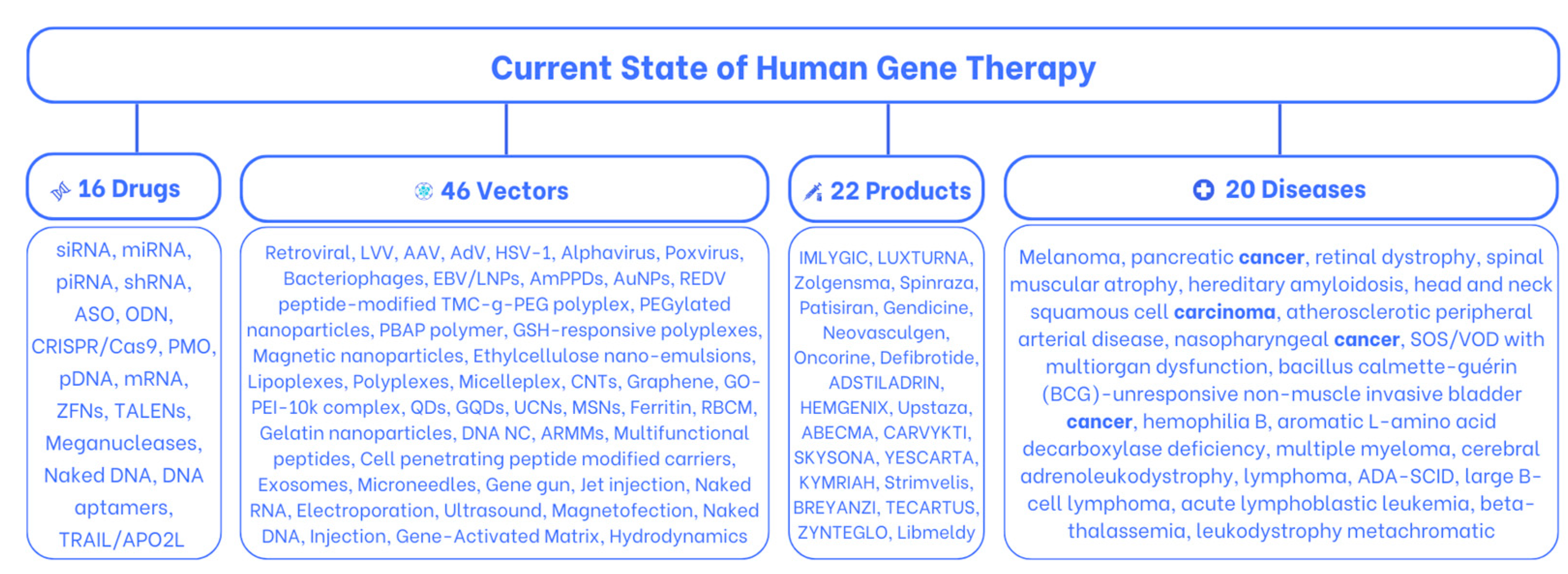
Current State of Human Gene Therapy: Approved Products and Vectors


Abstract
1. Introduction
2. Sixteen Gene Therapy Drugs
2.1. Small Interfering RNA (siRNA)
2.2. MicroRNAs (miRNAs)
2.3. PIWI-Interacting RNAs (piRNAs)
2.4. Short Hairpin RNA (shRNA)
2.5. Antisense Oligonucleotides (ASOs)
2.6. Oligodeoxynucleotides (ODNs)
2.7. Clustered Regularly Interspersed Short Palindromic Repeats (CRISPR)/CRISPR-Associated Protein 9 (Cas9)
2.8. Plasmid DNA (pDNA)
2.9. Messenger Ribonucleic Acid (mRNA)
2.10. Meganucleases
2.11. Zinc Finger Nucleases (ZFNs)
2.12. Transcription Activator-like Effector Nucleases (TALENs)
2.13. DNA Aptamers
2.14. Tumor Necrosis Factor-Related Apoptosis-Inducing Ligand (TRAIL/APO2L)
2.15. Phosphorodiamidate Morpholino Oligomers (PMOs)
2.16. Naked DNA
3. Forty-Six Gene Therapy Carriers
3.1. Viral Vectors
3.1.1. Retroviral
3.1.2. Lentiviral Vector (LVV)
3.1.3. Adeno-Associated Virus (AAV)
3.1.4. Adenovirus (AdV)
3.1.5. Herpes Simplex Virus Type 1 (HSV-1)
3.1.6. Alphavirus
3.1.7. Poxvirus
3.1.8. Bacteriophage
3.1.9. Epstein–Barr Virus (EBV), Human Gammaherpesvirus 4
3.2. Non-Viral Vectors
3.2.1. Lipid Nanoparticles (LNPs)
3.2.2. Amphiphilic Phospholipid Peptide Dendrimers (AmPPDs)
3.2.3. Gold Nanoparticles (AuNPs)
3.2.4. REDV Peptide-Modified TMC-g-PEG Polyplex
3.2.5. PEGylated Nanoparticles
3.2.6. PBAT (Polybutylene Adipate Terephthalate) Polymer
3.2.7. GSH-Responsive Polyplexes
3.2.8. Magnetic Nanoparticles
3.2.9. Ethylcellulose Nano-Emulsions
3.2.10. Lipoplexes
3.2.11. Polyplexes
3.2.12. Micelleplexes
3.2.13. Carbon Nanotubes (CNTs)
3.2.14. Graphene
3.2.15. GO-PEI-10k Complex
3.2.16. Quantum Dots (QDs)
3.2.17. Graphene Quantum Dots (GQDs)
3.2.18. Up-Conversion Nanoparticles (UCNs)
3.2.19. Mesoporous Silica Nanoparticles (MSNs)
3.2.20. Ferritin
3.2.21. Red Blood Cell Membrane (RBCM)
3.2.22. Gelatin Nanoparticles
3.2.23. DNA Nanoclews (NCs)
3.2.24. Arrestin Domain-Containing Protein 1 (ARRDC1)-Mediated Microvesicles (ARMMs)
3.2.25. Multifunctional Peptides
3.2.26. Cell-Penetrating Peptide (CPP), Cell-Permeable Peptides, Protein Transduction Domains (PTDs)
3.2.27. Exosomes (Endogenous Nanocarriers)
3.3. Physical Delivery
3.3.1. Microneedles (MNs)
3.3.2. Gene Gun (Biolistic Particle Delivery System)
3.3.3. Jet Injection
3.3.4. Electroporation
3.3.5. Ultrasound
3.3.6. Magnetofection
3.3.7. Naked RNA Injection
3.3.8. Naked DNA Injection
3.3.9. Gene-Activated Matrix (GAM)
3.3.10. Hydrodynamic High-Pressure Injection
4. Twenty-Two Approved Human Gene Therapy Products
4.1. Approved Human Gene Therapy Products and Their Applications for In Vivo Treatment
4.1.1. IMLYGIC/Melanoma, Pancreatic Cancer
4.1.2. LUXTURNA/Retinal Dystrophy
4.1.3. Zolgensma/Spinal Muscular Atrophy
4.1.4. Spinraza/Spinal Muscular Atrophy
4.1.5. Patisiran/Polyneuropathy
4.1.6. Gendicine/Carcinoma
4.1.7. Neovasculgen/Atherosclerotic Peripheral Arterial Disease
4.1.8. Oncorine/Nasopharyngeal Cancer
4.1.9. Defitelio/SOS/VOD
4.1.10. ADSTILADRIN/Cancer (BCG-NMIBC)
4.1.11. HEMGENIX/Hemophilia B
4.1.12. Upstaza/AADC Deficiency
4.2. Approved Human Gene Therapy Products and Their Applications for Ex Vivo Treatment
4.2.1. ABECMA/Multiple Myeloma
4.2.2. CARVYKTI/Multiple Myeloma
4.2.3. SKYSONA/Cerebral Adrenoleukodystrophy (CALD)
4.2.4. YESCARTA/Large B Cell Lymphoma
4.2.5. KYMRIAH/Lymphoma
4.2.6. Strimvelis/ADA-SCID
4.2.7. BREYANZI/Large B Cell Lymphoma (LBCL)
4.2.8. TECARTUS/Acute Lymphoblastic Leukemia (ALL)
4.2.9. ZYNTEGLO/Beta-Thalassemia
4.2.10. Libmeldy/Metachromatic Leukodystrophy
5. Future Directions
6. Conclusions
Author Contributions
Funding
Institutional Review Board Statement
Informed Consent Statement
Data Availability Statement
Conflicts of Interest
References
- Edelstein, M.L.; Abedi, M.R.; Wixon, J.; Edelstein, R.M. Gene therapy clinical trials worldwide 1989–2004—An overview. J. Gene Med. 2004, 6, 597–602. [Google Scholar] [CrossRef] [PubMed]
- Arabi, F.; Mansouri, V.; Ahmadbeigi, N. Gene therapy clinical trials, where do we go? An overview. Biomed. Pharmacother. 2022, 153, 113324. [Google Scholar] [CrossRef] [PubMed]
- O’Connor, C. Discovery of DNA as the hereditary material using Streptococcus pneumoniae. Nat. Educ. 2008, 1, 104. [Google Scholar]
- Scheller, E.L.; Krebsbach, P.H. Gene therapy: Design and prospects for craniofacial regeneration. J. Dent. Res. 2009, 88, 585–596. [Google Scholar] [CrossRef]
- Baltimore, D. Paul Berg (1926–2023). Science 2023, 379, 1095. [Google Scholar] [CrossRef]
- Approval Letter—KYMRIAH. Available online: https://www.fda.gov/media/106989/download (accessed on 4 July 2023).
- Shahryari, A.; Saghaeian Jazi, M.; Mohammadi, S.; Razavi Nikoo, H.; Nazari, Z.; Hosseini, E.S.; Burtscher, I.; Mowla, S.J.; Lickert, H. Development and Clinical Translation of Approved Gene Therapy Products for Genetic Disorders. Front. Genet. 2019, 10, 868. [Google Scholar] [CrossRef]
- Mullard, A. Parsing clinical success rates. Nat. Rev. Drug Discov. 2016, 15, 447. [Google Scholar] [CrossRef]
- Dana, H.; Chalbatani, G.M.; Mahmoodzadeh, H.; Karimloo, R.; Rezaiean, O.; Moradzadeh, A.; Mehmandoost, N.; Moazzen, F.; Mazraeh, A.; Marmari, V.; et al. Molecular mechanisms and biological functions of siRNA. Int. J. Biomed. Sci. 2017, 13, 48–57. [Google Scholar] [CrossRef]
- Xu, W.; Jiang, X.; Huang, L. RNA Interference Technology. Compr. Biotechnol. 2019, 5, 560–575. [Google Scholar] [CrossRef]
- Lam, J.K.W.; Chow, M.Y.T.; Zhang, Y.; Leung, S.W.S. siRNA Versus miRNA as Therapeutics for Gene Silencing. Mol. Ther. Nucleic Acids 2015, 4, e252. [Google Scholar] [CrossRef]
- Flemming, A. siRNA: Brain delivery breakthrough. Nat. Rev. Neurosci. 2007, 8, 570. [Google Scholar] [CrossRef]
- Jackson, A.L.; Linsley, P.S. Recognizing and avoiding siRNA off-target effects for target identification and therapeutic application. Nat. Rev. Drug Discov. 2010, 9, 57–67. [Google Scholar] [CrossRef]
- Robbins, M.; Judge, A.; MacLachlan, I. siRNA and innate immunity. Oligonucleotides 2009, 19, 89–102. [Google Scholar] [CrossRef] [PubMed]
- Ha, M.; Kim, V.N. Regulation of microRNA biogenesis. Nat. Rev. Mol. Cell Biol. 2014, 15, 509–524. [Google Scholar] [CrossRef] [PubMed]
- Brennecke, J.; Stark, A.; Russell, R.B.; Cohen, S.M. Principles of MicroRNA–Target Recognition. PLoS Biol. 2005, 3, e85. [Google Scholar] [CrossRef] [PubMed]
- Eslava-Avilés, E.; Arenas-Huertero, F. piRNAs: Nature, biogenesis, regulation, and their potential clinical utility. Bol. Med. Hosp. Infant. Mex. 2021, 78, 432–442. [Google Scholar] [CrossRef]
- Moore, C.B.; Guthrie, E.H.; Huang, M.T.-H.; Taxman, D.J. Short Hairpin RNA (shRNA): Design, Delivery, and Assessment of Gene Knockdown. Methods Mol. Biol. 2010, 629, 141–158. [Google Scholar] [CrossRef] [PubMed]
- Ge, Q.; Ilves, H.; Dallas, A.; Kumar, P.; Shorenstein, J.; Kazakov, S.A.; Johnston, B.H. Minimal-length short hairpin RNAs: The relationship of structure and RNAi activity. RNA 2010, 16, 106–117. [Google Scholar] [CrossRef]
- Larsson, E.; Sander, C.; Marks, D. mRNA turnover rate limits siRNA and microRNA efficacy. Mol. Syst. Biol. 2010, 6, 433. [Google Scholar] [CrossRef]
- Fellmann, C.; Lowe, S.W. Stable RNA interference rules for silencing. Nat. Cell Biol. 2014, 16, 10–18. [Google Scholar] [CrossRef]
- Mcintyre, G.J.; Yu, Y.-H.; Lomas, M.; Fanning, G.C. The effects of stem length and core placement on shRNA activity. BMC Mol. Biol. 2011, 12, 34. [Google Scholar] [CrossRef] [PubMed]
- Dhuri, K.; Bechtold, C.; Quijano, E.; Pham, H.; Gupta, A.; Vikram, A.; Bahal, R. Antisense Oligonucleotides: An Emerging Area in Drug Discovery and Development. J. Clin. Med. 2020, 9, 2004. [Google Scholar] [CrossRef] [PubMed]
- Scoles, D.R.; Minikel, E.V.; Pulst, S.M. Antisense oligonucleotides. Neurol. Genet. 2019, 5, e323. [Google Scholar] [CrossRef] [PubMed]
- McClorey, G.; Banerjee, S. Cell-Penetrating Peptides to Enhance Delivery of Oligonucleotide-Based Therapeutics. Biomedicines 2018, 6, 51. [Google Scholar] [CrossRef]
- Liang, X.-H.; Sun, H.; Nichols, J.G.; Crooke, S.T. RNase H1-Dependent Antisense Oligonucleotides Are Robustly Active in Directing RNA Cleavage in Both the Cytoplasm and the Nucleus. Mol. Ther. 2017, 25, 2075–2092. [Google Scholar] [CrossRef]
- Yoshida, T.; Naito, Y.; Yasuhara, H.; Sasaki, K.; Kawaji, H.; Kawai, J.; Naito, M.; Okuda, H.; Obika, S.; Inoue, T. Evaluation of off-target effects of gapmer antisense oligonucleotides using human cells. Genes Cells 2019, 24, 827–835. [Google Scholar] [CrossRef]
- Rinaldi, C.; Wood, M.J.A. Antisense oligonucleotides: The next frontier for treatment of neurological disorders. Nat. Rev. Neurol. 2018, 14, 9–21. [Google Scholar] [CrossRef]
- Rook, M.E.; Southwell, A.L. Antisense Oligonucleotide Therapy: From Design to the Huntington Disease Clinic. BioDrugs 2022, 36, 105–119. [Google Scholar] [CrossRef]
- Gardlík, R.; Pálffy, R.; Hodosy, J.; Lukács, J.; Turna, J.; Celec, P. Vectors and delivery systems in gene therapy. Med. Sci. Monit. 2005, 11, RA110–RA121. [Google Scholar]
- Asmamaw, M.; Zawdie, B. Mechanism and Applications of CRISPR/Cas-9-Mediated Genome Editing. Biologics 2021, 15, 353–361. [Google Scholar] [CrossRef]
- Xu, C.L.; Ruan, M.Z.C.; Mahajan, V.B.; Tsang, S.H. Viral Delivery Systems for CRISPR. Viruses 2019, 11, 28. [Google Scholar] [CrossRef] [PubMed]
- Naeem, M.; Majeed, S.; Hoque, M.Z.; Ahmad, I. Latest Developed Strategies to Minimize the Off-Target Effects in CRISPR-Cas-Mediated Genome Editing. Cells 2020, 9, 1608. [Google Scholar] [CrossRef] [PubMed]
- Rawashdeh, O.; Rawashdeh, R.Y.; Kebede, T.; Kapp, D.; Ralescu, A. Bio-informatic analysis of CRISPR protospacer adjacent motifs (PAMs) in T4 genome. BMC Genom. Data 2022, 23, 40. [Google Scholar] [CrossRef] [PubMed]
- Yang, Y.; Xu, J.; Ge, S.; Lai, L. CRISPR/Cas: Advances, Limitations, and Applications for Precision Cancer Research. Front. Med. 2021, 8, 649896. [Google Scholar] [CrossRef]
- Taniyama, Y.; Azuma, J.; Kunugiza, Y.; Iekushi, K.; Rakugi, H.; Morishita, R. Therapeutic option of plasmid-DNA based gene transfer. Curr. Top. Med. Chem. 2012, 12, 1630–1637. [Google Scholar] [CrossRef]
- Sousa, F.; Passarinha, L.; Queiroz, J.A. Biomedical application of plasmid DNA in gene therapy: A new challenge for chromatography. Biotechnol. Genet. Eng. Rev. 2009, 26, 83–116. [Google Scholar] [CrossRef]
- Dishart, K.L.; Work, L.M.; Denby, L.; Baker, A.H. Gene Therapy for Cardiovascular Disease. J. Biomed. Biotechnol. 2003, 2, 138–148. [Google Scholar] [CrossRef]
- Enghiad, B.; Xue, P.; Singh, N.; Boob, A.G.; Shi, C.; Petrov, V.A.; Liu, R.; Peri, S.S.; Lane, S.T.; Gaither, E.D.; et al. PlasmidMaker is a versatile, automated, and high throughput end-to-end platform for plasmid construction. Nat. Commun. 2022, 13, 2697. [Google Scholar] [CrossRef]
- Shintani, M.; Sanchez, Z.K.; Kimbara, K. Genomics of microbial plasmids: Classification and identification based on replication and transfer systems and host taxonomy. Front. Microbiol. 2015, 6, 242. [Google Scholar] [CrossRef]
- Barnhart, K.M.; Hartikka, J.; Manthorpe, M.; Norman, J.; Hobart, P. Enhancer and promoter chimeras in plasmids designed for intramuscular injection: A comparative in vivo and in vitro study. Hum. Gene Ther. 1998, 9, 2545–2553. [Google Scholar] [CrossRef]
- Eusébio, D.; Neves, A.R.; Costa, D.; Biswas, S.; Alves, G.; Cui, Z.; Sousa, Â. Methods to improve the immunogenicity of plasmid DNA vaccines. Drug Discov. Today 2021, 26, 2575–2592. [Google Scholar] [CrossRef] [PubMed]
- Antonenko, S.; Gurianov, D.; Telegeev, G. Colocalization of USP1 and РН domain of Bcr-Abl oncoprotein in terms of chronic myeloid leukemia cell rearrangements. Tsitol. Genet. 2016, 50, 352–356. [Google Scholar]
- Antonenko, S.; Telegeev, G. Inhibition of USP1, a new partner of bcr-abl, results in decrease of Bcr-Abl level in k562 cells. Exp. Oncol. 2020, 42, 109–114. [Google Scholar] [CrossRef] [PubMed]
- Antonenko, S.; Kravchuk, I.; Telegeev, G. GLG1 in K562 cells: Role in pathogenesis of chronic myeloid leukemia. Cytol. Genet. 2020, 54, 62–70. [Google Scholar] [CrossRef]
- Tang, X.; Zhang, S.; Fu, R.; Zhang, L.; Huang, K.; Peng, H.; Dai, L.; Chen, Q. Therapeutic Prospects of mRNA-Based Gene Therapy for Glioblastoma. Front. Oncol. 2019, 9, 2019. [Google Scholar] [CrossRef]
- Andreev, D.E.; Terenin, I.M.; Dmitriev, S.E.; Shatsky, I.N. Pros and cons of pDNA and mRNA transfection to study mRNA translation in mammalian cells. Gene 2016, 578, 1–6. [Google Scholar] [CrossRef]
- Damase, T.R.; Sukhovershin, R.; Boada, C.; Taraballi, F.; Pettigrew, R.I.; Cooke, J.P. The Limitless Future of RNA Therapeutics. Front. Bioeng. Biotechnol. 2021, 9, 628137. [Google Scholar] [CrossRef]
- Iqbal, Z.; Iqbal, M.S.; Ahmad, A.; Memon, A.G.; Ansari, M.I. New prospects on the horizon: Genome editing to engineer plants for desirable traits. Curr. Plant Biol. 2020, 24, 100171. [Google Scholar] [CrossRef]
- Flisikowska, T.; Kind, A.; Schnieke, A. 10—Production of Transgenic Rabbits. In Transgenic Animal Technology, 3rd ed.; Pinkert, C.A., Ed.; Elsevier: Amsterdam, The Netherlands, 2014; pp. 275–304. [Google Scholar] [CrossRef]
- Silva, G.; Poirot, L.; Galetto, R.; Smith, J.; Montoya, G.; Duchateau, P.; Pâques, F. Meganucleases and Other Tools for Targeted Genome Engineering: Perspectives and Challenges for Gene Therapy. Curr. Gene Ther. 2011, 11, 11–27. [Google Scholar] [CrossRef]
- Daboussi, F.; Stoddard, T.J.; Zhang, F. Engineering Meganuclease for Precise Plant Genome Modification. In Advances in New Technology for Targeted Modification of Plant Genomes; Zhang, F., Puchta, H., Thomson, J., Eds.; Springer: New York, NY, USA, 2015; pp. 21–38. [Google Scholar] [CrossRef]
- Epinat, J.C.; Arnould, S.; Chames, P.; Rochaix, P.; Desfontaines, D.; Puzin, C.; Patin, A.; Zanghellini, A.; Pâques, F.; Lacroix, E. A novel engineered meganuclease induces homologous recombination in yeast and mammalian cells. Nucleic Acids Res. 2003, 31, 2952–2962. [Google Scholar] [CrossRef]
- Urnov, F.D.; Rebar, E.J.; Holmes, M.C.; Zhang, H.S.; Gregory, P.D. Genome editing with engineered zinc finger nucleases. Nat. Rev. Genet. 2010, 11, 636–646. [Google Scholar] [CrossRef] [PubMed]
- Durai, S.; Mani, M.; Kandavelou, K.; Wu, J.; Porteus, M.H.; Chandrasegaran, S. Zinc finger nucleases: Custom-designed molecular scissors for genome engineering of plant and mammalian cells. Nucleic Acids Res. 2005, 33, 5978–5990. [Google Scholar] [CrossRef] [PubMed]
- Kim, Y.; Kweon, J.; Kim, A.; Chon, J.K.; Yoo, J.Y.; Kim, H.J.; Kim, S.; Lee, C.; Jeong, E.; Chung, E.; et al. A genome-wide TALEN resource. Nat. Methods 2013, 10, 286. [Google Scholar] [CrossRef]
- Heigwer, F.; Kerr, G.; Walther, N.; Glaeser, K.; Pelz, O.; Breinig, M.; Boutros, M. E-TALEN: A web tool to design TALENs for genome engineering. Nucleic Acids Res. 2013, 41, e190. [Google Scholar] [CrossRef] [PubMed]
- Zhang, G.; Lin, Y.; Qi, X.; Li, L.; Wang, Q.; Ma, Y. TALENs-Assisted Multiplex Editing for Accelerated Genome Evolution To Improve Yeast Phenotypes. ACS Synth. Biol. 2015, 4, 1101–1111. [Google Scholar] [CrossRef]
- Wang, L.; Li, F.; Dang, L.; Liang, C.; Wang, C.; He, B.; Liu, J.; Li, D.; Wu, X.; Xu, X.; et al. In Vivo Delivery Systems for Therapeutic Genome Editing. Int. J. Mol. Sci. 2016, 17, 626. [Google Scholar] [CrossRef]
- Lau, C.-H.; Zhu, H.; Tay, J.C.-K.; Li, Z.; Tay, F.C.; Chen, C.; Tan, W.-K.; Du, S.; Sia, V.-K.; Phang, R.-Z.; et al. Genetic rearrangements of variable di-residue (RVD)-containing repeat arrays in a baculoviral TALEN system. Mol. Ther. Methods Clin. Dev. 2014, 1, 14050. [Google Scholar] [CrossRef]
- Adachi, T.; Nakamura, Y. Aptamers: A Review of Their Chemical Properties and Modifications for Therapeutic Application. Molecules 2019, 24, 4229. [Google Scholar] [CrossRef]
- Mou, Q.; Xue, X.; Ma, Y.; Banik, M.; Garcia, V.; Guo, W.; Wang, J.; Song, T.; Chen, L.-Q.; Lu, Y. Efficient delivery of a DNA aptamer-based biosensor into plant cells for glucose sensing through thiol-mediated uptake. Sci. Adv. 2022, 8, eabo0902. [Google Scholar] [CrossRef]
- Kong, H.Y.; Byun, J. Nucleic Acid Aptamers: New Methods for Selection, Stabilization, and Application in Biomedical Science. Biomol. Ther. 2013, 21, 423–434. [Google Scholar] [CrossRef]
- Zhu, Q.; Liu, G.; Kai, M. DNA Aptamers in the Diagnosis and Treatment of Human Diseases. Molecules 2015, 20, 20979–20997. [Google Scholar] [CrossRef] [PubMed]
- Crivianu-Gaita, V.; Thompson, M. Aptamers, antibody scFv, and antibody Fab’ fragments: An overview and comparison of three of the most versatile biosensor biorecognition elements. Biosens. Bioelectron. 2016, 85, 32–45. [Google Scholar] [CrossRef] [PubMed]
- Miranda-Castro, R.; de-los-Santos-Álvarez, N.; Miranda-Ordieres, A.J.; Lobo-Castañón, M.J. Harnessing Aptamers to Overcome Challenges in Gluten Detection. Biosensors 2016, 6, 16. [Google Scholar] [CrossRef]
- Kawano, R.; Osaki, T.; Sasaki, H.; Takinoue, M.; Yoshizawa, S.; Takeuchi, S. Rapid detection of a cocaine-binding aptamer using biological nanopores on a chip. J. Am. Chem. Soc. 2011, 133, 8474–8477. [Google Scholar] [CrossRef]
- Xie, M.; Chen, Z.; Zhao, F.; Lin, Y.; Zheng, S.; Han, S. Selection and Application of ssDNA Aptamers for Fluorescence Biosensing Detection of Malachite Green. Foods 2022, 11, 801. [Google Scholar] [CrossRef]
- Neves, M.A.D.; Reinstein, O.; Saad, M.; Johnson, P.E. Defining the secondary structural requirements of a cocaine-binding aptamer by a thermodynamic and mutation study. Biophys. Chem. 2010, 153, 9–16. [Google Scholar] [CrossRef] [PubMed]
- Baugh, C.; Grate, D.; Wilson, C. 2.8 Å crystal structure of the malachite green aptamer. J. Mol. Biol. 2000, 301, 117–128. [Google Scholar] [CrossRef]
- Zhong, H.; Wang, H.; Li, J.; Huang, Y. TRAIL-based gene delivery and therapeutic strategies. Acta Pharmacol. Sin. 2019, 40, 1373–1385. [Google Scholar] [CrossRef]
- Griffith, T.S.; Stokes, B.; Kucaba, T.A.; Earel, J.K., Jr.; VanOosten, R.L.; Brincks, E.L.; Norian, L.A. TRAIL gene therapy: From preclinical development to clinical application. Curr. Gene Ther. 2009, 9, 9–19. [Google Scholar] [CrossRef]
- Deere, J.; Iversen, P.; Geller, B.L. Antisense phosphorodiamidate morpholino oligomer length and target position effects on gene-specific inhibition in Escherichia coli. Antimicrob. Agents Chemother. 2005, 49, 249–255. [Google Scholar] [CrossRef]
- Nan, Y.; Zhang, Y.-J. Antisense phosphorodiamidate morpholino oligomers as novel antiviral compounds. Front. Microbiol. 2018, 9, 750. [Google Scholar] [CrossRef] [PubMed]
- Nayerossadat, N.; Maedeh, T.; Ali, P.A. Viral and nonviral delivery systems for gene delivery. Adv. Biomed. Res. 2012, 1, 27. [Google Scholar] [CrossRef] [PubMed]
- Esposito, M.V.; Aveta, A.; Comegna, M.; Cernera, G.; Iacotucci, P.; Carnovale, V.; Taccetti, G.; Terlizzi, V.; Castaldo, G. Extensive CFTR Gene Analysis Revealed a Higher Occurrence of Cystic Fibrosis Transmembrane Regulator-Related Disorders (CFTR-RD) among CF Carriers. J. Clin. Med. 2020, 9, 3853. [Google Scholar] [CrossRef] [PubMed]
- Gillen, A.E.; Gosalia, N.; Leir, S.H.; Harris, A. MicroRNA regulation of expression of the cystic fibrosis transmembrane conductance regulator gene. Biochem. J. 2011, 438, 25–32. [Google Scholar] [CrossRef]
- De Palma, F.D.E.; Raia, V.; Kroemer, G.; Maiuri, M.C. The Multifaceted Roles of MicroRNAs in Cystic Fibrosis. Diagnostics 2020, 10, 1102. [Google Scholar] [CrossRef]
- Vargas, J.E.; Chicaybam, L.; Stein, R.T.; Tanuri, A.; Delgado-Cañedo, A.; Bonamino, M.H. Retroviral vectors and transposons for stable gene therapy: Advances, current challenges and perspectives. J. Transl. Med. 2016, 14, 288. [Google Scholar] [CrossRef]
- Ghosh, S.; Brown, A.M.; Jenkins, C.; Campbell, K. Viral Vector Systems for Gene Therapy: A Comprehensive Literature Review of Progress and Biosafety Challenges. Appl. Biosaf. 2020, 25, 7–18. [Google Scholar] [CrossRef]
- Naso, M.F.; Tomkowicz, B.; Perry, W.L., III; Strohl, W.R. Adeno-Associated Virus (AAV) as a Vector for Gene Therapy. BioDrugs 2017, 31, 317–334. [Google Scholar] [CrossRef]
- Hukkanen, V. Herpesvirus Vectors in Gene Therapy. Open Virol. J. 2010, 4, 94–95. [Google Scholar] [CrossRef]
- Lundstrom, K. Alphaviruses in Gene Therapy. Viruses 2015, 7, 2321–2333. [Google Scholar] [CrossRef]
- Kim, A.S.; Diamond, M.S. A molecular understanding of alphavirus entry and antibody protection. Nat. Rev. Microbiol. 2023, 21, 396–407. [Google Scholar] [CrossRef]
- Conrad, S.J.; Liu, J. Poxviruses as Gene Therapy Vectors: Generating Poxviral Vectors Expressing Therapeutic Transgenes. Methods Mol. Biol. 2019, 1937, 189–209. [Google Scholar] [CrossRef]
- Jenne, L.; Hauser, C.; Arrighi, J.F.; Saurat, J.H.; Hügin, A.W. Poxvirus as a vector to transduce human dendritic cells for immunotherapy: Abortive infection but reduced APC function. Gene Ther. 2000, 7, 1575–1583. [Google Scholar] [CrossRef]
- Oliveira, G.P.; Rodrigues, R.A.L.; Lima, M.T.; Drumond, B.P.; Abrahão, J.S. Poxvirus Host Range Genes and Virus–Host Spectrum: A Critical Review. Viruses 2017, 9, 331. [Google Scholar] [CrossRef] [PubMed]
- Saghazadeh, A.; Rezaei, N. Poxviruses and the immune system: Implications for monkeypox virus. Int. Immunopharmacol. 2022, 113, 109364. [Google Scholar] [CrossRef] [PubMed]
- Gibb, B.; Hyman, P.; Schneider, C.L. The Many Applications of Engineered Bacteriophages—An Overview. Pharmaceuticals 2021, 14, 634. [Google Scholar] [CrossRef] [PubMed]
- Kasman, L.M.; Porter, L.D. Bacteriophages. In StatPearls [Internet]; StatPearls Publishing: St. Petersburg, FL, USA, 2023. [Google Scholar]
- Sanger, F.; Coulson, A.R.; Hong, G.F.; Hill, D.F.; Petersen, G.B. Nucleotide sequence of bacteriophage λ DNA. J. Mol. Biol. 1982, 162, 729–773. [Google Scholar] [CrossRef]
- Dunn, J.J.; Studier, F.W.; Gottesman, M. Complete nucleotide sequence of bacteriophage T7 DNA and the locations of T7 genetic elements. J. Mol. Biol. 1983, 166, 477–535. [Google Scholar] [CrossRef]
- Petrov, V.M.; Ratnayaka, S.; Nolan, J.M.; Miller, E.S.; Karam, J.D. Genomes of the T4-related bacteriophages as windows on microbial genome evolution. Virol. J. 2010, 7, 292. [Google Scholar] [CrossRef]
- Hatfull, G.F. Bacteriophage genomics. Curr. Opin. Microbiol. 2008, 11, 447–453. [Google Scholar] [CrossRef]
- Principi, N.; Silvestri, E.; Esposito, S. Advantages and Limitations of Bacteriophages for the Treatment of Bacterial Infections. Front. Pharmacol. 2019, 10, 513. [Google Scholar] [CrossRef] [PubMed]
- Young, L.S. Epstein-Barr Virus: General Features. In Encyclopedia of Virology, 3rd ed.; Mahy, B.W.J., van Regenmortel, M.H.V., Eds.; Academic Press: Oxford, UK, 2008; pp. 148–157. [Google Scholar]
- Tomkinson, B.; Robertson, E.; Yalamanchili, R.; Longnecker, R.; Kieff, E. Epstein-Barr virus recombinants from overlapping cosmid fragments. J. Virol. 1993, 67, 7298–7306. [Google Scholar] [CrossRef] [PubMed]
- Zhang, N.; Zuo, Y.; Jiang, L.; Peng, Y.; Huang, X.; Zuo, L. Epstein-Barr Virus and Neurological Diseases. Front. Mol. Biosci. 2021, 8, 816098. [Google Scholar] [CrossRef] [PubMed]
- Wang, L.; Laing, J.; Yan, B.; Zhou, H.; Ke, L.; Wang, C.; Narita, Y.; Zhang, Z.; Olson, M.R.; Afzali, B.; et al. Epstein-Barr Virus Episome Physically Interacts with Active Regions of the Host Genome in Lymphoblastoid Cells. J. Virol. 2020, 94, e01390-20. [Google Scholar] [CrossRef]
- Wu, L.; Borza, C.M.; Hutt-Fletcher, L.M. Mutations of Epstein-Barr Virus gH That Are Differentially Able To Support Fusion with B Cells or Epithelial Cells. J. Virol. 2005, 79, 10923–10930. [Google Scholar] [CrossRef]
- Lujan, H.; Griffin, W.C.; Taube, J.H.; Sayes, C.M. Synthesis and characterization of nanometer-sized liposomes for encapsulation and microRNA transfer to breast cancer cells. Int. J. Nanomed. 2019, 14, 5159–5173. [Google Scholar] [CrossRef]
- Dong, Y.; Chen, Y.; Zhu, D.; Shi, K.; Ma, C.; Zhang, W.; Rocchi, P.; Jiang, L.; Liu, X. Self-assembly of amphiphilic phospholipid peptide dendrimer-based nanovectors for effective delivery of siRNA therapeutics in prostate cancer therapy. J. Control. Release 2020, 322, 416–425. [Google Scholar] [CrossRef]
- Paz, M.M.; Peinador Veiga, A.; Regueira, T.; Vázquez Vázquez, C.; López Quintela, M.A. Facile generation of surface diversity in gold nanoparticles. J. Colloid Interface Sci. 2023, 641, 719–728. [Google Scholar] [CrossRef]
- Si, P.; Razmi, N.; Nur, O.; Solanki, S.; Pandey, C.M.; Gupta, R.K.; Malhotra, B.D.; Willander, M.; de la Zerda, A. Gold nanomaterials for optical biosensing and bioimaging. Nanoscale Adv. 2021, 3, 2679–2698. [Google Scholar] [CrossRef]
- Jeong, E.H.; Jung, G.; Hong, C.A.; Lee, H. Gold nanoparticle (AuNP)-based drug delivery and molecular imaging for biomedical applications. Arch. Pharm. Res. 2014, 37, 53–59. [Google Scholar] [CrossRef]
- Ackerson, C.J.; Jadzinsky, P.D.; Sexton, J.Z.; Bushnell, D.A.; Kornberg, R.D. Synthesis and Bioconjugation of 2 and 3 nm-diameter Gold Nanoparticles. Bioconjug. Chem. 2010, 21, 214–218. [Google Scholar] [CrossRef] [PubMed]
- Hammami, I.; Alabdallah, N.M.; Al Jomaa, A.; Kamoun, M. Gold nanoparticles: Synthesis properties and applications. J. King Saud Univ.-Sci. 2021, 33, 101560. [Google Scholar] [CrossRef]
- Sani, A.; Cao, C.; Cui, D. Toxicity of gold nanoparticles (AuNPs): A review. Biochem. Biophys. Rep. 2021, 26, 100991. [Google Scholar] [CrossRef] [PubMed]
- Zhou, F.; Jia, X.; Yang, Q.; Yang, Y.; Zhao, Y.; Fan, Y.; Yuan, X. Targeted delivery of microRNA-126 to vascular endothelial cells via REDV peptide modified PEG-trimethyl chitosan. Biomater. Sci. 2016, 4, 849–856. [Google Scholar] [CrossRef] [PubMed]
- Suk, J.S.; Xu, Q.; Kim, N.; Hanes, J.; Ensign, L.M. PEGylation as a strategy for improving nanoparticle-based drug and gene delivery. Adv. Drug Deliv. Rev. 2016, 99 Pt A, 28–51. [Google Scholar] [CrossRef]
- Wang, Y.; Ye, M.; Xie, R.; Gong, S. Enhancing the In Vitro and In Vivo Stabilities of Polymeric Nucleic Acid Delivery Nanosystems. Bioconjug. Chem. 2019, 30, 325–337. [Google Scholar] [CrossRef]
- Wang, Y.; Ma, B.; Abdeen, A.A.; Chen, G.; Xie, R.; Saha, K.; Gong, S. Versatile Redox-Responsive Polyplexes for the Delivery of Plasmid DNA, Messenger RNA, and CRISPR-Cas9 Genome-Editing Machinery. ACS Appl. Mater. Interfaces 2018, 10, 31915–31927. [Google Scholar] [CrossRef]
- Jian, J.; Xiangbin, Z.; Xianbo, H. An overview on synthesis, properties and applications of poly(butylene-adipate-co-terephthalate)–PBAT. Adv. Ind. Eng. Polym. Res. 2020, 3, 19–26. [Google Scholar] [CrossRef]
- Weng, Y.-X.; Jin, Y.-J.; Meng, Q.-Y.; Wang, L.; Zhang, M.; Wang, Y.-Z. Biodegradation behavior of poly(butylene adipate-co-terephthalate) (PBAT), poly(lactic acid) (PLA), and their blend under soil conditions. Polym. Test. 2013, 32, 918–926. [Google Scholar] [CrossRef]
- Phothisarattana, D.; Wongphan, P.; Promhuad, K.; Promsorn, J.; Harnkarnsujarit, N. Biodegradable Poly(Butylene Adipate-Co-Terephthalate) and Thermoplastic Starch-Blended TiO2 Nanocomposite Blown Films as Functional Active Packaging of Fresh Fruit. Polymers 2021, 13, 4192. [Google Scholar] [CrossRef]
- Fukushima, K.; Rasyida, A.; Yang, M.-C. Characterization, degradation and biocompatibility of PBAT based nanocomposites. Appl. Clay Sci. 2013, 80–81, 291–298. [Google Scholar] [CrossRef]
- Chen, G.; Ma, B.; Wang, Y.; Gong, S. A Universal GSH-Responsive Nanoplatform for the Delivery of DNA, mRNA, and Cas9/sgRNA Ribonucleoprotein. ACS Appl. Mater. Interfaces 2018, 10, 18515–18523. [Google Scholar] [CrossRef] [PubMed]
- Ali, A.; Shah, T.; Ullah, R.; Zhou, P.; Guo, M.; Ovais, M.; Tan, Z.; Rui, Y. Review on Recent Progress in Magnetic Nanoparticles: Synthesis, Characterization, and Diverse Applications. Front. Chem. 2021, 9, 629054. [Google Scholar] [CrossRef]
- Ernst, C.; Bartel, A.; Elferink, J.W.; Huhn, J.; Eschbach, E.; Schönfeld, K.; Feßler, A.T.; Oberheitmann, B.; Schwarz, S. Improved DNA extraction and purification with magnetic nanoparticles for the detection of methicillin-resistant Staphylococcus aureus. Vet. Microbiol. 2019, 230, 45–48. [Google Scholar] [CrossRef]
- Savliwala, S.; Chiu-Lam, A.; Unni, M.; Rivera-Rodriguez, A.; Fuller, E.; Sen, K.; Threadcraft, M.; Rinaldi, C. Chapter 13—Magnetic nanoparticles. In Nanoparticles for Biomedical Applications; Chung, E.J., Leon, L., Rinaldi, C., Eds.; Elsevier: Amsterdam, The Netherlands, 2020; pp. 195–221. [Google Scholar] [CrossRef]
- Kim, I.; Yang, H.-M.; Park, C.W.; Yoon, I.-H.; Sihn, Y. 20—Environmental applications of magnetic nanoparticles. In Magnetic Nanoparticle-Based Hybrid Materials; Ehrmann, A., Nguyen, T.A., Ahmadi, M., Farmani, A., Nguyen-Tri, P., Eds.; Woodhead Publishing: Cambridge, UK, 2021; pp. 529–545. [Google Scholar] [CrossRef]
- Alagiri, M.; Muthamizhchelvan, C.; Ponnusamy, S. Structural and magnetic properties of iron, cobalt and nickel nanoparticles. Synth. Met. 2011, 161, 1776–1780. [Google Scholar] [CrossRef]
- Markides, H.; Rotherham, M.; El Haj, A.J. Biocompatibility and Toxicity of Magnetic Nanoparticles in Regenerative Medicine. J. Nanomater. 2012, 2012, 614094. [Google Scholar] [CrossRef]
- Busquets, M.A.; Espargaró, A.; Sabaté, R.; Estelrich, J. Magnetic Nanoparticles Cross the Blood-Brain Barrier: When Physics Rises to a Challenge. Nanomaterials 2015, 5, 2231–2248. [Google Scholar] [CrossRef]
- Leitner, S.; Solans, C.; García-Celma, M.J.; Morral-Ruíz, G.; Melgar-Lesmes, P.; Calderó, G. Ethylcellulose nanoparticles prepared from nano-emulsion templates as new folate binding haemocompatible platforms. Mater. Sci. Eng. C 2021, 120, 111682. [Google Scholar] [CrossRef]
- Tros de Ilarduya, C.; Sun, Y.; Düzgüneş, N. Gene delivery by lipoplexes and polyplexes. Eur J Pharm Sci. 2010, 40, 159–170. [Google Scholar] [CrossRef]
- Wasungu, L.; Hoekstra, D. Cationic lipids, lipoplexes and intracellular delivery of genes. J. Control. Release 2006, 116, 255–264. [Google Scholar] [CrossRef]
- Kim, N.J.; Yoo, J.J.; Atala, A.; Lee, S.J. Chapter 7—Small RNA Delivery for In Situ Tissue Regeneration. In Situ Tissue Regeneration; Academic Press: London, UK, 2016; pp. 111–135. [Google Scholar]
- Vasile, C. Chapter 1—Polymeric Nanomaterials: Recent Developments, Properties and Medical Applications. In Polymeric Nanomaterials in Nanotherapeutics; Vasile, C., Ed.; Elsevier: Amsterdam, The Netherlands, 2019; pp. 1–66. [Google Scholar] [CrossRef]
- Pereira-Silva, M.; Jarak, I.; Alvarez-Lorenzo, C.; Concheiro, A.; Santos, A.C.; Maleki, R.; Veiga, F.; Figueiras, A. Micelleplexes as nucleic acid delivery systems for cancer-targeted therapies. J. Control. Release 2020, 323, 442–462. [Google Scholar] [CrossRef] [PubMed]
- Grimme, C.J.; Hanson, M.G.; Corcoran, L.G.; Reineke, T.M. Polycation Architecture Affects Complexation and Delivery of Short Antisense Oligonucleotides: Micelleplexes Outperform Polyplexes. Biomacromolecules 2022, 23, 3257–3271. [Google Scholar] [CrossRef] [PubMed]
- Zare, H.; Ahmadi, S.; Ghasemi, A.; Ghanbari, M.; Rabiee, N.; Bagherzadeh, M.; Karimi, M.; Webster, T.J.; Hamblin, M.R.; Mostafavi, E. Carbon Nanotubes: Smart Drug/Gene Delivery Carriers. Int. J. Nanomed. 2021, 16, 1681–1706. [Google Scholar] [CrossRef]
- Ghuge, A.D.; Shirode, A.R.; Kadam, V.J. Graphene: A Comprehensive Review. Curr. Drug Targets 2017, 18, 724–733. [Google Scholar] [CrossRef] [PubMed]
- Han, X.M.; Zheng, K.W.; Wang, R.L.; Yue, S.F.; Chen, J.; Zhao, Z.W.; Song, F.; Su, Y.; Ma, Q. Functionalization and optimization-strategy of graphene oxide-based nanomaterials for gene and drug delivery. Am. J. Transl. Res. 2020, 12, 1515–1534. [Google Scholar]
- Devi, S.; Kumar, M.; Tiwari, A.; Tiwari, V.; Kaushik, D.; Verma, R.; Bhatt, S.; Sahoo, B.M.; Bhattacharya, T.; Alshehri, S.; et al. Quantum Dots: An Emerging Approach for Cancer Therapy. Front. Mater. 2022, 8, 798440. [Google Scholar] [CrossRef]
- Singh, S.; Dhawan, A.; Karhana, S.; Bhat, M.; Dinda, A.K. Quantum Dots: An Emerging Tool for Point-of-Care Testing. Micromachines 2020, 11, 1058. [Google Scholar] [CrossRef]
- Biswas, M.C.; Islam, M.T.; Nandy, P.K.; Hossain, M.M. Graphene Quantum Dots (GQDs) for Bioimaging and Drug Delivery Applications: A Review. ACS Mater. Lett. 2021, 3, 889–911. [Google Scholar] [CrossRef]
- Kumar, Y.R.; Deshmukh, K.; Sadasivuni, K.K.; Pasha, S.K.K. Graphene quantum dot based materials for sensing, bio-imaging and energy storage applications: A review. RSC Adv. 2020, 10, 23861–23898. [Google Scholar] [CrossRef]
- Henna, T.K.; Pramod, K. Graphene quantum dots redefine nanobiomedicine. Mater. Sci. Eng. C Mater. Biol. Appl. 2020, 110, 110651. [Google Scholar] [CrossRef]
- Tian, P.; Tang, L.; Teng, K.S.; Lau, S.P. Graphene quantum dots from chemistry to applications. Mater. Today Chem. 2018, 10, 221–258. [Google Scholar] [CrossRef]
- Ghafary, S.M.; Nikkhah, M.; Hatamie, S.; Hosseinkhani, S. Simultaneous Gene Delivery and Tracking through Preparation of Photo-Luminescent Nanoparticles Based on Graphene Quantum Dots and Chimeric Peptides. Sci. Rep. 2017, 7, 9552. [Google Scholar] [CrossRef] [PubMed]
- Fasbender, S.; Zimmermann, L.; Cadeddu, R.-P.; Luysberg, M.; Moll, B.; Janiak, C.; Heinzel, T.; Haas, R. The Low Toxicity of Graphene Quantum Dots is Reflected by Marginal Gene Expression Changes of Primary Human Hematopoietic Stem Cells. Sci. Rep. 2019, 9, 12028. [Google Scholar] [CrossRef] [PubMed]
- Chu, H.; Cao, T.; Dai, G.; Liu, B.; Duan, H.; Kong, C.; Tian, N.; Hou, D.; Sun, Z. Recent advances in functionalized upconversion nanoparticles for light-activated tumor therapy. RSC Adv. 2021, 11, 35472–35488. [Google Scholar] [CrossRef] [PubMed]
- Rajani, C.; Borisa, P.; Karanwad, T.; Borade, Y.; Patel, V.; Rajpoot, K.; Tekade, R.K. Cancer-targeted chemotherapy: Emerging role of the folate anchored dendrimer as drug delivery nanocarrier. In Pharmaceutical Applications of Dendrimers; Chauhan, A., Kulhari, H., Eds.; Elsevier: Amsterdam, The Netherlands, 2020; pp. 151–198. [Google Scholar] [CrossRef]
- Pednekar, P.P.; Godiyal, S.C.; Jadhav, K.R.; Kadam, V.J. Mesoporous silica nanoparticles: A promising multifunctional drug delivery system. In Nanostructures for Cancer Therapy; Elsevier: Amsterdam, The Netherlands, 2017; pp. 593–621. [Google Scholar] [CrossRef]
- Watermann, A.; Brieger, J. Mesoporous Silica Nanoparticles as Drug Delivery Vehicles in Cancer. Nanomaterials 2017, 7, 189. [Google Scholar] [CrossRef]
- Szulc, D.A.; Lee, X.A.; Cheng, H.-Y.M.; Cheng, H.-L.M. Bright Ferritin—A Reporter Gene Platform for On-Demand, Longitudinal Cell Tracking on MRI. iScience 2020, 23, 101350. [Google Scholar] [CrossRef]
- Tariq, H.; Batool, S.; Asif, S.; Ali, M.; Abbasi, B.H. Virus-Like Particles: Revolutionary Platforms for Developing Vaccines Against Emerging Infectious Diseases. Front. Microbiol. 2021, 12, 790121. [Google Scholar] [CrossRef]
- Kanekiyo, M.; Buck, C.B. Virus-Like Particle and Nanoparticle Vaccines. Hum. Vaccines 2017, 13, 87–98. [Google Scholar] [CrossRef]
- Khoshnejad, M.; Parhiz, H.; Shuvaev, V.V.; Dmochowski, I.J.; Muzykantov, V.R. Ferritin-based drug delivery systems: Hybrid nanocarriers for vascular immunotargeting. J. Control. Release 2018, 282, 13–24. [Google Scholar] [CrossRef]
- Xia, Q.; Zhang, Y.; Li, Z.; Hou, X.; Feng, N. Red blood cell membrane-camouflaged nanoparticles: A novel drug delivery system for antitumor application. Acta Pharm. Sin. B 2019, 9, 675–689. [Google Scholar] [CrossRef]
- Sun, W.; Ji, W.; Hall, J.M.; Hu, Q.; Wang, C.; Beisel, C.L.; Gu, Z. Self-assembled DNA nanoclews for the efficient delivery of CRISPR-Cas9 for genome editing. Angew Chem. Int. Ed. Engl. 2015, 54, 12029–12033. [Google Scholar] [CrossRef] [PubMed]
- Lin, Y.; Wagner, E.; Lächelt, U. Non-viral delivery of the CRISPR/Cas system: DNA versus RNA versus RNP. Biomater. Sci. 2022, 10, 1166–1192. [Google Scholar] [CrossRef] [PubMed]
- Wang, C.; Zhang, Z.; Liu, Y. Recent Progress on Nonviral Delivery Carriers for CRISPR/Cas9 Systems. Mater. Matters 2020, 15, 104–111. [Google Scholar]
- Wang, Q.; Yu, J.; Kadungure, T.; Beyene, J.; Zhang, H.; Lu, Q. ARMMs as a versatile platform for intracellular delivery of macromolecules. Nat. Commun. 2018, 9, 960. [Google Scholar] [CrossRef]
- ARRDC1-Mediated Microvesicles (ARMMs) and Uses Thereof. Available online: https://patents.google.com/patent/US9737480B2/en (accessed on 4 July 2023).
- Tarvirdipour, S.; Huang, X.; Mihali, V.; Schoenenberger, C.-A.; Palivan, C.G. Peptide-Based Nanoassemblies in Gene Therapy and Diagnosis: Paving the Way for Clinical Application. Molecules 2020, 25, 3482. [Google Scholar] [CrossRef] [PubMed]
- Torchilin, V.P. Cell penetrating peptide-modified pharmaceutical nanocarriers for intracellular drug and gene delivery. Biopolymers 2008, 90, 604–610. [Google Scholar] [CrossRef]
- Taylor, R.E.; Zahid, M. Cell Penetrating Peptides, Novel Vectors for Gene Therapy. Pharmaceutics 2020, 12, 225. [Google Scholar] [CrossRef]
- Johnsen, K.B.; Gudbergsson, J.M.; Skov, M.N.; Pilgaard, L.; Moos, T.; Duroux, M. A comprehensive overview of exosomes as drug delivery vehicles—Endogenous nanocarriers for targeted cancer therapy. Biochim. Biophys. Acta. 2014, 1846, 75–87. [Google Scholar] [CrossRef]
- Chen, W.; Li, H.; Shi, D.; Liu, Z.; Yuan, W. Microneedles as a delivery system for gene therapy. Front. Pharmacol. 2016, 7, 137. [Google Scholar] [CrossRef]
- O’Brien, J.A.; Lummis, S.C.R. Nano-biolistics: A method of biolistic transfection of cells and tissues using a gene gun with novel nanometer-sized projectiles. BMC Biotechnol. 2011, 11, 66. [Google Scholar] [CrossRef]
- Walther, W.; Stein, U.; Fichtner, I.; Malcherek, L.; Lemm, M.; Schlag, P.M. Nonviral in vivo gene delivery into tumors using a novel low volume jet-injection technology. Gene Ther. 2001, 8, 173–180. [Google Scholar] [CrossRef] [PubMed][Green Version]
- Gehl, J. Electroporation: Theory and methods, perspectives for drug delivery, gene therapy and research. Acta. Physiol. Scand. 2003, 177, 437–447. [Google Scholar] [CrossRef] [PubMed]
- Young, J.L.; Dean, D.A. Electroporation-mediated gene delivery. Adv. Genet. 2015, 89, 49–88. [Google Scholar] [CrossRef] [PubMed]
- Package Insert—Kymriah. Available online: https://www.fda.gov/media/107296/download (accessed on 4 July 2023).
- Shirley, S.A.; Heller, R.; Heller, L.C. Chapter 7—Electroporation Gene Therapy. In Gene Therapy of Cancer, 3rd ed.; Lattime, E.C., Gerson, S.L., Eds.; Academic Press: San Diego, CA, USA, 2014; pp. 93–106. [Google Scholar]
- Krut, Z.; Gazit, D.; Gazit, Z.; Pelled, G. Applications of Ultrasound-Mediated Gene Delivery in Regenerative Medicine. Bioengineering 2022, 9, 190. [Google Scholar] [CrossRef]
- Bez, M.; Foiret, J.; Shapiro, G.; Pelled, G.; Ferrara, K.W.; Gazit, D. Nonviral ultrasound-mediated gene delivery in small and large animal models. Nat. Protoc. 2019, 14, 1015–1026. [Google Scholar] [CrossRef]
- Huang, S.-L.; McPherson, D.D. Chapter 16—Ultrasound for Drug/Gene Delivery. In Cancer Theranostics, 1st ed.; Chen, X., Wong, S., Eds.; Academic Press: San Diego, CA, USA, 2014; pp. 269–283. [Google Scholar] [CrossRef]
- Walsh, A.P.G.; Gordon, H.N.; Peter, K.; Wang, X. Ultrasonic particles: An approach for targeted gene delivery. Adv. Drug Deliv. Rev. 2021, 179, 113998. [Google Scholar] [CrossRef]
- Scherer, F.; Anton, M.; Schillinger, U.; Henke, J.; Bergemann, C.; Krüger, A.; Gänsbacher, B.; Plank, C. Magnetofection: Enhancing and targeting gene delivery by magnetic force in vitro and in vivo. Gene Ther. 2002, 9, 102–109. [Google Scholar] [CrossRef]
- Prosen, L.; Prijic, S.; Music, B.; Lavrencak, J.; Cemazar, M.; Sersa, G. Magnetofection: A Reproducible Method for Gene Delivery to Melanoma Cells. BioMed Res. Int. 2013, 2013, 209452. [Google Scholar] [CrossRef]
- Ramachandran, S.; Satapathy, S.R.; Dutta, T. Delivery Strategies for mRNA Vaccines. Pharm. Med. 2022, 36, 11–20. [Google Scholar] [CrossRef]
- Wolff, J.A.; Malone, R.W.; Williams, P.; Chong, W.; Acsadi, G.; Jani, A.; Felgner, P.L. Direct gene transfer into mouse muscle in vivo. Science 1990, 247 Pt 1, 1465–1468. [Google Scholar] [CrossRef]
- Probst, J.; Weide, B.; Scheel, B.; Pichler, B.J.; Hoerr, I.; Rammensee, H.G.; Pascolo, S. Spontaneous cellular uptake of exogenous messenger RNA in vivo is nucleic acid-specific, saturable and ion dependent. Gene Ther. 2007, 14, 1175–1180. [Google Scholar] [CrossRef] [PubMed]
- Hotz, C.; Wagenaar, T.R.; Gieseke, F.; Bangari, D.S.; Callahan, M.; Cao, H.; Diekmann, J.; Diken, M.; Grunwitz, C.; Hebert, A.; et al. Local delivery of mRNA-encoded cytokines promotes antitumor immunity and tumor eradication across multiple preclinical tumor models. Sci. Transl. Med. 2021, 13, eabc7804. [Google Scholar] [CrossRef] [PubMed]
- Dirisala, A.; Uchida, S.; Tockary, T.A.; Yoshinaga, N.; Li, J.; Osawa, S.; Gorantla, L.; Fukushima, S.; Osada, K.; Kataoka, K. Precise tuning of disulphide crosslinking in mRNA polyplex micelles for optimising extracellular and intracellular nuclease tolerability. J. Drug Target. 2019, 27, 670–680. [Google Scholar] [CrossRef] [PubMed]
- Wang, C.; Ma, L.; Gao, C. Design of gene-activated matrix for the repair of skin and cartilage. Polym. J. 2014, 46, 476–482. [Google Scholar] [CrossRef]
- Suda, T.; Liu, D. Hydrodynamic Gene Delivery: Its Principles and Applications. Mol. Ther. 2007, 15, 2063–2069. [Google Scholar] [CrossRef]
- Zhang, G.; Gao, X.; Song, Y.K.; Vollmer, R.; Stolz, D.B.; Gasiorowski, J.Z.; Dean, D.A.; Liu, D. Hydroporation as the mechanism of hydrodynamic delivery. Gene Ther. 2004, 11, 675–682. [Google Scholar] [CrossRef]
- IMLYGIC. Available online: https://www.fda.gov/vaccines-blood-biologics/cellular-gene-therapy-products/imlygic (accessed on 4 July 2023).
- Detela, G.; Lodge, A. EU Regulatory Pathways for ATMPs: Standard, Accelerated and Adaptive Pathways to Marketing Authorisation. Mol. Ther. Methods Clin. Dev. 2019, 13, 205–232. [Google Scholar] [CrossRef]
- LUXTURNA. Available online: https://www.fda.gov/vaccines-blood-biologics/cellular-gene-therapy-products/luxturna (accessed on 4 July 2023).
- Beheshtizadeh, N.; Gharibshahian, M.; Pazhouhnia, Z.; Rostami, M.; Rajabi Zangi, A.; Maleki, R.; Kolahi Azar, H.; Zalouli, V.; Rajavand, H.; Farzin, A.; et al. Commercialization and regulation of regenerative medicine products: Promises, advances and challenges. Biomed. Pharmacother. 2022, 153, 113431. [Google Scholar] [CrossRef]
- ZOLGENSMA. Available online: https://www.fda.gov/vaccines-blood-biologics/zolgensma (accessed on 4 July 2023).
- Spinraza. Available online: https://www.ema.europa.eu/en/medicines/human/EPAR/spinraza (accessed on 4 July 2023).
- Onpattro. Available online: https://www.ema.europa.eu/en/medicines/human/EPAR/onpattro (accessed on 4 July 2023).
- Zhang, W.W.; Li, L.; Li, D.; Liu, J.; Li, X.; Li, W.; Xu, X.; Zhang, M.J.; Chandler, L.A.; Lin, H.; et al. The First Approved Gene Therapy Product for Cancer Ad-p53 (Gendicine): 12 Years in the Clinic. Hum. Gene Ther. 2018, 29, 160–179. [Google Scholar] [CrossRef]
- Zhang, S.-Y.; Lu, Y.-Y.; Peng, Z.-H. 10—Recombinant Adenoviral-p53 Agent (Gendicine®): Quality Control, Mechanism of Action, and Its Use for Treatment of Malignant Tumors. In Recent Advances in Cancer Research and Therapy; Elsevier: Amsterdam, The Netherlands, 2012; pp. 215–243. [Google Scholar] [CrossRef]
- Vinokurov, M.M.; Yakovlev, A.A.; Ushnitsky, I.D.; Palshin, G.A.; Petrov, A.P.; Popov, V.V.; Yalinskaya, T.V. The Experience of Using “Neovasculgen” in the Complex Treatment of Patients with Critical Lower Limb Ischemia. Int. J. Biomed. 2020, 10, 287–290. [Google Scholar] [CrossRef]
- Cao, G.; He, X.; Sun, Q.; Chen, S.; Wan, K.; Xu, X.; Feng, X.; Li, P.; Chen, B.; Xiong, M. The Oncolytic Virus in Cancer Diagnosis and Treatment. Front. Oncol. 2020, 10, 1786. [Google Scholar] [CrossRef] [PubMed]
- Liang, M. Oncorine, the World First Oncolytic Virus Medicine and its Update in China. Curr. Cancer Drug Targets 2018, 18, 171–176. [Google Scholar] [CrossRef] [PubMed]
- Defitelio. Available online: https://www.ema.europa.eu/en/medicines/human/EPAR/defitelio (accessed on 4 July 2023).
- ADSTILADRIN. Available online: https://www.fda.gov/vaccines-blood-biologics/cellular-gene-therapy-products/adstiladrin (accessed on 4 July 2023).
- Package Insert—ADSTILADRIN. Available online: https://www.fda.gov/media/164029/download (accessed on 4 July 2023).
- Hemgenix. Available online: https://www.ema.europa.eu/en/medicines/human/EPAR/hemgenix (accessed on 4 July 2023).
- HEMGENIX. Available online: https://www.fda.gov/vaccines-blood-biologics/vaccines/hemgenix (accessed on 4 July 2023).
- Package Insert—HEMGENIX. Available online: https://www.fda.gov/media/163467/download (accessed on 4 July 2023).
- Upstaza. Available online: https://www.ema.europa.eu/en/medicines/human/EPAR/upstaza (accessed on 4 July 2023).
- Upstaza (Eladocagene Exuparvovec). Available online: https://www.ema.europa.eu/en/documents/overview/upstaza-epar-medicine-overview_en.pdf (accessed on 4 July 2023).
- Abecma—Summary of Product Characteristics. Available online: https://www.ema.europa.eu/en/documents/product-information/abecma-epar-product-information_en.pdf (accessed on 4 July 2023).
- ABECMA (Idecabtagene Vicleucel). Available online: https://www.fda.gov/vaccines-blood-biologics/abecma-idecabtagene-vicleucel (accessed on 4 July 2023).
- Approval Letter—ABECMA. Available online: https://www.fda.gov/media/147062/download (accessed on 4 July 2023).
- Abecma. Available online: https://www.ema.europa.eu/en/medicines/human/EPAR/abecma (accessed on 4 July 2023).
- Carvykti. Available online: https://www.ema.europa.eu/en/medicines/human/EPAR/carvykti (accessed on 4 July 2023).
- Carvykti—Summary of Product Characteristics. Available online: https://www.ema.europa.eu/en/documents/product-information/carvykti-epar-product-information_en.pdf (accessed on 4 July 2023).
- Approval Letter—CARVYKTI. Available online: https://www.fda.gov/vaccines-blood-biologics/carvykti (accessed on 4 July 2023).
- Package Insert—CARVYKTI. Available online: https://www.fda.gov/media/156560/download (accessed on 4 July 2023).
- Approval Letter—SKYSONA. Available online: https://www.fda.gov/media/161665/download (accessed on 4 July 2023).
- Evans, C.H. The vicissitudes of gene therapy. Bone Joint Res. 2019, 8, 469–471. [Google Scholar] [CrossRef]
- Skysona. Available online: https://www.fda.gov/vaccines-blood-biologics/skysona (accessed on 4 July 2023).
- YESCARTA (Axicabtagene Ciloleucel). Available online: https://www.fda.gov/vaccines-blood-biologics/cellular-gene-therapy-products/yescarta-axicabtagene-ciloleucel (accessed on 4 July 2023).
- Package Insert—YESCARTA (Axicabtagene Ciloleucel). Available online: https://www.fda.gov/media/108377/download (accessed on 4 July 2023).
- KYMRIAH (Tisagenlecleucel). Available online: https://www.fda.gov/vaccines-blood-biologics/cellular-gene-therapy-products/kymriah-tisagenlecleucel (accessed on 4 July 2023).
- Kymriah. Available online: https://www.ema.europa.eu/en/medicines/human/EPAR/kymriah (accessed on 4 July 2023).
- Labbé, R.P.; Vessillier, S.; Rafiq, Q.A. Lentiviral Vectors for T Cell Engineering: Clinical Applications, Bioprocessing and Future Perspectives. Viruses 2021, 13, 1528. [Google Scholar] [CrossRef] [PubMed]
- Strimvelis. Available online: https://www.ema.europa.eu/en/medicines/human/EPAR/strimvelis (accessed on 4 July 2023).
- Breyanzi. Available online: https://www.ema.europa.eu/en/medicines/human/EPAR/breyanzi (accessed on 4 July 2023).
- Breyanzi. Available online: https://www.ema.europa.eu/en/documents/product-information/breyanzi-epar-product-information_en.pdf (accessed on 4 July 2023).
- Breyanzi (Lisocabtagene Maraleucel). Available online: https://www.fda.gov/vaccines-blood-biologics/cellular-gene-therapy-products/breyanzi-lisocabtagene-maraleucel (accessed on 4 July 2023).
- Tecartus (Brexucabtagene Autoleucel). Available online: https://www.fda.gov/vaccines-blood-biologics/cellular-gene-therapy-products/tecartus-brexucabtagene-autoleucel (accessed on 4 July 2023).
- Tecartus. Available online: https://www.ema.europa.eu/en/medicines/human/EPAR/tecartus (accessed on 4 July 2023).
- Zynteglo. Available online: https://www.fda.gov/vaccines-blood-biologics/zynteglo (accessed on 4 July 2023).
- Libmeldy. Available online: https://www.ema.europa.eu/en/medicines/human/EPAR/libmeldy (accessed on 4 July 2023).
- Ganesan, L.P.; Mohanty, S.; Kim, J.; Clark, K.R.; Robinson, J.M.; Anderson, C.L. Rapid and Efficient Clearance of Blood-borne Virus by Liver Sinusoidal Endothelium. PLoS Pathog. 2011, 7, e1002281. [Google Scholar] [CrossRef]
- Dirisala, A.; Uchida, S.; Toh, K.; Li, J.; Osawa, S.; Tockary, T.A.; Liu, X.; Abbasi, S.; Hayashi, K.; Mochida, Y.; et al. Transient stealth coating of liver sinusoidal wall by anchoring two-armed PEG for retargeting nanomedicines. Sci. Adv. 2020, 6, eabb8133. [Google Scholar] [CrossRef]
- Lindsay, K.E.; Bhosle, S.M.; Zurla, C.; Beyersdorf, J.; Rogers, K.A.; Vanover, D.; Xiao, P.; Araínga, M.; Shirreff, L.M.; Pitard, B.; et al. Visualization of early events in mRNA vaccine delivery in non-human primates via PET–CT and near-infrared imaging. Nat. Biomed. Eng. 2019, 3, 371–380. [Google Scholar] [CrossRef]
- Chen, J.; Ye, Z.; Huang, C.; Qiu, M.; Song, D.; Li, Y.; Xu, Q. Lipid nanoparticle-mediated lymph node-targeting delivery of mRNA cancer vaccine elicits robust CD8+ T cell response. Proc. Natl. Acad. Sci. USA 2022, 119, e2207841119. [Google Scholar] [CrossRef]
- Parhiz, H.; Brenner, J.S.; Patel, P.N.; Papp, T.E.; Shahnawaz, H.; Li, Q.; Shi, R.; Zamora, M.E.; Yadegari, A.; Marcos-Contreras, O.A.; et al. Added to pre-existing inflammation, mRNA-lipid nanoparticles induce inflammation exacerbation (IE). J. Control. Release 2022, 344, 50–61. [Google Scholar] [CrossRef]
- Dirisala, A.; Li, J.; Gonzalez-Carter, D.; Wang, Z. Editorial: Delivery systems in biologics-based therapeutics. Front. Bioeng. Biotechnol. 2023, 11, 1274210. [Google Scholar] [CrossRef]


| Drug | Full Name | Short Description, Functions | Size | Pros | Cons |
|---|---|---|---|---|---|
| siRNA | Short interfering RNA or silencing RNA | siRNA with a complementary sequence can be utilized to silence any gene. The functions of siRNA include RNA cleavage, regulation of protein-coding genes, and transposons. Additionally, siRNA plays a role in antiviral defense. | 20–25 bp | Exceptional target specificity due to its perfect complementarity. Promising results in delivering drugs to the brain. | Off-target effects and activation of the innate immune response [9,10,11,12,13,14,15]. |
| miRNA | microRNA | miRNAs are small non-coding RNAs that regulate gene expression post-transcriptionally through translational repression, mRNA degradation, and protein-coding gene regulation. | ~22 bp | Plant miRNA and target mRNA often show a high degree of complementarity. In animals, miRNAs have diverse functions, including developmental timing, tissue growth, and apoptosis. | Animal miRNA–target duplexes have more structural variability with gaps and mismatches in the short complementary sequence stretches. However, specific rules for functional miRNA–target pairing are yet to be established despite known functional targets [11,16]. |
| piRNA | PIWI-interacting RNA | piRNA performs diverse functions such as transcriptional or post-transcriptional repression of transposons, multigenerational epigenetic phenomena in worms, and pre-pachytene piRNA-mediated transposon silencing by binding to PIWI proteins. | 26–32 bp | Diverse functions: transcriptional or post-transcriptional repression of transposons, multigenerational epigenetic phenomena in worms, and pre-pachytene piRNA-mediated transposon silencing by binding to PIWI proteins. | Further research and clinical investigations are warranted [15,17]. |
| shRNA/Hairpin Vector | A short hairpin RNA or small hairpin RNA | The Hairpin Vector, or shRNA, is an engineered RNA molecule with a sharp turn that can silence gene expression through RNA interference. | 19–29 bp | Precise and enduring gene silencing, along with a slow rate of degradation and turnover. | In medicinal applications, the use of an expression vector may cause side effects [18,19,20,21,22]. |
| ASO | Antisense oligonucleotide | ASOs are short DNA or RNA strands that can prevent protein synthesis by binding to complementary mRNA strands. | 18–30 bp | Effective targeting of lncRNAs in both nuclear and cytoplasmic locations. | Potential off-target effects and suboptimal efficacy [23,24,25,26,27,28,29]. |
| ODNs | Oligodeoxynucleotides | ODNs can silence genes using the antisense or antigene strategy. | can vary | Easy synthesis and manipulation and the ability to target specific tissue transcription factors. | A short lifetime due to high degradation rates from endocytosis or nucleases [30]. |
| CRISPR/Cas9 | Clustered regularly interspersed short palindromic repeats (CRISPR)/CRISPR-associated protein 9 (Cas9), formerly called Cas5, Csn1, or Csx12 | CRISPR/Cas9 is a bacterial protein that cuts DNA, altering a cell’s genome and serving as a defense mechanism against DNA viruses and plasmids. | can vary | Simple design, delivery through plasmids or viral vectors. Off-target editing occurs frequently without additional homologous sequence. | Multiplexing difficulties and dependence on PAM for targeting [31,32,33,34,35]. |
| pDNA | Plasmid DNA | An extrachromosomal DNA molecule in cells that can replicate independently and is physically distinct from chromosomal DNA. | up to >106 Da | No substantial formulation or modification of plasmid molecules is necessary. Has potential use in treating cardiovascular disease, as transfer to cardiac or skeletal muscle is possible. | Immunogenicity. Plasmid molecules necessitate nuclear entry subsequent to cytoplasmic ingress [36,37,38,39,40,41,42,43,44,45]. |
| mRNA | A messenger ribonucleic acid | mRNA delivers correct genetic information to cells for healthy protein synthesis, correcting gene mutations that result in damaged proteins and diseases. | can vary | Simple to modify, quick and temporary protein production, and adaptable without causing mutations. | Its potential to trigger immune responses and the difficulty in regulating the concentration of reporter mRNA [46,47,48]. |
| Meganucleases | Meganucleases | Meganucleases are highly specific endodeoxyribonucleases that act as molecular DNA scissors, characterized by a large recognition site. They can be used to replace, eliminate, or modify sequences in a highly targeted way, and are considered the most specific naturally occurring restriction enzymes. | 18–30 bp | Tolerance of some mismatches, low off-target editing, and a small size that enables their use with a variety of viral vectors. | Challenging reengineering for novel specificities and design complexity [49,50,51,52,53]. |
| ZFNs | Zinc Finger Nucleases | ZFNs are artificial endonucleases combining a zinc finger protein (ZFP) with the cleavage domain of the FokI restriction enzyme. | can vary | Tolerating some mismatches and showing low to moderate off-target editing. | Difficulty with G-rich regions, challenging design, and limited multiplexing [54,55]. |
| TALENs | Transcription activator-like effector nucleases | TALENs are custom restriction enzymes designed for precise DNA cleavage. | 32–40 bp | Mismatches allowed, low off-target editing, moderate design complexity. | The need for a 5’ T for each TALEN, difficult multiplexing, and large size that limits viral vectors due to repetitive sequences leading to unwanted recombination [56,57,58,59,60]. |
| DNA aptamers | DNA aptamers | DNA aptamers are artificial DNA sequences with high affinity to specific target molecules due to their structural conformations. | 10–50 bp | Ease of generation and synthesis, stability, low immunogenicity, efficient penetration, less batch variation, easy modification, cost-effectiveness, and short production times. | Further research and clinical investigations are warranted [61,62,63,64,65,66,67,68,69,70]. |
| TRAIL/APO2L | Tumor necrosis factor-related apoptosis-inducing ligand | TRAIL/APO2L is a member of the tumor necrosis factor family that induces apoptosis of tumor cells through binding to death receptor 4 or 5 without affecting normal tissues. | can vary | Highly selective for inducing apoptosis in tumor cells while sparing normal cells and tissues. | The presence of TRAIL-resistant cancer cells [71,72]. |
| PMO | Phosphorodiamidate Morpholino Oligomer | PMO are single-stranded DNA analogs that block protein translation by binding to complementary sequences of target mRNA through steric blockade. They consist of morpholine rings connected by phosphorodiamidate linkages. | 6–22 bp | Resistant to biologic enzymes, ideal for in vivo applications. | Further research and clinical investigations are warranted [73,74]. |
| Naked DNA | Naked DNA | Direct injection of naked DNA can transfer genes to various tissues, including skin, thymus, cardiac muscle, skeletal muscle, and liver. | 2–19 kb | Naked DNA has a simple and safe method suitable for certain applications like DNA vaccination, with long-term expression observed in skeletal muscle for over 19 months. | Low gene delivery efficiency, with a single injection resulting in transgenic expression in less than 1% of muscle fibers. However, multiple injections can improve efficiency [75]. |
| Vector | Type | Short Description | Pros | Cons |
|---|---|---|---|---|
| Retroviral | Viral vector | RNA viruses that use a DNA intermediate for replication. | Suitability for ex vivo gene therapy, such as for transducing CD34+ bone marrow hematopoietic stem cells or peripheral blood lymphocytes. | Low vector concentration, in vitro transfection inefficiency, particle instability, inability to transduce non-dividing post-mitotic cells, and particles that only infect proliferating cells [75,79,80]. |
| Lentiviral vector (LVV) | Viral vector | A single-stranded RNA-based spherical structure. | Long-lasting gene transfer in proliferating cells. | The potential for non-target mutations and the risk of oncogenesis due to integration in both dividing and nondividing cells [75,79,80]. |
| Adeno-associated virus (AAV) | Viral vector | AAV, a non-enveloped DNA virus, boasts favorable biological features in gene therapy. | AAV is a stable, efficient, and easily scalable double-stranded DNA virus. It has high gene transduction rates and is non-immunogenic and non-pathogenic. | Expensive, complex to produce, limited packaging capacity [75,80,81]. |
| Adenovirus (AdV) | Viral vector | A non-enveloped virus with a double-stranded DNA and an icosahedral nucleocapsid. | Simple purification, genetic stability, large loading capacity for foreign genes, and effective transduction of various tissues. | Potential for strong inflammatory response [7]. |
| Herpesviral vector (HSV-1) | Viral vector | HSV-1, a human virus-causing labial herpes, has unique properties used for vector design, particularly in CNS disease therapy. | High loading capacity; neuron-specific targeting. | Potential for strong inflammation; transient gene expression outside neurons [80,82]. |
| Alphavirus | Viral vector | Alphaviruses are enveloped single-stranded RNA viruses, consisting of a protein capsid structure and spike membrane proteins. They recognize laminin and heparin receptors on mammalian and insect cells to deliver the RNA genome to the cell cytoplasm for immediate replication. | Wide host range and transient high expression of foreign genes. | Costly virus stock production; temporary gene expression [83,84]. |
| Poxvirus | Viral vector | A family of double-stranded DNA viruses, with 83 species among 22 genera, divided into two subfamilies. Smallpox is a disease associated with this family. | Prolonged immunological effects, and promising results as oncolytic therapy in early clinical trials for various cancers. | Undeveloped therapeutic potential for most species [85,86,87,88]. |
| Bacteriophages | Viral vector | Bacteriophages, also known as phages, are viruses that specifically target and replicate within bacterial cells. They are present everywhere and are considered the most prevalent biological agent on the planet. Phages consist of a genetic material surrounded by a shell of capsid proteins. | Advantages of bacteriophages include their specificity, low cost of production, stability, ability to package non-phage materials, ease of modification, and safety as generally recognized as safe (GRAS) agents. | Preparation of clinical formulations and mitigation of the risk of bacterial resistance via genetic material transmission remain unresolved. Additional focused investigations are imperative to address these concerns prior to the safe utilization of BPs in human subjects [89,90,91,92,93,94,95]. |
| Epstein–Barr virus (EBV)/Human gammaherpesvirus 4 | Viral vector | EBV, also known as human gammaherpesvirus 4, is a DNA virus. | EBV vector allows for controlled and long-term exogenous gene expression in neurons without integration into the host genome. | Further research and clinical investigations are warranted [96,97,98,99,100]. |
| Lipid nanoparticles (LNPs) | Non-viral | Lipid nanoparticles (LNPs) encapsulate both fat-soluble and water-soluble drugs and deliver genetic drugs to the body through fusion with cell membranes. They are formed by the orientation of phospholipid biomolecules. | Biocompatible, reduce drug toxicity, overcome drug resistance, and promote endosomal release. | Further research and clinical investigations are warranted [75,101]. |
| Amphiphilic phospholipid peptide dendrimers (AmPPDs) | Non-viral | Amphiphilic phospholipid peptide dendrimers (AmPPDs) compact siRNA into nanoparticles using natural lipid derivatives to protect against enzymatic degradation. | AmPPD advantages include an effective siRNA delivery system for anticancer therapy, and their simplicity in synthesis and versatility of functionalization. | Limited siRNA loading capacity and premature payload leakage [102]. |
| Gold nanoparticles (AuNPs) | Non-viral | Gold nanoparticles (AuNPs) are inorganic biomaterials suitable for drug delivery and molecular diagnostics due to their easy synthesis and surface modification capabilities. They can efficiently attach various biomacromolecules and are used as drug carriers and molecular nanoprobes for detection and monitoring of target molecules. | Gold nanoparticles (AuNPs) are non-toxic and non-immunogenic, with efficient biomolecule attachment through various methods. They have versatile applications as drug carriers and molecular nanoprobes due to their unique optical properties and facile surface modification. | Further research and clinical investigations are warranted [103,104,105,106,107,108]. |
| REDV peptide-modified TMC-g-PEG polyplex | Non-viral | A REDV peptide-modified TMC-g-PEG polyplex was developed for targeted delivery of miRNA-126 to VECs. The polyplex comprised of TMC, PEG and REDV peptide linked to TMC via PEG. | Could potentially be used as a miRNA carrier in artificial blood vessels for rapid endothelialization. | Further research and clinical investigations are warranted [109]. |
| PEGylated nanoparticles | Non-viral | PEGylation refers to the coating of nanoparticles with polyethylene glycol (PEG). | Advantages of PEGylated nanoparticles include an enhanced systemic circulation time and reduced immunogenicity. The PEG coating protects the surface from aggregation, opsonization, and phagocytosis, leading to a prolonged circulation time in the body. | Further research and clinical investigations are warranted [110]. |
| PBAP polymer (polybutylene adipate terephthalate polymer) | Non-viral | PBAP polymer, a biodegradable random copolymer, is made from adipic acid, 1,4-butanediol, and terephthalic acid. | Completely degradable. Efficiently delivers DNA, mRNA, Cas9 RNP, and S1mplex with low toxicity and high transfection efficacy. | Further research and clinical investigations are warranted [111,112,113,114,115,116]. |
| GSH-responsive polyplexes | Non-viral | GSH-responsive polyplexes form nanoplatforms for effective delivery of negatively charged genetic biomacromolecules, such as DNA, mRNA, and Cas9/sgRNA RNP, improving cellular uptake and endosomal escape. | Efficient delivery of genetic biomacromolecules, low cytotoxicity, and ease of surface functionalization. | Further research and clinical investigations are warranted [117]. |
| Magnetic nanoparticles | Non-viral | Magnetic nanoparticles are a class of nanoparticles that can be manipulated using magnetic fields. Such particles commonly consist of two components: a magnetic material, often iron, nickel or cobalt, and a chemical component that has functionality. | Intrinsically biocompatible. Magnetic moments can be manipulated using externally applied magnetic fields to control and leverage their nanoscale behavior. Ease of synthesis and modification. | Further research and clinical investigations are warranted [118,119,120,121,122,123,124]. |
| Ethylcellulose nano-emulsions | Non-viral | Ethylcellulose nano-emulsions are utilized to create folate–ethylcellulose nanoparticle complexes through a low-energy method with aqueous components. | Biocompatible and plentiful, classified by FDA as generally recognized as safe (GRAS), and can have a positive or negative surface charge. | Further research and clinical investigations are warranted [125]. |
| Lipoplexes | Non-viral | Lipoplexes consist of cationic lipids that have a hydrophilic and hydrophobic region. The lipids have a charged headgroup and a hydrocarbon chain or cholesterol derivative attached by a linker, such as glycerol. | Simple manufacturing process and do not typically cause immune responses like viral vectors. | Further research and clinical investigations are warranted [126,127]. |
| Polyplexes | Non-viral | Polyplexes are formed by electrostatic interactions between cationic polymers and genes, resulting in nanoscale complexes. These complexes contain condensed and/or complexed gene or siRNA. | Polyplexes have advantages such as low toxicity, low immunogenicity, high versatility, and biodegradability. They can form complexes with small RNAs, protecting them and facilitating intracellular release. | Further research and clinical investigations are warranted [128,129]. |
| Micelleplex | Non-viral | Micelleplexes, nanostructured NA/micelle-like complexes, draw their unique identity from the distinct chemical composition of amphiphilic copolymers, featuring domains that are cationic and hydrophilic/hydrophobic, thus facilitating the condensation of nucleic acids with one or more cationic blocks. | Effective binding, transportation, and targeted delivery of nucleic acids to cancer cells. The micelleplexes outperform their polyplex counterparts regarding gene silencing, internalization, toxicity, colloidal stability, and payload trafficking. | Nanosystems characterized by excessive positive charge density elicit substantive concerns pertaining to in vivo toxicity, necessitating critical appraisal [130,131] |
| Carbon nanotubes (CNTs) | Non-viral | CNTs are cylindrical carbon allotropes produced mainly via chemical vapor deposition. | CNTs have high surface areas, strength, biocompatibility, and can release therapeutic agents at specific locations. They also have a needle-like structure, flexible interaction with cargo, and a high drug loading capacity. Additionally, they possess outstanding optical and electrical properties and are highly stable. | CNTs have potential toxicity concerns in biological applications, necessitating careful evaluation of their safety profile [132]. |
| Graphene | Non-viral | Graphene is a single layer of carbon atoms arranged in a hexagonal honeycomb lattice. It has a molecular bond length of 0.142 nanometers and is stacked to form graphite. Van der Waals forces hold separate graphene layers together, which can be overcome during exfoliation. | Thinnest and lightest known material with exceptional strength. | Potential toxicity and improper release of therapeutic agents [133,134]. |
| GO-PEI-10k complex | Non-viral | The GO-PEI-10k complex combines graphene oxide with polyethyleneimine (PEI) to form a stable, positively charged complex that can bind with pDNA for intracellular transfection of the enhanced green fluorescence protein (EGFP) gene in HeLa cells, with reduced toxicity compared to bare PEI-10k polymer. | Minimal toxicity to cells and effective in gene transfer. | Further research and clinical investigations are warranted [134]. |
| Quantum dots (QDs) | Non-viral | Quantum dots (QDs) are nanocrystals with unique optical and electronic properties arising from quantum mechanics, which make them useful for various biomedical applications including imaging and sensing due to their narrow emission peak, size-dependent emission wavelength, and broad excitation range. | Compatibility with biological systems; stable and soluble in biomatrices. | Toxic [135,136]. |
| Graphene quantum dots (GQDs) | Non-viral | Graphene quantum dots (GQDs) are nanoparticles smaller than 100 nm with unique properties, including a low toxicity, chemical stability, and a strong quantum confinement effect. These properties make them promising for use in biological, opto-electronics, energy, and environmental applications. | GQDs advantages include low toxicity, dual functions for gene delivery and nuclear targeting, stable photoluminescence, chemical stability, and pronounced quantum confinement effect. | Further research and clinical investigations are warranted [137,138,139,140,141,142]. |
| Up-conversion nanoparticles (UCNs) | Non-viral | Upconversion nanoparticles (UCNPs) are nanocrystals containing lanthanide ions with optical properties. | Possess a photoactive delivery platform and can be surface modified to enhance their efficiency as gene carriers. | Further research and clinical investigations are warranted [143]. |
| Mesoporous silica nanoparticles (MSNs) | Non-viral | Mesoporous silica nanoparticles (MSNs) have a honeycomb-like, porous structure consisting of silica (SiO2). | High surface area, tunable particle and pore sizes, biocompatibility, and easily attachable functional groups. They also have a simple mesoporous or hollow structure, high pore volume, and stimuli-responsive drug release profiles. | Further research and clinical investigations are warranted [144,145,146]. |
| Ferritin | Non-viral | Ferritin forms a naturally occurring nanocage composed of heavy and light polypeptide chains, which can be used as a versatile transport vehicle for various cargo. | Uniform size, high stability in pH and temperature, can be tracked by MRI, and strong humoral immunogenicity. | Further research and clinical investigations are warranted [147,148,149,150]. |
| Red blood cell membrane (RBCM) | Non-viral | RBC membranes have been utilized in drug delivery systems due to their biocompatibility and long half-life. Nanoparticles coated with erythrocyte membranes mimic RBCs and interact with the environment to achieve long-term circulation. | Prolonged circulation, biocompatibility and biodegradability, reduced toxicity, long lifespan, high load capacity, and improved stability and storage. | Further research and clinical investigations are warranted [151]. |
| Gelatin nanoparticles | Non-viral | Gelatin nanoparticles can be hydrolyzed by gelatinases secreted by broad-spectrum bacteria. | Excellent immune evasion, susceptible to hydrolysis by bacteria’s gelatinases into small biomolecules. | Further research and clinical investigations are warranted [151]. |
| DNA nanoclews (DNA NC) | Non-viral | DNA nanoclews are made through rolling circle amplification and are bound to the guide segment of Cas9/sgRNA due to partially complementary sequences. They can be further coated with PEI for endosomal escape. | Precise targeting, consistent size, biodegradability, and efficient endosomal release. | Further research and clinical investigations are warranted [152,153,154]. |
| Arrestin domain-containing protein 1 (ARRDC1)-mediated microvesicles (ARMMs) | Non-viral | ARMMs, or arrestin domain-containing protein 1-mediated microvesicles, can transport various macromolecules, such as p53, RNAs, and CRISPR-Cas9/guide RNA, between cells. | The lipid bilayer membrane protects cargo from degradation and immune system detection. | Further research and clinical investigations are warranted [155,156]. |
| Multifunctional peptides | Non-viral | Multifunctional peptides are promising in nanocarrier design, as they can self-assemble into therapeutic or diagnostic delivery vehicles. | Compatibility with living organisms and ability to target specific sites, endless modification possibilities, and capacity for modular and multiplexed delivery systems. | Further research and clinical investigations are warranted [157]. |
| Cell-penetrating peptide-modified carriers | Non-viral | CPP-modified carriers: utilizing cell-penetrating peptides, small peptide domains with <40 amino acids, facilitates the uptake of various molecular cargo, including DNA, due to their ability to easily cross cell membranes. | Selectively targets specific cell types and traverse cellular membranes. | Further research and clinical investigations are warranted [158,159]. |
| Exosomes (endogenous nanocarriers) | Non-viral | Exosomes (natural nanocarriers) are nanoparticles that can transport RNA and proteins, characterized by their size, surface composition, and lipid and protein content. | Flexibility in donor cell, cargo, targeting peptide, loading technique, and delivery route. | Further research and clinical investigations are warranted [160]. |
| Microneedles (MNs) | Physical | Microneedles (MNs) are small needles that can easily penetrate the skin’s outer layer, making them a promising option for pain-free gene delivery. Different types of MNs, including metal, coated, and dissolving MNs, have shown potential in this regard. | MNs can deliver both low- and high-molecular-weight agents, including nucleic acids; are pain-free and patient-friendly; have the potential for self-administration; and are cost-effective. MNs have been used for transdermal delivery of a variety of agents, including siRNA, small molecular weight drugs, oligonucleotides, DNA, peptides, proteins, and inactivated viruses. | Microneedles are limited in that the application site is restricted to the arms, hands, and abdomen. The duration of application is also critical as some dissolving microneedles dissolve completely in 20 min. While microneedles may not be superior to intradermal injection, their combination with electroporation is more effective than injection alone [161]. |
| Gene gun or biolistic particle delivery system | Physical | The biolistic particle delivery system, also known as the gene gun, is a popular technique for delivering DNA or RNA into cells that are hard to transfect using traditional methods. The method employs microparticles that are ~1 μm in diameter, propelled into tissues via pressurized gas. | Efficient DNA delivery | Low efficiency in transfecting small cells and the potential for causing significant tissue damage [162]. |
| Jet injection | Physical | Jet injection is a needle-free physical method that delivers drugs or vaccines into the skin, subcutaneous or intramuscular tissues using high-pressure microdroplets of liquid. The pressure can reach up to 3–4 bar, with droplet velocities ranging from 100–200 ms−1. | Reduces patient discomfort and effective in DNA vaccination. | Further research and clinical investigations are warranted [163]. |
| Electroporation | Physical | Electroporation uses high-voltage pulses to temporarily disrupt cell membranes, allowing for the introduction of molecules such as DNA. The permeabilized state is reversible and can be used for loading cells with various molecules. | Improved gene delivery and expression by 20–1000-fold, decreased cost and time, high efficiency and safety, ability to deliver complex payloads to various tissues. | Optimization needed for new environments, variable efficiency, limited cell survival, non-uniform tissue regeneration, possible tissue harm [164,165,166,167,168]. |
| Ultrasound (Sonoporation) | Physical | Ultrasound waves create pores in cell membrane due to cavitation. Ultrasound-mediated gene transfer, referred to as sonoporation, occurs by the induction of transient membrane permeabilization and has been found to significantly increase the uptake and expression of DNA in cells across many organ systems. In addition, it offers a more favorable safety profile compared to other non-viral delivery methods. | Noninvasive, less tissue damage compared to electroporation, ultrasound is highly accepted in the clinical setting, more efficient than naked DNA injection, systemic injection is possible | Requires careful control of ultrasound parameters to avoid potential side effects [168,169,170,171]. |
| Magnetofection | Physical | Magnetofection is a nanoparticle-mediated approach for transfection of cells, tissues, and tumors. Specific interest is in using superparamagnetic iron oxide nanoparticles (SPIONs) as delivery system of therapeutic genes. Magnetic particles complexed with DNA and an external magnetic field | Magnetofection boosts gene vector efficacy and enables rapid delivery in minutes. It has shown high transduction efficiency in vitro and in vivo, including magnetic field-guided local transfection in the gastrointestinal tract and blood vessels. | Difficulties in localizing it in vivo and cytotoxicity, which may be impacted by particle size and cell entry [168,172,173]. |
| Naked RNA Injection | Physical | Messenger RNA can be conveyed via naked mRNA injection, bypassing the need for carrier molecules. | Naked mRNA offers streamlined preparation, storage, and cost efficiency. Mouse intramuscular injection yields diverse protein expression. Human intradermal injection demonstrates exogenous mRNA translation. Tumor growth inhibition is achieved through intratumoral saline-formulated mRNA encoding four cytokines. | Naked mRNA faces swift degradation by exo- and endonucleases, curtailing protein translation duration. However, PEG-conjugated cationic polymer packaging within polyplex micelles enhances mRNA protection over 10,000-fold compared to naked mRNA, even in ribonuclease-rich serum conditions [174,175,176,177,178]. |
| Naked DNA Injection | Physical | DNA directly injected to site of interest. | It is the most affordable and simple method for delivering DNA, and it can be used to target specific areas of the body for DNA uptake. | Inconsistent and low gene expression, tissue damage around injection area [168]. |
| Gene-Activated Matrix | Physical | Gene-Activated Matrix (GAM)—novel approach that combines gene therapy and tissue engineering to restore damaged tissues. | Targeted and prolonged gene expression, options for both in vivo and ex vivo methods, scaffold for tissue regeneration in three dimensions. | Additional vectors might be necessary to enhance expression, potential DNA damage during matrix construction [168,179]. |
| Hydrodynamics high pressure injection | Physical | Rapid injection of large pDNA volume. | Straightforward, user-friendly, adaptable, and effective. | Further research and clinical investigations are warranted [180,181]. |
| Product | Disease | Ex/In | Vector | Target | Approved by | Date | Mechanism of Action |
|---|---|---|---|---|---|---|---|
| IMLYGIC (Talimogenelaherparepvec) | Melanoma, pancreatic cancer | In vivo | HSV-1 oncolytic virus | GM-CSF | USA FDA | 10/2015 | IMLYGIC utilizes a modified herpes simplex virus type 1 (HSV-1) to target cancer cells. The virus has been engineered to have deletions in the y34.5 and a47 regions, and the GM-CSF gene has been inserted into the deleted y34.5 loci. This modification enables the virus to selectively replicate in cancer cells, leading to their destruction and the induction of an antitumor immune response [7,182,183]. |
| LUXTURNA (Voretigene Neparvovec) | Retinal dystrophy (biallelic RPE65 mutation-associated) | In vivo | AAV2 | RPE65 | USA FDA, EU FDA | 12/2017 | Luxturna utilizes AAV2 to target RPE cells and introduce a normal copy of the RPE65 gene, which compensates for biallelic mutation. The normal RPE65 protein produced as a result acts as isomerohydrolase, converting trans-retinyl esters to 11-cis-retinal, the natural chromophore of photoreceptor opsins, thereby restoring vision. It is applied via subretinal injection following vitrectomy [7,184,185]. |
| Zolgensma (Onasemnogene Abeparvovec) | Spinal muscular atrophy (Type I) | In vivo | AAV9 | SMN1 in motor neurons | USA FDA | 05/2019 | Zolgensma utilizes a non-replicating recombinant AAV9 vector to deliver a functional copy of the human SMN1 gene to motor neurons in patients with spinal muscular atrophy (SMA). The AAV9 capsid allows for efficient delivery across the blood–brain barrier through intravenous administration, and the modified AAV ITR produces a self-complementary DNA molecule to enhance transcription. The CMV enhancer and chicken-beta-actin-hybrid promoter (CB) control the expression of SMN1 specifically in motor neurons [7,186]. |
| Spinraza (Nusinersen) | Spinal muscular atrophy (Type I) | In vivo | ASO | Intron 7 on the SMN2 hnRNA | USA FDA, EU FDA | 12/2016 | Nusinersen works by targeting intron 7 on the SMN2 hnRNA through an ASO approach. By increasing inclusion of exon 7 in the final RNA, alternative splicing is modulated, resulting in higher levels of the functional SMN protein in the central nervous system (CNS) [7,187]. |
| Patisiran (Onpattro) | Polyneuropathy (familial amyloidotic polyneuropathy), Hereditary transthyretin-mediated (hATTR) amyloidosis = ATTRv amyloidosis | In vivo | An LNP containing an RNAi | Transthyretin (TTR) gene | EU FDA | 08/2018 | Patisiran, a siRNA, is designed to attach to and block the genetic material responsible for producing transthyretin. This reduces the production of defective transthyretin, resulting in less amyloid formation and symptom relief [7,188]. |
| Gendicine (rAd-p53) | Carcinoma (head and neck squamous cell carcinoma) | In vivo | Adenovirus | Tp53 tumour-suppressor gene | Chinese State FDA | 10/2003 | Gendicine uses an adenovirus vector to deliver wild-type p53 into cancer cells. This leads to an increase in p53 production, which can inhibit cell proliferation and induce apoptosis, ultimately helping to treat cancer [7,189,190]. |
| Neovasculgen (PI-VEGF-165) | Atherosclerotic peripheral arterial disease (PAD), including critical limb ischemia (CLI) transmuscular transfer (calf muscles) | In vivo | pDNA | 165-amino-acid isoform of human vascular endothelial growth factor (pCMV-VEGF165) | Russian Ministry of Healthcare | 12/2011 | Neovasculgen utilizes VEGF, an angiogenic effector, to induce angiogenesis and endothelial renovation. The recombinant DNA in Neovasculgen contains a transcription start site, the encoding VEGF165 isoform, a polyadenylation signal, a splicing signal, and SV40 transcription terminator [7,191]. |
| Oncorine (rAd5-H101) | Nasopharyngeal cancer (head and neck cancer, lung cancer, liver cancer, pancreatic cancer) | In vivo | Oncolytic adenovirus type 5 | E1B-55 kDa gene-deleted replication-selective adenovirus | Chinese State Food and Drug Administration | 11/2005 | Oncorine functions by eliminating the E1B-55 KD gene in adenovirus, which is responsible for p53 inactivation. This causes the Oncorine adenovirus to selectively propagate in P53-deficient cancer cells, while normal cells are unaffected. Oncorine-mediated cell cytotoxicity is initiated by the lysis of cancer cells, followed by the release and infection of neighboring cells by adenoviruses [7,192,193]. |
| Defibrotide (Defitelio, defibrotide sodium) | SOS/VOD with multiorgan dysfunction | In vivo | ODN | Vascular endothelial cells | EU FDA | 10/2013 | Defibrotide’s mechanism of action involves the use of single-stranded oligo DNAs derived from porcine mucosa tissue. These DNAs are designed with aptameric function that can bind to and activate vascular endothelial cells [7,194]. |
| ADSTILADRIN (nadofaragene firadenovec-vncg) | Bacillus Calmette–Guérin (BCG)-unresponsive non-muscle invasive bladder cancer (NMIBC) with carcinoma in situ (CIS) with or without papillary tumors | In vivo | Recombinant adenovirus serotype 5 vector | alfa-2b (IFNα2b) | USA FDA | 12/2022 | ADSTILADRIN uses a non-replicating adenoviral vector to deliver a copy of the gene that encodes for human interferon-alfa 2b (IFNα2b) to the bladder urothelium. Upon intravesical instillation, ADSTILADRIN causes cell transduction and temporary expression of IFNα2b, which is expected to have anti-tumor properties [195,196]. |
| HEMGENIX (etranacogene dezaparvovec-drlb) | Hemophilia B (congenital factor IX deficiency) | In vivo | Adeno-associated virus serotype 5 (AAV5) | hFIX598 Padua | USA FDA | 11/2022 | HEMGENIX is based on adeno-associated virus serotype 5 (AAV5), which is designed to deliver a copy of the Padua variant of human coagulation factor IX (hFIX Padua) gene. Upon a single intravenous infusion, HEMGENIX transduces cells and leads to an increase in circulating factor IX activity in patients with hemophilia B [197,198,199]. |
| Upstaza (eladocagene exuparvovec) | Aromatic L-amino acid decarboxylase (AADC) deficiency | In vivo | AAV | AADC | EU FDA | 07/2022 | Upstaza is designed to address aromatic L-amino acid decarboxylase (AADC) deficiency, a rare genetic disorder caused by mutations in the gene that produces the AADC enzyme. AADC is essential for producing dopamine, a neurotransmitter that controls movement. The therapy is based on an AAV virus carrying a functional copy of the AADC gene that can enter nerve cells, allowing them to produce the missing enzyme and improve dopamine production, leading to symptom relief [200,201]. |
| ABECMA (idecabtagene vicleucel) | Multiple myeloma | Ex vivo | Lentiviral vector (LVV) | A chimeric antigen receptor (CAR) | USA FDA, EU FDA | 03/2021 | ABECMA uses autologous human T cells, which are genetically modified outside of the body using a lentiviral vector that encodes a chimeric antigen receptor (CAR). This CAR recognizes the B cell maturation antigen (BCMA), which is expressed on the surface of multiple myeloma cells. Once infused back into the patient, the modified T cells can selectively recognize and eliminate the cancer cells expressing BCMA, providing a targeted treatment for multiple myeloma [202,203,204,205]. |
| CARVYKTI (ciltacabtagene autoleucel) | Multiple myeloma | Ex vivo | Lentiviral vector (LVV) | Anti-B cell maturation antigen (BCMA) chimeric antigen receptor (CAR) | USA FDA, EU FDA | 02/2022 | Carvykti is a gene therapy product containing ciltacabtagene autoleucel, which is composed of the patient’s own T cells that have been genetically modified to express a chimeric antigen receptor (CAR). This CAR is designed to bind to the B-cell maturation antigen (BCMA), a protein present on the surface of multiple myeloma cells, leading to the destruction of these cancerous cells by the modified T cells [206,207,208,209]. |
| SKYSONA (elivaldogene autotemcel) | Cerebral adrenoleukodystrophy (slows the progression of neurologic dysfunction with early, active cerebral adrenoleukodystrophy) (CALD) | Ex vivo | Lenti-D LVV | ABCD1 gene | USA FDA | 09/2022 (Accelerated Approval) | SKYSONA utilizes the patient’s own hematopoietic stem cells (HSCs) collected through apheresis procedures. The CD34+ cells are then transduced ex vivo with a replication-incompetent, self-inactivating lentiviral vector (Lenti-D LVV) carrying ABCD1 cDNA, which encodes normal ALDP. The modified viral promoter, internal MNDU3, controls expression of the ABCD1 gene in HSCs and their progeny across all lineages [210,211,212]. |
| YESCARTA (axicabtagene ciloleucel) | Melanoma, pancreatic cancer | Ex vivo | Gamma-retroviral vector | Anti-CD19 CAR gene | USA FDA | 10/2017 | Yescarta uses autologous T cells transfected with a gamma-retroviral vector expressing a chimeric antigen receptor (CAR) directed against CD19. This CAR consists of an extracellular murine anti-CD19 single-chain variable fragment fused to a cytoplasmic domain composed of CD28 and CD3-zeta co-stimulatory domains. After the autologous T cells are harvested from patients, they are enriched and activated before transduction with the retroviral vector in a closed system in the Yeskarta manufacturing center. The entire manufacturing process takes less than 10 days before the CAR T cells are ready for infusion back into the patient [7,213,214]. |
| KYMRIAH (tisagenlecleucel CTL 019) | Lymphoma (relapsed or refractory follicular) | Ex vivo | Lentiviral | Anti-CD19 chimeric antigen receptor (CAR) | USA FDA, EU FDA | 08/2018 | Kymriah is an autologous T cell therapy that utilizes a lentiviral delivery system to genetically engineer T cells to express a chimeric antigen receptor (CAR) consisting of a murine single-chain antibody fragment (scFv) that targets CD19, and an intracellular cytoplasmic domain for 4-1BB and CD3 zeta with a CD8 transmembrane hinge. The lentiviral vector is self-inactivating and pseudotyped with a VSV g envelope from the HIV-1 genome, resulting in the integration of the tisagenlecleucel CAR under the control of a constitutively active promoter. Upon binding to CD19-expressing cells, the activated tisagenlecleucel CAR initiates antitumor activity through the CD3 domain, with the intracellular 4-1BB costimulatory domain augmenting the response and promoting long-term persistence of CAR T cells [6,7,166,215,216,217]. |
| Strimvelis (GSK-2696273) | ADA-SCID (severe combined immunodeficiency due to adenosine deaminase deficiency) | Ex vivo | Retroviral | ADA cDNA | EU FDA | 05/2016 | Strimvelis is used to treat severe combined immunodeficiency due to adenosine deaminase deficiency (ADA-SCID). This rare inherited condition is caused by a mutation in the gene responsible for producing the ADA enzyme, which is essential for maintaining healthy white blood cells. Strimvelis is used in patients who cannot receive a bone marrow transplant due to lack of a suitable, matched donor. The treatment involves genetically modifying the patient’s own bone marrow cells to contain a working ADA gene and then delivering these cells back into the body [7,218]. |
| BREYANZI (lisocabtagene maraleucel) | Large B cell lymphoma (LBCL) | Ex vivo | Lentiviral | CD19 chimeric antigen receptor (CAR) | USA FDA, EU FDA | 04/2022 | BREYANZI involves the use of a lentiviral vector to genetically modify autologous human T cells. These modified T cells express an anti-CD19 chimeric antigen receptor (CAR) that is made up of CD8+ and CD4+ cell components. The therapy is administered at a strength of 1.1–70 × 106 CAR+ viable T cells/mL for each component [219,220,221]. |
| TECARTUS (brexucabtagene autoleucel) | Acute lymphoblastic leukemia (ALL) | Ex vivo | Retroviral | Anti-CD19 CD28/CD3-zeta chimeric antigen receptor (CAR) | USA FDA, EU FDA | 12/2020 | TECARTUS uses a patient’s own T cells which are genetically modified ex vivo with a retroviral vector. The vector contains an anti-CD19 chimeric antigen receptor (CAR) comprising a murine anti-CD19 single chain variable fragment (scFv) linked to the CD28 co-stimulatory domain and the CD3-zeta signaling domain [217,222,223,224]. |
| ZYNTEGLO (betibeglogene autotemcel) | Beta-thalassemia | Ex vivo | Lentiglobin BB305 lentiviral vector | Beta-A-T87Q-globin | USA FDA | 08/2022 | ZYNTEGLO uses autologous CD34+ cells, which contain hematopoietic stem cells that have been transduced with a lentiviral vector called lentiglobin BB305. This vector encodes the beta-A-T87Q-globin gene, which is intended to increase the production of hemoglobin in patients with transfusion-dependent beta-thalassemia [225]. |
| Libmeldy (atidarsagene autotemcel) | Leukodystrophy, Metachromatic | Ex vivo | Lentiviral | ARSA | EU FDA | 12/2020 | Libmeldy involves extracting CD34+ cells, which are capable of producing white blood cells, from the patient’s blood or bone marrow. Using a modified lentivirus, a gene that enables the production of ARSA is inserted into the CD34+ cells. Once administered back into the patient, the modified cells grow and produce normal white blood cells that can break down sulfatides in surrounding cells, alleviating disease symptoms. The therapy is expected to have long-lasting effects [226]. |
Disclaimer/Publisher’s Note: The statements, opinions and data contained in all publications are solely those of the individual author(s) and contributor(s) and not of MDPI and/or the editor(s). MDPI and/or the editor(s) disclaim responsibility for any injury to people or property resulting from any ideas, methods, instructions or products referred to in the content. |
© 2023 by the authors. Licensee MDPI, Basel, Switzerland. This article is an open access article distributed under the terms and conditions of the Creative Commons Attribution (CC BY) license (https://creativecommons.org/licenses/by/4.0/).
Share and Cite
Shchaslyvyi, A.Y.; Antonenko, S.V.; Tesliuk, M.G.; Telegeev, G.D. Current State of Human Gene Therapy: Approved Products and Vectors. Pharmaceuticals 2023, 16, 1416. https://doi.org/10.3390/ph16101416
Shchaslyvyi AY, Antonenko SV, Tesliuk MG, Telegeev GD. Current State of Human Gene Therapy: Approved Products and Vectors. Pharmaceuticals. 2023; 16(10):1416. https://doi.org/10.3390/ph16101416
Chicago/Turabian StyleShchaslyvyi, Aladdin Y., Svitlana V. Antonenko, Maksym G. Tesliuk, and Gennadiy D. Telegeev. 2023. "Current State of Human Gene Therapy: Approved Products and Vectors" Pharmaceuticals 16, no. 10: 1416. https://doi.org/10.3390/ph16101416
APA StyleShchaslyvyi, A. Y., Antonenko, S. V., Tesliuk, M. G., & Telegeev, G. D. (2023). Current State of Human Gene Therapy: Approved Products and Vectors. Pharmaceuticals, 16(10), 1416. https://doi.org/10.3390/ph16101416