
Discovery of Novel Dual Adenosine A2A and A1 Receptor Antagonists with 1H-Pyrazolo[3,4-d]pyrimidin-6-amine Core Scaffold as Anti-Parkinson’s Disease Agents

 , , ,
, , ,
Abstract
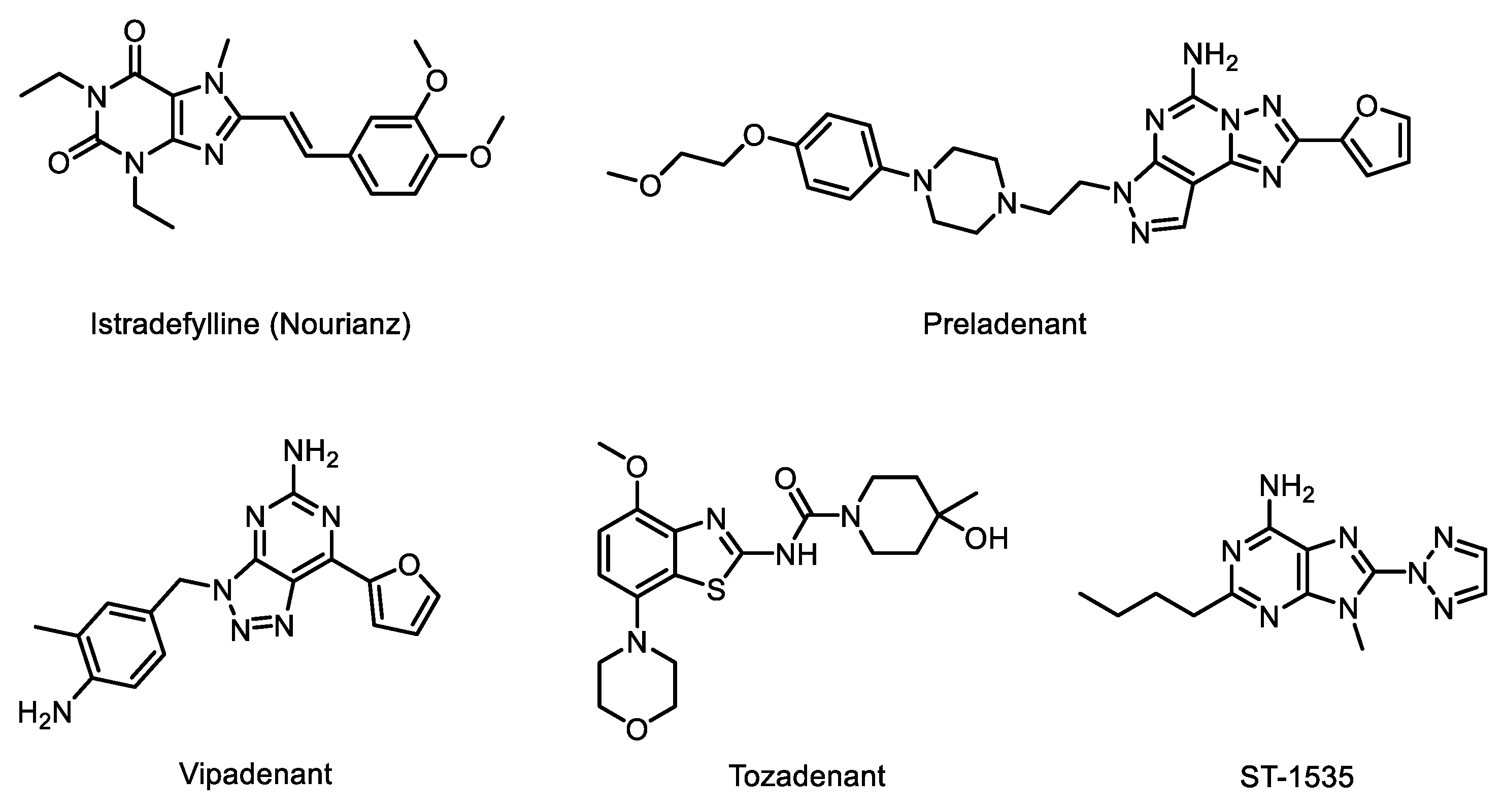
:1. Introduction
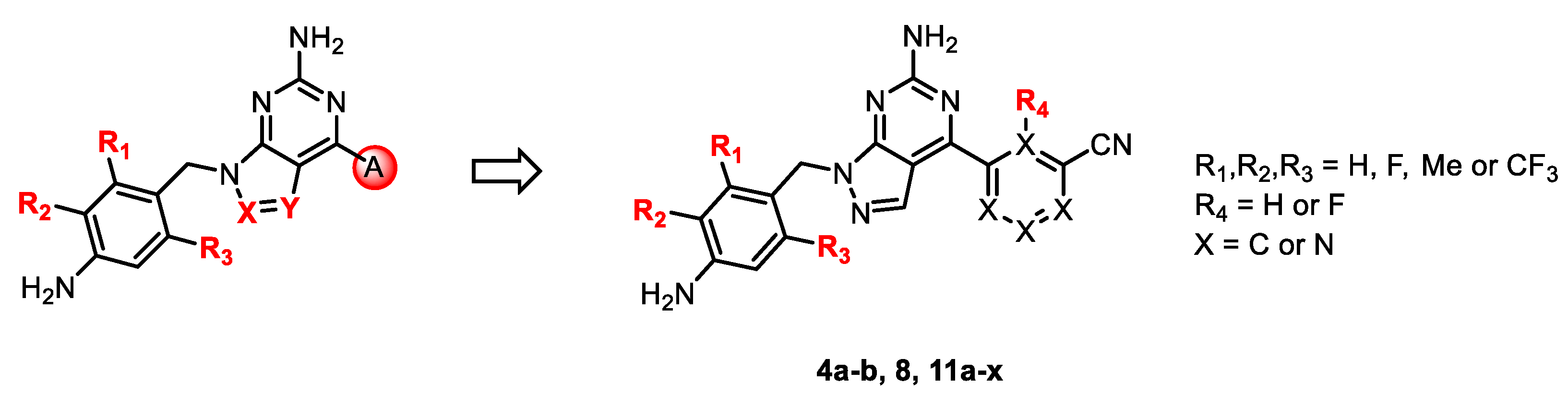
2. Results and Discussion
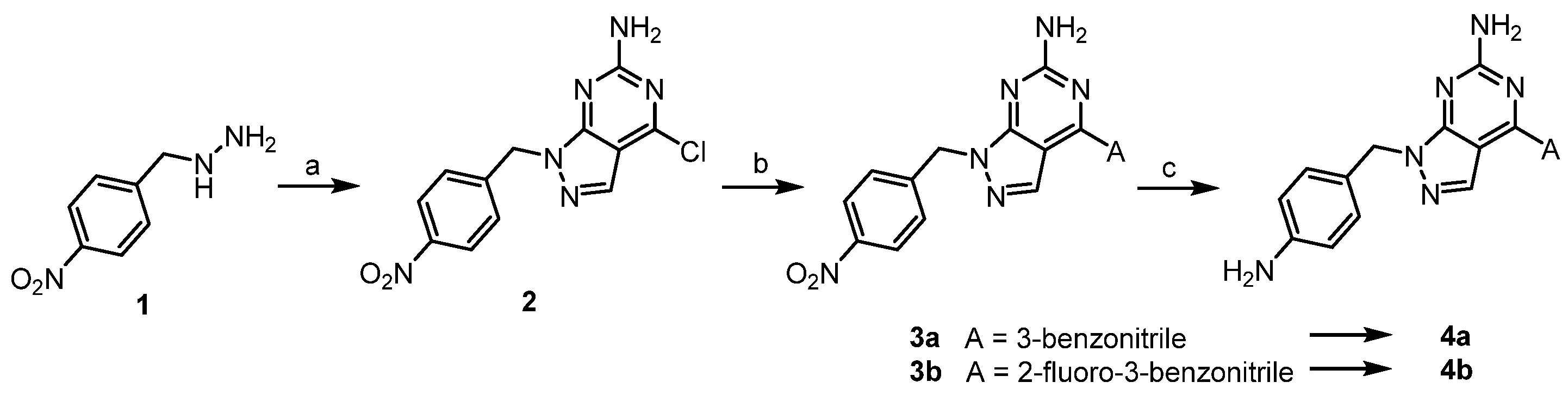
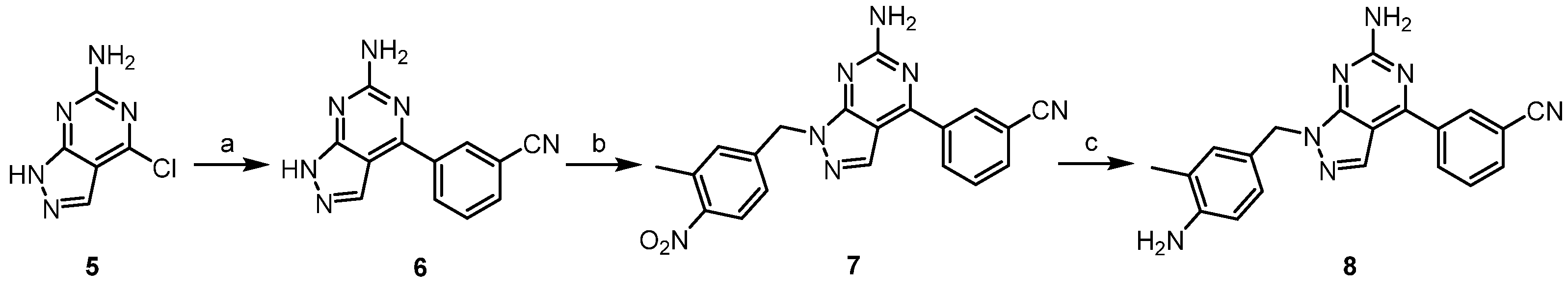
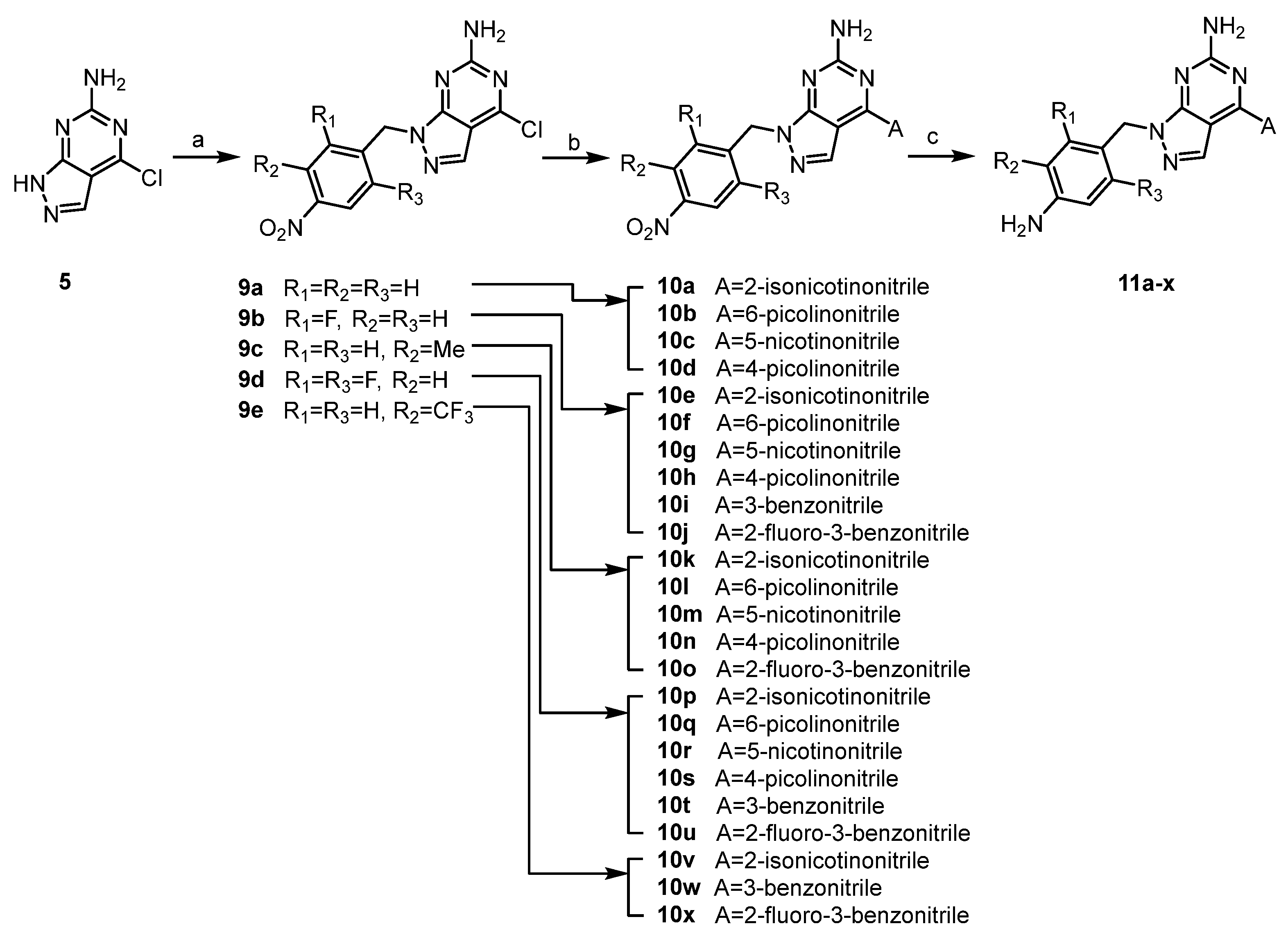
2.1. Chemistry
2.2. Biological Activities
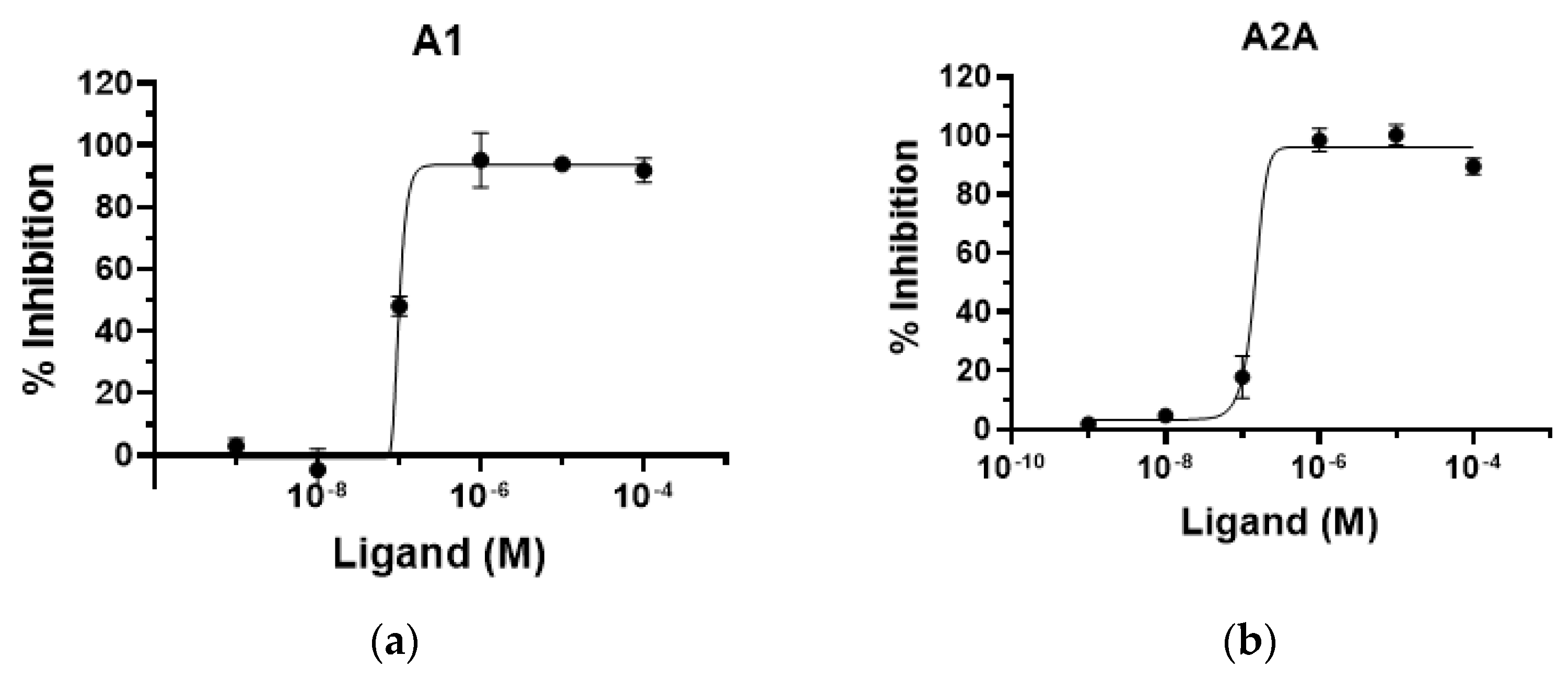
2.2.1. In Vitro Activity against Human A1 and A2A Receptors
2.2.2. Selectivity for Adenosine Receptor Subtypes
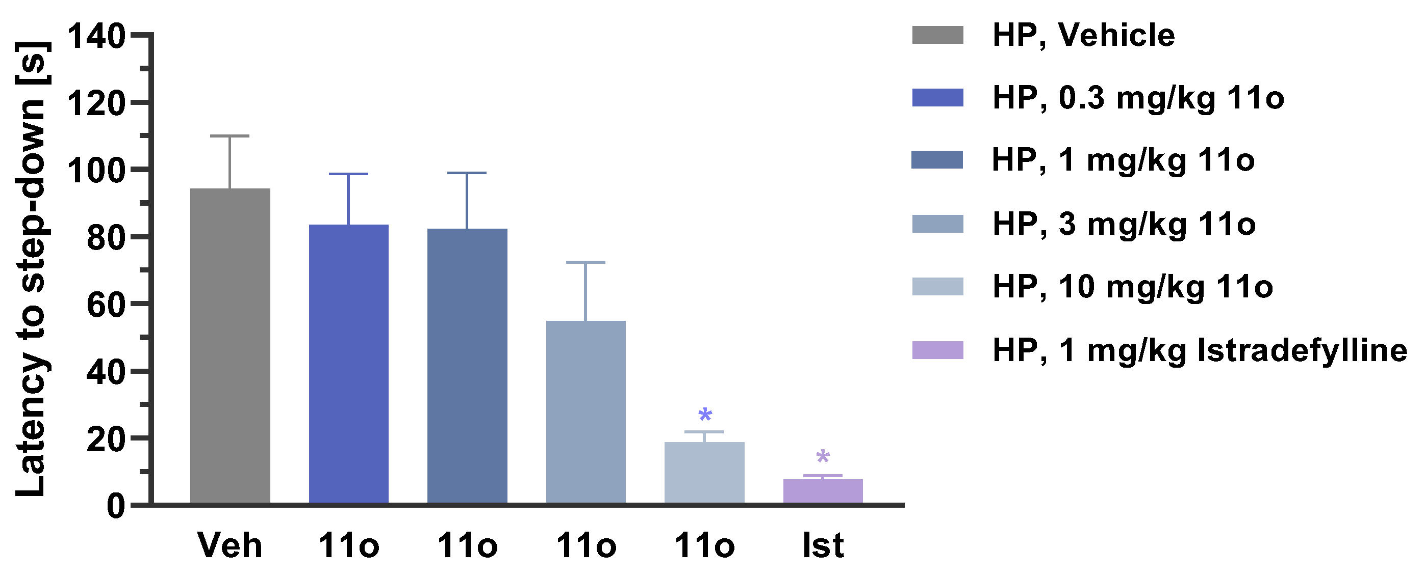
2.2.3. In Vivo Activity in Animal Models of Parkinson’s Disease
2.3. Pharmacokinetic Studies
2.3.1. In Vitro Metabolic Stability
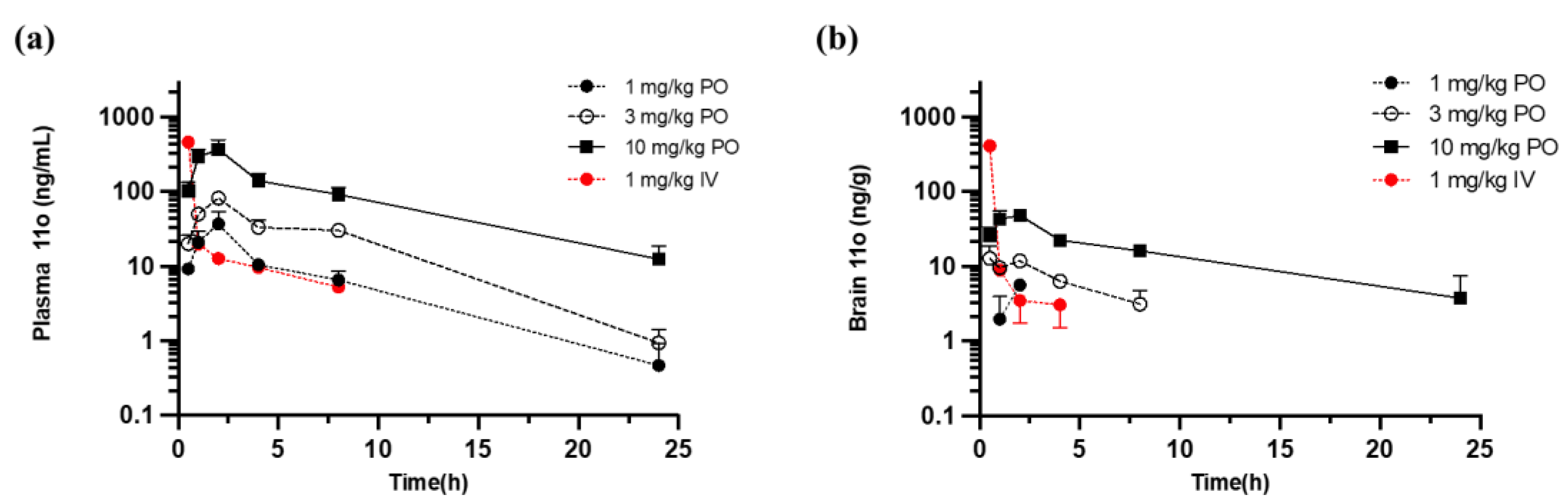
2.3.2. In Vivo Pharmacokinetic Study
2.4. Toxicity Studies
3. Materials and Methods
3.1. Chemistry
3.1.1. General Procedure A for 4a-4b
4-Chloro-1-(4-nitrobenzyl)-1H-pyrazolo[3,4-d]pyrimidin-6-amine (2)
3-(6-Amino-1-(4-nitrobenzyl)-1H-pyrazolo[3,4-d]pyrimidin-4-yl)benzonitrile (3a)
3-(6-Amino-1-(4-aminobenzyl)-1H-pyrazolo[3,4-d]pyrimidin-4-yl)benzonitrile (4a)
3-(6-Amino-1-(4-aminobenzyl)-1H-pyrazolo[3,4-d]pyrimidin-4-yl)-2-fluorobenzonitrile (4b)
3.1.2. General Procedure B for 8
3-(6-Amino-1H-pyrazolo[3,4-d]pyrimidin-4-yl)benzonitrile (6)
3-[6-Amino-1-[(3-methyl-4-nitro-phenyl)methyl]pyrazolo[3,4-d]pyrimidin-4-yl]benzonitrile (7)
3-(6-Amino-1-(4-amino-3-methylbenzyl)-1H-pyrazolo[3,4-d]pyrimidin-4-yl)benzonitrile (8)
3.1.3. General Procedure C for 11a~11w
4-Chloro-1-(3-methyl-4-nitrobenzyl)-1H-pyrazolo[3,4-d]pyrimidin-6-amine (9c)
3-(6-Amino-1-(4-amino-3-methylbenzyl)-1H-pyrazolo[3,4-d]pyrimidin-4-yl)-2-fluorobenzonitrile (10o)
3-(6-Amino-1-(4-amino-3-methylbenzyl)-1H-pyrazolo[3,4-d]pyrimidin-4-yl)-2-fluorobenzonitrile (11o)
2-(6-Amino-1-(4-aminobenzyl)-1H-pyrazolo[3,4-d]pyrimidin-4-yl)isonicotinonitrile (11a)
6-(6-Amino-1-(4-aminobenzyl)-1H-pyrazolo[3,4-d]pyrimidin-4-yl)picolinonitrile (11b)
5-(6-Amino-1-(4-aminobenzyl)-1H-pyrazolo[3,4-d]pyrimidin-4-yl)nicotinonitrile (11c)
4-(6-Amino-1-(4-aminobenzyl)-1H-pyrazolo[3,4-d]pyrimidin-4-yl)picolinonitrile (11d)
2-(6-Amino-1-(4-amino-2-fluorobenzyl)-1H-pyrazolo[3,4-d]pyrimidin-4-yl)isonicotinonitrile (11e)
6-[6-Amino-1-[(4-amino-2-fluoro-phenyl)methyl]pyrazolo[3,4-d]pyrimidin-4-yl]pyridine-2-carbonitrile (11f)
5-(6-Amino-1-(4-amino-2-fluorobenzyl)-1H-pyrazolo[3,4-d]pyrimidin-4-yl)nicotinonitrile (11g)
4-(6-Amino-1-(4-amino-2-fluorobenzyl)-1H-pyrazolo[3,4-d]pyrimidin-4-yl)picolinonitrile (11h)
3-(6-Amino-1-(4-amino-2-fluorobenzyl)-1H-pyrazolo[3,4-d]pyrimidin-4-yl)benzonitrile (11i)
3-(6-Amino-1-(4-amino-2-fluorobenzyl)-1H-pyrazolo[3,4-d]pyrimidin-4-yl)-2-fluorobenzonitrile (11j)
2-(6-Amino-1-(4-amino-3-methylbenzyl)-1H-pyrazolo[3,4-d]pyrimidin-4-yl)isonicotinonitrile (11k)
6-(6-Amino-1-(4-amino-3-methylbenzyl)-1H-pyrazolo[3,4-d]pyrimidin-4-yl)picolinonitrile (11l)
5-(6-Amino-1-(4-amino-3-methylbenzyl)-1H-pyrazolo[3,4-d]pyrimidin-4-yl)nicotinonitrile (11m)
4-(6-Amino-1-(4-amino-3-methylbenzyl)-1H-pyrazolo[3,4-d]pyrimidin-4-yl)picolinonitrile (11n)
2-[6-Amino-1-[(4-amino-2,6-difluoro-phenyl)methyl]pyrazolo[3,4-d]pyrimidin-4-yl]pyridine-4-carbonitrile (11p)
6-[6-Amino-1-[(4-amino-2,6-difluoro-phenyl)methyl]pyrazolo[3,4-d]pyrimidin-4-yl]pyridine-2-carbonitrile (11q)
5-[6-Amino-1-[(4-amino-2,6-difluoro-phenyl)methyl]pyrazolo[3,4-d]pyrimidin-4-yl]pyridine-3-carbonitrile (11r)
4-[6-Amino-1-[(4-amino-2,6-difluoro-phenyl)methyl]pyrazolo[3,4-d]pyrimidin-4-yl]pyridine-2-carbonitrile (11s)
3-[6-Amino-1-[(4-amino-2,6-difluoro-phenyl)methyl]pyrazolo[3,4-d]pyrimidin-4-yl]benzonitrile (11t)
3-(6-Amino-1-(4-amino-2,6-difluorobenzyl)-1H-pyrazolo[3,4-d]pyrimidin-4-yl)-2-fluorobenzonitrile (11u)
2-[6-Amino-1-[[4-amino-3-(trifluoromethyl)phenyl]methyl]pyrazolo[3,4-d]pyrimidin-4-yl]pyridine-4-carbonitrile (11v)
3-(6-Amino-1-(4-amino-3-(trifluoromethyl)benzyl)-1H-pyrazolo[3,4-d]pyrimidin-4-yl)benzonitrile (11w)
3-(6-Amino-1-(4-amino-3-(trifluoromethyl)benzyl)-1H-pyrazolo[3,4-d]pyrimidin-4-yl)-2-fluorobenzonitrile (11x)
3.2. In Vitro Human Adenosine Receptor-Binding and Functional Assays
3.3. hERG, AMES and Metabolic Stability Study
3.4. Animal Studies
3.4.1. MPTP-Induced Mouse Model of Parkinson’s Disease
3.4.2. Nest Building
3.4.3. Haloperidol-Induced Catalepsy in Female Rats
3.5. In Vivo Pharmacokinetics, Bioavailability and Brain Plasma Ratio Study in Mouse
3.6. Maximum Tolerated Dose Study in Rat and Dog
3.7. Statistical Analysis
4. Conclusions
Author Contributions
Funding
Institutional Review Board Statement
Informed Consent Statement
Data Availability Statement
Acknowledgments
Conflicts of Interest
References
- Tanner, C.M. Exploring the clinical burden of OFF periods in Parkinson Disease. Am. J. Manag. Care 2020, 26, S215–S264. [Google Scholar]
- Poewe, W.; Antonini, A. Novel formulations and modes of delivery of Levodopa. Mov. Disord. 2015, 30, 114–120. [Google Scholar] [CrossRef] [PubMed]
- Jenner, P.; Mori, A.; Aradi, S.D.; Hauser, R.A. Istradefylline—A first generation adenosine A2A antagonist for the treatment of Parkinson’s disease. Exp. Rev. Neurother. 2021, 21, 317–333. [Google Scholar] [CrossRef] [PubMed]
- LeWitt, P.A.; Aradi, S.D.; Hauser, R.A.; Rascol, O. The challenge of developing adenosine A2A antagonists for Parkinson disease: Istradefylline, preladenant, and tozadenant. Parkinsonism Relat. Disord. 2020, 80 (Suppl. 1), S54–S63. [Google Scholar]
- Saki, M.; Yamada, K.; Koshimura, E.; Sasaki, K.; Kanda, T. In vitro pharmacological profile of the A2A receptor antagonist istradefylline. Naunyn-Schmiedeberg’s Arch. Pharmacol. 2013, 386, 963–972. [Google Scholar] [CrossRef] [PubMed]
- Shook, C.; Rassnick, S.; Wallace, N.; Crooke, J.; Ault, M.; Chakravarty, D.; Barbay, K.; Wang, A.; Powell, M.T.; Leonard, K.; et al. Design and characterization of optimized adenosine A2A/A1 receptor antagonists for the treatment of Parkinson’s Disease. J. Med. Chem. 2012, 55, 1402–1417. [Google Scholar] [CrossRef]
- Mihara, T.; Iwashita, A.; Matsuoka, N. A novel adenosine A1 and A2A receptor antagonist ASP5854 ameliorates motor impairment in MPTP-treated marmosets; Comparison with existing anti-Parkinson’s disease drugs. Behav. Brain. Res. 2008, 194, 152–161. [Google Scholar] [CrossRef]
- Mihara, T.; Mihara, K.; Yarimizu, J.; Mitani, Y.; Matsuda, R.; Yamamoto, H.; Aoki, S.; Akahane, A.; Iwashita, A.; Matsuoka, N. Pharmacological characterization of a novel, potent adenosine A1 and A2A receptor dual antagonist, 5-[5-amino-3(4-fluorophenyl)pyrazin-2-yl]-1-isopropylpyridine-2(1H)-one (ASP5854), in models of Parkinson’s disease and cognition. J. Pharmacol. Exp. Ther. 2007, 323, 708–719. [Google Scholar] [CrossRef] [PubMed] [Green Version]
- Markovic, T.; Rocke, B.N.; Blakemore, D.C.; Mascitti, V.; Willis, M.C. Pyridine sulfinates as general nucleophilic coupling partners in palladium-catalyzed cross-coupling reactions with aryl halides. Chem. Sci. 2017, 8, 4437–4442. [Google Scholar] [CrossRef] [PubMed] [Green Version]
- Markovic, T.; Murray, P.R.D.; Rocke, B.N.; Shavnya, A.; Blakemre, D.C.; Willis, M.C. Heterocyclic allylsulfones as latent heteroaryl nucleophiles in palladium-catalyzed cross-coupling reactions. J. Am. Chem. Soc. 2018, 140, 15916–15923. [Google Scholar] [CrossRef] [PubMed] [Green Version]
- Varani, K.; Gessi, S.; Dalpiaz, A.; Borea, P.A. Pharmacological and biochemical characterization of purified A2A adenosine receptors in human platelet membranes by [3H]CGS21680 binding. Br. J. Pharmacol. 1996, 117, 1693–1701. [Google Scholar] [CrossRef] [PubMed] [Green Version]
- Gerlach, M.; Riederer, P. Animal models of Parkinson’s disease: An empirical comparison with the phenomenology of the disease in man. J. Neural Transm. 1996, 103, 987–1041. [Google Scholar] [CrossRef] [PubMed]
- Jackson-Lewis, V.; Przedborski, S. Protocol for the MPTP mouse model of Parkinson’s disease. Nat. Protoc. 2007, 2, 141–151. [Google Scholar] [CrossRef] [PubMed]
- Obach, R.S. Prediction of human clearance of twenty-nine drugs from hepatic microsomal intrinsic clearance data: An examination of in vitro half-life approach and nonspecific binding to microsomes. Drug. Met. Dis. 1999, 27, 1350–1359. [Google Scholar]
- Crumb, W.J., Jr.; Johannesen, V.J.; Strauss, D.G. An evaluation of 30 clinical drugs against the comprehensive in vitro proarrhythmia assay (CiPA) proposed ion channel panel. J. Pharmacol. Toxicol. Methods 2016, 81, 251–262. [Google Scholar] [CrossRef] [PubMed] [Green Version]
- Ames, B.N.; McCann, J.; Yamasaki, E. Methods for detecting carcinogens and mutagens with the Salmonella/mammalian microsome mutagenicity test. Mutat. Res. 1975, 31, 347–364. [Google Scholar] [CrossRef]
- Maron, D.M.; Ames, B.N. Revised methods for the Salmonella mutagenicity test. Mutat. Res. 1983, 113, 173–215. [Google Scholar] [CrossRef]
- Cheng, Y.; Prusoff, W.H. Relationship between the inhibition constant (K1) and the concentration of inhibitor which causes 50 per cent inhibition (I50) of an enzymatic reaction. Biochem. Pharmacol. 1973, 22, 3099–3108. [Google Scholar] [PubMed]
- OECD. OECD Guideline 471 (Genetic Toxicology: Bacterial Reverse Mutation Test), Ninth Addendum to the OECD Guidelines for the Testing of Chemicals, Adopted July 21, 1997; OECD: Paris, France, 1997. [Google Scholar]
- Detection of carcinogens as mutagens in the Salmonella/microsome test: Assay of 300 chemicals: Discussion. Proc. Natl. Acad. Sci. USA 1976, 73, 950–954. [CrossRef] [PubMed] [Green Version]
- Deacon, R.M.J. Assessing nest building in mice. Nat. Protoc. 2006, 1, 1117–1119. [Google Scholar] [CrossRef] [PubMed]


 | ||||||
|---|---|---|---|---|---|---|
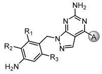
| Compounds | R1 | R2 | R3 | A2A Ki (nM) * | A1 Ki (nM) * | |
| Istradefylline | 8.64 | 610 | ||||
| 11a | 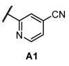 | H | H | H | 310 | 2470 |
| 11e | F | H | H | 120 | 710 | |
| 11k | H | Me | H | 65 | 510 | |
| 11p | F | H | F | 47 | 380 | |
| 11v | H | CF3 | H | 10.2 | 250 | |
| 11b | 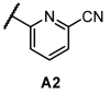 | H | H | H | 31 | 650 |
| 11f | F | H | H | 10.6 | 160 | |
| 11l | H | Me | H | 6.39 | 180 | |
| 11q | F | H | F | 6.84 | 150 | |
| 11c | 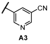 | H | H | H | 4790 | 32,000 |
| 11g | F | H | H | 2640 | 32,800 | |
| 11m | H | Me | H | 2480 | 21,200 | |
| 11r | F | H | F | 1220 | 10,000 | |
| 11d |  | H | H | H | 4720 | 8530 |
| 11h | F | H | H | 3190 | 28,000 | |
| 11n | H | Me | H | 1150 | 21,300 | |
| 11s | F | H | F | 4920 | - | |
| 4a |  | H | H | H | 16.38 | - |
| 11i | F | H | H | 12.1 | 160 | |
| 8 | H | Me | H | 11.1 | 110 | |
| 11t | F | H | F | 7.42 | 50 | |
| 11w | H | CF3 | H | 1.94 | 49 | |
| 4b |  | H | H | H | 19.4 | - |
| 11j | F | H | H | 16.4 | 63 | |
| 11o | H | Me | H | 13.3 | 55 | |
| 11u | F | H | F | 6.45 | 30 | |
| 11x | H | CF3 | H | 2.71 | 33 | |
| Adenosine Receptors | IC50 (nM) a | Ki (nM) a | nH b |
|---|---|---|---|
| A1 | 94 | 55 | 1.17 |
| A2A | 24 | 13.3 | 0.79 |
| A2B | 1220 | 400 | 0.98 |
| A3 | 1130 | 1050 | 0.44 |
| Species | Half-Life (min) | Intrinsic Clearance (mL/min/106 Cells) | Extrapolated Clearance, CL (mL/min/kg) | ||
|---|---|---|---|---|---|
| 11o | Midazolam b | 11o | Midazolam | 11o | |
| Mouse | 70.9 | 49.9 | 0.00977 | 0.0139 | 115 |
| Rat | 46.9 | 21.9 | 0.0148 | 0.0317 | 69.2 |
| Dog | ND a | 32.8 | ND a | 0.0211 | ND a |
| Monkey | 124 | 36.0 | 0.00558 | 0.0192 | 20.1 |
| Human | 184 | 68.3 | 0.00377 | 0.0101 | 9.58 |
| Dose (mg/kg) | Dose Route | Tmax (h) | T1/2 (h) | Cmax (ng/mL) | AUClast (h·ng/mL) | BA c (%) |
|---|---|---|---|---|---|---|
| 1 | PO | 2.00 | 4.38 | 36.8 | 176 | 53.0 |
| 3 | PO | 2.00 | 3.66 | 81.3 | 577 | 57.9 |
| 10 | PO | 2.00 | 5.69 | 367 | 2249 | 67.7 |
| 1 | IN | 0.50 | NC a | 23.3 | 74.9 | 22.6 |
| 3 | IN | 0.50 | NR b | 53.9 | 183 | 18.4 |
| 10 | IN | 0.50 | 7.40 | 88.5 | 444 | 13.4 |
| 1 | IV | 0.083 | 3.95 | 459 | 332 | 100 |
| Dose (mg/kg) | Dose Route | Tmax (h) | T1/2 (h) | Cmax (ng/mL) | AUClast (h·ng/mL) | B/P Ratio c |
|---|---|---|---|---|---|---|
| 1 | PO | 2.00 | NC a | 5.58 | 4.26 | 0.0242 |
| 3 | PO | 0.50 | 3.23 | 12.8 | 56.4 | 0.0977 |
| 10 | PO | 2.00 | 7.71 | 48.1 | 375 | 0.167 |
| 1 | IN | 0.50 | NC | 9.63 | 5.23 | 0.0698 |
| 3 | IN | 0.50 | NC | 15.9 | 14.2 | 0.0776 |
| 10 | IN | 0.50 | 3.74 | 38.2 | 81.3 | 0.183 |
| 1 | IV | 0.083 | NR b | 410 | 246 | 0.741 |
| Toxicity | |
|---|---|
| hERG inhibition | IC50 = 116 μM |
| AMES | no mutagenicity up to 1867 μg/plate |
| MTD (rat) | >1000 mg/kg |
| MTD (dog) | >400 mg/kg |
Publisher’s Note: MDPI stays neutral with regard to jurisdictional claims in published maps and institutional affiliations. |
© 2022 by the authors. Licensee MDPI, Basel, Switzerland. This article is an open access article distributed under the terms and conditions of the Creative Commons Attribution (CC BY) license (https://creativecommons.org/licenses/by/4.0/).
Share and Cite
Jung, J.; Lee, Y.; Moon, A.-N.; Ann, J.; Jeong, J.J.; Do, N.; Lee, J. Discovery of Novel Dual Adenosine A2A and A1 Receptor Antagonists with 1H-Pyrazolo[3,4-d]pyrimidin-6-amine Core Scaffold as Anti-Parkinson’s Disease Agents. Pharmaceuticals 2022, 15, 922. https://doi.org/10.3390/ph15080922
Jung J, Lee Y, Moon A-N, Ann J, Jeong JJ, Do N, Lee J. Discovery of Novel Dual Adenosine A2A and A1 Receptor Antagonists with 1H-Pyrazolo[3,4-d]pyrimidin-6-amine Core Scaffold as Anti-Parkinson’s Disease Agents. Pharmaceuticals. 2022; 15(8):922. https://doi.org/10.3390/ph15080922
Chicago/Turabian StyleJung, Juyoung, Yoonsuk Lee, An-Na Moon, Jihyae Ann, Jin Ju Jeong, Nayeon Do, and Jeewoo Lee. 2022. "Discovery of Novel Dual Adenosine A2A and A1 Receptor Antagonists with 1H-Pyrazolo[3,4-d]pyrimidin-6-amine Core Scaffold as Anti-Parkinson’s Disease Agents" Pharmaceuticals 15, no. 8: 922. https://doi.org/10.3390/ph15080922
APA StyleJung, J., Lee, Y., Moon, A.-N., Ann, J., Jeong, J. J., Do, N., & Lee, J. (2022). Discovery of Novel Dual Adenosine A2A and A1 Receptor Antagonists with 1H-Pyrazolo[3,4-d]pyrimidin-6-amine Core Scaffold as Anti-Parkinson’s Disease Agents. Pharmaceuticals, 15(8), 922. https://doi.org/10.3390/ph15080922