Article

α-Glucosidase and α-Amylase Inhibitory Potentials of Quinoline–1,3,4-oxadiazole Conjugates Bearing 1,2,3-Triazole with Antioxidant Activity, Kinetic Studies, and Computational Validation

1
School of Chemistry and Physics, University of KwaZulu-Natal, P/Bag X54001, Westville, Durban 4000, South Africa
2
Department of Biochemistry, School of Life Sciences, University of Kwazulu-Natal, Westville, Durban 4000, South Africa
*
Author to whom correspondence should be addressed.
Pharmaceuticals 2022, 15(8), 1035; https://doi.org/10.3390/ph15081035
Submission received: 21 July 2022 / Revised: 15 August 2022 / Accepted: 16 August 2022 / Published: 22 August 2022
(This article belongs to the Special Issue Hybrid Drugs: Design and Applications)

Abstract

:
Diabetes mellitus (DM) is a multifaceted metabolic disorder that remains a major threat to global health security. Sadly, the clinical relevance of available drugs is burdened with an upsurge in adverse effects; hence, inhibiting the carbohydrate-hydrolyzing enzymes α-glucosidase and α-amylase while preventing oxidative stress is deemed a practicable strategy for regulating postprandial glucose levels in DM patients. We report herein the α-glucosidase and α-amylase inhibition and antioxidant profile of quinoline hybrids 4at and 12at bearing 1,3,4-oxadiazole and 1,2,3-triazole cores, respectively. Overall, compound 4i with a bromopentyl sidechain exhibited the strongest α-glucosidase inhibition (IC50 = 15.85 µM) relative to reference drug acarbose (IC50 = 17.85 µM) and the best antioxidant profile in FRAP, DPPH, and NO scavenging assays. Compounds 4a and 12g also emerged as the most potent NO scavengers (IC50 = 2.67 and 3.01 µM, respectively) compared to gallic acid (IC50 = 728.68 µM), while notable α-glucosidase inhibition was observed for p-fluorobenzyl compound 4k (IC50 = 23.69 µM) and phenyl-1,2,3-triazolyl compound 12k (IC50 = 22.47 µM). Moreover, kinetic studies established the mode of α-glucosidase inhibition as non-competitive, thus classifying the quinoline hybrids as allosteric inhibitors. Molecular docking and molecular dynamics simulations then provided insights into the protein–ligand interaction profile and the stable complexation of promising hybrids at the allosteric site of α-glucosidase. These results showcase these compounds as worthy scaffolds for developing more potent α-glucosidase inhibitors with antioxidant activity for effective DM management.

1. Introduction

Diabetes Mellitus (DM) is a chronic systemic disorder characterized by dysfunctional glucose regulation. Typically, patients secrete insufficient insulin (i.e., type 1 diabetes mellitus–T1DM) or develop insulin resistance (i.e., type 2 diabetes mellitus–T2DM), which leads to elevated blood glucose levels (hyperglycemia) [1]. Complications associated with DM include nervous system damage, eye damage, cardiovascular disease, stroke, kidney disease, and gangrene [2]. The disease persists as a heavy burden on public health, having become the ninth leading cause of death with approximately 451 million incidences globally [3]. Recent estimations have shown increased incidences in Africa, with about 15.5 million cases in 2017, and that 69.2% of the adult population are ignorant of their diabetic status [4]. T2DM has a higher incidence rate globally, with about 90% or more of the cases relative to T1DM.
T2DM can be managed by regulating the key molecular targets involved in carbohydrate digestion i.e., α-glucosidase and pancreatic α-amylase [5]. These enzymes break down dietary carbohydrates into simple sugars which are absorbed from the small intestine into the bloodstream. Elevated levels of α-glucosidase and α-amylase are therefore associated with increased postprandial glucose levels seen in T2DM patients, making their inhibition a successful strategy for T2DM management [6]. Nevertheless, the availability of α-glucosidase inhibitors is currently restricted to N-heterocyclic carbasugars such as acarbose, voglibose, and miglitol [7]. These molecules present sugar-like moieties that compete with the enzyme’s natural substrates for binding at the active site to inhibit sugar hydrolysis, and consequently decrease postprandial hyperglycemia [8]. Although these drugs have rapid action, they are marred by efficacy problems and adverse side effects such as diarrhea, flatulence, and abdominal discomfort [9], which precipitates the need for new and safer α-glucosidase inhibitors.
Reactive oxygen species (ROS), including free radicals of superoxide anion (O2), hydroxyl (OH) radicals, and hydrogen peroxide (H2O2), play a prominent role in regulating cellular functions as their intermediates control various enzymatic reactions involved in signal transductions [10]. However, their excessive accumulation contributes to toxic cellular oxidative stress which induces the pathogenesis of diseases such as T2DM [11]. Therefore, the development of small molecules with antidiabetic and free radical scavenging potentials is considered an attractive option for T2DM management.
The medicinal chemistry significance of the quinoline scaffold has been reiterated in diverse therapeutic applications including antimicrobial [12], antimalarial [13], anticancer [14], antidiabetic [15], and antioxidant [16] therapies. Among the quinoline congeners, 8-hydroxyquinoline (8-HQ) has attracted attention due to its metal chelating properties, a characteristic which may be beneficial to attenuating the pathogenesis of several diseases including diabetes [17,18,19]. The 1,3,4-oxadiazole moiety is another relevant pharmacophore, and is a known bioisostere of amide and ester groups. The scaffold’s bioactivity has been linked to its strong hydrogen bond interactions with various targets [20,21]. The electronic effects of the azole stimulate ligand binding and improve lipophilicity for active transport through the cell membrane to various targets to elicit a biological response [20]. For example, Taha et al. [22] showed that a quinoline-1,3,4-oxadiazole hybrid A (Figure 1) had 15-fold superior α-glucosidase inhibition (IC50 = 2.6 µM) compared with acarbose (IC50 = 38.25 µM). Izgi et al. [23] reported the 1,3,4-oxadiazole compound B bearing an azaindole core, a potent α-glucosidase inhibitor with IC50 = 0.4 mM compared with acarbose IC50 = 1.75 mM. Furthermore, the 1,2,3-triazole core is renowned in medicinal chemistry as a five-membered ring with a wide range of biological activity [24,25]. The fame of the pharmacophore’s therapeutic potential has incessantly increased since the dawn of click chemistry, described by Sharpless [26]. As a result, several α-glucosidase inhibitors have been documented with 1,2,3-triazole moieties in their structures [27]. Asemanipoor et al. [28] reported the potent α-glucosidase inhibition (IC50 = 35.0 µM) of a 1,2,3-triazole derivative C compared with acarbose standard (IC50 = 750.0 µM).
Although quinoline, 1,3,4-oxadiazole, and 1,2,3-triazole moieties have been reported as α-glucosidase inhibitors in many molecular hybrid designs [29], these findings require further investigation to achieve optimal results. Consequently, in continuation of our exploits within the MH approach towards quinoline, 1,3,4-oxadiazole, and 1,2,3-triazole hybrids with therapeutic potentials [30,31], we herein incorporated these pharmacophores in a single molecular hybrid and evaluated the effect on the in vitro α-glucosidase and α-amylase inhibitory potencies and antioxidant activities. We also examined the mode of α-glucosidase inhibition of promising compounds via kinetic studies, then rationalized the results with molecular docking and molecular dynamics simulations.

2. Results and Discussion

2.1. Chemistry

The synthetic route to the targeted quinoline hybrids is depicted in Scheme 1, Scheme 2 and Scheme 3. First, compounds 4at were synthesized in four steps to examine the influence of 1,2,3-triazole moiety on the bioactivity of the present molecular design (Scheme 1). The reaction protocol started by O-alkylating 8-hydroxyquinoline (8-HQ) with ethyl chloroacetate in N,N-dimethylformamide (DMF) at room temperature to give ethyl 2-(quinolin-8-yloxy)acetate 1. The ester intermediate was then refluxed with hydrazine hydrate in ethanol to give 2-(quinolin-8-yloxy)acetohydrazide 2. A potassium hydroxide-promoted condensation of 2 with carbon disulfide afforded a potassium carbodithioate salt, which subsequently underwent hydrochloric acid-promoted intramolecular cyclization to yield 5-[(quinolin-8-yloxy)methyl]-1,3,4-oxadiazole-2-thiol 3 in 65% yield over 3 steps. A potassium carbonate-promoted S-alkylation of this thiol intermediate with different benzyl and alkyl halides, respectively, afforded the desired quinoline–1,3,4-oxadiazole hybrids 4at in moderate to quantitative yields.
To obtain hybrids 12at bearing the 1,2,3-triazole pharmacophore, the azide precursors needed for the Cu(I)-catalyzed azide–alkyne [3 + 2] cycloaddition reaction (click reaction) were synthesized according to their substrate demands (Scheme 2).
With the azides at hand, intermediate 3 was treated with propargyl bromide in DMF in the presence of potassium carbonate to give the corresponding terminal alkyne 11. This product was subjected to a click reaction with azides 6a, b, or 8ah, or 10aj in a DCM:H2O (1:1) solvent mixture to afford the target quinoline–1,3,4-oxadiazole–1,2,3-triazole hybrids 12at in 43–91% yields (Scheme 3).

2.2. Structural Elucidation

The synthetic protocol’s success was established using spectroscopic experiments, viz., NMR (1H, 13C and 2D) and HRMS. The emergence of -NH (δH 9.45) and -NH2 (δH 4.37) signals in the 1H NMR spectrum of compound 2 and the absence of ethoxy group quartet (δH 4.29, J = 7.1 Hz) and triplet (δH 1.28, J = 7.1 Hz) signals of compound 1, respectively, confirmed the ester group’s conversion to hydrazide. Likewise, the formation of the 2-mercapto-1,3,4-oxadiazole ring was evidenced by a broad singlet at δH 14.77 corresponding to the thiol group’s deshielded proton. The disappearance of this peak and the appearance of doublet (CH2, δH 4.17, J = 2.6 Hz) and (CH, δH 3.32, J = 2.6 Hz) triplet signals of an allylic alkyne unit confirmed the conversion of 3 to 11. Furthermore, the structural integrity of quinoline hybrids 4at and 12at was confirmed by 2D NMR experiments, i.e., heteronuclear single quantum coherence (HSQC) and heteronuclear multiple bond coherence (HMBC) correlations. For instance, in compound 4k, the correct linkage of quinoline to 1,3,4-oxadiazole was seen in the HMBC correlations of H-6′ (δH 5.61) to C-8 and C-5′quaternary carbons at 153.33 and 164.32 ppm respectively (Figure 2). H-7′ (δH 4.51) correlations to C-2′ (δC 164.82), C-8′ (δC 133.22, d, J = 3.0 Hz), and C-9′/C-13′ (δC 131.58, d, J = 8.3 Hz, C-9′,13′) showed that alkylation occurred at the sulfur atom of oxadiazole and not the nitrogen of a possible thione tautomer. In the 1,2,3-triazole series, e.g., 12e, the characteristic triazole H-5″ singlet peak at δH 7.70 showed two HMBC correlations with C-7″ (δC 53.43) and C-4″ (δC 143.25), while H-6″ (δH 4.51) correlated to C-2′ (δC 163.45), C-4″ (δC 143.25), and C-5″ (δC 123.24). Additionally, the molecular ion peaks corresponding to [M + Na] at m/z 390.0696 and 457.0851 in the HRMS spectra of 4k and 12e, respectively, also affirmed the quinoline hybrid’s structural integrity.

2.3. Antioxidant Activity Profiling

2.3.1. 2,2-Diphenyl-1-Picrylhydrazyl (DPPH) Radical Scavenging Activity

The DPPH assay is used conventionally to quantify a compound’s ability to trap free radicals. The relative efficacy of compounds 4at and 12at was evaluated in this regard; the results are shown in Table 1. Except for the hybrids’ precursor, compound 3 (IC50 of 25.15 µM), the compounds in both series exhibited moderate free radical scavenging activity relative to the reference scavenger, gallic acid (IC50 = 26.63 µM). The 1,3,4-oxadiazole series 4at were again more potent than the 1,2,3-triazoles 12at. SAR analysis showed that 2-mercapto-1,3,4-oxadiazole core was beneficial to DPPH scavenging activity, as seen in the two-fold improved potency of compound 3 as compared to 8-HQ (IC50 = 51.81 µM); however, S-alkylation was found generally detrimental to potency.

2.3.2. Ferric Reducing Antioxidant Power (FRAP)

FRAP is a ferric (Fe3+) to ferrous (Fe2+) ions reduction assay that measures a compound’s electron-donating capacity, i.e., the antioxidant’s reducing power [32]. The results in Table 2 reveal that compound 4i was the most promising hybrid in this regard; IC50 = 22.88 µM. The compound’s FRAP activity was four-fold superior to standard compound ascorbic acid (IC50 = 102.62 µM) and comparable to gallic acid (IC50 = 17.85 µM).

2.3.3. Nitric Oxide (NO) Activity

The screening of the present compound series for NO radical scavenging activity (Table 2) identified excellent NO scavengers. Compound 4a from the 1,3,4-oxadiazole series, bearing an ethyl chain, showed the highest NO scavenging activity with IC50 = 2.67 µM. Compound 4i, with IC50 = 197.93 µM, displayed a four-fold higher efficacy compared with gallic acid (IC50 = 782.68 µM), to emerge as the best antioxidant overall. From the 1,2,3-triazole series, para-nitro benzyl compound 12g showed excellent NO scavenging ability with IC50 = 3.01 µM, contrary to its 1,3,4-oxadiazole congener compound 4n (IC50 > 500 µM).

2.4. Enzyme Inhibition and Structure–Activity Relationship (SAR) Analysis

Abounding empirical evidence highlights the significance in the pathogenesis of T2DM of carbohydrate-digesting enzymes α-glucosidase and α-amylase; hence, inhibiting these enzymes is crucial to T2DM management [33]. The inhibitory potencies of quinoline hybrids 4at and 12at against these enzymes are presented in Table 2. The compounds show low micromolar α-glucosidase inhibition with IC50 values ranging between 15.85 to 63.59 µM, in contrast to their moderate α-amylase inhibition. The quinoline–1,3,4-oxadiazole series 4at are generally stronger α-glucosidase inhibitors compared with their 1,2,3-triazole congeners 12at. This SAR is conceivably due to the increased lipophilicity and polar surface area of the 1,2,3-triazole core. Interestingly, the less lipophilic phenyl compounds 12kt were also better α-glucosidase inhibitors compared with the more lipophilic benzyl compounds 12aj.
In the 1,3,4-oxadiazole series 4at, propyl was identified as the optimal alkane chain, as seen in the α-glucosidase inhibitory potencies of 4af. However, replacing these alkyl chains with a bromopentyl unit led to the most potent α-glucosidase inhibitor, compound 4i (IC50 = 15.85 µM) with a superior potency compared to standard drug acarbose (IC50 = 17.85 µM). Among the benzyl analogues, para-fluoro substituted compound 4k (IC50 = 23.69 µM) was twice as potent as the meta-fluoro analogue 4o (IC50 = 48.84 µM). The trend was seen in the chloro (4l, 4p) and bromo (4m, 4q) substituted compounds, respectively. This shows that altering the substituent’s position from para to meta is detrimental to α-glucosidase inhibition, and the order of potency in the para-substituted derivatives is F > Cl > Br > NO2. It is also inferable that inhibitory potency is dependent on the halogen’s electronegativity. The non-tolerance for meta or ortho positions was again highlighted in disubstituted compounds 4q4t which had inferior potencies relative to their monosubstituted congeners. Interestingly, those SARs were repeated in the 12at series. Compound 12d (IC50 = 38.11 µM) was more potent than 12h (IC50 = 63.59 µM) and 12i (IC50 = 42.79 µM). Conversely, both series exhibited poor α- amylase inhibition. A schematic SAR summary of the antioxidant profiling and α-glucosidase inhibition for the quinoline hybrids is presented in Figure 3.

2.5. Mode of α-Glucosidase Inhibition: Enzyme Kinetic Studies

Understanding a drug’s mode of inhibiting its target is crucial to improve the drug’s activity profile and target specificity. Although orthosteric inhibition, in which the ligand binds at the target’s active site, dominates among currently available drugs, allosteric inhibitors (i.e., drugs binding at regions close to the active site) are desirable due to their reduced vulnerability to active site mutation or to displacement through native substrate overload at the active site [34]. Accordingly, the mode of α-glucosidase inhibition was investigated for promising compounds 4b, 4i, 4j, 4k, 4l, 12k, and 12m, using a time-dependent para-nitrophenyl-β-D-glucopyranoside (pNPG) assay; the results are presented as Lineweaver–Burk plots (Figure 4). Clearly, all the compounds exhibited a non-competitive type of enzyme inhibition as the maximum reaction velocity (Vmax) changed for each inhibitor concentration tested, but the Michaelis–Menten constant (Km) remained unchanged. This indicates that the present compounds bind at the allosteric sites of free α-glucosidase, to inhibit its hydrolytic activity, and of the enzyme-substrate complex, to induce a slower release of the hydrolysis product glucose. Conceivably, this can result in decreased systemic glucose concentration thus alleviating postprandial hyperglycemia and its associated complications.

2.6. Homology Modeling and Molecular Docking

Having established from enzyme kinetic studies the allosteric mode of α-glucosidase inhibition for promising compounds 4b, 4i, 4j, 4k, 4l, 12k, and 12m, we sought to gain an in silico insight into the compounds’ binding profiles. However, the 3D-crystal structure of α-glucosidase remained unsolved; thus, we constructed a 3D model of α-glucosidase using the homology modeling module in the Schrödinger molecular modelling suite [35] and the FASTA sequence of Saccharomyces cerevisiae yeast α-glucosidase (UniProt entry: P38138) [36]. A BLAST search retuned the oligo-1,6-glucosidase isomaltase structure (PDB: 3A4A) which was selected as a template for model building. Notably, the yeast isomaltase shared a high sequence identity and similarity, 72% and 86%, respectively, with α-glucosidase; hence their 3D structures were similar (Figure 5).
Due to the non-competitive character (i.e., allosteric inhibition) shown by the selected compounds, SiteMap calculations [37] were performed to identify other potential binding sites on α-glucosidase. The calculations produced five binding sites (Table S1) which were evaluated in terms of site score, drugability score (>1), and site volume (>225). Site two was chosen as the most probable allosteric site using these scoring functions (Figure 4). Subsequently, molecular docking calculations were performed at this site using the induced fit docking protocol [38], with Prime and Glide algorithms to account for the protein’s conformational changes due to ligand binding.
Analysis of the α-glucosidase complexes of examined compounds (Figure 6) revealed crucial protein–ligand interactions which substantiated the enzyme inhibition data. Foremost, the most potent compound 4i found an excellent fit at the allosteric site, with the highest docking score of −6.938 in the series reflecting its potency (Table S2). The α-glucosidase complex was characterized by hydrogen bond interactions of the quinoline nitrogen and oxygens atoms with His245, as well as a similar interaction of the oxadiazole ring’s nitrogen with the amino side chain of Asn241. A face-to-face π–π stacking was present between the quinoline ring and Phe300. The ligand’s stability at the allosteric site was fostered by hydrophobic interactions with Lys155, Ser156, Phe157, Phe177, His239, Pro240, His245, Ala278, and Phe311 residues. More importantly, these interactions were anchored by a halogen bond interaction of the bromopentyl unit with Agr312, Tyr313, and Asn412, an interaction which conceivably accounts for the compound’s superior α-glucosidase inhibition.
The biochemical data indicated compound 12k as the second most potent α-glucosidase inhibitor, and this was reflected in the ligand’s docking score of −6.757. Interestingly, the imidazole ring of His245 flipped 180° to form a hydrogen bond interaction with the ligand’s quinoline nitrogen, while the oxadiazole ring nitrogen and oxygen atoms formed hydrogen bond interactions with Arg439 and amino group of Arg312, respectively. Also, the triazole ring nitrogen formed a hydrogen bond with Agr312 even as the quinoline ring established a π–π stacking interaction with Phe157. Hydrophobic interactions with Phe157, Phe177, Thr215, Leu218, Hie245, Ala278, Phe300, and Arg312 residues stabilized the 12k-α-glucosidase complex. Furthermore, the compound 4k-α-glucosidase complex exhibited a lower docking score (−6.498) compared with 4i & 12k complexes, aligning with the ligands’ IC50 values. Compound 4k’s interaction profile consisted of hydrogen bond interactions of 8-HQ oxygen with Phe300, oxadiazole ring hydrogen bond donors spanning to the opposite ends of Arg312, one on the side chain amino group, and peptide bond NH. The aromatic rings of Phe157 and Phe300 aligned in plane to establish a π–π stacking interaction with the quinoline ring.
For compound 4l-α-glucosidase complex, the hydrogen bond interaction of the quinoline nitrogen was present with His239, while oxygen atoms of the ether linkage and oxadiazole ring interacted in a like manner with Lys155. The oxadiazole nitrogen also formed additional hydrogen bonds with Lys155 and Gly160, respectively. Moreover, the quinoline ring underwent a π–π stacking interaction with the His239 imidazole ring, while the oxadiazole ring experienced a π–cation stacking interaction with the protonated nitrogen of Lys155. The aromatic ring chlorine formed a halogen bond with Asn314, and complex stability was reinforced by hydrophobic interactions with Ly155, Phe157, Phe310, and Glu426.
The lower inhibitory potencies of compounds 4j and 12m (relative to 4b, 4i, 4k, 4l and 12k) were highlighted in their docking scores of −5.040 and −4.961, respectively. The interaction profiles of these two compounds were not so impressive as other examined ligands, corroborating the biochemical data. Although the oxadiazole nitrogen formed hydrogen bond interactions with His239 and Arg312 in compound 4j, and Ser281 and Tyr286 in compound 12m, the absence of the quinoline nitrogen’s hydrogen bond interaction with allosteric site residues seen in other compounds presumably accounts for the low affinity that was recorded. These results jointly rationalize the compounds’ behavior as non-competitive inhibitors i.e., they may reversibly bind at the allosteric site to trigger altered conformational dynamics at the enzyme’s active site, consequently reducing catalytic activity without competing with the native substrate’s binding.

2.7. Molecular Dynamics Simulations

Molecular dynamics (MD) simulations of the two most promising compounds 4i and 12k were performed over 200 ns trajectory, to further evaluate the ligands’ stability at the allosteric site and the ligand-induced perturbation of protein conformation. Conformational stability was measured in terms of the root mean square deviations (RMSD) and root mean square fluctuations (RMSF) (Figure 7). RMSD indicates deviations of the protein–ligand complex from the reference structural conformation over the MD simulation trajectory, thus connoting the complex’s fluctuations. Smaller fluctuations suggest a stable conformation of the protein–ligand complex, and vice versa.
The RMSD of the protein’s C-α in the 4i complex experienced increased dynamics at the beginning of the MD simulation, highlighting the perturbation effect consequent to 4i binding at the allosteric site. RMSD for the protein and ligand reached equilibrium after 70 and 120 ns, respectively, with minimal fluctuations from the average values of 3.0 and 8.0 Å, thus indicating a stable complexation over the 200 ns simulation. In contrast, larger fluctuations persisted in the RMSD evolutions of the 12k–α-glucosidase model system over the 200 ns trajectory. This highlights the system’s reduced stability and, consequently, the weaker α-glucosidase inhibition of 12k compared to 4i. Nonetheless, the 12k model system equilibrated after 100 ns with no significant differences in fluctuation, hence the protein’s C-α and ligand’s RMSD converged at 2.4 and 6.5 Å, respectively. More importantly, the RMSD convergence of the protein’s C-α in both complexes was within the order of ≤3 Å showing no significantly large fluctuations, an indication of its excellent ability to maintain a stable residency, and ligands did not diffuse [39].
RMSF is significant for characterizing changes in protein residues’ flexibility within the chain. Herein, the ligands’ effect on the enzyme structure’s flexibility for each residue fluctuation was studied. Figure 7b shows that compound 4i efficiently induced fluctuations in the protein’s residues, as seen by its highest RMSF value at ~3.8 Å with residues Ala144 and Lys233 fluctuating the most during the 200 ns simulation. Meanwhile, the highest RMSF in the compound 12k complex occurred at 4.5 Å with residues Lys233 and Ala289. It is also inferable from Figure 7b that compound 4i perturbed the receptor more than 12k did. The number of green vertical bars denoting the residues interacting with the ligand were higher in the compound 4i–α-glucosidase complex compared with 12k. This further establishes the superior docking score, affinity for the target’s allosteric site, and consequently the α-glucosidase inhibitory potency of compound 4i relative to 12k.
The protein–ligand interactions were monitored over the simulation trajectory for contacts such as hydrogen bonds, hydrophobic, and water-mediated hydrogen bonding. Hydrogen bonds and hydrophobic contacts play significant roles at the binding site in ligands’ affinity and stability, respectively, to elicit a biological response; hence their occurrence is vital in MD simulations [40]. The analysis (Figure 7c) showed that hydrophobic interactions with α-glucosidase dominated in compound 4i compared to 12k; thus, reaffirming compound 4i’s improved α-glucosidase inhibition.

3. Conclusions

The antidiabetic potential of the quinoline pharmacophore via α-glucosidase inhibition and its antioxidant effects has been detailed in this contribution. Biological evaluations of the synthesized compounds (4at and 12at) suggested that the 1,3,4-oxadiazole core was more beneficial to the desired activity compared with 1,2,3-triazole. Compound 4i bearing a bromopentyl sidechain and compound 12k with an unsubstituted phenyl-1,2,3-triazole pendant emerged as the most promising α-glucosidase inhibitors overall. Enzyme kinetics further revealed a non-competitive mode of inhibition, thus classifying the present compounds as allosteric α-glucosidase inhibitors. This behavior is valuable to reducing systemic glucose levels while evading the limitations associated with the competitive type of target inhibition. Additionally, molecular docking and molecular dynamics simulations corroborated the compounds’ stability at the allosteric site, as evidenced by their strong interactions with the receptor. The results together illuminate the potential of the present structural template for developing new α-glucosidase inhibitors with improved efficacy for T2DM management.

4. Materials and Methods

All chemicals and reagents were purchased from Merck, Johannesburg, South Africa and were used without any purification. The reaction progress was monitored with thin layer chromatography (TLC) plates. Purification of the final products was achieved using column chromatography with silica gel (0.063–0.200 mm) at different gradients of EtOAc-hexane eluents. Melting points were determined with open-end capillary tubes in a Stuart melting point instrument (SMP-3) and were uncorrected. The 1H, 13C, and 2D-NMR spectra of all synthesized compounds were recorded on 400 and 600 MHz Bruker AvanceIII spectrometers. The chemical shifts were recorded in parts per million (ppm) with deuterated dimethyl sulfoxide-d6H 2.50 and δC 39.50 ppm) and chloroform-dH 7.26 and δC 77.00 ppm), wherein, tetramethylsilane (TMS) at δH = 0 was used as an internal standard. The splitting pattern is abbreviated as singlet (s), doublet (d), triplet (t), quartet (q), multiplet (m), pentet (p), doublet of triplet (dt), doublet of doublet (dd), triplet of doublet (td) and doublet of quartet (dq), while coupling constants J are reported in Hertz (Hz). High-resolution mass spectra were recorded using a Water Micromass LCT Premier TOF-MS spectrometer. The general synthetic procedure for azides is outlined in the supporting information.

4.1. Synthesis of 5-[(Quinolin-8-yloxy)methyl]-1,3,4-oxadiazole-2-thiol (3)

Anhydrous potassium carbonate 138mmol (2.0 eq) was added to a solution of 8-HQ (68.88 mmol) in DMF 40mL in a round bottom flask and stirred at room temperature (r.t). After a few minutes, ethyl chloroacetate was added with continued stirring for 6 h. The reaction was stopped after TLC showed a consumed 8-HQ, then the mixture was poured into a slurry of crushed ice and the resulting precipitate compound 1 was collected via vacuum filtration as a beige solid at 88% yield. Compound 1 (14.00 g 60 mmol) was dissolved in absolute ethanol 80 mL, and 120 mmol, 2.0 eq of hydrazine hydrate was added slowly. The whole mixture was refluxed at 95 °C overnight. Thereafter, the solvent’s volume was reduced and the reaction flask was cooled in an ice bath to precipitate 2-(quinolin-8-yloxy)acetohydrazide 2, which was filtered in vacuo while washing with cold diethyl ether to obtain 12.02 g of off-white solid (91% yield). Compound 2 (12.02 g, 55 mmol) was then poured into ethanolic potassium hydroxide and stirred at 80 °C for few a minutes before dropwise addition of carbon disulfide (221 mmol, 5eq). The mixture was refluxed at 80 °C for 12 h. Subsequently, the reaction mixture was evaporated to dryness and the crude product was treated with dilute HCl. The resulting precipitate was then poured into an ice slurry to yield a yellow solid. Recrystallization from ethanol afforded 5-((quinolin-8-yloxy)methyl)-1,3,4-oxadiazole-2-thiol as pale yellow solid at 80% yield.
Ethyl 2-(Quinolin-8-yloxy)acetate (1)
Beige solid; Chemical formula: C13H13NO3; Yield; 88%, Mol wt: 231.25 gmol−1.
1H NMR (400 MHz, Chloroform-d) δ 8.95 (dd, J = 4.2, 1.7 Hz, 1H, H-2), 8.15 (dd, J = 8.3, 1.8 Hz, 1H, H-4), 7.49–7.40 (m, 3H, H-3,5,6), 6.98 (dd, J = 6.7, 2.3 Hz, 1H, H-7), 4.96 (s, 2H, H-9), 4.29 (q, J = 7.1 Hz, 2H, H-11), 1.28 (t, J = 7.1 Hz, 3H, H-12).
13C NMR (101 MHz, Chloroform-d) δ 168.82 (C=O), 153.65 (C-8), 149.48 (C-2), 140.14 (C-4), 135.95 (C-8a), 129.58 (C-4a), 126.36 (C-6), 121.80 (C-3), 120.92 (C-5), 109.52 (C-7), 66.22 (O-CH2), 61.43 (C(O)O-CH2), 14.16 (CH3).
2-(Quinolin-8-yloxy)acetohydrazide (2)
Off-white solid; Chemical formula: C11H11N3O2, Yield; 91%, Mol wt: 217.22 gmol−1.
1H NMR (400 MHz, DMSO-d6) δ 9.45 (s, 1H, NH-1), 8.90 (dd, J = 4.1, 1.7 Hz, 1H, H-2), 8.35 (dd, J = 8.4, 1.7 Hz, 1H, H-4), 7.64–7.46 (m, 3H, H-3,5,6), 7.25 (dd, J = 7.6, 1.3 Hz, 1H, H-7), 4.74 (s, 2H, H-9), 4.37 (s, 2H, NH2).
13C NMR (101 MHz, DMSO) δ 166.80, 153.84, 149.29, 139.75, 136.02, 129.08, 126.72, 121.97, 120.90, 111.62, 68.24.
5-[(Quinolin-8-yloxy)methyl]-1,3,4-oxadiazole-2-thiol (3)
Dark yellow solid; Chemical formula: C12H9N3O2S, Yield; 80%, Mol wt: 259.28 g/mol
1H NMR (600 MHz, DMSO-d6) δ 14.77 (s, 1H, H-7′), 9.11 (dd, J = 4.9, 1.5 Hz, 1H, H-2), 8.97 (d, J = 8.4 Hz, 1H, H4), 7.98 (dd, J = 8.4, 5.0 Hz, 1H, H-3), 7.90 (d, J = 8.2 Hz, 1H, H-5), 7.81 (t, J = 8.1 Hz, 1H, H-6), 7.71 (d, J = 7.9 Hz, 1H, H-7), 5.61 (s, 2H, H-6′).
13C NMR (151 MHz, DMSO-d6) δ 178.65 (C-2′), 159.37 (C-5′), 149.81 (C-8), 147.28 (C-2), 143.50 (C-4), 133.23 (C-8a), 129.94 (C-4a), 129.26 (C-6), 123.19 (C-3), 122.13 (C-5), 114.22 (C-7), 61.27 (C-6′).
HRMS: (ESI+-MS, m/z) calcd for C12H8N3O2S (M − H)+: 258.0337; found: 258.0337.

4.2. General Synthetic Procedure for Quinoline-1,3,4-Oxadiazole Conjugates (4at)

To a solution of compound 3 (103.7 mg, 0.4 mmol) in DMF (3 mL), potassium carbonate (82.92 mg, 1.5 eq) was added and stirred until the reaction formed a paste in the round bottom flask. Then, 1.05 eq of appropriate benzyl bromides and aliphatic alkyl halides were added and stirring continued at r.t. for 1–2 h. Thereafter, the flask’s contents were poured in a slurry of ice and extracted with ethyl acetate. The crude product was purified with column chromatography using EtOAc-hexane eluents to obtain quinoline-1,3,4-oxadiazole conjugates in moderate to quantitative yields.
2-(Ethylthio)-5-[(quinolin-8-yloxy)methyl]-1,3,4-oxadiazole (4a)
Brown oil; Chemical formula: C14H13N3O2S, Yield; 53%, Mol wt: 287.34 gmol−1, M.p: 80.7–83.1 °C.
1H NMR (600 MHz, DMSO-d6) δ 8.88 (dd, J = 4.1, 1.8 Hz, 1H, H-2), 8.35 (dd, J = 8.3, 1.8 Hz, 1H, H-4), 7.62 (dd, J = 8.2, 1.2 Hz, 1H, H-5), 7.57 (dd, J = 8.3, 4.1 Hz, 1H, H-3), 7.54 (t, J = 7.9 Hz, 1H, H-6), 7.40 (dd, J = 7.7, 1.2 Hz, 1H, H-7), 5.63 (s, 2H H-6′), 3.26 (q, J = 7.3 Hz, 2H, H-7′), 1.37 (t, J = 7.3 Hz, 3H, H-8′).
13C NMR (151 MHz, DMSO-d6) δ 164.90, 163.58, 152.93, 149.39, 139.73, 135.93, 129.15, 126.53, 121.99, 121.49, 111.62, 60.56, 26.61, 14.70.
HRMS: (ESI+-MS, m/z) calcd for C14H13N3O2S (M + Na)+: 310.0626; found: 310.0631.
2-(Propylthio)-5-[(quinolin-8-yloxy)methyl]-1,3,4-oxadiazole (4b)
Brown oil; Chemical formula: C15H15N3O2S, Yield; 100%, Mol wt: 301.36 gmol−1.
1H NMR (600 MHz, DMSO-d6) δ 8.88 (dd, J = 4.1, 1.7 Hz, 1H, H-2), 8.35 (dd, J = 8.3, 1.7 Hz, 1H, H-4), 7.62 (dd, J = 8.3, 1.2 Hz, 1H, H-5), 7.57 (dd, J = 8.3, 4.1 Hz, 1H, H-3), 7.54 (t, J = 8.0 Hz, 1H, H-6), 7.39 (dd, J = 7.7, 1.2 Hz, 1H, H-7), 5.61 (s, 2H, H-6′), 3.23 (t, J = 7.1 Hz, 2H, H-7′), 1.73 (m, J = 7.3 Hz, 2H, H-8′), 0.96 (t, J = 7.4 Hz, 3H, H-9′).
13C NMR (151 MHz, DMSO-d6) δ 165.49 (C-2′), 164.08 (C-5′), 153.42 (C-8), 149.88 (C-2), 140.24 (C-8a), 136.43 (C-4), 129.65 (C-4a), 127.02 (C-6), 122.49 (C-3), 122.01 (C-5), 112.20 (C-7), 61.08 (C-6′), 34.38 (C-7′), 22.79 (C-8′), 13.19 (C-9′).
HRMS: (ESI+-MS, m/z) calcd for C15H15N3O2S (M + Na)+: 324.0783; found: 324.0786.
2-(Butylthio)-5-[(quinolin-8-yloxy)methyl]-1,3,4-oxadiazole (4c)
Yellow solid; Chemical formula: C16H17N3O2S, Yield; 53%, Mol wt: 315.39 gmol−1, M.p: 88.3–93.5 °C.
1H NMR (600 MHz, DMSO-d6) δ 8.88 (dd, J = 4.2, 1.8 Hz, 1H, H-2), 8.36 (dd, J = 8.3, 1.8 Hz, 1H, H-4), 7.63 (d, J = 8.4 Hz, 1H, H-5), 7.58 (dd, J = 8.3, 4.1 Hz, 1H, H-3), 7.54 (t, J = 8.0 Hz, 1H, H-6), 7.39 (dd, J = 7.8, 1.2 Hz, 1H, H-7), 5.62 (s, 2H H-6′), 3.25 (t, J = 7.3 Hz, 2H, H-7′), 1.74–1.65 (m, 2H, H-8′), 1.39 (hept, J = 7.7 Hz, 2H, H-9′), 0.87 (t, J = 7.4 Hz, 3H, H-10′).
13C NMR (151 MHz, DMSO-d6) δ 165.47 (C-2′), 164.10 (C-5′), 153.44 (C-8), 149.88 (C-2), 140.28 (C-8a), 136.43 (C-4), 129.66 (C-4a), 127.02 (C-6), 122.49 (C-3), 122.03 (C-5), 112.28 (C-7), 61.13 (C-6′), 32.23 (C-7′), 31.42 (C-8′), 21.44 (C-9′), 13.72 (C-10′).
HRMS: (ESI+-MS, m/z) calcd for C16H17N3O2S (M + Na)+: 338.0939; found: 338.0943.
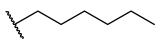
2-(Pentylthio)-5-[(quinolin-8-yloxy)methyl]-1,3,4-oxadiazole (4d)
Brown solid; Chemical formula: C17H19N3O2S, Yield; 77%, Mol wt: 329.42 gmol−1, M.p: 84.6–90.1 °C.
1H NMR (600 MHz, DMSO-d6) δ 8.88 (dd, J = 4.1, 1.7 Hz, 1H, H-2′), 8.36 (dd, J = 8.3, 1.7 Hz, 1H, H-4′), 7.63 (dd, J = 8.2, 1.2 Hz, 1H, H-5), 7.58 (dd, J = 8.3, 4.1 Hz, 1H, H-3), 7.54 (t, J = 7.9 Hz, 1H, H-6), 7.38 (dd, J = 7.7, 1.2 Hz, 1H, H-7), 5.62 (s, 2H, H-6′), 3.24 (t, J = 7.3 Hz, 2H, H-7′), 1.71 (p, J = 7.3 Hz, 2H, H-8′), 1.38–1.30 (m, 2H, H-9′), 1.27 (m, J = 14.6, 7.0 Hz, 2H, H-10′), 0.85 (t, J = 7.2 Hz, 3H, H-11′).
13C NMR (151 MHz, DMSO-d6) δ 165.49 (C-2′), 164.09 (C-5′), 153.42 (C-8), 149.88 (C-2), 140.25 (C-8a), 136.43 (C-4), 129.66 (C-4a), 127.02 (C-6), 122.49 (C-3), 122.01 (C-5), 112.22 (C-7), 61.07 (C-6′), 32.49 (C-7′), 30.40 (C-8′), 29.04 (C-9′), 21.94 (C-10′),14.18 (C-11′).
HRMS: (ESI+-MS, m/z) calcd for C17H19N3O2S (M + Na)+: 352.1096; found: 352.1101.
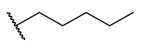
2-(Hexylthio)-5-[(quinolin-8-yloxy)methyl]-1,3,4-oxadiazole (4e)
Brown oil; Chemical formula: C18H21N3O2S, Yield; 60%, Mol wt: 343.44 gmol−1.
1H NMR (600 MHz, DMSO-d6) δ 8.87 (dd, J = 4.1, 1.7 Hz, 1H, H-2), 8.36 (dd, J = 8.3, 1.7 Hz, 1H, H-4), 7.62 (dd, J = 8.3, 1.2 Hz, 1H, H-5), 7.57 (dd, J = 8.3, 4.1 Hz, 1H, H-3), 7.54 (t, J = 7.9 Hz, 1H, H-6), 7.38 (dd, J = 7.7, 1.2 Hz, 1H, H-7), 5.61 (s, 2H, H-6′), 3.24 (t, J = 7.3 Hz, 2H, H-7′), 1.70 (p, J = 7.5 Hz, 2H, H-9′), 1.36 (q, J = 7.1, 6.6 Hz, 2H, H-11′), 1.25 (m, J = 7.5, 4.5 Hz, 4H, H-8′,10′), 0.84 (t, J = 7.0 Hz, 3H, H-12′).
13C NMR (151 MHz, DMSO-d6) δ 165.49 (C-2′), 164.08 (C-5′), 153.41 (C-8), 149.87 (C-2), 140.23 (C-8a), 136.43 (C-4), 129.65 (C-4a), 127.02 (C-6), 122.49 (C-3), 121.99 (C-5), 112.14 (C-7), 61.05 (C-6′), 32.51 (C-7′), 31.01 (C-8′), 29.32 (C-9′), 27.90 (C-11′), 22.35 (C-10′), 14.26 (C-12′).
HRMS: (ESI+-MS, m/z) calcd for C18H21N3O2S (M + Na)+: 366.1252; found: 366.1252.
2-(Allylthio)-5-[(quinolin-8-yloxy)methyl]-1,3,4-oxadiazole (4f)
Yellow oil; Chemical formula: C15H13N3O2S, Yield; 52%, Mol wt: 299.35 gmol−1.
1H NMR (400 MHz, DMSO-d6) δ 8.88 (dd, J = 4.1, 1.8 Hz, 1H, H-2), 8.36 (dd, J = 8.3, 1.7 Hz, 1H, H-4), 7.63 (dd, J = 8.3, 1.2 Hz, 1H, H-5), 7.58 (dd, J = 8.3, 4.2 Hz, 1H, H-3), 7.54 (t, J = 7.9 Hz, 1H, H-6), 7.38 (dd, J = 7.7, 1.3 Hz, 1H, H-7), 5.95 (m, J = 17.0, 10.0, 7.0 Hz, 1H, H-8′), 5.63 (s, 2H, H-6′), 5.30 (m, J = 17.0, 1.4 Hz, 2H, H-9b), 5.12 (m, J = 10.0, 1.7, 0.9 Hz, 2H, H-9a), 3.92 (dd, J = 6.9, 1.1 Hz, 2H, H-7′).
13C NMR (101 MHz, DMSO-d6) δ 164.75 (C-2′), 164.31 (C-5′), 153.37 (C-8), 149.90 (C-2), 140.16 (C-8a), 136.45 (C-4), 132.87 (C-8′), 129.63 (C-4a), 127.05 (C-6), 122.53 (C-3), 121.95 (C-5), 119.93 (C-9), 111.99 (C-7), 60.94 (C-6′), 35.04 (C-7′).
HRMS: (ESI+-MS, m/z) calcd for C15H13N3O2S (M + Na)+: 322.0626; found: 322.0636.
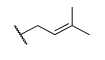
2-[(3-Methylbut-2-en-1-yl)thio]-5-[(quinolin-8-yloxy)methyl]-1,3,4-oxadiazole (4g)
Yellow oil; Chemical formula: C17H17N3O2S, Yield; 30%, Mol wt: 327.40 gmol−1.
1H NMR (400 MHz, DMSO-d6) δ 8.88 (dd, J = 4.2, 1.8 Hz, 1H, H-2), 8.36 (dd, J = 8.3, 1.7 Hz, 1H, H-4), 7.63 (dd, J = 8.2, 1.2 Hz, 1H, H-5), 7.60–7.51 (m, 2H, H-3,6), 7.38 (dd, J = 7.7, 1.3 Hz, 1H, H-7), 5.61 (s, 2H, H-6′), 5.35 (dd, J = 7.8, 1.5 Hz, 1H, H-8′), 3.91 (d, J = 7.8 Hz, 2H, H-7′), 1.66 (s, 6H, H-10′,11′).
13C NMR (101 MHz, DMSO-d6) δ 164.60 (C-2′), 164.11 (C-5′), 153.29 (C-8), 149.87 (C-2), 140.04 (C-8a), 136.60 (C-4), 129.64 (C-4a), 127.11 (C-6), 122.54 (C-3), 122.03 (C-5), 112.21 (C-7), 79.26 (C-8′), 75.51 (C-9′), 61.05 (C-6′), 21.19 (C-7′).
HRMS: (ESI+-MS, m/z) calcd for C17H17N3O2S (M + Na)+: 350.0939; found: 350.0950.
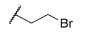
2-[(2-Bromoethyl)thio]-5-[(quinolin-8-yloxy)methyl]-1,3,4-oxadiazole (4h)
Yellow oil; Chemical formula: C14H12BrN3O2S, Yield; 54%, Mol wt: 326.33 gmol−1.
1H NMR (600 MHz, DMSO-d6) δ 8.88 (dd, J = 4.2, 1.7 Hz, 1H, H-2), 8.36 (dd, J = 8.3, 1.7 Hz, 1H, H-4), 7.63 (dd, J = 8.3, 1.2 Hz, 1H, H-5), 7.58 (dd, J = 8.3, 4.1 Hz, 1H, H-3), 7.55 (t, J = 7.9 Hz, 1H, H-6), 7.40 (dd, J = 7.7, 1.2 Hz, 1H, H-7), 5.62 (s, 2H, H-6′), 3.83 (t, J = 7.3 Hz, 2H, H-7′), 3.72 (t, J = 7.1 Hz, 2H, H-8′).
13C NMR (151 MHz, DMSO-d6) δ 164.59 (C-2′), 164.38 (C-5′), 153.43 (C-8), 149.90 (C-2), 140.25 (C-8a), 136.45 (C-4), 129.65 (C-4a), 127.04 (C-6), 122.50 (C-3), 122.05 (C-5), 112.30 (C-7), 61.13 (C-6′), 34.36 (C-7′), 31.19 (C-8′).
HRMS: (ESI+-MS, m/z) calcd for C14H12BrN3O2S (M + Na)+: 387.9731; found: 387.9747.
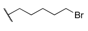
2-[(5-Bromopentyl)thio]-5-[(quinolin-8-yloxy)methyl]-1,3,4-oxadiazole (4i)
Yellow oil; Chemical formula: C17H18BrN3O2S, Yield; 61%, Mol wt: 408.31 gmol−1.
1H NMR (600 MHz, DMSO- d6) δ 8.89 (dd, J = 4.2, 1.6 Hz, 1H, H-2), 8.42 (dd, J = 8.3, 1.7 Hz, 1H, H-4), 7.65 (d, J = 8.2 Hz, 1H, H-5), 7.62 (dd, J = 8.3, 4.2 Hz, 1H, H-3), 7.57 (t, J = 8.0 Hz, 1H, H-6), 7.42 (d, J = 7.8 Hz, 1H, H-7), 5.63 (s, 2H, H-6′), 3.51 (t, J = 6.7 Hz, 2H, H-11′), 3.26 (t, J = 7.3 Hz, 2H, H-7′), 1.81 (p, J = 6.9 Hz, 2H, H-9′), 1.74 (p, J = 7.4 Hz, 2H, H-8′), 1.49 (p, J = 7.7 Hz, 2H, H-10′).
13C NMR (151 MHz, DMSO-d6) δ 165.48 (C-2′), 164.02 (C-5′), 152.99 (C-8), 149.58 (C-2), 139.88 (C-8a), 137.27 (C-4), 129.68 (C-4a), 127.30 (C-6), 122.59 (C-3), 122.01 (C-5), 112.40 (C-7), 61.07 (C-6′), 35.26 (C-11′), 32.30 (C-7′), 31.97 (C-9′), 28.46 (C-8′), 26.82 (C-10′).
HRMS: (ESI+-MS, m/z) calcd for C17H18BrN3O2S (M + Na)+: 430.0201; found: 430.0212.
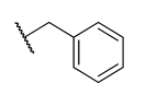
2-(Benzylthio)-5-[(quinolin-8-yloxy)methyl]-1,3,4-oxadiazole (4j)
Light brown solid; Chemical formula: C19H15N3O2S, Yield; 50%, Mol wt: 349.41 gmol−1, M.p: 75.3–79.8 °C.
1H NMR (600 MHz, DMSO-d6) δ 8.87 (dd, J = 4.1, 1.8 Hz, 1H, H-2), 8.36 (dd, J = 8.3, 1.8 Hz, 1H, H-4), 7.63 (dd, J = 8.2, 1.2 Hz, 1H, H-5), 7.58 (dd, J = 8.3, 4.1 Hz, 1H, H-3), 7.54 (t, J = 8.0 Hz, 1H, H-6), 7.42 (dd, J = 7.6, 1.7 Hz, 2H, H-9′, 13′), 7.37 (dd, J = 7.8, 1.2 Hz, 1H, H-7), 7.33–7.24 (m, 3H, H-10′, 11′,12′), 5.62 (s, 2H, H-6′), 4.52 (s, 2H, H-7′).
13C NMR (151 MHz, DMSO-d6) δ 164.87 (C-2′), 164.31 (C-5′), 153.38 (C-8), 149.90 (C-2), 140.21 (C-8a), 136.82 (C-8′), 136.44 (C-4), 129.65 (C-4a), 129.43 (C-9′,13′), 129.01 (C-10′,12′), 128.23 (C-11′), 127.04 (C-6), 122.52 (C-3), 121.98 (C-5), 112.05 (C-7), 60.98 (C-6′), 36.34 (C-7′).
HRMS: (ESI+-MS, m/z) calcd for C19H15N3O2S (M + Na)+: 372.0783; found: 372.0790.
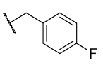
2-[(4-Fluorobenzyl)thio]-5-[(quinolin-8-yloxy)methyl]-1,3,4-oxadiazole (4k)
Yellow crystals; Chemical formula: C19H16FN3O2S, Yield; 32%, Mol wt: 367.40 gmol−1, M.p: 106.9–109.4 °C.
1H NMR (600 MHz, DMSO-d6) δ 8.87 (dd, J = 4.1, 1.7 Hz, 1H, H-2), 8.36 (dd, J = 8.3, 1.7 Hz, 1H, H-4), 7.63 (dd, J = 8.3, 1.2 Hz, 1H, H-5), 7.58 (dd, J = 8.3, 4.1 Hz, 1H, H-3), 7.54 (t, J = 7.9 Hz, 1H, H-6), 7.47 (dd, J = 8.5, 5.6 Hz, 2H, H-9′,13′), 7.36 (dd, J = 7.7, 1.2 Hz, 1H, H-7), 7.09 (d, J = 8.9 Hz, 2H, H-10′,12′), 5.61 (s, 2H, H-6′), 4.51 (s, 2H, H-7′).
13C NMR (151 MHz, DMSO-d6) δ 164.82 (C-2′), 164.32 (C-5′), 162.05 (d, J = 244.2 Hz, C-11′), 153.33 (C-8), 149.90 (C-2), 140.12 (C-8a), 136.46 (C-4), 133.22 (d, J = 3.0 Hz, C-8′), 131.58 (d, J = 8.3 Hz, C-9′,13′), 129.63 (4a), 127.05 (C-6), 122.55 (C-3), 121.92 (C-5), 115.80 (d, J = 21.5 Hz, C-10′,12′), 111.82 (C-7), 60.85 (C-6′), 35.46 (C-7′).
HRMS: (ESI+-MS, m/z) calcd for C19H14FN3O2S (M + Na)+: 390.0688; found: 390.0696.
2-[(4-Chlorobenzyl)thio]-5-[(quinolin-8-yloxy)methyl]-1,3,4-oxadiazole (4l)
Cream crystals; Chemical formula: C19H14ClN3O2S, Yield; 85%, Mol wt: 383.85 gmol−1, M.p: 101.4–104.2 °C.
1H NMR (600 MHz, DMSO-d6) δ 8.87 (dd, J = 4.1, 1.7 Hz, 1H, H-2), 8.36 (dd, J = 8.3, 1.8 Hz, 1H, H-4), 7.63 (dd, J = 8.3, 1.2 Hz, 1H, H-5), 7.58 (dd, J = 8.3, 4.1 Hz, 1H, H-3), 7.53 (t, J = 8.0 Hz, 1H, H-6), 7.45 (d, J = 8.4 Hz, 2H, H-9′,13′), 7.36 (dd, J = 7.8, 1.2 Hz, 1H, H-7), 7.32 (d, J = 8.4 Hz, 2H, H-10′,12′), 5.61 (s, 2H, H-6′), 4.51 (s, 2H, H-7′).
13C NMR (151 MHz, DMSO-d6) δ 164.71 (C-2′), 164.37 (C-5′), 153.37 (C-8), 149.89 (C-2), 140.19 (C-8a), 136.45 (C-4), 136.13 (C-8′), 132.86 (C-11′), 131.32 (C-9′,13′), 129.65 (C-4a), 128.94 (C-10′,12′), 127.04 (C-6), 122.53 (C-3), 121.98 (C-5), 112.00 (C-7), 60.96 (C-6′), 35.52 (C-7′).
HRMS: (ESI+-MS, m/z) calcd for C19H14ClN3O2S (M + Na)+: 406.0393; found: 406.0407.
2-[(4-Bromobenzyl)thio]-5-[(quinolin-8-yloxy)methyl]-1,3,4-oxadiazole (4m)
Yellow solid; Chemical formula: C19H14BrN3O2S, Yield; 27%, Mol wt: 428.30 gmol−1, M.p: 120.9–124.1 °C.
1H NMR (600 MHz, DMSO-d6) δ 8.87 (dd, J = 4.1, 1.7 Hz, 1H, H-2), 8.36 (dd, J = 8.3, 1.7 Hz, 1H, H-4), 7.63 (dd, J = 8.3, 1.2 Hz, 1H, H-5), 7.58 (dd, J = 8.3, 4.1 Hz, 1H, H-3), 7.53 (t, J = 8.0 Hz, 1H, H-6), 7.46 (d, J = 8.4 Hz, 2H, H-10′,12′), 7.38 (d, J = 8.4 Hz, 2H, H-9′,13′), 7.36 (dd, J = 7.8, 1.2 Hz, 1H, H-7), 5.61 (s, 2H, H-6′), 4.49 (s, 2H, H-7′).
13C NMR (151 MHz, DMSO-d6) δ 164.69 (C-2′), 164.38 (C-5′), 153.38 (C-8), 149.89 (C-2), 140.20 (C-8a), 136.57 (C-8′), 136.45 (C-4), 131.87 (C-10′,12′), 131.64 (C-9′,13′), 129.65 (C-4a), 127.04 (C-6), 122.52 (C-3), 121.98 (C-5), 121.40 (C-11′), 112.02 (C-7), 60.97 (C-6′), 35.57 (C-7′).
HRMS: (ESI+-MS, m/z) calcd for C19H14BrN3O2S (M + Na)+: 449.9888; found: 449.9895.
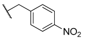
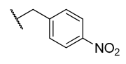
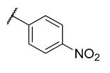
2-[(4-Nitrobenzyl)thio]-5-[(quinolin-8-yloxy)methyl]-1,3,4-oxadiazole (4n)
Yellow crystals; Chemical formula: C19H14N4O4S, Yield; 104%, Mol wt: 394.40 gmol−1, M.p: 156.6–160.1 °C.
1H NMR (600 MHz, DMSO-d6) δ 8.88 (dd, J = 4.2, 1.8 Hz, 1H, H-2), 8.35 (dd, J = 8.3, 1.8 Hz, 1H, H-4), 8.13 (dd, J = 8.7, 6.8 Hz, 2H, H-10′,12′), 7.71 (dd, J = 8.7, 6.7 Hz, 2H, H-9′,13′), 7.63 (dd, J = 8.3, 1.2 Hz, 1H, H-5), 7.57 (dd, J = 8.3, 4.1 Hz, 1H, H-3), 7.53 (t, J = 8.0 Hz, 1H, H-6), 7.37 (dd, J = 7.8, 1.2 Hz, 1H, H-7), 5.62 (s, 2H, H-6′), 4.65 (s, 2H, H-7′).
13C NMR (151 MHz, DMSO-d6) δ 163.98 (C-2′), 163.97 (C-5′), 152.88 (C-8), 149.38 (C-2), 146.85 (C-11′), 144.68 (C-8′), 139.67 (C-8a), 135.95 (C-4), 130.25 (C-9′,13′), 129.14 (4a), 126.52 (C-6), 123.53 (C-10′,12′), 122.02 (C-3), 121.45 (C-5), 111.41 (C-7), 60.43 (C-6′), 34.86 (C-7′).
HRMS: (ESI+-MS, m/z) calcd for C19H14N4O4S (M + Na)+: 417.0633; found: 417.0645.
2-[(3-Fluorobenzyl)thio]-5-[(quinolin-8-yloxy)methyl]-1,3,4-oxadiazole (4o)
Yellow oil; Chemical formula: C19H14FN3O2S, Yield; 63%, Mol wt: 367.40 gmol−1.
1H NMR (600 MHz, DMSO-d6) δ 8.91 (d, J = 4.2 Hz, 1H, H-2), 8.48 (d, J = 8.2 Hz, 1H, H-4), 7.68 (d, J = 8.2 Hz, 1H, H-5), 7.65 (dd, J = 8.3, 4.3 Hz, 1H, H-3), 7.59 (t, J = 8.0 Hz, 1H, H-6), 7.43 (d, J = 7.7 Hz, 1H, H-7), 7.33 (q, J = 7.5 Hz, 1H, H-10′), 7.31–7.24 (m, 2H, H-9′,13′), 7.10 (td, J = 8.6, 2.6 Hz, 1H, H-11′), 5.64 (s, 2H, H-6′), 4.54 (s, 2H, H-7′).
13C NMR (151 MHz, DMSO-d6) δ 164.79 (d, J = 3.9 Hz, C-2′), 164.26 (d, J = 3.4 Hz, C-5′), 162.42 (d, J = 244.1 Hz, C-12′), 152.59 (C-8), 149.30 (C-2), 139.79 (d, J = 7.9 Hz, C-8a), 137.98 (C-4), 130.94 (d, J = 8.4 Hz, C-10′), 129.71 (d, J = 2.4 Hz, C-4a), 127.51 (d, J = 3.9 Hz, C-6), 125.57 (t, J = 3.2 Hz, C-9′), 122.65 (d, J = 3.5 Hz, C-3), 122.04 (d, J = 7.7 Hz, C-5), 116.21 (dd, J = 21.9, 2.9 Hz, C-13′), 115.09 (dd, J = 21.0, 1.4 Hz, C-11′), 122.65 (d, J = 3.5 Hz, C-7), 61.13 (C-6′), 35.62 (C-7′).
HRMS: (ESI+-MS, m/z) calcd for C19H14FN3O2S (M + Na)+: 390.0688; found: 390.0697.
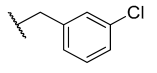
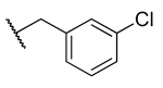
2-[(3-Chlorobenzyl)thio]-5-[(quinolin-8-yloxy)methyl]-1,3,4-oxadiazole (4p)
Yellow oil; Chemical formula: C19H14ClN3O2S, Yield; 27%, Mol wt: 383.85 gmol−1.
1H NMR (600 MHz, DMSO-d6) δ 8.87 (dd, J = 4.2, 1.8 Hz, 1H, H-2), 8.36 (dd, J = 8.3, 1.8 Hz, 1H, H-4), 7.62 (d, J = 8.2 Hz, 1H, H-5), 7.57 (dd, J = 8.3, 4.1 Hz, 1H, H-3), 7.54 (s, 1H, H-13′), 7.52 (d, J = 8.0 Hz, 1H, H-6), 7.39 (dd, J = 7.1, 1.7 Hz, 1H, H-9′), 7.36 (dd, J = 7.7, 1.2 Hz, 1H, H-7), 7.34–7.26 (m, 2H, H-10′,11′), 5.60 (s, 2H, H-6′), 4.53 (s, 2H, H-7′).
13C NMR (151 MHz, DMSO-d6) δ 164.72 (C-2′), 164.40 (C-5′), 153.38 (C-8), 149.90 (C-2), 140.20 (C-8a), 139.59 (C-8′), 136.45 (C-4), 133.47 (C-12′), 130.82 (C-10′), 129.64 (C-4a), 129.29 (C-13′), 128.19 (C-9′), 128.14 (C-11′), 127.03 (C-6), 122.51 (C-3), 122.02 (C-5), 112.11 (C-7), 61.04 (C-6′), 35.51 (C-7′).
HRMS: (ESI+-MS, m/z) calcd for C19H14ClN3O2S (M + Na)+: 406.0393; found: 406.0405.
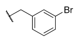
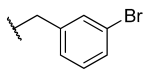
2-[(3-Bromobenzyl)thio]-5-[(quinolin-8-yloxy)methyl]-1,3,4-oxadiazole (4q)
Yellow oil; Chemical formula: C19H14BrN3O2S, Yield; 51%, Mol wt: 428.30 gmol−1.
1H NMR (600 MHz, DMSO-d6) δ 8.90 (d, J = 4.0 Hz, 1H, H-2), 8.45 (d, J = 8.2 Hz, 1H, H-4), 7.69 (s, 1H, H-13′), 7.67 (d, J = 8.2 Hz, 1H, H-5), 7.64 (dd, J = 8.3, 4.2 Hz, 1H, H-3), 7.58 (t, J = 8.0 Hz, 1H, H-6), 7.47 (dd, J = 8.0, 1.9 Hz, 1H, H-11′), 7.44 (d, J = 7.7 Hz, 1H, H-9′), 7.42 (d, J = 7.7 Hz, 1H, H-7), 7.25 (t, J = 7.8 Hz, 1H, H-10′), 5.63 (s, 2H, H-6′), 4.52 (s, 2H, H-7′).
13C NMR (151 MHz, DMSO-d6) δ 164.76 (C-2′), 164.37 (C-5′), 153.30 (C-8), 149.47 (C-2), 139.86 (C-8a), 139.82 (C-8′), 137.58 (C-4), 137.47 (C-10′), 132.15 (C-13′), 131.08 (C-11′), 129.73 (C-4a), 128.50 (C-9′), 127.36 (C-6), 122.58 (C-3), 122.07 (C-5), 122.02 (C-12′), 112.95 (C-7), 61.38 (C-6′), 35.61 (C-7′).
HRMS: (ESI+-MS, m/z) calcd for C19H14BrN3O2S (M + Na)+: 449.9888; found: 449.9903.
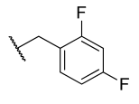
2-[(2,4-Difluorobenzyl)thio]-5-[(quinolin-8-yloxy)methyl]-1,3,4-oxadiazole (4r)
Cream solid; Chemical formula: C19H13F2N3O2S, Yield; 29%, Mol wt: 385.39 gmol−1, M.p: 98.7–105.2 °C.
1H NMR (600 MHz, DMSO-d6) δ 8.87 (dd, J = 4.1, 1.7 Hz, 1H, H-2), 8.36 (dd, J = 8.3, 1.8 Hz, 1H, H-4), 7.63 (dd, J = 8.2, 1.2 Hz, 1H, H-5), 7.61–7.51 (m, 3H, H-3.9′,6), 7.37 (dd, J = 7.7, 1.2 Hz, 1H, H-7), 7.24 (ddd, J = 10.6, 9.2, 2.6 Hz, 1H, H-12′), 6.99 (td, J = 8.5, 2.4 Hz, 1H, H-10′), 5.61 (s, 2H, H-6′), 4.53 (s, 2H, H-7′).
13C NMR (151 MHz, DMSO-d6) δ 164.58 (d, J = 7.5 Hz, C-5′), 164.31 (C-2′), 161.97 (d, J = 7.8 Hz, C-11′), 161.10 (d, J = 26.8 Hz, C-8′), 153.45 (d, J = 13.5 Hz, C-8), 149.89 (C-2), 140.32 (d, J = 25.4 Hz, C-8a), 136.43 (d, J = 3.8 Hz, C-4), 132.96 (dd, J = 9.9, 5.2 Hz, C-9′), 129.68 (d, J = 7.7 Hz, C-4a), 127.01 (d, J = 5.3 Hz, C-6), 122.48 (d, J = 8.6 Hz, C-3), 122.09 (d, J = 20.1 Hz, C-5), 120.44–120.17 (m, C-13′), 112.60 (C-7), 112.05 (dd, J = 21.5, 3.6 Hz, C-10′), 104.57 (dd, J = 26.1, 25.4 Hz, C-12′), 61.20 (d, J = 40.4 Hz, C-6′), 29.97–29.67 (m, C-7′).
HRMS: (ESI+-MS, m/z) calcd for C19H13F2N3O2S (M + Na)+: 408.0594; found: 408.0604.
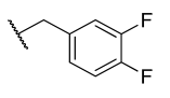
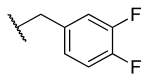
2-[(3,4-Difluorobenzyl)thio]-5-[(quinolin-8-yloxy)methyl]-1,3,4-oxadiazole (4s)
Cream solid; Chemical formula: C19H13F2N3O2S, Yield; 62%, Mol wt: 385.39 gmol−1, M.p: 98.8–101.2 °C.
1H NMR (600 MHz, DMSO-d6) δ 8.88 (dd, J = 4.1, 1.8 Hz, 1H, H-2), 8.35 (dd, J = 8.3, 1.7 Hz, 1H, H-4), 7.63 (dd, J = 8.3, 1.2 Hz, 1H, H-5), 7.61–7.50 (m, 3H, H-3,13′,6), 7.37 (dd, J = 7.7, 1.2 Hz, 1H, H-7), 7.20 (td, J = 9.9, 2.5 Hz, 1H, H-12′), 6.98 (td, J = 8.5, 2.6 Hz, 1H, H-9′), 5.62 (s, 2H, H-6′), 4.52 (s, 2H, H-7′).
13C NMR (151 MHz, DMSO-d6) δ 164.56 (C-5′), 164.31 (C-2′), 162.55 (dd, J = 236.3, 11.0 Hz, C-10′), 160.90 (dd, J = 241.0, 12.0 Hz, C-11′), 153.42 (C-8), 149.88 (C-2), 140.25 (C-8a), 136.43 (C-4), 132.98 (dd, J = 10.0, 4.9 Hz, C-13′), 129.66 (C-4a), 127.01 (C-6), 122.49 (C-3), 122.03 (C-5), 120.29 (dd, J = 14.6, 3.6 Hz, C-8′), 112.42–112.16 (m, C-9), 112.04 (dd, J = 21.4, 3.6 Hz, C-7), 104.57 (t, J = 25.8 Hz, C-12′), 61.10 (C-6′), 29.81 (C-7′).
HRMS: (ESI+-MS, m/z) calcd for C19H13F2N3O2S (M + Na)+: 408.0594; found: 408.0606.
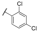
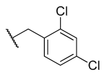
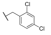
2-[(2,4-Dichlorobenzyl)thio]-5-[(quinolin-8-yloxy)methyl]-1,3,4-oxadiazole (4t)
Cream solid; Chemical formula: C19H13Cl2N3O2S, Yield; 40%, Mol wt: 418.30 gmol−1, M.p: 116.7–119.7 °C.
1H NMR (400 MHz, DMSO-d6) δ 8.86 (dd, J = 4.2, 1.7 Hz, 1H, H-2), 8.36 (dd, J = 8.3, 1.7 Hz, 1H, H-4), 7.64 (d, J = 2.2 Hz, 1H, H-12′), 7.62 (dd, J = 8.3, 1.2 Hz, 1H, H-5), 7.60–7.55 (m, 2H, H-9′,3), 7.53 (t, J = 8.0 Hz, 1H, H-6), 7.36 (dd, J = 7.7, 1.2 Hz, 1H, H-6), 7.32 (dd, J = 8.3, 2.2 Hz, 1H, H-10′), 5.61 (s, 2H, H-6′), 4.57 (s, 2H, H-7′).
13C NMR (101 MHz, DMSO-d6) δ 169.37 (C-5′), 168.94 (C-2′), 158.14 (C-8), 154.71 (C-2), 145.02 (C-8a), 141.19 (C-4), 139.51 (C-11′), 138.81 (C-13′), 138.15 (C-8′), 137.94 (C-9′), 134.40 (C-4a), 134.30 (C-12′), 132.75 (C-10′), 131.78 (C-6), 127.26 (C-3), 126.74 (C-5), 116.77 (C-7), 65.77 (C-6′), 38.86 (C-7′).
HRMS: (ESI+-MS, m/z) calcd for C19H13Cl2N3O2S (M + Na)+: 440.0003; found: 440.0016.

4.3. Synthesis of 2-(Prop-2-yn-1-ylthio)-5-[(quinolin-8-yloxy)methyl]-1,3,4-oxadiazole (11)

A measure of 0.722 g (3.4 mmol 1 eq) of compound 3 was dissolved in a stirring mixture of DMF (5 mL) and pre-activated K2CO3 (0.769 g, 2.0 eq). A sticky mass was formed after few minutes, then 0.36 mL of propargyl bromide (80% in toluene) in DMF (2 mL) was added and stirring continued at r.t for 4 h. On completion of the reaction as evidenced by TLC, the mixture was poured into a slurry of ice and stirred vigorously for 30 min. The resulting brown precipitate was filtered in vacuo to obtain 0.66 g compound 11 (79% yield).
Brown solid; Chemical formula: C15H11N3O2S, Yield; 79%, Mol wt: 297.33 gmol−1, M.p: 79.1–82.7 °C.
1H NMR (400 MHz, DMSO-d6) δ 8.89 (dd, J = 4.2, 1.7 Hz, 1H, H-2), 8.38 (dd, J = 8.3, 1.8 Hz, 1H, H-4), 7.64 (dd, J = 8.3, 1.2 Hz, 1H, H-5), 7.59 (dd, J = 8.3, 4.1 Hz, 1H, N-3), 7.55 (t, J = 7.9 Hz, 1H, H-6), 7.40 (dd, J = 7.7, 1.2 Hz, 1H, H-7), 5.64 (s, 2H, H-6′), 4.17 (d, J = 2.6 Hz, 2H, H-7′), 3.32 (t, J = 2.6 Hz, 1H, H-8′).
13C NMR (101 MHz, DMSO-d6) δ 164.60 (C-5′), 164.11 (C-2′), 153.29 (C-8), 149.87 (C-2), 140.04 (C-8a), 136.60 (C-4), 129.64 (C-4a), 127.11 (C-6), 122.54 (C-3), 122.03 (C-5), 112.21 (C-7), 79.26 (C-8′), 75.51 (C-9′), 61.05 (C-6′), 21.19 (C-7′).
HRMS: (ESI+-MS, m/z) calcd for C15H11N3O2S (M + Na)+: 320.0470; found: 320.0476.

4.4. General Synthetic Procedure for Quinoline-1,3,4-Oxadiazole-1,2,3-Triazole Hybrids (12at)

To a solution of alkyne intermediate 11 (0.15 g) in DCM (10 mL) in a round bottom flask, sodium ascorbate (22 mol%) and copper(II) sulphate pentahydrate (10 mol%) in water (10 mL) were added and stirred at r.t for few minutes. Then, the appropriate azides (1.1 eq) in DCM were added and the mixture was stirred for 2–4 h. After this time, TLC analysis showed the alkyne was consumed, then the reaction was diluted with water and filtered to remove residual salts. The resulting filtrate was extracted with DCM, washed with brine and dried over anhydrous sodium sulphate, filtered, and concentrated under reduced pressure. The crude product was purified with column chromatography in EtOAc-hexane eluents to obtain pure quinoline-1,3,4-oxadiazole-1,2,3-triazole hybrids 12at in excellent yields (43–91%).
2-{[(1-Pentyl-1H-1,2,3-triazol-4-yl)methyl]thio-5-[(quinolin-8-yloxy)methyl]-1,3,4-oxadiazole (12a)
Yellow solid; Chemical Formula: C20H22N6O2S, Yield; 59%, Mol wt: 410.49 gmol−1, M.p: 90–94 °C.
1H NMR (400 MHz, DMSO-d6) δ 8.88 (dd, J = 4.2, 1.8 Hz, 1H), 8.37 (dd, J = 8.3, 1.8 Hz, 1H), 8.13 (s, 1H), 7.63 (dd, J = 8.3, 1.3 Hz, 1H), 7.60–7.57 (m, 1H), 7.54 (d, J = 8.1 Hz, 1H), 7.40 (dd, J = 7.7, 1.3 Hz, 1H), 5.62 (s, 2H), 4.60 (s, 2H), 4.28 (t, J = 7.1 Hz, 2H), 1.73 (p, J = 7.2 Hz, 2H), 1.31–1.05 (m, 4H), 0.80 (t, J = 7.2 Hz, 3H).
13C NMR (101 MHz, DMSO-d6) δ 164.70, 164.35, 153.42, 149.92, 142.24, 140.15, 136.49, 129.65, 127.08, 124.24, 122.55, 121.97, 111.98, 61.02, 49.82, 29.78, 28.37, 27.30, 21.91, 14.18.
HRMS: (ESI+-MS, m/z) calcd for C20H22N6O2S (M + Na)+: 433.1423; found: 433.1413.
2-{[(1-Hexyl-1H-1,2,3-triazol-4-yl)methyl]thio}-5-[(quinolin-8-yloxy)methyl]-1,3,4-oxadiazole (12b)
Yellow solid; Chemical Formula: C21H24N6O2S, Yield; 75%, Mol wt: 424.52 gmol−1, M.p: 72–78 °C.
1H NMR (400 MHz, DMSO-d6) δ 8.88 (dd, J = 4.1, 1.7 Hz, 1H), 8.37 (dd, J = 8.3, 1.7 Hz, 1H), 8.13 (s, 1H), 7.67–7.51 (m, 3H), 7.40 (dd, J = 7.7, 1.3 Hz, 1H), 5.62 (s, 2H), 4.60 (s, 2H), 4.28 (t, J = 7.1 Hz, 2H), 1.72 (p, J = 7.2 Hz, 2H), 1.41–1.04 (m, 6H), 0.84–0.76 (m, 3H).
13C NMR (101 MHz, DMSO-d6) δ 164.70, 164.34, 153.41, 149.92, 142.25, 140.14, 136.49, 129.64, 127.09, 124.25, 122.56, 121.96, 111.96, 61.01, 49.83, 30.96, 30.04, 27.29, 25.85, 22.32, 14.25.
HRMS: (ESI+-MS, m/z) calcd for C21H24N6O2S (M + Na)+: 447.1579; found: 447.1568.
2-{[(1-Benzyl-1H-1,2,3-triazol-4-yl)methyl]thio}-5-[(quinolin-8-yloxy)methyl]-1,3,4-oxadiazole (12c)
Yellow solid; Chemical Formula: C22H18N6O2S, Yield; 82%, Mol wt: 430.48 gmol−1, M.p: 68–71 °C.
1H NMR (400 MHz, Chloroform-d) δ 8.86 (dd, J = 4.2, 1.7 Hz, 1H), 8.08 (dd, J = 8.3, 1.7 Hz, 1H), 7.59 (s, 1H), 7.46–7.34 (m, 3H), 7.25 (dd, J = 5.1, 2.0 Hz, 3H), 7.23–7.16 (m, 1H), 7.20–7.10 (m, 2H), 5.45 (s, 2H), 5.37 (s, 2H), 4.44 (s, 2H).
13C NMR (101 MHz, Chloroform-d) δ 165.77, 163.48, 153.07, 149.59, 143.05, 139.95, 136.32, 134.43, 129.65, 129.11, 128.76, 128.01, 126.58, 123.27, 121.95, 121.73, 110.75, 60.75, 54.19, 26.91.
HRMS: (ESI+-MS, m/z) calcd for C20H22N6O2S (M + Na)+: 453.1110; found: 453.1098.
2-{[(1-(4-Chlorobenzyl)-1H-1,2,3-triazol-4-yl)methyl]thio}-5-[(quinolin-8-yloxy)methyl]-1,3,4-oxadiazole (12d)
Cream white solid; Chemical Formula: C22H17ClN6O2S, Yield; 63%, Mol wt: 464.93 gmol−1, M.p: 92–96 °C.
1H NMR (400 MHz, Chloroform-d) δ 8.87–8.81 (m, 1H), 8.08 (dd, J = 8.3, 1.7 Hz, 1H), 7.64 (s, 1H), 7.45–7.32 (m, 3H), 7.26–7.14 (m, 3H), 7.11–7.01 (m, 2H), 5.44 (s, 2H), 5.33 (s, 2H), 4.43 (s, 2H).
13C NMR (101 MHz, Chloroform-d) δ 165.60, 163.58, 153.19, 149.57, 143.27, 140.19, 136.15, 134.76, 133.00, 129.67, 129.27, 129.26, 126.51, 123.25, 121.87, 121.83, 111.10, 60.92, 53.37, 26.90.
HRMS: (ESI+-MS, m/z) calcd for C22H17ClN6O2S (M + Na)+: 487.0720; found: 487.0706.
2-{[(1-(4-Fluorobenzyl)-1H-1,2,3-triazol-4-yl)methyl]thio}-5-[(quinolin-8-yloxy)methyl]-1,3,4-oxadiazole (12e)
Yellow solid; Chemical Formula: C22H17FN6O2S, Yield; 62%, Mol wt: 448.47 gmol−1, M.p: 96–101 °C.
1H NMR (400 MHz, Chloroform-d) δ 8.95 (d, J = 3.7 Hz, 1H, H-2), 8.20 (dd, J = 8.2, 1.7 Hz, 1H, H-4), 7.70 (s, 1H, H-5″), 7.56–7.42 (m, 3H, H-3,H-5, H-6), 7.28 (dd, J = 7.3, 1.6 Hz, 1H, H-7), 7.21 (dd, J = 8.5, 5.3 Hz, 2H, H-2′″&H-6′″), 7.07–6.96 (m, 2H, H-3′″&H-5′″), 5.54 (s, 2H, H-6′), 5.42 (s, 2H, H-7″), 4.51 (s, 2H, H-6″).
13C NMR (101 MHz, Chloroform-d) δ 165.81, C-5′, 164.08, C-4′″, 163.45, C-2′, 161.61, C-4′″, 152.92, C-8, 149.42, C-2, 143.25,C-4″, 139.60, C-8a, 136.66, C-4, 130.34 (d, J = 3.4 Hz, C-1′″), 129.92 (d, J = 8.4 Hz, C-2′″&6′″), 129.68, C-4a, 126.72, C-6, 123.24. C-5″, 121.96, C-3, 121.73, C-5, 116.11 (d, J = 21.9 Hz, C-3′″&5′″), 110.84, C-7, 60.74, C-6′, 53.43, C-7″, 26.87, C-6″.
HRMS: (ESI+-MS, m/z) calcd for C22H17ClN6O2S (M + Na)+: 471.1004; found: 471.0999.
2-{[(1-(4-Bromobenzyl)-1H-1,2,3-triazol-4-yl)methyl]thio}-5-[(quinolin-8-yloxy)methyl]-1,3,4-oxadiazole (12f)
Cream solid; Chemical Formula: C22H17BrN6O2S, Yield; 53%, Mol wt: 509.38 gmol−1, M.p: 78–82 °C.
1H NMR (400 MHz, Chloroform-d) δ 8.89–8.83 (m, 1H), 8.10 (dd, J = 8.3, 1.6 Hz, 1H), 7.65 (s, 1H), 7.47–7.35 (m, 5H), 7.20 (dd, J = 7.4, 1.5 Hz, 1H), 7.05–6.97 (m, 2H), 5.46 (s, 2H), 5.33 (s, 2H), 4.45 (s, 2H).
13C NMR (101 MHz, Chloroform-d) δ 165.75, 163.50, 153.07, 149.59, 143.33, 139.94, 136.34, 133.48, 132.26, 129.66, 129.60, 126.59, 123.36, 122.91, 121.96, 121.75, 110.69, 60.73, 53.46, 26.85.
HRMS: (ESI+-MS, m/z) calcd for C22H17BrN6O2S (M + Na)+: 517.0058; found: 517.0046.
2-{[(1-(4-Nitrobenzyl)-1H-1,2,3-triazol-4-yl)methyl]thio}-5-[(quinolin-8-yloxy)methyl]-1,3,4-oxadiazole (12g)
Bright yellow solid; Chemical Formula: C22H17N7O4S, Yield; 74%, Mol wt: 475.48 gmol−1, M.p: 100–106 °C.
1H NMR (400 MHz, Chloroform-d) δ 8.83 (dd, J = 4.2, 1.7 Hz, 1H), 8.12–8.04 (m, 3H), 7.77 (s, 1H), 7.46–7.34 (m, 3H), 7.28–7.14 (m, 2H), 5.49 (s, 2H), 5.44 (s, 2H), 4.45 (s, 2H).
13C NMR (101 MHz, Chloroform-d) δ 165.55, 163.66, 149.53, 136.23, 129.72, 128.54, 126.52, 124.21, 123.70, 121.89, 111.17, 60.98, 53.06, 26.87.
HRMS: (ESI+-MS, m/z) calcd for C22H17N7O4S (M + Na)+: 498.0960; found: 498.0951.
2-{[(1-(3-Chlorobenzyl)-1H-1,2,3-triazol-4-yl)methyl]thio}-5-[(quinolin-8-yloxy)methyl]-1,3,4-oxadiazole (12h)
Cream white solid; Chemical Formula: C22H17ClN6O2S, Yield; 76%, Mol wt: 464.93 gmol−1, M.p: 78–84 °C.
1H NMR (600 MHz, Chloroform-d) δ 8.87 (dd, J = 4.3, 1.7 Hz, 1H), 8.11 (dd, J = 8.2, 1.7 Hz, 1H), 7.67 (s, 1H), 7.46–7.37 (m, 3H), 7.25–7.16 (m, 3H), 7.13 (d, J = 1.9 Hz, 1H), 7.01 (dt, J = 7.4, 1.5 Hz, 1H), 5.47 (s, 2H), 5.35 (s, 2H), 4.46 (s, 2H).
13C NMR (101 MHz, Chloroform-d) δ 165.69, 163.56, 153.17, 149.70, 143.33, 140.19, 136.38, 136.11, 134.95, 130.41, 129.65, 128.97, 128.03, 126.48, 126.02, 123.44, 121.95, 121.77, 110.63, 60.75, 53.43, 26.85.
HRMS: (ESI+-MS, m/z) calcd for C22H17ClN6O2S (M + Na)+: 487.0720; found: 487.0730.
2-{[(1-(2,4-Dichlorobenzyl)-1H-1,2,3-triazol-4-yl)methyl]thio}-5-[(quinolin-8-yloxy)methyl]-1,3,4-oxadiazole (12i)
Cream white solid; Chemical Formula: C22H16Cl2N6O2S, Yield; 57%, Mol wt: 499.37 gmol−1, M.p: 124–130 °C.
1H NMR (400 MHz, Chloroform-d) δ 8.97 (dd, J = 4.3, 1.7 Hz, 1H), 8.22 (dd, J = 8.4, 1.7 Hz, 1H), 7.82 (s, 1H), 7.59–7.46 (m, 3H), 7.42 (d, J = 2.1 Hz, 1H), 7.31 (dd, J = 7.4, 1.6 Hz, 1H), 7.22 (dd, J = 8.3, 2.1 Hz, 1H), 7.04 (d, J = 8.3 Hz, 1H), 5.57 (d, J = 3.4 Hz, 4H), 4.56 (s, 2H).
13C NMR (101 MHz, Chloroform-d) δ 165.73, 163.47, 152.89, 149.41, 143.17, 139.57, 136.67, 134.04, 130.95, 130.91, 129.74, 129.67, 127.93, 126.72, 123.76, 121.96, 121.72, 110.83, 60.73, 50.84.
HRMS: (ESI+-MS, m/z) calcd for C22H16Cl2N6O2S (M + Na)+: 521.0411; found: 521.0421.
2-{[(1-(3,4-Difluorobenzyl)-1H-1,2,3-triazol-4-yl)methyl]thio}-5-[(quinolin-8-yloxy)methyl]-1,3,4-oxadiazole (12j)
Cream white solid; Chemical Formula: C22H16F2N6O2S, Yield; 61%, Mol wt: 466.46 gmol−1, M.p: 70–75 °C.
1H NMR (400 MHz, Chloroform-d) δ 8.97 (dd, J = 4.2, 1.7 Hz, 1H), 8.20 (dd, J = 8.4, 1.7 Hz, 1H), 7.78 (s, 1H), 7.56–7.45 (m, 3H), 7.34–7.19 (m, 2H), 6.92–6.80 (m, 2H), 5.56 (s, 2H), 5.49 (s, 2H), 4.54 (s, 2H).
13C NMR (101 MHz, Chloroform-d) δ 165.76, 164.55 (dd, J = 250.5, 12.1 Hz), 163.49, 160.65 (dd, J = 251.3, 27.0 Hz), 153.00, 149.52, 143.18, 139.79, 136.47, 131.51 (dd, J = 9.9, 4.7 Hz), 129.67, 126.64, 123.41, 121.95, 121.73, 117.78 (dt, J = 14.6, 3.7 Hz), 112.15 (dd, J = 21.5, 3.8 Hz), 110.77, 104.40 (t, J = 25.4 Hz), 60.74, 47.17 (d, J = 3.8 Hz), 26.84.
HRMS: (ESI+-MS, m/z) calcd for C22H16Cl2N6O2S (M + Na)+: 521.0411; found: 521.0421.
2-{[(1-Phenyl-1H-1,2,3-triazol-4-yl)methyl]thio}-5-[(quinolin-8-yloxy)methyl]-1,3,4-oxadiazole (12k)
Light brown solid; Chemical Formula: C21H16N6O2S, Yield; 91%, Mol wt: 416.46 gmol−1, M.p: 122–128 °C.
1H NMR (400 MHz, Chloroform-d) δ 8.83 (dd, J = 4.3, 1.7 Hz, 1H), 8.11 (s, 1H), 8.05 (dd, J = 8.3, 1.8 Hz, 1H), 7.60 (dd, J = 7.6, 1.8 Hz, 2H), 7.45–7.28 (m, 6H), 7.18 (dd, J = 7.3, 1.7 Hz, 1H), 5.47 (s, 2H), 4.54 (s, 2H).
13C NMR (101 MHz, Chloroform-d) δ 165.75, 163.64, 153.18, 149.71, 143.46, 140.23, 136.82, 136.06, 129.70, 129.63, 128.85, 126.44, 121.91, 121.79, 121.59, 120.60, 110.77, 60.83, 26.83.
HRMS: (ESI+-MS, m/z) calcd for C21H16N6O2S (M + Na)+: 439.0953; found: 439.0959.
2-{[(1-(4-Chlorophenyl)-1H-1,2,3-triazol-4-yl)methyl]thio}-5-[(quinolin-8-yloxy)methyl]-1,3,4-oxadiazole (12l)
Yellow solid; Chemical Formula: C21H15ClN6O2S, Yield; 79%, Mol wt: 450.90 gmol−1, M.p: 132–136 °C.
1H NMR (400 MHz, Chloroform-d) δ 8.85 (dd, J = 4.3, 1.7 Hz, 1H), 8.15–8.05 (m, 2H), 7.61–7.52 (m, 2H), 7.48–7.33 (m, 5H), 7.20 (dd, J = 7.3, 1.6 Hz, 1H), 5.48 (s, 2H), 4.54 (s, 2H).
13C NMR (101 MHz, Chloroform-d) δ 165.73, 163.64, 153.06, 149.59, 143.79, 139.94, 136.34, 135.30, 134.62, 129.87, 129.66, 126.56, 121.96, 121.79, 121.76, 121.57, 110.77, 60.80, 26.74.
HRMS: (ESI+-MS, m/z) calcd for C21H16N6O2S (M + Na)+: 473.0563; found: 473.0560.
2-{[(1-(4-Fluorophenyl)-1H-1,2,3-triazol-4-yl)methyl]thio}-5-[(quinolin-8-yloxy)methyl]-1,3,4-oxadiazole (12m)
Brown crystalline; Chemical Formula: C21H15FN6O2S, Yield; 84%, Mol wt: 434.45 gmol−1, M.p: 134–140 °C.
1H NMR (400 MHz, Chloroform-d) δ 8.83 (dd, J = 4.3, 1.8 Hz, 1H), 8.10–8.02 (m, 2H), 7.64–7.53 (m, 2H), 7.44–7.31 (m, 3H), 7.19 (d, J = 7.4 Hz, 1H), 7.13–7.03 (m, 2H), 5.47 (s, 2H), 4.53 (s, 2H).
13C NMR (101 MHz, Chloroform-d) δ 165.72, 161.21, 153.20, 149.72, 143.64, 140.23, 136.05, 133.09, 129.64, 126.44, 122.65, 122.56, 121.93, 121.80, 116.76, 116.53, 110.71, 60.81, 26.76.
HRMS: (ESI+-MS, m/z) calcd for C21H16N6O2S (M + Na)+: 457.0859; found: 457.0851.
2-{[(1-(4-Bromophenyl)-1H-1,2,3-triazol-4-yl)methyl]thio}-5-[(quinolin-8-yloxy)methyl]-1,3,4-oxadiazole (12n)
Brown solid; Chemical Formula: C21H15BrN6O2S, Yield; 77%, Mol wt: 495.35 gmol−1, M.p: 126–132 °C.
1H NMR (400 MHz, Chloroform-d) δ 8.84 (dd, J = 4.2, 1.7 Hz, 1H), 8.13–8.03 (m, 2H), 7.56–7.46 (m, 4H), 7.45–7.32 (m, 3H), 7.19 (dd, J = 6.8, 2.2 Hz, 1H), 5.48 (s, 2H), 4.54 (s, 2H).
13C NMR (101 MHz, Chloroform-d) δ 165.71, 163.68, 153.20, 149.73, 143.83, 140.24, 136.07, 135.79, 132.84, 129.65, 126.45, 122.51, 121.98, 121.94, 121.82, 121.47, 110.73, 60.82, 26.74.
HRMS: (ESI+-MS, m/z) calcd for C21H15BrN6O2S (M + Na)+: 517.0058; found: 517.0046.
2-{[(1-(4-Nitrophenyl)-1H-1,2,3-triazol-4-yl)methyl]thio}-5-[(quinolin-8-yloxy)methyl]-1,3,4-oxadiazole (12o)
Bright yellow crystals; Chemical Formula: C21H15N7O4S, Yield; 83%, Mol wt: 461.45 gmol−1, M.p: 150–156 °C.
1H NMR (400 MHz, Chloroform-d) δ 8.94 (dd, J = 4.3, 1.7 Hz, 1H), 8.42 (s, 1H), 8.40–8.32 (m, 2H), 8.20 (dd, J = 8.3, 1.7 Hz, 1H), 8.00–7.92 (m, 2H), 7.56–7.44 (m, 3H), 7.30 (dd, J = 7.3, 1.6 Hz, 1H), 5.57 (s, 2H), 4.65 (s, 2H).
13C NMR (101 MHz, Chloroform-d) δ 165.62, 163.67, 152.99, 149.52, 147.24, 144.57, 140.96, 139.75, 136.53, 129.68, 126.64, 125.44, 122.00, 121.83, 121.67, 120.61, 110.76, 60.78, 26.59.
HRMS: (ESI+-MS, m/z) calcd for C21H15N7O4S (M + Na)+: 484.0804; found: 484.0799.
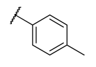
2-[(Quinolin-8-yloxy)methyl]-5-{[(1-(p-tolyl)-1h-1,2,3-triazol-4-yl)methyl]thio}-1,3,4-oxadiazole (12p)
Light brown solid; Chemical Formula: C22H18N6O2S, Yield; 73%, Mol wt: 430.48 gmol−1, M.p: 78–80 °C.
1H NMR (400 MHz, Chloroform-d) δ 8.87 (dd, J = 4.2, 1.8 Hz, 1H), 8.10 (dd, J = 8.3, 1.7 Hz, 1H), 8.07 (s, 1H), 7.52–7.45 (m, 2H), 7.40 (dddd, J = 9.7, 8.3, 5.9, 1.5 Hz, 3H), 7.21 (dd, J = 8.7, 7.3 Hz, 3H), 5.50 (d, J = 1.8 Hz, 2H), 4.55 (s, 2H), 2.33 (s, 3H).
13C NMR (101 MHz, Chloroform-d) δ 165.83, 163.58, 152.96, 149.43, 143.29, 139.71, 138.99, 136.58, 134.58, 130.19, 129.68, 126.68, 121.92, 121.77, 121.55, 111.09, 60.88, 26.89, 21.08.
HRMS: (ESI+-MS, m/z) calcd for C21H15N7O4S (M + Na)+: 453.1110; found: 453.1097.
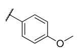
2-{[(1-(4-Methoxyphenyl)-1H-1,2,3-triazol-4-yl)methyl]thio}-5-[(quinolin-8-yloxy)methyl]-1,3,4-oxadiazole (12q)
Black solid; Chemical Formula: C22H18N6O3S, Yield; 76%, Mol wt: 446.48 gmol−1, M.p: 122–128 °C.
1H NMR (400 MHz, Chloroform-d) δ 8.84 (dd, J = 4.3, 1.7 Hz, 1H), 8.07 (dd, J = 8.3, 1.7 Hz, 1H), 8.02 (s, 1H), 7.53–7.45 (m, 2H), 7.44–7.32 (m, 3H), 7.20 (dd, J = 7.3, 1.6 Hz, 1H), 6.93–6.84 (m, 2H), 5.48 (s, 2H), 4.53 (s, 2H), 3.76 (s, 3H).
13C NMR (101 MHz, Chloroform-d) δ 165.80, 163.60, 159.87, 153.12, 149.62, 143.21, 140.08, 136.20, 130.28, 129.64, 126.52, 122.23, 121.91, 121.77, 121.71, 114.71, 110.84, 60.83, 55.62, 26.88.
HRMS: (ESI+-MS, m/z) calcd for C22H18N6O3S (M + Na)+: 469.1059; found: 469.1049.
2-{[(1-(2,4-Dichlorophenyl)-1H-1,2,3-triazol-4-yl)methyl]thio}-5-[(quinolin-8-yloxy)methyl]-1,3,4-oxadiazole (12r)
Light brown solid; Chemical Formula: C21H14Cl2N6O2S, Yield; 73%, Mol wt: 485.35 gmol−1, M.p: 112–116 °C.
1H NMR (400 MHz, Chloroform-d) δ 8.85 (dd, J = 4.3, 1.7 Hz, 1H), 8.14 (s, 1H), 8.08 (dd, J = 8.3, 1.7 Hz, 1H), 7.55 (d, J = 2.4 Hz, 1H), 7.45–7.30 (m, 5H), 7.20 (dd, J = 7.2, 1.7 Hz, 1H), 5.48 (s, 2H), 4.56 (s, 2H).
13C NMR (101 MHz, Chloroform-d) δ 165.59, 163.61, 153.04, 149.57, 142.75, 139.91, 136.31, 135.29, 133.67, 131.59, 130.81, 129.64, 127.79, 126.74, 126.55, 125.39, 121.93, 121.74, 110.69, 60.73, 26.68.
HRMS: (ESI+-MS, m/z) calcd for C21H14Cl2N6O2S (M + Na)+: 521.0411; found: 521.0421.
2-{[(1-(2,6-Dichlorophenyl)-1H-1,2,3-triazol-4-yl)methyl]thio}-5-[(quinolin-8-yloxy)methyl]-1,3,4-oxadiazole (12s)
Light brown solid; Chemical Formula: C21H14Cl2N6O2S, Yield; 77 %, Mol wt: 485.35 gmol−1, M.p: 113–118 °C.
1H NMR (400 MHz, Chloroform-d) δ 8.84 (dd, J = 4.3, 1.7 Hz, 1H), 8.07 (dd, J = 8.3, 1.7 Hz, 1H), 7.89 (s, 1H), 7.44–7.26 (m, 6H), 7.23–7.16 (m, 1H), 5.49 (s, 2H), 4.58 (s, 2H).
13C NMR (101 MHz, Chloroform-d) δ 163.63, 153.09, 149.53, 142.67, 140.02, 136.23, 131.72, 129.63, 128.76, 126.54, 125.80, 121.89, 121.74, 110.92, 60.88, 26.91.
HRMS: (ESI+-MS, m/z) calcd for C21H14Cl2N6O2S (M + Na)+: 508.01; found: 508.35.
2-{[(1-(2,6-Dimethylphenyl)-1H-1,2,3-triazol-4-yl)methyl]thio}-5-[(quinolin-8-yloxy)methyl]-1,3,4-oxadiazole (12t)
Brown solid; Chemical Formula: C23H20N6O2S, Yield; 43%, Mol wt: 444.51 gmol−1, M.p: 110–116 °C.
1H NMR (400 MHz, Chloroform-d) δ 8.82 (dd, J = 4.2, 1.7 Hz, 1H), 8.05 (dd, J = 8.4, 1.7 Hz, 1H), 7.71 (s, 1H), 7.43–7.31 (m, 2H), 7.25–7.15 (m, 1H), 7.05 (d, J = 7.6 Hz, 2H), 5.49 (s, 2H), 4.57 (s, 2H), 1.83 (s, 6H).
13C NMR (101 MHz, Chloroform-d) δ 165.71, 163.61, 153.09, 149.58, 142.63, 140.05, 136.19, 135.69, 135.34, 130.03, 129.63, 128.41, 126.48, 125.09, 121.90, 121.77, 110.82, 60.81, 27.03, 17.31.
HRMS: (ESI+-MS, m/z) calcd for C23H20N6O2S, (M + Na)+: 467.1266; found: 467.1267.

4.5. α-Glucosidase Inhibitory Activity

The α-glucosidase inhibitory activity was evaluated using a previous method [41] with slight modifications. Briefly, 100 µL aliquot of each compound or acarbose (62.5–500 µM) was added to α-glucosidase (1.0 U/mL) solution in 100 mM sodium phosphate buffer (pH 6.8). The reaction was incubated at 37 °C for 15 min before adding 50 µL of 5 mM pNPG solution. After further incubation at 37 °C for another 30 min, the absorbance of the resulting solution was measured at 405 nm. Each compound’s inhibitory activity was calculated as a percentage of the control sample using the expression below:
%   Inhibition = 1 Absorbance   of   compound Absorbance   of   control × 100

4.6. α-Amylase Inhibitory Activity

α-Amylase inhibitory activity was determined according to Ibitoye et al. [42] with slight modifications. Briefly, 200 μL of each compound or acarbose (62.5–500 μM) was incubated at 25 °C for 10 min, with 200 μL solution of porcine pancreatic amylase (0.5 mg/mL) prepared in 6 μM NaCl and 20 mM sodium phosphate buffer (pH 6.9). Then, 500 μL of 1% starch solution was added before further incubation at 25 °C for 15 min. The reaction in the mixture was terminated with a 1 mL dinitrosalicylate reagent before boiling for 10 min. The cooled mixture was diluted with 5 mL distilled water, and the absorbance was read at 540 nm. The inhibitory activity was calculated using the formula below:
%   Inhibition = 1 Absorbance   of   compound Absorbance   of   control × 100

4.7. Antioxidant Activity

4.7.1. Nitric Oxide Scavenging Activity

The antioxidant capacity of the compounds to scavenge nitric oxide radicals was determined using a modified version of the protocol by Kurian et al. [43]. Briefly, 250 μL of sodium nitroprusside (10 mM) prepared in sodium phosphate buffer (pH 7.4) was added to 500 μL of the compound solution (62.5–500 μM) or distilled water (control). The resulting solution was incubated at 37 °C for 2 h. Then, 250 μL of Griess reagent was added to the reaction mixture before the absorbance was measured at 546 nm. The nitric oxide scavenging activity of the compounds was calculated with the formula:
Nitric   oxide   scavenging   ( % ) = 1 Absorbance   of   compound Absorbance   of   control × 100

4.7.2. 2,2′-Diphenyl-1-Picrylhydrazyl (DPPH) Scavenging Activity

The compounds were evaluated for their ability to scavenge stable DPPH radicals, using a modification of the method by Turkoglu et al. [44]. 2 mL of varied concentrations (62.5–500 μM) of compound or gallic acid was added to 2 mL 0.3 mM DPPH prepared in methanol. After thorough mixing, the solution was kept in a dark chamber at room temperature (25 °C) for 30 min. Then, the absorbance was measured at 517 nm, and the DPPH radical scavenging activity was calculated as follows:
DPPH   scavenging   activity   ( % ) = 1 Absorbance   of   compound Absorbance   of   control × 100

4.7.3. Ferric Reducing Antioxidant Power

500 μL of varying concentrations of the compounds or gallic acid (62.5–500 μM) was added to 250 μL distilled water, 100 μL of 200 mM phosphate buffer (pH = 7.3), and 100 μL of 1% potassium ferricyanide [K3Fe(CN)6]. The mixture was incubated at 50 °C for 20 min, followed by acidification with 100 μL trichloroacetic acid (10%). After centrifugation at 3500 rpm for 10 min, 200 μL of the supernatant was transferred into another test tube containing 200 μL of distilled water and 0.8 mL of FeCl3 (0.1%) [45]. Finally, the absorbance was read at 700 nm, and the total reductive antioxidant power was calculated thus:
Ferric   reducing   antioxidant   power   ( % ) = Absorbance   of   compound Absorbance   of   500 µ M   gallic   acid × 100

4.7.4. Mode of α-Glucosidase Inhibition

The mode of inhibition of α-glucosidase of the 4b, 4i, 4j, 4k, 4l, 12k, and 12m compounds was determined using the method described by Kazeem et al. 2013, with modifications [46]. Briefly, 50 µL of the 4b, 4i, 4j, 4k, 4l, 12k, and 12m compounds at (0 µM, 75 µM, 150 µM and 300 µM) concentrations were preincubated with 100 µL of α-glucosidase solution for 10 min at 25 °C in one set of tubes. In another set of tubes, α-glucosidase was preincubated with 50 µL of phosphate buffer (pH 6.9). 25 µL of pNPG at increasing concentrations (0.125–4.0 mg/mL) was added to both sets of solution to start the reaction. Immediately, the absorbance of the resulting solution was monitored for 2 min in a spectrophotometer at 405 nm. The amount of 4-nitrophenol released was determined from a 4-nitrophenol standard curve and then converted to reaction velocities.

4.8. Molecular Modeling

All in silico calculations were performed with the Schrodinger molecular modeling suite (version 2021-2) using the OPLS4 force field.

4.8.1. Homology Modeling of α-Glucosidase and SiteMap

Using the homology modeling module in Schrödinger, the FASTA format sequence of Saccharomyces cerevisiae (UniProt entry: P38138) was downloaded and a Protein BLAST was run to obtain a structure template for the homology model building. From the retuned templates, the native structure of oligo-1,6-glucosidase isomaltase yeast (PDB: 3A4A) was selected. The sequences of the target and template were aligned, and the model was built and its energy minimized with Prime. The structural precision of the model was evaluated with a Ramachandran plot and structure reliability report. Thereafter, SiteMap calculations were performed using the default settings to identify potential allosteric sites.

4.8.2. Molecular Docking

The lowest-energy 3D conformations of the identified inhibitors were generated with LigPrep [47], assigning their protonation and ionization states with Epik [48]. The minimized 3D coordinates of the built α-glucosidase model were generated with Protein Preparation Wizard [49] by adding hydrogens, filling in missing loops, and generating heteroatoms and tautomers with Epik at pH 7.2 ± 0.2. This was followed by hydrogen bond network optimization and restrained minimization to an RMSD of 0.30 Å. The prepared ligands were then docked at the allosteric site deduced from SiteMap analysis using the induced-fit docking module (IFD) [50]. In brief, the employed standard protocol included defining the ligand grid box as the centroid of the SiteMap points, setting the ligand box size to ≤20 Å and conformational sampling at an energy window of 2.5 kcalmol−1. The initial glide docking stage produced poses which were processed for Prime side chain refinement. Finally, complexes with energy of 30 kcalmol−1 were redocked using the extra precision mode to avoid false positives and the 10 best poses were selected for analysis. The protein–ligand interactions were characterized by hydrogen bonds, hydrophobic interactions, and π–π stacking.

4.8.3. Molecular Dynamics Simulations

All MD simulations were performed with the Desmond [51] module. The required model systems for compounds 4i and 12k were generated with the system builder in an orthorhombic box in the SPC water model, and periodic boundary conditions of 10 Å for all dimensions. Before model relaxation with NVT and NPT ensembles, 0.15 M of Na+ and Cl- ions were added to neutralize the system, and minimization with 1000 steps of steepest descent was performed using the NPT ensemble. The MD simulation was thereafter conducted for 200 ns at a constant temperature (300.15 K) and pressure (1.01325 bar), while recording the trajectory 200 ps intervals. Trajectory analysis was achieved with the simulation interaction diagram tool.

Supplementary Materials

The following supporting information can be downloaded at: https://www.mdpi.com/article/10.3390/ph15081035/s1, Table S1: Details of SiteMap analysis to select the putative allosteric site; Table S2: Induced docking results at the selected allosteric site.

Author Contributions

N.C. designed and executed all synthetic and molecular modeling protocols, data analysis, and wrote the manuscript. P.A. analyzed the data and cowrote the manuscript. P.S. (Pule Seboletswe) contributed to molecular modeling and data analysis. K.O. performed biological assays and contributed to manuscript writing. M.S.I. designed the biological experiments and edited the manuscript. P.S. (Parvesh Singh) is a supervisor of the first author who conceptualized and designed the research and edited the manuscript. All authors have read and agreed to the published version of the manuscript.

Funding

This work was funded by grants from the NRF South Africa (Grant Nos. 113402, 126963, 121276).

Institutional Review Board Statement

Not applicable.

Informed Consent Statement

Not applicable.

Data Availability Statement

All the research data are available in the supporting information section.

Acknowledgments

N.C. gratefully acknowledges the National Integrated Cyber Infrastructure System (NICIS) for Centre for High-Performance Computing (CHPC), Cape Town (South Africa), for their supercomputing facilities.

Conflicts of Interest

The authors declare no conflict of interest.

References

  1. Ignatavicius, D.D.; Workman, M.L. Medical-Surgical Nursing-E-Book: Patient-Centered Collaborative Care; Elsevier Health Sciences: Gurgaon, India, 2015. [Google Scholar]
  2. Barrett, E.J.; Liu, Z.; Khamaisi, M.; King, G.L.; Klein, R.; Klein, B.E.K.; Hughes, T.M.; Craft, S.; Freedman, B.I.; Bowden, D.W. Diabetic microvascular disease: An endocrine society scientific statement. J. Clin. Endocrinol. Metab. 2017, 102, 4343–4410. [Google Scholar] [CrossRef] [PubMed]
  3. WHO. The Top 10 Causes of Death. Available online: https://www.who.int/en/news-room/fact-sheets (accessed on 14 December 2021).
  4. International Diabetes Federation. IDF Diabetes Atlas, 8th ed.; International Diabetes Federation: Brussels, Belgium, 2017; pp. 905–911. [Google Scholar]
  5. Kerru, N.; Singh-Pillay, A.; Awolade, P.; Singh, P. Current anti-diabetic agents and their molecular targets: A review. Eur. J. Med. Chem. 2018, 152, 436–488. [Google Scholar] [CrossRef] [PubMed]
  6. Aroua, L.M.; Almuhaylan, H.R.; Alminderej, F.M.; Messaoudi, S.; Chigurupati, S.; Al-Mahmoud, S.; Mohammed, H.H. A facile approach synthesis of benzoylaryl benzimidazole as potential α-amylase and α-glucosidase inhibitor with antioxidant activity. Bioorg. Chem. 2021, 114, 105073. [Google Scholar] [CrossRef]
  7. Dash, R.P.; Babu, R.J.; Srinivas, N.R. Reappraisal and perspectives of clinical drug–drug interaction potential of α-glucosidase inhibitors such as acarbose, voglibose and miglitol in the treatment of type 2 diabetes mellitus. Xenobiotica 2018, 48, 89–108. [Google Scholar] [CrossRef]
  8. Hossain, U.; Das, A.K.; Ghosh, S.; Sil, P.C. An overview on the role of bioactive α-glucosidase inhibitors in ameliorating diabetic complications. Food Chem. Toxicol. 2020, 145, 111738. [Google Scholar] [CrossRef]
  9. Subramaniam, K.; Joseph, M.P.; Babu, L.A. A Common drug causing a common side effect at an uncommon time: Metformin-induced chronic diarrhea and weight loss after years of treatment. Clin. Diabetes 2021, 39, 237. [Google Scholar] [CrossRef] [PubMed]
  10. Collin, F. Chemical basis of reactive oxygen species reactivity and involvement in neurodegenerative diseases. Int. J. Mol. Sci. 2019, 20, 2407. [Google Scholar] [CrossRef] [Green Version]
  11. Volpe, C.M.O.; Villar-Delfino, P.H.; Dos Anjos, P.M.F.; Nogueira-Machado, J.A. Cellular death, reactive oxygen species (ROS) and diabetic complications. Cell Death Dis. 2018, 9, 119. [Google Scholar] [CrossRef]
  12. Eissa, S.I.; Farrag, A.M.; Abbas, S.Y.; El Shehry, M.F.; Ragab, A.; Fayed, E.A.; Ammar, Y.A. Novel structural hybrids of quinoline and thiazole moieties: Synthesis and evaluation of antibacterial and antifungal activities with molecular modeling studies. Bioorg. Chem. 2021, 110, 104803. [Google Scholar] [CrossRef]
  13. Marinho, J.A.; Guimarães, D.S.M.; Glanzmann, N.; Pimentel, G.d.A.; Nunes, I.K.d.C.; Pereira, H.M.G.; Navarro, M.; Varotti, F.d.P.; da Silva, A.D.; Abramo, C. In vitro and in vivo antiplasmodial activity of novel quinoline derivative compounds by molecular hybridization. Eur. J. Med. Chem. 2021, 215, 113271. [Google Scholar] [CrossRef]
  14. Shah, S.R.; Katariya, K.D.; Reddy, D. Quinoline-1,3-oxazole hybrids: Syntheses, anticancer activity and molecular docking studies. Chem. Sel. 2020, 5, 1097–1102. [Google Scholar] [CrossRef]
  15. Nikookar, H.; Mohammadi-Khanaposhtani, M.; Imanparast, S.; Faramarzi, M.A.; Ranjbar, P.R.; Mahdavi, M.; Larijani, B. Design, synthesis and in vitro α-glucosidase inhibition of novel dihydropyrano[3,2-c]quinoline derivatives as potential anti-diabetic agents. Bioorg. Chem. 2018, 77, 280–286. [Google Scholar] [CrossRef] [PubMed]
  16. Vivekanand, B.; Raj, K.M.; Mruthyunjayaswamy, B.H.M. Synthesis, characterization, antimicrobial, DNA-cleavage and antioxidant activities of 3-((5-chloro-2-phenyl-1H-indol-3-ylimino)methyl)quinoline-2(1H)-thione and its metal complexes. J. Mol. Struct. 2015, 1079, 214–224. [Google Scholar] [CrossRef]
  17. Suwanjang, W.; Prachayasittikul, S.; Prachayasittikul, V. Effect of 8-hydroxyquinoline and derivatives on human neuroblastoma SH-SY5Y cells under high glucose. PeerJ 2016, 4, e2389. [Google Scholar] [CrossRef] [Green Version]
  18. Prachayasittikul, V.; Prachayasittikul, S.; Ruchirawat, S. 8-Hydroxyquinolines: A review of their metal chelating properties and medicinal applications. Drug Des. Dev. Ther. 2013, 7, 1157–1178. [Google Scholar] [CrossRef] [Green Version]
  19. Zurawski, J.R.; Vincent, R.; Stout, D.M.; Nitz, T.J.; Youdim, M.B.H.; Weinreb, O. Neuroprotective and Neuro-Restorative Iron Chelators and Monoamine Oxidase Inhibitors and Uses Thereof. U.S. Patent 20140186280a1, 3 July 2014. [Google Scholar]
  20. Boström, J.; Hogner, A.; Llinàs, A.; Wellner, E.; Plowright, A.T. Oxadiazoles in medicinal chemistry. J. Med. Chem. 2012, 55, 1817–1830. [Google Scholar] [CrossRef]
  21. Nirogi, R.; Mohammed, A.R.; Shinde, A.K.; Gagginapally, S.R.; Kancharla, D.M.; Middekadi, V.R.; Bogaraju, N.; Ravella, S.R.; Singh, P.; Birangal, S.R.; et al. Synthesis, structure–activity relationships, and preclinical evaluation of heteroaromatic amides and 1,3,4-oxadiazole derivatives as 5-HT4 receptor partial agonists. J. Med. Chem. 2018, 61, 4993–5008. [Google Scholar] [CrossRef]
  22. Taha, M.; Ismail, N.H.; Imran, S.; Wadood, A.; Rahim, F.; Ali, M.; Rehman, A.U. Novel quinoline derivatives as potent in vitro α-glucosidase inhibitors: In silico studies and SAR predictions. MedChemComm 2015, 6, 1826–1836. [Google Scholar] [CrossRef]
  23. Izgi, S.; Sengul, I.F.; Şahin, E.; Koca, M.S.; Cebeci, F.; Kandemir, H. Synthesis of 7-azaindole based carbohydrazides and 1,3,4-oxadiazoles; antioxidant activity, α-glucosidase inhibition properties and docking study. J. Mol. Struct. 2022, 1247, 131343. [Google Scholar] [CrossRef]
  24. Kumar, S.; Sharma, B.; Mehra, V.; Kumar, V. Recent accomplishments on the synthetic/biological facets of pharmacologically active 1H-1,2,3-triazoles. Eur. J. Med. Chem. 2021, 212, 113069. [Google Scholar] [CrossRef]
  25. Bozorov, K.; Zhao, J.; Aisa, H.A. Aisa, 1,2,3-Triazole-containing hybrids as leads in medicinal chemistry: A recent overview. Bioorg. Med. Chem. 2019, 27, 3511–3531. [Google Scholar] [CrossRef] [PubMed]
  26. Kolb, H.C.; Sharpless, K.B. The growing impact of click chemistry on drug discovery. Drug Discov. Today 2003, 8, 1128–1137. [Google Scholar] [CrossRef]
  27. Fallah, Z.; Tajbakhsh, M.; Alikhani, M.; Larijani, B.; Faramarzi, M.A.; Hamedifar, H.; Mohammadi-Khanaposhtani, M.; Mahdavi, M. A review on synthesis, mechanism of action, and structure-activity relationships of 1,2,3-triazole-based α-glucosidase inhibitors as promising anti-diabetic agents. J. Mol. Struct. 2022, 1255, 132469. [Google Scholar] [CrossRef]
  28. Asemanipoor, N.; Mohammadi-Khanaposhtani, M.; Moradi, S.; Vahidi, M.; Asadi, M.; Faramarzi, M.A.; Mahdavi, M.; Biglar, M.; Larijani, B.; Hamedifar, H.; et al. Synthesis and biological evaluation of new benzimidazole-1,2,3-triazole hybrids as potential α-glucosidase inhibitors. Bioorg. Chem. 2020, 95, 103482. [Google Scholar] [CrossRef]
  29. Dhameja, M.; Gupta, P. Synthetic heterocyclic candidates as promising α-glucosidase inhibitors: An overview. Eur. J. Med. Chem. 2019, 176, 343–377. [Google Scholar] [CrossRef]
  30. Awolade, P.; Cele, N.; Kerru, N.; Singh, P. Synthesis, antimicrobial evaluation, and in silico studies of quinoline—1H-1,2,3-triazole molecular hybrids. Mol. Divers. 2020, 25, 2201–2218. [Google Scholar] [CrossRef]
  31. Dhawan, S.; Kerru, N.; Awolade, P.; Singh-Pillay, A.; Saha, S.T.; Kaur, M.; Jonnalagadda, S.B.; Singh, P. Synthesis, computational studies and antiproliferative activities of coumarin-tagged 1,3,4-oxadiazole conjugates against MDA-MB-231 and MCF-7 human breast cancer cells. Bioorg. Med. Chem. 2018, 26, 5612–5623. [Google Scholar] [CrossRef]
  32. Nussinov, R.; Tsai, C.-J. Allostery in disease and in drug discovery. Cell 2013, 153, 293–305. [Google Scholar] [CrossRef] [Green Version]
  33. Saddique, F.A.; Ahmad, M.; Ashfaq, U.A.; Muddassar, M.; Sultan, S.; Zaki, M.E.A. Identification of cyclic sulfonamides with an N-arylacetamide group as α-glucosidase and α-amylase inhibitors: Biological evaluation and molecular modeling. Pharmaceuticals 2022, 15, 106. [Google Scholar] [CrossRef]
  34. Xing, R.; Liu, S.; Guo, Z.; Yu, H.; Wang, P.; Li, C.; Li, Z.; Li, P. Relevance of molecular weight of chitosan and its derivatives and their antioxidant activities in vitro. Bioorg. Med. Chem. 2005, 13, 1573–1577. [Google Scholar] [CrossRef]
  35. Schrödinger. Release 2021-2: Prime, S.; LLC: New York, NY, USA, 2021. [Google Scholar]
  36. Uniprot.org. Available online: https://www.uniprot.org/ (accessed on 12 April 2022).
  37. Schrödinger. Release 2021-2: SiteMap, S.; LLC: New York, NY, USA, 2021. [Google Scholar]
  38. Broughton, H.B.; Watson, I.A. Selection of heterocycles for drug design. J. Mol. Graph. Model. 2004, 23, 51–58. [Google Scholar] [CrossRef] [PubMed]
  39. Maier, J.A.; Martinez, C.; Kasavajhala, K.; Wickstrom, L.; Hauser, K.E.; Simmerling, C. ff14SB: Improving the accuracy of protein side chain and backbone parameters from ff99SB. J. Chem. Theory Comput. 2015, 11, 3696–3713. [Google Scholar] [CrossRef] [Green Version]
  40. Wade, R.C.; Goodford, P.J. The role of hydrogen-bonds in drug binding. Prog. Clin. Biol. Res. 1989, 289, 433–444. [Google Scholar] [PubMed]
  41. Ademiluyi, A.O.; Oboh, G. Soybean phenolic-rich extracts inhibit key-enzymes linked to type 2 diabetes (α-amylase and α-glucosidase) and hypertension (angiotensin I converting enzyme) in vitro. Exp. Toxicol. Pathol. 2013, 65, 305–309. [Google Scholar] [CrossRef] [PubMed]
  42. Ibitoye, O.B.; Olofinsan, K.A.; Teralı, K.; Ghali, U.M.; Ajiboye, T.O. Bioactivity-guided isolation of antidiabetic principles from the methanolic leaf extract of Bryophyllum pinnatum. J. Food Biochem. 2018, 42, e12627. [Google Scholar] [CrossRef]
  43. Kurian, G.A.; Suryanarayanan, S.; Raman, A.; Padikkala, J. Antioxidant effects of ethyl acetate extract of Desmodium gangeticum root on myocardial ischemia reperfusion injury in rat hearts. Chin. Med. 2010, 5, 3. [Google Scholar] [CrossRef] [Green Version]
  44. Turkoglu, A.; Duru, M.E.; Mercan, N.; Kivrak, I.; Gezer, K. Antioxidant and antimicrobial activities of Laetiporus sulphureus (Bull.) Murrill. Food Chem. 2007, 101, 267–273. [Google Scholar] [CrossRef]
  45. Oyaizu, M. Studies on products of browning reaction antioxidative activities of products of browning reaction prepared from glucosamine. Jpn. J. Nutr. Diet. 1986, 44, 307–315. [Google Scholar] [CrossRef] [Green Version]
  46. Kazeem, M.I.; Adamson, J.O.; Ogunwande, I.A. Modes of inhibition of α-amylase and α-glucosidase by aqueous extract of Morinda lucida Benth leaf. BioMed Res. Int. 2013, 2013, 527570. [Google Scholar] [CrossRef] [Green Version]
  47. Schrödinger. Release 2021-2: LigPrep, S.; LLC: New York, NY, USA, 2021. [Google Scholar]
  48. Schrödinger. Release 2021-2: Epik, S.; LLC: New York, NY, USA, 2021. [Google Scholar]
  49. Schrödinger. Release 2021-2: Protein Preparation Wizard, Epik, S.; LLC: New York, NY, USA, 2021. [Google Scholar]
  50. Schrödinger. Release 2021-2: Induced Fit Docking Protocol, Glide, S.; LLC: New York, NY, USA, 2021. [Google Scholar]
  51. Schrödinger. Release 2021-2: Desmond Molecular Dynamics System; D. E. Shaw Research: New York, NY, USA, 2021; Maestro-Desmond Interoperability Tools; Schrödinger: New York, NY, USA, 2021. [Google Scholar]
Figure 1. Reported α-glucosidase inhibitors based on quinoline, 1,3,4-oxadiazole, and 1,2,3-triazole cores.
Pharmaceuticals 15 01035 g001
Scheme 1. Synthetic route to quinoline-1,3,4-oxadiazole hybrids.
Pharmaceuticals 15 01035 sch001
Scheme 2. Synthesis of requisite azide substrates for the 1,3-dipolar cycloaddition reaction.
Pharmaceuticals 15 01035 sch002
Scheme 3. Synthesis of quinoline-1,3,4-oxadiazole-1,2,3-triazole hybrids via Cu(I)-catalyzed azide-alkyne [3 + 2] cycloaddition reaction.
Pharmaceuticals 15 01035 sch003
Figure 2. Schematic representation of key HMBC correlations in compounds 4k and 12e.
Pharmaceuticals 15 01035 g002
Figure 3. SAR summary of α-glucosidase inhibition and antioxidant activity.
Pharmaceuticals 15 01035 g003
Figure 4. Lineweaver–Burk plots of promising α-glucosidase inhibitors.
Pharmaceuticals 15 01035 g004aPharmaceuticals 15 01035 g004b
Figure 5. (A) Aligned protein sequences of S. cerevisiae yeast α-glucosidase and yeast isomaltase (PDB: 3A4A) showing the sequences similarity and identity. (B) α-Glucosidase homology model built from the 3A4A structure. The most probable allosteric site obtained from SiteMap calculations is depicted as a mesh surface while glucose bound at the active site is shown in CPK representation.
Pharmaceuticals 15 01035 g005
Figure 6. 3D representation of the α-glucosidase complexes of promising compounds docked at the allosteric site. (H-bond yellow, π–π stacking blue, halogen purple, and π–cation green).
Pharmaceuticals 15 01035 g006
Figure 7. MD simulation trajectory analysis of compounds 4i and 12k. (A) RMSD and (B) RMSF plots. (C) Protein–ligand interactions over the 200 ns trajectory.
Pharmaceuticals 15 01035 g007
Table 1. IC50 (µM) for antioxidant profiling of compounds 4at and 12at.
EntryRDPPHFRAPNOEntryRDPPHFRAPNO
8-HQn/a51.81183.903H25.15463.85
4a Pharmaceuticals 15 01035 i00170.942.6712a Pharmaceuticals 15 01035 i00289.98
4b Pharmaceuticals 15 01035 i00378.2012b Pharmaceuticals 15 01035 i00480.00
4c Pharmaceuticals 15 01035 i00579.0312c Pharmaceuticals 15 01035 i00680.17
4d Pharmaceuticals 15 01035 i00782.90457.3812d Pharmaceuticals 15 01035 i00880.40
4e Pharmaceuticals 15 01035 i00975.7412e Pharmaceuticals 15 01035 i01092.82
4f Pharmaceuticals 15 01035 i01172.1112f Pharmaceuticals 15 01035 i01291.78481.76
4g Pharmaceuticals 15 01035 i01386.3912g Pharmaceuticals 15 01035 i01481.913.01
4h Pharmaceuticals 15 01035 i01575.3512h Pharmaceuticals 15 01035 i01690.69
4i Pharmaceuticals 15 01035 i01754.8622.88197.9312i Pharmaceuticals 15 01035 i01895.38
4j Pharmaceuticals 15 01035 i01954.86399.3212j Pharmaceuticals 15 01035 i020101.73
4k Pharmaceuticals 15 01035 i02181.1312k Pharmaceuticals 15 01035 i02285.72
4l Pharmaceuticals 15 01035 i02377.7112l Pharmaceuticals 15 01035 i02481.74
4m Pharmaceuticals 15 01035 i02585.1512m Pharmaceuticals 15 01035 i02688.69
4n Pharmaceuticals 15 01035 i02777.2312n Pharmaceuticals 15 01035 i02892.19
4o Pharmaceuticals 15 01035 i02976.8512o Pharmaceuticals 15 01035 i03088.92
4p Pharmaceuticals 15 01035 i03167.9912p Pharmaceuticals 15 01035 i03296.04
4q Pharmaceuticals 15 01035 i03378.5912q Pharmaceuticals 15 01035 i03498.50
4r Pharmaceuticals 15 01035 i03564.5212r Pharmaceuticals 15 01035 i036100.96
4s Pharmaceuticals 15 01035 i03787.2212s Pharmaceuticals 15 01035 i03884.48
4t Pharmaceuticals 15 01035 i03993.9712t Pharmaceuticals 15 01035 i04090.02
Gallic acid26.6317.85728.68Ascorbic acid 102.62
“–” = greater than 500 μM.
Table 2. IC50 (µM) for α-glucosidase and α-amylase inhibition by compounds 4at and 12at.
EntryRα-Glucosidaseα-AmylaseEntryRα-Glucosidaseα-Amylase
8-HQn/a29.283H34.70
4a Pharmaceuticals 15 01035 i04141.6012a Pharmaceuticals 15 01035 i04235.19
4b Pharmaceuticals 15 01035 i04324.8712b Pharmaceuticals 15 01035 i04436.74
4c Pharmaceuticals 15 01035 i04541.3512c Pharmaceuticals 15 01035 i04646.86
4d Pharmaceuticals 15 01035 i04746.6612d Pharmaceuticals 15 01035 i04838.11
4e Pharmaceuticals 15 01035 i04948.08210.2612e Pharmaceuticals 15 01035 i05039.34
4f Pharmaceuticals 15 01035 i05134.5012f Pharmaceuticals 15 01035 i05256.36
4g Pharmaceuticals 15 01035 i05334.0912g Pharmaceuticals 15 01035 i05446.51
4h Pharmaceuticals 15 01035 i05546.0712h Pharmaceuticals 15 01035 i05663.59
4i Pharmaceuticals 15 01035 i05715.8512i Pharmaceuticals 15 01035 i05842.79
4j Pharmaceuticals 15 01035 i05926.6712j Pharmaceuticals 15 01035 i06059.02
4k Pharmaceuticals 15 01035 i06123.6912k Pharmaceuticals 15 01035 i06222.47
4l Pharmaceuticals 15 01035 i06325.2312l Pharmaceuticals 15 01035 i06438.02
4m Pharmaceuticals 15 01035 i06528.3112m Pharmaceuticals 15 01035 i06626.81
4n Pharmaceuticals 15 01035 i06732.7612n Pharmaceuticals 15 01035 i06835.22
4o Pharmaceuticals 15 01035 i06948.8412o Pharmaceuticals 15 01035 i07038.30
4p Pharmaceuticals 15 01035 i07129.5612p Pharmaceuticals 15 01035 i07244.07494.76
4q Pharmaceuticals 15 01035 i07329.64426.7712q Pharmaceuticals 15 01035 i07462.05
4r Pharmaceuticals 15 01035 i07528.2212r Pharmaceuticals 15 01035 i07648.54
4s Pharmaceuticals 15 01035 i07735.3712s Pharmaceuticals 15 01035 i07838.79
4t Pharmaceuticals 15 01035 i07927.6712t Pharmaceuticals 15 01035 i08044.12
Acarbose17.8584.26
“–” = greater than 500 μM.
Publisher’s Note: MDPI stays neutral with regard to jurisdictional claims in published maps and institutional affiliations.

