Article

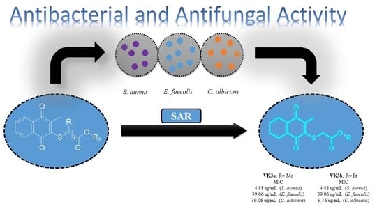
Promising Antibacterial and Antifungal Agents Based on Thiolated Vitamin K3 Analogs: Synthesis, Bioevaluation, Molecular Docking

by
Hatice Yıldırım
1,
Mahmut Yıldız
2,
Nilüfer Bayrak
1,
Emel Mataracı-Kara
3,
Mohamed Osman Radwan
4,5,
Ayse Tarbin Jannuzzi
6,
Masami Otsuka
4,7,
Mikako Fujita
4 and
Amaç Fatih TuYuN
8,*
1
Department of Chemistry, Faculty of Engineering, Istanbul University-Cerrahpasa, Avcilar, Istanbul 34320, Turkey
2
Department of Chemistry, Gebze Technical University, Gebze, Kocaeli 41400, Turkey
3
Department of Pharmaceutical Microbiology, Faculty of Pharmacy, Istanbul University, Beyazit, Istanbul 34116, Turkey
4
Medicinal and Biological Chemistry Science Farm Joint Research Laboratory, Faculty of Life Sciences, Kumamoto University, 5-1 Oe-honmachi, Chuo-ku, Kumamoto 862-0973, Japan
5
Chemistry of Natural Compounds Department, Pharmaceutical and Drug Industries Research Division, National Research Centre, Dokki, Cairo 12622, Egypt
6
Department of Pharmaceutical Toxicology, Faculty of Pharmacy, Istanbul University, Beyazit, Istanbul 34116, Turkey
7
Department of Drug Discovery, Science Farm Ltd., 1-7-30 Kuhonji, Chuo-ku, Kumamoto 862-0976, Japan
8
Department of Chemistry, Faculty of Science, Istanbul University, Fatih, Istanbul 34126, Turkey
*
Author to whom correspondence should be addressed.
Pharmaceuticals 2022, 15(5), 586; https://doi.org/10.3390/ph15050586
Submission received: 25 March 2022 / Revised: 20 April 2022 / Accepted: 24 April 2022 / Published: 10 May 2022
(This article belongs to the Special Issue Privileged Structures as Leads in Medicinal Chemistry)

Abstract

:
In the present study, we designed and synthesized thiolated VK3 analogs (VK3a–g) along with an extensive antimicrobial study. After the evaluation of the antibacterial and antifungal activity against various bacterial and fungal strains, we presented an initial structure–activity relationship study on these VK3 analogs. In particular, four thiolated VK3 analogs exhibited superior biological potency against some Gram-positive bacterial strains, including Staphylococcus aureus (ATCC® 29213) and Enterococcus faecalis (ATCC® 29212). Next, all thiolated VK3 analogs were evaluated for their potential of cell growth inhibition on the NCI-60 cancer cell lines panel. This screening underlined that the thiolated VK3 analogs have no visible cytotoxicity on different cancer cell lines. The selected two thiolated VK3 analogs (VK3a and VK3b), having minimal hemolytic activity, which also have the lowest MIC values on S. aureus and E. faecalis, were further evaluated for their inhibition capacities on biofilm formation after evaluating their potential in vitro antimicrobial activity against each of the 20 clinically obtained resistant strains of Staphylococcus aureus. VK3b showed excellent antimicrobial activity against clinically resistant S. aureus isolates. Furthermore, the tested molecules showed nearly two log10 reduction in the viable cell count at six hours according to the time kill curve studies. Although these molecules decreased biofilm attachment about 50%, when sub-MIC concentrations were used these molecules increased the percentage of biofilm formation. The molecular docking of VK3a and VK3b in S. aureus thymidylate kinase was conducted in order to predict their molecular interactions. VK3a and VK3b exhibited excellent lead-likeness properties and pharmacokinetic profiles that qualify them for further optimization and development. In conclusion, since investigating efficient novel antimicrobial molecules is quite difficult, these studies are of high importance, especially in the present era of antimicrobial resistance.

Graphical Abstract

1. Introduction

Vitamins are micronutrients comprised of organic structures that cannot be synthesized by the human body, but must be taken exogenously for the body in order to perform its normal functions and prevent metabolic disorders [1]. It is known that a diet rich in vitamins strengthens the immune system and plays an important role in fighting against disease-causing microorganisms. Additionally, although data on the protective factors of the disease are limited in the Coronavirus epidemic, which has turned into a worldwide pandemic, vitamins are among the preventive health measures that can reduce the risk of infection in elderly and immunocompromised individuals [2,3]. The most significant vitamins for the human body are vitamin A [4], vitamin B complex [5], vitamin C [6], vitamin D [7], and Vitamin K [8] etc. Vitamin K is an important component in the system playing a key role in blood coagulation in addition to strengthening bones and promoting the calcification of arteries and soft tissues [9]. The structure of the Vitamin K family consists of a common motif structure 2-methyl-1,4-naphthoquinone analogs, as shown in Figure 1. Vitamin K3 (VK3), named as menadione, is a synthetic analog of natural Vitamin K, unlike the other two forms, which exist in nature (Figure 1) [10,11]. VK3 analogs are attractive to medicinal and/or organic chemists as a result of their unique chemical and broad range of biological activities [12,13]. Such research indicates that the VK3 moiety is an important framework with good biological potency, which effectively shows anticancer [14,15], antimalarial [16,17], antioxidant [18], and catalase inhibition activity [19,20]. In addition to inhibition of in vitro and in vivo cancer cell lines [21,22,23,24,25], iron-VK3 analog was shown to synergistically promote ferroptotic therapy for inhibiting tumor growth, preventing metastasis and tackling radioresistance [26].
For some time in our laboratory, we have focused on the construction of 1,4-quinones with different substitution patterns, along with detailed studies on their potential biological performances, in order to discover new compounds that have the potential to be lead molecules in the fight against cancer and antimicrobial resistance [27,28,29]. To date, the biologically important lead structures have been obtained and gathered for the tuning of pharmacological efficacy as an antimicrobial and/or anticancer agent. It is mainly understood that incorporating amino groups (i.e., aryl amines, piperazines, and piperidines) into 1,4-quinone moiety with different side groups (i.e., phenyl, dimethyl, and pyridinyl) has mainly increased the biological potency of the target molecules [27,28,29]. On the other hand, we have previously accessed the C2- and/or C2C3-thiolated 1,4-quinones with different side groups, such as the dimethyl or pyridinyl group [30,31]. A key aspect of the biological activity in our previous studies is the certain chemical modifications of 1,4-quinone moiety. Naphthoquinones [19,32,33,34,35], quinolinequinones [27,30,36,37], and dimethylbenzoquinones [28,38,39] are the most widely used motifs belonging to the quinone family, being biologically important lead structures, which have been extensively used over the years [40,41]. Some of these C2- and/or C2C3-thiolated 1,4-quinones with different side groups were more potent than the reference drug(s). In these studies, alkyl chain thiols were connected to a dichloronaphthoquinone [32,33] and dimethylbenzoquinone [28,38,39,42] via different substrates [30,31]. As seen in these studies, the used alkyl chain or aromatic thiols as substrates, connected to dimethylbenzoquinone, did not exhibit any significant antibacterial and/or antifungal activity. Last but not least, the introduction of the dichloronaphthoquinone in alkyl chain thiols gave the powerful lead structures for antimicrobial (antibacterial and antifungal) studies [32,33]. Another well-explored moiety of dichloronaphthoquinone is the second halogen atom, which easily undergoes substitution reactions with additional amines or thiols. Replacement of the chlorine atom with either amines or thiols decreased the antimicrobial potency. Some works were focused on the synthesis of these mixed thiolated naphthoquinones and aminated thiolated naphthoquinones (Figure 2) [32,33,43,44,45,46]. In general, the presence of the alkyl chain thiol and the chlorine atom is more important for the activity than fully thiolated naphthoquinones [32,33]. Thus, to reveal the effect of the quinone moiety and the second substituent (chloro, amino, or thio group) on the antimicrobial potency, VK3 (Menadione) was selected as a parent molecule of the substrates to synthesize some analogs of VK3. Motivated by these studies and given our long-standing interest in these specific structures, we envisioned that the used alkyl chain thiols as the substrates might offer new lead structures against antimicrobial resistance. In this paper, efforts for the design, synthesis, and antimicrobial evaluation of target analogs of VK3 were made to exploit the effect of structural modification on their potency obtained from the commercially available VK3 under mild conditions. Indeed, some members of thiolated VK3 were investigated for their antibiofilm activity, along with their potential antimicrobial activity against each of the 20 clinically obtained strains of Methicillin resistant Staphylococcus aureus, and bactericidal time-kill kinetic study.

2. Results and Discussion

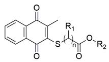
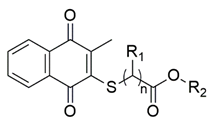
2.1. Library Design and Synthesis

Due to their high electrophilicity, versatile 1,4-quinones are very reactive towards thiols, which are soft nucleophiles according to the Pearson’s scale, unlike primary and secondary amines. Our synthesis studies suggest that VK3 has only one capacity to react with alkyl chain thiols under mild condition. The synthetic strategy adopted for the synthesis of the target VK3 analogs (VK3a–g) is depicted in one step (Scheme 1). The commercially available VK3 was directly thiolated using the corresponding alkyl chain thiols to give the target VK3 analogs (VK3a–g) in ethanol in one step (Scheme 1) by adopting the procedure with a slight modification (i.e., heating) from that mentioned in the literature [47]. The reaction mixture was then refluxed for three–six hours to yield the final desired VK3 analogs. (VK3a–g) were purified via silica gel column chromatography. All of the thiolated VK3 analogs were fully characterized by mass spectroscopy (MS), Fourier-transform infrared spectroscopy (FTIR), nuclear magnetic resonance (NMR), and high-resolution mass spectrometry (HRMS). The structure of methyl 2-(3-methyl-1,4-dioxo-1,4-dihydronaphthalen-2-ylthio)acetate (VK3a) was determined by single-crystal X-ray diffraction analysis (Figure 3). The absorption bands from vibrations of the following bands are observed at around 1720 and 1650 cm−1 for C=O; at around 1590 cm−1 for C=C; 2900 cm−1 for C-Haliphatic in the IR spectra. The methylene protons attached to either the oxygen or sulfur atom were observed at around 4.00 ppm. In some cases, the methylene protons attached to the sulfur atom were seen at around 3.50 ppm. The singlet in the region of 3.70 ppm was assigned to the protons of the methyl group attached to the oxygen atom, and multiplets or doublets in the region of 6.90–8.00 ppm to the protons of aromatic rings. The 13C NMR shifts of the carbonyl carbon atoms, within 1,4-quinone of the target analogs, appeared downfield at around 182 and 181 ppm as two peaks, while the other carbonyl carbon atoms, which belong to thiol moiety, had resonance at around 170 ppm. The individual -OCH2 and -OCH3 carbon atoms in each molecule of the library provided chemical shift values of around 63 ppm and 53 ppm, respectively. The methyl carbon atoms connected to the 1,4-quinone moiety have resonanced at upfield around 14 ppm. Additionally, the methylene carbons attached to the sulfur atom within thiol moiety were observed at around 40 ppm. Moreover, in the high-resolution mass spectra of the thiolated VK3 analogs, the molecular ion peak with a proton adduct were detected in strong abundance.
To produce VK3a in high purity, the compound was dissolved in ethanol and stored for at least one week for crystallization. The structure of VK3a was analyzed by the X-ray diffractometer. The ORTEP drawing (displacement parameters drawn at 50% probability level) of the VK3a is shown in Figure 3. The crystal system of VK3a is monoclinic (space group P 1 21/n 1). It is clear from the X-ray analysis data that the bond lengths of O1-C2 and O2-C2 belonging to the ester group of VK3a are 1.19 Å and 1.32 Å, respectively (Table 1). In the data of X-ray analysis, the bond lengths of O1-C2 and O2-C2 of the ester group of VK3a are 1.19 Å and 1.32 Å, respectively. It is clear that the bond between O2 and C2 atoms is longer than the bond between C2 and O1 atoms, and that the O1-C2 bond has a characteristic double bond and the O2-C2 bond has a characteristic single bond. By experimentally observing that the torsion angle of S1-C4-C14-O3 is 5.4°, it can be determined that the sulfur and oxygen atoms are approximately in the same plane. From the determination of the torsional angle of C3-S1-C4-C14 as −49.5°, it can be deduced that the methylene group may have intramolecular and intermolecular H-bonding interaction with the carbonyl group. The X-ray data of VK3a also shows H-bonding between the oxygen atom as an acceptor and methylene hydrogens as donors. The selected bond lengths, bond angles, torsion angles, hydrogen bond distances, and angles are given in Table 2, Table 3, Table 4 and Table 5.

2.2. Biological Activity

2.2.1. Evaluation of Antimicrobial Activity of the Thiolated VK3 Analogs (VK3a–g)

The obtained thiolated VK3 analogs (VK3a–g) were initially tested in vitro for their antibacterial and antifungal profile by determining their minimum inhibitory concentration (MIC) values, listed in Table 6 and Table 7 and compared with the commercially available reference drugs, against four Gram-negative bacteria (Pseudomonas aeruginosa ATCC 27853, Escherichia coli ATCC 25922, Klebsiella pneumoniae ATCC 4352, and Proteus mirabilis ATCC 14153), three Gram-positive bacteria (Staphylococcus aureus ATCC 29213, Staphylococcus epidermidis ATCC 12228, and Enterococcus faecalis ATCC 29212), and three fungi (Candida albicans ATCC 10231, Candida parapsilosis ATCC 22019, and Candida tropicalis ATCC 750) by the broth microdilutions technique using the Clinical Laboratory Standards Institute (CLSI) recommendations [48,49]. The MIC values were determined by comparison with standard agents.
According to the obtained results, the thiolated VK3 analogs (VK3a–g) did not have any antibacterial activity against Gram-negative bacterial strains (P. aeruginosa, E. coli, K. pneumoniae, and P. mirabilis) in addition to one Gram-positive bacterial strain (S. epidermidis). It is noteworthy to mention that three of the thiolated VK3 analogs (VK3a-b and VK3d) were the most active analogs on S. aureus and E. faecalis with MIC values of 4.88 and 39.06 μg/mL, respectively. Furthermore, two of them (VK3c and VK3f) were the other remarkable VK3 analogs against S. aureus with MIC of 9.76 μg/mL. VK3c was another remarkable VK3 analog on E. faecalis. When the obtained thiolated VK3 analogs (VK3a–g) are evaluated in terms of the antifungal activity, VK3b showed high antifungal activity against C. albicans possessing the MIC value of 9.76 μg/mL. Besides, VK3a and VK3d were observed to be the most active VK3 analogs as antifungal agents against C. tropicalis with 9.76 μg/mL MIC value. Among the tested VK3 analogs, two VK3 analogs (VK3a and VK3b) showed strong antibacterial and antifungal potency. Moreover, the antibacterial activity of these two analogs seems to be strongly specific to S. aureus and E. faecalis since their MIC values against other tested bacterial strains were relatively better. Thus, we mainly focused on these two VK3 analogs for the best antimicrobial potency against clinically obtained Methicillin-resistant strains of Staphylococcus aureus. The in vitro activities of the VK3a and VK3b analogs against 20 clinical isolates of MRSA are summarized in Table 8. Although the in vitro activity of VK3a was found to have a moderate effect on the studied clinical strains, VK3b showed excellent activity on these isolates, similar to be the standard S. aureus ATCC isolate’s MIC results. Moreover, there was no major difference between the bactericidal and inhibitory endpoints; the MBCs were generally two-fold higher than those of the MICs. With these excellent activity results, especially from VK3b, we further analyzed the anticancer property of these antimicrobials.
Although the biological dataset is small, some structure–activity relationships can be extracted. Herein, to examine how the alkyl chain thiols affect the antibacterial and antifungal activity, we have varied the backbone group attached to the oxygen atom, while keeping the methylene bridge(s) or methine bridge between the sulfur and carbonyl group of the substrates. Keeping the methylene bridge unchanged, the group attached to the oxygen atom differed from the methyl or ethyl group. In this case, the activity was not affected. The activity against S. aureus was decreased with the insertion of an additional methyl group instead of a hydrogen atom in the methylene group between the sulfur atom and carbonyl group in the thiolated VK3 analog (VK3c). When the methylene bridge was held with one additional methylene group, the analogs were diversified with alkyl groups such as methyl, ethyl, or butyl group. Thus, only one methylene bridge between the sulfur and carbonyl group are allowed in the analogs, with a methyl or ethyl group favoring inhibition rather than molecules having two methylene bridges between the sulfur and carbonyl group (VK3a vs VK3d; VK3b vs. VK3e). Replacement of a hydrogen atom with methyl group in the methylene bridge between the sulfur and carbonyl group decreased activity slightly (VK3b vs VK3c) against S. aureus, but had no effect against E. faecalis. In general, we could conclude that a remarkable decrease in inhibitory activity was observed when the length of the alkyl chain attached to the oxygen atom was changed from methyl to ethyl or butyl group. Upon increasing the alkyl group attached to the oxygen atom to varied groups, there was a remarkable decrease in activity against all the tested bacteria. In other words, bulkier substituents, such as long-chained alkyl groups in VK3g, are detrimental to the activity. Both the introduction of the second methylene bridge between the sulfur and carbonyl group and the length of the alkyl chain attached to the oxygen atom diminished the activity. The detrimental effect is stronger at the long alkyl chain attached to the oxygen atom than the methylene bridge. The same parabolic pattern was followed for the thiolated VK3 analogs for antifungal activity.

2.2.2. Toxicity Evaluation

In order to check the cell toxicity of the tested thiolated VK3 analogs presented herein, all thiolated VK3 analogs were submitted to the National Cancer Institute (NCI) of Bethesda within the Developmental Therapeutics Program (DTP). After selection, they were screened for evaluation of their cell growth inhibition potential on the NCI-60 cancer cell lines panel [50] at a single high dose concentration (10 µM) on nine different cancer types, namely: leukemia; lung; colon; CNS; melanoma; ovarian; renal; prostate; and breast cancer cell lines [51] with the protocol of the Drug Evaluation Branch, NCI [52]. Herein, testing of those analogs (VK3a–g) against the NCI panel of 60 cell lines provides us with comprehensive information on the growth inhibitory effects of analogs. The single-dose results, released in the form of a mean graph of the cell growth percentage (GP) of each of the tested thiolated VK3 analogs, were reported. Their one-dose mean graphs are presented in the Supplementary Material as Figures S3–S9. The thiolated VK3 analogs displayed slight, to completely no cytotoxic effects towards all of the cancer cell lines.

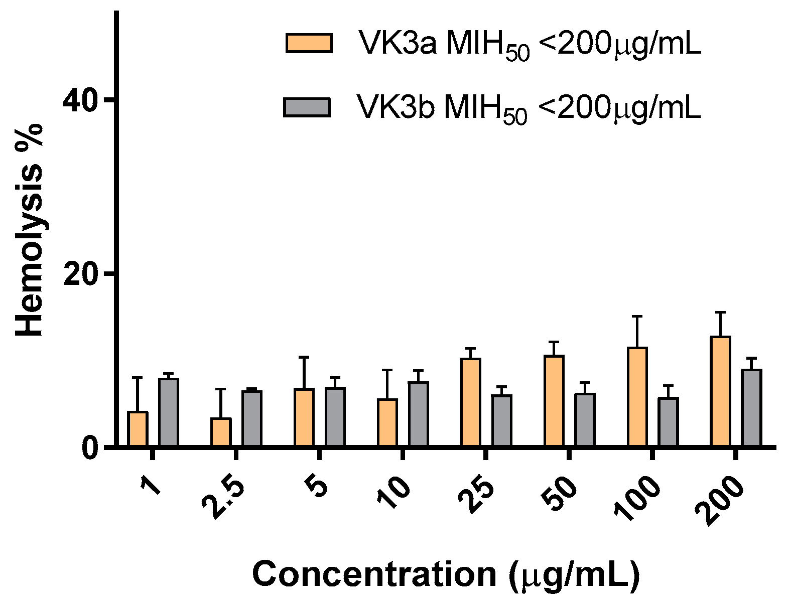
2.2.3. Hemolytic Activity

The toxicity of VK3a and VK3b on erythrocytes was assayed with hemolysis assay. Even at the highest concentrations, which were 70 fold higher than the dose for the toxicity evaluation with NCI-60 cancer cell lines panel, both of the analogs had minimal hemolytic activity and the minimal concentrations inducing 50% hemolysis (MIH50) were <200 µg/mL for VK3a and VK3b (Figure 4). These results indicate that both VK3a and VK3b analogs showed a good safety profile and were proved to be devoid of hemolytic activity in a broad range of concentrations, which also covered their antimicrobial concentrations.

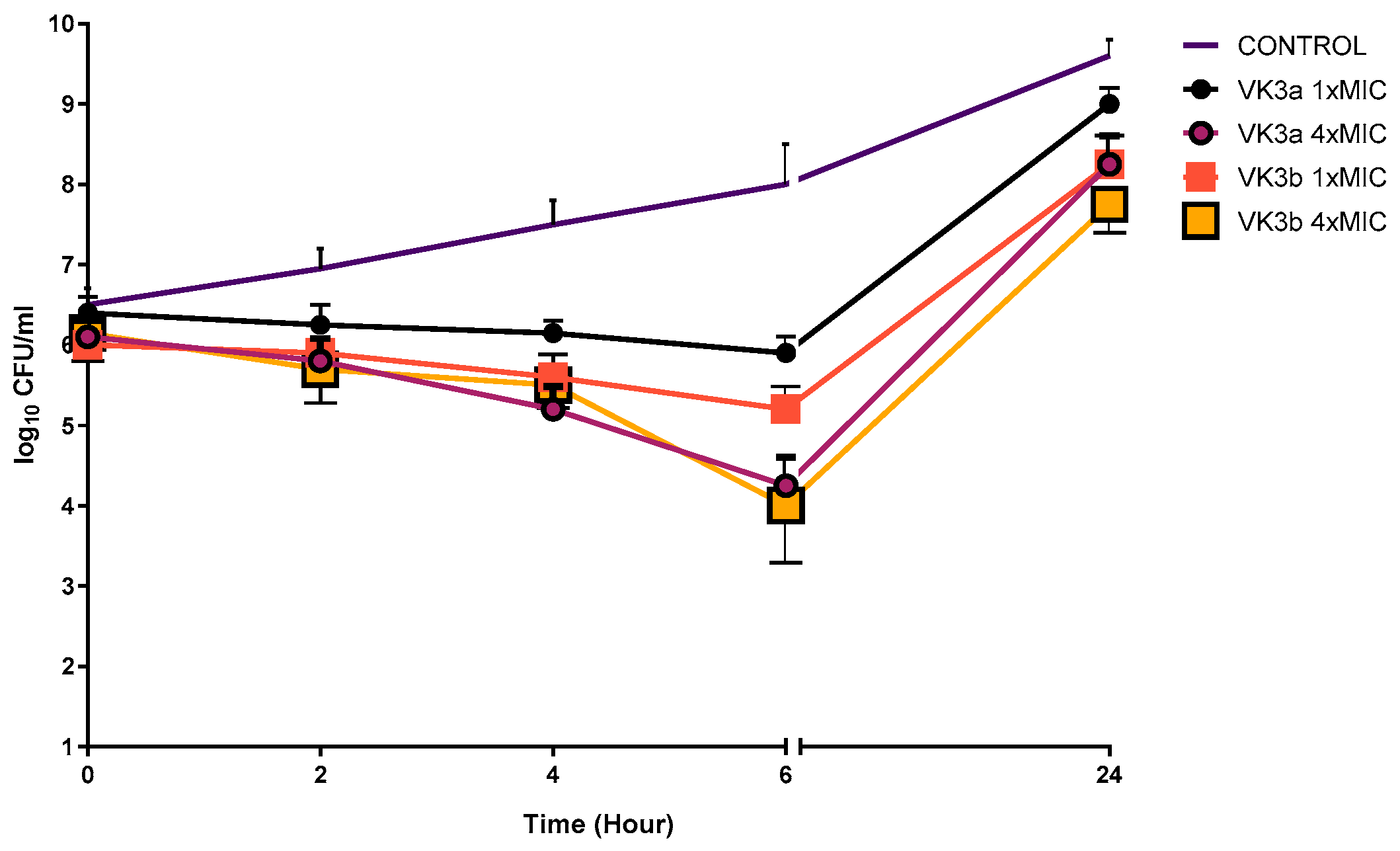
2.2.4. Time-Kill Kinetic Study

Due to their significant in vitro antimicrobial activity results, VK3a and VK3b were chosen between the tested molecules for further investigation of the mode of action. Time-kill studies for VK3a and VK3b were studied on one clinically obtained Methicillin resistant Staphylococcus aureus isolates and the results are given in Figure 5.
According to our Time-kill curve results, VK3a and VK3b did not show bactericidal activity (with a 3-log10 kill determined) against the studied strain at 1× and 4× MIC concentrations within 24 h. (Figure 5). Even if the tested molecules decreased the viable cell count at six hours for the tested strain, within 24 h, bacterial regrowth was seen when the tested molecules were used alone. Apart from our study, Ravichandiran et al. [44,45] showed that newly synthesized 1,4-naphthoquinone molecules had excellent bactericidal activity in a time-dependent manner at 24 h on the studied Escherichia coli and Staphylococcus aureus strains by using the time-kill curve method. The differences between the reduction in the bacterial cell count could be associated with the resistance profile of the tested microorganisms. Due to the promising antimicrobial activity especially on the clinically resistant isolates, even if bacterial regrowth was seen at 24 h in the time-kill curve studies, these molecules provide a chance to try the synergistic activity in combination with antibiotics frequently used in clinics to inhibit bacterial regrowth in studied strains in further investigations.

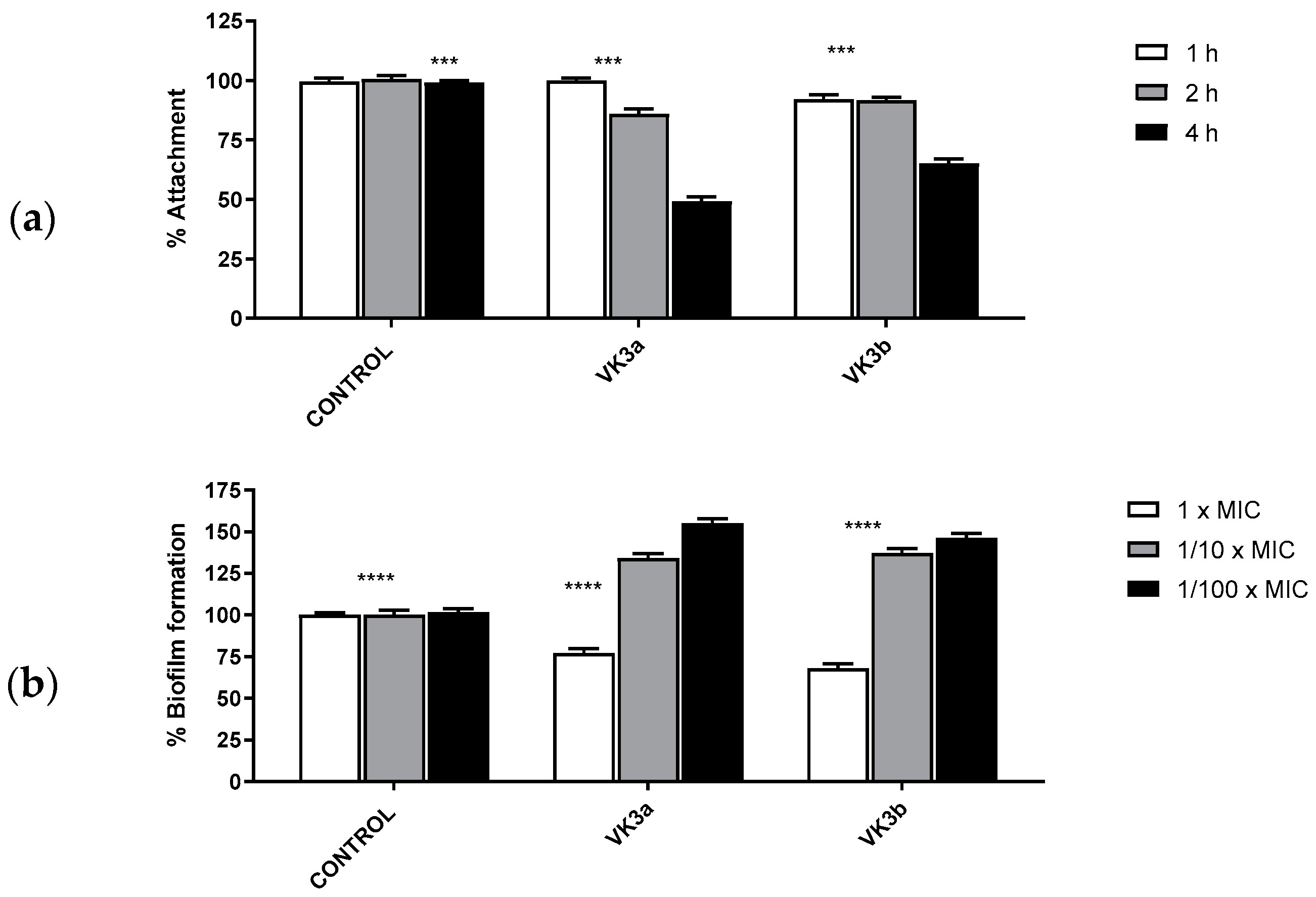
2.2.5. Evaluation of the In Vitro Antibiofilm Activity

The occurrence of many biofilm-based infections and their multiple antimicrobial tolerance are major concerns in healthcare. The elevated rate of resistance to antibiotics in biofilm leads to the discovery of novel anti-biofilm agents. Biofilm formation mainly occurs in four stages: (1) bacterial attachment to a surface; (2) microcolony formation; (3) biofilm maturation; and (4) detachment (also termed dispersal) of bacteria, which may then colonize new areas [53,54]. Due to the increased antimicrobial tolerance with the matured biofilm, it is easier to target the elimination of the biofilm formation at the first two stages.
For this reason, we purposed to determine the antibiofilm effects of VK3a and VK3b against the first two stages of clinically obtained MRSA biofilms. When the 1/10× MICs of tested molecules were examined for 1, 2, or 4 h at 37 °C for MRSA’s adherence to the wells of tissue culture microtiter plates, the tested agents inhibited biofilm attachment processes at least 50% for VK3a at four hours. In general, the inhibition rates of adhesion showed a time-dependent effect for MRSA (Figure 6). When we evaluated the biofilm formation percentage of the studied strains, the rates of biofilm formation inhibition were dependent on concentration, and the highest inhibition rates were shown at 1× MICs for the tested molecules (Figure 6). However, when 1/10× MIC and 1/100× MIC concentrations were used, these molecules acted like substrates for bacterial growth. From the antibiofilm activity results, even if these molecules could achieve an almost 50% decrease in the biofilm attachment process after 4 h for the tested concentrations, at 1/10× MIC and 1/100× MIC (concentrations), these molecules seemed to increase biofilm formation. Our results, similar to Silva et al. [55] and Novais et al. [56] showed time and dose dependent antibiofilm activity against tested isolates.

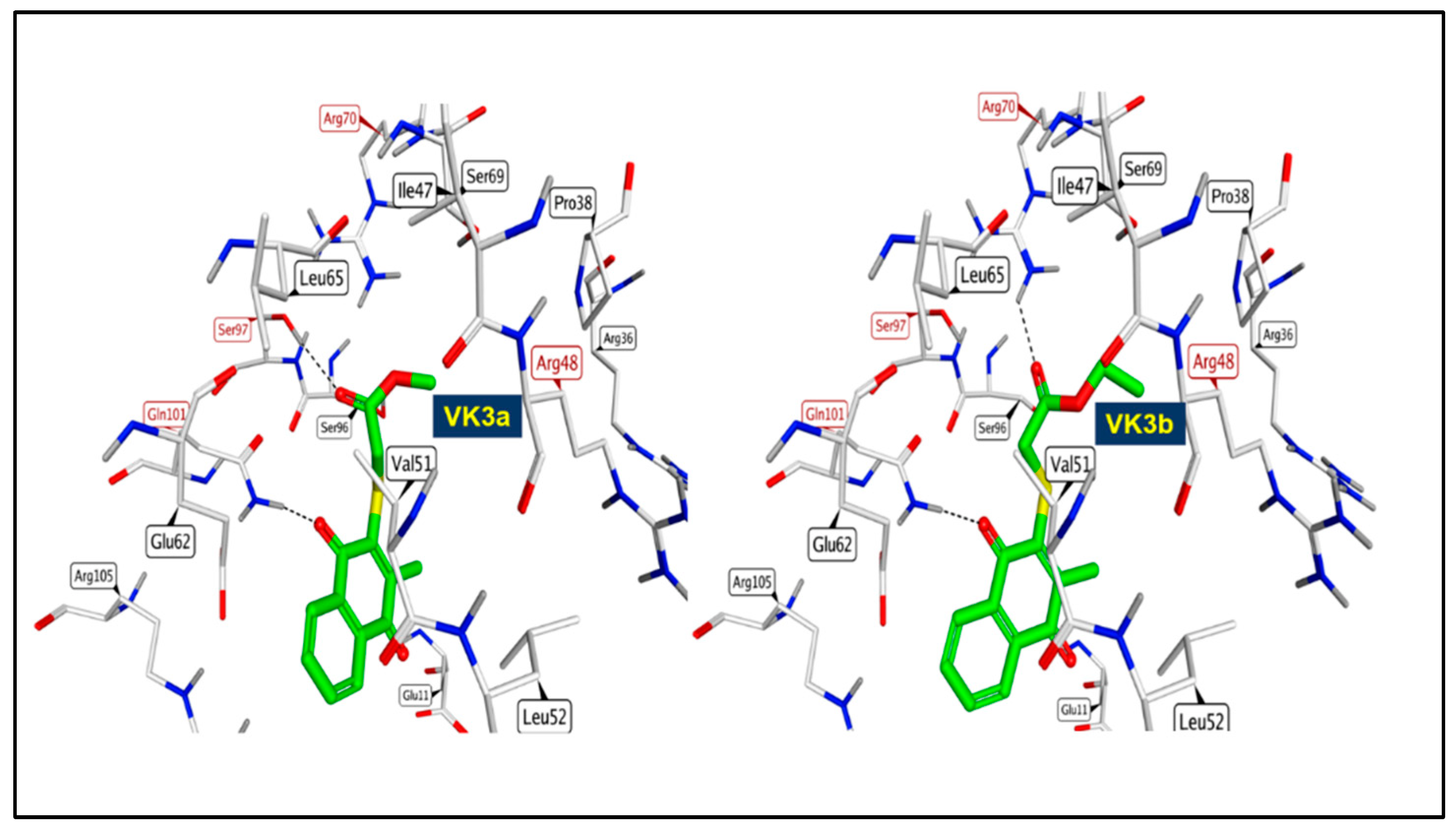
2.2.6. Molecular Docking Study

Thymidylate kinase (TMK) is an enzyme that delivers phosphate from ATP to thymidine monophosphate to produce thymidine diphosphate [57,58]. Inhibition of bacterial TMK function blocks DNA biosynthesis and consequently leads to cell death [59]. To get more insights into the mechanism of action of VK3a and VK3b, we docked them into S. aureus thymidylate kinase (TMK), a nucleotide kinase for a bacterial DNA synthesis pathway and a valid target for the development of new antibacterial drugs [60,61,62]. The thymidine monophosphate site is deep and shares fewer common residues with human TMK, which makes it more druggable than the ATP binding site. Visualization of docked conformations of VK3a and VK3b into TMK from S. aureus showed that they are likely to adopt the same orientation and interactions. Side chain C=O group of VK3a and VK3b are involved in H bonding with key amino acids Ser97 and Arg70, respectively, whereas their C=O of the 1,4 quinone scaffold forms H bonds with Gln101 [63] (Figure 7). However, both compounds lack crucial interactions with Arg48. It seems that the extension of the side chain by using longer R2 leads to an atomic clash with the pocket amino acids, which explains the low activity of VK3g and VK3h.

2.2.7. In Silico Drug-Likeness and ADME Analysis of Selected Thiolated VK3 Analogs

Drug-likeness prediction is a quantitative concept used to study the probability that a chemical compound could be a potential drug. SwissADME server, a free tool for drug-likeness prediction, was employed for calculating several molecular and structural features of VK3a and VK3b. Both compounds can be easily transported in the body due to their low molecular weight. This small molecular weight makes them ideal lead compounds that can be modified to enhance their affinity within the target enzyme. Their octanol-water partition coefficient (log P), which indicates lipophilicity, is in the acceptable range (−0.4–5.6) (Table 9). Their total polar surface area TPSA is less than 160 Å2 indicating good bioavailability. Furthermore, the number of hydrogen bond acceptors (HBA < 10) and the number of hydrogen bond donors (HBD < 5), are in the acceptable ranges (Table 9). SwissADME calculations revealed that the titled compounds have leadlikeness properties [64] and they obey all of Lipinski [65], Ghose [66], Veber [67], Egan [68], and Muegge [69] rules for druglikeness without any violation.
The ADME properties of VK3a and VK3b displayed promising profiles as well. In SwissADME, they use a BOILED-Egg model that showed that both compounds cannot pass through the blood brain barrier (BBB), which helps avoid detrimental effects on the central nervous system (CNS). They are also likely to have a good gastrointestinal (GI) absorption, which is attributed to their ability to be passively absorbed by the GIT. They have quite good solubility and bioavailability scores as shown in Table 9. In addition, they may interfere with the function of the metabolic enzymes cytochrome P450 isoforms CYP1A2, CYP2C19, and CYP2C9 without having an effect on the other isozymes CYP2D6 and CYP3A4.

3. Materials and Methods

3.1. Chemicals and Apparatus

All the solvents and reagents used in the present study were purchased from commercial suppliers with a high purity standard, and used without any purification prior to use. Thin-layer chromatography (TLC) was carried out with silica gel coated aluminum sheets purchased from Merck KGaA. Visualization was achieved using UV light (254 nm). All molecules were purified by column chromatography with Silica gel 60 (63–200 µm particle-sized). Melting points (mp) were measured in a capillary tube in an electrical melting point (Büchi B-540) and are uncorrected. Purified analogs were characterized by nuclear magnetic resonance spectroscopy (1H—500 MHz, 13C—125 MHz). The 1H NMR spectra data is expressed in the form: chemical shifts in units of parts per million (ppm) in CDCl3 and coupling constants (J) are in hertz (Hz). The infrared (IR) spectra of all analogs were obtained on a FTIR spectrometer, using the single reflection diamond ATR module. Mass spectra were performed on a BRUKER Microflex LT equipped with a MALDI (Matrix Assisted Laser Desorption Ionization)-TOF technique by the addition of 1,8,9-anthracenetriol (DIT, dithranol) as a matrix. High-resolution mass spectra electrospray ionization (HRMS-ESI) was obtained on a Waters SYNAPT G1 MS by dissolving analogs (2–3 mg) in acetonitrile. Prior to biological activity, the purity of the thiolated VK3 analogs (VK3a–g) was confirmed by HPLC with hexane/2-propanol = 95:5 as the mobile phase at a flow rate of 1.0 mL/min. Purity of all thiolated VK3 analogs (VK3a–g) was further confirmed to be ≥95% by HPLC analyses with Shimadzu/DGU-20A5 HPLC apparatus fitted with a 25 cm Chiralpac AD-H chiral column. Their chromatograms are provided as Supplementary Material (Figures S8–S14).

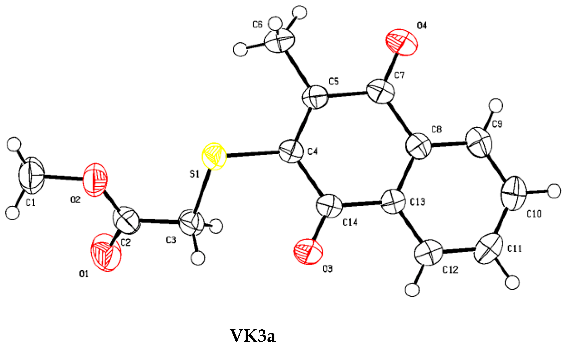
3.2. X-ray Diffraction Analysis

A Bruker APEX II QUAZAR three-circle diffractometer was used to obtain the data for the single crystal compounds. APEX2 was used to perform indexing [70]. SAINT was used for data integration and reduction [71]. Absorption corrections were performed by the multi-scan method implemented in SADABS [72]. The structure was solved by direct methods using the Bruker SHELXTL [73] software package. Structure refinement: full matrix least-squares methods on F2 using SHELXTL all non-hydrogen atoms with anisotropic displacement parameters. Platon software was used to validate the crystal structure and to calculate [74]. The .cif files were processed with Mercury software [75] for the visualization. Table 1 shows the crystallographic and structure refinement data in addition to Table 2, Table 3, Table 4 and Table 5, which present the selected bond lengths, bond angles, torsion angles, hydrogen bond distances, and angles, respectively. The crystallographic data have been deposited with the Cambridge Crystallographic Data Centre as the supplementary publication number is 2150435 for VK3a. Copies of the data can be obtained free of charge on application to CCDC, 12 Union Road, Cambridge CB2 1EZ, UK [fax: +44(1223)336033, e-mail: [email protected]].

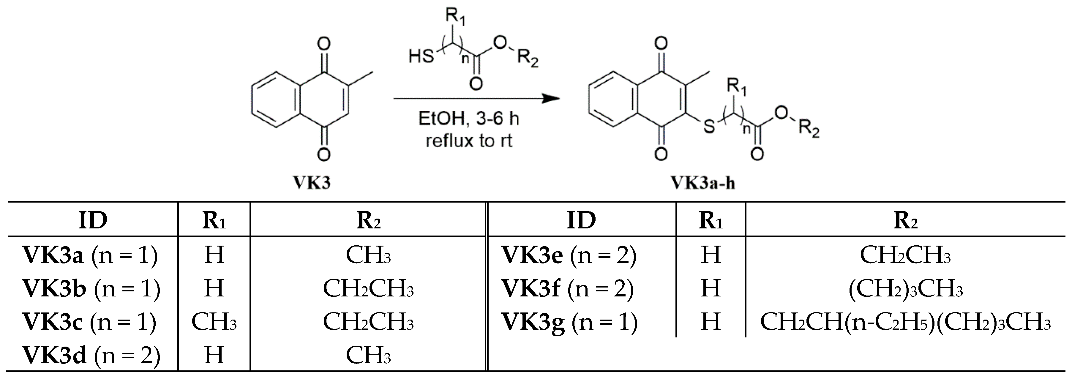
3.3. General Procedure for the Synthesis of the Thiolated Vitamin K3 (VK3) Analogs

To a stirred solution of VK3 (100 mg, 1 eq.) in ethanol (20 mL) was added corresponding thiol (1 eq.) dropwise at room temperature. The reaction mixture was then refluxed until the consumption of the starting material as reported in the literature [47]. The reaction mixture was cooled to an ambient temperature. After the reaction mixture was concentrated under reduced pressure, the residue was dissolved with CH2Cl2 (50 mL), and the solution was washed sequentially with water (3 × 30 mL). The organic layer was dried over CaCl2, filtered, and concentrated under reduced pressure, and the residue was purified by means of column chromatography on silica gel to afford the desired products (VK3 analogs).

3.3.1. Methyl 2-(3-Methyl-1,4-dioxo-1,4-dihydronaphthalen-2-ylthio)acetate (VK3a)

The general procedure was followed using methyl thioglycolate (68 mg, 1 eq.) and VK3. Purification by column chromatography on silica gel (PET/EtAc, v/v 4:1) yielded VK3a (75%) [9] as a yellow crystal, mp 92.5–93.3 °C. FTIR (ATR) υ (cm−1): 2944, 2852 (CHaliphatic), 1721, 1660, 1588, 1562 (>C=O), 1435, 1368, 1289, 1265, 1184, 1112, 1001; 1H NMR (500 MHz, CDCl3) δ (ppm): 8.14–7.98 (m, 2H, CHaromatic), 7.76–7.62 (m, 2H, CHaromatic), 4.00 (s, 2H, SCH2), 3.71 (s, 3H, OCH3), 2.38 (s, 3H, CH3); 13C NMR (125 MHz, CDCl3) δ (ppm): 182.1, 181.1, 169.7 (>C=O), 147.1, 144.5, 133.7, 133.4, 132.7, 132.0, 126.8, 126.6 (CH and Cq), 52.6 (OCH3), 35.0 (SCH2), 15.1 (CH3); HRMS (TOF MS ES+) m/z calcd for C14H13O4S [M + H]+: 277.0535; found: 277.0535.

3.3.2. Ethyl 2-(3-Methyl-1,4-dioxo-1,4-dihydronaphthalen-2-ylthio)acetate (VK3b)

The general procedure was followed using ethyl thioglycolate (77 mg, 1 eq.) and VK3. Purification by column chromatography on silica gel (PET/EtAc, v/v 4:1) yielded VK3b (70%) [76] as a yellow crystal, 68.6–70.2 °C. FTIR (ATR) υ (cm−1): 2985, 2914 (CHaliphatic), 1732, 1661, 1639 (>C=O), 1587, 1548, 1455, 1390, 1310, 1283, 1206, 1150, 1115, 1021; 1H NMR (500 MHz, CDCl3) δ (ppm): 8.21–7.94 (m, 2H, CHaromatic), 7.86–7.58 (m, 2H, CHaromatic), 4.17–4.08 (m, 2H, OCH2), 3.98 (s, 2H, SCH2), 2.39 (s, 3H, CH3), 1.20 (t, J = 7.1 Hz, 3H, CH3); 13C NMR (125 MHz, CDCl3) δ (ppm): 182.1, 181.1, 169.1 (>C=O), 147.1, 144.8, 133.7, 133.4, 132.8, 132.0, 126.8, 126.6 (CH and Cq), 61.6 (OCH2), 35.2 (SCH2), 15.1, 14.1 (CH3); HRMS (TOF MS ES+) m/z calcd for C15H15O4S [M + H]+: 291.0691; found: 291.0707; calcd for C15H14O4SNa [M + Na]+: 313.0511; found: 313.0511.

3.3.3. Ethyl 2-(3-Methyl-1,4-dioxo-1,4-dihydronaphthalen-2-ylthio)propanoate(VK3c)

The general procedure was followed using ethyl 2-mercaptopropanoate (86 mg, 1 eq.) and VK3. Purification by column chromatography on silica gel (PET/EtAc, v/v 4:1) yielded VK3c (92%) as a yellow oil. FTIR (ATR) υ (cm−1): 2981, 2933 (CHaliphatic), 1732, 1661, 1592 (>C=O), 1568, 1448, 1370, 1320, 1280, 1250, 1158, 1062, 1021; 1H NMR (500 MHz, CDCl3) δ (ppm): 8.26–7.92 (m, 2H, CHaromatic), 7.82–7.57 (m, 2H, CHaromatic), 4.70–4.35 (m, 1H, SCH), 4.30–3.89 (m, 2H, OCH2), 2.38 (s, 3H, CH3), 1.58 (d, J = 7.2 Hz, 3H, CH3), 1.08 (t, J = 7.1 Hz, 3H, CH3); 13C NMR (125 MHz, CDCl3) δ (ppm): 182.5, 181.0, 172.0 (>C=O), 148.1, 145.4, 133.7, 133.5, 132.8, 132.0, 126.9, 126.6 (CH and Cq), 61.3 (OCH2), 43.3 (SCH), 16.9, 15.4, 14.0 (CH3); HRMS (TOF MS ES+) m/z calcd for C16H17O4S [M + H]+: 305.0848; found: 305.0849.

3.3.4. Methyl 3-(3-Methyl-1,4-dioxo-1,4-dihydronaphthalen-2-ylthio)propanoate (VK3d)

The general procedure was followed using methyl 3-mercaptopropanoate (77 mg, 1 eq.) and VK3. Purification by column chromatography on silica gel (PET/EtAc, v/v 4:1) yielded VK3d (62%) [77] as a yellow crystal, 76.2–77.9 °C. FTIR (ATR) υ (cm−1): 2996, 2951, 2848 (CHaliphatic), 1717, 1666, 1649 (>C=O), 1590, 1561, 1436, 1370, 1317, 1280, 1245, 1181, 1108; 1H NMR (500 MHz, CDCl3) δ (ppm): 8.20–7.98 (m, 2H, CHaromatic), 7.79–7.62 (m, 2H, CHaromatic), 3.69 (s, 3H, OCH3), 3.46 (t, J = 7.0 Hz, 2H, SCH2), 2.72 (t, J = 6.9 Hz, 2H, CH2), 2.36 (s, 3H, CH3); 13C NMR (125 MHz, CDCl3) δ (ppm): 182.2, 181.2, 171.9 (>C=O), 147.4, 145.8, 133.7, 133.4, 132.8, 132.0, 126.8, 126.6 (CH and Cq), 51.9 (OCH2), 35.5, 29.3 (SCH2CH2), 15.3 (CH3); HRMS (TOF MS ES+) m/z calcd for C15H15O4S [M + H]+: 291.0691; found: 291.0691; calcd for C15H14O4SNa [M + Na]+: 313.0511; found: 313.0512.

3.3.5. Ethyl 3-(3-Methyl-1,4-dioxo-1,4-dihydronaphthalen-2-ylthio)propanoate (VK3e)

The general procedure was followed using ethyl 3-mercaptopropanoate (86 mg, 1 eq.) and VK3. Purification by column chromatography on silica gel (PET/EtAc, v/v 4:1) yielded VK3e (95%) as a yellow oil. FTIR (ATR) υ (cm−1): 2979, 2904 (CHaliphatic), 1726, 1661 (>C=O), 1589, 1553, 1481, 1411, 1371, 1340, 1284, 1254, 1209, 1153, 1108, 1030, 1015; 1H NMR (500 MHz, CDCl3) δ (ppm): 8.17–7.95 (m, 2H, CHaromatic), 7.77–7.62 (m, 2H, CHaromatic), 4.34–3.95 (m, 2H, OCH2), 3.53–3.35 (m, 2H, SCH2), 2.76–2.55 (m, 2H, CH2(C=O)), 2.36 (s, 3H, CH3), 1.31–1.16 (m, 3H, CH3); 13C NMR (125 MHz, CDCl3) δ (ppm): 182.2, 181.1, 171.4 (>C=O), 147.4, 145.9, 133.7, 133.4, 132.8, 132.0, 126.8, 126.6 (CH and Cq), 60.8 (OCH2CH3), 35.7 (SCH2), 29.3 (SCH2(C=O)), 15.3, 14.2 (CH3); HRMS (TOF MS ES+) m/z calcd for C16H17O4S [M + H]+: 305.0848; found: 305.0847; calcd for C16H16O4SNa [M + Na]+: 327.0667; found: 327.0673.

3.3.6. Butyl 3-(3-Methyl-1,4-dioxo-1,4-dihydronaphthalen-2-ylthio)propanoate (VK3f)

The general procedure was followed using butyl 3-mercaptopropanoate (104 mg, 1 eq.) and VK3. Purification by column chromatography on silica gel (PET/EtAc, v/v 4:1) yielded VK3f (66%) as a yellow oil. FTIR (ATR) υ (cm−1): 2959, 2930, 2867 (CHaliphatic), 1731, 1661, 1592 (>C=O), 1559, 1459, 1420, 1322, 1280, 1248, 1180, 1032; 1H NMR (500 MHz, CDCl3) δ (ppm): 8.20–8.00 (m, 2H, CHaromatic), 7.83–7.62 (m, 2H, CHaromatic), 4.18–3.97 (m, 2H, OCH2), 3.52–3.33 (m, 2H, SCH2), 2.80–2.62 (m, 2H, CH2(C=O)), 2.36 (s, 3H, CH3), 1.69–1.51 (m, 2H, CH2), 1.44–1.18 (m, 2H, CH2), 1.03–0.76 (m, 3H, CH3); 13C NMR (125 MHz, CDCl3) δ (ppm): 182.2, 181.2, 171.5 (>C=O), 147.4, 145.8, 133.7, 133.4, 132.8, 132.0, 126.8, 126.6 (CH and Cq), 64.7 (OCH2), 35.7 (SCH2), 30.6 (SCH2(C=O)), 29.3, 19.1 (CH2), 15.3, 13.7 (CH3); HRMS (TOF MS ES+) m/z calcd for C18H21O4S [M + H]+: 333.1161; found: 333.1170; calcd for C18H20O4SNa [M + Na]+: 355.0980; found: 355.0988.

3.3.7. 2-Ethylhexyl 2-(3-Methyl-1,4-dioxo-1,4-dihydronaphthalen-2-ylthio)acetate (VK3g)

The general procedure was followed using 2-ethylhexyl thioglycolate (131 mg, 1 eq.) and VK3. Purification by column chromatography on silica gel (PET/EtAc, v/v 4:1) yielded VK3g (72%) as a yellow oil. FTIR (ATR) υ (cm−1): 2959, 2929, 2863 (CHaliphatic), 1732, 1661, 1592 (>C=O), 1564, 1461, 1320, 1280, 1159, 1032; 1H NMR (500 MHz, CDCl3) δ (ppm): 8.21–7.88 (m, 2H, CHaromatic), 7.86–7.60 (m, 2H, CHaromatic), 4.26–3.89 (m, 4H, OCH2+SCH2), 2.38 (s, 3H, CH3), 1.54–1.43 (m, 1H, CH), 1.35–1.09 (m, 8H, CH2), 0.86 (t, J = 6.7 Hz, 3H, CH3), 0.80 (t, J = 7.4 Hz, 3H, CH3); 13C NMR (125 MHz, CDCl3) δ (ppm): 182.0, 181.1, 169.4 (>C=O), 146.8, 144.8, 133.7, 133.4, 132.8, 132.0, 126.8, 126.6 (CH and Cq), 68.0 (OCH2), 38.7 (SCH2), 35.2 (CH), 30.2, 28.8, 23.6, 22.9 (CH2), 15.1, 14.0, 10.9 (CH3); HRMS (TOF MS ES+) m/z calcd for C21H27O4S [M + H]+: 375.1630; found: 375.1630.

3.4. Biological Evaluation

3.4.1. MIC Evaluation

MICs of the molecules were examined by the broth microdilution technique approved by the Clinical and Laboratory Institute (CLSI) [48,49]. In total, ten different standard ATCC isolates were prepared according to the CLSI recommendations. The stock solutions of the tested molecules were prepared in DMSO. Serial two-fold dilutions ranging from 1250 to 0.06 μg/mL were prepared in Mueller Hinton Broth for the tested bacteria and an RPMI-1640 medium for the yeast, respectively.
According to the antimicrobial activity results, we aimed to identify in vitro activities of the VK3a and VK3b against clinically obtained strains by the broth microdilution dilution technique, as described by the CLSI recommendations [48,49]. For this assay, 20 nonduplicates, nosocomially acquired Methicillin-Resistant Staphylococcus aureus isolated from blood specimens between April and September 2017 were obtained from the Department of Infectious Diseases and Clinical Microbiology, Faculty of Medicine, Istanbul Medipol University. All strains were identified using API STAPH (bioM’erieux). Then, all the tested S. aureus isolates were chosen by using Oxacillin susceptibility to determine the Methicillin resistant isolates, approved by CLSI (MIC ≥ 4 µg/mL) [48]. The MIC was defined as the lowest concentration of tested extracts, giving complete inhibition of visible growth. Experiments were performed in triplicate. MBCs were determined at the end of the incubation period by removing two 0.01 mL samples from each well demonstrating no visible growth and plated onto TSA. Resultant colonies were counted after an overnight incubation at 37 °C. The MBC was defined as the lowest concentration of molecules giving at least a 99.9% killing of the initial inoculums [78].

3.4.2. Determination of Time-Kill Curves

For the evaluation of the bactericidal activity of the selected molecules (VK3a and VK3b), the time-kill curve (TKC) method was performed at one and four times the MIC against one (1) MRSA clinical strains. Molecule-free controls were included for the tested strain. The inocula were quantified spectrophotometrically and added to the flasks to yield a final concentration of 1 × 10⁶ CFU/mL. The test tubes containing MHB with and without (growth control) molecules in a final volume of 10 mL were incubated in a 37 °C calibrated shaking water bath, and viable counts were determined at 0, 2, 4, 6, and 24 h intervals after inoculation, by subculturing 0.1 mL serial dilutions onto TSA plates. All tests were performed in duplicate. The lower limit of detection for the time-kill assay was 1log10 CFU/mL. Bactericidal activity was defined as a ≥3 log10 CFU/mL decrease from the initial inoculum.

3.4.3. Determination of the Antibiofilm Activities

Biofilm attachment and inhibition of biofilm formation assays were performed by the previously described method with some modifications [36]. For biofilm attachment, an overnight culture of strong, biofilm-producing clinically MRSA isolate was made, which was diluted 1/50 to obtain 1 × 10⁶–1 × 10⁷ CFU/200 mL for bacteria in TSB supplemented with 1% glucose. Then the strain was added to each well of 96-well tissue culture microtiter plates with 1/10× MIC of tested molecules. The plates were allowed to incubate for 1, 2, and 4 h at 37 °C. The positive control was the studied strain using only the medium. After incubation, each well was washed with PBS solution three times and measured at OD 595 nm.
For inhibition of the biofilm formation, the tested strain was incubated in its medium containing the molecule at 1× and 1/10× in addition to 1/100× MIC at 37 °C for 24 h in microtiter plates. Six wells were used for each molecule. The positive control was the tested strain in its medium without molecules. After incubation, each well was washed with PBS solution three times and measured at OD 595 nm.

3.4.4. Hemolysis Assay

Hemolytic activities of the VK3a and VK3b analogs were detected as previously reported with some modifications [79]. In brief, 2% human red erythrocyte suspension was prepared with PBS. Then, 1, 2.5, 5, 10, 25, 50, 100, and 200 µg/mL VK3a and VK3b were added to the erythrocyte suspension. The positive control erythrocyte suspension was prepared with distilled water and the negative control was prepared in 1% DMSO. The samples were incubated at 37 °C for 1 h. After centrifugation at 1500 rpm for 5 min, the resulting supernatants were transferred to a 96-well plate and the absorbance was read at 405 nm using a microplate reader (Epoc, BioTek). The percent of the hemolysis was calculated as follows: Hemolysis % = [(OD405nm(analogs) − OD405nm(negative control))/(OD405nm(positive control) − OD405nm(negative control))].

3.5. Statistical Analysis

All experiments were performed in two independent assays. Two-way ANOVA-Tukey’s multiple comparison test was used to compare differences between control and antimicrobials treated biofilms. p value < 0.005 was considered as statistically significant.

3.6. Molecular Docking

TMK X-ray crystal structure (PDB code 4GFD) [58] was retrieved from Protein Data Bank to be utilized as a model in the present study. The protein structure was prepared using QuickPrep module of MOE (Version 2019.01, Chemical Computing Group Inc., Montreal, QC, Canada). Only one monomer, chain A, was selected and water residues were deleted. The docking study was conducted using the rigid-receptor method [80]. The co-crystallized ligand TK-666 was defined as the center of the binding site. Using the MOE build suite, the chemical structures were drawn, and then energy-minimized using the MOE default force field [81]. All other docking options were kept at their default values. Fifteen docking positions were generated for each ligand. The generated docking positions were visualized using MOE.

3.7. In Silico Drug-Likeness and ADMET Analysis

The online SwissADME database was used to predict the drug-likeness and ADME parameters [82].

4. Conclusions

Today, antimicrobial resistance is still a main health problem in the world. Although many antibiotic drugs with adverse effects are available, novel drugs with different activity mechanisms are needed as soon as possible. In summary, a series of the thiolated VK3 analogs (VK3a–g) were designed and synthesized by a simple, fast, high-efficiency, and applicable method. In vitro antimicrobial activity assays were performed on seven different bacterial strains in addition to three different fungal strains by the serial broth microdilution method. The obtained results indicated that introducing alkyl chain thiols into the VK3 core led to an improvement in the antibacterial activity of the VK3 analogs. Of these, two thiolated VK3 analogs (VK3a and VK3b) were also tested for additional studies to reveal the antimicrobial and safety profile. Furthermore, they were tested for understanding the mode of action with evaluation by the time-kill kinetic study. Furthermore, we carried out another study on two analogs, active on S. aureus and E. faecalis, via determining their biofilm inhibition capacities. The present study is the first record to evaluate the antibiofilm and bactericidal profile against clinically resistant species. In line with this, VK3b showed the most potent antimicrobial activity against the clinically obtained MRSA strains with 4.88 µg/mL MIC50 value. Therefore, in accordance with the time-kill curve study results, these two molecules, VK3a and VK3b, showed bacterial regrowth at 1× and 4× MIC concentrations used within 24 h against the Methicillin-Resistant S. aureus isolate, and these molecules decreased the viable cell count at 6 h. Therefore, to achieve synergistic activity, it would be possible to use these molecules in combination with conventional antibiotics, for reducing antibiotic toxicity and antimicrobial resistance. Furthermore, VK3a and VK3b had no obvious hemolytic activity. The molecular docking of VK3a and VK3b into S. aureus thymidylate kinase showed similar placement and interactions with critical amino acids. Their interesting leadlikeness and pharmacokinetics make them worthy of further attention. Although finding safe and effective antimicrobial drugs are quite difficult, it is indeed necessary to conduct these studies continuously, especially in the era of antimicrobial resistance.

Supplementary Materials

The following supporting information can be downloaded at: https://www.mdpi.com/article/10.3390/ph15050586/s1, Figure S1: The cell growth percentage (GP) of VK3a; Figure S2: The cell growth percentage (GP) of VK3b; Figure S3: The cell growth percentage (GP) of VK3c; Figure S4: The cell growth percentage (GP) of VK3d; Figure S5: The cell growth percentage (GP) of VK3e; Figure S6: The cell growth percentage (GP) of VK3f; Figure S7: The cell growth percentage (GP) of VK3g; Figure S8: Purity chromatogram of the VK3a; Figure S9: Purity chromatogram of the VK3b; Figure S10: Purity chromatogram of the VK3c; Figure S11: Purity chromatogram of the VK3d; Figure S12: Purity chromatogram of the VK3e; Figure S13: Purity chromatogram of the VK3f; Figure S14: Purity chromatogram of the VK3g; Figure S15: 1H NMR (500 MHz) spectrum of the VK3a in CDCl3-d1; Figure S16: 13C NMR (125 MHz) spectrum of the VK3a in CDCl3-d1; Figure S17: 1H NMR (500 MHz) spectrum of the VK3b in CDCl3-d1; Figure S18: 13C NMR (125 MHz) spectrum of the VK3b in CDCl3-d1; Figure S19: 1H NMR (500 MHz) spectrum of the VK3c in CDCl3-d1; Figure S20: 13C NMR (125 MHz) spectrum of the VK3c in CDCl3-d1; Figure S21: 1H NMR (500 MHz) spectrum of the VK3d in CDCl3-d1; Figure S22: 13C NMR (125 MHz) spectrum of the VK3d in CDCl3-d1; Figure S23: 1H NMR (500 MHz) spectrum of the VK3e in CDCl3-d1; Figure S24: 13C NMR (125 MHz) spectrum of the VK3e in CDCl3-d1; Figure S25: 1H NMR (500 MHz) spectrum of the VK3f in CDCl3-d1; Figure S26: 13C NMR (125 MHz) spectrum of the VK3f in CDCl3-d1; Figure S27: 1H NMR (500 MHz) spectrum of the VK3g in CDCl3-d1; Figure S28: 13C NMR (125 MHz) spectrum of the VK3g in CDCl3-d1; Table S1: Bond lengths (Å) for VK3a; Table S2: Bond angles (°) for VK3a; Table S3: Torsion angles (°) for VK3a.

Author Contributions

Conceptualization, E.M.-K., M.O., M.F. and A.F.T.; Data curation, H.Y., M.Y., N.B., E.M.-K., M.O.R., A.T.J., M.O., M.F. and A.F.T.; Formal analysis, H.Y., M.Y., N.B., E.M.-K., M.O.R., A.T.J. and A.F.T.; Funding acquisition, A.F.T.; Investigation, H.Y., M.Y., N.B., E.M.-K., M.O.R., A.T.J. and A.F.T.; Methodology, H.Y., E.M.-K., A.T.J. and A.F.T.; Project administration, A.F.T.; Resources, E.M.-K., M.O., M.F. and A.F.T.; Software, H.Y., E.M.-K., M.O.R., M.O., M.F. and A.F.T.; Validation, H.Y., E.M.-K., M.O.R. and A.T.J.; Supervision, A.F.T.; Visualization, H.Y., M.Y., N.B., E.M.-K., M.O.R., A.T.J., M.O., M.F. and A.F.T.; Writing—review & editing, H.Y., M.Y., N.B., E.M.-K., M.O.R., A.T.J., M.O., M.F. and A.F.T.; Writing—original draft, E.M.-K., M.O.R. and A.F.T. All authors have read and agreed to the published version of the manuscript.

Funding

This work was financially supported by the Scientific Research Projects Coordination Unit of Istanbul University (Project numbers: FBA-2021-37260) for supplying the equipment and materials.

Institutional Review Board Statement

Not applicable.

Informed Consent Statement

Not applicable.

Data Availability Statement

Data is contained within the article and supplementary material.

Acknowledgments

The authors present their thanks to the National Cancer Institute (NCI), Bethesda, Maryland, USA for carrying out the antiproliferative activity by the Developmental Therapeutics Program (DTP), Division of Cancer Treatment and Diagnosis, National Cancer Institute (http://dtp.cancer.gov).

Conflicts of Interest

The authors declare no conflict of interest.

References

  1. Fairfield, K.M.; Fletcher, R.H. Vitamins for chronic disease prevention in adults: Scientific review. JAMA 2002, 287, 3116–3126. [Google Scholar] [CrossRef] [PubMed]
  2. Rezaei, H.; Khiali, S.; Rezaei, H.; Rezaee, H.; Bannazadeh Baghi, H.; Pourghasem, M.; Entezari-Maleki, T. Potential Roles of Vitamins in the Management of COVID-19: A Comprehensive Review. Pharm. Sci. 2021, 27, S29–S49. [Google Scholar] [CrossRef]
  3. Jovic, T.H.; Ali, S.R.; Ibrahim, N.; Jessop, Z.M.; Tarassoli, S.P.; Dobbs, T.D.; Holford, P.; Thornton, C.A.; Whitaker, I.S. Could Vitamins Help in the Fight Against COVID-19? Nutrients 2020, 12, 2550. [Google Scholar] [CrossRef] [PubMed]
  4. Li, X.; Wang, G.; Chen, D.; Lu, Y. β-Carotene and astaxanthin with human and bovine serum albumins. Food Chem. 2015, 179, 213–221. [Google Scholar] [CrossRef] [PubMed]
  5. Bourassa, P.; Hasni, I.; Tajmir-Riahi, H.A. Folic acid complexes with human and bovine serum albumins. Food Chem. 2011, 129, 1148–1155. [Google Scholar] [CrossRef] [PubMed]
  6. Xu, H.; Liu, Q.; Zuo, Y.; Bi, Y.; Gao, S. Spectroscopic Studies on the Interaction of Vitamin C with Bovine Serum Albumin. J. Solut. Chem. 2009, 38, 15–25. [Google Scholar] [CrossRef]
  7. Haddad, J.G. Human serum binding protein for vitamin D and its metabolites (DBP): Evidence that actin is the DBP binding component in human skeletal muscle. Arch. Biochem. Biophys. 1982, 213, 538–544. [Google Scholar] [CrossRef]
  8. Dahlbäck, B. Purification of human vitamin K-dependent protein S and its limited proteolysis by thrombin. Biochem. J. 1983, 209, 837–846. [Google Scholar] [CrossRef] [Green Version]
  9. Suganthi, M.; Elango, K.P. Synthesis, characterization and serum albumin binding studies of vitamin K3 derivatives. J. Photochem. Photobiol. B 2017, 166, 126–135. [Google Scholar] [CrossRef]
  10. Hamidi, M.S.; Cheung, A.M. Vitamin K and musculoskeletal health in postmenopausal women. Mol. Nutr. Food Res. 2014, 58, 1647–1657. [Google Scholar] [CrossRef]
  11. Scheiber, D.; Veulemans, V.; Horn, P.; Chatrou, M.L.; Potthoff, S.A.; Kelm, M.; Schurgers, L.J.; Westenfeld, R. High-Dose Menaquinone-7 Supplementation Reduces Cardiovascular Calcification in a Murine Model of Extraosseous Calcification. Nutrients 2015, 7, 6991–7011. [Google Scholar] [CrossRef] [PubMed] [Green Version]
  12. Yerramsetti, N.; Dampanaboina, L.; Mendu, V.; Battula, S. Synergistic factors ensue high expediency in the synthesis of menaquinone [K-2] analogue MK-6: Application to access an efficient one-pot protocol to MK-9. Tetrahedron 2020, 76, 131696. [Google Scholar] [CrossRef]
  13. Li, X.Y.; Himes, R.A.; Prosser, L.C.; Christie, C.F.; Watt, E.; Edwards, S.F.; Metcalf, C.S.; West, P.J.; Wilcox, K.S.; Chan, S.S.L.; et al. Discovery of the First Vitamin K Analogue as a Potential Treatment of Pharmacoresistant Seizures. J. Med. Chem. 2020, 63, 5865–5878. [Google Scholar] [CrossRef] [PubMed]
  14. Wellington, K.W.; Hlatshwayo, V.; Kolesnikova, N.I.; Saha, S.T.; Kaur, M.; Motadi, L.R. Anticancer activities of vitamin K3 analogues. Investig. New Drug 2020, 38, 378–391. [Google Scholar] [CrossRef] [PubMed]
  15. Doroshow, J.H. Effect of Anticancer Quinones on Reactive Oxygen Production by Adult Rat Heart Myocytes. Oxidative Med. Cell. Longev. 2020, 2020, 8877100. [Google Scholar] [CrossRef]
  16. Bauer, H.; Fritz-Wolf, K.; Winzer, A.; Kuhner, S.; Little, S.; Yardley, V.; Vezin, H.; Palfey, B.; Schirmer, R.H.; Davioud-Charvet, E. A fluoro analogue of the menadione derivative 6-[2′-(3′-methyl)-1′,4′-naphthoquinolyl]hexanoic acid is a suicide substrate of glutathione reductase. Crystal structure of the alkylated human enzyme. J. Am. Chem. Soc. 2006, 128, 10784–10794. [Google Scholar] [CrossRef] [PubMed]
  17. Patel, O.P.S.; Beteck, R.M.; Legoabe, L.J. Antimalarial application of quinones: A recent update. Eur. J. Med. Chem. 2021, 210, 113084. [Google Scholar] [CrossRef]
  18. Majiene, D.; Kuseliauskyte, J.; Stimbirys, A.; Jekabsone, A. Comparison of the Effect of Native 1,4-Naphthoquinones Plumbagin, Menadione, and Lawsone on Viability, Redox Status, and Mitochondrial Functions of C6 Glioblastoma Cells. Nutrients 2019, 11, 1294. [Google Scholar] [CrossRef] [Green Version]
  19. Deniz, N.G.; Abdassalam, A.F.S.; Ozyurek, M.; Yesil, E.A.; Sayil, C. New vitamin K3 (menadione) analogues: Synthesis, characterization, antioxidant and catalase inhibition activities. J. Chem. Sci. 2020, 132, 138. [Google Scholar] [CrossRef]
  20. Hileman, E.O.; Liu, J.S.; Albitar, M.; Keating, M.J.; Huang, P. Intrinsic oxidative stress in cancer cells: A biochemical basis for therapeutic selectivity. Cancer Chemoth. Pharm. 2004, 53, 209–219. [Google Scholar] [CrossRef]
  21. Tetef, M.; Margolin, K.; Ahn, C.; Akman, S.; Chow, W.; Leong, L.; Morgan, R.J., Jr.; Raschko, J.; Somlo, G.; Doroshow, J.H. Mitomycin C and menadione for the treatment of lung cancer: A phase II trial. Investig. New Drugs 1995, 13, 157–162. [Google Scholar] [CrossRef] [PubMed]
  22. Osada, S.; Tomita, H.; Tanaka, Y.; Tokuyama, Y.; Tanaka, H.; Sakashita, F.; Takahashi, T. The utility of vitamin K3 (menadione) against pancreatic cancer. Anticancer Res. 2008, 28, 45–50. [Google Scholar] [PubMed]
  23. Verrax, J.; Cadrobbi, J.; Marques, C.; Taper, H.; Habraken, Y.; Piette, J.; Calderon, P.B. Ascorbate potentiates the cytotoxicity of menadione leading to an oxidative stress that kills cancer cells by a non-apoptotic caspase-3 independent form of cell death. Apoptosis 2004, 9, 223–233. [Google Scholar] [CrossRef] [PubMed]
  24. Suresh, S.; Raghu, D.; Karunagaran, D. Menadione (Vitamin K3) Induces Apoptosis of Human Oral Cancer Cells and Reduces their Metastatic Potential by Modulating the Expression of Epithelial to Mesenchymal Transition Markers and Inhibiting Migration. Asian Pac. J. Cancer P 2013, 14, 5461–5465. [Google Scholar] [CrossRef] [PubMed] [Green Version]
  25. Ngo, E.O.; Sun, T.P.; Chang, J.Y.; Wang, C.C.; Chi, K.H.; Cheng, A.L.; Nutter, L.M. Menadione-Induced DNA Damage in a Human Tumor-Cell Line. Biochem. Pharmacol. 1991, 42, 1961–1968. [Google Scholar] [CrossRef]
  26. Zhang, Z.; Ding, Y.; Li, J.; Wang, L.; Xin, X.; Yan, J.; Wu, J.; Yuan, A.; Hu, Y. Versatile iron-vitamin K3 derivative-based nanoscale coordination polymer augments tumor ferroptotic therapy. Nano Res. 2021, 14, 2398–2409. [Google Scholar] [CrossRef]
  27. Bayrak, N.; Ciftci, H.I.; Yildiz, M.; Yildirim, H.; Sever, B.; Tateishi, H.; Otsuka, M.; Fujita, M.; Tuyun, A.F. Structure based design, synthesis, and evaluation of anti-CML activity of the quinolinequinones as LY83583 analogs. Chem. Biol. Interact. 2021, 345, 109555. [Google Scholar] [CrossRef]
  28. Mataraci-Kara, E.; Bayrak, N.; Yildirim, H.; Yildiz, M.; Ataman, M.; Ozbek-Celik, B.; Tuyun, A.F. Plastoquinone analogs: A potential antimicrobial lead structure intensely suppressing Staphylococcus epidermidis and Candida albicans growth. Med. Chem. Res. 2021, 30, 1728–1737. [Google Scholar] [CrossRef]
  29. Jannuzzi, A.T.; Yıldız, M.; Bayrak, N.; Yıldırım, H.; Shilkar, D.; Jayaprakash, V.; TuYuN, A.F. Anticancer agents based on Plastoquinone analogs with N-phenylpiperazine: Structure-activity relationship and mechanism of action in breast cancer cells. Chem.-Biol. Interact. 2021, 349, 109673. [Google Scholar] [CrossRef]
  30. Yildirim, H. Arylthio analogs of 5,8-quinolinedione: Synthesis, characterization, antibacterial, and antifungal evaluations. Phosphorus Sulfur 2020, 195, 516–522. [Google Scholar] [CrossRef]
  31. Yildirim, H. Synthesis, characterization, and biological evaluation of a set of new alkylthio substituted plastoquinones containing ester group. J. Mol. Struct. 2020, 1203, 127433. [Google Scholar] [CrossRef]
  32. Ibis, C.; Tuyun, A.F.; Bahar, H.; Ayla, S.S.; Stasevych, M.V.; Musyanovych, R.Y.; Komarovska-Porokhnyavets, O.; Novikov, V. Synthesis of novel 1,4-naphthoquinone derivatives: Antibacterial and antifungal agents. Med. Chem. Res. 2013, 22, 2879–2888. [Google Scholar] [CrossRef]
  33. Ibis, C.; Tuyun, A.F.; Bahar, H.; Ayla, S.S.; Stasevych, M.V.; Musyanovych, R.Y.; Komarovska-Porokhnyavets, O.; Novikov, V. Nucleophilic substitution reactions of 1,4-naphthoquinone and biologic properties of novel S-, S,S-, N-, and N,S-substituted 1,4-naphthoquinone derivatives. Med. Chem. Res. 2014, 23, 2140–2149. [Google Scholar] [CrossRef]
  34. Espinosa-Bustos, C.; Vázquez, K.; Varela, J.; Cerecetto, H.; Paulino, M.; Segura, R.; Pizarro, J.; Vera, B.; González, M.; Zarate, A.M.; et al. New aryloxy-quinone derivatives with promising activity on Trypanosoma cruzi. Arch. Pharm. 2020, 353, 1900213. [Google Scholar] [CrossRef] [PubMed]
  35. De Almeida, P.D.O.; Jobim, G.D.B.; Ferreira, C.C.D.; Bernardes, L.R.; Dias, R.B.; Sales, C.B.S.; Valverde, L.D.; Rocha, C.A.G.; Soares, M.B.P.; Bezerra, D.P.; et al. A new synthetic antitumor naphthoquinone induces ROS-mediated apoptosis with activation of the JNK and p38 signaling pathways. Chem.-Biol. Interact. 2021, 343, 109444. [Google Scholar] [CrossRef]
  36. Mataraci-Kara, E.; Bayrak, N.; Yildiz, M.; Yildirim, H.; TuYu, N.A. Active Quinolinequinones against Methicillin-Resistant Staphylococcus spp. Chem. Biodivers 2021, 19, e202100616. [Google Scholar] [CrossRef]
  37. Ciftci, H.I.; Bayrak, N.; Yıldız, M.; Yıldırım, H.; Sever, B.; Tateishi, H.; Otsuka, M.; Fujita, M.; Tuyun, A.F. Design, synthesis and investigation of the mechanism of action underlying anti-leukemic effects of the quinolinequinones as LY83583 analogs. Bioorg. Chem. 2021, 114, 105160. [Google Scholar] [CrossRef]
  38. Bayrak, N.; Yildiz, M.; Yildirim, H.; Mataraci-Kara, E.; Tuyun, A.F. Novel plastoquinone analogs containing benzocaine and its analogs: Structure-based design, synthesis, and structural characterization. Res. Chem. Intermediat. 2021, 47, 2125–2141. [Google Scholar] [CrossRef]
  39. Bayrak, N.; Yildiz, M.; Yildirim, H.; Kara, E.M.; Celik, B.O.; Tuyun, A.F. Brominated plastoquinone analogs: Synthesis, structural characterization, and biological evaluation. J. Mol. Struct. 2020, 1219, 128560. [Google Scholar] [CrossRef]
  40. Wellington, K.W.; Kolesnikova, N.I.; Nyoka, N.B.P.; McGaw, L.J. Investigation of the antimicrobial and anticancer activity of aminonaphthoquinones. Drug Dev. Res. 2019, 80, 138–146. [Google Scholar] [CrossRef] [Green Version]
  41. Wellington, K.W. Understanding cancer and the anticancer activities of naphthoquinones—A review. Rsc. Adv. 2015, 5, 20309–20338. [Google Scholar] [CrossRef]
  42. Ryu, C.K.; Song, A.L.; Lee, J.Y.; Hong, J.A.; Yoon, J.H.; Kim, A. Synthesis and antifungal activity of benzofuran-5-ols. Bioorg. Med. Chem. Lett. 2010, 20, 6777–6780. [Google Scholar] [CrossRef] [PubMed]
  43. Ibis, C.; Ozsoy-Gunes, Z.; Tuyun, A.F.; Ayla, S.S.; Bahar, H.; Stasevych, M.V.; Musyanovych, R.Y.; Komarovska-Porokhnyavets, O.; Novikov, V. Synthesis, antibacterial and antifungal evaluation of thio- or piperazinyl-substituted 1,4-naphthoquinone derivatives. J. Sulfur. Chem. 2016, 37, 477–487. [Google Scholar] [CrossRef]
  44. Ravichandiran, P.; Maslyk, M.; Sheet, S.; Janeczko, M.; Premnath, D.; Kim, A.R.; Park, B.H.; Han, M.K.; Yoo, D.J. Synthesis and Antimicrobial Evaluation of 1,4-Naphthoquinone Derivatives as Potential Antibacterial Agents. Chemistryopen 2019, 8, 589–600. [Google Scholar] [CrossRef] [PubMed] [Green Version]
  45. Ravichandiran, P.; Sheet, S.; Premnath, D.; Kim, A.R.; Yoo, D.J. 1,4-Naphthoquinone Analogues: Potent Antibacterial Agents and Mode of Action Evaluation. Molecules 2019, 24, 1437. [Google Scholar] [CrossRef] [PubMed] [Green Version]
  46. Ravichandiran, P.; Subramaniyan, S.A.; Kim, S.Y.; Kim, J.S.; Park, B.H.; Shim, K.S.; Yoo, D.J. Synthesis and Anticancer Evaluation of 1,4-Naphthoquinone Derivatives Containing a Phenylaminosulfanyl Moiety. Chemmedchem 2019, 14, 532–544. [Google Scholar] [CrossRef] [PubMed] [Green Version]
  47. Tandon, V.K.; Maurya, H.K.; Tripathi, A.; ShivaKeshava, G.B.; Shukla, P.K.; Srivastava, P.; Panda, D. 2,3-Disubstituted-1,4-naphthoquinones, 12H-benzo[b]phenothiazine-6, 11-diones and related compounds: Synthesis and Biological evaluation as potential antiproliferative and antifungal agents. Eur. J. Med. Chem. 2009, 44, 1086–1092. [Google Scholar] [CrossRef]
  48. Clinical and Laboratory Standards Institute (CLSI). Performance Standards for Antimicrobial Susceptibility Testing; Clinical and Laboratory Standards Institute: Wayne, PA, USA, 2020; p. 19087. [Google Scholar]
  49. Clinical and Laboratory Standards Institute (CLSI). Reference Method for Broth Dilution Antifungal Susceptibility Testing of Yeasts; Approved Standard, 2nd ed.; Clinical and Laboratory Standards Institute: Wayne, PA, USA, 1997. [Google Scholar]
  50. Ricci, M.S.; Zong, W.-X. Chemotherapeutic Approaches for Targeting Cell Death Pathways. Oncologist 2006, 11, 342–357. [Google Scholar] [CrossRef] [Green Version]
  51. Boyd, M.R.; Pauli, K.D. Some Practical Considerations and Applications of the National-Cancer-Institute in-Vitro Anticancer Drug Discovery Screen. Drug Develop. Res. 1995, 34, 91–109. [Google Scholar] [CrossRef]
  52. Razaghi, A.; Heimann, K.; Schaeffer, P.M.; Gibson, S.B. Negative regulators of cell death pathways in cancer: Perspective on biomarkers and targeted therapies. Apoptosis 2018, 23, 93–112. [Google Scholar] [CrossRef]
  53. Da Silva, R.E.; Ribeiro, F.d.O.S.; de Carvalho, A.M.A.; Daboit, T.C.; Marinho-Filho, J.D.B.; Matos, T.S.; Pessoa, O.D.L.; de Souza de Almeida Leite, J.R.; de Araújo, A.R.; dos Santos Soares, M.J. Antimicrobial and antibiofilm activity of the benzoquinone oncocalyxone A. Microb. Pathogenesis. 2020, 149, 104513. [Google Scholar] [CrossRef] [PubMed]
  54. Novais, J.S.; Carvalho, M.F.; Ramundo, M.S.; Beltrame, C.O.; Geraldo, R.B.; Jordao, A.K.; Ferreira, V.F.; Castro, H.C.; Figueiredo, A.M.S. Antibiofilm effects of N,O-acetals derived from 2-amino-1,4-naphthoquinone are associated with downregulation of important global virulence regulators in methicillin-resistant Staphylococcus aureus. Sci. Rep 2020, 10, 19631. [Google Scholar] [CrossRef] [PubMed]
  55. Mataraci-Kara, E.; Ozbek-Celik, B. Investigation of the effects of various antibiotics against Klebsiella pneumoniae biofilms on in vitro catheter model. J. Chemotherapy 2018, 30, 82–88. [Google Scholar] [CrossRef] [PubMed]
  56. Crouzet, M.; Le Senechal, C.; Brözel, V.S.; Costaglioli, P.; Barthe, C.; Bonneu, M.; Garbay, B.; Vilain, S. Exploring early steps in biofilm formation: Set-up of an experimental system for molecular studies. BMC Microbiol. 2014, 14, 253. [Google Scholar] [CrossRef] [Green Version]
  57. Petit, C.M.; Koretke, K.K. Characterization of Streptococcus pneumoniae thymidylate kinase: Steady-state kinetics of the forward reaction and isothermal titration calorimetry. Biochem. J. 2002, 363, 825–831. [Google Scholar] [CrossRef]
  58. Keating, T.A.; Newman, J.V.; Olivier, N.B.; Otterson, L.G.; Andrews, B.; Boriack-Sjodin, P.A.; Breen, J.N.; Doig, P.; Dumas, J.; Gangl, E.; et al. In Vivo Validation of Thymidylate Kinase (TMK) with a Rationally Designed, Selective Antibacterial Compound. Acs Chem. Biol. 2012, 7, 1866–1872. [Google Scholar] [CrossRef]
  59. Kawatkar, S.P.; Keating, T.A.; Olivier, N.B.; Breen, J.N.; Green, O.M.; Guler, S.Y.; Hentemann, M.F.; Loch, J.T.; McKenzie, A.R.; Newman, J.V.; et al. Antibacterial Inhibitors of Gram-Positive Thymidylate Kinase: Structure–Activity Relationships and Chiral Preference of a New Hydrophobic Binding Region. J. Med. Chem. 2014, 57, 4584–4597. [Google Scholar] [CrossRef]
  60. Vanheusden, V.; Munier-Lehmann, H.; Froeyen, M.; Busson, R.; Rozenski, J.; Herdewijn, P.; Van Calenbergh, S. Discovery of Bicyclic Thymidine Analogues as Selective and High-Affinity Inhibitors of Mycobacterium tuberculosis Thymidine Monophosphate Kinase. J. Med. Chem. 2004, 47, 6187–6194. [Google Scholar] [CrossRef] [Green Version]
  61. Abo-Salem, H.M.; Abd El Salam, H.A.; Abdel-Aziem, A.; Abdel-Aziz, M.S.; El-Sawy, E.R. Synthesis, Molecular Docking, and Biofilm Formation Inhibitory Activity of Bis(Indolyl)Pyridines Analogues of the Marine Alkaloid Nortopsentin. Molecules 2021, 26, 4112. [Google Scholar] [CrossRef]
  62. Dechouk, L.F.; Bouchoucha, A.; Abdi, Y.; Si Larbi, K.; Bouzaheur, A.; Terrachet-Bouaziz, S. Coordination of new palladium (II) complexes with derived furopyran-3,4-dione ligands: Synthesis, characterization, redox behaviour, DFT, antimicrobial activity, molecular docking and ADMET studies. J. Mol. Struct. 2022, 1257, 132611. [Google Scholar] [CrossRef]
  63. Martínez-Botella, G.; Loch, J.T.; Green, O.M.; Kawatkar, S.P.; Olivier, N.B.; Boriack-Sjodin, P.A.; Keating, T.A. Sulfonylpiperidines as novel, antibacterial inhibitors of Gram-positive thymidylate kinase (TMK). Bioorg. Med. Chem. Lett. 2013, 23, 169–173. [Google Scholar] [CrossRef] [PubMed]
  64. Teague, S.J.; Davis, A.M.; Leeson, P.D.; Oprea, T. The Design of Leadlike Combinatorial Libraries. Angew. Chem. Int. Ed. Engl. 1999, 38, 3743–3748. [Google Scholar] [CrossRef]
  65. Lipinski, C.A.; Lombardo, F.; Dominy, B.W.; Feeney, P.J. Experimental and computational approaches to estimate solubility and permeability in drug discovery and development settings. Adv. Drug Deliv. Rev. 2001, 46, 3–26. [Google Scholar] [CrossRef]
  66. Ghose, A.K.; Viswanadhan, V.N.; Wendoloski, J.J. A knowledge-based approach in designing combinatorial or medicinal chemistry libraries for drug discovery. 1. A qualitative and quantitative characterization of known drug databases. J. Comb. Chem. 1999, 1, 55–68. [Google Scholar] [CrossRef] [PubMed]
  67. Veber, D.F.; Johnson, S.R.; Cheng, H.-Y.; Smith, B.R.; Ward, K.W.; Kopple, K.D. Molecular Properties That Influence the Oral Bioavailability of Drug Candidates. J. Med. Chem. 2002, 45, 2615–2623. [Google Scholar] [CrossRef]
  68. Egan, W.J.; Merz, K.M., Jr.; Baldwin, J.J. Prediction of drug absorption using multivariate statistics. J. Med. Chem. 2000, 43, 3867–3877. [Google Scholar] [CrossRef]
  69. Muegge, I.; Heald, S.L.; Brittelli, D. Simple Selection Criteria for Drug-like Chemical Matter. J. Med. Chem. 2001, 44, 1841–1846. [Google Scholar] [CrossRef]
  70. Bruker. APEX2, Version 2014.1-1; Bruker AXS Inc.: Madison, WI, USA, 2014. [Google Scholar]
  71. Bruker. SAINT, Version 8.34A; Bruker AXS Inc.: Madison, WI, USA, 2013. [Google Scholar]
  72. Bruker. SADABS, Version 2012/2; Bruker AXS Inc.: Madison, WI, USA, 2012. [Google Scholar]
  73. Bruker. SHELXTL, Version 6.14; Bruker AXS Inc.: Madison, WI, USA, 2000. [Google Scholar]
  74. Spek, A.L. Structure validation in chemical crystallography. Acta Crystallogr. D 2009, 65, 148–155. [Google Scholar] [CrossRef]
  75. Macrae, C.F.; Edgington, P.R.; McCabe, P.; Pidcock, E.; Shields, G.P.; Taylor, R.; Towler, M.; van De Streek, J. Mercury: Visualization and analysis of crystal structures. J. Appl. Crystallogr. 2006, 39, 453–457. [Google Scholar] [CrossRef] [Green Version]
  76. Habib, N.S.; Mahran, M.A. Synthesis and biological evaluation of novel naphthoquinone derivatives as potential anticancer and antimicrobial agents. Boll. Chim. Farm. 2004, 143, 299–307. [Google Scholar]
  77. Nagar, B.; Dhar, B.B. Visible Light-Mediated Thiolation of Substituted 1,4-Naphthoquinones Using Eosin Y as a Photoredox Catalyst. J. Org. Chem. 2022, 87, 3195–3201. [Google Scholar] [CrossRef] [PubMed]
  78. National Committee for Clinical Laboratory Standards. Methods for Determining Bactericidal Activity of Antimicrobial Agents; NCCLS: Wayne, MI, USA, 1999. [Google Scholar]
  79. Oddo, A.; Hansen, P.R. Hemolytic Activity of Antimicrobial Peptides. In Antimicrobial Peptides: Methods and Protocols; Hansen, P.R., Ed.; Springer New York: New York, NY, USA, 2017; pp. 427–435. [Google Scholar]
  80. Tateishi, H.; Tateishi, M.; Radwan, M.O.; Masunaga, T.; Kawatashiro, K.; Oba, Y.; Oyama, M.; Inoue-Kitahashi, N.; Fujita, M.; Okamoto, Y.; et al. A New Inhibitor of ADAM17 Composed of a Zinc-Binding Dithiol Moiety and a Specificity Pocket-Binding Appendage. Chem. Pharm. Bull. 2021, 69, 1123–1130. [Google Scholar] [CrossRef] [PubMed]
  81. Mingle, D.; Ospanov, M.; Radwan, M.O.; Ashpole, N.; Otsuka, M.; Ross, S.A.; Walker, L.A.; Shilabin, A.G.; Ibrahim, M.A. First in class (S,E)-11-[2-(arylmethylene)hydrazono]-PBD analogs as selective CB2 modulators targeting neurodegenerative disorders. Med. Chem. Res. 2021, 30, 98–108. [Google Scholar] [CrossRef] [PubMed]
  82. Daina, A.; Michielin, O.; Zoete, V. SwissADME: A free web tool to evaluate pharmacokinetics, drug-likeness and medicinal chemistry friendliness of small molecules. Sci. Rep. 2017, 7, 42717. [Google Scholar] [CrossRef] [Green Version]
Figure 1. The most important members of Vitamin K family.
Pharmaceuticals 15 00586 g001
Figure 2. Design strategy by the incorporation of VK3 and alkyl chain thiols as the substrates, based on previous results from the literature.
Pharmaceuticals 15 00586 g002
Scheme 1. Construction of the thiolated VK3 analogs (VK3ag).
Pharmaceuticals 15 00586 sch001
Figure 3. ORTEP drawings of VK3a at 50% probability level.
Pharmaceuticals 15 00586 g003
Figure 4. Hemolytic activity (% of hemolysis) of VK3a and VK3b in human erythrocytes within the concentration range of 1–200 µg/mL. (n = 3, MIH50: The minimal concentrations inducing 50% hemolysis).
Pharmaceuticals 15 00586 g004
Figure 5. Time-kill determinations for clinically resistant MRSA isolate after treatment with VK3a and VK3b at 1× and 4× MIC, respectively. The x-axis represents the killing time, and the y-axis represents the logarithmic MRSA survival.
Pharmaceuticals 15 00586 g005
Figure 6. Inhibition of MRSA: (a) surface attachment to the wells contained 1/10× MIC of molecules and an inoculum of 1 × 10⁶ CFU/200 μL, incubated for 1, 2, or 4 h at 37 °C for MRSA; (b) biofilm formation in each well contained 1×, 1/10×, or 1/100× MIC of molecules and an inoculum of 1 × 10⁶–1 × 10⁷ CFU/200 μL, incubated for 24 h at 37 °C for MRSA. Control bars accepted as 100% indicate microorganisms without molecules. Six wells were used for the tested molecule. Each experiment is representative of two independent tests. All differences between the control and molecule treated biofilms were statistically significant (**** p < 0.005; *** not significant).
Pharmaceuticals 15 00586 g006
Figure 7. Binding mode of compounds VK3a (left) and VK3b (right) into S. aureus TMK (PDB:4GFD) showing similarity in their placement and interactions with the key amino acids.
Pharmaceuticals 15 00586 g007
Table 1. Crystallographic data for the VK3a.
Identification CodeVK3a
Chemical formulaC14H12O4S
Formula weight (g mol−1)276.30
Temperature (K)273
Radiation λ (Å)0.71073
Crystal systemMonoclinic
Space groups, ZP 1 21/n 1, 4
Unit cell dimensions (Å)a = 17.1194(16)
b = 3.9184(4)
c = 19.3277(18)
α, γ = 90°
β = 101.4320(10)°
Volume (Å3)1270.8(2)
Crystal sizes (mm)0.087 × 0.143 × 0.360
dcalc (g cm−3)1.444
Absorption coefficient (mm−1)0.261
Absorption correction, Tmin, Tmaxnone, 0.9780 and 0.9120
θmax, deg1.77 to 27.48
Goodness-of-fit on F21.018
Index ranges−22 ≤ h ≤ 22
−5 ≤ k ≤ 5
−25 ≤ l ≤ 25
Reflections collected15,493
Independent reflections2896 [R(int) = 0.0558]
Final R indices [I > 2σ(I)]R1 = 0.0417
wR2 = 0.1064
R indices (all data)R1 = 0.0587
wR2 = 0.1162
Refinement methodFull-matrix least-squares on F2
Data/restraints/parameters2896/0/174
Largest diff. peak and hole (eÅ−3)0.279 and −0.231
Table 2. Selected bond lengths (Å) for VK3a.
S1-C41.7583(18)S1-C31.8145(18)
O2-C21.325(2)O2-C11.449(2)
O3-C141.218(2)O1-C21.195(2)
O4-C71.216(2)C2-C31.500(3)
Table 3. Selected bond angles (°) for VK3a.
C4-S1-C3102.70(8)C2-O2-C1116.90(16)
C5-C4-C14121.34(16)C5-C4-S1119.68(13)
C14-C4-S1118.61(12)C12-C13-C8119.37(17)
C4-C5-C6123.16(17)C7-C5-C6116.23(15)
O1-C2-O2124.19(18)O1-C2-C3122.32(18)
O2-C2-C3113.48(16)C2-C3-S1112.18(12)
Table 4. Selected torsion angles (°) for VK3a.
C3-S1-C4-C5137.42(16)C3-S1-C4-C14−49.49(16)
C1-O2-C2-O10.2(3)C8-C13-C14-O3−169.01(19)
C6-C5-C7-O40.8(3)S1-C4-C14-O3−5.4(3)
C9-C8-C7-O4−1.6(3)C1-O2-C2-C3−178.98(18)
O1-C2-C3-S1164.08(18)O2-C2-C3-S1−16.7(2)
C4-S1-C3-C2−151.94(14)C4-C5-C7-O4179.9(2)
Table 5. Hydrogen-bond geometry (Å, °) for VK3a.
.D-H···AD-HH···AD···AD-H···A
C3-H3A···O3 i0.972.563.513 (2)168
Symmetry code: i x, y−1, z.
Table 6. The minimal inhibitory concentration (MIC) value of the thiolated VK3 analogs (VK3a–g) for antibacterial activity a.
Thiolated VK3SubstituentsGram-Negative Bacteria a
(MIC, μg/mL)
Gram-Positive Bacteria b
(MIC, μg/mL)
General FormulaIDR1R2PaEcKpPmSaSeEf
Pharmaceuticals 15 00586 i001VK3a (n = 1)H-CH3----4.88-39.06
VK3b (n = 1)H-CH2CH3----4.88-39.06
VK3c (n = 1)CH3-CH2CH3----9.76-39.06
VK3d (n = 2)H-CH3----4.88-39.06
VK3e (n = 2)H-CH2CH3----312.50--
VK3f (n = 2)H-(CH2)3CH3----9.76-312.50
VK3g (n = 1)H-CH2CH(n-C2H5)(CH2)3CH3----312.50--
Ceftazidime2.44
Cefuroxime-Na 4.884.882.441.22
Cefuroxime 9.76
Amikacin 128.00
a Gram-negative bacteria: Pseudomonas aeruginosa (ATCC 27853, Pa), Escherichia coli (ATCC 25922, Ec), Klebsiella penumoniae (ATCC 4352, Kp), and Proteus mirabilis (ATCC 14153, Pm). b Gram-positive bacteria: Staphylococcus aureus (ATCC 29213, Sa), Staphylococcus epidermidis (ATCC 12228, Se), and Enterococcus faecalis (ATCC 29212, Ef). “-” means no activity.
Table 7. The minimal inhibitory concentration (MIC) value of the thiolated VK3 analogs (VK3a–g) for antifungal activity a.
Thiolated VK3SubstituentsFungi a
(MIC, μg/mL)
General FormulaIDR1R2CaCpCt
Pharmaceuticals 15 00586 i002VK3a (n = 1)H-CH339.0678.129.76
VK3b (n = 1)H-CH2CH39.7678.1219.53
VK3c (n = 1)CH3-CH2CH339.0678.1239.06
VK3d (n = 2)H-CH339.06156.259.76
VK3e (n = 2)H-CH2CH3---
VK3f (n = 2)H-(CH2)3CH378.12-19.53
VK3g (n = 1)H-CH2CH(n-C2H5)(CH2)3CH3--625
Clotrimazole4.9
Amphotericin B 0.51
a Fungi: Candida albicans (ATCC 10231, Ca), Candida parapsilosis (ATCC 22019, Cp), and Candida tropicalis (ATCC 750, Ct). “-” means no activity.
Table 8. The MIC and MBC distributions of VK3a and VK3b against 20 clinically obtained Methicillin-Resistant Staphylococcus aureus isolates.
MoleculesNumber of Strains
MIC
(MBC)
MIC RangeMIC50MIC90MBC RangeMBC50MBC90
Concentrations (µg/mL)
>625625312.50156.2578.1239.0619.539.764.882.44
VK3a-
(1)
4
(3)
-
(1)
1
(4)
1
(3)
10
(6)
2
(1)
2
(1)
9.76–62539.066259.76–>62578.12625
VK3b -
(1)
-
(1)
1
(4)
1
(2)
6
(4)
8
(6)
4
(2)
2.44–39.064.889.762.44–156.259.7639.06
Table 9. In silico drug-likeness and the ADME analysis of selected thiolated VK3 analogs.
Property/RuleVK3aVK3b
Physico-chemical properties and drug-likeness predictionMW276290
Log P2.342.62
TPSA Å285.7485.74
HBA44
HBD00
LipiniskiYes, 0 violationYes, 0 violation
GhoseYesYes
VeberYesYes
EganYesYes
MueggeYesYes
LeadlikenessYesYes
ADME PredictionBBB permeabilityNoNo
GI absorptionHighhigh
Log S−3.08−3.31
SolubilitySolubleSoluble
Bioavailability score0.550.55
CYP1A2, CYP2C19, CYP2C9YesYes
CYP2D6, CYP3A4NoNo
Publisher’s Note: MDPI stays neutral with regard to jurisdictional claims in published maps and institutional affiliations.

