Article

Malva parviflora Leaves and Fruits Mucilage as Natural Sources of Anti-Inflammatory, Antitussive and Gastro-Protective Agents: A Comparative Study Using Rat Models and Gas Chromatography

1
Department of Pharmacy Practice, Faculty of Pharmacy, King Abdulaziz University, P.O. Box 80260, Jeddah 21589, Saudi Arabia
2
Department of Pharmacognosy, College of Pharmacy, University of the Punjab, Lahore 54000, Pakistan
3
Department of Pathology, Lahore Medical and Dental College, University of Health Sciences, Lahore 54600, Pakistan
4
Department of Pharmacognosy, Faculty of Pharmacy, Bahauddin Zakariya University, Multan 60800, Pakistan
5
Department of Clinical Nutrition, Faculty of Applied Medical Sciences, Umm Al-Qura University, P.O. Box 7067, Makkah 21955, Saudi Arabia
6
Department of Natural Products, Faculty of Pharmacy, King Abdulaziz University, P.O. Box 80260, Jeddah 21589, Saudi Arabia
7
Department of Pharmaceutics, Faculty of Pharmacy, King Abdulaziz University, P.O. Box 80260, Jeddah 21589, Saudi Arabia
8
Center of Excellence for Drug Research and Pharmaceutical Industries, King Abdulaziz University, P.O. Box 80200, Jeddah 21589, Saudi Arabia
9
Department of Pharmacognosy, Faculty of Pharmacy, Ain-Shams University, Abbasia, Cairo 11566, Egypt
10
Department of Pharmaceutical Sciences, Pharmacy Program, Batterjee Medical College, P.O. Box 6231, Jeddah 21442, Saudi Arabia
*
Author to whom correspondence should be addressed.
Pharmaceuticals 2022, 15(4), 427; https://doi.org/10.3390/ph15040427
Submission received: 7 March 2022 / Revised: 24 March 2022 / Accepted: 28 March 2022 / Published: 31 March 2022

Abstract

:
Malva parviflora L., Little mallow, has been traditionally used as an alternative food source. It acts as a medicinal herb containing a potential source of mucilage thus herein; we aimed to assess the toxicity, anti-inflammatory, antitussive and gastro-protective actions of M. parviflora mucilage extracted from its leaves (MLM) and fruit (MFM). Toxicity studies were investigated by in vitro hemolytic assay whereas acute anti-inflammatory and antitussive activities were assessed by carrageenan-induced paw edema and sulphur dioxide induced cough model in rats, respectively. Gastro-protective effects were studied using ethanol induced acute and chronic gastric ulcer rat models. Their metabolic profiles were determined using gas chromatography. The results revealed that MLM and MFM were non-toxic towards human erythrocytes and their lethal doses were found to be greater than 5 g/kg. Pretreatment with MLM (500 mg/kg) and MFM (500 mg/kg) significantly reduced the carrageenan-induced paw thickness (p < 0.001). Maximum edema inhibition (%) was observed at 4 h in diclofenac sodium (39.31%) followed by MLM (27.35%) and MFM (15.68%). Animals pretreated with MLM (500 mg/kg) significantly lower the cough frequency in SO2 gas induced cough models in contrast to control. Moreover, MLM at doses of 250 and 500 mg/kg reduced the ethanol induced gastric mucosal injuries in acute gastric ulcer models presenting ulcer inhibition of 23.04 and 38.74%, respectively. The chronic gastric ulcer model MFM (500 mg/kg) demonstrated a remarkable gastro-protective effect showing 63.52% ulcer inhibition and results were closely related to standard drug sucralfate. In both models, MLM and MFM decreased gastric juice volume and total acidity in addition to an increased gastric juice pH and gastric mucous content justifying an anti-secretary role of this mucilage that was further confirmed by histopathological examination. Meanwhile, GC analyses of the mucilage revealed their richness with natural as well as acidic monosaccharides. It is concluded that MLM and MFM can be used therapeutically for the management of inflammation, cough and gastric ulcer.

1. Introduction

Demulcents form a protective coating over mucous membranes alleviating pain and inflammation becoming slimy upon contact water [1]. They are indicated to heal hot, irritated, dry and inflamed mucous membranes and for ulcers, sore throat, inflammation in bowels and upper respiratory tract, and for bladder infections [2]. They can also act as prebiotics and normalize intestinal flora population. They repair the mucosa by reducing the irritation of bowel and decreasing sensitivity to gastric acid [3]. Histological studies have revealed that consumption of demulcent herbs results in the presence of polysaccharides layers on the membrane surface [4].
Demulcents are medicinally important as they contain high amounts of mucilage. Plant mucilage is a gelatinous substance chiefly comprising polysaccharides and uronic acid, as well as glycoproteins with other biologically active substances including tannins, alkaloids and steroids [5,6]. Mucilage is a renewable and inexpensive source of non-toxic, bioactive and eco-friendly compounds and have extensive applications in pharmaceutical, food and nutraceutical, cosmetics, textile, paper and paint industries [7]. The biologically active and nutraceutical characteristics, along with their possible health blessings, mark mucilage as an important ingredient in a healthy diet. They have been used as an antioxidant, antidiabetic, anticancer, antifungal, antimicrobial, anti-inflammatory, wound healer, ACE enzyme inhibitor, hypolipidemic agent and immune-stimulator. Further, they are used to treat skin, gastrointestinal, respiratory and urinary disorders [8,9]. It is worthy to highlight that a number of plant mucilages are available in the literature that have been approved by FDA for their additive roles in food, cosmetics and pharmaceutical industries i.e., Fenugreek seed mucilage, Hibiscus rosa-sinensis mucilage Lepidium sativum mucilage, Aloe vera, Plantago ovata seed mucilage [10].
Moreover, inflammation is the first response of body’s immune system to stress, injury and infection. Prolonged and persistent inflammation could be harmful leading to the development of diseases such as fever, asthma, arthritis, atherosclerosis, auto-immune disorders and cancer [11,12,13]. The treatment of inflammation usually depends on steroidal and non-steroidal anti-inflammatory drugs that reveal side effects such as gastrointestinal ulcer, hypertension, osteoporosis, hepatotoxicity, kidney disorders as well as allergies [14]. Gastric ulcers are a long-lasting illness affecting thousands of people globally. This not only disturbs everyday life of affected individuals but is also sometimes accompanied by fatal complications i.e., gastric bleeding and perforations [15].
Traditionally, natural products were utilized to cure and manage stomach ulcers [16,17,18]. They proposed effective, inexpensive and easily available forms of treatment for individuals affected by gastric ulcers [19]. Thus, unveiling of the most active and safe ulcer healing and protective agent from natural products is of great value [20]. This suggests that seeking new natural anti-inflammatory and gastro-protective agents with limited side effects is needed for human health [21]. Furthermore, mucilage polysaccharides obtained from different plants are used in children with acute cough due to their emollient and demulcent properties. Their mucus-protective and bio-adhesive properties are useful in oral and gastric disorders [22].
Little mallow plant (Malva parviflora L.) are among the edible crops and have been part of the Mediterranean diet for a long time [23]. It is generally recognized as cheese-weed, small whorl mallow and in Pakistan as Sonchal. Traditionally decoction of the entire plant has been used as a remedy for fever, cold and cough meanwhile its leaves are used as a vegetable and an emollient. They are used to treat inflammation, wounds, gastritis, bladder ulcers, diuretic, constipation, abdominal pain, diarrhea, anthelmintic, hair loss, ocular disease, scorpion sting and profuse menstruation [24]. Immature fruits are used as snacks. Mallows have a long history of medicinal use due to their high antioxidant activity and anti-inflammatory potential. Pharmacologically, M. parviflora have been evaluated for its antioxidant, anti-inflammatory, antimicrobial, antiulcer, antidiabetic, anti-irritant, hepatoprotective, neuroprotective, analgesic and wound healing properties [25]. Although anti-inflammatory and gastro-protective effects of crude methanol and ethanol extracts of M. parviflora leaves have been reported, no research was found in the literature regarding such activities of M. parviflora leaves and fruit mucilage [26,27,28,29].
Hence, our previous study reported the potent antioxidant, DNA damage and skin protection activity of M. parviflora leaves mucilage [25], thus, herein our main objective was to compare both M. parviflora leaves and fruit mucilage in virtue of their toxicity, anti-inflammatory, antitussive and gastro-protective activities aiming to use them as potential food sources to alleviate many health disorders. In vitro and in vivo toxicity was studied by hemolytic assay and following the OECD 425 guidelines, respectively. Acute antitussive and anti-inflammatory activities were investigated using sulphur dioxide induced cough models and carrageenan-induced paw edema models in rats, respectively. In addition, ethanol induced acute and chronic gastric ulcer models were used to study the gastro-protective effects of M. parviflora leaves (MLM) and fruit (MFM) mucilage. Moreover, their comparative metabolic profiles were determined using gas chromatography. Meanwhile, it is noteworthy to mention that this is the first study to report the toxicity and antitussive activity of M. parviflora leaves and fruit mucilage. Previously, no data were available regarding M. parviflora mucilage, meanwhile the clinical role of mucilage is an emerging research area and remains still under investigation.

2. Results

2.1. In Vitro Hemolytic Activity

Hemolysis assay is suitable to investigate whether cytotoxicity is associated with direct damage to cell membrane or not. Hemolytic activity of MLM and MFM was screened against human erythrocytes at four different concentrations which are 125, 250, 500 and 1000 µg/mL. Full hemolysis (100%) was attained using 0.1% Triton X-100 whereas no hemolysis was observed with Phosphate-buffered saline (PBS). Both mucilages displayed little hemolytic result towards human RBCs. Very weak hemolysis was observed for MLM and MFM even at high concentrations showing 3.81 and 2.43% hemolysis at 1000 µg/mL respectively. Results for hemolytic activity as depicted in Figure 1 supports the safety of MLM and MFM towards human erythrocytes.

2.2. Acute Toxicity Study

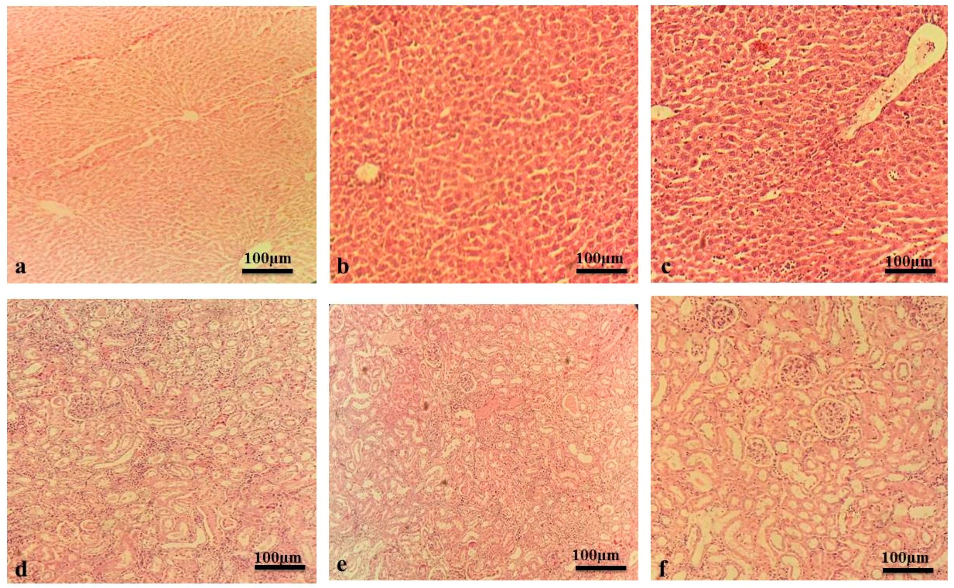
In acute toxicity studies, all rats remained alive showing no signs of toxicity or abnormalities observed at a dose of 5 g/kg of MLM and MFM, administered orally. For the period of fourteen days, there were no signs of abnormalities evidenced by no behavioral changes or alteration in body weight. There was a non-significant difference in serum biochemical parameters of treated group and control group (Table 1). Histological analysis showed no signs of hepatic or renal toxicity as compared to control group (Figure 2). Thus, it was concluded that the oral lethal dose for MLM and MFM would be greater than 5 g/kg. These results support the safety of MLM and MFM even at high dose.

2.3. Acute Anti-Inflammatory Activity

The carrageenan-induced paw edema model was carried out to investigate the anti-inflammatory potential of mucilage. Sub plantar injection of 1% carrageenan (0.1 mL) to rat hind paw steadily increased the paw thickness that extends to maximum after 4 h. Pretreatment with MLM (500 mg/kg) and MFM (500 mg/kg) significantly reduced the paw thickness with maximum inhibition at 4 h as compared to the disease control group (p < 0.001). MLM exhibited better inhibition of paw thickness as compared to MFM but not as much as diclofenac sodium (Table 2). The maximum percentage of anti-inflammatory activity was observed in diclofenac sodium (39.31%) at 4 h followed by MLM (27.35%) and MFM (15.68%) as depicted in Figure 3. Percentage inhibition of edema observed by MLM was significantly comparable with diclofenac sodium (p < 0.05).

2.4. Acute Antitussive Activity

Codeine (10 mg/kg) reduced cough frequency from 127.6 to 53.8; marking a 57.8% inhibition in the incidence of cough. Oral administration of 500 mg/kg of MLM significantly decreased the number of coughs induced by SO2 gas from 127.4 to 66.2; marking a 47.99 % inhibition in the incidence of cough. The least antitussive activity was observed in animals treated with 500 mg/kg of MFM that reduced the cough frequency from 133 to 91.66 with 31.37% inhibition of cough (Figure 4). There was a non-significant difference in percentage inhibition of cough in MLM treated rats when compared with standard drug.

2.5. Antiulcer Activity

2.5.1. Macroscopic Analysis of Stomach Mucosa

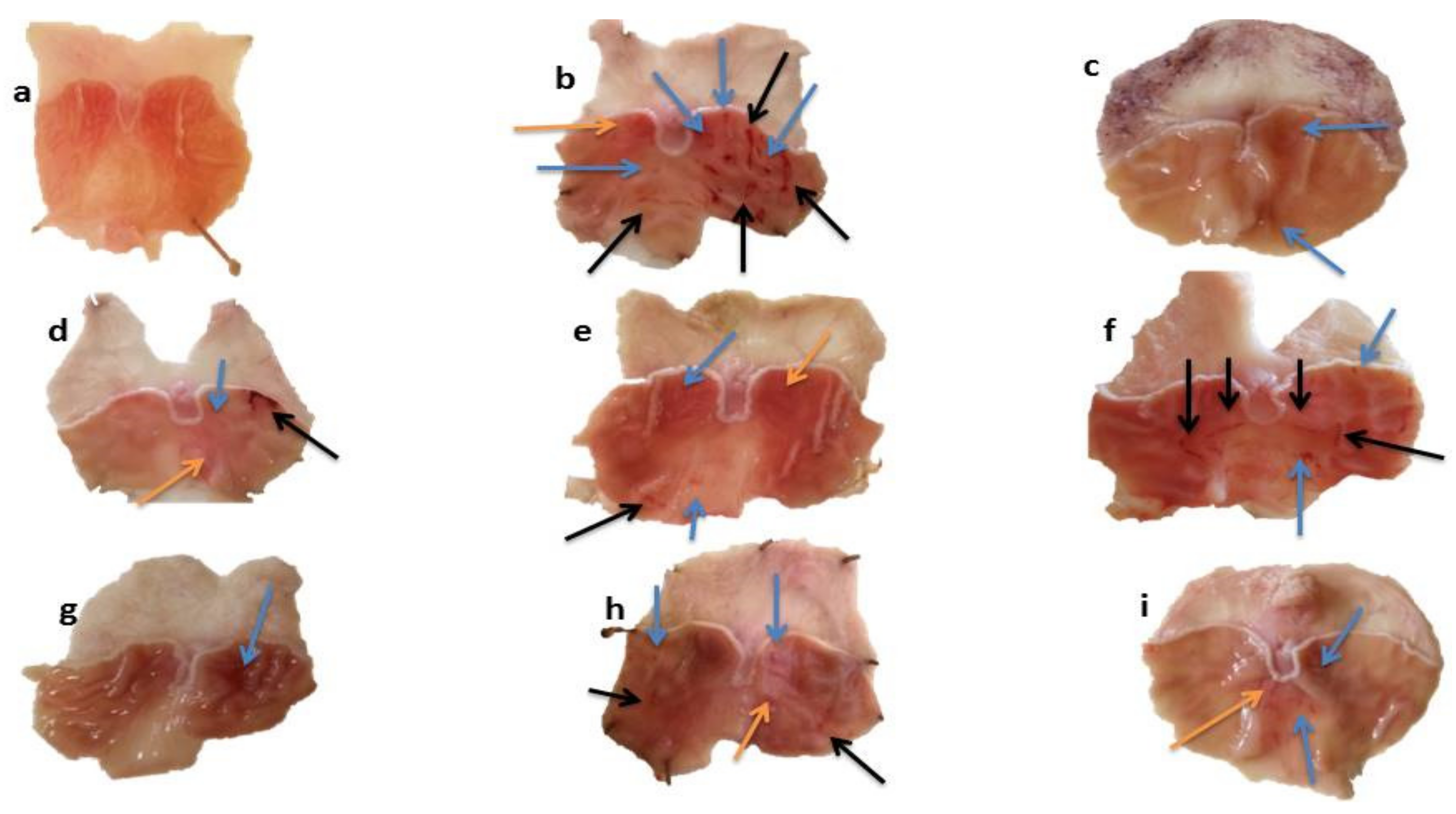
The stomach walls of each rat was observed by the naked eye and then by hand lens. Macroscopic view of gastric walls of normal group showed smooth surface without noticeable scars. In acute gastric ulcer models, severe lesions with extensive visible hemorrhagic streaks were observed in the ulcer control group. Moderate lesions were seen in rats pretreated with MLM (250 mg/kg) and MFM (250 mg/kg and 500 mg/kg). Low moderate lesions were seen in rats pretreated with ranitidine, sucralfate and MLM (500 mg/kg). However, mild lesions were observed in omeprazole treated group which directs high protection against gastric ulcer (Figure 5).
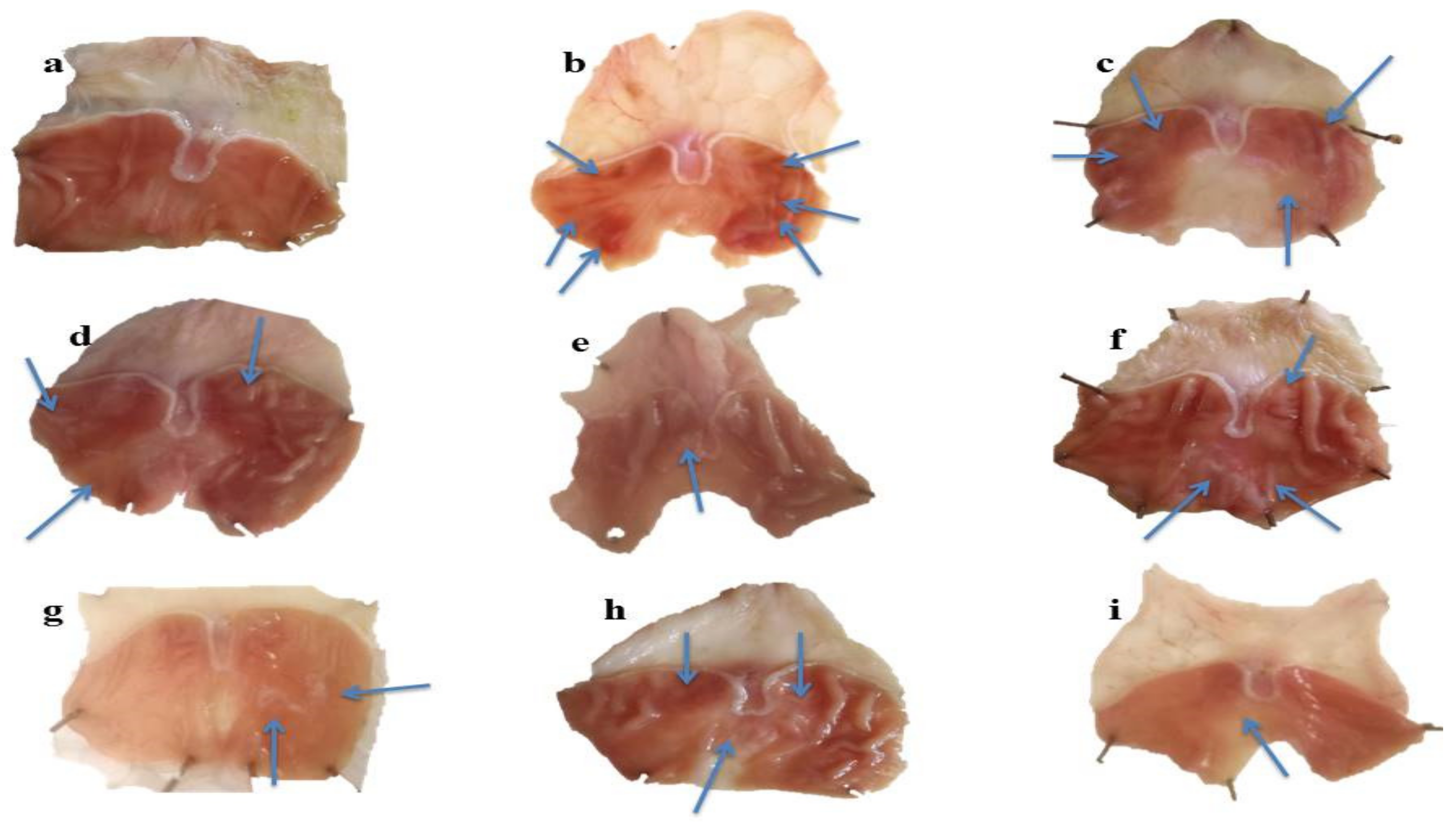
In chronic gastric ulcer models, ethanol induced gastric ulcer scars and lesions that were visible, but the nature of lesions was different as compared to the acute ulcer models. The lesions are generally categorized by the existence of white rounded scars with rare hemorrhage. Moderate scars were present in gastric mucosa of rats pretreated with ranitidine, omeprazole and MLM (250 mg/kg). However, rare scars were observed in rates pretreated with sucralfate, MLM (500 mg/kg) and MFM (Figure 6).

2.5.2. Determination of Ulcer Score, Ulcer Index and Percentage Inhibition

The ulcer index (UI) and % ulcer inhibition of experimental and control groups in acute gastric ulcer are given in Table 3. UI for the ethanol group was 11.36 meanwhile omeprazole, ranitidine and sucralfate considerably reduced the UI to 3.46, 5.21 and 5.27 with ulcer protection of 69.56%, 54.21% and 53.63%, respectively, where omeprazole showed maximum ulcer protection. Pretreatment with MLM at dosage of 250 mg/kg and 500 mg/kg decreased the UI to 8.74 and 6.96 and gave ulcer protection of 23.04% and 38.74%, respectively. Pretreatment with MFM at dosage of 250 mg/kg and 500 mg/kg decreased the UI to 10.47 and 7.03 and gave ulcer protection 7.85% and 38.08%, respectively. Thus, it was clear that pretreatment with MFM and MLM resulted in an increased ulcer inhibition activity in a dose–dependent manner (Table 3).
Meanwhile, in chronic gastric ulcer model, UI of ulcer control group, ethanol group, was 10.8. Pretreatment with omeprazole, ranitidine and sucralfate considerably reduced the UI to 5.22, 3.5 and 3.51 showing ulcer protection of 51.66, 67.59 and 67.5%, respectively. As compared to acute ulcer model, in chronic gastric ulcer model ranitidine and sucralfate showed maximum ulcer protection estimated by 3.5 and 3.51 accounting for 67.59 and 67.50%. Pretreatment with MLM at dosages of 250 mg/kg and 500 mg/kg decreased the UI to 7.08 and 5.67 and gave ulcer protection of 34.44 and 47.44%, respectively. Pretreatment with MFM at dosages of 250 mg/kg and 500 mg/kg decreased the UI to 6.95 and 3.95 and gave ulcer protection of 35.65 and 63.52%, respectively (Table 4). As observed in acute gastric ulcer models, dose–dependent ulcer inhibition was also observed in chronic gastric ulcer model; however, in chronic ulcer MFM was found to be more effective in contrast to acute gastric ulcer models where MLM revealed higher efficacy.

2.5.3. Histology of Stomach Wall

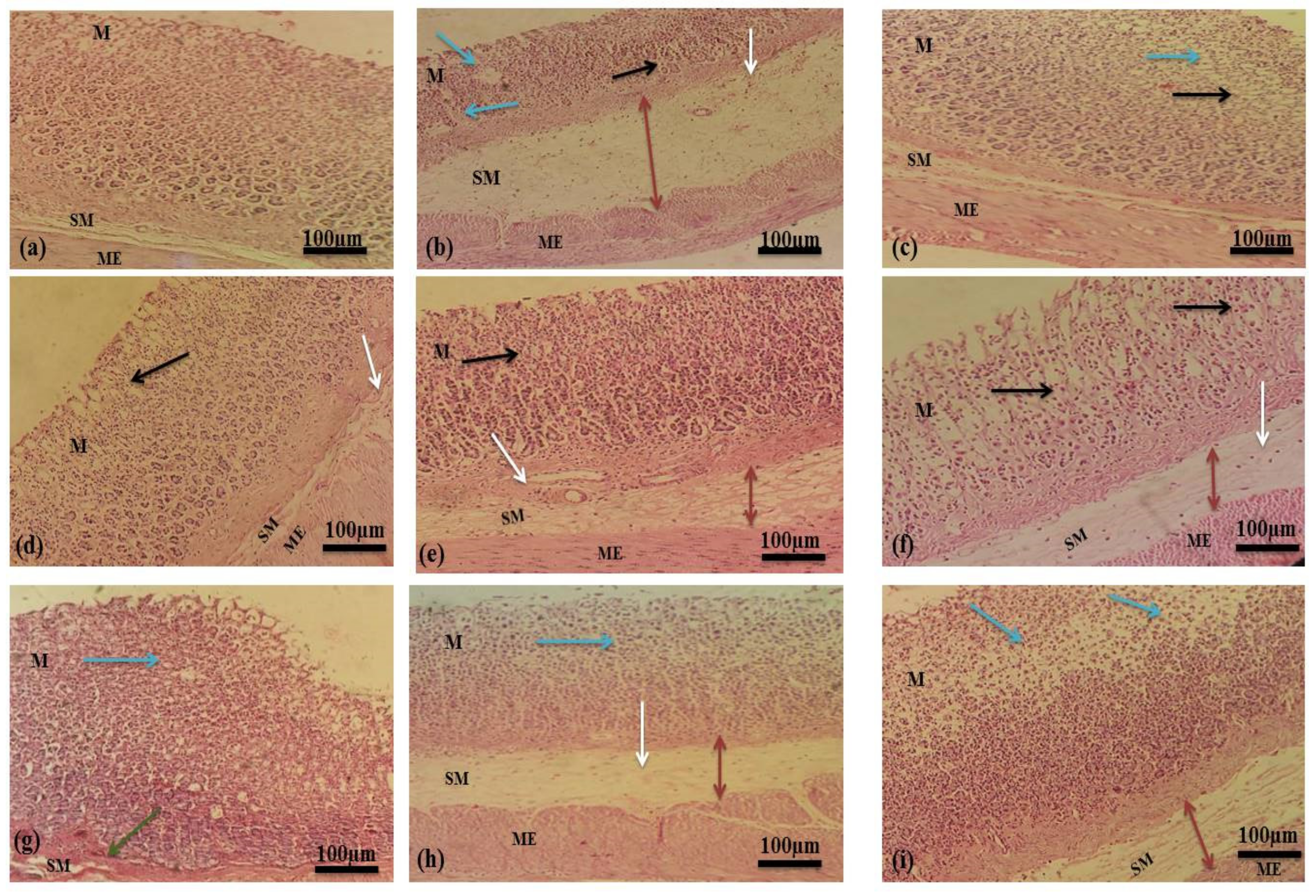
Histological examination of gastric mucosa revealed that ethanol caused severe disruption in the gastric mucosa that pierced deeply, accompanied by mucosal and sub-mucosal edema and leukocyte infiltration. Thickness of mucosal layer decreased extensively in chronic ulcer models due to the daily interruption of ethanol with gastric mucosa. The standard drugs and mucilage established enhanced protection of gastric mucosa in a dose–dependent manner accompanied by a pronounced reduction in edema and leukocytes infiltration of sub-mucosal layers. Histological observations in acute and chronic ulcer model are shown in Figure 7 and Figure 8.

2.5.4. Mechanism of Action

Effect on Gastric Juice Parameters

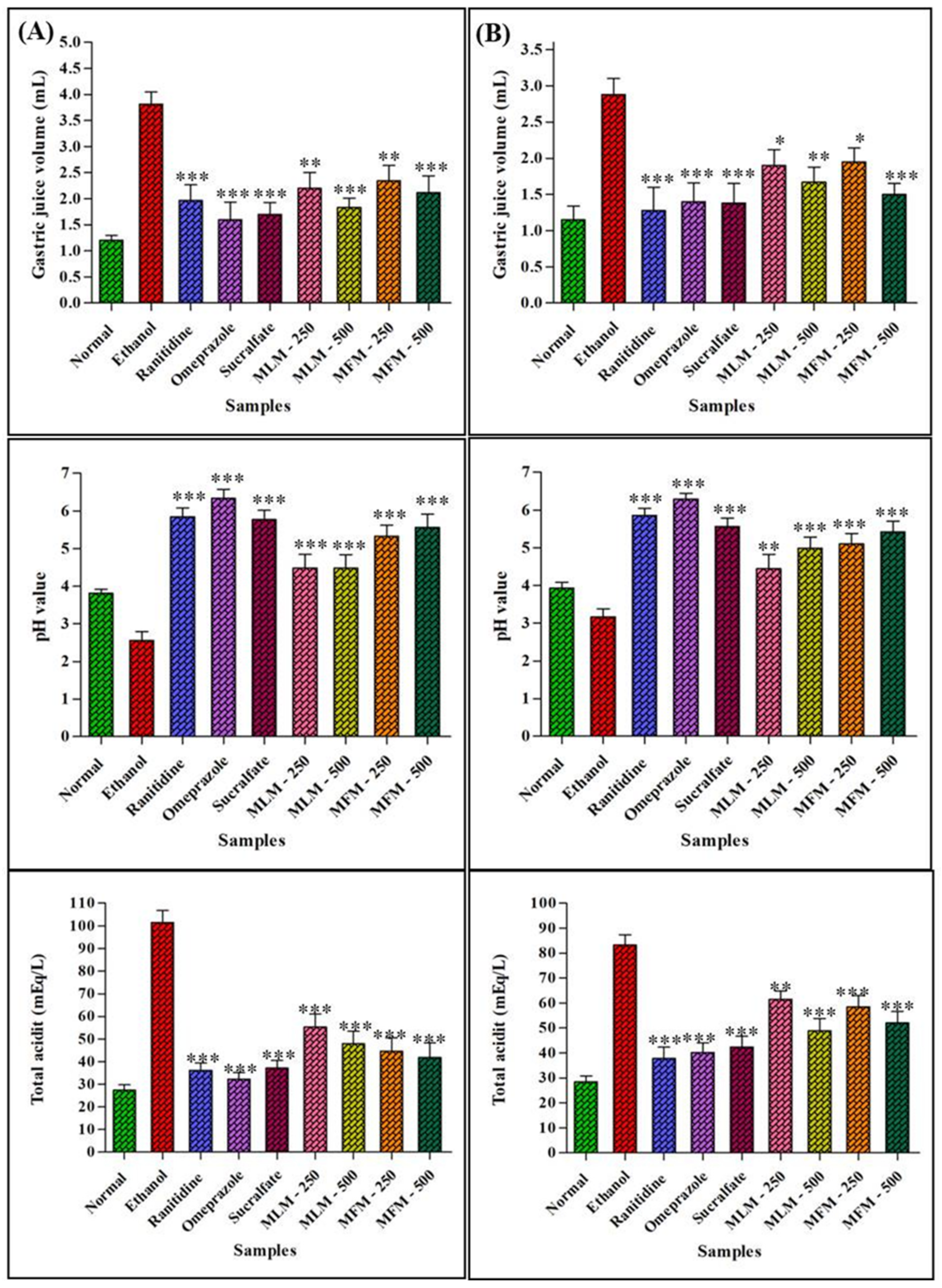
Gastric juice of ulcer control group displayed higher gastric juice volume, lower pH and high total acidity in contrast to normal animals. Pretreatment with MLM (500 mg/kg) and MFM (500 mg/kg) significantly (p < 0.001) reduced the gastric juice volume and total acidity but increased pH when compared with ulcer control group as illustrated in Figure 9. Similar changes were also observed in chronic gastric ulcer models (Table 5).

Effect on Total Protein and Total Mucus Content

The Alcian blue binding capacity was used as a marker for quantification of gastric mucus content. Ethanol treatment reduced the total gastric mucus content and ultimately Alcian blue binding capacity. Pretreatment with MLM (500 mg/kg) and MFM (500 mg/kg) in acute gastric ulcer models significantly (p < 0.01) protected the gastric mucosa increasing the mucus content in contrast to the ulcer control group. However, at low dosages (250 mg/kg), a non-significant difference was observed in total mucus content of MLM and MFM with ulcer control group. Ranitidine and MLM (500 mg/kg) had total protein content significantly dissimilar from ulcer control group (p < 0.01). However, in all other treated group a non-significant difference was observed in comparison to ulcer control group. In chronic gastric ulcers, there was a dramatic rise of total mucous content in animals pretreated with MLM (500 mg/kg), MFM (500 mg/kg), MFM (250 mg/kg) and sucralfate (100 mg/kg) significantly different from ulcer control group (p < 0.001). A non-significant difference was observed between total mucus content of animals pretreated with MLM (250 mg/kg) and ranitidine when compared with ulcer control group. Total protein content was also significantly different in the treated group as compared to the ulcer control group (Table 5).

2.6. Gas Chromatography Analyses of MLM and MFM

GC/MS analysis of MFM revealed the existence of four neutral monosaccharides which are galactose, rhamnose, arabinose as well as glucose constituting about 50.21, 7.61, 6.18 1.90 mg/g of MFM, respectively, whereas galacturonic acid constitutes the identified acidic monosaccharide showing 16.02 mg/g of MFM (Figure S1). Regarding the main monosaccharides detected in MLM, they are previously analyzed by the authors and reported [25] where GC/MS analysis of MLM showed the presence of five neutral monosaccharides together with one acidic monosaccharide which are galactose, rhamnose, arabinose, mannose as well as glucose and galacturonic acid accounting for 51.09, 10.24, 8.90, 1.80, 0.90 and 15.06 mg/g of MLM, respectively. Results showed that the leaves mucilage showed slightly higher levels of neutral monosaccharides compared to the fruit mucilage in contrast to the acidic monosaccharide represented by galacturonic acid that displayed a higher level in the fruit mucilage.

3. Discussion

Plant derived natural therapeutic agents have become part of primary healthcare in developing countries. Because of their natural origin some of them are mistakenly considered as absolute safe drugs. Thus, there is a great need of scientific studies on toxicities of drugs obtained from natural sources [30]. Erythrocytes are abundantly present in the human body, and they show vast biological and structural attributes playing an important role in drug transport. Breakdown of erythrocytes is known as hemolysis that occurs upon exposure of erythrocytes to toxicants. Some phytochemicals can cause hemolysis that in turn reflects that given substance is cytotoxic to RBCs [31,32].
Erythrocyte membranes are made up of polyunsaturated fatty acids and proteins, therefore, more vulnerable to peroxidation. Hemoglobin present in RBCs also catalyzes the oxidation process. Oxidation of RBCs represents oxidative damage of other biological membranes. Thus, the chemicals which produced free radicals can damage the erythrocyte membrane and serve as hemolytic agent [33]. In vitro hemolytic activity of MLM and MFM was assessed in an effort to check their toxicity to red blood cells. The results clearly justified the safety of MLM and MFM for RBCs and hence, can be used for further in vivo studies. Furthermore, acute oral toxicity of extracted mucilage was also performed to identify the dose that can be safely used for in-vivo studies. It was concluded that MLM and MFM are totally safe for systemic use and lethal dose is above 5 g/kg. As 5 g/kg showed no signs of toxicities therefore 1/20 and 1/10 of this lethal dose were selected in the current study (250 mg/kg and 500 mg/kg, respectively) for in vivo studies.
Regarding inflammation, it is defined as a localized reaction accompanied by soreness, warmth, swelling, tenderness and loss of function. Inflammatory mediators are released upon exposure to inflammatory agents. These inflammatory mediators widen the blood vessels and cause chemotaxis and ultimately lead to many disorders [34]. Red seaweeds (Rhodophyceae) are the source of carrageenan which is a sulphated polyglactan with esterified sulphate group that is responsible for its chemical activity. Carrageenan-induced paw edema is the most often practiced model for acute anti-inflammatory activity [34].
Carrageenan generates oxygen free radicals by changing the neutrophil membrane. Neutrophils are responsible for acute external inflammation and rapidly move towards the site of inflammation. They engulfed the pathogens and trashes of damaged tissues and consequently produce the reactive oxygen species and proteolytic enzyme to tear down the engulfed particles. Host tissues are also damaged during this process. As a result of these activities, a deficiency of ATP occurs resulting in the loss of cell function ultimately leading to necrosis [35]. The current study determined the anti-inflammatory activity of mucilage. The results showed that maximum edema inhibition occurred at the fourth hour by diclofenac sodium followed by MLM and MFM where MLM showed better anti-inflammatory activity when compared to MFM. Anti-inflammatory activity of MLM increased with increase in time with maximum results at the fourth hour. This might be due to the radical scavenging effects of MLM and MFM against free radicals produced at site of inflammation. The polysaccharides present in this mucilage may be the active ingredient against inflammation. Previously, mucilage extracted from lemon demonstrated pronounced anti-inflammatory activity in carrageenan-induced paw edema model in rats [35]. Okra and Baobab belongs to the family Malvaceae and their mucilage polysaccharides have been reported to possess anti-inflammatory properties [36].
Concerning the antitussive activity, there are various causes of cough that can be induced by any type of irritation to bronchi and trachea. This irritation can be the result of exposure to light, allergens, chemicals or any other foreign matter. As a result, airway sensory nerves are activated followed by increased mucous secretion, release of inflammatory mediators and damage to airway epithelium. Antitussives suppress cough either by acting centrally or peripherally. Centrally acting antitussive agents suppress the cough center in the brain and peripherally acting agents work as a demulcent or local anesthetic [37]. In this study, SO2 gas was used as an irritant to induce cough in rats. SO2 was produced by chemical reaction between sodium hydrogen sulphite and concentrated sulphuric acid. As all conditions were kept the same, it was therefore supposed that the quantity and saturation of SO2 would remain same during each exposure. Therefore, quantification of SO2 was not performed. The results showed that MLM exhibited 47.99% inhibition against chemical-induced cough. MLM was a better cough suppressant than MFM thus supporting the traditional use of leaves of M. parviflora as an antitussive. MLM and MFM might act peripherally due to the demulcent properties of mucilage. Folk use of M. parviflora is as a demulcent with mucilage responsible for this demulcent action [38]. Hence, this study consolidated the demulcent action of mucilage of M. parviflora based on scientific evidence. Rhamnogalacturonan was previously isolated from Marshmallow (Althea officinalis) mucilage that also belongs to family Malvaceae and showed high cough suppressant action in guinea pigs [39]. Similarly in an open trial, patients suffering from cough were treated successfully with herbal cough syrup containing marshmallow root mucilage [40].
Gastric ulcer is the chief health issue of GIT where gastric acid, pepsin and H. pylori are among the destructive factors that aggravate gastric ulcers, meanwhile the protective factors include mucin, NO, bicarbonates, blood flow, prostaglandins and growth factors. When a disturbance occurs between destructive and defensive factors, gastric ulcers occur [41]. The mucosal layer of stomach releases mucous that contains glycoprotein and lipids. Aside from suppressing the oxygen-derived free radicals it stimulates smooth movement of food and protects the stomach against the damaging effects of HCl and pepsin by preventing their penetration in the mucosal membrane [42,43]. Ethanol causes gastric ulcers as it solubilizes the mucous and diffuses into gastric mucosa. As a result, HCl and pepsin come in contact with gastric mucosa and injure it, in addition, ethanol increases gastric acid secretion. Moreover, it ruptures vascular endothelium, increases vascular permeability, increases oxidative stress in tissue and decreases gastric mucous secretion and thus ethanol induced gastric ulcer models were found to be suitable [44].
Hence, the antiulcer activities of MLM and MFM were evaluated in ethanol induced acute and chronic gastric ulcer models. It has been reported that antacids, anti-secretory agents and cytoprotective agents are the main classes of drugs used for the management of ulcers [45]. Therefore, in this study omeprazole, ranitidine and sucralfate were used as standard drugs. The results showed that M. parviflora mucilage and standard drugs significantly reduced the ulcer count and ulcer index with increased cytoprotective effects in both acute and chronic models, whereas the ulcer control group presented severe hemorrhagic streaks. The necrotic and petechial lesions in gastric mucosa leading to gastric ulcers are attributed to ethanol treatment [43]. Extreme stomach mucosa disruption leads to lower release of bicarbonate ions that in turn lowers the pH via increased production of gastric contents. Hence, ethanol increases gastric acid secretion that may result in increased gastric volume, decreased pH, increased total acidity and increased ulcer index [46]. A significant increase in pH and reduction in gastric volume and acidity was noticed in rats pretreated with MLM, MFM and standard drugs when compared with ulcer control groups as represented in Figure 9. This predicted the anti-secretory and antacid mechanism of the gastroprotective effects of MLM and MFM.
In addition, a reduced amount of protein content in stomach tissue homogenate is marked as a sign of damage to normal cellular functions. Therefore, treatments that cause an increase in protein content may be considered to contain auto-healing agents that support the mucosal regeneration process [40]. The low protein content in the disease control group of acute and chronic models was a sign of cellular dysfunction. The protein content in experimental and standard groups was higher compared with the disease control group thus supporting the presence of auto-healing effects. Treatment with MLM and MFM augmented the regeneration of epithelial cells and thus considerably amplified the protein concentration. Meanwhile, gastric mucus also acts as a shield for the protection of gastric walls against aggressive factors and as a first line of mucosal protection from luminal acid [47]. It contains viscid, flexible and translucent gel composed of glycoproteins (5%) and water (95%) and can be identified by the extent of Alcian blue binding. The defensive role of the mucus shield relies on the structure as well as the density of the layer protecting the mucosal surface. Furthermore, mucus can also act as an antioxidant and can diminish free radical-induced mucosal damage [48]. Administration of MLM and MFM significantly raised the amount of mucous in contrast to the disease control group. MLM and MFM treatment increased the gastric mucous content directly dependent on dosage. Interestingly, production of mucus in chronic ulcer models have remarkably been increased. The highest mucus content was observed in rats pretreated with MFM (500 mg/kg) followed by MLM (500 mg/kg) and results were comparable with rats pretreated with sucralfate. This unusual increase in gastric mucus suggested that MLM and MFM may trigger the discharge of chemical mediators responsible for the gastric mucus production i.e., gastrin, prostaglandin, secretin and acetylcholine [49]. Hence, it can be predicted that an increased secretion of mucus by administering MLM and MFM may be one of the potential mechanisms of their gastroprotective action. Mucilage contains polysaccharides where previous studies reported that polysaccharides possess antiulcer activity. These mucilaginous polysaccharides may form a protective covering on gastric mucosa or regenerate it [19]. This can be justified by the high mucus content in both acute and chronic gastric ulcer models.

4. Materials and Methods

4.1. Plant Material

Malva parviflora was collected in February 2018 from Lahore, Punjab, Pakistan. Plant Taxonomist, Dr. Zaheer-ud-din khan at Department of Botany, Government College University, Lahore authenticated the plant and issued a voucher specimen number GC.Herb.Bot.3533. Dusty material was removed by washing the leaves and unripe fruits with tap water. Leaves were dried in shade and concomitantly powdered in a mechanical grinder. The powdered leaves and fresh unripe fruits were used for the extraction. The mucilage was dried in an oven at 45 °C till it was completely dried. The dried mucilage was then powdered by mortar and pestle. The powdered mucilage was passed through a sieve#80. Particle size of the obtained powder was uniform [50,51,52].

4.2. Drugs and Chemicals

Omeprazole, sucralfate and ranitidine were used as standard anti-ulcer drugs and were kindly provided by Schazoo Zaka (Pvt.) Ltd. (Sheikhupura, Punjab, Pakistan), Highnoon Laboratories (Pvt.) Ltd. (Lahore, Punjab, Pakistan) and Surge Laboratories (Pvt.) Ltd. (Sheikhupura, Punjab, Pakistan), respectively. Alcian blue dye was obtained from Unichem, china, bovine serum albumin from Bioshop (Burlington, Ontario, Canada) and normal saline from Otsuka (Karachi, Pakistan. Diethyl ether and sucrose were acquired from Labscan (Bangkok, Thailand). Sodium hydrogen sulphite, sodium hydroxide, Sodium carbonate, potassium chloride, Tris HCl and phenolphthalein were purchased from BDH chemicals (Poole, UK). All other chemicals used in this study were obtained from Sigma-Aldrich (Steinheim, Germany). All the reagents were prepared freshly and were of pharmaceutical grade.

4.3. Animals

Wistar albino rats of both sexes (180–200 g) were maintained under standard conditions at (22–24 °C). The relative humidity of 50–60% and photoperiod of 12 h light and 12 h dark cycle was retained. Animals were fed on commercial pellet food with unrestricted supply of water. The experiments were performed following the guidelines set by the Institutional Ethical Committee for animal care and experimentation, College of Pharmacy, University of the Рunjаb, Lаhore, Раkistаn (AEC/PUCP/1094 dated 11 February 2019).

4.4. Preparation of the Mucilage

Mucilage was extracted from dried powdered leaves and fresh fruits following the previously described method by Munir et al. [25]. Malva parviflora leaves and fruit mucilage was labelled as MLM and MFM respectively.

4.5. Evaluation of the Biological Activities of the Mucilage Extracts

4.5.1. In Vitro Hemolytic Activity

Blood (3 mL) was collected in an anticoagulant tube from a volunteer human (male, 26 years, Blood group O+) by a Phlebotomist in Punjab University Healthcentre lab (Allama Iqbal campus, University of Punjab, Lahore, Pakistan). The sample was centrifuged at 850 rpm for 5 min. The clear supernatant was poured off followed by washing of residue pellets with 5 mL of chilled (4 °C) phosphate buffer saline (PBS) solution (pH 7.4). Washed cells suspension was made in 20 mL cool sterilized PBS. Cells were calculated using hemocytometer and for each assay 7.068 × 10⁸ cell/mL were used. 20 µL of plant mucilage were taken in an Eppendorf tube and PBS and 0.1% TritonX-100 were used as negative and positive control respectively. 180 µL of diluted blood cell suspension were added into each tube followed by incubation at 37 °C. After incubation for 35 min, suspension was allowed to cool for 5 min followed by centrifugation at 1500 rpm for 5 min. 100 µL of supernatant were collected and diluted with 900 µL chilled sterile phosphate buffer saline. An aliquot of 200 µL of all these samples including positive and negative control were transferred to 96-well plate. ELISA microplate reader was used to measure the absorbance at 630 nm [53].
%   Hemolysis = A b s S a m p l e   a b s o r b a n c e A b s   c o n t r o l   a b s o r b a n c e × 100

4.5.2. Acute Toxicity Study

Acute oral toxicity of MLM and MFM was determined in Wistar albino rats according to OECD guidelines 425 [54]. Limit test was performed at 5000 mg/kg where the control group was given only distilled water. The animals were observed for death or any noxious outcome in earliest four hours after the dosing and regularly for fourteen days. During this period, parameters such as weight, physical appearance, behavioral changes, injury, illness signs and mortality were observed. On the 15th day animals were anesthetized and sacrificed with an overdose of xylazine and ketamine anaesthetic drugs and blood samples were obtained by cardiac puncture. Animals were dissected and organs such as liver and kidney were obtained for histopathological examination.

4.5.3. Anti-Inflammatory Activity

Acute anti-inflammatory activity of MLM and MFM was evaluated by carrageenan-induced paw edema in rats [55]. The rats were arbitrarily separated in four groups (n = 5). In the first group, the animals received a dose of 10 mL/kg of distilled water (carrageenan control); meanwhile in the second group, the animals were orally administered with diclofenac sodium in a dose of 10 mg/kg (standard group) [56]. In the third and fourth groups, the animals were orally given 500 mg/kg of MLM (MLM experimental group) and MFM (MFM experimental group), respectively. After 1 h, all the animals were injected with 0.1 mL of carrageenan in normal saline (1% (w/v)) into subplantar region of left hind paw of each rat. Thickness of paw was measured using digital vernier caliper at 0 h, 1 h, 2 h, 3 hand 4 h intervals. Edema inhibition (%) was calculated using the following formula:
%   inhibition   of   edema = T c T t T c   ×   100
where T c = Paw thickness of control group and T t = Paw thickness of experimental group.

4.5.4. Antitussive Activity

Acute antitussive activity of MLM and MFM was evaluated by SO2 induced cough model in rats [57]. Each animal served his own control due to variation in number of coughs in each individual animal. Groups were treated in the following manner where in the first group, the animals were orally given codeine phosphate in a dose of 10 mg/kg. In the second and third groups, the animals were orally given 500 mg/kg of MLM and MFM respectively. The animals were exposed to SO2 for 60 s after an hour of receiving the treatment and the number of coughs was counted. The number of coughs was compared before and after the treatment to determine the reduction in cough. The percentage inhibition frequency of cough was determined using the following formula:
%   inhibition   of   frequency   of   cough = C C C T   C C × 100
where C C is the cough frequency in control animal and C T   is the cough frequency in treated animal [57].

4.5.5. Antiulcer Activity

Study Design

The animals were arbitrarily separated into 9 groups (n = 6) where the first group (normal group) was orally treated with distilled water (DW) 1 mL/100g [58]. The second group was orally treated with 1 mL/100 g of ethanol (ethanol group) meanwhile from 3–5 groups, the animals received orally standard drugs which are ranitidine (50 mg/kg) [59], omeprazole (20 mg/kg) [60] and sucralfate (100 mg/kg) [61], respectively. Groups 6–7 were orally administered MLM in doses of 250 and 500 mg/kg, respectively whereas groups 8–9 were orally administered MFM in doses of 250 and 500 mg/kg, respectively.

Ethanol Induced Acute Gastric Ulcer

Animals were prevented from food for 24 h with unrestricted excess toward water. They were deprived of water just 2 h prior starting the experimental procedure. Animals of all groups were pretreated with drugs and extract as mentioned above. After 30 min all groups were orally administered 90% ethanol (1 mL/100 g) except normal group. Animals were euthanized with an overdose of xylazine and ketamine anaesthetic drugs after 1 h of ethanol administration [62]. Animals were dissected, removed the stomachs and incised along the larger curve. Washed with ice cold normal saline and gastric mucosa was examined [63].

Ethanol Induced Chronic Gastric Ulcer

Animals were prevented from food for 24 h with unrestricted excess toward water. They were deprived of water just 2 h prior to starting the experimental procedure. All animals were treated with drug and extract for fourteen days as mentioned above. On the first day of experiment after 30 min above treatment animals were orally administered with 90% ethanol (1 mL/100 g) except normal group. From the 2nd day 30% (v/v) ethanol was orally given to all groups for fourteen days except normal group. On 14th day, all the animals were anesthetized and sacrificed with an overdose of xylazine and ketamine anaesthetic drugs after 1 h of ethanol administration [62]. Animals were dissected, removed stomachs and incised along larger curve and washed with ice cold normal saline then gastric mucosa was examined [64].

Parameters of Gastric Ulcer Evaluation

  • Macroscopic and Microscopic Evaluation
Normal saline was used to wash the stomach and the apparent changes in inner walls of stomach were carefully examined macroscopically by using magnifying glass and microscope. The number of lesions was carefully noted and was used for the determination of ulcer index. Photos were taken using mobile camera (Oppo A57, Guangdong Oppo Mobile Telecommunications Corp., Ltd., Dongguan, Guangdong, China) [65].

Ulcer Scoring

Based on severity, the ulcers were given scores as previously reported by Gupta et al. [66] as follows: 0 = no ulcer; 0.5 = reddish mucosa; 1 = red spots; 1.5 = hemorrhagic streaks; 2 = deep ulcers and 3 = perforations.

Ulcer Index

Ulcer index can be calculated as previously described by Gul et al. using the following formula [67].
Ulcer   index = UN + US + UP × 10 1
where UN = average number of ulcers per animal; US = average of severity score; UP = percentage of animals with ulcer.

Ulcer Protection (%)

Ulcer protection (%) was calculated described by Gul et al. using the following formula [67].
%   Protection = Ulcer   index   of   ethanol   treated   group ulcer   index   of   treated   group ulcer   index   of   ethanol   treated   group   × 100

Histological Analysis

Samples of stomach walls were fixed in formalin solution (10%) for 48 h and then dehydrated by washing with ascending grades of ethanol. Samples were cleaned by xylene and embedded in paraffin wax. The rotary microtome was used to cut sections of 5–6 mm thickness followed by staining with hematoxylin and eosin. The sections were examined under a microscope for histopathological variations such as ulceration, congestion, edema, necrosis and leukocyte infiltration [68].

Mode of Gastro-Protective Activity

  • Estimation of the Gastric Volume
After opening the stomach gastric content was squeezed out in a falcon tube followed by centrifugation at 1000 rpm) for 15 min, then the supernatant was collected, and its volume was determined [69].

Determination of the pH Value and Total Acidity

Gastric juice pH was determined using a digital pH meter as previously described by Sen et.al [68]. A 1 mL volume of gastric juice was obtained in a vial then 2 drops of phenolphthalein were added as indicator. Then, it was titrated against 0.1 N NaOH until the end point that is the change from colorless to light pink. The volume of NaOH used was determined then the following formula was used to calculate total acidity where results were expressed in terms of clinical unit mEq/L [70].
Total   acidity = Volume   of   NaOH   used × Normality   of   NaOH   used × 100 0.1

Determination of Gastric Mucin Content

The glandular portion of stomach was excised and weighed, and this portion was soaked for 2 h in 10 mL of 0.1% Alcian blue solution in 0.16 M sucrose buffered with 0.05 M sodium acetate adjusted to pH = 5. Excess uncomplexed dye was removed by rinsing with 0.25 M sucrose solution at an interval of 15 min and 45 min respectively. Dye forms a complex with mucus that was extracted with 10 mL of 0.5 M magnesium chloride solution for 2 h with consecutive shaking for one minute after 30 min interval. A 4 mL volume of this solution was shaken with an equal volume of diethyl ether. The resulting emulsion was centrifuged at 3000 rpm and the absorbance of aqueous layer was taken at 580 nm. The mucin content of the sample was determined from the standard curve, which was expressed in microgram of Alcian blue extracted per gram of wet gland tissue [71].

Determination of the Total Protein Content

Total protein of glandular tissues homogenate was estimated by Lowry method [72]. The tissue homogenate of glandular portion of stomach was mixed with 1 mL of 0.1 M Tris-HCl buffer (pH 7.4) by a homogenizer followed by centrifugation at 1500 rpm for 15 min at 4 °C. The homogenate was diluted with 0.1 M tris-HCl buffer to reach 10 mL. Bovine serum albumin (BSA) was used as standard and 0.1 N of NaOH was used as blank. A 1 mL volume of the sample was mixed with 4.5 mL of reagent I that is formed by mixing 48 mL of 2% Na2CO3 in 0.1 N NaOH with 1 mL of 1% NaK Tartrate in H2O and 1 mL of 0.5% CuSO4·5 H2O in H2O followed by addition of 0.5 mL of reagent II (1 part Folin-Phenol [2 N]: 1 part water) after 10 min. The test tubes were placed in the dark for 30 min and the absorbance was taken at 750 nm. BSA standard calibration curve was used to interpret the results.

4.6. Gas Chromatography Coupled with Mass Spectrometry Analysis

Monosaccharide composition of MLM and MFM was analyzed using a modified GC-MS analytical procedure previously adopted by Xia et al. [73] depending upon trimethylsilyl dithioacetal (TMSD) derivatization. A 1 µL volume of the sample was applied to Agilent 7890A Gas Chromatography coupled to Agilent 5975C Mass spectrometer (Agilent Technologies, Santa Clara, CA, USA) with HP-5MS column using a temperature range of 80 °C for 0 min, 80–90 °C at 2.5 °C/min, 190–252 °C at 2 °C/min, 252–300 °C at 25 °C/min, 300–310 °C at 25 °C/min and held for 15 min. Mass spectra were recorded employing total ion chromatogram (TIC) mode and interpreted using NIST 5 software [25].

4.7. Statistical Analysis

GraphPad prism 8.4.3 was used for statistical analysis. Statistical significance difference was calculated using one way ANOVA followed by Dunnett’s test. The values p < 0.05 *, 0.01 ** and 0.001 *** were considered as statistically significant.

5. Conclusions

In conclusion, the current findings suggest that mucilage extracted from the leaves and fruit of M. parviflora are safe for in vivo studies and could be incorporated in many significant therapeutic applications after clinical trials. Treatment with mucilage reduced cough, inflammation and ulcers in animal models. The underlying mechanism of gastro protection is in the anti-secretory and mucus protective potential of mucilage. However, further in vivo studies are recommended to comprehensively understand its exact mechanism of action. This study clearly highlights the application of M. parviflora mucilage in adjuvant therapy of gastric ulcers. Further studies are in progress to isolate bioactive polysaccharides and glycoprotein from this mucilage.

Supplementary Materials

The following supporting information can be downloaded at: https://www.mdpi.com/article/10.3390/ph15040427/s1, Figure S1: GC/MS chromatograms of neutral (A) and acidic (B) polysaccharides in M. parviflora mucilage obtained from the fruits (MFM).

Author Contributions

Conceptualization, A.M. and S.I.; methodology A.M., S.I., M.R., S.S.E. and K.A.; software, F.S.Y.; resources, A.E.A., O.K., W.Y.R. and S.S.E.; writing—original draft preparation, A.M. and S.I.; writing—review and editing, F.S.Y., S.S.E. and M.L.A.; supervision, F.S.Y., S.S.E. and M.L.A.; funding acquisition, W.Y.R., A.E.A., O.K. and S.S.E. All authors have read and agreed to the published version of the manuscript.

Funding

This research was funded by the Deanship of Scientific Research (DSR) at King Abdulaziz University, Jeddah, Saudi Arabia under grant no. (G-68-166-38).

Institutional Review Board Statement

The animal study protocol was approved by the Punjab University College of Pharmacy, (AEC/PUCP/1094 dated 11 February 2019).

Informed Consent Statement

Informed consent was obtained from all subjects involved in the study.

Data Availability Statement

Data is contained within the article and Supplementary Materials.

Acknowledgments

This project was funded by the Deanship of Scientific Research (DSR) at King Abdulaziz University, Jeddah, Saudi Arabia under grant no. (G-68-166-38). All the authors, therefore, acknowledge with thanks DSR for technical and financial support.

Conflicts of Interest

The authors declare no conflict of interest.

References

  1. Eccles, R.; Mallefet, P. Soothing properties of glycerol in cough syrups for acute cough due to common cold. Pharmacy 2017, 5, 4. [Google Scholar] [CrossRef] [PubMed]
  2. Romm, A.; Ganora, L.; Hoffmann, D.; Yarnell, E.; Abascal, K.; Coven, M. Chapter 3—Fundamental Principles of Herbal Medicine. In Botanical Medicine for Women’s Health; Room, A., Hardy, M.L., Mills, S., Eds.; Churchill Livingstone: Saint Louis, France, 2010; pp. 24–74. [Google Scholar]
  3. Czibulka, A. Probiotics and Herbal Therapies. In Laryngopharyngeal Reflux Disease; Springer: Berlin/Heidelberg, Germany, 2019; pp. 103–113. [Google Scholar]
  4. Schmidgall, J.; Schnetz, E.; Hensel, A. Evidence for bioadhesive effects of polysaccharides and polysaccharide-containing herbs in an ex vivo bioadhesion assay on buccal membranes. Planta Med. 2000, 66, 48–53. [Google Scholar] [CrossRef] [PubMed]
  5. Tosif, M.M.; Najda, A.; Bains, A.; Kaushik, R.; Dhull, S.B.; Chawla, P.; Walasek-Janusz, M. A Comprehensive review on plant-derived mucilage: Characterization, functional properties, applications, and its utilization for nanocarrier fabrication. Polymers 2021, 13, 1066. [Google Scholar] [CrossRef] [PubMed]
  6. Nazari, M.; Riebeling, S.; Banfield, C.C.; Akale, A.; Crosta, M.; Mason-Jones, K.; Dippold, M.A.; Ahmed, M.A. Mucilage polysaccharide composition and exudation in maize from contrasting climatic regions. Front. Plant Sci. 2020, 11, 1968. [Google Scholar] [CrossRef] [PubMed]
  7. Dybka-Stępień, K.; Otlewska, A.; Góźdź, P.; Piotrowska, M. The renaissance of plant mucilage in health promotion and industrial applications: A review. Nutrients 2021, 13, 3354. [Google Scholar] [CrossRef]
  8. El-Shiekh, R.A.; Salama, A.; Al-Mokaddem, A.K.; Abdel-Sattar, E.A. Gastroprotective effect of mucilage fraction from Solenostemma argel via cytoprotection and attenuation of oxidative stress, inflammation and apoptosis. J. Herb. Pharmacol. 2021, 10, 232–240. [Google Scholar] [CrossRef]
  9. Wadhwa, J.; Nair, A.; Kumria, R. Potential of plant mucilages in pharmaceuticals and therapy. Curr. Drug Deliv. 2013, 10, 198–207. [Google Scholar] [CrossRef]
  10. Alam, M.T.; Parvez, N.; Sharma, P.K. FDA-approved natural polymers for fast dissolving tablets. J. Pharm. 2014, 2014, 952970. [Google Scholar] [CrossRef]
  11. Hou, C.; Chen, L.; Yang, L.; Ji, X. An insight into anti-inflammatory effects of natural polysaccharides. Int. J. Biol. Macromol. 2020, 153, 248–255. [Google Scholar] [CrossRef]
  12. Aboulwafa, M.M.; Youssef, F.S.; Gad, H.A.; Altyar, A.E.; Al-Azizi, M.M.; Ashour, M.L. A comprehensive insight on the health benefits and phytoconstituents of Camellia sinensis and recent approaches for its quality control. Antioxidants 2019, 8, 455. [Google Scholar] [CrossRef] [Green Version]
  13. Ashour, M.L.; Youssef, F.S.; Gad, H.A.; El-Readi, M.Z.; Bouzabata, A.; Abuzeid, R.M.; Sobeh, M.; Wink, M. Evidence for the anti-inflammatory activity of Bupleurum marginatum (Apiaceae) extracts using in vitro and in vivo experiments supported by virtual screening. J. Pharm. Pharmacol. 2018, 70, 952–963. [Google Scholar] [CrossRef] [PubMed]
  14. El Senousy, A. Immunomodulatory and anti-inflammatory activities of the defatted alcoholic extract and mucilage of Hibiscus sabdariffa L. leaves, and their chemical characterization. J. Pharmacog. Phytochem. 2019, 8, 982–990. [Google Scholar]
  15. Carlotto, J.; Maria-Ferreira, D.; de Souza, L.M.; da Luz, B.B.; Dallazen, J.L.; de Paula Werner, M.F.; Cipriani, T.R. A polysaccharide fraction from “ipê-roxo” (Handroanthus heptaphyllus) leaves with gastroprotective activity. Carbohydr. Polym. 2019, 226, 115239. [Google Scholar] [CrossRef]
  16. Nascimento, A.M.; Maria-Ferreira, D.; de Souza, E.F.J.; de Souza, L.M.; Sassaki, G.L.; Iacomini, M.; Werner, M.F.d.P.; Cipriani, T.R. Gastroprotective effect and chemical characterization of a polysaccharide fraction from leaves of Croton cajucara. Int. J. Biol. Macromol. 2017, 95, 153–159. [Google Scholar] [CrossRef]
  17. El-Din, M.I.G.; Youssef, F.S.; Said, R.S.; Ashour, M.L.; Eldahshan, O.A.; Singab, A.N.B. Chemical constituents and gastro-protective potential of Pachira glabra leaves against ethanol-induced gastric ulcer in experimental rat model. Inflammopharmacology 2021, 29, 317–332. [Google Scholar] [CrossRef] [PubMed]
  18. El-Din, M.I.G.; Youssef, F.S.; Ashour, M.L.; Eldahshan, O.A.; Singab, A.N.B. New γ-pyrone glycoside from Pachira glabra and assessment of its gastroprotective activity using an alcohol-induced gastric ulcer model in rats. Food Funct. 2020, 11, 1958–1965. [Google Scholar] [CrossRef] [PubMed]
  19. Khan, M.S.A.; Khundmiri, S.U.K.; Khundmiri, S.R.; Al-Sanea, M.M.; Mok, P.L. Fruit-derived polysaccharides and terpenoids: Recent update on the gastroprotective effects and mechanisms. Front. Pharmacol. 2018, 9, 569. [Google Scholar] [CrossRef]
  20. Lajili, S.; Ammar, H.H.; Mzoughi, Z.; Amor, H.B.H.; Muller, C.D.; Majdoub, H.; Bouraoui, A. Characterization of sulfated polysaccharide from Laurencia obtusa and its apoptotic, gastroprotective and antioxidant activities. Int. J. Biol. Macromol. 2019, 126, 326–336. [Google Scholar] [CrossRef]
  21. Fathy, S.; Emam, M.; Agwa, S.A.; Zahra, F.A.; Youssef, F.; Sami, R. The antiproliferative effect of Origanum majorana on human hepatocarcinoma cell line: Suppression of NF-kB. Cell. Mol. Biol. 2016, 62, 80–84. [Google Scholar]
  22. Murgia, V.; Ciprandi, G.; Votto, M.; De Filippo, M.; Tosca, M.A.; Marseglia, G.L. Natural remedies for acute post-viral cough in children. Allergol. Immunopathol. 2021, 49, 173–184. [Google Scholar] [CrossRef]
  23. Kahramanoğlu, İ.; Wan, C. Determination and improvement of the postharvest storability of Little Mallow (Malva parviflora L.): A novel crop for a sustainable diet. HortScience 2020, 55, 1378–1386. [Google Scholar]
  24. Ododo, M.M.; Choudhury, M.K.; Dekebo, A.H. Structure elucidation of β-sitosterol with antibacterial activity from the root bark of Malva parviflora. SpringerPlus 2016, 5, 1210. [Google Scholar] [CrossRef] [PubMed] [Green Version]
  25. Munir, A.; Youssef, F.S.; Ishtiaq, S.; Kamran, S.H.; Sirwi, A.; Ahmed, S.A.; Ashour, M.L.; Elhady, S.S. Malva parviflora leaves mucilage: An eco-friendly and sustainable biopolymer with antioxidant properties. Polymers 2021, 13, 4251. [Google Scholar] [CrossRef] [PubMed]
  26. Dugani, A.; Dakhil, B.; Treesh, S. Protective effect of the methanolic extract of Malva parviflora L. leaves on acetic acid-induced ulcerative colitis in rats. Saudi J. Gastroenterol. 2016, 22, 226–233. [Google Scholar]
  27. El-Naggar, M.E.; Hussein, J.; El-sayed, S.M.; Youssef, A.M.; El Bana, M.; Latif, Y.A.; Medhat, D. Protective effect of the functional yogurt based on Malva parviflora leaves extract nanoemulsion on acetic acid-induced ulcerative colitis in rats. J. Mat. Res. Technol. 2020, 9, 14500–14508. [Google Scholar] [CrossRef]
  28. Afolayan, A.J.; Aboyade, O.M.; Adedapo, A.A.; Sofidiya, M.O. Anti-inflammatory and analgesic activity of the methanol extract of Malva parviflora Linn (Malvaceae) in rats. Afr. J. Biotechnol. 2010, 9, 1225–1229. [Google Scholar]
  29. Bouriche, H.; Meziti, H.; Senator, A.; Arnhold, J. Anti-inflammatory, free radical-scavenging, and metal-chelating activities of Malva parviflora. Pharm. Biol. 2011, 49, 942–946. [Google Scholar] [CrossRef] [Green Version]
  30. Jothy, S.L.; Zakaria, Z.; Chen, Y.; Lau, Y.L.; Latha, L.Y.; Sasidharan, S. Acute oral toxicity of methanolic seed extract of Cassia fistula in Mice. Molecules 2011, 16, 5268–5282. [Google Scholar] [CrossRef]
  31. Afsar, T.; Razak, S.; Khan, M.R.; Mawash, S.; Almajwal, A.; Shabir, M.; Haq, I.U. Evaluation of antioxidant, anti-hemolytic and anticancer activity of various solvent extracts of Acacia hydaspica R. Parker aerial parts. BMC Complement. Altern. Med. 2016, 16, 258. [Google Scholar]
  32. Lakshmi, G.; Smitha, N.; Ammu, S.; Priya, C.; Bhaskara Rao, K. Phytochemical profile, in vitro antioxidant and hemolytic activities of various leaf extract of Nymphaea Nouchali Linn: An in vitro study. Int. J. Pharm. Pharm. Sci. 2014, 6, 548–552. [Google Scholar]
  33. Vidhya, R.; Udayakumar, R. Phytochemical screening and evaluation of in vitro haemolytic, thrombolytic and antiinflammatory activities of Aerva lanata (L.). IAJPS 2016, 6, 6–7. [Google Scholar]
  34. Elmajdoub, A.A.; Awidat, S.K.; El-Mahmoudy, A.M. Anti-inflammatory potential of Agaricus in carrageenan-induced model of local inflammation in rats. Int. J. Basic Clin. Pharmacol. 2015, 4, 497. [Google Scholar] [CrossRef] [Green Version]
  35. Galati, E.; Cavallaro, A.; Ainis, T.; Tripodo, M.; Bonaccorsi, I.; Contartese, G.; Taviano, M.F.; Fimiani, V. Anti-Inflammatory effect of lemon mucilage: In vivo and in vitro studies. Immunopharmacol. Immunotoxicol. 2005, 27, 661–670. [Google Scholar] [CrossRef] [PubMed]
  36. Alba, K.; Nguyen, P.T.; Kontogiorgos, V. Sustainable polysaccharides from Malvaceae family: Structure and functionality. Food Hydrocolloid. 2021, 118, 106749. [Google Scholar] [CrossRef]
  37. Kaur, H.B.; Ruknuddin, G.; Nariya, M.; Patgiri, B.; Bedarkar, P.; Prajapati, P.K. Anti-tussive activity of Ashtangavaleha prepared by two different methods against sulphur dioxide induced cough in mice. Med. J. 2018, 11, 471–475. [Google Scholar] [CrossRef]
  38. Ridh, D.A.A.M.; Sarheed, N.M.; Kokaz, O. Activity of ethanolic extraction of Malva parviflora and liqourice as antifungal and antioxidant in male rats. J. Pharm. Sci. Res. 2018, 10, 777–781. [Google Scholar]
  39. Sutovska, M.; Capek, P.; Franova, S.; Joskova, M.; Sutovsky, J.; Marcinek, J.; Kalman, M. Antitussive activity of Althaea officinalis L. polysaccharide rhamnogalacturonan and its changes in guinea pigs with ovalbumine-induced airways inflammation. Bratisl. Lek. Listy 2011, 112, 670–675. [Google Scholar]
  40. Büechi, S.; Vögelin, R.; Eiff, M.; Ramos, M.; Melzer, J. Open trial to assess aspects of safety and efficacy of a combined herbal cough syrup with Ivy and Thyme. Res. Complement. Nat. Class. Med. 2006, 12, 328–332. [Google Scholar] [CrossRef]
  41. Adefisayo, M.A.; Akomolafe, R.O.; Akinsomisoye, S.O.; Alabi, Q.K.; Ogundipe, O.L.; Omole, J.G.; Olamilosoye, K.P. Gastro-protective effect of methanol extract of Vernonia amygdalina (del.) leaf on aspirin-induced gastric ulcer in Wistar rats. Toxicol. Rep. 2017, 4, 625–633. [Google Scholar] [CrossRef]
  42. Chen, S.H.; Liang, Y.C.; Chao, J.C.; Tsai, L.H.; Chang, C.C.; Wang, C.C.; Pan, S. Protective effects of Ginkgo biloba extract on the ethanol-induced gastric ulcer in rats. World J. Gastroenterol. 2005, 11, 3746–3750. [Google Scholar] [CrossRef]
  43. Rahim, N.A.; Hassandarvish, P.; Golbabapour, S.; Ismail, S.; Tayyab, S.; Abdulla, M.A. Gastroprotective effect of ethanolic extract of Curcuma xanthorrhiza leaf against ethanol-induced gastric mucosal lesions in Sprague-Dawley rats. BioMed Res. Int. 2014, 2014, 416409. [Google Scholar] [CrossRef] [PubMed] [Green Version]
  44. Adinortey, M.; Ansah, C.; Galyuon, I.; Nyarko, A. In vivo models used for evaluation of potential antigastroduodenal Ulcer Agents. Ulcers 2013, 2013, 489–512. [Google Scholar] [CrossRef] [Green Version]
  45. Sharifi-Rad, M.; Fokou, P.V.T.; Sharopov, F.; Martorell, M.; Ademiluyi, A.O.; Rajkovic, J.; Salehi, B.; Martins, N.; Iriti, M.; Sharifi-Rad, J. Antiulcer agents: From plant extracts to phytochemicals in healing promotion. Molecules 2018, 23, 1751. [Google Scholar] [CrossRef] [PubMed] [Green Version]
  46. Balogun, M.E.; Besong, E.E.; Obimma, J.N.; Djobissie, S.F.; Mbamalu, O.S. Gastroprotective effect of ethanolic extract of Vigna subterranea in ethanol-induced gastric mucosal ulceration in Rats. Ind. J. Physiol. Pharmacol. 2018, 62, 347–358. [Google Scholar]
  47. Zakaria, Z.A.; Balan, T.; Azemi, A.K.; Omar, M.H.; Mohtarrudin, N.; Ahmad, Z.; Abdullah, M.N.H.; Desa, M.N.M.; Teh, L.K.; Salleh, M.Z. Mechanism (s) of action underlying the gastroprotective effect of ethyl acetate fraction obtained from the crude methanolic leaves extract of Muntingia calabura. BMC Complement. Altern. Med. 2016, 16, 78. [Google Scholar] [CrossRef] [PubMed] [Green Version]
  48. Arawwawala, L.; Arambewela, L.; Ratnasooriya, W. Gastroprotective effect of Piper betle Linn. leaves grown in Sri Lanka. J. Ayurveda Integ. Med. 2014, 5, 38. [Google Scholar] [CrossRef] [Green Version]
  49. Onoja, S.O.; Chinagorom, O.; Ikpa, C.B.; Madubuike, K.G.; Ezeigbo, I.I.; Ijioma, S.N.; Anaga, A.O.; Ezeja, M.I. Gastroprotective effects of methanol extract of Eremomastax speciosa leaf harvested in Southern part of Nigeria in rat. EuroBiotech J. 2018, 2, 200–208. [Google Scholar] [CrossRef] [Green Version]
  50. Monrroy, M.; García, E.; Ríos, K.; García, J.R. Extraction and physicochemical characterization of mucilage from Opuntia cochenillifera (L.) Miller. J. Chem. 2017, 2017, 4301901. [Google Scholar] [CrossRef] [Green Version]
  51. Patel, D.; Prajapati, D.; Patel, N. Seed mucilage from Ocimum americanum linn. as disintegrant in tablets: Separation and evaluation. Ind. J. Pharm. Sci. 2007, 69, 431. [Google Scholar] [CrossRef] [Green Version]
  52. Kamble, M.S. Studies on isolation and evaluation of Ocimum tenuiflorum linn seed mucilage. J. Drug Del. Therap. 2012, 2, 6. [Google Scholar] [CrossRef] [Green Version]
  53. Zubair, M.; Rizwan, K.; Rashid, U.; Saeed, R.; Saeed, A.A.; Rasool, N.; Riaz, M. GC/MS profiling, in vitro antioxidant, antimicrobial and haemolytic activities of Smilax macrophylla leaves. Arab. J. Chem. 2017, 10, S1460–S1468. [Google Scholar] [CrossRef] [Green Version]
  54. OECD. Test No. 425: Acute oral toxicity—Up-and-down procedure. In The OECD Guidelines for the Testing of Chemicals; OECD: Paris, France, 2001; ISBN 9789264071049. [Google Scholar]
  55. Igbe, I.; Ching, F.P.; Eromon, A. Anti-inflammatory activity of aqueous fruit pulp extract of Hunteria umbellata K. schum in acute and chronic inflammation. Acta Pol. Pharm. Drug Res. 2010, 67, 81–85. [Google Scholar]
  56. Singh, M.; Kumar, V.; Singh, I.; Gauttam, V.; Kalia, A.N. Anti-inflammatory activity of aqueous extract of Mirabilis jalapa Linn. leaves. Pharmacog. Res. 2010, 2, 364. [Google Scholar] [CrossRef] [PubMed] [Green Version]
  57. Gupta, R.; Gupta, M.; Bhandari, A.; Gupta, J. Evaluation of antitussive activity of polyherbomineral formulation on cough reflex induced by different cough induced models in mice. Int. J. Drug Dev. Res. 2014, 6, 280–289. [Google Scholar]
  58. Ateufack, G.; Domgnim Mokam, E.C.; Mbiantcha, M.; Dongmo Feudjio, R.B.; David, N.; Kamanyi, A. Gastroprotective and ulcer healing effects of piptadeniastrum Africanum on experimentally induced gastric ulcers in rats. BMC Complement. Altern. Med. 2015, 15, 214. [Google Scholar] [CrossRef] [PubMed] [Green Version]
  59. Katary, M.; Salahuddin, A. Gastroprotective Effect of punicalagin against ethanol-induced gastric Ulcer: The possible underlying Mechanisms. Biomark. J. 2017, 3, 1. [Google Scholar] [CrossRef]
  60. Rafiei, M.; Namazi, F.; Rajaian, H.; Nazifi, S. Gastroprotective effects of various Scrophularia striata extracts on ethanol-induced gastric ulcer in Rats. Turk. J. Pharm. Sci. 2016, 13, 328–334. [Google Scholar] [CrossRef]
  61. Arab, H.H.; Salama, S.A.; Omar, H.A.; Arafa, E.-S.A.; Maghrabi, I.A. Diosmin protects against ethanol-induced gastric injury in rats: Novel anti-ulcer actions. PLoS ONE 2015, 10, e0122417. [Google Scholar] [CrossRef] [Green Version]
  62. Tamaddonfard, E.; Erfanparast, A.; Farshid, A.A.; Imani, M.; Mirzakhani, N.; Salighedar, R.; Tamaddonfard, S. Safranal, a constituent of saffron, exerts gastro-protective effects against indomethacin-induced gastric ulcer. Life Sci. 2019, 224, 88–94. [Google Scholar] [CrossRef]
  63. Sharma, A.L.; Bhot, M.A.; Chandra, N. Gastroprotective effect of aqueous extract and mucilage from Bryophyllum pinnatum (Lam.) Kurz. Anci. Sci. Life. 2014, 33, 252–258. [Google Scholar] [CrossRef]
  64. Das, A.K.; Bigoniya, P.; Verma, N.K.; Rana, A. Gastroprotective effect of Achyranthes aspera Linn. leaf on rats. Asian Pac. J. Trop. Med. 2012, 5, 197–201. [Google Scholar] [CrossRef] [Green Version]
  65. Dashputre, N.; Naikwade, N. Evaluation of anti-ulcer activity of methanolic extract of Abutilon indicum Linn. leaves in experimental rats. Int. J. Pharm. Sci. Drug Res. 2011, 3, 97–100. [Google Scholar]
  66. Gupta, J.; Kumar, D.; Gupta, A. Evaluation of gastric anti–ulcer activity of methanolic extract of Cayratia trifolia in experimental animals. Asian Pac. J. Trop. Dis. 2012, 2, 99–102. [Google Scholar] [CrossRef]
  67. Gul, H.; Abbas, K.; Qadir, M.I. Gastro-protective effect of ethanolic extract of Mentha longifolia in alcohol-and aspirin-induced gastric ulcer models. Bang. J. Pharmacol. 2015, 10, 241–245. [Google Scholar] [CrossRef] [Green Version]
  68. Sen, S.; Asokkumar, K.; Umamaheswari, M.; Sivashanmugam, A.; Subhadradevi, V. Antiulcerogenic effect of gallic acid in rats and its effect on oxidant and antioxidant parameters in stomach tissue. Ind. J. Pharm. Sci. 2013, 75, 149. [Google Scholar]
  69. Kore Kakasaheb, J.; Shete Rajkumar, V.; Patel Apsari, J.; Kulkarni Jitendra, B. Antiulcer activity of aqueous extract of Spinacia oleracia in rats. IJRPC 2011, 13, 29–31. [Google Scholar]
  70. Shukla, P.; Porwal, A.; Roy, S.; Chaturvedi, S.; Tripathi, S.; Arya, N. Preliminary study on antiulcer effect of agomelatine and its potentiation with pyridoxine. Int. J. Basic Clin. Pharmacol. 2017, 6, 2566–2570. [Google Scholar] [CrossRef] [Green Version]
  71. AlRashdi, A.S.; Salama, S.M.; Alkiyumi, S.S.; Abdulla, M.A.; Hadi, A.H.A.; Abdelwahab, S.I.; Taha, M.M.; Hussiani, J.; Asykin, N. Mechanisms of gastroprotective effects of ethanolic leaf extract of Jasminum sambac against HCl/ethanol-induced gastric mucosal injury in rats. Evid. Based Complement. Altern. Med. 2012, 2012, 786426. [Google Scholar] [CrossRef] [Green Version]
  72. Lowry, O.H.; Rosebrough, N.J.; Farr, A.L.; Randall, R.J. Protein measurement with the Folin phenol reagent. J. Biol. Chem. 1951, 193, 265–275. [Google Scholar] [CrossRef]
  73. Xia, Y.G.; Sun, H.M.; Wang, T.L.; Liang, J.; Yang, B.Y.; Kuang, H.X. A modified GC-MS analytical procedure for separation and detection of multiple classes of carbohydrates. Molecules 2018, 23, 1284. [Google Scholar] [CrossRef] [Green Version]
Figure 1. Hemolytic activity of MLM and MFM at different concentrations, 125, 250, 500 and 1000 μg/mL; PBS (negative control) showed no hemolysis whereas Triton-X (positive control) showed 100% hemolysis; Significant at p < 0.05 * and 0.01 **.
Pharmaceuticals 15 00427 g001
Figure 2. Liver and kidney histology of acute toxicity assay (a) Liver of vehicle control group (b) Liver of MLM treated group (c) Liver of MFM treated group (d) Kidney of vehicle control group (e) kidney of MLM treated group (f) Kidney of MFM treated group.
Pharmaceuticals 15 00427 g002
Figure 3. Percentage inhibition of edema by MLM and MFM (500 mg/kg) at different time intervals; Significant at p < 0.05 *, 0.01 ** and 0.001 ***.
Pharmaceuticals 15 00427 g003
Figure 4. Number of coughs in control and treated groups with codeine phosphate (10 mg/kg), MLM (500 mg/kg) and MFM (500 mg/kg); Significant at p < 0.001 ***, ns = not significant.
Pharmaceuticals 15 00427 g004
Figure 5. Macroscopic view of gastric mucosa in acute gastric ulcer model of (a) Normal (b) Ethanol (c) Omeprazole (d) Ranitidine (e) Sucralfate (f) MLM 250 mg/kg (g) MLM 500 mg/kg (h) MFM 250 mg/kg (i) MFM 500 mg/kg groups. Black arrows = hemorrhagic streaks, Blue arrows = Ulcer spots, Orange arrows = reddish mucosa.
Pharmaceuticals 15 00427 g005
Figure 6. Macroscopic view of gastric mucosa in chronic gastric ulcer model of (a) Normal (b) Ethanol (c) Omeprazole (d) Ranitidine (e) Sucralfate (f) MLM 250 mg/kg (g) MLM 500 mg/kg (h) MFM 250 mg/kg (i) MFM 500 mg/kg groups. Blue arrows = Ulcer spots.
Pharmaceuticals 15 00427 g006
Figure 7. Histological evaluations in acute ulcer model (a) Normal control group (b) Ethanol (Ulcer control group) (c) Omeprazole (d) Ranitidine (e) Sucralfate (f) MLM 250 mg/kg (g) MLM 500 mg/kg (h) MFM 250 mg/kg (i) MFM 500 mg/kg. M represents mucosa, SM represents submucosa and ME represent muscularis externa. Blue arrows indicate areas of ulceration and focal erosion. Black arrows indicate the mucosal edema. Dark red double headed arrows indicate submucosal edema. White arrows indicate leukocyte infiltration. Green arrows indicate congestions.
Pharmaceuticals 15 00427 g007
Figure 8. Histological evaluations in chronic ulcer model (a) Normal control group (b) Ethanol (Ulcer control group) (c) Omeprazole (d) Ranitidine (e) Sucralfate (f) MLM 250 mg/kg (g) MLM 500 mg/kg (h) MFM 250 mg/kg (i) MFM 500 mg/kg. M represents mucosa, SM represents submucosa and ME represent muscularis externa. Blue arrows indicate areas of ulceration and focal erosion. Black arrows indicate the mucosal edema. Dark red double headed arrows indicate submucosal edema. White arrows indicate leukocyte infiltration. Green arrows indicate congestions.
Pharmaceuticals 15 00427 g008
Figure 9. Effect of MLM and MFM on gastric juice volume, pH and total acidity in acute (A) and chronic ulcer (B); The results are expressed in the form of Mean ± SEM. Significant at p < 0.05 *, 0.01 ** and 0.001 ***.
Pharmaceuticals 15 00427 g009
Table 1. Results of biochemical parameters from acute toxicity studies performed using MLM and MFM (5 g/Kg) treated rats.
GroupsUrea
(mg/dL)
Creatinine
(mg/dL)
Bilirubin
(mg/dL)
SGPT
(u/L)
SGOT
(u/L)
ALP
(u/L)
Normal42.83 ± 1.050.9 ± 0.050.89 ± 0.039107.5 ± 9.98118 ± 8.88139 ± 6.96
MLM38.83 ± 1.8 ns0.82 ± 0.06 ns0.76 ± 0.05 ns105.83 ± 11 ns114.67 ± 7.8 ns128.5 ± 5.5 ns
MFM40.5 ± 1.57 ns0.73 ± 0.05 ns0.79 ± 0.04 ns86.67 0± 6.66 ns114.67 ± 7.8 ns133 ± 3.21 ns
All values are represented as (Mean ± SEM), ns = not significant.
Table 2. Paw thickness at different time intervals in rats treated with MLM and MFM at a dose of 500 mg/kg.
Group NamePaw Thickness (mm)
0 h1 h2 h3 h4 h
Control2.09 ± 0.193.0 ± 0.163.40 ± 0.163.52 ± 0.143.55 ± 0.15
Standard2.10 ± 0.26 ns2.30 ± 0.25 ***2.38 ± 0.17 ***2.28 ± 0.20 ***2.15 ± 0.25 ***
MLM2.07 ± 0.25 ns2.68 ± 0.18 *2.80 ± 0.09 ***2.71 ± 0.1 ***2.58 ± 0.17 ***
MFM2.05 ± 0.16 ns2.73 ± 0.18 ns2.99 ± 0.20 **2.97 ± 0.18 ***2.99 ± 0.16 ***
Results are expressed as mean ± S.D; Control is distilled water (10 mL/kg); the standard is diclofenac sodium (10 mg/kg). Significant at p < 0.05 *, 0.01 ** and 0.001 ***, ns = not significant.
Table 3. Effect of MLM and MFM on ulcer score, ulcer index and percentage inhibition in acute ulcer.
Group NameUlcer No.Ulcer ScoreIncidence of Ulcer (%)Ulcer IndexInhibition of Ulcer (%)
Normal (10 mL/kg p. o)-----
Ethanol (10 mL/kg p. o)7.83 ± 0.605.75 ± 0.5710011.36-
Ranitidine (50 mg/kg p. o)1 ± 0.52 ***1.0 ± 0.47 ***505.2154.21
Omeprazole (20 mg/kg p. o)0.67 ± 0.42 ***0.58 ± 0.37 ***33.333.4669.56
Sucralfate (100 mg/kg p. o)1.16 ± 0.54 ***1.5 ± 0.67 ***505.2753.63
MLM (250 mg/kg p. o)2.0 ± 0.73 ***2.08 ± 0.69 ***83.338.7423.04
MLM (500 mg/kg p. o)1.16 ± 0.60 ***1.75 ± 0.70 ***66.666.9638.74
MFM (250 mg/kg p. o)2.17 ± 0.48 ***2.5 ± 0.56 **10010.477.85
MFM (500 mg/kg p. o)1.5 ± 0.5 ***2.17 ± 0.70 ***66.667.0338.08
The results are shown in the form of Mean ± SEM. Significant at p < 0.01 ** and 0.001 ***.
Table 4. Effect of MLM and MFM on ulcer score, ulcer index and percentage inhibition in chronic ulcer.
Group NameUlcer No.Ulcer ScoreIncidence of Ulcer (%)Ulcer IndexInhibition of Ulcer (%)
Normal (10 mL/kg p. o)-----
Ethanol (10 mL/kg p. o)3.67 ± 0.714.33 ± 0.7910010.8-
Ranitidine (50 mg/kg p. o)0.67 ± 0.49 ***1 ± 0.74 ***33.333.567.59
Omeprazole (20 mg/kg p. o)0.83 ± 0.40 ***1.42 ± 0.70 ***505.2251.66
Sucralfate (100 mg/kg p. o)0.83 ± 0.54 ***0.92 ± 0.58 ***33.333.5167.5
MLM (250 mg/kg p. o)1.83 ± 0.79 ***2.33 ± 1.08 ***66.677.0834.44
MLM (500 mg/kg p. o)0.83 ± 0.40 ***1.58 ± 0.78 ***505.6747.44
MFM (250 mg/kg p. o)1.00 ± 3.7 ***1.83 ± 0.69 **66.676.9535.65
MFM (500 mg/kg p. o)0.83 ± 0.54 ***1.08 ± 0.76 ***33.333.9463.52
The results are shown in the form of Mean± SEM. Significant at p < 0.01 ** and 0.001 ***.
Table 5. Effect of MLM and MFM on mucous content and protein content in acute and chronic gastric ulcer.
Acute Gastric UlcerChronic Gastric Ulcer
Group NameMucous ContentTotal ProteinMucous ContentTotal Protein
Normal (10 mL/kg p.o)482.5 ± 15.5952.16 ± 3.66504.17 ± 14.5274.64 ± 3.71
Ethanol (10 mL/kg p. o)411.17 ± 9.1319.55 ± 3.44295 ± 37.1338.68 ± 3.26
Ranitidine (50 mg/kg p. o)452.5 ± 13.14 ns44.27 ± 3.66 **445 ± 21.29 ns72.72± 4.62 ***
Omeprazole (20 mg/kg p. o)501.67 ± 13.52 ***38.58 ± 6.37 ns637.67 ± 32.44 ***68.41 ± 3.55 ***
Sucralfate (100 mg/kg p. o)432.5 ± 12.63 ns28.52 ± 5.93 ns947.17 ± 65.21 ***70.46 ± 4.09 ***
MLM (250 mg/kg p. o)440.5 ± 12.63 ns23.54 ± 4.46 ns440.5 ± 12.63 ns68.58 ± 4.37 ***
MLM (500 mg/kg p. o)520 ± 17.89 ***47.47 ± 6.50 **960.33 ± 58.24 ***69.35 ± 3.40 ***
MFM (250 mg/kg p. o)430 ± 12.11 ns26.73 ± 5.03 ns840.17 ± 26.24 ***48.64 ± 3.31 ns
MFM (500 mg/kg p. o)488.16 ± 17.09 **30.28 ± 3.95 ns1239.33 ± 54.29 ***70.05 ± 4.02 ***
Results are expressed in the form of Mean ± SEM.; Significant at p < 0.01 ** and 0.001 ***, ns = not significant; Mucous content are expressed as µg of Alcian blue/g wet tissue; Total protein is expressed as µg/mL.
Publisher’s Note: MDPI stays neutral with regard to jurisdictional claims in published maps and institutional affiliations.