Article

The Synthetic Curcumin Analog HO-3867 Rescues Suppression of PLAC1 Expression in Ovarian Cancer Cells

by
Eric J. Devor
1,2,*,
Brandon M. Schickling
1,
Jace R. Lapierre
1,
David P. Bender
1,2,
Jesus Gonzalez-Bosquet
1,2 and
Kimberly K. Leslie
1,2
1
Department of Obstetrics and Gynecology, University of Iowa, Carver College of Medicine, Iowa City, IA 52242, USA
2
Holden Comprehensive Cancer Center, University of Iowa Hospitals and Clinics, Iowa City, IA 52242, USA
*
Author to whom correspondence should be addressed.
Pharmaceuticals 2021, 14(9), 942; https://doi.org/10.3390/ph14090942
Submission received: 20 July 2021 / Revised: 6 September 2021 / Accepted: 9 September 2021 / Published: 21 September 2021
(This article belongs to the Section Pharmacology)

Abstract

:
Elevated expression of placenta-specific protein 1 (PLAC1) is associated with the increased proliferation and invasiveness of a variety of human cancers, including ovarian cancer. Recent studies have shown that the tumor suppressor p53 directly suppresses PLAC1 transcription. However, mutations in p53 lead to the loss of PLAC1 transcriptional suppression. Small molecules that structurally convert mutant p53 proteins to wild-type conformations are emerging. Our objective was to determine whether the restoration of the wild-type function of mutated p53 could rescue PLAC1 transcriptional suppression in tumors harboring certain TP53 mutations. Ovarian cancer cells OVCAR3 and ES-2, both harboring TP53 missense mutations, were treated with the p53 reactivator HO-3867. Treatment with HO-3867 successfully rescued PLAC1 transcriptional suppression. In addition, cell proliferation was inhibited and cell death through apoptosis was increased in both cell lines. We conclude that the use of HO-3867 as an adjuvant to conventional therapeutics in ovarian cancers harboring TP53 missense mutations could improve patient outcomes. Validation of this conclusion must, however, come from an appropriately designed clinical trial.

1. Introduction

Placenta-specific protein 1 (PLAC1) is a unique 212 amino acid secreted cell surface protein. PLAC1 was initially identified because it is located in a region of the human and mouse X-chromosome that contains other loci suspected of being involved in fetal and placental pathologies [1]. Under normal circumstances, the expression of PLAC1 is almost exclusively limited to the apical surfaces of placental trophoblasts [2]. Placenta-specific expression of PLAC1 has led to its implication in a variety of gestational disorders, including pre-term birth and preeclampsia [3,4,5,6,7,8,9,10,11,12,13,14,15]. In the two decades since PLAC1 was initially reported, many papers have demonstrated its co-opted expression in numerous human cancers [16,17,18,19,20,21,22,23,24,25,26,27,28,29,30,31,32,33,34,35,36,37,38]. In all of these studies, increased PLAC1 expression is linked to increased invasiveness, proliferation and/or aggressiveness, as well as poor prognosis, though the precise mechanisms remain poorly defined. Regardless, it is well established that PLAC1 is an oncogene in addition to being a fetal–placental gene.
Studies of the regulation of PLAC1 expression identified two promoters, termed P1 (distal), located at exon 1, and P2 (proximal), located at exon 4. Transcripts are produced from both promoters simultaneously in cells that express PLAC1 mRNAs [39,40]. However, in placental tissues, PLAC1 is predominantly transcribed from the P2 promoter, while in tumors and fetal tissues, PLAC1 is predominantly transcribed from the P1 promoter. Chen et al. [39] identified RXRα and LXRβ as promoters of P1 transcription, and later, Chen et al. [41] showed in vitro that p53 protein suppresses PLAC1 transcription by blocking the binding of these transcription factors to the PLAC1 P1 promoter. We subsequently confirmed the p53 suppression of PLAC1 transcription in a high-grade serous ovarian cancer (HGSOC) tumor panel [32]. However, in that study, we found that PLAC1 transcriptional suppression was dependent upon the mutation status of the TP53 gene. Specifically, only wild-type p53 suppressed PLAC1 transcription, while any TP53 mutation appears to permit P1 PLAC1 transcription. We identified a non-canonical p53 binding site just upstream from the P1 RXRα and LXRβ binding sites. These results led us to propose that P1 PLAC1 transcriptional suppression is due to steric hindrance when wild-type p53 is bound. Conversely, P1 PLAC1 transcription in the presence of mutant p53 is permitted due to the failure of mutated p53 to bind and block the RXRα and LXRβ binding sites.
Recognition that mutant p53 permits P1 PLAC1 transcription in HGSOC tumors and that increased PLAC1 expression in these cancers leads to poor patient outcomes suggested that altering P1 PLAC1 transcription can be accomplished by targeting p53 itself. TP53 harbors both missense mutations, which result in single amino acid changes, as well as frameshift/splice site mutations that result in the loss of protein expression. p53 missense mutants can be targeted by molecules that reactivate their wild-type function. One agent that appears to be well suited to this task is the synthetic curcumin analog HO-3867. In the most extensive study to date, the treatment of various cancer cells harboring non-truncating TP53 mutations with HO-3867 restored the wild-type transcriptional regulation of a number of key p53 target genes [42]. Herein we report that HO-3867 treatment rescues PLAC1 transcriptional suppression in TP53-mutated ovarian cancer cells. Reestablishment of PLAC1 downregulation via p53 reactivation could be used in combination with other therapies to improve outcomes for patients presenting with ovarian carcinomas harboring missense TP53 mutations.

2. Results

2.1. HO-3867 Treatment of Ovarian Cancer Cells Restores Suppression of PLAC1 Transcription

Our studies were carried out on two well-characterized ovarian cancer cell lines that contain TP53 missense mutations, OVACR3 and ES-2. OVCAR3 cells contain the frequent p53 gain-of-function mutant R248Q and were derived from a patient presenting with high-grade serous ovarian cancer, the most common and deadly type. ES-2 ovarian cancer cells are a model of clear-cell ovarian carcinoma and harbor a different p53 binding domain mutation, S241F. While not regarded as a gain-of-function mutant, S241F is classified as pathogenic in COSMIC (https://cancer.sanger.ac.uk/cosmic (accessed on 11 October 2019)) and is a DNA contact mutant overrepresented in ovarian cancers [43].
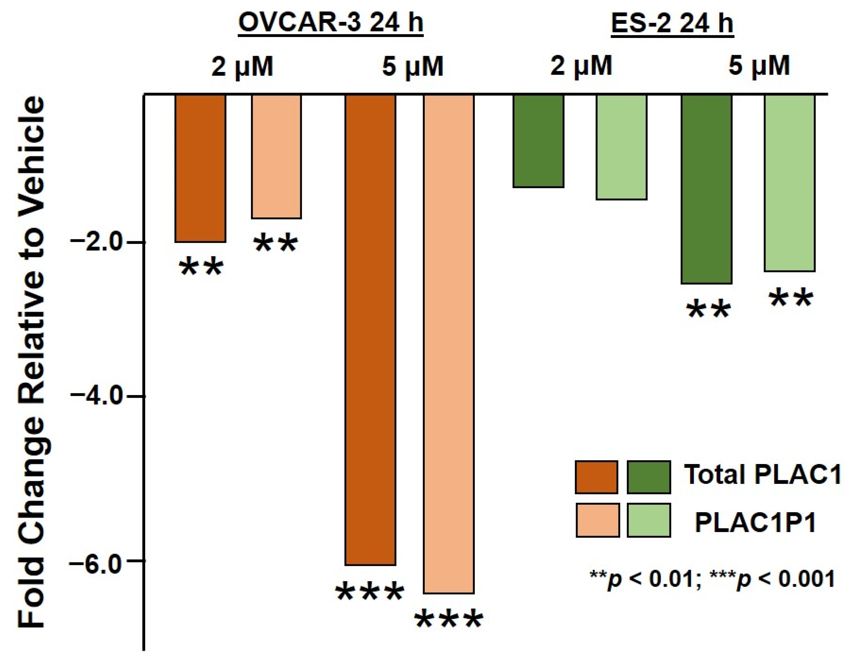
First, the OVCAR3 cells were exposed to 2 µM and 5 µM HO-3867 for 24 h and 48 h. PLAC1 expression relative to untreated OVCAR3 cells is shown in Figure 1. In all cases, HO-3867-treated cells displayed reduced PLAC1 transcription. Moreover, this suppression was focused at the P1 promoter.
Treatment of the ES-2 cells with 2 µM and 5 µM HO-3867 for 24 h and 48 h produced a similar, though not as dramatic, rescue of PLAC1 transcriptional suppression (Figure 2). Again, transcriptional suppression appeared to be focused on transcripts originating from the PLAC1 P1 promoter.

2.2. HO-3867 Treatment of Both OVCAR3 and ES-2 Ovarian Cancer Cells Results in a Reduction in Proliferation and an Increase in Apoptosis

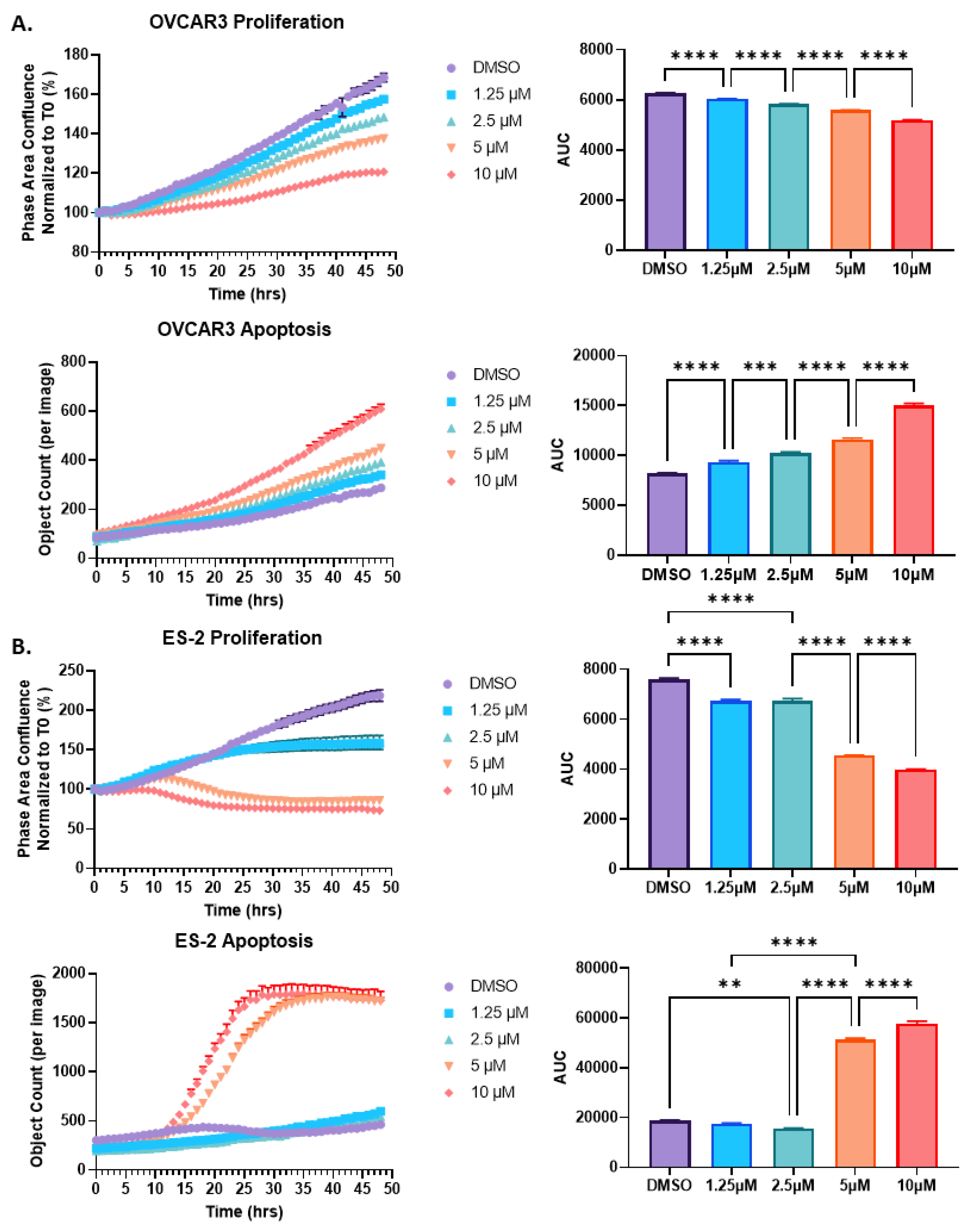
Given the importance of p53 in proliferation and apoptosis, we next assessed the functional effect of HO-3867 exposure on both the OVCAR3 and ES-2 cells. Overall, the HO-3867 treatment revealed a dose-dependent decrease in proliferation and a dose-dependent increase in apoptosis in both cell lines (Figure 3). Specifically, treatment of the ES-2 cells with HO-367 showed decreased proliferation at 1.25 µM and 2.5 µM with no change in apoptosis, whereas with higher doses of HO-3867 (5 µM and 10 µM), the ES-2 cells had decreased proliferation and increased apoptosis. Interestingly, HO-3867 had a greater effect on the ES-2 cells compared to the OVCAR3 cells for both proliferation and apoptosis, especially at higher concentrations of HO-3867. The results indicate that HO-3867 is capable of arresting cell growth and inducing cell death through the reactivation of p53 and that the effect of HO-3867 is not only cell-type-specific but also putatively dependent on p53 mutation status.

3. Discussion

It has long been recognized that a TP53 mutation is a molecular vulnerability awaiting therapeutic exploitation [44,45,46,47]. Wild-type p53 has a wide variety of cellular roles, including being a powerful tumor suppressor. Thus, it follows that the re-introduction of wild-type p53 into tumor cells would result in a positive anti-tumor effect [48]. However, attempts to do this have so far proved unsuccessful [49,50]. For example, gene therapy with p53 failed in clinical trials due to its insufficient accumulation in tumor tissue [49]. A promising alternative strategy would be to introduce a compound that is specific for mutant p53 and would also restore wild-type function. To this end, a number of such compounds have been developed, including quinuclidines, 2-sulfonylpyrimidines, zinc metallochaperones and thiosemicarbizones [51]. Some of these are well known, such as COTI-2 [52], PRIMA1 and PRIMA1met, now designated as APR-246 [53,54]. Here, we have chosen to assess the less well-known synthetic diarylidenylpiperidone curcumin analog, designated HO-3867, for the purpose of rescuing the suppression of the expression of the oncogene PLAC1 in ovarian cancer cells harboring a missense TP53 mutation.
HO-3867 binds p53 at Cys182 and Cys277, creating a “clamp” that holds the β-sandwich in place in a proper DNA-binding configuration while also stabilizing the Zn++ binding pocket [42]. The mature p53 protein in this configuration is thus able to function like wild-type p53, including in binding DNA and inducing the transcription of canonical p53 target genes. This was shown by Madan et al. [42] in a panel of 29 human cancer cell lines representing 11 different cancers. These cell lines harbor a total of 26 different missense TP53 mutations, including six classified as “gain of function” mutants [55]. The effect of exposing these cell lines to HO-3867 was assessed via qPCR expression assays on fourteen known p53 transcriptional target loci [42,56,57]. In the vast majority of these 406 assays (29 cell lines by 14 loci), HO-3867 exposure rescued the appropriate up- or down-regulation of transcription. Indeed, the overall pattern of transcriptional changes was highly significant (5 × 10−211) [42]. Our study adds to the literature by providing evidence for the appropriate down-regulation of another p53-regulated gene, PLAC1, as well as demonstrating the effect in ovarian cancer cells, which were not included in the Madan et al. study [42].
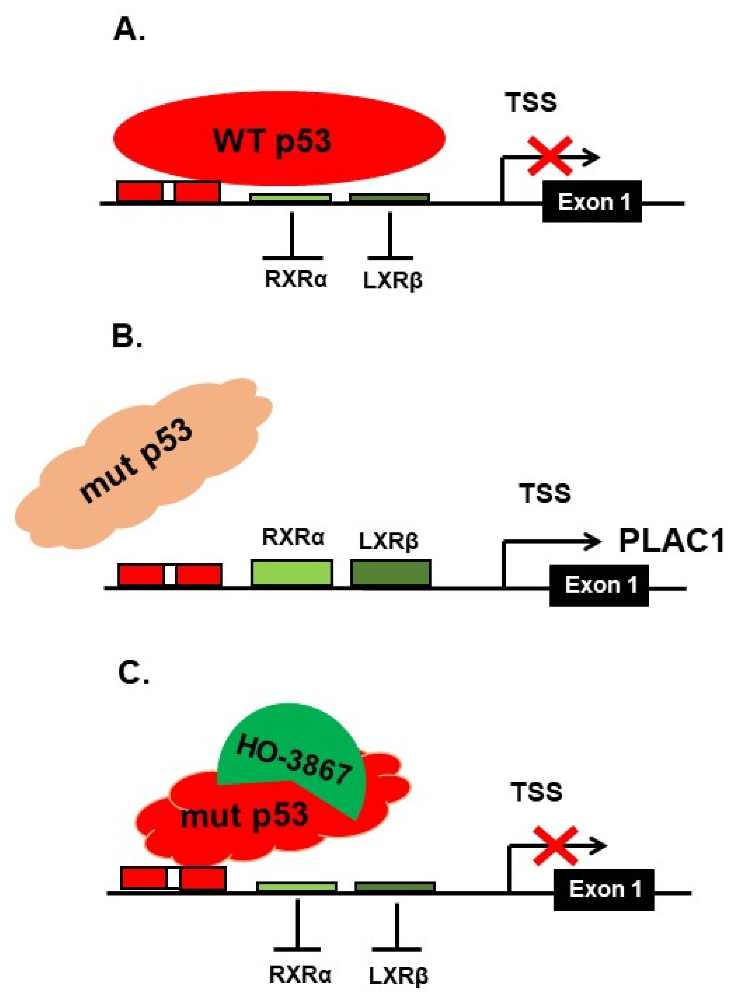
A model describing the effect of HO-3867 exposure on PLAC1 transcription in cancer cells harboring DNA-binding domain missense mutations is presented in Figure 4. In this model the P1 promoter is sterically blocked when wild-type p53 binds to its nearby binding site. This leads to reduced PLAC1 transcription. However, mutant p53 is unable to bind to that site, thus opening up PLAC1 transcription. Exposure to HO-3867 will, in some mutants, restore wild-type configuration, once again sterically blocking the binding site and reducing PLAC1 transcription.
Where ovarian cancer has been the focus of the effect of HO-3867 is a series of papers originating from The Ohio State University [58,59,60,61]. In those papers, it has been demonstrated that HO-3867 is cytotoxic to ovarian cancer cells through the induction of apoptosis and the abrogation of cisplatin resistance. Their primary readout was STAT3 protein expression and STAT3 phosphorylation. It should be noted that STAT3 is also a direct transcriptional target of p53 [56], though it remains unclear if the p53 regulation of STAT3 is the mechanism of action of HO-3867. Thus, it is possible that the changes in proliferation and apoptosis in our study may be due to the restoration of p53 regulation of genes in addition to PLAC1.
Finally, while it can be argued that the treatment of ovarian cancer patients with HO-3867—preferably as an adjuvant to conventional therapies—could improve outcomes for these patients, its effect is only possible for those cancers that harbor a TP53 missense mutation. Obviously, nonsense mutants will often result in the truncation of transcripts that are likely to be eliminated by nonsense-mediated RNA decay [62] or another mechanism that would render the cells devoid of p53 protein expression (i.e., p53 null). Further, the small number of TP53 missense mutations that occur outside the DNA-binding domain will also not be rescued by HO-3867, as the mechanism of HO-3867 action would not, for example, affect p53 tetramerization. However, taking all of the potential exceptions to the effect of HO-3867 exposure on ovarian cancer into account, there remains around 50% of all ovarian cancers that will qualify. Given the fact that the American Cancer Society estimates that there are roughly 22,000 new cases of ovarian cancer in the United States each year, some 11,000 patients could potentially benefit from adding HO-3867 to their treatment regimens. Naturally, the efficacy of using such an adjuvant therapy can only be assessed through an appropriately designed clinical trial.

4. Materials and Methods

4.1. Cell Culture

NIH:OVCAR3 cells were established from ascites from a 60-year-old patient presenting with a poorly differentiated ovarian adenocarcinoma [63]. We obtained these cells from the American Type Culture Collection (HTB-161) and propagated them in RPMI-1640 media supplemented with 20% fetal bovine serum, 0.01 mg/mL bovine insulin and 1% pen/strep antibiotic at 37 °C and 5% CO2.
ES-2 cells were established from primary tumor tissue from a 47-year-old patient presenting with clear-cell adenocarcinoma [64]. We obtained these cells from AddexBio (C0017006) and propagated them in McCoy’s 5A media supplemented with 10% fetal bovine serum and 1% pen/strep antibiotic at 37 °C and 5% CO2.
Cell line identities were verified via CODIS DNA profiling at BioSynthesis, Inc. (Lewisville, TX, USA) and by direct TP53 sequencing. Sequencing confirmed that our OVCAR3 cells harbor the R248Q gain-of-function mutant and that our ES-2 cells harbor the unknown function S241F missense mutant as expected.

4.2. HO-3867 Treatment

The HO-3867 reagent was purchased from Cayman Chemical (Ann Arbor, MI, USA, Item No. 21581). Five milligrams of HO-3867 crystals were dissolved in 10.76 mL of DMSO to produce a 1 mM stock solution that was then added to the appropriate cell media to generate the final experimental concentrations.
Depending upon the assay to be carried out, the OVCAR3 and ES-2 cells were seeded in optimum media in 6-well or 96-well culture plates (200,000 cells and 5000 cells per well, respectively) for 24 h prior to the HO-3867 treatment.

4.3. RNA Purification and Quality Control

The total cellular RNA was purified from cells using the RNeasy Plus Kit following the manufacturer’s instructions (QIAGEN, Germantown, MD, USA). The RNA yield and purity were assessed in the Genomics Division of the Iowa Institute of Human Genetics (IIHG) using an Agilent Model 2100 DNA Analyzer and a Trinean DropSense 16 spectrophotometer. Samples with an RNA integrity number (RIN) above 7.0 were used for subsequent protocols [65].

4.4. Real-Time PCR

Equal-mass RNA aliquots (500 ng) from treated cells were reverse transcribed in the presence of SuperScript III Reverse Transcriptase and an oligo (dT) primer (Thermo Fisher Scientific, Waltham, MA, USA). The resulting cDNAs were then amplified in the presence of Power SYBR Green (Thermo Fisher) on an Applied Biosystems Model 7900HT platform in the Genomics Division of the Iowa Institute of Human Genetics (IIHG) using the locus-specific primer pairs shown in Table 1.
The raw expression Ct values were normalized against 18S rRNA (ΔCt). The fold change relative to the untreated cells was calculated via the conventional ΔΔCt method, where fold change is 2−ΔΔCt [66,67]. The statistical significance of fold changes was assessed via a t-test with unequal variances [68]. All experiments were carried out in triplicate.

4.5. Cell Proliferation and Apoptosis

Proliferation: The OVCAR3 and ES-2 cells were plated on 96-well plates. Following a 24 h incubation, the cells were treated with increasing amounts of HO-3867. The cells were imaged using the Incucyte S3 live-cell imaging platform (Sartorius AG, Göttingen, Germany) every hour for up to 48 h. The phase-contrast images were processed using masks and filters to determine cell confluence with the Incucyte Analysis Software (2019B Rev3). Proliferation was plotted as percent confluence normalized to time 0.
Apoptosis: The OVCAR3 and ES-2 cells were plated on 96-well plates. Following a 24 h incubation, the cells were treated with increasing amounts of HO-3867 in combination with 5 uM Incucyte Caspase-3/7 Green Dye (Sartorius # 4440). The cells were imaged with the Incucyte S3 fluorescence module every hour for up to 48 h. The fluorescent images were processed with the Incucyte Analysis software (2019B Rev3). Caspase 3/7 activation was plotted as the number of fluorescent objects per image. The graphs were created using GraphPad Prism (v9.1.0). All experiments were carried out in triplicate.

5. Conclusions

We have demonstrated that the synthetic curcumin analog HO-3867 successfully rescues the p53- mediated suppression of PLAC1 transcription in ovarian cancer cells harboring non-truncating TP53 mutations. As a high expression of PLAC1 is well known to be associated with poor patient outcomes in a variety of cancers, the use of this compound in conjunction with conventional therapies may serve to improve these outcomes.

Author Contributions

E.J.D. conceived and initiated this project; cell culture, treatments and cell biology assays were carried out by E.J.D., B.M.S. and J.R.L.; RNA purifications and qPCR were performed by E.J.D., B.M.S. and J.R.L.; manuscript writing, reviewing and editing was done by all authors; funding was provided by K.K.L., B.M.S., J.G.-B. and D.P.B. All authors have read and agreed to the published version of the manuscript.

Funding

This work was supported by NIH R01CA99908 and R01CA184101 (K.K.L.), Department of Defense Ovarian Cancer Research Program OC190352 (K.K.L.), NIH32HL007101 (B.M.S.), 2018 ABOG/AAOGF Bridge Funding Award (J.G.-B.), the University of Iowa Department of Obstetrics and Gynecology Research Development Fund and by the Iowa cancer patient advocacy group CCKMA (D.P.B.). The Genome Core Facility of the Iowa Institute of Human Genetics is funded, in part, through NIH/NCI P30CA086862 to the Holden Comprehensive Cancer Center at the University of Iowa Hospitals and Clinics. Funders did not participate in the study design, data collection or analysis, or preparation of the manuscript.

Institutional Review Board Statement

Not applicable.

Informed Consent Statement

Not applicable.

Data Availability Statement

Data are contained within the article.

Acknowledgments

The authors wish to acknowledge the continued support and encouragement from UIHC gynecologic cancer patients and their families, as well as the invaluable support provided by the Genome Core Facility, particularly Mary Boes and Garry Hauser, without whom much of this work would not have been possible.

Conflicts of Interest

The authors declare no conflict of interest. The funders had no role in the design of the study; in the collection, analyses or interpretation of data; in the writing of the manuscript or in the decision to publish the results.

References

  1. Cocchia, M.; Huber, R.; Pantano, S.; Chen, E.Y.; Ma, P.; Forabosco, A.; Ko, M.S.; Schlessinger, D. PLAC1, an Xq26 gene with placenta-specific expression. Genomics 2000, 68, 305–312. [Google Scholar] [CrossRef] [Green Version]
  2. Fant, M.; Barerra-Saldana, H.; Dubinsky, W.; Poindexter, B.; Bick, R. The PLAC1 protein localizes to membranous compartments in the apical region of the syncytiotrophoblast. Mol. Reprod. Dev. 2007, 74, 922–929. [Google Scholar] [CrossRef] [PubMed]
  3. Farina, A.; Concu, M.; Banzola, I.; Tempesta, A.; Vagnoni, S.; Gabrielli, S.; Mattioli, M.; Carinci, P.; Pilu, G.; Morano, D.; et al. PLAC1 mRNA in maternal blood correlates with Doppler waveform in uterine arteries in normal pregnancies at the second and third trimester. Ann. N. Y. Acad. Sci. 2006, 1075, 130–136. [Google Scholar] [CrossRef]
  4. Fujito, N.; Samura, O.; Miharu, N.; Tanigawa, M.; Hyodo, M.; Kudo, Y. Increased plasma mRNAs of placenta-specific 1 (PLAC1) and glial cells-missing 1 (GCM1) in mothers with pre-eclampsia. Hiroshima J. Med. Sci. 2006, 55, 9–15. [Google Scholar] [PubMed]
  5. Purwosunu, Y.; Sekizawa, A.; Farina, A.; Wibowo, N.; Okazaki, S.; Nakamura, M.; Samura, O.; Fujito, N.; Okai, T. Cell-free mRNA concentrations of CRH, PLAC1, and selectin-P are increased in the plasma of pregnant women with preeclampsia. Prenat. Diagn. 2007, 27, 772–777. [Google Scholar] [CrossRef]
  6. Kodama, M.; Miyoshi, H.; Fujito, N.; Samura, O.; Kudo, Y. Plasma mRNA concentrations of placenta-specific 1 (PLAC1) and pregnancy associated plasma protein A (PAPP-A) are higher in early-onset than late-onset pre-eclampsia. J. Obstet. Gynecol. Res. 2011, 37, 313–318. [Google Scholar] [CrossRef] [PubMed]
  7. Kotto-Kome, A.C.; Silva, C.; Whiteman, V.; Kong, X.; Fant, M.E. Circulating anti-PLAC1 antibodies during pregnancy and in women with reproductive failure: A preliminary analysis. ISRN Immunol. 2011, 530491. [Google Scholar] [CrossRef] [Green Version]
  8. Jackman, S.M.; Kong, X.; Fant, M.E. Plac1 (placenta-specific 1) is essential for normal placental and embryonic development. Mol. Reprod. Dev. 2012, 79, 564–572. [Google Scholar] [CrossRef] [PubMed] [Green Version]
  9. Matteo, M.; Greco, P.; Levi Setti, P.E.; Morenghi, E.; De Rosario, F.; Massenzio, F.; Albani, E.; Totaro, P.; Liso, A. Preliminary evidence for high anti-PLAC1 antibody levels in infertile patients with repeated unexplained implantation failure. Placenta 2013, 34, 335–339. [Google Scholar] [CrossRef] [PubMed]
  10. Zanello, M.; Sekizawa, A.; Purwosunu, Y.; Curti, A.; Farina, A. Circulating mRNA for the PLAC1 gene as a second trimester marker (14–18 weeks’ gestation) in the screening for late preeclampsia. Fetal Diagn. Ther. 2014, 36, 196–201. [Google Scholar] [CrossRef] [PubMed]
  11. Ibanoglu, M.C.; Ozgu-Erdinc, A.S.; Uygur, D. Maternal Plac1 protein levels in early- and late-onset preeclampsia. Ginekol. Pol. 2018, 89, 147–152. [Google Scholar] [CrossRef] [PubMed] [Green Version]
  12. Ibanoglu, M.C.; Ozgu-Erdinc, A.S.; Kara, O.; Topcu, H.O.; Uygur, D. Association of Higher Maternal Serum Levels of Plac1 Protein with Intrauterine Growth Restriction. Z. Geburtshilfe Neonatol. 2019, 223, 285–288. [Google Scholar] [CrossRef] [Green Version]
  13. Wan, L.; Sun, D.; Xie, J.; Du, M.; Wang, P.; Wang, M.; Lei, Y.; Wang, H.; Wang, H.; Dong, M. Declined placental PLAC1 expression is involved in preeclampsia. Medicine. 2019, 98, e17676. [Google Scholar] [CrossRef] [PubMed]
  14. Levine, L.; Habertheuer, A.; Ram, C.; Korutla, L.; Schwartz, N.; Hu, R.W.; Reddy, S.; Freas, A.; Zielinski, P.D.; Harmon, J.; et al. Syncytiotrophoblast extracellular microvesicle profiles in maternal circulation for noninvasive diagnosis of preeclampsia. Sci. Rep. 2020, 10, 6398. [Google Scholar] [CrossRef] [PubMed]
  15. Yilmaz, N.; Timur, H.; Ugurlu, E.N.; Yilmaz, S.; Ozgu-Erdinc, A.S.; Erkilinc, S.; Inal, H.A. Placenta specific protein-1 in recurrent pregnancy loss and in In Vitro Fertilisation failure: A prospective observational case-control study. J. Obstet. Gynaecol. 2020, 40, 843–848. [Google Scholar] [CrossRef]
  16. Chen, J.; Pang, X.W.; Liu, F.F.; Dong, X.Y.; Wang, H.C.; Wang, S.; Zhang, Y.; Chen, W.F. [PLAC1/CP1 gene expression and autologous humoral immunity in gastric cancer patients]. Beijing Da Xue Xue Bao Yi Xue Ban 2006, 38, 124–127. (In Chinese) [Google Scholar]
  17. Koslowski, M.; Sahin, U.; Mitnacht-Kraus, R.; Seitz, G.; Huber, C.; Türeci, O. A placenta-specific gene ectopically activated in many human cancers is essentially involved in malignant cell processes. Cancer Res. 2007, 67, 9528–9534. [Google Scholar] [CrossRef] [Green Version]
  18. Silva, W.A., Jr.; Gnjatic, S.; Ritter, E.; Chua, R.; Cohen, T.; Hsu, M.; Jungbluth, A.A.; Altorki, N.K.; Chen, Y.T.; Old, L.J.; et al. PLAC1, a trophoblast-specific cell surface protein, is expressed in a range of human tumors and elicits spontaneous antibody responses. Cancer Immunol. 2007, 7, 18. [Google Scholar]
  19. Dong, X.Y.; Peng, J.R.; Ye, Y.J.; Chen, H.S.; Zhang, L.J.; Pang, X.W.; Li, Y.; Zhang, Y.; Wang, S.; Fant, M.E.; et al. Plac1 is a tumor-specific antigen capable of eliciting spontaneous antibody responses in human cancer patients. Int. J. Cancer 2008, 122, 2038–2043. [Google Scholar] [CrossRef]
  20. Liu, F.F.; Dong, X.Y.; Pang, X.W.; Xing, Q.; Wang, H.C.; Zhang, H.G.; Li, Y.; Yin, Y.H.; Fant, M.; Ye, Y.J.; et al. The specific immune response to tumor antigen CP1 and its correlation with improved survival in colon cancer patients. Gastroenterology 2008, 134, 998–1006. [Google Scholar] [CrossRef]
  21. Tchabo, N.E.; Mhawech-Fauceglia, P.; Caballero, O.L.; Villella, J.; Beck, A.F.; Miliotto, A.J.; Liao, J.; Andrews, C.; Lele, S.; Old, L.J.; et al. Expression and serum immunoreactivity of developmentally restricted differentiation antigens in epithelial ovarian cancer. Cancer Immunol. 2009, 9, 6. [Google Scholar]
  22. Koslowski, M.; Türeci, O.; Biesterfeld, S.; Seitz, G.; Huber, C.; Sahin, U. Selective activation of trophoblast-specific PLAC1 in breast cancer by CCAAT/enhancer-binding protein beta (C/EBPbeta) isoform 2. J. Biol. Chem. 2009, 284, 28607–28615. [Google Scholar] [CrossRef] [Green Version]
  23. Devor, E.J.; Leslie, K.K. The oncoplacental gene placenta-specific protein 1 is highly expressed in endometrial tumors and cell lines. Obstet. Gynecol. Int. 2013, 807849. [Google Scholar] [CrossRef] [PubMed] [Green Version]
  24. Yuan, H.; Lu, J.; Xiao, J.; Upadhyay, G.; Umans, R.; Kallakury, B.; Yin, Y.; Fant, M.E.; Kopelovich, L.; Glazer, R.I. PPARδ induces estrogen receptor-positive mammary neoplasia through an inflammatory and metabolic phenotype linked to mTOR activation. Cancer Res. 2013, 73, 4349–4361. [Google Scholar] [CrossRef] [Green Version]
  25. Devor, E.J.; Reyes, H.D.; Santillan, D.A.; Santillan, M.K.; Onukwugha, C.; Goodheart, M.J.; Leslie, K.K. Placenta-specific protein 1: A potential key to many oncofetal-placental OB/GYN research questions. Obstet. Gynecol. Int. 2014, 678984. [Google Scholar] [CrossRef] [PubMed] [Green Version]
  26. Ghods, R.; Ghahremani, M.H.; Madjd, Z.; Asgari, M.; Abolhasani, M.; Tavasoli, S.; Mahmoudi, A.R.; Darzi, M.; Pasalar, P.; Jeddi-Tehrani, M.; et al. High placenta-specific 1/low prostate-specific antigen expression pattern in high-grade prostate adenocarcinoma. Cancer Immunol. Immunother. 2014, 63, 1319–1327. [Google Scholar] [CrossRef] [PubMed]
  27. Liu, F.; Zhang, H.; Shen, D.; Wang, S.; Ye, Y.; Chen, H.; Pang, X.; Song, Q.; He, P. Identification of two new HLA-A*0201-restricted cytotoxic T lymphocyte epitopes from colorectal carcinoma-associated antigen PLAC1/CP1. J. Gastroenterol. 2014, 49, 419–426. [Google Scholar] [CrossRef] [PubMed]
  28. Liu, F.; Shen, D.; Kang, X.; Zhang, C.; Song, Q. New tumour antigen PLAC1/CP1, a potentially useful prognostic marker and immunotherapy target for gastric adenocarcinoma. J. Clin. Pathol. 2015, 68, 913–916. [Google Scholar] [CrossRef] [PubMed]
  29. Wu, Y.; Lin, X.; Di, X.; Chen, Y.; Zhao, H.; Wang, X. Oncogenic function of Plac1 on the proliferation and metastasis in hepatocellular carcinoma cells. Oncol. Rep. 2017, 37, 465–473. [Google Scholar] [CrossRef] [Green Version]
  30. Guo, L.; Xu, D.; Lu, Y.; Peng, J.; Jiang, L. Detection of circulating tumor cells by reverse transcription-quantitative polymerase chain reaction and magnetic activated cell sorting in the peripheral blood of patients with hepatocellular carcinoma. Mol. Med. Rep. 2017, 16, 5894–5900. [Google Scholar] [CrossRef] [PubMed] [Green Version]
  31. Yin, Y.; Zhu, X.; Huang, S.; Zheng, J.; Zhang, M.; Kong, W.; Chen, Q.; Zhang, Y.; Chen, X.; Lin, K.; et al. Expression and clinical significance of placenta-specific 1 in pancreatic ductal adenocarcinoma. Tumour Biol. 2017, 39, 1–8. [Google Scholar] [CrossRef] [Green Version]
  32. Devor, E.J.; Gonzalez-Bosquet, J.; Warrier, A.; Reyes, H.D.; Ibik, N.V.; Schickling, B.M.; Newtson, A.; Goodheart, M.J.; Leslie, K.K. p53 mutation status is a primary determinant of placenta-specific protein 1 expression in serous ovarian cancers. Int. J. Oncol. 2017, 50, 1721–1728. [Google Scholar] [CrossRef] [PubMed] [Green Version]
  33. Devor, E.J.; Reyes, H.D.; Gonzalez-Bosquet, J.; Warrier, A.; Kenzie, S.A.; Ibik, N.V.; Miller, M.D.; Schickling, B.M.; Goodheart, M.J.; Thiel, K.W.; et al. Placenta-specific protein 1 expression in human papillomavirus 16/18-positive cervical cancers is associated with tumor histology. Int. J. Gynecol. Cancer 2017, 27, 784–790. [Google Scholar] [CrossRef]
  34. Li, Y.; Chu, J.; Li, J.; Feng, W.; Yang, F.; Wang, Y.; Zhang, Y.; Sun, C.; Yang, M.; Vasilatos, S.N.; et al. Cancer/testis antigen-Plac1 promotes invasion and metastasis of breast cancer through Furin/NICD/PTEN signaling pathway. Mol. Oncol. 2018, 12, 1233–1248. [Google Scholar] [CrossRef] [PubMed]
  35. Yang, L.; Zha, T.Q.; He, X.; Chen, L.; Zhu, Q.; Wu, W.B.; Nie, F.Q.; Wang, Q.; Zang, C.S.; Zhang, M.L. Placenta-specific protein 1 promotes cell proliferation and invasion in non-small cell lung cancer. Oncol. Rep. 2018, 39, 53–60. [Google Scholar] [CrossRef] [PubMed] [Green Version]
  36. Yuan, H.; Wang, X.; Shi, C.; Jin, L.; Hu, J.; Zhang, A.; Li, J.; Vijayendra, N.; Doodala, V.; Weiss, S.; et al. Plac1 is a key regulator of the inflammatory response and immune tolerance in mammary tumorigenesis. Sci. Rep. 2018, 8, 5717. [Google Scholar] [CrossRef]
  37. Lin, C.; Qian, P.; Zhang, Y.; Liu, Z.; Dai, K.; Sun, D. Plac1 promotes nasopharyngeal carcinoma cells proliferation, migration and invasion via Furin/NICD/PTEN pathway. Tissue Cell Ress. 2021, 69, 101480. [Google Scholar] [CrossRef] [PubMed]
  38. Ma, J.; Li, L.; Du, J.; Pan, C.; Zhang, C.; Chen, Y. Placenta-specific protein 1 enhances liver metastatic potential and is associated with the PI3K/AKT/NF-κB signaling pathway in colorectal cancer. Eur. J. Cancer Prev. 2021, 30, 220–231. [Google Scholar] [CrossRef]
  39. Chen, Y.; Moradin, A.; Schlessinger, D.; Nagaraja, R. RXRα and LXR activate two promoters in placenta- and tumor-specific expression of PLAC1. Placenta 2011, 32, 877–884. [Google Scholar] [CrossRef] [PubMed] [Green Version]
  40. Devor, E.J. Placenta-specific protein 1 (PLAC1) is a unique onco-fetal-placental protein and an underappreciated therapeutic target in cancer. Integr. Cancer Sci. Ther. 2016, 3, 479–483. [Google Scholar] [CrossRef] [Green Version]
  41. Chen, Y.; Schlessinger, D.; Nagaraja, R. T antigen transformation reveals TP53/RB-dependent route to PLAC1 transcription activation in primary fibroblasts. Oncogenesis 2013, 2, e67. [Google Scholar] [CrossRef] [Green Version]
  42. Madan, E.; Parker, T.M.; Bauer, M.R.; Dhiman, A.; Pelham, C.J.; Nagane, M.; Kuppusamy, M.L.; Holmes, M.; Holmes, T.R.; Shaik, K.; et al. The curcumin analog HO-3867 selectively kills cancer cells by converting mutant p53 protein to transcriptionally active wildtype p53. J. Biol. Chem. 2018, 293, 4262–4276. [Google Scholar] [CrossRef] [PubMed] [Green Version]
  43. Mullany, L.K.; Wong, K.-K.; Marciano, D.C.; Katsonis, P.; King-Crane, E.R.; Ren, Y.A.; Lichtarge, O.; Richards, J.S. Specific TP53 mutants overrepresented in ovarian cancer impact CNV, TP53 activity, responses to Nutlin-3a, and cell survival. Neoplasia 2015, 17, 789–803. [Google Scholar] [CrossRef] [Green Version]
  44. Joerger, A.C.; Fersht, A.R. Structure-function-rescue: The diverse nature of common p53 cancer mutants. Oncogene 2007, 26, 2226–2242. [Google Scholar] [CrossRef] [Green Version]
  45. Emerling, B.M.; Hurov, J.B.; Poulogiannis, G.; Tsukazawa, K.S.; Choo-Wing, R.; Wulf, G.M.; Bell, E.L.; Shim, H.-S.; Lamia, K.A.; Rameh, L.E.; et al. Depletion of a putatively druggable class of phosphatidylinositol kinases inhibits growth of p53-null tumors. Cell 2013, 155, 844–857. [Google Scholar] [CrossRef] [PubMed] [Green Version]
  46. Parrales, A.; Iwakuma, T. Targeting oncogenic mutant p53 for cancer therapy. Front. Oncol. 2015, 5, 288. [Google Scholar] [CrossRef] [PubMed] [Green Version]
  47. Bykov, V.J.N.; Issaeva, N.; Shilov, A.; Hultcrantz, M.; Pugacheva, E.; Chumakov, P.; Bergman, J.; Wiman, K.; Selivanova, G. Restoration of the tumor suppressor function to mutant p53 by a low molecular-weight compound. Nat. Med. 2002, 8, 282–288. [Google Scholar] [CrossRef] [PubMed]
  48. Kastan, M.B. Wild-type p53: Tumors can’t stand it. Cell 2007, 128, 837–840. [Google Scholar] [CrossRef] [Green Version]
  49. Vinyals, A.; Peinado, M.A.; Gonzalez-Garrigues, M.; Monzo, M.; Binfil, R.D.; Fabra, A. Failure of wild-type p53 gene therapy in human cancer cells expressing a mutant p53 protein. Gene Ther. 1999, 6, 22–33. [Google Scholar] [CrossRef] [Green Version]
  50. Watanabe, T.; Sullenger, B.A. Introduction of wild-type p53 activity in human cancer cells by ribozymes that repair mutant p53 transcripts. Proc. Natl. Acad. Sci. USA 2000, 97, 8490–8494. [Google Scholar] [CrossRef] [PubMed] [Green Version]
  51. Duffy, M.J.; Synnott, N.C.; Crown, J. Mutant p53 as a target for cancer treatment. Eur. J. Cancer 2017, 83, 258–265. [Google Scholar] [CrossRef]
  52. Vareki, S.M.; Salim, K.Y.; Danter, W.R.; Koropatnick, J. Novel anti-cancer drug COTI-2 synergizes with therapeutic agents and does not induce resistance or exhibit cross-resistance in human cancer cell lines. PLoS ONE 2018, 13, e0191766. [Google Scholar] [CrossRef] [PubMed] [Green Version]
  53. Lambert, J.M.R.; Gorzov, P.; Veprintsev, D.B.; Soderqvist, M.; Segerback, D.; Bergman, J.; Fersht, A.R.; Hainault, P.; Wiman, K.G.; Bykov, V.J.N. PRIMA-1 reactivates mutant p53 by covalently binding to the core domain. Cancer Cell 2019, 15, 376–388. [Google Scholar] [CrossRef] [PubMed] [Green Version]
  54. Zhang, Q.; Bykov, V.J.N.; Wiman, K.; Zawacka-Pankua, J. APR-246 reactivates mutant p53 by targeting cysteines 124 and 277. Cell Death Dis. 2018, 9, 439. [Google Scholar] [CrossRef] [PubMed] [Green Version]
  55. Brachova, P.; Mueting, S.R.; Devor, E.J.; Leslie, K.K. Oncomorphic TP53 mutations in gynecologic cancers lose the normal protein:protein interactions with the microRNA microporcessing complex. J. Cancer Ther. 2014, 5, 506–516. [Google Scholar] [CrossRef] [Green Version]
  56. Fischer, M. Census and evaluation of p53 target genes. Oncogene 2017, 36, 3943–3956. [Google Scholar] [CrossRef] [Green Version]
  57. Nguyen, T.-A.T.; Grimm, S.A.; Bushel, P.R.; Li, J.; Li, Y.; Bennett, B.D.; Lavender, C.A.; Ward, J.M.; Fargo, D.C.; Anderson, C.W.; et al. Revealing a human p53 universe. Nucleic Acids Res. 2018, 46, 8153–8167. [Google Scholar] [CrossRef] [Green Version]
  58. Selvendiran, K.; Tong, L.; Bratasz, A.; Kuppusamy, M.L.; Ahmed, S.; Ravi, Y.; Trigg, N.J.; Rivera, B.K.; Kalai, T.; Hideg, K.; et al. Anticancer efficacy of a difluorodiarylidenyl piperidone (HO-3867) in human ovarian cancer cells and tumor xenographs. Mol. Cancer Ther. 2010, 9, 1169–1179. [Google Scholar] [CrossRef] [Green Version]
  59. Selvendiran, K.; Ahmed, S.; Dayton, A.; Kuppusamy, M.L.; Rivera, B.K.; Kalai, T.; Hideg, K.; Kuppusamy, P. HO-3867, a curcumin analog, sensitizes cisplatin-resistant ovarian carcinoma, leading to therapeutic synergy through STAT3 inhibition. Cancer Biol. Ther. 2011, 12, 837–845. [Google Scholar] [CrossRef] [Green Version]
  60. Rath, K.S.; Naidu, S.K.; Lata, P.; Bid, H.K.; Rivera, B.K.; McCann, G.A.; Tierney, B.J.; ElNaggar, A.C.; Bravo, V.; Leone, G.; et al. HO-3867, a safe STAT3 inhibitor, is selectively cytotoxic to ovarian cancer. Cancer Res. 2014, 74, 2316–2327. [Google Scholar] [CrossRef] [Green Version]
  61. Bixel, K.; Saini, U.; Bid, H.K.; Fowler, J.; Riley, M.; Wanner, R.; Dorayappan, K.D.P.; Rajendran, S.; Konishi, I.; Matsumura, N.; et al. Targeting STAT3 by HO-3867 induces apoptosis in ovarian clear cell carcinoma. Int. J. Cancer 2017, 141, 1856–1866. [Google Scholar] [CrossRef] [Green Version]
  62. Lindeboom, R.G.H.; Supek, F.; Lehner, B. The rules and impact of nonsense-mediated mRNA decay in human cancers. Nat. Genet. 2017, 48, 1112–1118. [Google Scholar] [CrossRef] [Green Version]
  63. Hamilton, T.C.; Young, R.C.; McKoy, W.M.; Grotzinger, K.R.; Green, J.A.; Chu, E.W.; Whang-Peng, J.; Rogan, A.M.; Green, W.R.; Ozols, R.F. Characterization of a human ovarian carcinoma cell line (NIH:OVCAR3) with androgen and estrogen receptors. Cancer Res. 1983, 43, 5379–5398. [Google Scholar] [PubMed]
  64. Lau, D.H.; Lewis, A.D.; Ehsan, M.N.; Sikic, B.I. Multifactorial mechanisms associated with broad cross-resistance of ovarian carcinoma cells selected by cyanomorpholino doxorubicin. Cancer Res. 1991, 51, 5181–5187. [Google Scholar] [PubMed]
  65. Schroeder, A.; Mueller, O.; Stocker, S.; Salowsky, R.; Leiber, M.; Gassmann, M.; Lightfoot, S.; Menzel, W.; Granzow, M.; Ragg, T. The RIN: An RNA integrity number for assigning integrity values to RNA measurements. BMC Mol. Biol. 2006, 7, 3. [Google Scholar] [CrossRef] [PubMed] [Green Version]
  66. Livak, K.J.; Schmittgen, T.D. Analysis of relative gene expression data using real-time quantitative PCR and the 2(-Delta Delta, C.(T)) Method. Methods 2001, 25, 402–408. [Google Scholar] [CrossRef] [PubMed]
  67. Schmittgen, T.D.; Livak, K.J. Analyzing real-time PCR data by the comparative Ct method. Nat. Protoc. 2008, 3, 1101–1108. [Google Scholar] [CrossRef]
  68. Snedecor, G.W.; Cochran, W.G. Statistical Methods, 8th ed.; State University Press: Ames, IA, USA, 1989. [Google Scholar]
Figure 1. Total PLAC1 and PLAC1P1-specific expression in OVCAR3 ovarian cancer cells treated with 2 µM and 5 µM HO-3867 for 24 h and 48 h.
Pharmaceuticals 14 00942 g001
Figure 2. Total PLAC1 and PLAC1P1-specific expression in ES-2 ovarian cancer cells treated with 2 µM and 5 µM HO-3867 for 24 h. ES-2 cells exposed to 2 µM HO-3867 for 48 h did not yield RNA of sufficient quality to perform qPCR, and ES-2 cells exposed to 5 µM HO-3867 for 48 h did not survive. The 24 h results for OVCAR3 cells are shown on the left for comparison.
Pharmaceuticals 14 00942 g002
Figure 3. Proliferation and apoptosis in OVCAR3 (A) and ES-2 (B) cells. Cells were treated with increasing amounts of HO-3867, as shown. Proliferation was measured by phase contrast, and apoptosis was assessed by caspase 3/7 activation. Cells were monitored hourly for 48 h using the Incucyte S3 live-cell imaging platform. Area under the curve (AUC) values were calculated automatically by the Incucyte Software and analyzed via ANOVA. Significance values are indicated as ** p < 0.01; *** p < 0.001; **** p < 0.0001.
Pharmaceuticals 14 00942 g003
Figure 4. A model of the effect of HO-3867 on p53 binding in the P1 promoter of PLAC1. In the top panel (A), wild-type p53 freely binds to its binding site, sterically hindering access of the P1 transcription factors RXRα and LXRβ to their binding sites. This has the effect of suppressing PLAC1 transcription. However, when p53 is mutated, as in the middle panel (B), p53 is unable to bind to its binding site, thus freeing the P1 transcription factors RXRα and LXRβ to bind to theirs and promoting PLAC1 transcription. When exposed to HO-3867, as in the lower panel (C), certain p53 mutants are reactivated and will bind to the p53 binding site, once again sterically hindering access of the P1 transcription factors RXRα and LXRβ to their binding sites and hindering PLAC1 transcription. TSS is the transcription start site.
Pharmaceuticals 14 00942 g004
Table 1. Primer pairs used for SYBR Green qPCR in this study. Amplification conditions are 95 °C (10:00); 40 (95 °C (0:15), 60 °C (1:00)). A dissociation curve is run at 95 °C (0:15); 60 °C (0:15); 95 °C (0:15) following the amplification.
LocusSequenceAmpliconTm (°C) *
PLAC15′-CACCAGTGAGCACAAAGCCACATT-3232 bp60.3
5′-CCATGAACCAGTCTATGGAG-3′ 52.3
PLAC1P15′-AAACTTACACGAGGAGTCTGTC-3′371 bp #57.2
5′-CTGTGACCATGAACCAGTCTAT-3′285 bp #54.2
18S rRNA5′-AACTTTCGATGGTAGTCGCCG-3′104 bp57.2
5′-CCTTGGATGTGGTAGCCGTTT-3′ 54.2
* Tm calculated at 1.5 mM MgCl2. # Amplicons are from the simultaneously produced P1Long and P1Short transcripts [40].
Publisher’s Note: MDPI stays neutral with regard to jurisdictional claims in published maps and institutional affiliations.