
Systematic Review and Meta-Analysis of In Vitro Anti-Human Cancer Experiments Investigating the Use of 5-Aminolevulinic Acid (5-ALA) for Photodynamic Therapy

 , ,
, , 
Abstract
1. Introduction
2. Methods
2.1. Literature Search and Selection
2.2. Consistency of Terminology
2.3. Data Collection, Processing, and Statistics
3. Results
3.1. Collection of In Vitro 5-ALA PDT Experiments
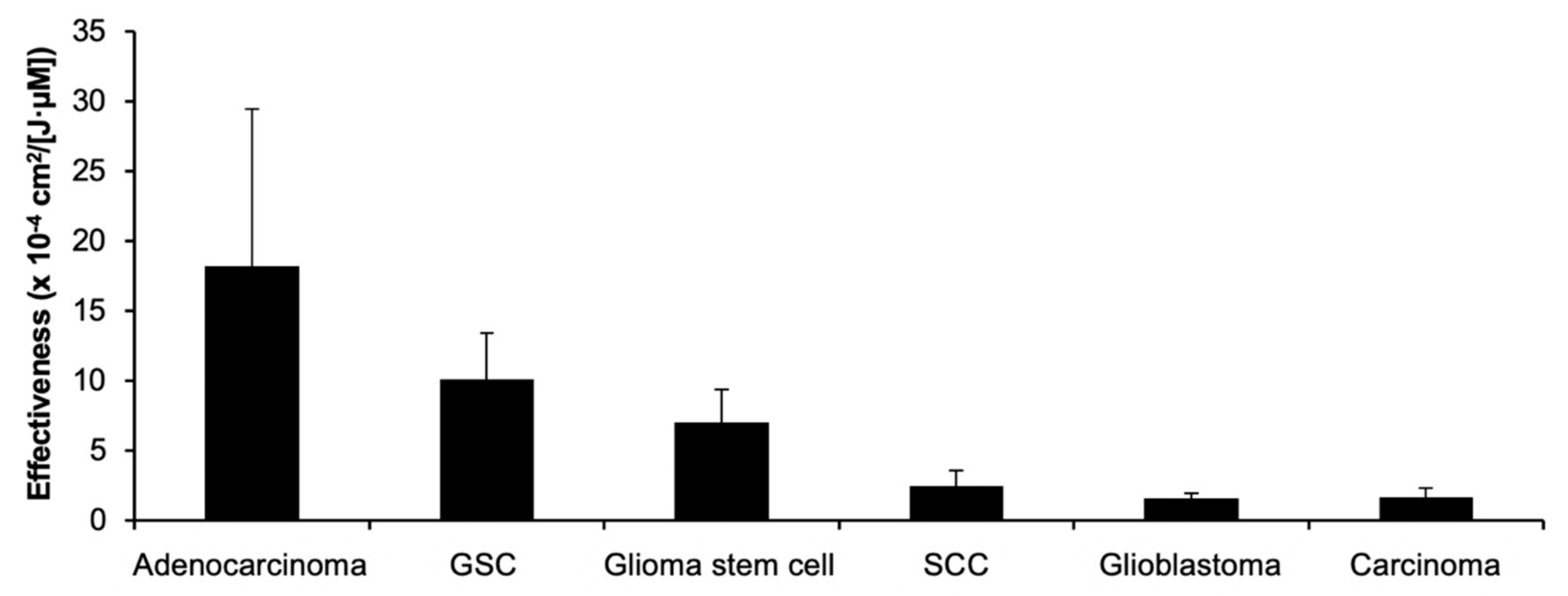
3.2. 5-ALA PDT Effect on Cells of Different Cancer Classifications
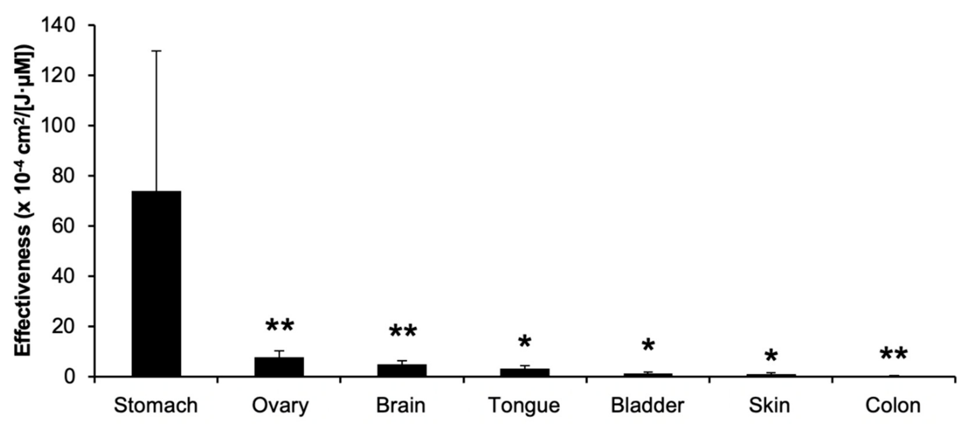
3.3. 5-ALA PDT Effect on Cells of Different Cancer Origins
4. Discussion and Future Perspective
Author Contributions
Funding
Acknowledgments
Conflicts of Interest
References
- Berlin, N.I.; Neuberger, A.; Scott, J.J. The metabolism of delta -aminolaevulic acid. 1. Normal pathways, studied with the aid of 15N. Biochem. J. 1956, 64, 80–90. [Google Scholar] [CrossRef] [PubMed]
- Berlin, N.I.; Neuberger, A.; Scott, J.J. The metabolism of delta -aminolaevulic acid. 2. Normal pathways, studied with the aid of 14C. Biochem. J. 1956, 64, 90–100. [Google Scholar] [CrossRef]
- Fukuda, H.; Casas, A.; Batlle, A. Aminolevulinic acid: From its unique biological function to its star role in photodynamic therapy. Int. J. Biochem. Cell Biol. 2005, 37, 272–276. [Google Scholar] [CrossRef]
- Kennedy, J.C.; Pottier, R.H. Endogenous protoporphyrin IX, a clinically useful photosensitizer for photodynamic therapy. J. Photochem. Photobiol. B 1992, 14, 275–292. [Google Scholar] [CrossRef]
- Kennedy, J.C.; Pottier, R.H.; Pross, D.C. Photodynamic therapy with endogenous protoporphyrin IX: Basic principles and present clinical experience. J. Photochem. Photobiol. B 1990, 6, 143–148. [Google Scholar] [CrossRef]
- Krieg, R.C.; Messmann, H.; Rauch, J.; Seeger, S.; Knuechel, R. Metabolic characterization of tumor cell-specific protoporphyrin IX accumulation after exposure to 5-aminolevulinic acid in human colonic cells. Photochem. Photobiol. 2002, 76, 518–525. [Google Scholar] [CrossRef]
- Zenzen, V.; Zankl, H. Protoporphyrin IX-accumulation in human tumor cells following topical ALA- and h-ALA-application in vivo. Cancer Lett. 2003, 202, 35–42. [Google Scholar] [CrossRef] [PubMed]
- Berg, K.; Selbo, P.K.; Weyergang, A.; Dietze, A.; Prasmickaite, L.; Bonsted, A.; Engesaeter, B.O.; Angell-Petersen, E.; Warloe, T.; Frandsen, N.; et al. Porphyrin-related photosensitizers for cancer imaging and therapeutic applications. J. Microsc. 2005, 218, 133–147. [Google Scholar] [CrossRef] [PubMed]
- Casas, A. Clinical uses of 5-aminolaevulinic acid in photodynamic treatment and photodetection of cancer: A review. Cancer Lett. 2020, 490, 165–173. [Google Scholar] [CrossRef] [PubMed]
- Ishizuka, M.; Abe, F.; Sano, Y.; Takahashi, K.; Inoue, K.; Nakajima, M.; Kohda, T.; Komatsu, N.; Ogura, S.; Tanaka, T. Novel development of 5-aminolevurinic acid (ALA) in cancer diagnoses and therapy. Int. Immunopharmacol. 2011, 11, 358–365. [Google Scholar] [CrossRef]
- Mahmoudi, K.; Garvey, K.L.; Bouras, A.; Cramer, G.; Stepp, H.; Jesu Raj, J.G.; Bozec, D.; Busch, T.M.; Hadjipanayis, C.G. 5-aminolevulinic acid photodynamic therapy for the treatment of high-grade gliomas. J. Neurooncol. 2019, 141, 595–607. [Google Scholar] [CrossRef] [PubMed]
- Cramer, S.W.; Chen, C.C. Photodynamic Therapy for the Treatment of Glioblastoma. Front. Surg. 2020, 6, 81. [Google Scholar] [CrossRef] [PubMed]
- Friesen, S.A.; Hjortland, G.O.; Madsen, S.J.; Hirschberg, H.; Engebraten, O.; Nesland, J.M.; Peng, Q. 5-Aminolevulinic acid-based photodynamic detection and therapy of brain tumors (review). Int. J. Oncol. 2002, 21, 577–582. [Google Scholar] [CrossRef]
- Stepp, H.; Stummer, W. 5-ALA in the management of malignant glioma. Lasers Surg. Med. 2018, 50, 399–419. [Google Scholar] [CrossRef]
- Nordmann, N.J.; Michael, A.P. 5-Aminolevulinic acid radiodynamic therapy for treatment of high-grade gliomas: A systematic review. Clin. Neurol. Neurosurg. 2020, 201, 106430. [Google Scholar] [CrossRef]
- Tetard, M.C.; Vermandel, M.; Mordon, S.; Lejeune, J.P.; Reyns, N. Experimental use of photodynamic therapy in high grade gliomas: A review focused on 5-aminolevulinic acid. Photodiagnosis Photodyn. Ther. 2014, 11, 319–330. [Google Scholar] [CrossRef] [PubMed]
- Champeau, M.; Vignoud, S.; Mortier, L.; Mordon, S. Photodynamic therapy for skin cancer: How to enhance drug penetration? J. Photochem. Photobiol. B 2019, 197, 111544. [Google Scholar] [CrossRef]
- Blume, J.E.; Oseroff, A.R. Aminolevulinic acid photodynamic therapy for skin cancers. Dermatol. Clin. 2007, 25, 5–14. [Google Scholar] [CrossRef]
- Naidoo, C.; Kruger, C.A.; Abrahamse, H. Simultaneous Photodiagnosis and Photodynamic Treatment of Metastatic Melanoma. Molecules 2019, 24, 3153. [Google Scholar] [CrossRef] [PubMed]
- Zou, Y.; Zhao, Y.; Yu, J.; Luo, X.; Han, J.; Ye, Z.; Li, J.; Lin, H. Photodynamic therapy versus surgical excision to basal cell carcinoma: Meta-analysis. J. Cosmet. Dermatol. 2016, 15, 374–382. [Google Scholar] [CrossRef]
- Marmur, E.S.; Schmults, C.D.; Goldberg, D.J. A review of laser and photodynamic therapy for the treatment of nonmelanoma skin cancer. Dermatol. Surg. 2004, 30, 264–271. [Google Scholar] [CrossRef] [PubMed]
- Zeitouni, N.C.; Oseroff, A.R.; Shieh, S. Photodynamic therapy for nonmelanoma skin cancers. Current review and update. Mol. Immunol. 2003, 39, 1133–1136. [Google Scholar] [CrossRef]
- De Vijlder, H.C.; Middelburg, T.; De Bruijn, H.S.; Martino Neumann, H.A.; Sterenborg, H.C.; Robinson, D.J.; De Haas, E.R. Optimizing ALA-PDT in the management of non-melanoma skin cancer by fractionated illumination. G. Ital. Dermatol. Venereol. 2009, 144, 433–439. [Google Scholar] [PubMed]
- Jin, X.; Xu, H.; Deng, J.; Dan, H.; Ji, P.; Chen, Q.; Zeng, X. Photodynamic therapy for oral potentially malignant disorders. Photodiagnosis Photodyn. Ther. 2019, 28, 146–152. [Google Scholar] [CrossRef]
- Oka, T.; Matsuoka, K.I.; Utsunomiya, A. Sensitive Photodynamic Detection of Adult T-cell Leukemia/Lymphoma and Specific Leukemic Cell Death Induced by Photodynamic Therapy: Current Status in Hematopoietic Malignancies. Cancers 2020, 12, 335. [Google Scholar] [CrossRef]
- Gross, S.A.; Wolfsen, H.C. The role of photodynamic therapy in the esophagus. Gastrointest. Endosc. Clin. N. Am. 2010, 20, 35–53. [Google Scholar] [CrossRef]
- Fukuhara, H.; Yamamoto, S.; Karashima, T.; Inoue, K. Photodynamic diagnosis and therapy for urothelial carcinoma and prostate cancer: New imaging technology and therapy. Int. J. Clin. Oncol. 2021, 26, 18–25. [Google Scholar] [CrossRef]
- Matoba, Y.; Banno, K.; Kisu, I.; Aoki, D. Clinical application of photodynamic diagnosis and photodynamic therapy for gynecologic malignant diseases: A review. Photodiagnosis Photodyn. Ther. 2018, 24, 52–57. [Google Scholar] [CrossRef]
- Moher, D.; Liberati, A.; Tetzlaff, J.; Altman, D.G. Preferred reporting items for systematic reviews and meta-analyses: The PRISMA statement. PLoS Med. 2009, 6, e1000097. [Google Scholar] [CrossRef] [PubMed]
- Berlanda, J.; Kiesslich, T.; Engelhardt, V.; Krammer, B.; Plaetzer, K. Comparative in vitro study on the characteristics of different photosensitizers employed in PDT. J. Photochem. Photobiol. B 2010, 100, 173–180. [Google Scholar] [CrossRef] [PubMed]
- Hartl, B.A.; Hirschberg, H.; Marcu, L.; Cherry, S.R. Characterizing low fluence thresholds for in vitro photodynamic therapy. Biomed. Opt. Express 2015, 6, 770–779. [Google Scholar] [CrossRef]
- Riesenberg, R.; Fuchs, C.; Kriegmair, M. Photodynamic effects of 5-aminolevulinic acid-induced porphyrin on human bladder carcinoma cells in vitro. Eur. J. Cancer 1996, 32A, 328–334. [Google Scholar] [CrossRef]
- Krieg, R.C.; Herr, A.; Raupach, K.; Ren, Q.; Schwamborn, K.; Knuechel, R. Analyzing effects of photodynamic therapy with 5-aminolevulinic acid (ALA) induced protoporphyrin IX (PPIX) in urothelial cells using reverse phase protein arrays. Photochem. Photobiol. Sci. 2007, 6, 1296–1305. [Google Scholar] [CrossRef]
- Wild, P.J.; Krieg, R.C.; Seidl, J.; Stoehr, R.; Reher, K.; Hofmann, C.; Louhelainen, J.; Rosenthal, A.; Hartmann, A.; Pilarsky, C.; et al. RNA expression profiling of normal and tumor cells following photodynamic therapy with 5-aminolevulinic acid-induced protoporphyrin IX in vitro. Mol. Cancer Ther. 2005, 4, 516–528. [Google Scholar] [CrossRef] [PubMed]
- Cornelius, J.F.; Eismann, L.; Ebbert, L.; Senger, B.; Petridis, A.K.; Kamp, M.A.; Sorg, R.V.; Steiger, H.J. 5-Aminolevulinic acid-based photodynamic therapy of chordoma: In vitro experiments on a human tumor cell line. Photodiagnosis Photodyn. Ther. 2017, 20, 111–115. [Google Scholar] [CrossRef] [PubMed]
- Yanase, S.; Nomura, J.; Matsumura, Y.; Nagai, K.; Kinoshita, M.; Nakanishi, H.; Ohnishi, Y.; Tokuda, T.; Tagawa, T. Enhancement of the effect of 5-aminolevulinic acid-based photodynamic therapy by simultaneous hyperthermia. Int. J. Oncol. 2005, 27, 193–201. [Google Scholar] [CrossRef]
- Nomura, J.; Yanase, S.; Tokuda, T.; Matsumura, Y.; Sekida, M.; Tagawa, T. Griseofulvin enhances the effect of aminolevulinic acid-based photodynamic therapy in vitro. Photomed. Laser Surg. 2006, 24, 186–191. [Google Scholar] [CrossRef] [PubMed]
- Schwake, M.; Nemes, A.; Dondrop, J.; Schroeteler, J.; Schipmann, S.; Senner, V.; Stummer, W.; Ewelt, C. In-Vitro Use of 5-ALA for Photodynamic Therapy in Pediatric Brain Tumors. Neurosurgery 2018, 83, 1328–1337. [Google Scholar] [CrossRef]
- Shi, L.; Buchner, A.; Pohla, H.; Pongratz, T.; Ruhm, A.; Zimmermann, W.; Gederaas, O.A.; Zhang, L.; Wang, X.; Stepp, H.; et al. Methadone enhances the effectiveness of 5-aminolevulinic acid-based photodynamic therapy for squamous cell carcinoma and glioblastoma in vitro. J. Biophotonics 2019, 12, e201800468. [Google Scholar] [CrossRef] [PubMed]
- Hirschberg, H.; Sun, C.H.; Tromberg, B.J.; Yeh, A.T.; Madsen, S.J. Enhanced cytotoxic effects of 5-aminolevulinic acid-mediated photodynamic therapy by concurrent hyperthermia in glioma spheroids. J. Neurooncol. 2004, 70, 289–299. [Google Scholar] [CrossRef]
- Fahey, J.M.; Emmer, J.V.; Korytowski, W.; Hogg, N.; Girotti, A.W. Antagonistic Effects of Endogenous Nitric Oxide in a Glioblastoma Photodynamic Therapy Model. Photochem. Photobiol. 2016, 92, 842–853. [Google Scholar] [CrossRef]
- Albert, I.; Hefti, M.; Luginbuehl, V. Physiological oxygen concentration alters glioma cell malignancy and responsiveness to photodynamic therapy in vitro. Neurol. Res. 2014, 36, 1001–1010. [Google Scholar] [CrossRef]
- Schimanski, A.; Ebbert, L.; Sabel, M.C.; Finocchiaro, G.; Lamszus, K.; Ewelt, C.; Etminan, N.; Fischer, J.C.; Sorg, R.V. Human glioblastoma stem-like cells accumulate protoporphyrin IX when subjected to exogenous 5-aminolaevulinic acid, rendering them sensitive to photodynamic treatment. J. Photochem. Photobiol. B 2016, 163, 203–210. [Google Scholar] [CrossRef]
- Fisher, C.J.; Niu, C.; Foltz, W.; Chen, Y.; Sidorova-Darmos, E.; Eubanks, J.H.; Lilge, L. ALA-PpIX mediated photodynamic therapy of malignant gliomas augmented by hypothermia. PLoS ONE 2017, 12, e0181654. [Google Scholar] [CrossRef]
- Fisher, C.; Obaid, G.; Niu, C.; Foltz, W.; Goldstein, A.; Hasan, T.; Lilge, L. Liposomal Lapatinib in Combination with Low-Dose Photodynamic Therapy for the Treatment of Glioma. J. Clin. Med. 2019, 8, 2214. [Google Scholar] [CrossRef]
- Ritz, R.; Scheidle, C.; Noell, S.; Roser, F.; Schenk, M.; Dietz, K.; Strauss, W.S. In vitro comparison of hypericin and 5-aminolevulinic acid-derived protoporphyrin IX for photodynamic inactivation of medulloblastoma cells. PLoS ONE 2012, 7, e51974. [Google Scholar] [CrossRef]
- Hefti, M.; Albert, I.; Luginbuehl, V. Phenytoin reduces 5-aminolevulinic acid-induced protoporphyrin IX accumulation in malignant glioma cells. J. Neurooncol. 2012, 108, 443–450. [Google Scholar] [CrossRef] [PubMed]
- Sailer, R.; Strauss, W.S.; Wagner, M.; Emmert, H.; Schneckenburger, H. Relation between intracellular location and photodynamic efficacy of 5-aminolevulinic acid-induced protoporphyrin IX in vitro. Comparison between human glioblastoma cells and other cancer cell lines. Photochem. Photobiol. Sci. 2007, 6, 145–151. [Google Scholar] [CrossRef] [PubMed]
- Cornelius, J.F.; Slotty, P.J.; El Khatib, M.; Giannakis, A.; Senger, B.; Steiger, H.J. Enhancing the effect of 5-aminolevulinic acid based photodynamic therapy in human meningioma cells. Photodiagnosis Photodyn. Ther. 2014, 11, 1–6. [Google Scholar] [CrossRef] [PubMed]
- Wu, S.M.; Ren, Q.G.; Zhou, M.O.; Peng, Q.; Chen, J.Y. Protoporphyrin IX production and its photodynamic effects on glioma cells, neuroblastoma cells and normal cerebellar granule cells in vitro with 5-aminolevulinic acid and its hexylester. Cancer Lett. 2003, 200, 123–131. [Google Scholar] [CrossRef]
- Fahey, J.M.; Girotti, A.W. Nitric oxide-mediated resistance to photodynamic therapy in a human breast tumor xenograft model: Improved outcome with NOS2 inhibitors. Nitric Oxide 2017, 62, 52–61. [Google Scholar] [CrossRef]
- Calvo, G.; Saenz, D.; Simian, M.; Sampayo, R.; Mamone, L.; Vallecorsa, P.; Batlle, A.; Casas, A.; Di Venosa, G. Reversal of the Migratory and Invasive Phenotype of Ras-Transfected Mammary Cells by Photodynamic Therapy Treatment. J. Cell. Biochem. 2017, 118, 464–477. [Google Scholar] [CrossRef]
- Ziegler, V.G.; Knaup, J.; Stahl, D.; Krammer, B.; Plaetzer, K. Fluorescence detection and depletion of T47D breast cancer cells from human mononuclear cell-enriched blood preparations by photodynamic treatment: Basic in vitro experiments towards the removal of circulating tumor cells. Lasers Surg. Med. 2011, 43, 548–556. [Google Scholar] [CrossRef]
- Krieg, R.C.; Messmann, H.; Schlottmann, K.; Endlicher, E.; Seeger, S.; Scholmerich, J.; Knuechel, R. Intracellular localization is a cofactor for the phototoxicity of protoporphyrin IX in the gastrointestinal tract: In vitro study. Photochem. Photobiol. 2003, 78, 393–399. [Google Scholar] [CrossRef]
- Hatakeyama, T.; Murayama, Y.; Komatsu, S.; Shiozaki, A.; Kuriu, Y.; Ikoma, H.; Nakanishi, M.; Ichikawa, D.; Fujiwara, H.; Okamoto, K.; et al. Efficacy of 5-aminolevulinic acid-mediated photodynamic therapy using light-emitting diodes in human colon cancer cells. Oncol. Rep. 2013, 29, 911–916. [Google Scholar] [CrossRef]
- Wawrzyniec, K.; Kawczyk-Krupka, A.; Czuba, Z.P.; Krol, W.; Sieron, A. The influence of ALA-mediated photodynamic therapy on secretion of selected growth factors by colon cancer cells in hypoxia-like environment in vitro. Photodiagnosis Photodyn. Ther. 2015, 12, 598–611. [Google Scholar] [CrossRef] [PubMed]
- Kawczyk-Krupka, A.; Czuba, Z.P.; Kwiatek, B.; Kwiatek, S.; Krupka, M.; Sieron, K. The effect of ALA-PDT under normoxia and cobalt chloride (CoCl2)-induced hypoxia on adhesion molecules (ICAM-1, VCAM-1) secretion by colorectal cancer cells. Photodiagnosis Photodyn. Ther. 2017, 19, 103–115. [Google Scholar] [CrossRef] [PubMed]
- Chen, X.; Zhao, P.; Chen, F.; Li, L.; Luo, R. Effect and mechanism of 5-aminolevulinic acid-mediated photodynamic therapy in esophageal cancer. Lasers Med. Sci. 2011, 26, 69–78. [Google Scholar] [CrossRef]
- Zhang, X.; Cai, L.; He, J.; Li, X.; Li, L.; Chen, X.; Lan, P. Influence and mechanism of 5-aminolevulinic acid-photodynamic therapy on the metastasis of esophageal carcinoma. Photodiagnosis Photodyn. Ther. 2017, 20, 78–85. [Google Scholar] [CrossRef] [PubMed]
- Yang, T.H.; Chen, C.T.; Wang, C.P.; Lou, P.J. Photodynamic therapy suppresses the migration and invasion of head and neck cancer cells in vitro. Oral Oncol. 2007, 43, 358–365. [Google Scholar] [CrossRef] [PubMed]
- Ahn, J.C.; Biswas, R.; Mondal, A.; Lee, Y.K.; Chung, P.S. Cisplatin enhances the efficacy of 5-aminolevulinic acid mediated photodynamic therapy in human head and neck squamous cell carcinoma. Gen. Physiol. Biophys. 2014, 33, 53–62. [Google Scholar] [CrossRef]
- Yow, C.M.; Wong, C.K.; Huang, Z.; Ho, R.J. Study of the efficacy and mechanism of ALA-mediated photodynamic therapy on human hepatocellular carcinoma cell. Liver. Int. 2007, 27, 201–208. [Google Scholar] [CrossRef]
- Vonarx-Coinsman, V.; Foultier, M.T.; de Brito, L.X.; Morlet, L.; Gouyette, A.; Patrice, T. HepG2 human hepatocarcinoma cells: An experimental model for photosensitization by endogenous porphyrins. J. Photochem. Photobiol. B 1995, 30, 201–208. [Google Scholar] [CrossRef]
- Campbell, D.L.; Gudgin-Dickson, E.F.; Forkert, P.G.; Pottier, R.H.; Kennedy, J.C. Detection of early stages of carcinogenesis in adenomas of murine lung by 5-aminolevulinic acid-induced protoporphyrin IX fluorescence. Photochem. Photobiol. 1996, 64, 676–682. [Google Scholar] [CrossRef] [PubMed]
- Su, G.C.; Wei, Y.H.; Wang, H.W. NADH fluorescence as a photobiological metric in 5-aminolevlinic acid (ALA)-photodynamic therapy. Opt. Express 2011, 19, 21145–21154. [Google Scholar] [CrossRef]
- Boehncke, W.H.; Ruck, A.; Naumann, J.; Sterry, W.; Kaufmann, R. Comparison of sensitivity towards photodynamic therapy of cutaneous resident and infiltrating cell types in vitro. Lasers Surg. Med. 1996, 19, 451–457. [Google Scholar] [CrossRef]
- Betz, C.S.; Lai, J.P.; Xiang, W.; Janda, P.; Heinrich, P.; Stepp, H.; Baumgartner, R.; Leunig, A. In vitro photodynamic therapy of nasopharyngeal carcinoma using 5-aminolevulinic acid. Photochem. Photobiol. Sci. 2002, 1, 315–319. [Google Scholar] [CrossRef] [PubMed]
- Wang, X.; Jin, J.; Li, W.; Wang, Q.; Han, Y.; Liu, H. Differential in vitro sensitivity of oral precancerous and squamous cell carcinoma cell lines to 5-aminolevulinic acid-mediated photodynamic therapy. Photodiagnosis Photodyn. Ther. 2020, 29, 101554. [Google Scholar] [CrossRef] [PubMed]
- Teshigawara, T.; Mizuno, M.; Ishii, T.; Kitajima, Y.; Utsumi, F.; Sakata, J.; Kajiyama, H.; Shibata, K.; Ishizuka, M.; Kikkawa, F. Novel potential photodynamic therapy strategy using 5-Aminolevulinic acid for ovarian clear-cell carcinoma. Photodiagnosis Photodyn. Ther. 2018, 21, 121–127. [Google Scholar] [CrossRef] [PubMed]
- Chakrabarti, P.; Orihuela, E.; Egger, N.; Neal, D.E., Jr.; Gangula, R.; Adesokun, A.; Motamedi, M. Delta-aminolevulinic acid-mediated photosensitization of prostate cell lines: Implication for photodynamic therapy of prostate cancer. Prostate 1998, 36, 211–218. [Google Scholar] [CrossRef]
- Robertson, C.A.; Abrahamse, H.; Evans, D. The in vitro PDT efficacy of a novel metallophthalocyanine (MPc) derivative and established 5-ALA photosensitizing dyes against human metastatic melanoma cells. Lasers Surg. Med. 2010, 42, 766–776. [Google Scholar] [CrossRef] [PubMed]
- Cordoba, F.; Braathen, L.R.; Weissenberger, J.; Vallan, C.; Kato, M.; Nakashima, I.; Weis, J.; von Felbert, V. 5-aminolaevulinic acid photodynamic therapy in a transgenic mouse model of skin melanoma. Exp. Dermatol. 2005, 14, 429–437. [Google Scholar] [CrossRef]
- Gilmore, B.F.; McCarron, P.A.; Morrow, D.I.; Murphy, D.J.; Woolfson, A.D.; Donnelly, R.F. In vitro phototoxicity of 5-aminolevulinic acid and its methyl ester and the influence of barrier properties on their release from a bioadhesive patch. Eur. J. Pharm. Biopharm. 2006, 63, 295–309. [Google Scholar] [CrossRef]
- Novak, B.; Heesen, L.; Schary, N.; Lubbert, H. The influence of different illumination parameters on protoporphyrin IX induced cell death in squamous cell carcinoma cells. Photodiagnosis Photodyn. Ther. 2018, 21, 385–392. [Google Scholar] [CrossRef]
- Shi, L.; Wang, X.; Zhao, F.; Luan, H.; Tu, Q.; Huang, Z.; Wang, H.; Wang, H. In vitro evaluation of 5-aminolevulinic acid (ALA) loaded PLGA nanoparticles. Int. J. Nanomed. 2013, 8, 2669–2676. [Google Scholar] [CrossRef]
- Liu, H.; Daly, L.; Rudd, G.; Khan, A.P.; Mallidi, S.; Liu, Y.; Cuckov, F.; Hasan, T.; Celli, J.P. Development and evaluation of a low-cost, portable, LED-based device for PDT treatment of early-stage oral cancer in resource-limited settings. Lasers Surg. Med. 2019, 51, 345–351. [Google Scholar] [CrossRef]
- Mei, L.H.; Yang, G.; Fang, F. Hyperbaric Oxygen Combined with 5-Aminolevulinic Acid Photodynamic Therapy Inhibited Human Squamous Cell Proliferation. Biol. Pharm. Bull. 2019, 42, 394–400. [Google Scholar] [CrossRef] [PubMed]
- Ishida, N.; Watanabe, D.; Akita, Y.; Nakano, A.; Yamashita, N.; Kuhara, T.; Yanagishita, T.; Takeo, T.; Tamada, Y.; Matsumoto, Y. Etretinate enhances the susceptibility of human skin squamous cell carcinoma cells to 5-aminolaevulic acid-based photodynamic therapy. Clin. Exp. Dermatol. 2009, 34, 385–389. [Google Scholar] [CrossRef] [PubMed]
- Hagiya, Y.; Endo, Y.; Yonemura, Y.; Takahashi, K.; Ishizuka, M.; Abe, F.; Tanaka, T.; Okura, I.; Nakajima, M.; Ishikawa, T.; et al. Pivotal roles of peptide transporter PEPT1 and ATP-binding cassette (ABC) transporter ABCG2 in 5-aminolevulinic acid (ALA)-based photocytotoxicity of gastric cancer cells in vitro. Photodiagnosis Photodyn. Ther. 2012, 9, 204–214. [Google Scholar] [CrossRef] [PubMed]
- Takahashi, H.; Nakajima, S.; Sakata, I.; Ishida-Yamamoto, A.; Iizuka, H. Photodynamic therapy using a novel photosensitizer, ATX-S10(Na): Comparative effect with 5-aminolevulinic acid on squamous cell carcinoma cell line, SCC15, ultraviolet B-induced skin tumor, and phorbol ester-induced hyperproliferative skin. Arch. Dermatol. Res. 2005, 296, 496–502. [Google Scholar] [CrossRef] [PubMed]
- Yang, D.F.; Chen, J.H.; Chiang, C.P.; Huang, Z.; Lee, J.W.; Liu, C.J.; Chang, J.L.; Hsu, Y.C. Improve efficacy of topical ALA-PDT by calcipotriol through up-regulation of coproporphyrinogen oxidase. Photodiagnosis Photodyn. Ther. 2014, 11, 331–341. [Google Scholar] [CrossRef]
- Yang, D.F.; Lee, J.W.; Chen, H.M.; Huang, Z.; Hsu, Y.C. Methotrexate enhances 5-aminolevulinic acid-mediated photodynamic therapy-induced killing of human SCC4 cells by upregulation of coproporphyrinogen oxidase. J. Formos. Med. Assoc. 2014, 113, 88–93. [Google Scholar] [CrossRef]
- JalalKamali, M.; Nematollahi-Mahani, S.N.; Shojaei, M.; Shamsoddini, A.; Arabpour, N. Effect of light polarization on the efficiency of photodynamic therapy of basal cell carcinomas: An in vitro cellular study. Lasers Med. Sci. 2018, 33, 305–313. [Google Scholar] [CrossRef] [PubMed]
- Wei, X.Q.; Ma, H.Q.; Liu, A.H.; Zhang, Y.Z. Synergistic anticancer activity of 5-aminolevulinic acid photodynamic therapy in combination with low-dose cisplatin on Hela cells. Asian Pac. J. Cancer Prev. 2013, 14, 3023–3028. [Google Scholar] [CrossRef] [PubMed][Green Version]
- Zhou, X.; Wang, Y.; Si, J.; Zhou, R.; Gan, L.; Di, C.; Xie, Y.; Zhang, H. Laser controlled singlet oxygen generation in mitochondria to promote mitochondrial DNA replication in vitro. Sci. Rep. 2015, 5, 16925. [Google Scholar] [CrossRef] [PubMed]
- He, G.F.; Bian, M.L.; Zhao, Y.W.; Xiang, Q.; Li, H.Y.; Xiao, C. Apoptosis-inducing effect of 5-aminolevulinic acid-mediated photodynamic therapy (5-ALA-PDT) on cervical cancer cell lines. Chin. J. Cancer 2008, 27, 195–200. [Google Scholar] [PubMed]
- McCarron, P.A.; Donnelly, R.F.; Gilmore, B.F.; Woolfson, A.D.; McClelland, R.; Zawislak, A.; Price, J.H. Phototoxicity of 5-aminolevulinic acid in the HeLa cell line as an indicative measure of photodynamic effect after topical administration to gynecological lesions of intraepithelial form. Pharm. Res. 2004, 21, 1871–1879. [Google Scholar] [CrossRef] [PubMed]
- Xie, J.; Wang, S.; Li, Z.; Ao, C.; Wang, J.; Wang, L.; Peng, X.; Zeng, K. 5-aminolevulinic acid photodynamic therapy reduces HPV viral load via autophagy and apoptosis by modulating Ras/Raf/MEK/ERK and PI3K/AKT pathways in HeLa cells. J. Photochem. Photobiol. B 2019, 194, 46–55. [Google Scholar] [CrossRef]
- He, G.F.; Bian, M.L.; Zhao, Y.W.; Xiang, Q.; Li, H.Y.; Xiao, C. A study on the mechanism of 5-aminolevulinic acid photodynamic therapy in vitro and in vivo in cervical cancer. Oncol. Rep. 2009, 21, 861–868. [Google Scholar] [CrossRef][Green Version]
- Layer, P.G.; Robitzki, A.; Rothermel, A.; Willbold, E. Of layers and spheres: The reaggregate approach in tissue engineering. Trends Neurosci. 2002, 25, 131–134. [Google Scholar] [CrossRef]
- Jensen, C.; Teng, Y. Is It Time to Start Transitioning From 2D to 3D Cell Culture? Front. Mol. Biosci. 2020, 7, 33. [Google Scholar] [CrossRef]
- Knight, E.; Przyborski, S. Advances in 3D cell culture technologies enabling tissue-like structures to be created in vitro. J. Anat. 2015, 227, 746–756. [Google Scholar] [CrossRef]
- Edmondson, R.; Broglie, J.J.; Adcock, A.F.; Yang, L. Three-dimensional cell culture systems and their applications in drug discovery and cell-based biosensors. Assay. Drug. Dev. Technol. 2014, 12, 207–218. [Google Scholar] [CrossRef] [PubMed]
- Wright, K.E.; MacRobert, A.J.; Phillips, J.B. Inhibition of specific cellular antioxidant pathways increases the sensitivity of neurons to meta-tetrahydroxyphenyl chlorin-mediated photodynamic therapy in a 3D co-culture model. Photochem. Photobiol. 2012, 88, 1539–1545. [Google Scholar] [CrossRef] [PubMed]
- Orlandi, V.T.; Martegani, E.; Bolognese, F. Catalase A is involved in the response to photooxidative stress in Pseudomonas aeruginosa. Photodiagnosis Photodyn. Ther. 2018, 22, 233–240. [Google Scholar] [CrossRef] [PubMed]
- Girotti, A.W.; Fahey, J.M.; Korytowski, W. Nitric oxide-elicited resistance to anti-glioblastoma photodynamic therapy. Cancer. Drug. Resist. 2020, 3, 401–414. [Google Scholar] [CrossRef] [PubMed]
- Theodossiou, T.A.; Olsen, C.E.; Jonsson, M.; Kubin, A.; Hothersall, J.S.; Berg, K. The diverse roles of glutathione-associated cell resistance against hypericin photodynamic therapy. Redox. Biol. 2017, 12, 191–197. [Google Scholar] [CrossRef] [PubMed]
- Yokoyama, Y.; Shigeto, T.; Miura, R.; Kobayashi, A.; Mizunuma, M.; Yamauchi, A.; Futagami, M.; Mizunuma, H. Differences in the sensitivity of ovarian cancer to photodynamic therapy and the mechanisms for those differences. Oncol. Lett. 2017, 13, 4933–4938. [Google Scholar] [CrossRef]
- Takahashi, T.; Misawa, S.; Suzuki, S.; Saeki, N.; Shinoda, Y.; Tsuneoka, Y.; Akimoto, J.; Fujiwara, Y. Possible mechanism of heme oxygenase-1 expression in rat malignant meningioma KMY-J cells subjected to talaporfin sodium-mediated photodynamic therapy. Photodiagnosis Photodyn. Ther. 2020, 32, 102009. [Google Scholar] [CrossRef] [PubMed]
- Franchi, L.P.; de Freitas Lima, J.E.B.; Piva, H.L.; Tedesco, A.C. The redox function of apurinic/apyrimidinic endonuclease 1 as key modulator in photodynamic therapy. J. Photochem. Photobiol. B 2020, 211, 111992. [Google Scholar] [CrossRef]
- Mikesova, L.; Mikes, J.; Koval, J.; Gyuraszova, K.; Culka, L.; Vargova, J.; Valekova, B.; Fedorocko, P. Conjunction of glutathione level, NAD(P)H/FAD redox status and hypericin content as a potential factor affecting colon cancer cell resistance to photodynamic therapy with hypericin. Photodiagnosis Photodyn. Ther. 2013, 10, 470–483. [Google Scholar] [CrossRef] [PubMed]
- Bunke, A.; Zerbe, O.; Schmid, H.; Burmeister, G.; Merkle, H.P.; Gander, B. Degradation mechanism and stability of 5-aminolevulinic acid. J. Pharm. Sci. 2000, 89, 1335–1341. [Google Scholar] [CrossRef]
- Ma, Y.; Qu, S.; Xu, L.; Lu, H.; Li, B. An in vitro study of the effect of 5-ALA-mediated photodynamic therapy on oral squamous cell carcinoma. BMC Oral Health 2020, 20, 258. [Google Scholar] [CrossRef] [PubMed]
- Liu, B.; Farrell, T.J.; Patterson, M.S. Comparison of photodynamic therapy with different excitation wavelengths using a dynamic model of aminolevulinic acid-photodynamic therapy of human skin. J. Biomed. Opt. 2012, 17, 088001. [Google Scholar] [CrossRef]
- Helander, L.; Krokan, H.E.; Johnsson, A.; Gederaas, O.A.; Plaetzer, K. Red versus blue light illumination in hexyl 5-aminolevulinate photodynamic therapy: The influence of light color and irradiance on the treatment outcome in vitro. J. Biomed. Opt. 2014, 19, 088002. [Google Scholar] [CrossRef] [PubMed][Green Version]
- Di Venosa, G.; Vallecorsa, P.; Giuntini, F.; Mamone, L.; Batlle, A.; Vanzuli, S.; Juarranz, A.; MacRobert, A.J.; Eggleston, I.M.; Casas, A. The use of dipeptide derivatives of 5-aminolaevulinic acid promotes their entry to tumor cells and improves tumor selectivity of photodynamic therapy. Mol. Cancer Ther. 2015, 14, 440–451. [Google Scholar] [CrossRef]
- Perotti, C.; Fukuda, H.; DiVenosa, G.; MacRobert, A.J.; Batlle, A.; Casas, A. Porphyrin synthesis from ALA derivatives for photodynamic therapy. In vitro and in vivo studies. Br. J. Cancer 2004, 90, 1660–1665. [Google Scholar] [CrossRef][Green Version]
- Yamamoto, J.; Yamamoto, S.; Hirano, T.; Li, S.; Koide, M.; Kohno, E.; Okada, M.; Inenaga, C.; Tokuyama, T.; Yokota, N.; et al. Monitoring of singlet oxygen is useful for predicting the photodynamic effects in the treatment for experimental glioma. Clin. Cancer Res. 2006, 12, 7132–7139. [Google Scholar] [CrossRef] [PubMed]
- Angell-Petersen, E.; Spetalen, S.; Madsen, S.J.; Sun, C.H.; Peng, Q.; Carper, S.W.; Sioud, M.; Hirschberg, H. Influence of light fluence rate on the effects of photodynamic therapy in an orthotopic rat glioma model. J. Neurosurg. 2006, 104, 109–117. [Google Scholar] [CrossRef]
- Guney Eskiler, G.; Deveci Ozkan, A.; Sozen Kucukkara, E.; Kamanli, A.F.; Gunoglu, B.; Yildiz, M.Z. Optimization of 5-aminolevulinic acid-based photodynamic therapy protocol for breast cancer cells. Photodiagnosis Photodyn. Ther. 2020, 31, 101854. [Google Scholar] [CrossRef] [PubMed]
- Levine, E.M.; Becker, Y.; Boone, C.W.; Eagle, H. Contact Inhibition, Macromolecular Synthesis, and Polyribosomes in Cultured Human Diploid Fibroblasts. Proc. Natl. Acad. Sci. USA 1965, 53, 350–356. [Google Scholar] [CrossRef] [PubMed]
- Fujishiro, T.; Nonoguchi, N.; Pavliukov, M.; Ohmura, N.; Kawabata, S.; Park, Y.; Kajimoto, Y.; Ishikawa, T.; Nakano, I.; Kuroiwa, T. 5-Aminolevulinic acid-mediated photodynamic therapy can target human glioma stem-like cells refractory to antineoplastic agents. Photodiagnosis Photodyn. Ther. 2018, 24, 58–68. [Google Scholar] [CrossRef] [PubMed]
- Tsai, J.C.; Hsiao, Y.Y.; Teng, L.J.; Chen, C.T.; Kao, M.C. Comparative study on the ALA photodynamic effects of human glioma and meningioma cells. Lasers Surg. Med. 1999, 24, 296–305. [Google Scholar] [CrossRef]
- Iinuma, S.; Farshi, S.S.; Ortel, B.; Hasan, T. A mechanistic study of cellular photodestruction with 5-aminolaevulinic acid-induced porphyrin. Br. J. Cancer 1994, 70, 21–28. [Google Scholar] [CrossRef] [PubMed]


| Organ | Classification | Name [s]: Sphere | Effectiveness (×10−4 cm2/(J·µM)) | LC50 (μM) | Duration of Incubation (h) | Irradiation Wavelength (nm) | Fluence (J/cm2) | Duration between Irradiation and Viability Assay (h) | Ref. |
|---|---|---|---|---|---|---|---|---|---|
| Bladder | Carcinoma | HCV-29 | 0.2 | 597 | 4 | 635 | 100 | 24 | [32] |
| Carcinoma | J82 | 1.1 | 597 | 4 | 635 | 15 | 24 | [32] | |
| Carcinoma | J82 | 12.0 | 597 | 3 | 590–700 | 1.4 | 48 | [33] | |
| Carcinoma | RT112 | 41.9 | 597 | 3 | 590–700 | 0.4 | 48 | [33] | |
| Carcinoma | RT4 | 2.2 | 298 | 4 | 635 | 15 | 24 | [32] | |
| Carcinoma | RT4 | 41.9 | 597 | 3 | 590–700 | 0.4 | 48 | [33] | |
| Carcinoma | RT4 [s] | 20.9 | 597 | 3 | 400–700 | 0.8 | 24 | [34] | |
| Bone | Chordoma | U-CH2 | 3.0 | 181 | 6 | 635 | 18.75 | 24 | [35] |
| Osteosarcoma | HOSM-1 | 2.5 | 200 | 6 | 580–740 | 20 | 24 | [36] | |
| Osteosarcoma | HOSM-2 | 0.5 | 1000 | 12 | 600–1600 | 20 | 24 | [37] | |
| Brain | AT/RT | BT-16 | 1.1 | 370 | 4 | 635 | 25 | 12 | [38] |
| Glioblastoma | A172 | 3.3 | 1000 | 24 | 635 | 3 | 24 | [39] | |
| Glioblastoma | ACBT [s] | 0.6 | 597 | 4 | 635 | 30 | 24 | [40] | |
| Glioblastoma | U251MG | 3.3 | 1000 | 0.5 | All (white) | 3 | 20-24 | [41] | |
| Glioblastoma | U251MG | 5.0 | 1000 | 4 | 627 | 2 | O/N | [42] | |
| Glioblastoma | U373 | 3.7 | 144 | 4 | 635 | 18.8 | 24 | [43] | |
| Glioblastoma | U373 | 1.2 | 650 | 4 | 635 | 12.75 | 24 | [44] | |
| Glioblastoma | U373 | 3.2 | 315 | 4 | 635 ± 20 | 10 | 24 | [45] | |
| Glioblastoma | U373MG | 3.5 | 500 | 2 | 635 | 5.7 | 48 | [46] | |
| Glioblastoma | U373MG | 7.1 | 1000 | 4 | 627 | 1.4 | O/N | [47] | |
| Glioblastoma | U373MG | 5.1 | 1000 | 4 | 635 | 1.95 | 48 | [48] | |
| Glioblastoma | U373vIII | 0.7 | 1100 | 4 | 635 | 12.75 | 24 | [44] | |
| Glioblastoma | U373vIII | 2.5 | 407 | 4 | 635 ± 20 | 10 | 24 | [45] | |
| Glioblastoma | U87 | 1.5 | 510 | 4 | 635 | 12.75 | 24 | [44] | |
| Glioblastoma | U87 | 1.1 | 931 | 4 | 635 ± 20 | 10 | 24 | [45] | |
| Glioblastoma | U87MG | 2.5 | 1000 | 0.5 | All (white) | 4 | 20–24 | [41] | |
| Glioblastoma | U87MG | 2.4 | 1000 | 6 | 634 ± 7 | 4.1 | 48 | [31] | |
| Glioblastoma | U87MG | 4.2 | 1000 | 4 | 627 | 2.4 | O/N | [47] | |
| Glioblastoma | U87MG | 3.3 | 1000 | 4 | 627 | 3 | O/N | [42] | |
| Glioblastoma | U87vIII | 0.3 | 2800 | 4 | 635 | 12.75 | 24 | [44] | |
| Glioblastoma | U87vIII | 0.9 | 1161 | 4 | 635 ± 20 | 10 | 24 | [45] | |
| GSC | BT273 [s] | 4.4 | 122 | 4 | 635 | 18.8 | 24 | [43] | |
| GSC | BT275 [s] | 10.7 | 49.5 | 4 | 635 | 18.8 | 24 | [43] | |
| GSC | BT379 [s] | 8.8 | 60.3 | 4 | 635 | 18.8 | 24 | [43] | |
| GSC | GS3 [s] | 4.3 | 124 | 4 | 635 | 18.8 | 24 | [43] | |
| GSC | GS5 [s] | 22.3 | 23.9 | 4 | 635 | 18.8 | 24 | [43] | |
| Glioma stem cell | GS2 | 2.6 | 298 | 4 | 635 | 12.75 | 24 | [44] | |
| Glioma stem cell | GS2 | 7.7 | 130 | 4 | 635 ± 20 | 10 | 24 | [45] | |
| Glioma stem cell | GSC30 [s] | 10.8 | 93 | 4 | 635 ± 20 | 10 | 24 | [45] | |
| Medulloblastoma | D283 Med | 5.3 | 500 | 2 | 635 | 3.8 | 48 | [46] | |
| Medulloblastoma | Daoy | 1.7 | 239 | 4 | 635 | 25 | 12 | [38] | |
| Meningioma | KT21-MG1 | 1.2 | 448 | 24 | 635 | 18.75 | 1.5 | [49] | |
| Neuroblastoma | SK-N-SH | 1.4 | 1000 | 8 | 500– | 7.2 | 48 | [50] | |
| PNET | PFSK-1 | 1.7 | 239 | 4 | 635 | 25 | 12 | [38] | |
| Breast | Adenocarcinoma | MDA-MB-231 | 5.0 | 1000 | 0.5 | 633 ± 6 | 2 | 20 | [51] |
| Adenocarcinoma | MDA-MB-231 | 4.3 | 1000 | 6 | 634 ± 7 | 2.3 | 48 | [31] | |
| Carcinoma | HB4a-Ras | 166.7 | 1000 | 3 | 400-700 | 0.06 | 19 | [52] | |
| Carcinoma | T47D | 5.0 | 500 | 24 | 624 ± 5 | 4 | 24 | [53] | |
| Carcinoma | T47D | 16.7 | 1000 | 4 | 635 | 0.6 | 48 | [48] | |
| Colon | Adenocarcinoma | Caco-2 | 9.9 | 597 | 3 | 590–700 | 1.7 | 48 | [54] |
| Adenocarcinoma | HT-29 | 2.5 | 1000 | 3 | 635 | 4 | 24 | [55] | |
| Adenocarcinoma | HT-29 | 4.3 | 597 | 3 | 590–700 | 3.9 | 48 | [54] | |
| Adenocarcinoma | SW480 | 0.1 | 1500 | 4 | 600–720 | 50 | 24 | [56] | |
| Adenocarcinoma | SW480 | 0.1 | 1500 | 4 | 600–720 | 53 | 24 | [57] | |
| Adenocarcinoma | SW480 | 4.9 | 597 | 3 | 590–700 | 3.4 | 48 | [54] | |
| Adenocarcinoma | SW620 | 0.4 | 1500 | 4 | 600–720 | 18 | 24 | [56] | |
| Adenocarcinoma | SW620 | 0.4 | 1000 | 4 | 600–720 | 24 | 24 | [57] | |
| Esophagus | SCC | Eca-109 | 1.0 | 1000 | 24 | 630 | 10 | 24 | [58] |
| SCC | Eca-109 | 0.1 | 750 | 6 | 630 | 100 | 24 | [59] | |
| Gingiva | SCC | Ca9-22 | 20.8 | 1000 | 3 | 633 | 0.48 | 24 | [60] |
| Hypopharynx | SCC | FADU | 8.9 | 1000 | 24 | 635 | 1.12 | 24 | [39] |
| Kidney | Carcinoma | A498 | 1.2 | 1000 | 6 | 634 ± 7 | 8.2 | 48 | [31] |
| Larynx | SCC | AMC-HN3 | 7.0 | 239 | 24 | 632 | 6 | 24 | [61] |
| Liver | Carcinoma | HepG2 | 10.0 | 1000 | 28 | 600–800 | 1 | 2 | [62] |
| Carcinoma | HepG2 | 2.2 | 185 | 3 | 632 | 25 | 24 | [63] | |
| Lung | Adenocarcinoma | LC-T | 1.0 | 5000 | 9 | 600–700 | 2.1 | 0 | [64] |
| Carcinoma | H1299 | 5.0 | 1000 | 4 | 633 | 2 | 2.3 | [65] | |
| Carcinoma | QU-DB | 0.8 | 5000 | 9 | 600–700 | 2.5 | 0 | [64] | |
| Lymph | Lymphoma | HuT78 | 41.9 | 59.7 | 2 | 630 | 4 | 24 | [66] |
| Lymphoma | Ramos (RA1) | 16.8 | 59.7 | 2 | 630 | 10 | 24 | [66] | |
| Nasopharynx | Carcinoma | HNE-1 | 3.0 | 328 | 4 | 630 | 10 | 24 | [67] |
| Carcinoma | KJ-1 | 3.6 | 1000 | 3 | 633 | 2.8 | 24 | [60] | |
| Oral Cavity | Dysplasia | DOK | 1.2 | 810 | 4 | 635 | 10 | 24 | [68] |
| Ovary | Adenocarcinoma | ES2 | 1.1 | 882 | 4 | 631 | 10.4 | 24 | [69] |
| Adenocarcinoma | KOC7C | 1.1 | 857 | 4 | 631 | 10.4 | 24 | [69] | |
| Adenocarcinoma | OV2774 | 1.3 | 1000 | 4 | 635 | 8 | 48 | [48] | |
| Adenocarcinoma | OVMANA | 9.9 | 97 | 4 | 631 | 10.4 | 24 | [69] | |
| Adenocarcinoma | OVTOKO | 3.9 | 244 | 4 | 631 | 10.4 | 24 | [69] | |
| Adenocarcinoma | RMG1 | 17.1 | 56 | 4 | 631 | 10.4 | 24 | [69] | |
| Adenocarcinoma | RMG2 | 17.1 | 56 | 4 | 631 | 10.4 | 24 | [69] | |
| Adenocarcinoma | TOV21G | 2.9 | 330 | 4 | 631 | 10.4 | 24 | [69] | |
| Prostate | Adenocarcinoma | LNCaP | 11.2 | 298 | 4 | 631 | 3 | 24 | [70] |
| Skin | Melanoma | A375 | 2.0 | 500 | 4 | 636 | 10 | 24 | [71] |
| Melanoma | A375 | 0.6 | 358 | 4 | 420–1400 | 45 | 24 | [72] | |
| Melanoma | LOX | 0.03 | 4000 | 4 | 635 | 100 | 20 | [73] | |
| SCC | A431 | 0.6 | 6000 | 3 | 635 ± 9 | 3 | 0 | [74] | |
| SCC | A431 | 17.0 | 393 | 20 | 630 | 1.5 | 24 | [30] | |
| SCC | A431 | 12.5 | 100 | 24 | 632.8 | 8 | 24 | [75] | |
| SCC | A431 | 0.1 | 2000 | 4 | 635 | 40 | 24 | [76] | |
| SCC | A431 | 1131.9 | 1.77 | 48 | 630 ± 15 | 5 | 48 | [77] | |
| SCC | HSC-5 | 1.0 | 200 | 2 | 545–700 | 50 | 3 | [78] | |
| SCC | SCC-13 | 0.1 | 6000 | 1 | 635 ± 9 | 12.21 | 0 | [74] | |
| Stomach | Adenocarcinoma | KKLS | 13.2 | 700 | 4 | 630 | 1.08 | 24 | [79] |
| Adenocarcinoma | MKN28 | 23.1 | 400 | 4 | 630 | 1.08 | 24 | [79] | |
| Adenocarcinoma | MKN45 | 185.2 | 50 | 4 | 630 | 1.08 | 24 | [79] | |
| Tongue | SCC | CAL-27 | 1.6 | 620 | 4 | 635 | 10 | 24 | [68] |
| SCC | SCC-15 | 11.2 | 59.7 | 12 | 630 | 15 | 6 | [80] | |
| SCC | SCC-4 | 5.3 | 187 | 4 | 640 | 10 | 24 | [81] | |
| SCC | SCC-4 | 2.7 | 375 | 4 | 640 | 10 | 24 | [82] | |
| Uterus | Adenocarcinoma | BCC | 16.7 | 500 | 4.5 | 532 ± 20 | 1.2 | 20 | [83] |
| Adenocarcinoma | HeLa | 4.0 | 500 | 4 | 635 | 5 | 24 | [84] | |
| Adenocarcinoma | HeLa | 100.0 | 200 | 8 | 630 | 0.5 | 24 | [85] | |
| Adenocarcinoma | HeLa | 98.0 | 10.2 | 6 | 630 | 10 | 3 | [86] | |
| Adenocarcinoma | HeLa | 0.3 | 300 | 4 | 635 | 100 | 20 | [87] | |
| Adenocarcinoma | HeLa | 16.7 | 1000 | 24 | 635 | 0.6 | 24 | [88] | |
| Adenocarcinoma | KB | 1.3 | 200 | 6 | 580–740 | 40 | 24 | [36] | |
| SCC | C-33A | 588.2 | 1.7 | 6 | 630 | 10 | 3 | [86] | |
| SCC | C-4 I | 12.9 | 77.7 | 6 | 630 | 10 | 3 | [86] | |
| SCC | Ca Ski | 28.4 | 35.2 | 6 | 630 | 10 | 3 | [86] | |
| SCC | HT-3 | 3.0 | 332 | 6 | 630 | 10 | 3 | [86] | |
| SCC | Me-180 | 1373.6 | 0.728 | 6 | 630 | 10 | 3 | [86] | |
| SCC | Me-180 | 660.1 | 0.505 | 4 | 632.8 | 30 | 4 | [89] | |
| SCC | SiHa | 3.0 | 332 | 6 | 630 | 10 | 3 | [86] |
Publisher’s Note: MDPI stays neutral with regard to jurisdictional claims in published maps and institutional affiliations. |
© 2021 by the authors. Licensee MDPI, Basel, Switzerland. This article is an open access article distributed under the terms and conditions of the Creative Commons Attribution (CC BY) license (http://creativecommons.org/licenses/by/4.0/).
Share and Cite
Shinoda, Y.; Kato, D.; Ando, R.; Endo, H.; Takahashi, T.; Tsuneoka, Y.; Fujiwara, Y. Systematic Review and Meta-Analysis of In Vitro Anti-Human Cancer Experiments Investigating the Use of 5-Aminolevulinic Acid (5-ALA) for Photodynamic Therapy. Pharmaceuticals 2021, 14, 229. https://doi.org/10.3390/ph14030229
Shinoda Y, Kato D, Ando R, Endo H, Takahashi T, Tsuneoka Y, Fujiwara Y. Systematic Review and Meta-Analysis of In Vitro Anti-Human Cancer Experiments Investigating the Use of 5-Aminolevulinic Acid (5-ALA) for Photodynamic Therapy. Pharmaceuticals. 2021; 14(3):229. https://doi.org/10.3390/ph14030229
Chicago/Turabian StyleShinoda, Yo, Daitetsu Kato, Ryosuke Ando, Hikaru Endo, Tsutomu Takahashi, Yayoi Tsuneoka, and Yasuyuki Fujiwara. 2021. "Systematic Review and Meta-Analysis of In Vitro Anti-Human Cancer Experiments Investigating the Use of 5-Aminolevulinic Acid (5-ALA) for Photodynamic Therapy" Pharmaceuticals 14, no. 3: 229. https://doi.org/10.3390/ph14030229
APA StyleShinoda, Y., Kato, D., Ando, R., Endo, H., Takahashi, T., Tsuneoka, Y., & Fujiwara, Y. (2021). Systematic Review and Meta-Analysis of In Vitro Anti-Human Cancer Experiments Investigating the Use of 5-Aminolevulinic Acid (5-ALA) for Photodynamic Therapy. Pharmaceuticals, 14(3), 229. https://doi.org/10.3390/ph14030229