Article

The Antiarrhythmic Activity of Novel Pyrrolidin-2-one Derivative S-75 in Adrenaline-Induced Arrhythmia

1
Department of Pharmacodynamics, Faculty of Pharmacy, Jagiellonian University Medical College, Medyczna 9, 30-688 Krakow, Poland
2
Department of Physiochemical Drug Analysis, Faculty of Pharmacy, Jagiellonian University Medical College, Medyczna 9, 30-688 Krakow, Poland
3
Department of Pharmacobiology, Faculty of Pharmacy, Jagiellonian University Medical College, Medyczna 9, 30-688 Krakow, Poland
*
Authors to whom correspondence should be addressed.
Pharmaceuticals 2021, 14(11), 1065; https://doi.org/10.3390/ph14111065
Submission received: 26 August 2021 / Revised: 13 October 2021 / Accepted: 14 October 2021 / Published: 20 October 2021
(This article belongs to the Section Pharmacology)

Abstract

:
Arrhythmia is a quivering or irregular heartbeat that can often lead to blood clots, stroke, heart failure, and other heart-related complications. The limited efficacy and safety of antiarrhythmic drugs require the design of new compounds. Previous research indicated that pyrrolidin-2-one derivatives possess an affinity for α1-adrenergic receptors. The blockade of α1-adrenoceptor may play a role in restoring normal sinus rhythm; therefore, we aimed to verify the antiarrhythmic activity of novel pyrrolidin-2-one derivative S-75. In this study, we assessed the influence on sodium, calcium, potassium channels, and β1-adrenergic receptors to investigate the mechanism of action of S-75. Lack of affinity for β1-adrenoceptors and weak effects on ion channels decreased the role of these adrenoceptors and channels in the pharmacological activity of S-75. Next, we evaluated the influence of S-75 on normal ECG in rats and isolated rat hearts, and the tested derivative did not prolong the QTc interval, which may confirm the lack of the proarrhythmic potential. We tested antiarrhythmic activity in adrenaline-, aconitine- and calcium chloride-induced arrhythmia models in rats. The studied compound showed prophylactic antiarrhythmic activity in the adrenaline-induced arrhythmia, but no significant activity in the model of aconitine- or calcium chloride-induced arrhythmia. In addition, S-75 was not active in the model of post-reperfusion arrhythmias of the isolated rat hearts. Conversely, the compound showed therapeutic antiarrhythmic properties in adrenaline-induced arrhythmia, reducing post-arrhythmogen heart rhythm disorders, and decreasing animal mortality. Thus, we suggest that the blockade of α1-adrenoceptor might be beneficial in restoring normal heart rhythm in adrenaline-induced arrhythmia.

1. Introduction

Cardiovascular diseases are the leading cause of death worldwide—around 17.9 million people die each year from coronary heart disease, cerebrovascular disease, rheumatic heart disease, and other conditions [1]. One of the most severe cardiac disorders is arrhythmia. We distinguish several classifications of arrhythmia, considering either its origin (ventricular or supraventricular) or its effect on the heart rate (tachycardia or bradycardia) [2]. Despite the high prevalence of this heart disease, we still do not have effective and safe antiarrhythmic drugs. All available drugs lead to more or less pronounced proarrhythmic activity manifested by the occurrence of torsades de pointes, the increased number of premature beats, subsequent beats, or reentry disorders [3,4,5,6]. Any compound that increases the duration of the action potential may lead to the development of ventricular tachycardia, and any compound that changes the conduction rate or the refractory period may increase the recurrence of arrhythmias [7]. Either the elderly, or patients with an impaired heart muscle contractility or people with electrolyte disturbances are susceptible to arrhythmogenic effects of antiarrhythmic drugs [8]. Thus, there is still an urgent need to search for novel compounds with better safety profiles that would effectively stop arrhythmia attacks.
All subtypes of α1 receptor are expressed in the cardiomyocytes and play an important role in heart functioning [9]. The activation of these receptors mediates physiologic or adaptive hypertrophy in both in vitro [10,11,12,13,14] and in vivo [15,16,17] studies, prevents cardiac myocyte death [18,19,20,21], augments the contractility [22,23,24], and induces ischemic preconditioning [25,26,27,28]. Moreover, the α1 receptors may be involved in the etiology of the ischemia- and reperfusion-related arrhythmias [29,30]. Furthermore, α1 receptors antagonists such as prazosin and phentolamine showed antiarrhythmic properties in ischemia-induced arrhythmias in animal models [31,32,33,34,35]. In addition, many novel compounds targeting α1 receptors possess a strong antiarrhythmic activity e.g., in the adrenaline- or barium chloride-induced model of arrhythmia or in the rat coronary artery ligation-reperfusion model, which is due to the α1-adrenolytic properties [36,37,38].
Numerous studies have shown that pyrrolidin-2-one derivatives have significant antiarrhythmic and hypotensive properties [39,40,41,42,43,44,45,46]. This effect is possibly mediated via α1 receptor blockade as these derivatives bind strongly to adrenergic receptors, especially when they have arylpiperazine fragment and hydrogen bond acceptor [47,48,49]. In the previous studies, we showed that S-75, a novel arylpiperazine derivative of pyrrolidine-2-one, possesses a high affinity toward α1 (pKi = 7.3) and α2 (pKi = 6.01) receptors [50]. Moreover, it turned out that the tested compound acts as an antagonist of both α1A and α1B receptor subtypes (EC50 equals 138.2 ± 21.9 nM and 2.53 ± 0.5 nM, respectively) [50]. Considering its receptor profile, S-75 has already been tested for its potential hypotensive properties—it significantly decreased the blood pressure at doses 5, 1, and 0.5 mg/kg in normotensive rats.
As the antiarrhythmic effect of S-75 has never been tested, here, we examined whether this compound prevented the occurrence of an abnormal heart rhythm in adrenaline-, calcium chloride-, and aconitine-induced arrhythmia and reversed the adrenaline-caused irregularities of the heart rhythm. Moreover, we evaluated the potential antiarrhythmic properties of S-75 in post-reperfusion arrhythmia in the isolated rat’s heart. To present a complete picture of how S-75 affects heart functioning, we investigated the influence of the novel tested compound on ECG both in vivo and ex vivo (in the isolated heart). In addition, to obtain a detailed pharmacological profile, we assessed the affinity of S-75 toward β1 receptors and sodium, potassium, and calcium channels.

2. Results

2.1. S-75 Showed Low Affinity toward Sodium, Potassium, and Calcium Channels

S-75 possesses low affinity toward sodium, potassium, and calcium channels. Moreover, the tested compound did not bind to β1 receptors (Table 1).

2.2. S-75 Prolonged the PQ and Decreased Heart Rate in the Normal ECG in Rats

S-75 given at dose 5 mg/kg had no influence on QRS (Column factor: F (3,15) = 1.611, ns; Row factor: F (5,15) = 3.184, p < 0.05), QTc (Column factor: F (3,15) = 1.312, ns; Row factor: F (5,15) = 18.86, p < 0.0001) and QT (Column factor: F (3,15) = 1.095, ns; Row factor: F (5,15) = 3.297, p < 0.05) (Table 2). After 15 min the tested compound prolonged the PQ interval by 12.2% (Column factor: F (3,15) = 6.116, p < 0.01; Row factor: F (5,15) = 5.442 p < 0.01), but also decreased the heart rate by 7.4% (after 10 min) and 8.6% (after 15 min) (Column factor: F (3,15) = 9.272, p = 0.001; Row factor: F (5,15) = 82.7, p < 0.0001) (Table 2).

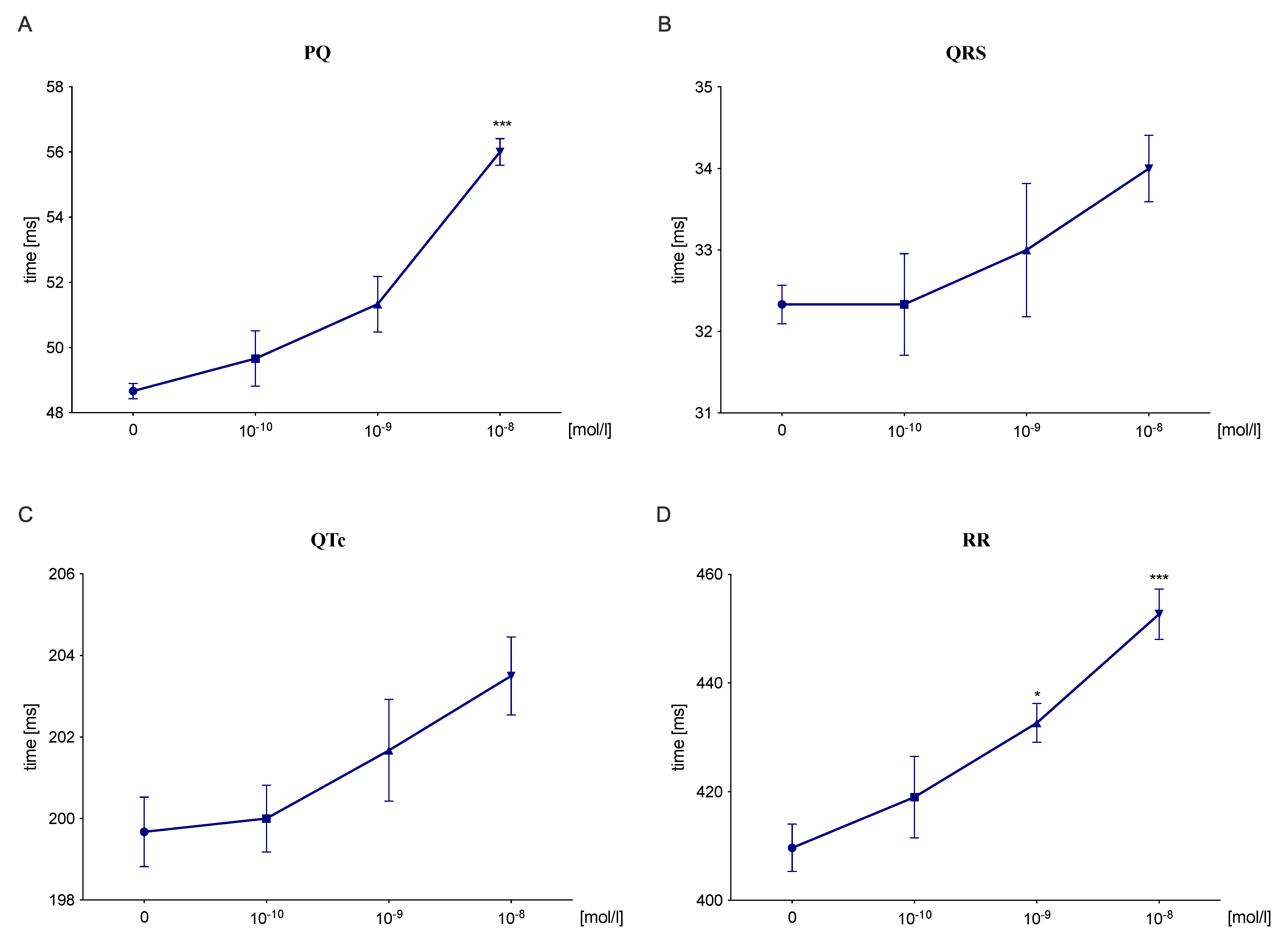
2.3. S-75 Prolonged the PQ and RR Interval in an Isolated Rat’s Heart

S-75 at dose 10−8 mol/L significantly prolonged the PQ interval by 15.1% (Column factor: F (3,9) = 22.55, p < 0.001; Row factor: F (3,9) = 0.5644, ns) (Figure 1A) and the RR interval by 10.5% (Column factor: F (3,9) = 12.19, p < 0.01; Row factor: F (3,9) = 0.8253, ns) (Figure 1D), not affecting either QRS complex (Column factor: F (3,9) = 2.738, ns; Row factor: F (3,9) = 2.633, ns) (Figure 1B) or QTc interval (Column factor: F (3,9) = 3.43, ns; Row factor: F (3,9) = 1.281, ns) (Figure 1C).

2.4. S-75 Showed Prophylactic Antiarrhythmic Properties in Arrythmia Models Caused by Adrenaline, but Not Calcium Chloride and Aconitine

The intravenous injections of either adrenaline (20 μg/kg), calcium chloride (140 mg/kg) or aconitine (20 μg/kg) caused atrioventricular disturbances and ventricular and supraventricular extrasystoles (Table 3).
The administration of the tested compound at dose 0.5 mg/kg before the arrhythmogenic substance (adrenaline) prevented or decreased the number of extrasystoles and atrioventricular blocks (ED50 = 0.05 (0.02–0.11). ED50 value for carvedilol was 0.36 (0.16–0.8).
S-75 did not significantly reduce the amount of calcium chloride- and aconitine-induced heart rhythm disturbances and showed no activity is these models of arrhythmia.

2.5. S-75 Did Not Possess the Antiarrhythmic Activity in the Post-Reperfusion Model in Isolated Rat Heart

S-75 did not show the activity in the post-reperfusion model of arrhythmia and the arrhythmia severity index was higher compared to the control group (F (4,15) = 6.854, p < 0.01) (Table 4).

2.6. S-75 Showed Therapeutic Antiarrhythmic Properties in Adrenaline-Induced Arrythmia Model

S-75 significantly decreased the number of adrenaline-induced extrasystoles by 72.6% compared to the vehicle-treated group (H (3,18) = 11.93, p < 0.001) (Figure 2). The reference compound, carvedilol, diminished the number of extrasystoles by 95.2%.
S-75 also diminished the number of bradycardia and block episodes in adrenaline-treated rats and this effect was more pronounced than that for carvedilol (Table 5).

3. Discussion

We showed for the first time that the studied pyrrolidine-2-one derivative S-75 possessed prophylactic and therapeutic antiarrhythmic activity in the model of adrenaline-induced arrhythmia. Furthermore, its prophylactic antiarrhythmic activity was stronger than that of carvedilol—a commonly used β-blocker with α1-adrenolytic properties [53]. However, S-75 did not prevent arrhythmia caused by either aconitine or calcium chloride administration, nor did it abolish the heart rhythm abnormalities in the post-reperfusion model of arrhythmia.
Pyrrolidyn-2-one derivatives have been shown to interact with adrenergic receptors [39,45,54]. Due to the highest affinity for α1-adrenergic receptors assessed in our previous study, we chose the compound S-75 for further experimentation [50]. Our functional experiments revealed that S-75 antagonized α1A- and α1B receptors [50]. The α1A, α1B, and α1D subtypes of α1-adrenoceptor have been found in the human heart. α1D receptors play a marginal role, whereas α1A mediate a positive inotropic response of atrial cardiomyocytes, and α1B regulate the myocytes contractility [55]. Given previous in vitro studies demonstrated that S-75 blocked α1A- and α1B-adrenoceptors, and these receptors can contribute to the development of cardiac arrhythmias induced by catecholamines, we aimed to evaluate the compound’s antiarrhythmic activity.
As we mentioned already, commonly used antiarrhythmic drugs possess limited efficacy and safety. However, an ideal antiarrhythmic agent administered in an emergency should effectively stop an attack of arrhythmia, and if taken regularly, should prevent its recurrence. The most often used antiarrhythmic drugs nowadays are divided according to the Vaughan Williams classification. This classification is based on the mechanism of action of drugs and divides them into four classes: sodium channel blockers, β-blockers, potassium channel blockers, and calcium channel blockers [56]. Thus, as a first step we investigated whether other than α1 receptors might contribute to the potential antiarrhythmic effect of a novel compound—S-75.
In order to evaluate the effect of the tested compound on β1-adrenergic receptors, we performed the radioligand studies. Lack of affinity for β1-adrenergic receptors excluded the role of those adrenoceptors in the mechanism of action of S-75. Next, we decided to determine the effect of the tested derivative on ion channels. First, we investigated the influence of S-75 on voltage-gated sodium channels (site 2). The sodium channels in the cardiomyocytes’ sarcolemma enable the conduction of depolarizing sodium current in the 0 phase of the action potential [57]. The blockade of these channels is used in the pharmacotherapy of arrhythmias; however, despite the therapeutic value of the sodium channel blockers, their pro-arrhythmic activity should be kept in mind [58]. The affinity of S-75 toward sodium channel was about 3.5 times lower than veratridine used as a reference compound. Therefore, this ion channel can at least partially contribute to the pharmacological effect of the tested compound.
Next, we investigated whether S-75 targets the potassium hERG channel. The potassium channels encoded by the hERG gene play an important role in controlling the repolarization of ventricular cardiomyocytes. The blockage of potassium channels inhibits the rapidly activating delayed rectifier potassium current. As a result, the duration of an action potential is prolonged, which can induce the formation of early subsequent depolarizations and torsades de pointes [59]. Our experiments revealed that S-75 showed 1172 times lower affinity toward potassium channel hERG than withdrawn from market terfenadine, which can indicate a low potential of S-75 to cause torsades de pointes [60].
Finally, we evaluated the effect of S-75 on L-type calcium channels (verapamil site). Since Ringer’s first observation in 1883 that Ca2+ ions are essential for myocardial contraction, their function in the heart has become increasingly appreciated. Every action potential induced by Na+ influx through voltage-gated sodium channels causes Ca2+ flux through the voltage-activated L-type calcium channels [61]. However, the role of the calcium channels in the mechanism of action of S-75 is marginal since it showed 20 times lower affinity than the reference compound D-600.
Since many antiarrhythmic agents tend to possess life-threatening proarrhythmic potential, we assessed the influence of the studied compound on normal ECG in rats. Most importantly, S-75 did not prolong the QTc interval, which may suggest no potential for the induction of ventricular tachyarrhythmias, in particular torsades de pointes. Instead, the tested compound prolonged the PQ interval by 12.2%, and this effect may be associated with slower conduction in the atrioventricular node [62]. S-75 also decreased the heart rate by 7.4% (after 10 min) and 8.6% (after 15 min), which can be linked to the blockade of α1 receptors. Our in vivo findings mostly align with the results obtained on the isolated rat heart. S-75 did not affect either QRS complex or QTc interval, yet the dose of 10−8 mol/L significantly prolonged the PQ interval by 15.1%, which may be due to the interaction with L-type Ca2+channels. Moreover, the tested compound prolonged the RR interval by 10.5%, decreasing the heart rhythm and showing a negative chronotropic effect.
Although α1-adrenoceptor antagonists do not have a well-established position in the pharmacotherapy of arrhythmias, the α1-adrenergic blockade may be beneficial in restoring normal heart rhythm and may also reduce the risk of arrhythmia [63]. According to research conducted by Billman and colleagues in dogs after myocardial infarction, prazosin and a tested α1-receptor antagonist prevented an arrhythmia caused by cardiac ischemia, accordingly, in 78% and 93% of the animals [64]. In the study by Tölg et al [33], a non-selective α-adrenergic receptor antagonist (phentolamine) reduced the incidence of ventricular fibrillation in isolated ischemic rat hearts by 32%. In comparison, a selective α1 receptor antagonist (prazosin) decreased the risk of cardiac arrhythmias by 83% and a tested α1A-blocker (WB 4101) limited the incidence of arrhythmias by 49% [33].
Considering the α1-adrenolytic properties of S-75, we determined its antiarrhythmic activity in the adrenaline-induced model of arrhythmia. An arrhythmogen, adrenaline, induced arrhythmias manifested by extrasystoles, atrioventricular blocks, bradycardia, and mortality. S-75 showcased prophylactic antiarrhythmic activity in this model, reducing the amount of post-adrenaline extrasystoles, conduction blocks and decreasing mortality of rats. The tested compound possessed stronger prophylactic antiarrhythmic properties than carvedilol (a reference drug). Considering that carvedilol blocks both β1- and α1-adrenoceptors and presented weaker antiarrhythmic activity than S-75 in the adrenaline-induced model of arrhythmia, we think that the potent blockade of α1-adrenoceptors is essential to treat adrenaline-induced arrhythmia. The obtained results are promising, as a novel compound with such pharmacological properties can be used to prevent attacks of arrhythmia due to emotional or physical stress.
In the next stage of our research, we evaluated antiarrhythmic properties in arrhythmia models, i.e., aconitine- and calcium chloride-induced. Aconitine and calcium chloride induce the changes in intracellular ion concentration (accordingly Na+ and Ca2+), which subsequently leads to arrhythmia [65]. Our results indicate that S-75 did not significantly reduce the number of cardiac rhythm disturbances or animal mortality in both arrhythmia models. Consequently, the studies confirmed our in vitro results, and we may presume that the blockade of sodium or calcium channels was not responsible for the mechanism of the antiarrhythmic effect of S-75.
Subsequently, we assessed the activity of the tested compound in the post-reperfusion model in isolated rat heart. This model recreates the clinical situation of a patient with myocardial infarction. After restoring coronary flow in the previously ischemic area, as a result of metabolic changes (e.g., increased adrenergic stimulation, overproduction of reactive oxygen species, disturbance in calcium ion homeostasis) post-reperfusion arrhythmias develop [66]. In ischemic conditions, the number of α1-adrenergic receptors increases, and their activation induces heart rhythm disturbances [29]. Despite the effectiveness of α1-blockers in eliminating post-reperfusion arrhythmias, our tested compound was not effective in this model of arrhythmia. There are several explanations to why S-75 did not show an antiarrhythmic effect in this model but was effective in the adrenaline-induced arrhythmia. These arrhythmia models differ in etiology—in adrenaline-induced arrhythmia, the abnormalities in the heart rhythm result from adrenergic receptors’ stimulation, and in the post-reperfusion model, arrhythmia develops after ischemia, reactive oxygen species production, and inflammation. We speculate that S-75 may not be effective in the post-reperfusion model of arrhythmia due to the lack of anti-inflammatory properties–however, this hypothesis needs further confirmation. Moreover, we need to highlight that one model was performed in vivo and another, ex vivo, and this methodology difference might underlie the differences in results. Finally, the antiarrhythmic effect of S-75 in the post-reperfusion model might be observed in different dosage schemes.
Since S-75 showed prophylactic antiarrhythmic properties in the adrenaline-induced arrhythmia, we next investigated whether it demonstrates therapeutic antiarrhythmic activity (a situation where a tested compound is given immediately after the administering the arrhythmogen). An effective antiarrhythmic drug should restore normal sinus rhythm as quickly as possible when used in a patient during the attack of arrhythmia. S-75 showed significant therapeutic antiarrhythmic activity by reducing the amount of post-adrenaline extrasystoles, atrioventricular blocks, bradycardia, and decreasing animal mortality.
Limitations to our study include evaluating the antiarrhythmic activity of pyrrolidyn-2-one derivative S-75 only after a single administration. Assessing the ability of the tested compound to prevent the attacks of arrhythmia after the repeated administration would provide information about its mechanism of the antiarrhythmic effect and if S-75 possesses the potential of long-acting antiarrhythmic. Moreover, significant activity in the adrenaline-induced arrhythmia model may relate to antioxidant effects, which needs further investigation. Therefore, our future studies will focus on assessing the effects of prolonged administration of S-75 and evaluating its extensive pharmacological profile.

4. Materials and Methods

4.1. Drugs

The studied compound: 1-(4-(4-(2-chlorophenyl)piperazin-1-yl)butyl)pyrrolidin-2-one hydrochloride (S-75) was synthesized in the Department of Physiochemical Drug Analysis, Faculty of Pharmacy, Jagiellonian University Medical College [50]. The investigated compound was dissolved in saline and administered intravenously (iv).
Thiopental (Sandoz GmbH, Kundl, Austria) and propafenone (Polpharma, Starogard Gdański, Poland) were dissolved in saline and administered intraperitoneally (ip). Adrenaline (Polfa S.A., Warsaw, Poland), aconitine (Merck, Darmstadt, Germany), calcium chloride (P.O.Ch. S.A., Gliwice, Poland), amiodarone (Sanofi- Aventis, Paris, France), carvedilol (Sigma-Aldrich, Steinheim, Germany), and verepamil (Abbott, Abbot Park, IL, USA) were dissolved in saline and administered intravenously (iv). Heparin (Polfa S.A., Warsaw, Poland) was used as anticoagulant during experiments. The control groups received saline as a vehicle.

4.2. Animals

Male normotensive Wistar rats, weighing 200–250 g, were used in experiments. Animals were kept in groups of 3 in standard plastic cages (42.7 cm × 26.7 cm) at constant room temperature of 22 ± 2 °C, with 12:12 h light/dark cycle and ad libitum access to food and water. Six rats were used in each experimental and control groups. All injections were administered in a volume of 1 ml/kg. Immediately after each experiment, animals were killed by cervical dislocation. All conducted procedures were approved by the Local Ethics Committee for Experiments on Animals of the Jagiellonian University in Krakow, Poland.

4.3. Affinity for β1-Adrenergic Receptors

Radioligand binding assay was performed on tissue rat cortex. A volume of 50 µL working solution of the tested compound, 50 µL [3H]-CGP-12177 (with final concentration 0.9 nM) and 150 µL tissue suspension prepared in assay buffer (50 mM Tris-HCl, pH 7.6) were transferred to polypropylene 96-well microplate using 96-wells pipetting station Rainin Liquidator (Mettler Toledo, Columbus, USA). In order to measure the unspecific binding 1 μM of propranolol was applied. Microplate was covered with a sealing tape, mixed and incubated for 60 minutes at 37 °C. The incubation was terminated by rapid filtration through GF/B filter mate. Ten rapid washes with 200 µL 50 mM Tris buffer (4 °C, pH 7.6) were completed using 96-well FilterMate harvester (PerkinElmer, Boston, MA, USA). The filter mates were dried at 37 °C in forced air fan incubator and then solid scintillator MeltiLex was melted on filter mates at 90 °C for 4 min. The radioactivity on the filter was measured in MicroBeta TriLux 1450 scintillation counter (PerkinElmer, Boston, MA, USA). Data were fitted to a one-site curve-fitting equation with Prism 6 (GraphPad Software, San Diego, CA, USA) and Ki values were estimated from the Cheng−Prusoff equation [51].

4.4. Effect on Sodium, Potassium, and Calcium Channels

Radioligand binding assay for Na+ channel was carried out according to the method of Callaway et al., with slight modifications [67]. Weighed rat cerebral cortex was homogenized at concentration of 10% (w/v) in ice-cold 0.32 M sucrose, 10 mM phosphate buffer (pH 7.4) using an ULTRA TURRAX homogenizer and next centrifuged at 1000× g for 10 min (0–4 °C). Collected supernatant was centrifuged at 40,000× g for 45 min (0–4 °C) to obtain membrane fraction. The resulting pellet was resuspended in incubation buffer (containing (mM): 50 Tris base, 50 HEPES, 130 choline chloride, 5.4 KCl, 0.8 MgSO4, 5.5 glucose). A volume of 200 µL of the tissue suspension (300 µg/well) containing LQ scorpion venom (10 µg/well), 50 µL of [3H]-BTX solution (final concentration 10 nM, PerkinElmer, Boston, MA, USA) and 50 µL of the tested compound were incubated at 37 °C for 1 h. The reaction was terminated by rapid filtration over glass fiber filters (FilterMate B, PerkinElmer, Boston, MA, USA) using 96-well harvester (PerkinElmer, Boston, MA, USA). Eight rapid washes with 500 µL of ice-cold 50 mM Tris-HCl buffer, pH 7.4, were completed. Veratridine (300 μM) was used to define nonspecific binding. Filter mates were dried at 37 °C in forced-air incubator and solid scintillator (MeltiLex, PerkinElmer, Boston, MA, USA) was then melted on them at 100 °C for 5 min. The radioactivity on the filters was measured in MicroBeta TriLux 1450 scintillation counter (PerkinElmer, Boston, MA, USA). The compound was tested in a screening assay at final concentration of 100 µM and result was expressed as percent inhibition of [3H]-batrachotoxin binding.
The radioligand binding studies for K+ and Ca2+ channels were performed commercially by Eurofins Cerep (Celle l’Evescault, France) using testing procedures described elsewhere [68,69]. Briefly, radioligand binding assay for K+ channel was performed on human recombinant HEK-293 cells using [3H]-dofetilide as specific ligand and non-specific binding was determined in the presence of 25 μM terfenadine [69], whereas radioligand binding assay for Ca2+ channel (L, verapamil site) was carried out in the rat cerebral cortex using [3H]-D888 as a specific ligand and non-specific binding was determined in the presence of 10 μM D600 [68].

4.5. Effect on Normal Electrocardiogram in Rats

To eliminate the influence of the tested derivative on normal electrocardiogram (ECG), the experiment was performed. ECG tests were carried out with Aspel ASCARD apparatus (standard II lead, with the tape speed 50 mm/s and voltage calibration 1 Mv = 1 cm). The ECG was recorded prior and 5, 10, 15 min after iv administration of tested compound. The influence on PQ, QT, QTc interval, QRS complex and heart rate (RR) was defined. The Bazzett’s formula: QTc = QT/√RR was used to calculate QTc [52].

4.6. Effect on Normal ECG of Isolated Rat Heart

Hearts form Wistar rats were isolated and immersed in Chenoweth-Koelle solution (120 mM NaCl; 5 mM KCl; 2.2 mM MgCl2; 19 mM KCl; 2.4 mM CaCl2; 10 mM glucose). Hearts were perfused according to the Langendorff model under a constant pressure of 70 cm H2O (6.87 kPa) with a solution at 37 °C, pH 7.4, constantly oxygenated (O2/CO2, 19:1) using the MyoHEART Langendorff System-900MH (Danish Myo Technology, Hinnerup, Denmark), which simultaneously monitored the ECG. After 20-min stabilization period, the tested compound was administered directly to the nutrient fluid every 15 min at increasing concentrations of 10−10–10−8. The influence on PQ, QTc interval, QRS complex and heart rate (RR) was assessed. The Bazzett’s formula: QTc = QT/√RR was used to calculate QTc [52].

4.7. Prophylactic Antiarrhythmic Activity in Adrenaline-, Aconitine-, and Calcium Chloride-Induced Arrhythmias

The experiments were carried out according to the method described by Szekeres and Papp (1975) [70]. The intravenous administration of adrenaline (20 μg/kg), aconitine (20 μg/kg) or calcium chloride (140 mg/kg) solution into the caudal vein of anesthetized rats (thiopental, 75 mg/kg) induced the heart rhythm disturbances. The studied compounds were injected iv 15 min before the arrhythmogen. The ECG was recorded during the first 2 min and in the 5th, 10th, and 15th min after the adrenaline, aconitine or calcium chloride injection. The criterion of antiarrhythmic activity was the decrease or complete absence of extrasystoles, atrioventricular blocks and bradycardia in the ECG recording in comparison with the control group (in case of aconitine- and calcium chloride-induced arrhythmia the absence of fibrillation was also taken into account). The ED50 (a dose producing a 50% inhibition of ventricular contractions) was calculated using the method of Litchfield and Wilcoxon [71]. The compound was administered at the dose 5 mg/kg, and we decreased the dose by half until the disappearance of antiarrhythmic activity.

4.8. Antiarrhythmic Activity in the Post-Reperfusion Model in Isolated Rat Heart

Hearts form Wistar rats were isolated and immersed in Chenoweth–Koelle solution (120 mM NaCl; 5 mM KCl; 2.2 mM MgCl2; 19 mM KCl; 2.4 mM CaCl2; 10 mM glucose). Hearts were perfused according to the Langendorff model under a constant pressure of 70 cm H2O (6.87 kPa) with a solution at 37 °C, pH 7.4, constantly oxygenated (O2/CO2, 19:1) using the MyoHEART Langendorff System-900MH (Danish Myo Technology, Hinnerup, Denmark), which monitored the ECG. After 20 min stabilization period, acute myocardial ischemia was induced locally by installing a clamp on the left coronary artery (ischemic period). After 15 min, the clamp was removed, and ECG changes were monitored for 30 min (reperfusion period). The tested compound was added to the perfusion solution 15 min before coronary artery clamping [39]. The point scale described by Bernauer [35] was used to assess the antiarrhythmic activity in this arrhythmia model. The occurrence of a specific type of heart rhythm disturbance during the reperfusion period was scored according to the following scale:
  • occurrence of less than 10 extrasystoles—1 point;
  • occurrence of more than 10 extrasystoles—2 points;
  • presence of ventricular tachycardia—3 points;
  • presence of fibrillation—4 points.

4.9. Therapeutic Antiarrhythmic Activity in Adrenaline-Induced Arrhythmia

The experiments were carried out according to the method described by Szekeres and Papp [70]. The heart rhythm disturbances were evoked by intravenous administration of adrenaline (20 µg/kg) to anesthetized rats (thiopental, 75 mg/kg). The tested compound was administered iv immediately after the injection of adrenaline, at dose 5 mg/kg. The ECG was recorded during the first 2 min and in the 5th, 10th, and 15th min after the adrenaline injection. The ECG was recorded for the first 2 min and then at 5, 10 and 15 min of the experiment. The criterion of antiarrhythmic activity was the decrease or complete absence of extrasystoles, atrioventricular blocks and bradycardia in the ECG recording in comparison with the control group [39].

4.10. Statistical Analysis

The number of animals in groups was based on our previous studies [50]. Results are presented as either means ± SEM or as a percentage of occurrence of specific events (like extrasystoles, fibrillations, tachy- or bradycardias, mortality). Where it was necessary, the normality of data sets and homogeneity of variance were determined using Shapiro–Wilk and Brown–Forsythe test, respectively. In our analysis, we used one-way repeated measures ANOVA followed by Dunnett’s post hoc (ECG measurements). The reported p values of all ANOVAs used the Geisser–Greenhouse correction when the sphericity assumption was not met. In cases when assumptions for normal distribution of data were not fulfilled (therapeutic arrhythmia), we used Kruskal–Wallis with Dunn’s post hoc test.

5. Conclusions

This work presents the pharmacological profile of pyrrolidine-2-one derivative S-75 and its effect on the cardiovascular system. The tested compound possessed prophylactic and therapeutic antiarrhythmic activity in adrenaline-induced arrhythmia, which is most likely mediated via α1-adrenoceptors. Our study indicates that S-75 can be a model structure for synthesizing compounds with the potential to treat or prevent arrhythmia attacks.

Author Contributions

Conceptualization, K.L., J.S. and K.P.; resources: P.Z.; performance of experiments: K.L., K.S. and A.S.; data analysis: K.L., K.S., K.P., A.S. and J.S.; writing-original draft preparation, K.L., K.S. and K.P.; writing-review and editing, K.L., K.S. and K.P.; supervision, K.P. and J.S.; project administration, J.S. and K.P.; funding acquisition, J.S. and K.L. All authors have read and agreed to the published version of the manuscript.

Funding

This study was supported by Jagiellonian University grants number K/DSC/003508.

Institutional Review Board Statement

All studies were approved by the Institutional Animal Care and Ethics Committee of the Jagiellonian University.

Informed Consent Statement

Not applicable.

Data Availability Statement

Data is contained within the article.

Acknowledgments

We would like to thank Joanna Knutelska for the technical assistance during experiments and Magdalena Kotańska for her help with the experiments on the isolated rat heart.

Conflicts of Interest

The authors declare no conflict of interest.

References

  1. WHO. Cardiovascular Diseases. Available online: https://www.who.int/en/news-room/fact-sheets/detail/cardiovascular-diseases-(cvds) (accessed on 26 August 2021).
  2. Institute of Medicine (US) Committee on Social Security. Cardiovascular Disability Criteria, D. Arrhythmias. In Cardiovascular Disability: Updating the Social Security Listings; National Academies Press (US): Washington, DC, USA, 2010; ISBN 978-0-309-15698-1. [Google Scholar]
  3. Feld, G.K.; Chen, P.S.; Nicod, P.; Fleck, R.P.; Meyer, D. Possible atrial proarrhythmic effects of class 1C antiarrhythmic drugs. Am. J. Cardiol. 1990, 66, 378–383. [Google Scholar] [CrossRef]
  4. Wang, D.; Patel, C.; Cui, C.; Yan, G.-X. Preclinical assessment of drug-induced proarrhythmias: Role of the arterially perfused rabbit left ventricular wedge preparation. Pharmacol. Ther. 2008, 119, 141–151. [Google Scholar] [CrossRef] [PubMed]
  5. Cardiac Arrhythmia Suppression Trial (CAST) Investigators. Preliminary report: Effect of encainide and flecainide on mortality in a randomized trial of arrhythmia suppression after myocardial infarction. N. Engl. J. Med. 1989, 321, 406–412. [Google Scholar] [CrossRef] [PubMed]
  6. Naccarelli, G.V.; Wolbrette, D.L.; Khan, M.; Bhatta, L.; Hynes, J.; Samii, S.; Luck, J. Old and new antiarrhythmic drugs for converting and maintaining sinus rhythm in atrial fibrillation: Comparative efficacy and results of trials. Am. J. Cardiol. 2003, 91, 15D–26D. [Google Scholar] [CrossRef]
  7. Gillis, A.M. Effects of antiarrhythmic drugs on QT interval dispersion--relationship to antiarrhythmic action and proarrhythmia. Prog. Cardiovasc. Dis. 2000, 42, 385–396. [Google Scholar] [CrossRef]
  8. Jaszczyszyn, E.; Panaszek, B. Podstawy elektrofizjologiczne, przyczyny i klasyfikacja zaburzeń rytmu serca—Implikacje rokownicze i terapeutyczne. Fam. Med. Prim. Care Rev. 2013, 15, 573–579. [Google Scholar]
  9. O’Connell, T.D.; Jensen, B.C.; Baker, A.J.; Simpson, P.C. Cardiac alpha1-adrenergic receptors: Novel aspects of expression, signaling mechanisms, physiologic function, and clinical importance. Pharmacol. Rev. 2014, 66, 308–333. [Google Scholar] [CrossRef] [Green Version]
  10. Ikeda, U.; Tsuruya, Y.; Yaginuma, T. Alpha 1-adrenergic stimulation is coupled to cardiac myocyte hypertrophy. Am. J. Physiol. 1991, 260, H953-6. [Google Scholar] [CrossRef]
  11. Fuller, S.J.; Gaitanaki, C.J.; Sugden, P.H. Effects of catecholamines on protein synthesis in cardiac myocytes and perfused hearts isolated from adult rats. Stimulation of translation is mediated through the alpha 1-adrenoceptor. Biochem. J. 1990, 266, 727–736. [Google Scholar] [CrossRef]
  12. Simpson, P. Stimulation of hypertrophy of cultured neonatal rat heart cells through an alpha 1-adrenergic receptor and induction of beating through an alpha 1- and beta 1-adrenergic receptor interaction. Evidence for independent regulation of growth and beating. Circ. Res. 1985, 56, 884–894. [Google Scholar] [CrossRef] [Green Version]
  13. Simpson, P. Norepinephrine-stimulated hypertrophy of cultured rat myocardial cells is an alpha 1 adrenergic response. J. Clin. Investig. 1983, 72, 732–738. [Google Scholar] [CrossRef] [Green Version]
  14. Simpson, P.; McGrath, A.; Savion, S. Myocyte hypertrophy in neonatal rat heart cultures and its regulation by serum and by catecholamines. Circ. Res. 1982, 51, 787–801. [Google Scholar] [CrossRef] [Green Version]
  15. Milano, C.A.; Dolber, P.C.; Rockman, H.A.; Bond, R.A.; Venable, M.E.; Allen, L.F.; Lefkowitz, R.J. Myocardial expression of a constitutively active alpha 1B-adrenergic receptor in transgenic mice induces cardiac hypertrophy. Proc. Natl. Acad. Sci. USA 1994, 91, 10109–10113. [Google Scholar] [CrossRef] [Green Version]
  16. O’Connell, T.D.; Ishizaka, S.; Nakamura, A.; Swigart, P.M.; Rodrigo, M.C.; Simpson, G.L.; Cotecchia, S.; Rokosh, D.G.; Grossman, W.; Foster, E.; et al. The alpha(1A/C)- and alpha(1B)-adrenergic receptors are required for physiological cardiac hypertrophy in the double-knockout mouse. J. Clin. Investig. 2003, 111, 1783–1791. [Google Scholar] [CrossRef]
  17. Papay, R.S.; Shi, T.; Piascik, M.T.; Naga Prasad, S.V.; Perez, D.M. α₁A-adrenergic receptors regulate cardiac hypertrophy in vivo through interleukin-6 secretion. Mol. Pharmacol. 2013, 83, 939–948. [Google Scholar] [CrossRef] [PubMed] [Green Version]
  18. Chaulet, H.; Lin, F.; Guo, J.; Owens, W.A.; Michalicek, J.; Kesteven, S.H.; Guan, Z.; Prall, O.W.; Mearns, B.M.; Feneley, M.P.; et al. Sustained augmentation of cardiac alpha1A-adrenergic drive results in pathological remodeling with contractile dysfunction, progressive fibrosis and reactivation of matricellular protein genes. J. Mol. Cell. Cardiol. 2006, 40, 540–552. [Google Scholar] [CrossRef] [PubMed]
  19. Du, X.-J.; Gao, X.-M.; Kiriazis, H.; Moore, X.-L.; Ming, Z.; Su, Y.; Finch, A.M.; Hannan, R.A.; Dart, A.M.; Graham, R.M. Transgenic alpha1A-adrenergic activation limits post-infarct ventricular remodeling and dysfunction and improves survival. Cardiovasc. Res. 2006, 71, 735–743. [Google Scholar] [CrossRef] [PubMed] [Green Version]
  20. Huang, Y.; Wright, C.D.; Merkwan, C.L.; Baye, N.L.; Liang, Q.; Simpson, P.C.; O’Connell, T.D. An alpha1A-adrenergic-extracellular signal-regulated kinase survival signaling pathway in cardiac myocytes. Circulation 2007, 115, 763–772. [Google Scholar] [CrossRef] [PubMed] [Green Version]
  21. Zhu, H.; McElwee-Witmer, S.; Perrone, M.; Clark, K.L.; Zilberstein, A. Phenylephrine protects neonatal rat cardiomyocytes from hypoxia and serum deprivation-induced apoptosis. Cell Death Differ. 2000, 7, 773–784. [Google Scholar] [CrossRef] [Green Version]
  22. Terzic, A.; Pucéat, M.; Clément, O.; Scamps, F.; Vassort, G. Alpha 1-adrenergic effects on intracellular pH and calcium and on myofilaments in single rat cardiac cells. J. Physiol. 1992, 447, 275–292. [Google Scholar] [CrossRef]
  23. Hirano, S.; Kusakari, Y.; O-Uchi, J.; Morimoto, S.; Kawai, M.; Hongo, K.; Kurihara, S. Intracellular mechanism of the negative inotropic effect induced by alpha1-adrenoceptor stimulation in mouse myocardium. J. Physiol. Sci. 2006, 56, 297–304. [Google Scholar] [CrossRef] [Green Version]
  24. Lin, F.; Owens, W.A.; Chen, S.; Stevens, M.E.; Kesteven, S.; Arthur, J.F.; Woodcock, E.A.; Feneley, M.P.; Graham, R.M. Targeted alpha(1A)-adrenergic receptor overexpression induces enhanced cardiac contractility but not hypertrophy. Circ. Res. 2001, 89, 343–350. [Google Scholar] [CrossRef] [Green Version]
  25. Kitakaze, M.; Hori, M.; Sato, H.; Iwakura, K.; Gotoh, K.; Inoue, M.; Kitabatake, A.; Kamada, T. Beneficial effects of alpha 1-adrenoceptor activity on myocardial stunning in dogs. Circ. Res. 1991, 68, 1322–1339. [Google Scholar] [CrossRef] [Green Version]
  26. Kitakaze, M.; Hori, M.; Morioka, T.; Minamino, T.; Takashima, S.; Sato, H.; Shinozaki, Y.; Chujo, M.; Mori, H.; Inoue, M. Alpha 1-adrenoceptor activation mediates the infarct size-limiting effect of ischemic preconditioning through augmentation of 5′-nucleotidase activity. J. Clin. Investig. 1994, 93, 2197–2205. [Google Scholar] [CrossRef]
  27. Tsuchida, A.; Liu, Y.; Liu, G.S.; Cohen, M.V.; Downey, J.M. alpha 1-adrenergic agonists precondition rabbit ischemic myocardium independent of adenosine by direct activation of protein kinase C. Circ. Res. 1994, 75, 576–585. [Google Scholar] [CrossRef] [Green Version]
  28. Bankwala, Z.; Hale, S.L.; Kloner, R.A. Alpha-adrenoceptor stimulation with exogenous norepinephrine or release of endogenous catecholamines mimics ischemic preconditioning. Circulation 1994, 90, 1023–1028. [Google Scholar] [CrossRef] [Green Version]
  29. Heusch, G. Alpha-adrenergic mechanisms in myocardial ischemia. Circulation 1990, 81, 1–13. [Google Scholar] [CrossRef] [Green Version]
  30. Corr, P.B.; Witkowski, F.X. Arrhythmias associated with reperfusion: Basic insights and clinical relevance. J. Cardiovasc. Pharmacol. 1984, 6 (Suppl. S6), S903–S909. [Google Scholar] [CrossRef] [PubMed]
  31. Lamontagne, D.; Yamaguchi, N.; Nadeau, R.; De Champlain, J.; Godin, D.; Campeau, N. Effects of sotalol, (-)-propranolol and prazosin on reperfusion-induced arrhythmias and increased cardiac norepinephrine release. Eur. J. Pharmacol. 1986, 123, 1–10. [Google Scholar] [CrossRef]
  32. Colucci, W.S. Alpha-adrenergic receptor blockade with prazosin. Consideration of hypertension, heart failure, and potential new applications. Ann. Intern. Med. 1982, 97, 67–77. [Google Scholar] [CrossRef] [PubMed]
  33. Tölg, R.; Kurz, T.; Ungerer, M.; Schreieck, J.; Görge, B.; Richardt, G. Influence of alpha- and beta-adrenoceptor antagonists on ventricular fibrillation in ischemic rat hearts. Naunyn Schmiedebergs Arch. Pharmacol. 1997, 356, 62–68. [Google Scholar] [CrossRef]
  34. Bralet, J.; Didier, J.; Moreau, D.; Opie, L.H.; Rochette, L. Effect of alpha-adrenoceptor antagonists (phentolamine, nicergoline and prazosin) on reperfusion arrhythmias and noradrenaline release in perfused rat heart. Br. J. Pharmacol. 1985, 84, 9–18. [Google Scholar]
  35. Bernauer, W.; Ernenputsch, I. Antagonistic effects of alpha-adrenoceptor blocking agents on arrhythmias, enzyme release, and myocardial necrosis in isolated rat hearts with coronary occlusion and reperfusion. Naunyn Schmiedebergs Arch. Pharmacol. 1988, 338, 88–95. [Google Scholar] [CrossRef]
  36. Kubacka, M.; Mogilski, S.; Filipek, B.; Marona, H. Antiarrhythmic properties of some 1,4-disubstituted piperazine derivatives with α1-adrenoceptor affinities. Eur. J. Pharmacol. 2013, 720, 237–246. [Google Scholar] [CrossRef]
  37. Kubacka, M.; Szkaradek, N.; Mogilski, S.; Pańczyk, K.; Siwek, A.; Gryboś, A.; Filipek, B.; Żmudzki, P.; Marona, H.; Waszkielewicz, A.M. Design, synthesis and cardiovascular evaluation of some aminoisopropanoloxy derivatives of xanthone. Bioorgan. Med. Chem. 2018, 26, 3773–3784. [Google Scholar] [CrossRef]
  38. Handzlik, J.; Bajda, M.; Zygmunt, M.; MacIg, D.; Dybała, M.; Bednarski, M.; Filipek, B.; Malawska, B.; Kieć-Kononowicz, K. Antiarrhythmic properties of phenylpiperazine derivatives of phenytoin with α 1-adrenoceptor affinities. Bioorgan. Med. Chem. 2012, 20, 2290–2303. [Google Scholar] [CrossRef]
  39. Sapa, J.; Nowaczyk, A.; Kulig, K. Antiarrhythmic and antioxidant activity of novel pyrrolidin-2-one derivatives with adrenolytic properties. Naunyn Schmiedebergs Arch. Pharmacol. 2011, 383, 13–25. [Google Scholar] [CrossRef] [Green Version]
  40. Malawska, B.; Kulig, K.; Filipek, B.; Sapa, J.; Macia̧g, D.; Zygmunt, M.; Antkiewicz-Michaluk, L. Synthesis, antiarrhythmic, and antihypertensive effects of novel 1-substituted pyrrolidin-2-one and pyrrolidine derivatives with adrenolytic activity. Eur. J. Med. Chem. 2002, 37, 183–195. [Google Scholar] [CrossRef]
  41. Malawska, B.; Kulig, K.; Gippert, A.; Filipek, B.; Sapa, J.; Maciag, D. Synthesis and development of new 2-substituted 1-[3-(4-arylpiperazin-1-yl)propyl]-pyrrolidin-2-one derivatives with antiarrhythmic, hypotensive, and alpha-adrenolytic activity. Farmaco 2005, 60, 793–803. [Google Scholar] [CrossRef] [PubMed]
  42. Kulig, K.; Sapa, J.; Maciag, D.; Filipek, B.; Malawska, B. Synthesis and pharmacological evaluation of new 1-[3-(4-arylpiperazin-1-yl)-2-hydroxypropyl]-pyrrolidin-2-one derivatives with anti-arrhythmic, hypotensive, and alpha-adrenolytic activity. Arch. Pharm. Weinh. 2007, 340, 466–475. [Google Scholar] [CrossRef] [PubMed]
  43. Kulig, K.; Sapa, J.; Nowaczyk, A.; Filipek, B.; Malawska, B. Design, synthesis and pharmacological evaluation of new 1-[3-(4-arylpiperazin-1-yl)-2-hydroxy-propyl]-3,3-diphenylpyrrolidin-2-one derivatives with antiarrhythmic, antihypertensive, and alpha-adrenolytic activity. Eur. J. Med. Chem. 2009, 44, 3994–4003. [Google Scholar] [CrossRef]
  44. Kulig, K.; Sapa, J.; Nowaczyk, A.; Filipek, B.; Malawska, B. Synthesis and pharmacological evaluation of new 1-[3-(4-phenylpiperazin-1-yl)-propyl]- and 1-[3-(4-phenylpiperidine)-propyl]-3-aryl-3-alkyl-pyrrolidin-2-one derivatives with antiarrhythmic and antihypertensive activity. Acta Pol. Pharm. 2009, 66, 649–662. [Google Scholar]
  45. Kulig, K.; Spieces, C.; Sapa, J.; Caspers, C.; Filipek, B.; Malawska, B. Synthesis and pharmacological evaluation of pyrrolidin-2-one derivatives as antiarrhythmic, antihypertensive and alpha-adrenolytic agents. Pharmacol. Rep. 2010, 62, 68–85. [Google Scholar] [CrossRef]
  46. Sapa, J. Antiarrhythmic activity of novel S-enantiomers of pyrrolidin-2-one derivatives with adrenolytic properties. Acta Pol. Pharm. 2010, 67, 537–542. [Google Scholar]
  47. Betti, L.; Zanelli, M.; Giannaccini, G.; Manetti, F.; Schenone, S.; Strappaghetti, G. Synthesis of new piperazine-pyridazinone derivatives and their binding affinity toward alpha1-, alpha2-adrenergic and 5-HT1A serotoninergic receptors. Bioorgan. Med. Chem. 2006, 14, 2828–2836. [Google Scholar] [CrossRef]
  48. Li, M.-Y.; Tsai, K.-C.; Xia, L. Pharmacophore identification of alpha(1A)-adrenoceptor antagonists. Bioorgan. Med. Chem. Lett. 2005, 15, 657–664. [Google Scholar] [CrossRef] [PubMed]
  49. Li, M.-Y.; Fang, H.; Xia, L. Pharmacophore-based design, synthesis, biological evaluation, and 3D-QSAR studies of aryl-piperazines as alpha(1)-adrenoceptor antagonists. Bioorgan. Med. Chem. Lett. 2005, 15, 3216–3219. [Google Scholar] [CrossRef]
  50. Zaręba, P.; Dudek, M.; Lustyk, K.; Siwek, A.; Starowicz, G.; Bednarski, M.; Nowiński, L.; Raźny, K.; Sapa, J.; Malawska, B.; et al. α-Adrenoceptor antagonistic and hypotensive properties of novel arylpiperazine derivatives of pyrrolidin-2-one. Bioorgan. Med. Chem. 2015, 23, 2104–2111. [Google Scholar] [CrossRef]
  51. Cheng, Y.; Prusoff, W.H. Relationship between the inhibition constant (K1) and the concentration of inhibitor which causes 50 percent inhibition (I50) of an enzymatic reaction. Biochem. Pharmacol. 1973, 22, 3099–3108. [Google Scholar] [PubMed]
  52. De Clerck, F.; Van de Water, A.; D’Aubioul, J.; Lu, H.R.; van Rossem, K.; Hermans, A.; Van Ammel, K. In vivo measurement of QT prolongation, dispersion and arrhythmogenesis: Application to the preclinical cardiovascular safety pharmacology of a new chemical entity. Fundam. Clin. Pharmacol. 2002, 16, 125–140. [Google Scholar] [CrossRef]
  53. Nichols, A.J.; Sulpizio, A.C.; Ashton, D.J.; Hieble, J.P.; Ruffolo, R.R.J. In vitro pharmacologic profile of the novel beta-adrenoceptor antagonist and vasodilator, carvedilol. Pharmacology 1989, 39, 327–336. [Google Scholar] [CrossRef]
  54. Sapa, J.; Filipek, B.; Nowiński, L. Antiarrhythmic and hypotensive activities of 1-[2-hydroxy-3-(4-phenyl-1-piperazinyl)propyl]-pyrrolidin-2-one (MG-1(R,S)) and its enantiomers. Pharmacol. Rep. 2011, 63, 455–463. [Google Scholar] [CrossRef]
  55. Ruffolo, R.R.J.; Nichols, A.J.; Stadel, J.M.; Hieble, J.P. Structure and function of alpha-adrenoceptors. Pharmacol. Rev. 1991, 43, 475–505. [Google Scholar] [PubMed]
  56. Vaughan Williams, E.M. Classification of antidysrhythmic drugs. Pharmacol. Ther. B 1975, 1, 115–138. [Google Scholar] [CrossRef] [PubMed]
  57. Amin, A.S.; Asghari-Roodsari, A.; Tan, H.L. Cardiac sodium channelopathies. Pflug. Arch. 2010, 460, 223–237. [Google Scholar] [CrossRef] [PubMed] [Green Version]
  58. Clancy, C.E.; Zhu, Z.I.; Rudy, Y. Pharmacogenetics and anti-arrhythmic drug therapy: A theoretical investigation. Am. J. Physiol. Heart Circ. Physiol. 2007, 292, H66–H75. [Google Scholar] [CrossRef]
  59. Raschi, E.; Vasina, V.; Poluzzi, E.; De Ponti, F. The hERG K+ channel: Target and antitarget strategies in drug development. Pharmacol. Res. 2008, 57, 181–195. [Google Scholar] [CrossRef]
  60. Brown, A.M. Drugs, hERG and sudden death. Cell Calcium 2004, 35, 543–547. [Google Scholar] [CrossRef]
  61. Landstrom, A.P.; Dobrev, D.; Wehrens, X.H.T. Calcium Signaling and Cardiac Arrhythmias. Circ. Res. 2017, 120, 1969–1993. [Google Scholar] [CrossRef]
  62. Krumpl, G.; Todt, H.; Krejcy, K.; Raberger, G. Antiarrhythmic efficacy of labetalol as assessed by programmed electrical stimulation. Br. J. Pharmacol. 1990, 100, 855–861. [Google Scholar] [CrossRef] [Green Version]
  63. Hieble, J.P. Adrenoceptor subclassification: An approach to improved cardiovascular therapeutics. Pharm. Acta Helv. 2000, 74, 163–171. [Google Scholar] [CrossRef]
  64. Billman, G.E. Effect of alpha 1-adrenergic receptor antagonists on susceptibility to malignant arrhythmias: Protection from ventricular fibrillation. J. Cardiovasc. Pharmacol. 1994, 24, 394–402. [Google Scholar] [CrossRef]
  65. Pytka, K.; Lustyk, K.; Żmudzka, E.; Kotańska, M.; Siwek, A.; Zygmunt, M.; Dziedziczak, A.; Śniecikowska, J.; Olczyk, A.; Gałuszka, A.; et al. Chemically Homogenous Compounds with Antagonistic Properties at All α1-Adrenoceptor Subtypes but not β1-Adrenoceptor Attenuate Adrenaline-Induced Arrhythmia in Rats. Front. Pharmacol. 2016, 7, 229. [Google Scholar] [CrossRef] [PubMed] [Green Version]
  66. Corr, P.B.; Heathers, G.P.; Yamada, K.A. Mechanisms contributing to the arrhythmogenic influences of alpha 1-adrenergic stimulation in the ischemic heart. Am. J. Med. 1989, 87, 19S–25S. [Google Scholar] [CrossRef]
  67. Callaway, J.K.; Castillo-Melendez, M.; Giardina, S.F.; Krstew, E.K.; Beart, P.M.; Jarrott, B. Sodium channel blocking activity of AM-36 and sipatrigine (BW619C89): In vitro and in vivo evidence. Neuropharmacology 2004, 47, 146–155. [Google Scholar] [CrossRef]
  68. Reynolds, I.J.; Snowman, A.M.; Snyder, S.H. (-)-[3H] desmethoxyverapamil labels multiple calcium channel modulator receptors in brain and skeletal muscle membranes: Differentiation by temperature and dihydropyridines. J. Pharmacol. Exp. Ther. 1986, 237, 731–738. [Google Scholar] [PubMed]
  69. Huang, X.-P.; Mangano, T.; Hufeisen, S.; Setola, V.; Roth, B.L. Identification of human Ether-à-go-go related gene modulators by three screening platforms in an academic drug-discovery setting. Assay Drug Dev. Technol. 2010, 8, 727–742. [Google Scholar] [CrossRef] [Green Version]
  70. Szekeres, L.; Papp, J. Experimental cardiac arrhythmias. In Experimental Production of Diseases, Part 3, Heart and Circulation, Handbook of Experimental Pharmacology; Springer: Berlin/Heidelberg, Germany, 1975. [Google Scholar]
  71. Litchfield, J.T.; Wilcoxon, F. A simplified method of evaluating dose-effect experiments. J. Pharmacol. Exp. Ther. 1949, 96, 99–113. [Google Scholar]
Figure 1. The effect of S-75 on the ECG components of the isolated rat heart. After stabilization period, the tested compound was administered directly to the nutrient fluid, in which heart was submerged, every 15 min at increasing concentrations of 10−10–10−8 mol/L. The influence on PQ (A), QRS complex (B), QTc (C), and RR interval (D) was assessed. The results are presented as mean ± SEM. Statistical analysis: Shapiro–Wilk test for data normality, and one-way repeated measures ANOVA (Dunnett’s post hoc); * p < 0.05, *** p < 0.001; n = 4 hearts.
Pharmaceuticals 14 01065 g001
Figure 2. The therapeutic effect of S-75 in the adrenaline-induced arrhythmia model. After intravenous (iv) injection of adrenaline (20 µg/kg), the tested compound was immediately administered iv at dose 5 mg/kg. Control group received no additional treatment. The ECG was recorded for the first 2 min and then at 5, 10 and 15 min of the experiment. The criterion of antiarrhythmic activity was the decrease or complete absence of extrasystoles in the ECG recording in comparison with the control group. The results are presented as box plots showing the following data: mean (‘+’), median (horizontal line), upper and lower quartile (the width of the box shows interquartile range), upper and lower extreme (whiskers). Statistical analysis: Kruskal–Wallis test with Dunn’s post hoc; * p < 0.05, ** p < 0.01; n = 6 rats.
Pharmaceuticals 14 01065 g002
Table 1. The affinity of S-75 for adrenergic β1 receptors, Na+, Ca2+ and K+ channels.
Treatmentβ1 a,c
(pKi [nM])
Na+ a,d
(IC50 [μM])
K+ hERG b,e
(IC50 [μM])
Ca2+ a,f
(IC50 [μM])
S-75n.c.5829.30.757
Propranolol15.0---
Veratridine-16.7--
Terfenadine--0.025-
D-600---0.037
Data are represented as pKi, that is, −logKi or IC50 and expressed as means from three independent experiments performed in duplicates. Inhibition constants (Ki) and IC50 were calculated according to the equation of Cheng and Prusoff [51]. a Radioligand binding was performed using rat cortex tissue. b Radioligand binding was performed using HEK-293 cells. c The affinity values were determined using [3H]-CGP-12177. d The affinity values were determined using [3H]-batrachotoxin. e The affinity values were determined using [3H]-dofetilide. f The affinity values were determined using [3H]-D888.
Table 2. Effects of an intravenous injection of the S-75 at dose 5 mg/kg on heart rate and ECG intervals.
ParametersTime of Observation [min]
051015
PQ46.44 ± 1.35247.00 ± 1.53749.78 ± 1.04252.11 ± 2.061 **
QRS33.78 ± 1.38435.56 ± 1.70133.89 ± 1.62837.22 ± 1.637
QTc182.9 ± 4.805192.3 ± 6.176189.3 ± 9.543190.9 ± 11.46
QT81.57 ± 1.35681.53 ± 1.81282.47 ± 2.18683.90 ± 1.730
Rate310.8 ± 17.07307.9 ± 18.93287.8 ± 22.94 **284.0 ± 23.50 **
Statistical analysis: Shapiro–Wilk test for normality, and repeated measures one-way ANOVA (Dunnett’s post hoc) ** p < 0.01; n = 6 rats. QTc– calculated QT interval according to Bazzett’s formula: QTc = QT/√RR [52].
Table 3. The effect of the tested compound on chosen parameters in arrythmia models in rats.
TreatmentDose (mg/kg)Fibrillations (%)Extrasystoles (%)Bradycardia (%)Blocks (%)Mortality (%)
Adrenaline-Induced Arrythmia
Control--100100100100
S-750.5-16.7033.30
0.25-66.716.7500
0.125-66.733.333.350
0.06-83.3100500
0.03-10010010033.3
Carvedilol0.25-10016.610066.7
Calcium Chloride-Induced Arrythmia
Control-100100100100100
S-75566.710010010083.3
Verapamil2.533.366.710083.316.7
Aconitine-Induced Arrythmia
Control-100100100100100
S-755100100100100100
Propafenone533.366.710083.316.6
Except for propafenone (ip), the tested or reference compounds were administered intravenously (iv) 15 min before the experiment. Control group received no treatment except the administration of arrhythmogen. The observation was performed for 15 min after the iv injection of adrenaline (20 μg/kg), aconitine (20 μg/kg) or calcium chloride (140 mg/kg) i.e., during the first 2 min, at 5, 10, and 15 min. Results are presented as a percentage of the occurrence of specific events (fibrillations, extrasystoles, bradycardia, blocks, mortality). n = 5−6 animals.
Table 4. The arrhythmic and antiarrhythmic activity of tested and reference compounds in post-reperfusion irregularities of heart rhythm.
TreatmentConcentration (M)Extrasystoles (%)Tachycardia
(%)
Fibrillations
(%)
Arrhythmia Severity Index
Control-10010033.36.3
S-7510−101001001009
Amiodarone10−910010066.77.7
Propafenone10−1110066.733.36.7
Quinidine10−683.35002.3 *
After stabilization period, acute myocardial ischemia was induced locally by installing a clamp on the left coronary artery (ischemic period) in the isolated rat heart. After 15 min, the clamp was removed, and ECG changes were monitored for 30 min (reperfusion period). The tested or reference compounds was added to the perfusion solution 15 min before coronary artery clamping. Control group received no treatment. Results are presented as a percentage of the occurrence of specific events (fibrillations, extrasystoles, bradycardia, blocks, mortality). Arrhythmia severity index was calculated according to Bernauer scale [35]. Statistical analysis: Shapiro–Wilk test for data normality, and one-way repeated measures ANOVA (Dunnett’s post hoc); * p < 0.05; n = 4 hearts.
Table 5. The therapeutic antiarrhythmic effect of the tested compound and carvedilol in adrenaline-induced arrhythmia model.
TreatmentDose (mg/kg)Bradycardia (%)Blocks
(%)
Mortality
(%)
Control-10010067.7
S-75533.333.316.7
Carvedilol567.783.316.7
The tested or reference compound was administered intravenously (iv) immediately after the injection of adrenaline, at dose 5 mg/kg. Control group received no treatment except the administration of arrhythmogen. The observation was performed for 15 min. Results are presented as a percentage of the occurrence of specific events (bradycardia, blocks, mortality). n = 5−6 animals.
Publisher’s Note: MDPI stays neutral with regard to jurisdictional claims in published maps and institutional affiliations.

