Article

Synthesis, Computational Analysis, and Antiproliferative Activity of Novel Benzimidazole Acrylonitriles as Tubulin Polymerization Inhibitors: Part 2

1
Department of Organic Chemistry, Faculty of Chemical Engineering and Technology, University of Zagreb, Marulićev trg 19, HR-10000 Zagreb, Croatia
2
Laboratory for the Computational Design and Synthesis of Functional Materials, Division of Organic Chemistry and Biochemistry, Ruđer Bošković Institute, Bijenička cesta 54, HR-10000 Zagreb, Croatia
3
KU Leuven, Department of Microbiology, Immunology and Transplantation, Laboratory of Virology and Chemotherapy, Rega Institute, 3000 Leuven, Belgium
*
Authors to whom correspondence should be addressed.
These authors contributed equally to this work.
Pharmaceuticals 2021, 14(10), 1052; https://doi.org/10.3390/ph14101052
Submission received: 6 September 2021 / Revised: 9 October 2021 / Accepted: 13 October 2021 / Published: 17 October 2021
(This article belongs to the Section Medicinal Chemistry)

Abstract

:
We used classical linear and microwave-assisted synthesis methods to prepare novel N-substituted, benzimidazole-derived acrylonitriles with antiproliferative activity against several cancer cells in vitro. The most potent systems showed pronounced activity against all tested hematological cancer cell lines, with favorable selectivity towards normal cells. The selection of lead compounds was also tested in vitro for tubulin polymerization inhibition as a possible mechanism of biological action. A combination of docking and molecular dynamics simulations confirmed the suitability of the employed organic skeleton for the design of antitumor drugs and demonstrated that their biological activity relies on binding to the colchicine binding site in tubulin. In addition, it also underlined that higher tubulin affinities are linked with (i) bulkier alkyl and aryl moieties on the benzimidazole nitrogen and (ii) electron-donating substituents on the phenyl group that allow deeper entrance into the hydrophobic pocket within the tubulin’s β-subunit, consisting of Leu255, Leu248, Met259, Ala354, and Ile378 residues.

Graphical Abstract

1. Introduction

Microtubules, being key dynamic structural components in cells, have attracted considerable attention from medicinal chemists as targets for anticancer drug discovery [1,2,3]. These protein biopolymers, formed through the polymerization of heterodimers of α- and β-tubulins, play an important role in cellular shape organization, cell division, mitosis, and intracellular movement. Potent microtubule-targeting drugs such as paclitaxel, vinblastine, colchicine, and vincristine are structurally complex natural products that are widely used in anticancer therapy [4]. These products alter the dynamics of tubulin, such as the polymerization and depolymerization [5], by binding to specific sites on the tubulin heterodimers [6], of which the most important are those for paclitaxel, vinblastine, and colchicine; thus, within the binding to the tubulin heterodimers, inhibitors could suppress tubulin dynamic instability and interfere with microtubule functions, including the mitotic spindle formation.
Inhibitors that bind to the vinblastine and colchicine binding sites are known as inhibitors of tubulin polymerization, while inhibitors interacting with the paclitaxel binding site are known as microtubule-stabilizing compounds [7].
During the past decades, there has been an increase in the development of novel tubulin polymerization inhibitors, while versatile classes of organic derivatives, both natural and synthetic, have been extensively studied [8,9,10,11,12]. Nowadays, considerable interest is focused on the development, design, and biological activity of novel heteroaromatic systems, whereby nitrogen heterocycles have become the essential structural motifs in medicinal and pharmaceutical chemistry, being very important in drug discovery. Benzimidazoles can easily interact with essential biomolecules of living systems due to the structural similarity of their nuclei with naturally occurring purines. Benzimidazoles are, therefore, very prominent scaffolds for the development of novel derivatives with a wide range of diverse biological activities. Considering all of the biological activities displayed by benzimidazoles, one of the most important is their antitumor activity, which is exerted by acting on numerous biological targets (Figure 1). For example, they can interact with DNA and RNA as intercalators or minor groove binders [13]; they can inhibit topoisomerases I and II [14]; and they can act as antiangiogenic agents [15], androgen receptor antagonists [16], and inhibitors of kinases [17,18]. Some benzimidazoles were also recognized as tubulin polymerization inhibitors, mostly binding to the colchicine binding site [19,20,21,22]. Nocodazole (Figure 1a) is a well-known anticancer agent, which significantly inhibits tubulin polymerization at low nanomolar concentrations [23]. Additionally, benzimidazole-5-carboxylate derivatives causes mitotic catastrophes by specifically targeting the microtubule system [24]. Several studies have reported on the biological activity of 2-aryl-1,2,4-oxadiazolo-benzimidazole derivatives (Figure 1b) with different mechanisms of biological action, such as binding to the colchicine binding site [25]. A series of benzimidazole-2-urea derivatives (Figure 1c) has been described as novel β-tubulin inhibitors that might bind in a new binding site different from the three well-known ones [26]. Novel 2-aryl-benzimidazole derivatives of dehydroabietic acid have been reported as tubulin polymerization inhibitors, which significantly disrupt the intracellular microtubule network by binding to the colchicine site of tubulin [26].
Recently, as a continuation of our previous efforts aimed at the design and discovery of novel benzimidazoles with promising antitumor activities, we prepared novel N-substituted, benzimidazole-derived acrylonitriles as potential tubulin polymerization inhibitors. N,N-dimethylamino-substituted acrylonitriles I and II (Figure 2) with submicromolar inhibitory concentrations (IC50 0.2–0.6 μM) were chosen as lead compounds, while their interference with the tubulin activity was confirmed by in vitro studies of the tubulin polymerization inhibition and the computational analysis [27].
Encouraged by our findings and the fact that some of the tested compounds showed strong and selective antiproliferative activity, we further optimized the presented structure by designing and synthesizing novel N-substituted benzimidazole acrylonitriles. Here, we present the synthesis, biological activity, and tubulin polymerization inhibition of the most active compounds and demonstrate their binding within the colchicine site of tubulin via computational docking analysis and molecular dynamics simulations.

2. Results and Discussion

2.1. Chemistry

The synthesis of novel N-substituted, benzimidazole-derived acrylonitriles 3271 is illustrated in Scheme 1, starting from the ortho-chloronitrobenzenes 12.
By using uncatalyzed microwave-assisted amination in acetonitrile with an excess of desired amine, N-substituted precursors 38 bearing i-butyl, methyl, phenyl, and n-hexyl substituents were obtained in good reaction yields. Within the reduction of nitro-substituted compounds 38 with SnCl2·2H2O in MeOH followed by cyclocondensation with 2-cyanoacetamide at high temperatures, corresponding N-substituted 2-(cyanomethyl)-benzimidazoles 1725 as main precursors were obtained in moderate yields [27]. Targeted acrylonitrile derivatives 3271 were synthesized in the condensation with the chosen unsubstituted and methoxy-, N,N-dimethyl-, and N,N-diethyl-substituted aromatic aldehydes 2631 in absolute ethanol by using a few drops of piperidine as a weak base. All acrylonitriles were obtained in moderate to high reaction yields, while some of them were obtained as mixtures of E- and Z-isomers (38, 41, 45, 61, 64, 67, and 68), which could not be efficiently separated by column chromatography.
The structural determination of newly prepared acrylonitriles was performed using 1H and 13C NMR spectroscopies and elemental analysis. The structural characterization was performed based on the chemical shifts in both spectra and H-H coupling constant values in the 1H spectra. The successful nucleophilic substitution was confirmed within the signals in the aliphatic part of both 1H and 13C NMR spectra of compounds 38. The structure of amino-substituted derivatives 916 was confirmed via the observation of signals related to the protons of amino groups placed in the range of 5.25–4.44 ppm in the 1H NMR spectra. A singlet of acrylonitrile protons in the range of 8.54–7.20 ppm confirmed the formation of acrylonitrile derivatives.

2.2. Biological Evaluation

2.2.1. Antiproliferative Activity In Vitro

All newly prepared compounds were tested for their antiproliferative activity in vitro. The results are displayed in Table 1 as IC50 values (50% inhibitory concentrations) and are compared to known antiproliferative agents combretastatin A4 (CA4) and docetaxel.
For the biological evaluation, eight human cancer cells from different cancer types were used (LN-229—glioblastoma; Capan-1—pancreatic adenocarcinoma; HCT-116—colorectal carcinoma; NCI-H460—lung carcinoma; DND-41—acute lymphoblastic leukemia; HL-60—acute myeloid leukemia; K-562—chronic myeloid leukemia; Z-138—non-Hodgkin lymphoma). The majority of the compounds exerted weak to moderate antiproliferative activity on the tested cell lines, while compounds 50, 64, 68, and 69 showed exceptionally strong antiproliferative activity against some, but not all, of the cancer cells.
Thirteen derivatives did not show any activity against the tested cell lines. Some of the tested derivatives exhibited strong and selective antiproliferative activity but were less active in comparison to the used standard drugs. Among the most active compounds, the N,N-diethylamino-substituted derivative with the i-butyl substituent placed at the nitrogen atom of benzimidazole core 64 did not show any significant selectivity towards the tested cancer cell lines and was the most potent system elucidated here. Compound 50 substituted with cyano and phenyl rings at the benzimidazole core bearing two methoxy groups, 68 substituted with cyano and i-butyl substituents at the benzimidazole core bearing a N,N-diethylamino group, and 69 substituted with cyano and methyl substituents at the benzimidazole core bearing a N,N-diethylamino group showed selectivity against the hematological cancer cell lines (acute lymphoblastic leukemia (DND-41), acute myeloid leukemia (HL-60), chronic myeloid leukemia (K-562), and non-Hodgkin lymphoma (Z-138). Among other derivatives with moderate activities, compound 48 substituted with cyano and i-butyl at the benzimidazole core bearing two methoxy groups showed some selectivity against lung carcinoma (NCI-H460) and colorectal carcinoma (HCT-116).
In general, comparing the unsubstituted and cyano-substituted derivatives bearing the same substituents at the nitrogen atom of the benzimidazole core and at the phenyl ring, it was observed that some cyano-substituted derivatives showed slightly improved antiproliferative activity, while for some others the presence of the –CN moiety reduced the activity.
The latter also holds for the most potent compound 64, whose cyano derivative 68 is generally less active. Among the tested systems, i-butyl-substituted derivatives showed improved activity relative to methyl, phenyl or n-hexyl-substituted analogues. According to the obtained results, it is obvious that the most significant impact on the antiproliferative activity enhancement relates to the introduction of the N,N-diethylamino group at the para-position of the phenyl ring. In comparison to the previously published results [27], it can be concluded that the introduction of the N,N-diethylamino group instead of the N,N-dimethylamino group decreased the antiproliferative activity against Capan-1, HCT-116, NCI-H460, DND-41, and HL-60 cancer cells from a submicromolar to micromolar range of inhibitory concentrations.
In order to establish whether the observed antiproliferative activity is selective towards cancer cells, normal peripheral blood mononuclear cells (PBMCs) from two healthy donors were treated with compounds 50, 64, 68, and 69 (Figure 3). The compounds with the best antiproliferative activity in hematological cancer cells (compound 50, 68, and 69) also induced apoptosis in normal cells, although this was limited to the highest concentration tested (20 µM) for compounds 68 and 69, resulting in a favorable selectivity window. Derivative 64, which was inhibitory against all tested cancer cell lines, had no effect on the viability of normal PBMC and is, thus, a selective anticancer compound.

2.2.2. In Vitro Inhibition of the Tubulin Polymerization

The ability of derivatives 50, 64, 68, and 69 to inhibit the polymerization of tubulin was confirmed in vitro in a purified protein system. All tested compounds showed effective activity in a dose-dependent manner (Figure 4).
At the highest concentrations tested (30 and 10 µM), these four derivatives were all able to inhibit tubulin polymerization. Even when lowering the dose to 3 µM, compounds 50 (to a lesser extent) and 68 still showed some inhibitory activity in the in vitro assay.

2.3. Computational Analysis

Computational analysis was performed to interpret the observed biological properties and to identify structural and electronic features responsible for the highest activity of 64, which should aid in the subsequent design of even more effective ligands based on the utilized organic skeleton. To do so, we considered a representative set of compounds, including 50, 63, 64, 66, 68, and 69, together with two model systems m1 and m2, as well as colchicine, which was taken as a typical ligand for the colchicine binding site in tubulin (Figure 5), in line with our previous results [27].
The systems investigated here consisted of variously substituted benzimidazole and phenyl fragments, linked together with an acrylonitrile unit, which can formally exist as either E- or Z-isomers. Our DFT analysis showed that all systems were between 0.5 and 4.0 kcal mol−1 more stable as Z-isomers (Table 2 and Figure S120), where the cyano group and the benzimidazole moiety reside on the same side of the C=C double bond. Although these values neglected any kinetic aspects of the isomer formation, the obtained thermodynamic data were found to be in good quantitative agreement with the experiments, which generally predict the predominance of Z-isomers. The differences in the stability among isomers were higher for the systems with bulkier N-substituents due to larger unfavorable steric interactions with the attached phenyl group occurring in their E-analogues. Interestingly, the only exception was provided by the most active system, 64, which was 1.2 kcal mol−1 more stable as an E-analogue. This confirms its experimentally observed preference for the E-isomer at a 2:1 ratio, yet such the small energy difference observed here, and in other evaluated systems, allows for the presence of the other isomer as well. With this in mind, we will focus our discussion on the most stable Z-isomers of all systems, while both isomers will be considered for the most active compound 64.
The docking procedure produced the binding free energies shown in Table 2. The highest affinity was obtained for colchicine, ΔGbind = −9.3 kcal mol−1, closely matching the value of ΔGbind = −9.0 kcal mol−1 reported by Silva-García and co-workers obtained using the AutoDock docking software [28]. Moreover, both of these values were in excellent agreement with the experimental value of ΔGbind,EXP = −8.3 kcal mol−1 calculated from the colchicine binding constant Kbind,EXP = 6.3 × 10⁵ L mol−1 measured by Wilson and Meza [29]. In addition, the predicted colchicine binding position very closely matched that in the crystal structure, suggesting that the docking procedure correctly positioned it within the colchicine binding site (Figure S121). Such an agreement in terms of both the binding energy and the position of the ligand leads us to conclude that these results lend firm credence to the employed computational methodology and support the reliability of the other results as well.
In certain cases, the values correspond to allosteric positions on tubulin (Figure S122), which is why we also analyzed the most favorable orthosteric poses within the colchicine binding site, since the latter are responsible for a potential tubulin polymerization inhibition. The lowest binding affinity was displayed by the least substituted m1, ΔGbind = −8.0 kcal mol−1, corresponding to the allosteric binding, which suggests a lack of antitumor activity. This already suggests that the substitution of the used organic framework is likely crucial for the binding and that specific protein–ligand interactions govern the activity. Since m1 is a reference system, let us note that a binding pose within the colchicine binding site comes with a further lower affinity, ΔGbind = −7.6 kcal mol−1, being the least exergonic site here. In that case (Figure S123), m1 is oriented so that its phenyl unit is immersed into the β-subunit close to Cys241, but without any significant interaction with it. In contrast, the importance of the benzimidazole unit is seen in favorable N–H∙∙∙π interactions with Lys352, yet this is outperformed by the unfavorable steric contacts between N-i-butyl, Lys254, and Asn258, which decrease the binding and disfavor such orthosteric positions for m1.
The addition of electron donors on the phenyl unit, such as the p-OMe group in m2, improves the binding. This allows deeper penetration of the β-subunit (Figure S123) facilitated by the S–H∙∙∙O(Me) hydrogen bonding with Cys241, which is absent in m1. This maintains favorable contacts with Lys352, while offering reduced steric interactions with Lys254 and Asn258, with the latter being allowed to engage in N–H∙∙∙N hydrogen bonds with the benzimidazole moiety. All of this contributes around 0.4 kcal mol−1 to the binding within the colchicine binding site, yet still promotes the allosteric binding as being the most favorable at ΔGbind = −8.4 kcal mol−1. Replacing the p-OMe group with p-NEt2 immediately offers the most potent system 64. With its better hydrogen-bond-accepting properties, 64 forms a stronger S–H∙∙∙N(Et2) interaction with Cys241 (Figure S123), which is evident in the reduced S∙∙∙N distance of 0.4 Å from that observed for the matching S∙∙∙O distance in m2. This allows 64 to rotate and avoid the unfavorable steric contacts with Lys254 and Asn258, enabling both to bind the benzimidazole fragment—the former through the N–H∙∙∙π interactions, while the latter through the N–H∙∙∙N hydrogen bonds. The attached cyano group in 64 accepts hydrogen bonding from Lys352, further promoting the binding.
All of this positions 64 within the colchicine binding site as the most favorable binding location, linked with the most exergonic binding energy of ΔGbind = −8.7 kcal mol−1. This confirms its high activity and promotes the tubulin polymerization inhibition as its likely biological mechanism of action.
The presence of the bulky N-i-butyl group is also significant in this activity. In general, this allows the investigated ligands to better position themselves within the hydrophobic interior of the β-subunit. If this is replaced by a smaller N-Me group as in 63, the system is reverted back to the allosteric binding as being most favorable, confirming its reduced activity, while its potential binding within the colchicine binding is also reduced to ΔGbind = −8.0 kcal mol−1 (Figure S124). Along these lines, the introduction of the aromatic N-phenyl unit in 66 improves the binding within the colchicine binding site to ΔGbind = −8.6 kcal mol−1, mostly through favorable N–H∙∙∙π interactions with this substituent, while positive contributions from Lys254 remain limited, yet this binding pose is also dominated by the allosteric binding that is 0.2 kcal mol−1 higher, making 66 a non-active compound.
Lastly, the presence of the electron-withdrawing cyano group on the benzimidazole core generally leads to reduced activities in the investigated cases. As illustrative examples, both 68 and 69 are associated with lower affinities than 64, regardless of having either N-i-butyl or N-methyl groups attached to the benzimidazole unit. In 68, this results in promoting the allosteric binding and allowing for only a moderate orthosteric binding at ΔGbind = −8.1 kcal mol−1, while in 69 the effect is smaller, although seen in reduced orthosteric binding at ΔGbind = −8.3 kcal mol−1 due to a notable departure from the β-subunit interior (Figure S124). In both cases, the reduced affinity likely comes as a result of a depleted electron density within the benzimidazole unit, which makes it less susceptible for the N–H∙∙∙π interactions with either Lys254 or Lys352 residues and favors ligand departure from the colchicine binding site.
The results presented so far confirm 64 as the most potent ligand and reveal its position within the colchicine binding site as the most favorable binding location. Knowing that it was isolated as a mixture of both isomers, we decided to further support its prevalence for the E-isomer and its likely biological activity through a series of MD simulations considering both isomers. It turned out that when a less stable Z-isomer is placed within the colchicine binding site, it leaves this location after 130 ns of the simulation time, only to remain allosterically bound for the rest of the simulations (Figure S125). The MM-GBSA analysis reveals that during the first part of the simulation, while orthosterically bound, its binding free energy is 2.1 kcal mol−1 lower than during the second part, when it is positioned outside the colchicine binding site, thereby providing the driving force for the departure (Figure S126). On the other hand, its E-isomer remains within the colchicine binding site throughout the MD simulations (Figure S127), while the decomposition of the obtained binding energy into contributions from individual residues demonstrates interesting trends (Figure 6). This confirms the hydrophobic nature of the β-subunit interior and the orthosteric binding site, as Leu255 and Leu248 dominate the binding, being solely responsible for over 40% of the binding energy. This is followed by the mentioned Lys254 and Lys352 residues, which establish hydrogen bonds mainly with the cyano group and the unsaturated benzimidazole nitrogen atom, respectively, with the former occasionally being supported through Asn258 as well (Figure S128).
In concluding this section, we can emphasize that docking simulations confirmed 64 as the most potent ligand studied here, while MD simulations support E-isomer as its biologically active form. The investigated ligands compete between orthosteric binding into the colchicine binding site responsible for the observed antitumor activities and other allosteric positions, where the latter prevails in several cases, leading to compounds that are inactive against tubulin polymerization; however, the obtained insights regarding the most potent systems suggest that higher tubulin affinities are associated with (i) bulkier alkyl and aryl moieties on the benzimidazole nitrogen and (ii) electron-donating substituents on the phenyl group that allow deeper entrance into the hydrophobic pocket within the β-subunit predominantly consisting of Leu255, Leu248, Met259, Ala354, and Ile378 residues.

3. Experimental Section

3.1. Chemistry

3.1.1. General Methods

All chemicals and solvents used for the synthesis were obtained from the commercial suppliers Aldrich and Acros. Melting points were determined on an SMP11 Bibby and Büchi 535 apparatus and were uncorrected. NMR spectra were taken in DMSO-d6 solutions with TMS as an internal standard. The 1H and 13C NMR spectra were recorded on a Varian Bruker Advance III HD 400 MHz/54 mm Ascend instrument. Chemical shifts are given in ppm (δ) relative to TMS. All prepared compounds were checked by TLC with Merck silica gel 60F-254 glass plates. Microwave-assisted synthesis was performed in a Milestone start S microwave oven using quartz cuvettes under a pressure of 40 bar. Elemental analyses for C, H, and N were performed on a Perkin-Elmer 2400 elemental analyzer. Where analyses are indicated only as symbols of elements, the analytical results obtained were within 0.4% of the theoretical value. NMR spectra of newly prepared compounds are presented in Supporting Information (Figures S1–S119).

3.1.2. General Method for Preparation of Compounds 38

Compounds 38 were prepared using microwave irradiation at optimized reaction times at 170 °C, with a power level of 800 W and 40 bar pressure from 1 or 2 in acetonitrile (10 mL), with an excess of the corresponding amine. After cooling, the resulting product was purified by column chromatography on SiO2 using dichloromethane/methanol at 200:1 as the eluent. The synthesis of the previously published derivatives 36 is outlined in the Supporting Materials.
N-Hexyl-2-nitroaniline 7
Compound 7 was prepared from 1 (0.50 g, 3.2 mmol) and hexylamine (2.90 mL, 22.2 mmol) after 2 h of irradiation to yield 0.69 g (98%) of orange oil. 1H NMR (DMSO-d6, 400 MHz): δ/ppm = 8.12 (t, 1H, J = 5.1 Hz, NH), 8.06 (dd, 1H, J = 8.6, 1.6 Hz, Harom), 7.54 (td, 1H, J = 7.8, 1.3 Hz, Harom), 7.05 (d, 1H, J = 8.0 Hz, Harom), 6.68 (td, 1H, J = 7.8, 1.3 Hz, Harom), 3.37–3.34 (m, 2H, CH2), 1.67–1.58 (m, 2H, CH2), 1.41–1.26 (m, 6H, CH2), 0.87 (t, 3H, J = 7.1 Hz, CH3); 13C NMR (DMSO-d6, 101 MHz): δ/ppm = 145.7, 137.1, 131.3, 126.7, 115.5, 115.0, 42.7, 31.4, 28.7, 26.5, 22.5, 14.3; Anal. Calcd. for C12H18N2O2: C, 64.84; H, 8.16; N, 12.60; O, 14.39. Found: C, 64.79; H, 8.12; N, 12.63; O, 14.32%.
3-N-(hexylamino)-4-nitrobenzonitrile 8
Compound 8 was prepared from 2 (0.50 g, 2.7 mmol) and hexylamine (1.80 mL, 13.7 mmol) after 2 h of irradiation to yield 0.68 g (99%) of yellow oil. 1H NMR (DMSO-d6, 300 MHz): δ/ppm = 8.58 (t, 1H, J = 5.4 Hz, NH), 8.50 (d, 1H, J= 2.0 Hz, Harom), 7.81 (dd, 1H, J = 9.1, 1.6 Hz, Harom), 7.18 (d, 1H, J = 9.1 Hz, Harom), 3.41 (q, 2H, J = 6.7 Hz, CH2), 1.65–1.55 (m, 2H, CH2), 1.38–1.23 (m, 6H, CH2), 0.87 (t, 3H, J = 6.7 Hz, CH3); 13C NMR (DMSO-d6, 75 MHz): δ/ppm = 146.8, 137.5, 131.9, 130.5, 118.2, 115.7, 96.0, 42.3, 30.8, 27.9, 25.8, 21.9, 13.7; Anal. Calcd. for C13H17N3O2: C, 63.14; H, 6.93; N, 16.99; O, 12.94. Found: C, 63.10; H, 6.97; N, 17.04; O, 12.88%.

3.1.3. General Method for Preparation of Compounds 911

Derivatives 3, 7, and 8 and a solution of SnCl2·2H2O in MeOH and concentrated HCl were refluxed for 0.5 h. After cooling, the reaction mixture was evaporated under vacuum conditions and dissolved in water (20 mL). The resulting solution was treated with 20% NaOH to pH = 14. The resulting precipitate was filtered off, washed with hot ethanol, then filtered again. The filtrate was evaporated at a reduced pressure and extracted with ethyl acetate. The organic layer was dried over anhydrous MgSO4 and concentrated at reduced pressure. The synthesis of the previously published derivative 9 is outlined in Supporting Materials.
N-Hexylbenzene-1,2-diamine 10
Compound 10 was prepared from 7 (3.52 g, 15.8 mmol), SnCl2·2H2O (29.70 g, 131.6 mmol), HClconc. (49 mL), and MeOH (49 mL) to yield 2.14 g (70%) of brown oil. 1H NMR (DMSO-d6, 600 MHz): δ/ppm = 6.52 (dd, 1H, J = 7.8, 1.5 Hz, Harom), 6.48 (td, 1H, J = 7.6, 1.5 Hz, Harom), 6.41–6.37 (m, 2H, Harom), 4.44 (s, 2H, NH2), 4.28 (t, 1H, J = 5.2 Hz, NH), 2.99 (q, 2H, J = 6.9 Hz, CH2), 1.61–1.55 (m, 2H, CH2), 1.41–1.36 (m, 2H, CH2), 1.31–1.28 (m, 4H, CH2), 0.88 (t, 3H, J = 7.0 Hz, CH3); 13C NMR (DMSO-d6, 151 MHz): δ/ppm = 136.1, 135.0, 117.54, 116.5, 114.0, 109.6, 43.4, 31.2, 28.8, 26.5, 22.1, 13.2. Anal. Calcd. for C12H20N2: C, 74.95; H, 10.48; N, 14.57. Found: C, 74.89; H, 10.54; N, 14.63%.
4-Amino-3-(hexylamino)benzonitrile 11
Compound 11 was prepared from 8 (2.71 g, 10.9 mmol), SnCl2·2H2O (14.85 g, 131.6 mmol), HClconc. (29 mL), and MeOH (29 mL) to yield 1.65 g (69%) of yellow oil. 1H NMR (DMSO-d6, 300 MHz): δ/ppm = 6.91 (dd, 1H, J = 8.2, 1.9 Hz, Harom), 6.76 (d, 1H, J = 2.0 Hz, Harom), 6.44 (d, 1H, J = 8.2 Hz, Harom), 5.32 (t, 1H, J = 5.0 Hz, NH), 4.96 (s, 2H, NH2), 6.41–6.37 (m, 2H, Harom), 4.44 (s, 2H, NH2), 3.08 (q, 2H, J = 6.4 Hz, CH2), 1.64–1.51 (m, 2H, CH2), 1.39–1.27 (m, 6H, CH2), 0.89 (t, 3H, J = 6.6 Hz, CH3);
13C NMR (DMSO-d6, 75 MHz): δ/ppm = 140.5, 135.5, 123.4, 121.6, 115.2, 108.8, 96.7, 43.2, 31.6, 28.8, 26.8, 22.6, 14.4. Anal. Calcd. for C13H19N3: C, 71.85; H, 8.81; N, 19.34. Found: C, 71.81; H, 8.76; N, 19.37%.

3.1.4. General Method for Preparation of Compounds 1416

Benzonitrile derivatives 4–6 and a solution of SnCl2·2H2O in MeOH and concentrated HCl were refluxed for 0.5 h. After cooling, the reaction mixture was evaporated under vacuum conditions and dissolved in water (20 mL). The resulting solution was treated with 20% NaOH to pH = 14. The resulting precipitate was filtered off, washed with hot ethanol, then filtered again. The filtrate was evaporated at a reduced pressure, a small amount of water was added, then the product was filtered again. The synthesis of the previously published derivatives 1416 is outlined in the Supporting Materials.

3.1.5. General Method for Preparation of Compounds 1821

A mixture of the corresponding substituted 1,2-phenylenediamines 9, 10, 12, 13, and 2-cyanoacetamide was heated in an oil bath for 35–50 min at 185 °C. After cooling, the resulting product was purified by column chromatography on SiO2 using dichloromethane/methanol at 200:1 as the eluent. The synthesis of the previously published derivatives 1820 is outlined in the Supporting Materials.
2-Cyanomethyl-N-hexylbenzimidazole 21
Compound 21 was prepared from N-hexyl-1,2-phenylenediamine 10 (1.00 g, 5.2 mmol) and 2-cyanoacetamide (0.87 g, 10.4 mmol) after 40 min of heating to yield 0.24 g (20%) of brown oil. 1H NMR (DMSO-d6, 600 MHz): δ/ppm = 7.63 (d, 1H, J = 7.9 Hz, Harom), 7.56 (d, 1H, J = 7.9 Hz, Harom), 7.26 (td, 1H, J = 7.6, 1.0 Hz, Harom), 7.21 (td, 1H, J = 7.6, 1.0 Hz, Harom), 4.53 (s, 2H, NH2), 4.20 (t, 2H, J = 7.5 Hz, CH2), 1.75–1.67 (m, 2H, CH2), 1.31–1.23 (m, 6H, CH2), 0.84 (t, 3H, J = 7.0 Hz, CH3); 13C NMR (DMSO-d6, 75 MHz): δ/ppm = 145.6, 142.3, 135.7, 123.0, 122.3, 119.4, 116.9, 110.9, 43.7, 31.3, 29.6, 26.2, 22.4, 17.8, 14.3. Anal. Calcd. For C15H19N3: C, 74.65; H, 7.94; N, 17.41. Found: C, 74.71; H, 7.90; N, 17.55%.

3.1.6. General Method for Preparation of Compounds 2225

A mixture of substituted benzonitriles 11, 1416, and 2-cyanoacetamide was heated for 5–20 min at 280 °C. After cooling, the resulting product was purified by column chromatography on SiO2 using dichloromethane/methanol at 200:1 as the eluent. The synthesis of the previously published derivatives 2224 is outlined in the Supporting Materials.
6-Cyano-2-cyanomethyl-N-hexylbenzimidazole 25
Compound 25 was prepared from 3-amino-4-N-hexylaminobenzonitrile 11 (0.50 g, 2.3 mmol) and 2-cyanoacetamide (0.39 g, 4.6 mmol) after 30 min of heating to yield 0.07 g (12%) of brown oil. 1H NMR (DMSO-d6, 400 MHz): δ/ppm = 8.04 (d, 1H, J = 1.1 Hz, Harom), 7.73 (d, 1H, J = 8.4 Hz, Harom), 7.59 (dd, 1H, J = 8.4, 1.5 Hz, Harom), 4.23 (t, 2H, J = 7.4 Hz, CH2), 2.59 (s, 2H, CH2), 1.74–1.66 (m, 2H, CH2), 1.29–1.24 (m, 6H, CH2), 0.83 (t, 3H, J = 7.0 Hz, CH3); 13C NMR (DMSO-d6, 101 MHz): δ/ppm = 155.4, 142.3, 138.7, 125.5, 123.4, 120.5 (2C), 111.9, 103.8, 43.8, 31.3, 29.6, 26.2, 22.4, 14.0. Anal. Calcd. For C16H18N4: C, 72.15; H, 6.81; N, 21.04. Found: C, 72.23; H, 6.74; N, 20.93%.

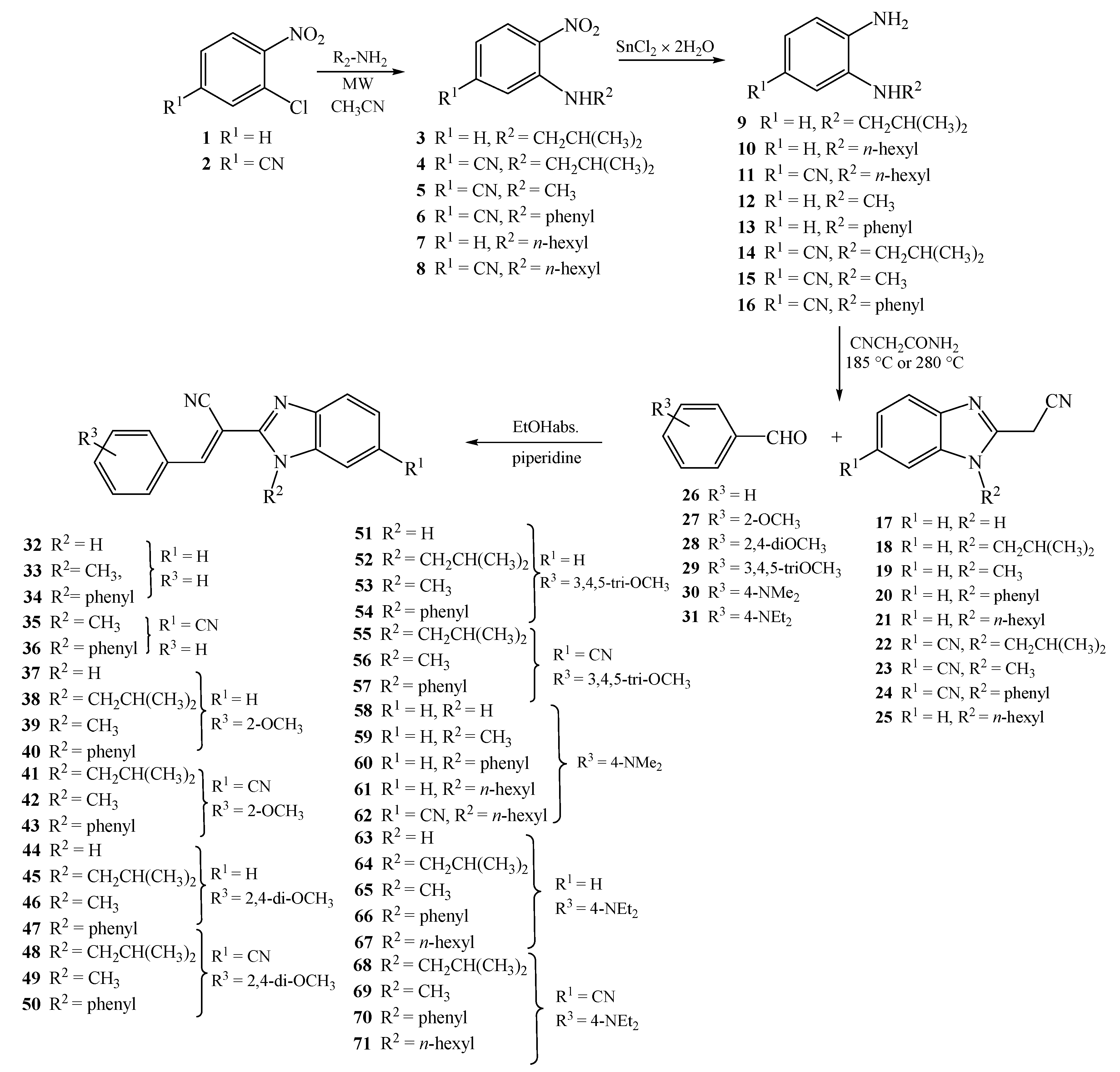
3.1.7. General Method for Preparation of Compounds 32–71

A solution of equimolar amounts of 2-(cyanomethyl)-benzimidazoles 1725, corresponding aromatic aldehydes 2530, and few drops of piperidine in absolute ethanol was refluxed for 2–4 h. The cooled reaction mixture was filtered, and if necessary the product was purified by column chromatography on SiO2 using dichloromethane/methanol at 200:1 as the eluent.
(E)-2-(1H-benzimidazol-2-yl)-3-phenylacrylonitrile 32
Compound 32 was prepared from 17 (0.10 g, 0.6 mmol) and 26 (0.07 g, 0.6 mmol) in absolute ethanol (2 mL) after refluxing for 2 h to yield 0.12 g (78%) of light yellow powder; m.p 224–228 °C; 1H NMR (DMSO-d6, 400 MHz): δ/ppm = 13.10 (s, 1H, NHbenz), 8.36 (s, 1H, Harom), 8.02–7.97 (m, 2H, Harom), 7.71 (d, 1H, J = 7.9 Hz, Harom), 7.64–7.55 (m, 4H, Harom), 7.30 (t, 1H, J = 7.6 Hz, Harom), 7.25 (t, 1H, J = 7.6 Hz, Harom); 13C NMR (DMSO-d6, 101 MHz): δ/ppm = 147.9, 145.8, 143.8, 135.3, 133.2, 132.2, 130.0 (2C), 129.9 (2C), 129.8, 124.2, 122.8, 119.7, 116.6, 112.0, 102.9; Anal. Calcd. for C16H11N3: C, 78.35; H, 4.52; N, 17.13. Found: C, 78.43; H, 4.61; N, 17.07%.
(E)-2-(N-methylbenzimidazol-2-yl)-3-phenylacrylonitrile 33
Compound 33 was prepared from 19 (0.10 g, 0.6 mmol) and 26 (0.06 g, 0.6 mmol) in absolute ethanol (2 mL) after refluxing for 2 h to yield 0.07 g (46%) of red oil. 1H NMR (DMSO-d6, 400 MHz): δ/ppm = 8.19 (s, 1H, Harom), 7.97–7.93 (m, 2H, Harom), 7.80–7.76 (m, 1H, Harom), 7.61–7.57 (m, 3H, Harom), 7.34–7.31 (m, 3H, Harom), 3.33 (s, 3H, CH3); 13C NMR (DMSO-d6, 101 MHz): δ/ppm = 166.8, 163.2, 151.0, 132.8, 132.4, 130.5, 130.2, 129.7, 129.5, 128.9, 128.3, 127.5, 119.3, 118.9, 116.9, 107.2, 30.3; Anal. Calcd. for C17H13N3: C, 78.74; H, 5.05; N, 16.20. Found: C, 78.69; H, 4.95; N, 16.24%.
(E)-2-(N-phenylbenzimidazol-2-yl)-3-phenylacrylonitrile 34
Compound 34 was prepared from 20 (0.10 g, 0.4 mmol) and 26 (0.05 g, 0.4 mmol) in absolute ethanol (2 mL) after refluxing for 2 h to yield 0.04 g (%) of red oil. 1H NMR (DMSO-d6, 400 MHz): δ/ppm = 8.19 (s, 1H, Harom), 7.96–7.93 (m, 3H, Harom), 7.78 (bs, 1H, Harom), 7.61–7.56 (m, 4H, Harom), 7.50 (bs, 1H, Harom), 7.42–7.26 (m, 5H, Harom); 13C NMR (DMSO-d6, 101 MHz): δ/ppm = 166.8, 163.2, 151.0 (2C), 137.4, 132.8 (2C), 132.4, 130.5 (2C), 129.7 (2C), 129.5, 129.2, 129.1, 129.1, 128.9, 128.8, 128.3, 127.5, 118.9, 117.0, 107.2; Anal. Calcd. for C22H15N3: C, 82.22; H, 4.70; N, 13.08. Found: C, 82.15; H, 4.59; N, 13.24%.
(E)-2-(6-cyano-N-methylbenzimidazol-2-yl)-3-phenylacrylonitrile 35
Compound 35 was prepared from 23 (0.10 g, 0.5 mmol) and 26 (0.05 g, 0.6 mmol) in absolute ethanol (2 mL) after refluxing for 2 h to yield 0.11 g (77%) of light brown powder; m.p 197–202 °C; 1H NMR (DMSO-d6, 400 MHz): δ/ppm = 8.30 (d, 1H, J = 0.9 Hz, Harom), 8.27 (s, 1H, Harom), 8.09–8.04 (m, 2H, Harom), 7.91 (d, 1H, J = 8.1 Hz, Harom), 7.76 (dd, 1H, J = 8.5, 1.5 Hz, Harom), 7.65–7.59 (m, 3H, Harom), 4.07 (s, 3H, CH3); 13C NMR (DMSO-d6, 101 MHz): δ/ppm = 152.0, 150.6, 141.7, 139.9, 133.0, 132.7, 130.3 (2C), 129.7 (2C), 127.0, 124.8, 120.1, 116.9, 113.0, 105.4, 100.6, 32.6; Anal. Calcd. for C18H12N4: C, 76.04; H, 4.25; N, 19.71. Found: C, 76.11; H, 4.14; N, 19.68%.
(E)-2-(6-cyano-N-phenylbenzimidazol-2-yl)-3-phenylacrylonitrile 36
Compound 36 was prepared from 24 (0.10 g, 0.4 mmol) and 26 (0.04 g, 0.4 mmol) in absolute ethanol (2 mL) after refluxing for 2 h to yield 0.10 g (73%) of light yellow powder; m.p 195–200 °C; 1H NMR (DMSO-d6, 400 MHz): δ/ppm = 8.44 (s, 1H, Harom), 7.98 (s, 1H, Harom), 7.84–7.80 (m, 2H, Harom), 7.73 (dd, 1H, J = 8.4, 1.3 Hz, Harom), 7.71–7.66 (m, 5H, Harom), 7.59–7.53 (m, 3H, Harom), 7.39 (d, 1H, J = 8.5 Hz, Harom); 13C NMR (DMSO-d6, 101 MHz): δ/ppm = 152.3, 149.8, 141.9, 140.0, 134.8, 132.9, 132.6, 130.8 (2C), 130.5, 130.2 (2C), 129.7 (2C), 128.1 (2C), 128.0, 125.2, 119.9, 115.6, 112.8, 106.2, 100.3; Anal. Calcd. for C23H14N4: C, 79.75; H, 4.07; N, 16.17. Found: C, 79.71; H, 4.14; N, 16.22%.
(E)-2-(1H-benzimidazol-2-yl)-3-(2-methoxyphenyl)acrylonitrile 37
Compound 37 was prepared from 17 (0.10 g, 0.6 mmol) and 27 (0.09 g, 0.6 mmol) in absolute ethanol (3 mL) after refluxing for 3 h to yield 0.18 g (48%) of yellow powder; m.p 260–264 °C; 1H NMR (DMSO-d6, 600 MHz): δ/ppm = 13.11 (s, 1H, NHbenz), 8.54 (s, 1H, Harom), 8.10 (dd, 1H, J = 7.8, 1.2 Hz, Harom), 7.62 (bs, 1H, Harom), 7.57 (td, 1H, J = 7.9, 1.6 Hz, Harom), 7.28–7.24 (m, 2H, Harom), 7.22 (d, 1H, J = 8.4 Hz, Harom), 7.15 (t, 1H, J = 7.6 Hz, Harom), 3.94 (s, 3H, OCH3);
13C NMR (DMSO-d6, 75 MHz): δ/ppm = 158.5, 148.0, 140.8, 133.9, 128.7, 121.9, 121.2, 116.7, 112.3, 103.3, 56.4; Anal. Calcd. for C17H13N3O: C, 74.17; H, 4.76; N, 15.26; O, 5.81. Found: C, 74.11; H, 4.74; N, 15.31; O, 5.77%.
E(Z)-3-(2-methoxyphenyl)-2-(N-isobutylbenzimidazol-2-yl)acrylonitrile 38
Compound 38 was prepared from 18 (0.10 g, 0.5 mmol) and 27 (0.06 g, 0.5 mmol) in absolute ethanol (3 mL) after refluxing for 4.5 h to yield 0.15 g (67%) of yellow oil in the form of a mixture of E- and Z-isomers at the ratio of 38a/38b = 2:1; 38a: 1H NMR (DMSO-d6, 400 MHz): δ/ppm = 8.42 (s, 1H, Harom), 8.09 (dd, 1H, J = 7.8, 1.4 Hz, Harom), 7.74–7.71 (m, 2H, Harom), 7.62–7.58 (m, 1H, Harom), 7.32–7.30 (m, 1H, Harom), 7.28 (dd, 1H, J = 7.9, 1.3 Hz, Harom), 7.23 (d, 1H, J = 7.9 Hz, Harom), 7.18 (d, 1H, J = 7.7 Hz, Harom), 4.35 (d, 2H, J = 7.5 Hz, CH2), 3.92 (s, 3H, CH3), 2.24–2.15 (m, 1H, CH), 0.87 (d, 6H, J = 6.7 Hz, CH3); 13C NMR (DMSO-d6, 101 MHz): δ/ppm = 158.6, 147.2, 146.4, 142.2, 136.9, 134.2, 128.6, 123.8, 123.1, 121.8, 121.2, 119.8, 117.1, 112.4, 111.2, 102.5, 56.5, 51.3, 29.8, 20.0; 38b: 1H NMR (DMSO-d6, 400 MHz): δ/ppm = 8.15 (s, 1H, Harom), 7.76–7.73 (m, 1H, Harom), 7.63 (dd, 1H, J = 6.9, 1.4 Hz, Harom), 7.41 (td, 1H, J = 7.9, 1.7 Hz, Harom), 7.36–7.32 (m, 2H, Harom), 6.70 (t, 1H, J = 7.4 Hz, Harom), 6.57 (dd, 1H, J = 7.8, 1.6 Hz, Harom), 3.84 (s, 3H, CH3), 3.64 (d, 2H, J = 7.6 Hz, CH2), 2.06–2.00 (m, 1H, CH), 0.74 (d, 6H, J = 6.7 Hz, CH3); 13C NMR (DMSO-d6, 101 MHz): δ/ppm = 158.2, 146.8, 145.1, 142.8, 135.4, 134.0, 128.8, 124.0, 123.0, 121.8, 121.2, 121.0, 120.2, 118.6, 112.5, 112.1, 101.4, 56.5, 51.1, 29.0, 19.9; Anal. Calcd. for C21H21N3O: C, 76.11; H, 6.39; N, 12.68; O, 4.83. Found: C, 76.28; H, 6.35; N, 12.71; O, 4.74%.
(E)-3-(2-methoxyphenyl)-2-(N-methylbenzimidazol-2-yl)acrylonitrile 39
Compound 39 was prepared from 19 (0.10 g, 0.6 mmol) and 27 (0.08 g, 0.6 mmol) in absolute ethanol (3 mL) after refluxing for 3 h to yield 0.12 g (71%) of yellow powder; m.p 144–146 °C; 1H NMR (DMSO-d6, 400 MHz): δ/ppm = 8.33 (s, 1H, Harom), 8.12 (dd, 1H, J = 7.8, 1.4 Hz, Harom), 7.72 (d, 1H, J = 7.6 Hz, Harom), 7.65 (d, 1H, J = 7.9 Hz, Harom), 7.60 (td, 1H, J = 7.9, 1.4 Hz, Harom), 7.36 (td, 1H, J = 7.6, 1.2 Hz, Harom), 7.30 (td, 1H, J = 7.6, 1.1 Hz, Harom), 7.22 (d, 1H, J = 8.4 Hz, Harom), 7.17 (t, 1H, J = 7.6 Hz, Harom), 4.01 (s, 3H, OCH3), 3.92 (s, 3H, CH3); 13C NMR (DMSO-d6, 101 MHz): δ/ppm = 158.5, 147.8, 145.9, 142.3, 137.1, 134.1, 128.7, 123.8, 123.1, 121.9, 121.1, 119.7, 117.1, 112.3, 111.3, 101.4, 56.5, 32.1; Anal. Calcd. for C18H15N3O: C, 74.72; H, 5.23; N, 14.52; O, 5.53. Found: C, 74.86; H, 5.04; N, 14.31; O, 5.77%.
(E)-3-(2-methoxyphenyl)-2-(N-phenylbenzimidazol-2-yl)acrylonitrile 40
Compound 40 was prepared from 20 (0.10 g, 0.4 mmol) and 27 (0.06 g, 0.4 mmol) in absolute ethanol (3 mL) after refluxing for 2 h to yield 0.10 g (70%) of orange powder; m.p 169–173 °C; 1H NMR (DMSO-d6, 400 MHz): δ/ppm = 8.02 (dd, 1H, J = 7.8, 1.4 Hz, Harom), 7.98 (s, 1H, Harom), 7.84 (dd, 1H, J = 6.7, 1.5 Hz, Harom), 7.72–7.65 (m, 3H, Harom), 7.65–7.62 (m, 2H, Harom), 7.53 (td, 1H, J = 7.8, 1.5 Hz, Harom), 7.37 (td, 1H, J = 7.4, 1.5 Hz, Harom), 7.33 (td, 1H, J = 7.5, 1.5 Hz, Harom), 7.18 (dd, 1H, J = 6.9, 1.5 Hz, Harom), 7.11 (d, 1H, J = 7.9 Hz, Harom), 7.08 (d, 1H, J = 7.6 Hz, Harom), 3.75 (s, 3H, CH3); 13C NMR (DMSO-d6, 101 MHz): δ/ppm = 158.5, 147.2, 144.5, 142.4, 137.6, 135.8, 134.3, 130.8 (2C), 129.9, 128.1, 128.0 (2C), 126.0, 124.8, 123.8, 121.4 (2C), 121.1, 120.1, 116.3 (2C), 112.3, 111.0, 101.3, 56.2; Anal. Calcd. for C23H17N3O: C, 78.61; H, 4.88; N, 11.96; O, 4.55. Found: C, 78.51; H, 4.74; N, 11.81; O, 4.67%.
E(Z)-2-(6-cyano-N-isobutylbenzimidazol-2-yl)-3-(2-methoxyphenyl)acrylonitrile 41
Compound 41 was prepared from 22 (0.10 g, 0.4 mmol) and 27 (0.06 g, 0.4 mmol) in absolute ethanol (3 mL) after refluxing for 4.5 h to yield 0.15 g (30%) of yellow oil in the form of a mixture of E- and Z-isomers at a ratio of 41a/41b = 2:1; 41a: 1H NMR (DMSO-d6, 600 MHz): δ/ppm = 8.33 (d, 1H, J = 1.08 Hz, Harom), 8.21 (s, 1H, Harom), 7.88 (d, 1H, J = 8.45 Hz, Harom), 7.75–7.73 (m, 1H, Harom), 7.42 (td, 1H, J = 7.9, 1.6 Hz, Harom), 7.13 (d, 1H, J = 8.3 Hz, Harom), 6.73 (t, 1H, J = 7.5 Hz, Harom), 6.60 (dd, 1H, J = 7.8, 1.3 Hz, Harom), 3.77 (s, 3H, CH3), 3.69 (d, 2H, J = 7.6 Hz, CH2), 2.05–1.96 (m, 1H, CH), 0.73 (d, 6H, J = 6.6 Hz, CH3);
13C NMR (DMSO-d6, 151 MHz): δ/ppm = 158.2, 149.4, 147.2, 141.1, 139.3, 134.1, 128.2, 126.4, 124.5, 121.1, 120.7, 119.6, 116.3, 113.1, 112.0, 105.0, 100.0, 56.0, 51.1, 29.3, 19.4 (2C); 41b: 1H NMR (DMSO-d6, 600 MHz): δ/ppm = 8.51 (s, 1H, Harom), 8.30 (d, 1H, J = 1.0 Hz, Harom), 8.10 (dd, 1H, J1 = 7.7, 1.3 Hz, Harom), 7.96 (d, 1H, J = 8.6 Hz, Harom), 7.73–7.71 (m, 1H, Harom), 7.61 (td, 1H, J = 7.7, 1.6 Hz, Harom), 7.23 (d, 1H, J = 8.4 Hz, Harom), 7.17 (t, 1H, J = 7.5 Hz, Harom), 4.40 (d, 2H, J = 7.5 Hz, CH2), 3.92 (s, 3H, CH3), 2.22–2.16 (m, 1H, CH), 0.86 (d, 6H, J = 6.6 Hz, CH3); 13C NMR (DMSO-d6, 151 MHz): δ/ppm = 158.8, 148.8, 147.7, 141.7, 137.8, 133.8, 128.6, 126.7, 124.9, 121.1, 120.5, 119.4, 117.7, 113.2, 112.0, 105.0, 101.2, 56.0, 50.6, 28.6, 19.4 (2C); Anal. Calcd. for C22H20N4O: C, 74.14; H, 5.66; N, 15.72; O, 4.49. Found: C, 74.11; H, 5.74; N, 15.75; O, 4.54%.
(E)-2-(6-cyano-N-methylbenzimidazol-2-yl)-3-(2-methoxyphenyl)acrylonitrile 42
Compound 42 was prepared from 23 (0.10 g, 0.5 mmol) and 27 (0.10 g, 0.5 mmol) in absolute ethanol (3 mL) after refluxing for 3 h to yield 0.13 g (80%) of brown powder; m.p 178–181 °C; 1H NMR (DMSO-d6, 400 MHz): δ/ppm = 8.41 (s, 1H, Harom), 8.30 (d, 1H, J = 1.0 Hz, Harom), 8.13 (dd, 1H, J = 7.4, 1.4 Hz, Harom), 7.88 (d, 1H, J = 8.4 Hz, Harom), 7.75 (dd, 1H, J = 8.5, 1.4 Hz, Harom), 7.62 (td, 1H, J = 7.9, 1.4 Hz, Harom), 7.23 (d, 1H, J = 8.1 Hz, Harom), 7.18 (t, 1H, J = 7.6 Hz, Harom), 4.05 (s, 3H, OCH3), 3.92 (s, 3H, CH3); 13C NMR (DMSO-d6, 101 MHz): δ/ppm = 158.7, 150.6, 147.1, 141.7, 139.9, 134.5, 128.7, 126.9, 124.8, 121.7, 121.2, 120.2, 116.8, 113.0, 112.4, 105.3, 100.7, 56.5, 32.6; Anal. Calcd. for C19H14N4O: C, 72.60; H, 4.49; N, 17.82; O, 5.09. Found: C, 72.53; H, 4.41; N, 17.75; O, 4.94%.
(E)-2-(6-cyano-N-phenylbenzimidazol-2-yl)-3-(2-methoxyphenyl)acrylonitrile 43
Compound 43 was prepared from 24 (0.10 g, 0.4 mmol) and 27 (0.05 g, 0.4 mmol) in absolute ethanol (3 mL) after refluxing for 2 h to yield 0.15 g (77%) of yellow powder; m.p 222–225 °C; 1H NMR (DMSO-d6, 400 MHz): δ/ppm = 8.44 (d, 1H, J = 0.9 Hz, Harom), 8.06–8.01 (m, 2H, Harom), 7.73–7.67 (m, 6H, Harom), 7.55 (td, 1H, J = 7.9, 1.4 Hz, Harom), 7.32 (dd, 1H, J = 8.4, 0.5 Hz, Harom), 7.12 (d, 1H, J = 8.6 Hz, Harom), 7.08 (d, 1H, J = 7.5 Hz, Harom), 3.75 (s, 3H, CH3); 13C NMR (DMSO-d6, 101 MHz): δ/ppm = 158.6, 149.9, 145.8, 141.9, 140.3, 135.1, 134.7, 130.9 (2C), 130.5, 128.2, 128.1 (2C), 127.9, 126.1, 125.1, 121.1, 119.9, 116.0, 112.6, 112.4, 106.0 (2C), 100.5, 56.2; Anal. Calcd. for C24H16N4O: C, 76.58; H, 4.28; N, 14.88; O, 4.25. Found: C, 76.61; H, 4.24; N, 15.05; O, 4.34%.
(E)-2-(1H-benzimidazol-2-yl)-3-(2,4-dimethoxyphenyl)acrylonitrile 44
Compound 44 was prepared from 17 (0.10 g, 0.6 mmol) and 28 (0.10 g, 0.6 mmol) in absolute ethanol (3 mL) after refluxing for 3 h to yield 0.19 g (80%) of yellow powder; m.p 205–209 °C; 1H NMR (DMSO-d6, 600 MHz): δ/ppm = 13.00 (s, 1H, NHbenz), 8.47 (s, 1H, Harom), 8.17 (d, 1H, J = 8.7 Hz, Harom), 7.62 (bs, 2H, Harom), 7.23 (bs, 2H, Harom), 6.76 (dd, 1H, J = 8.8, 2.3 Hz, Harom), 6.74 (d, 1H, J = 2.3 Hz, Harom), 3.95 (s, 3H, OCH3), 3.88 (s, 3H, OCH3); 13C NMR (DMSO-d6, 151 MHz): δ/ppm = 164.0, 160.0, 148.0, 139.5, 129.2, 116.9, 114.3, 106.4, 98.9, 98.4, 56.1, 55.7; Anal. Calcd. for C18H15N3O2: C, 70.81; H, 4.95; N, 13.76; O, 10.48. Found: C, 70.78; H, 4.74; N, 13.75; O, 10.54%.
E(Z)-3-(2,4-dimethoxyphenyl)-2-(N-isobutylbenzimidazol-2-yl)acrylonitrile 45
Compound 45 was prepared from 18 (0.10 g, 0.5 mmol) and 28 (0.08 g, 0.5 mmol) in absolute ethanol (3 mL) after refluxing for 4 h to yield 0.12 g (70%) of yellow oil in the form of a mixture of E- and Z-isomers at a ratio of 45a/45b = 5:1; 45a: 1H NMR (DMSO-d6, 600 MHz): δ/ppm = 8.33 (s, 1H, Harom), 8.16 (d, 1H, J = 8.8 Hz, Harom), 7.67–7.63 (m, 2H, Harom), 7.28–7.26 (m, 1H, Harom), 7.25–7.21 (m, 1H, Harom), 6.75 (dd, 1H, J = 8.8, 2.3 Hz, Harom), 6.71 (d, 1H, J = 2.3 Hz, Harom), 4.29 (d, 2H, J = 7.4 Hz, CH2), 3.88 (s, 3H, OCH3), 3.86 (s, 3H, OCH3), 2.19–2.11 (m, 1H, CH), 0.82 (d, 6H, J = 6.6 Hz, CH3); 13C NMR (DMSO-d6, 151 MHz): δ/ppm = 164.3, 160.2, 147.4, 144.7, 141.7, 136.4, 129.2, 123.0, 122.5, 119.1, 117.4, 114.1, 111.3, 106.5, 98.4, 96.6, 56.1, 55.8, 50.8, 29.2, 19.5 (2C); 45b: 1H NMR (DMSO-d6, 600 MHz): δ/ppm = 7.98 (s, 1H, Harom), 7.70 (d, 1H, J = 7.8 Hz, Harom), 7.61 (d, 1H, J = 8.0 Hz, Harom), 7.31–7.28 (m, 2H, Harom), 6.61 (d, 1H, J = 2.3 Hz, Harom), 6.43 (d, 1H, J = 8.8 Hz, Harom), 6.29 (dd, 1H, J = 8.8, 2.3 Hz, Harom), 6.71 (d, 1H, J = 2.3 Hz, Harom), 3.81 (s, 3H, OCH3), 3.71 (s, 3H, OCH3), 3.68 (d, 2H, J = 7.4 Hz, CH2), 2.05–1.98 (m, 1H, CH), 0.72 (d, 6H, J = 6.6 Hz, CH3); 13C NMR (DMSO-d6, 151 MHz): δ/ppm = 164.0, 159.8, 145.1, 144.9, 142.4, 134.9, 129.5, 129.2, 123.0, 119.7, 118.7, 114.0, 111.5, 106.5, 98.4, 56.2, 55.6, 50.7, 28.6, 19.5 (2C); Anal. Calcd. for C22H23N3O2: C, 73.11; H, 6.41; N, 11.63; O, 8.85. Found: C, 73.19; H, 6.51; N, 11.58; O, 8.75%.
(E)-3-(2,4-dimethoxyphenyl)-2-(N-methylbenzimidazol-2-yl)acrylonitrile 46
Compound 46 was prepared from 19 (0.10 g, 0.6 mmol) and 28 (0.09 g, 0.6 mmol) in absolute ethanol (3 mL) after refluxing for 3 h to yield 0.19 g (89%) of yellow powder; m.p 168–171 °C; 1H NMR (DMSO-d6, 600 MHz): δ/ppm = 8.26 (s, 1H, Harom), 8.20 (d, 1H, J = 8.7 Hz, Harom), 7.68 (d, 1H, J = 8.0 Hz, Harom), 7.61 (d, 1H, J = 8.0 Hz, Harom), 7.32 (td, 1H, J = 7.7, 1.0 Hz, Harom), 7.27 (td, 1H, J = 7.8, 1.0 Hz, Harom), 6.78 (dd, 1H, J = 8.8, 2.4 Hz, Harom), 6.73 (d, 1H, J = 2.3 Hz, Harom), 3.97 (s, 3H, OCH3), 3.91 (s, 3H, OCH3), 3.89 (s, 3H, CH3) 13C NMR (DMSO-d6, 151 MHz): δ/ppm = 164.2, 160.1, 148.0, 144.4, 141.9, 136.6, 129.2, 123.0, 122.5, 119.0, 117.3, 114.2, 110.6, 106.5, 98.4, 96.7, 56.1, 55.7, 31.5; Anal. Calcd. for C19H17N3O2: C, 71.46; H, 5.37; N, 13.16; O, 10.02. Found: C, 71.39; H, 5.43; N, 13.07; O, 10.13%.
(E)-3-(2,4-dimethoxyphenyl)-2-(N-phenylbenzimidazol-2-yl)acrylonitrile 47
Compound 47 was prepared from 20 (0.10 g, 0.4 mmol) and 28 (0.07 g, 0.4 mmol) in absolute ethanol (3 mL) after refluxing for 4 h to yield 0.16 g (75%) of yellow powder; m.p 164–166 °C; 1H NMR (DMSO-d6, 600 MHz): δ/ppm = 8.10 (d, 1H, J = 8.8 Hz, Harom), 7.95 (s, 1H, Harom), 7.80 (d, 1H, J = 7.9 Hz, Harom), 7.69–7.62 (m, 3H, Harom), 7.59 (dd, 2H, J = 6.9, 1.5 Hz, Harom), 7.34 (td, 1H, J = 8.1, 1.1 Hz, Harom), 7.30 (td, 1H, J = 7.9, 1.0 Hz, Harom), 7.14 (d, 1H, J = 7.9 Hz, Harom), 6.70 (dd, 1H, J = 8.9, 2.3 Hz, Harom), 6.63 (d, 1H, J = 2.3 Hz, Harom), 3.85 (s, 3H, OCH3), 3.75 (s, 3H, OCH3); 13C NMR (DMSO-d6, 75 MHz): δ/ppm = 164.7, 160.5, 147.9, 143.5, 142.4, 137.6, 136.0, 130.7, 129.9, 129.2, 128.0, 124.5, 123.7, 119.9, 117.0, 114.3, 110.9, 107.0, 98.7, 97.2, 56.4, 56.2, 56.1; Anal. Calcd. for C24H19N3O2: C, 75.57; H, 5.02; N, 11.02; O, 8.39. Found: C, 75.51; H, 4.92; N, 11.08; O, 8.22%.
(E)-2-(6-cyano-N-isobutylbenzimidazol-2-yl)-3-(2,4-dimethoxyphenyl)acrylonitrile 48
Compound 48 was prepared from 22 (0.10 g, 0.4 mmol) and 28 (0.07 g, 0.4 mmol) in absolute ethanol (3 mL) after refluxing for 4 h to yield 0.16 g (84%) of orange powder; m.p 120–125 °C; 1H NMR (DMSO-d6, 600 MHz): δ/ppm = 8.43 (s, 1H, Harom), 8.23 (d, 1H, J = 0.9 Hz, Harom), 8.18 (d, 1H, J = 8.8 Hz, Harom), 7.89 (d, 1H, J = 8.5 Hz, Harom), 7.67 (dd, 1H, J = 8.5, 1.4 Hz, Harom), 6.75 (dd, 1H, J = 8.8, 2.3 Hz, Harom), 6.71 (d, 1H, J = 2.3 Hz, Harom), 4.35 (d, 2H, J = 7.6 Hz, CH2), 3.89 (s, 3H, OCH3), 3.86 (s, 3H, OCH3), 2.18–2.10 (m, 1H, CH), 0.82 (d, 6H, J = 6.6 Hz, CH3); 13C NMR (DMSO-d6, 151 MHz): δ/ppm = 164.2, 159.8, 148.2, 146.2, 141.8, 137.8, 129.9, 126.6, 124.8, 119.5, 118.4, 113.8, 113.2, 106.7, 104.9, 98.4, 97.2, 56.2, 55.7, 50.9, 28.7, 19.4 (2C); Anal. Calcd. for C23H22N4O2: C, 71.48; H, 5.74; N, 14.50; O, 8.28. Found: C, 71.54; H, 5.61; N, 14.58; O, 8.15%.
(E)-2-(6-cyano-N-methylbenzimidazol-2-yl)-3-(2,4-dimethoxyphenyl)acrylonitrile 49
Compound 49 was prepared from 23 (0.10 g, 0.5 mmol) and 28 (0.08 g, 0.5 mmol) in absolute ethanol (3 mL) after refluxing for 2 h to yield 0.17 g (77%) of yellow powder; m.p 228–231 °C; 1H NMR (DMSO-d6, 400 MHz): δ/ppm = 8.37 (s, 1H, Harom), 8.26 (d, 1H, J = 0.9 Hz, Harom), 8.23 (d, 1H, J = 8.8 Hz, Harom), 7.85 (d, 1H, J = 8.4 Hz, Harom), 7.72 (dd, 1H, J = 8.5, 1.4 Hz, Harom), 6.80 (dd, 1H, J = 8.8, 2.3 Hz, Harom), 6.75 (d, 1H, J = 2.4 Hz, Harom), 4.03 (s, 3H, OCH3), 3.93 (s, 3H, OCH3), 3.91 (s, 3H, CH3); 13C NMR (DMSO-d6, 151 MHz): δ/ppm = 165.1, 160.9, 151.3, 146.0, 141.8, 140.0, 129.9, 126.6, 124.5, 120.2, 117.6, 114.5, 112.8, 107.1, 105.1, 98.9, 96.2, 56.7, 56.3, 32.5; Anal. Calcd. for C20H16N4O2: C, 69.76; H, 4.68; N, 16.27; O, 9.29. Found: C, 69.71; H, 4.51; N, 16.38; O, 9.15%.
(E)-2-(6-cyano-N-phenylbenzimidazol-2-yl)-3-(2,4-dimethoxyphenyl)acrylonitrile 50
Compound 50 was prepared from 24 (0.10 g, 0.4 mmol) and 28 (0.06 g, 0.4 mmol) in absolute ethanol (3 mL) after refluxing for 2 h to yield 0.16 g (98%) of yellow powder; m.p 213–217 °C; 1H NMR (DMSO-d6, 400 MHz): δ/ppm = 8.39 (d, 1H, J = 0.8 Hz, Harom), 8.13 (d, 1H, J = 8.9 Hz, Harom), 8.02 (s, 1H, Harom), 7.70–7.65 (m, 6H, Harom), 7.28 (d, 1H, J = 8.5 Hz, Harom), 6.71 (dd, 1H, J = 8.9, 2.3 Hz, Harom), 6.64 (d, 1H, J = 2.4 Hz, Harom), 3.86 (s, 3H, OCH3), 3.76 (s, 3H, OCH3); 13C NMR (DMSO-d6, 151 MHz): δ/ppm = 164.4, 153.4, 150.9, 133.1 (2C), 119.2, 118.7, 112.1 (2C), 97.7, 40.0 (2C); Anal. Calcd. for C25H18N4O2: C, 73.88; H, 4.46; N, 13.78; O, 7.87. Found: C, 73.73; H, 4.51; N, 13.69; O, 7.75%.
(E)-2-(1H-benzimidazol-2-yl)-3-(3,4,5-trimethoxyphenyl)acrylonitrile 51
Compound 51 was prepared from 17 (0.10 g, 0.6 mmol) and 29 (0.12 g, 0.6 mmol) in absolute ethanol (3 mL) after refluxing for 2.5 h to yield 0.05 g (22%) of yellow powder; m.p 188–193 °C; 1H NMR (DMSO-d6, 400 MHz): δ/ppm = 8.17 (s, 1H, Harom), 7.85 (bs, 1H, Harom), 7.74 (bs, 2H, Harom), 7.36 (s, 3H, Harom), 3.83 (s, 6H, OCH3), 3.77 (s, 3H, OCH3); 13C NMR (DMSO-d6, 101 MHz): δ/ppm = 163.2, 153.4, 151.3, 141.5, 127.6, 117.3, 108.4, 105.5, 60.8, 56.5; Anal. Calcd. for C19H17N3O3: C, 68.05; H, 5.11; N, 12.53; O, 14.31. Found: C, 68.11; H, 4.96; N, 12.38; O, 14.36%.
(E)-2-(N-isobutylbenzimidazol-2-yl)-3-(3,4,5-trimethoxyphenyl)acrylonitrile 52
Compound 52 was prepared from 18 (0.10 g, 0.5 mmol) and 29 (0.09 g, 0.5 mmol) in absolute ethanol (3 mL) after refluxing for 4 h to yield 0.18 g (64%) of orange oil; 1H NMR (DMSO-d6, 400 MHz): δ/ppm = 8.25 (s, 1H, Harom), 7.72 (t, 2H, J = 8.3 Hz, Harom), 7.50 (s, 1H, Harom), 7.35–7.28 (m, 2H, Harom), 4.36 (d, 2H, J = 7.4 Hz, CH2), 3.87 (s, 6H, OCH3), 3.79 (s, 3H, OCH3), 2.21–2.13 (m, 1H, CH), 0.84 (d, 6H, J = 6.7 Hz, CH3); 13C NMR (DMSO-d6, 101 MHz): δ/ppm = 153.4, 153.0, 151.5, 147.3, 142.2, 136.9, 128.4, 128.1, 123.8, 123.1, 119.7, 117.6, 112.0, 108.2, 107.8, 99.1, 60.8, 56.5 (2C), 55.7, 51.3, 20.0 (2C); Anal. Calcd. for C23H25N3O3: C, 70.57; H, 6.44; N, 10.73; O, 12.26. Found: C, 70.62; H, 6.41; N, 10.81; O, 12.36%.
(E)-3-(3,4,5-trimethoxyphenyl)-2-(N-methylbenzimidazol-2-yl)acrylonitrile 53
Compound 53 was prepared from 19 (0.10 g, 0.6 mmol) and 29 (0.11 g, 0.6 mmol) in absolute ethanol (3 mL) after refluxing for 4 h to yield 0.20 g (49%) of yellow powder; m.p 134–137 °C; 1H NMR (DMSO-d6, 400 MHz): δ/ppm = 8.13 (s, 1H, Harom), 7.71 (d, 1H, J = 7.7 Hz, Harom), 7.67 (d, 1H, J = 7.9 Hz, Harom), 7.49 (s, 2H, Harom), 7.36 (td, 1H, J = 7.6, 1.1 Hz, Harom), 7.30 (td, 1H, J = 7.5, 1.1 Hz, Harom), 4.02 (s, 3H, OCH3), 3.87 (s, 3H, OCH3), 3.79 (s, 3H, OCH3); 13C NMR (DMSO-d6, 101 MHz): δ/ppm = 153.4, 150.6, 147.9, 142.4, 141.2, 137.1, 128.5, 123.8, 123.1, 119.7, 117.6, 111.3, 108.1, 99.7, 60.8, 56.5 (2C), 32.2; Anal. Calcd. for C20H19N3O3: C, 68.75; H, 5.48; N, 12.03; O, 13.74. Found: C, 68.72; H, 5.36; N, 12.08; O, 13.66%.
(E)-2-(N-phenylbenzimidazol-2-yl)-3-(3,4,5-trimethoxyphenyl)acrylonitrile 54
Compound 54 was prepared from 20 (0.10 g, 0.4 mmol) and 29 (0.08 g, 0.4 mmol) in absolute ethanol (3 mL) after refluxing for 4 h to yield 0.17 g (71%) of orange oil; 1H NMR (DMSO-d6, 300 MHz): δ/ppm = 7.99 (s, 1H, Harom), 7.81 (d, 1H, J = 6.8 Hz, Harom), 7.67–7.62 (m, 3H, Harom), 7.51–7.46 (m, 1H, Harom), 7.38–7.33 (m, 2H, Harom), 7.27 (s, 2H, Harom), 7.22 (d, 1H, J = 7.2 Hz, Harom), 7.13–7.10 (m, 1H, Harom), 3.79 (s, 3H, OCH3), 3.75 (s, 3H, OCH3); 13C NMR (DMSO-d6, 101 MHz): δ/ppm = 153.3, 153.0, 152.5, 151.0, 135.5, 134.6, 130.7, 130.6, 130.2, 130.2, 129.2, 128.0, 126.4, 126.3, 125.2, 124.0, 123.9, 120.4, 107.6, 107.5, 60.8, 60.7, 56.5, 55.8, 55.7; Anal. Calcd. for C25H21N3O3: C, 72.98; H, 5.14; N, 10.21; O, 11.67. Found: C, 72.86; H, 5.06; N, 10.18; O, 11.59%.
(E)-2-(6-cyano-N-isobutylbenzimidazol-2-yl)-3-(3,4,5-trimethoxyphenyl)acrylonitrile 55
Compound 55 was prepared from 22 (0.10 g, 0.4 mmol) and 29 (0.08 g, 0.4 mmol) in absolute ethanol (3 mL) after refluxing for 4 h to yield 0.17 g (56%) of yellow powder; m.p 163–167 °C; 1H NMR (DMSO-d6, 600 MHz): δ/ppm = 8.33 (s, 1H, Harom), 8.28 (d, 1H, J = 1.2 Hz, Harom), 7.98 (d, 1H, J = 8.4 Hz, Harom), 7.74 (dd, 1H, J = 8.5, 1.4 Hz, Harom), 7.51 (s, 2H, Harom), 4.43 (d, 2H, J = 7.6 Hz, CH2), 3.86 (s, 6H, OCH3), 3.80 (s, 3H, OCH3), 2.20–2.15 (m, 1H, CH), 0.84 (d, 6H, J = 6.6 Hz, CH3); 13C NMR (DMSO-d6, 151 MHz): δ/ppm = 152.9, 152.3, 149.5, 141.2, 141.1, 139.3, 127.6, 126.4, 124.3, 119.5, 116.7, 113.1, 108.0, 105.0, 97.7, 60.3, 56.0 (2C), 51.0, 29.3, 19.4; Anal. Calcd. for C24H24N4O3: C, 69.21; H, 5.81; N, 13.45; O, 11.52. Found: C, 69.19; H, 5.86; N, 13.38; O, 11.56%.
(E)-2-(6-cyano-N-methylbenzimidazol-2-yl)-3-(3,4,5-trimethoxyphenyl)acrylonitrile 56
Compound 56 was prepared from 23 (0.10 g, 0.5 mmol) and 29 (0.09 g, 0.5 mmol) in absolute ethanol (3 mL) after refluxing for 4 h to yield 0.16 g (88%) of yellow powder; m.p 259–262 °C; 1H NMR (DMSO-d6, 400 MHz): δ/ppm = 8.28 (d, 1H, J = 1.0 Hz, Harom), 8.19 (s, 1H, Harom), 7.89 (d, 1H, J = 8.5 Hz, Harom), 7.75 (dd, 1H, J = 8.5, 1.5 Hz, Harom), 7.50 (s, 2H, Harom), 4.06 (s, 3H, OCH3), 3.87 (s, 3H, OCH3), 3.80 (s, 3H, OCH3); 13C NMR (DMSO-d6, 101 MHz): δ/ppm = 153.4, 151.8, 150.7, 141.7, 141.5, 139.9, 128.2, 126.9, 124.7, 120.2, 117.3, 113.0, 108.3, 105.3, 98.9, 60.8, 56.5 (2C), 32.6; Anal. Calcd. for C21H18N4O3: C, 67.37; H, 4.85; N, 14.96; O, 12.82. Found: C, 67.29; H, 4.89; N, 14.88; O, 12.86%.
(E)-2-(6-cyano-N-phenylbenzimidazol-2-yl)-3-(3,4,5-trimethoxyphenyl)acrylonitrile 57
Compound 57 was prepared from 24 (0.10 g, 0.4 mmol) and 29 (0.08 g, 0.4 mmol) in absolute ethanol (3 mL) after refluxing for 4 h to yield 0.17 g (83%) of yellow powder; m.p 147–150 °C; 1H NMR (DMSO-d6, 600 MHz): δ/ppm = 8.39 (d, 1H, J = 0.9 Hz, Harom), 8.06 (s, 1H, Harom), 7.71 (dd, 1H, J = 8.5, 1.4 Hz, Harom), 7.69–7.66 (m, 5H, Harom), 7.36 (d, 1H, J = 8.4 Hz, Harom), 7.29 (s, 2H, Harom), 3.79 (s, 3H, OCH3), 3.76 (s, 3H, OCH3); 13C NMR (DMSO-d6, 75 MHz): δ/ppm = 153.4, 152.3, 145.0, 141.9, 140.1, 134.8, 130.7, 130.5, 128.1, 127.9, 127.9, 124.9, 119.9, 115.9, 112.7, 108.2, 106.1, 98.4, 60.8, 56.5 (2C); Anal. Calcd. for C26H20N4O3: C, 71.55; H, 4.62; N, 12.84; O, 11.00. Found: C, 71.48; H, 4.65; N, 12.78; O, 11.07%.
(E)-2-(1H-benzimidazol-2-yl)-3-(4-N,N-dimethylaminophenyl)acrylonitrile 58
Compound 58 was prepared from 17 (0.10 g, 0.6 mmol) and 30 (0.09 g, 0.6 mmol) in absolute ethanol (2 mL) after refluxing for 3 h to yield 0.14 g (76%) of orange powder; m.p 272–277 °C; 1H NMR (DMSO-d6, 300 MHz): δ/ppm = 12.78 (s, 1H, NHbenz), 8.13 (s, 1H, Harom), 7.91 (d, 1H, J = 9.0 Hz, Harom), 7.63 (d, 1H, J = 7.4 Hz, Harom), 7.50 (d, 1H, J = 7.1 Hz, Harom), 7.25–7.16 (m, 2H, Harom), 6.87 (d, 2H, J = 9.1 Hz, Harom), 3.07 (s, 6H, CH3); 13C NMR (DMSO-d6, 75 MHz): δ/ppm = 164.4, 152.9, 150.9, 149.4, 145.9, 133.1 (2C), 132.3, 120.3, 119.2, 118.2, 112.3 (2C), 112.1 (2C), 94.2; Anal. Calcd. for C18H16N4: C, 74.98; H, 5.59; N, 19.43. Found: C, 74.91; H, 5.76; N, 19.38%.
(E)-3-(4-N,N-dimethylaminophenyl)-2-(N-methylbenzimidazol-2-yl)acrylonitrile 59
Compound 59 was prepared from 19 (0.10 g, 0.6 mmol) and 30 (0.09 g, 0.6 mmol) in absolute ethanol (2 mL) after refluxing for 3.5 h to yield 0.10 g (58%) of red powder; m.p 202–206 °C; 1H NMR (DMSO-d6, 400 MHz): δ/ppm = 7.97 (s, 1H, Harom), 7.86 (d, 3H, J = 9.1 Hz, Harom), 7.60–7.45 (m, 2H, Harom), 6.83 (d, 3H, J = 9.1 Hz, Harom), 3.06 (s, 9H, CH3); 13C NMR (DMSO-d6, 101 MHz): δ/ppm = 164.4, 153.4, 150.9, 133.1 (2C), 119.2 (2C), 118.7 (2C), 112.1 (2C), 97.7, 40.0 (3C); Anal. Calcd. for C19H18N4: C, 75.47; H, 6.00; N, 18.53. Found: C, 75.56; H, 5.94; N, 18.47%.
(E)-3-(4-N,N-dimethylaminophenyl)-2-(N-phenylbenzimidazol-2-yl)acrylonitrile 60
Compound 60 was prepared from 20 (0.10 g, 0.4 mmol) and 30 (0.06 g, 0.4 mmol) in absolute ethanol (2 mL) after refluxing for 3.5 h to yield 0.11 g (69%) of yellow powder; m.p 206–209 °C; 1H NMR (DMSO-d6, 300 MHz): δ/ppm = 7.77 (dd, 1H, J = 7.0, 1.1 Hz, Harom), 7.73 (d, 3H, J = 8.8 Hz, Harom), 7.68–7.56 (m, 5H, Harom), 7.34 (td, 1H, J = 7.4, 1.4 Hz, Harom), 7.27 (td, 1H, J = 7.9, 1.4 Hz, Harom), 7.17 (dd, 1H, J = 7.1, 1.4 Hz, Harom), 6.79 (d, 2H, J = 9.1 Hz, Harom), 3.04 (s, 3H, CH3); 13C NMR (DMSO-d6, 75 MHz): δ/ppm = 153.1, 150.6, 148.7, 142.6, 137.4, 136.0, 133.1, 132.4, 130.6, 129.8, 128.0, 124.0, 123.5, 120.0, 119.5, 117.6, 112.1, 110.8, 91.9, 40.0 (2C); Anal. Calcd. for C24H20N4: C, 79.10; H, 5.53; N, 15.37. Found: C, 79.17; H, 5.47; N, 15.43%.
E(Z)-3-(4-N,N-dimethylaminophenyl)-2-(N-hexylbenzimidazol-2-yl)acrylonitrile 61
Compound 61 was prepared from 21 (0.07 g, 0.3 mmol) and 30 (0.04 g, 0.3 mmol) in absolute ethanol (2 mL) after refluxing for 3 h to yield 0.10 g (94%) of brown oil in the form of a mixture of E- and Z-isomers at a ratio of 61a/61b = 2:1; 61a: 1H NMR (DMSO-d6, 400 MHz): δ/ppm = 7.99 (s, 1H, Harom), 7.97 (d, 1H, J = 9.0 Hz, Harom), 7.67 (d, 1H, J = 7.6 Hz, Harom), 7.63 (d, 1H, J = 7.9 Hz, Harom), 7.33–7.27 (m, 1H, Harom), 7.27–7.23 (m, 1H, Harom), 6.88 (t, 2H, J = 8.7 Hz, Harom), 6.56 (d, 1H, J = 9.1 Hz, Harom), 4.43 (t, 2H, J = 7.5 Hz, CH2), 3.08 (s, 6H, CH3), 1.84–1.76 (m, 2H, CH2), 1.30–1.20 (m, 6H, CH2), 0.80 (t, 6H, J = 7.0 Hz, CH3); 13C NMR (DMSO-d6, 101 MHz): δ/ppm = 152.8, 151.7, 146.0, 143.1, 135.2, 132.6 (2C), 123.8, 122.8, 120.2, 120.1, 119.9, 112.1 (2C), 111.9, 93.3, 44.2, 31.1, 29.6, 26.1, 22.4, 14.2 (2C); 61b: 1H NMR (DMSO-d6, 400 MHz): δ/ppm = 7.97 (d, 2H, J = 9.0 Hz, Harom), 7.87 (s, 1H, Harom), 7.74 (d, 1H, J = 7.8 Hz, Harom), 7.63 (d, 1H, J = 7.9 Hz, Harom), 7.38–7.32 (m, 1H, Harom), 7.33–7.27 (m, 1H, Harom), 6.88 (t, 2H, J = 8.7 Hz, Harom), 4.03 (t, 2H, J = 7.1 Hz, CH2), 2.92 (s, 6H, CH3), 1.65–1.59 (m, 2H, CH2), 1.14–1.08 (m, 6H, CH2), 0.76 (t, 6H, J = 6.7 Hz, CH3); 13C NMR (DMSO-d6, 101 MHz): δ/ppm = 153.1, 151.3, 148.7, 142.6, 136.4, 132.4 (2C), 123.8, 123.2, 120.3, 119.9, 119.4, 111.9 (2C), 111.8, 91.2, 44.4, 31.1, 29.7, 26.2, 22.3, 14.2 (2C); Anal. Calcd. for C24H28N4: C, 77.38; H, 7.58; N, 15.04. Found: C, 77.41; H, 7.66; N, 15.08%.
(E)-2-(5-cyano-N-hexylbenzimidazol-2-yl)-3-(4-N,N-dimethylaminophenyl)acrylonitrile 62
Compound 62 was prepared from 25 (0.05 g, 0.2 mmol) and 30 (0.03 g, 0.2 mmol) in absolute ethanol (1.5 mL) after refluxing for 3 h to yield 0.3 g (76%) of orange oil; 1H NMR (DMSO-d6, 600 MHz): δ/ppm = 8.20 (d, 1H, J = 1.0 Hz, Harom), 8.08 (s, 1H, Harom), 7.98 (d, 2H, J = 9.1 Hz, Harom), 7.87 (d, 1H, J = 9.1 Hz, Harom), 7.69 (dd, 1H, J = 8.4, 1.4 Hz, Harom), 6.87 (d, 2H, J = 9.1 Hz, Harom), 4.49 (t, 2H, J = 7.5 Hz, CH2), 3.08 (s, 6H, CH3), 1.81–1.78 (m, 2H, CH2), 1.26–1.23 (m, 6H, CH2), 0.80 (t, 3H, J = 6.7 Hz, CH3); 13C NMR (DMSO-d6, 151 MHz): δ/ppm = 153.4, 152.4, 151.5, 142.0, 139.3, 133.0 (2C), 126.4, 124.2, 120.2, 120.1, 118.6, 112.8, 112.2 (2C), 105.1, 89.9, 44.8, 31.0, 29.7, 26.0, 22.4, 14.2 (2C); Anal. Calcd. for C25H27N5: C, 75.54; H, 6.85; N, 17.62. Found: C, 75.61; H, 6.83; N, 17.58%.
(E)-2-(1H-benzimidazol-2-yl)-3-(4-N,N-diethylaminophenyl)acrylonitrile 63
Compound 63 was prepared from 17 (0.10 g, 0.6 mmol) and 31 (0.11 g, 0.6 mmol) in absolute ethanol (2 mL) after refluxing for 3 h to yield 0.14 g (68%) of orange powder; m.p 151–156 °C; 1H NMR (DMSO-d6, 300 MHz): δ/ppm = 12.75 (s, 1H, NHbenz), 8.10 (s, 1H, Harom), 7.97–7.81 (m, 3H, Harom), 7.53 (bs, 2H, Harom), 7.23–7.17 (m, 1H, Harom), 6.87–6.77 (m, 2H, Harom), 3.47 (q, 4H, J = 6.4 Hz, CH2), 1.15 (t, 6H, J = 6.7 Hz, CH3); 13C NMR (DMSO-d6, 151 MHz): δ/ppm = 150.6, 149.5, 145.9, 144.0, 135.3, 133.5, 132.8, 123.2, 122.3, 119.6, 119.0, 118.4 (2C), 111.8, 111.6, 93.4, 44.4 (2C), 12.9 (2C); Anal. Calcd. for C20H20N4: C, 75.92; H, 6.37; N, 17.71. Found: C, 75.97; H, 6.46; N, 17.76%.
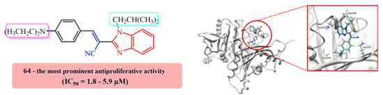
E(Z)-3-(4-N,N-diethylaminophenyl)-2-(N-isobutylbenzimidazol-2-yl)acrylonitrile 64
Compound 64 was prepared from 18 (0.10 g, 0.5 mmol) and 31 (0.08 g, 0.5 mmol) in absolute ethanol (2 mL) after refluxing for 3 h to yield 0.10 g (61%) of light red oil in the form of a mixture of E- and Z-isomers at a ratio of 64a/64b = 2:1; 64a: 1H NMR (DMSO-d6, 600 MHz): δ/ppm = 8.02 (s, 1H, Harom), 7.95 (d, 2H, J = 9.1 Hz, Harom), 7.67–7.63 (m, 2H, Harom), 7.31–7.29 (m, 2H, Harom), 7.24 (td, 1H, J = 7.6, 1.1 Hz, Harom), 6.85–6.81 (m, 1H, Harom), 6.52 (d, 1H, J = 9.2 Hz, Harom), 4.31 (d, 2H, J = 7.5 Hz, CH2), 3.47 (q, 4H, J = 7.0 Hz, CH2), 2.22–2.09 (m, 1H, CH), 1.15 (t, 6H, J = 6.9 Hz, CH3), 0.83 (d, 6H, J = 6.7 Hz, CH3); 13C NMR (DMSO-d6, 151 MHz): δ/ppm = 151.3, 150.9, 148.8, 142.4, 136.9, 133.0 (2C), 123.1, 122.7, 119.7, 119.3, 119.0, 111.7 (2C), 111.5, 90.6, 51.2, 44.4 (2C), 29.6, 20.1 (2C), 12.9 (2C); 64b: 1H NMR (DMSO-d6, 600 MHz): δ/ppm = 7.79 (s, 1H, Harom), 7.73 (d, 1H, J = 7.9 Hz, Harom), 7.69 (d, 1H, J = 8.0 Hz, Harom), 7.35 (td, 1H, J = 7.5, 1.0 Hz, Harom), 7.31–7.29 (m, 1H, Harom), 6.85–6.81 (m, 4H, Harom), 3.84 (d, 2H, J = 7.6 Hz, CH2), 3.31 (q, 4H, J = 6.8 Hz, CH2), 2.12–2.08 (m, 1H, CH), 1.02 (t, 6H, J = 6.9 Hz, CH3), 0.79 (d, 6H, J = 6.6 Hz, CH3); 13C NMR (DMSO-d6, 151 MHz): δ/ppm = 151.3, 150.5, 146.3, 143.0, 135.5, 132.8 (2C), 123.7, 122.8, 120.2, 120.1, 119.4, 112.1, 111.6 (2C), 92.6, 51.3, 44.2 (2C), 29.1, 20.2 (2C), 12.8 (2C); Anal. Calcd. for C24H28N4: C, 77.38; H, 7.58; N, 15.04. Found: C, 77.42; H, 7.63; N, 15.10%.
(E)-3-(4-N,N-diethylaminophenyl)-2-(N-methylbenzimidazol-2-yl)acrylonitrile 65
Compound 65 was prepared from 19 (0.10 g, 0.6 mmol) and 31 (0.10 g, 0.6 mmol) in absolute ethanol (2 mL) after refluxing for 2.5 h to yield 0.12 g (65%) of orange oil; 1H NMR (DMSO-d6, 400 MHz): δ/ppm = 7.95 (s, 1H, Harom), 7.84 (d, 3H, J = 9.1 Hz, Harom), 7.60–7.45 (m, 2H, Harom), 6.80 (d, 3H, J = 9.1 Hz, Harom), 3.45 (q, 4H, J = 7.0 Hz, Harom), 1.13 (t, 6H, J = 7.0 Hz, Harom); 13C NMR (DMSO-d6, 101 MHz): δ/ppm = 164.5, 151.2, 150.8 (2C), 147.5, 133.5 (2C), 133.2, 118.9, 118.6, 111.7 (2C), 111.2, 96.9, 44.4 (2C), 12.9 (3C); Anal. Calcd. for C21H22N4: C, 76.33; H, 6.71; N, 16.96. Found: C, 76.27; H, 6.76; N, 16.88%.
(E)-3-(4-N,N-diethylaminophenyl)-2-(N-phenylbenzimidazol-2-yl)acrylonitrile 66
Compound 66 was prepared from 20 (0.10 g, 0.4 mmol) and 31 (0.08 g, 0.4 mmol) in absolute ethanol (2.5 mL) after refluxing for 2 h to yield 0.06 g (50%) of red powder; m.p 142–147 °C; 1H NMR (DMSO-d6, 600 MHz): δ/ppm = 7.75 (d, 1H, J = 7.9 Hz, Harom), 7.70 (d, 2H, J = 9.0 Hz, Harom), 7.67–7.63 (m, 3H, Harom), 7.61–7.59 (m, 1H, Harom), 7.59–7.56 (m, 2H, Harom), 7.32 (td, 1H, J = 7.6, 0.9 Hz, Harom), 7.27 (td, 1H, J = 8.1, 1.0 Hz, Harom), 7.15 (d, 1H, J = 8.0 Hz, Harom), 6.75 (d, 2H, J = 9.0 Hz, Harom), 3.43 (q, 4H, J = 6.9 Hz, CH2), 1.11 (t, 6H, J = 7.0 Hz, CH3); 13C NMR (DMSO-d6, 151 MHz): δ/ppm = 150.8, 150.4, 148.9, 142.6, 137.4, 136.1, 132.9 (2C), 130.6 (2C), 129.7, 128.0 (2C), 124.0, 123.5, 119.5, 119.5, 117.8, 111.7 (2C), 110.7, 91.2, 44.4 (2C), 12.9 (2C); Anal. Calcd. for C26H24N4: C, 79.56; H, 6.16; N, 14.27. Found: C, 79.51; H, 6.26; N, 14.19%.
E(Z)-3-(4-N,N-diethylaminophenyl)-2-(N-hexylbenzimidazol-2-yl)acrylonitrile 67
Compound 67 was prepared from 21 (0.07 g, 0.3 mmol) and 31 (0.05 g, 0.3 mmol) in absolute ethanol (2 mL) after refluxing for 3 h to yield 0.10 g (87%) of light red oil in the form of a mixture of E- and Z-isomers at a ratio of 67a/67b = 2:1; 67a: 1H NMR (DMSO-d6, 400 MHz): δ/ppm = 7.96 (s, 2H, Harom), 7.69–7.65 (m, 1H, Harom), 7.65–7.61 (m, 1H, Harom), 7.27–7.22 (m, 1H, Harom), 6.87 (d, 1H, J = 9.1 Hz, Harom), 6.83 (d, 2H, J = 9.1 Hz, Harom), 6.52 (d, 1H, J = 9.2 Hz, Harom), 4.43 (t, 2H, J = 7.4 Hz, CH2), 3.48 (q, 4H, J = 7.0 Hz, CH2), 1.84–1.75 (m, 2H, CH2), 1.30–1.20 (m, 6H, CH2), 1.15 (t, 6H, J = 7.0 Hz, CH3), 0.80 (t, 3H, J = 7.0 Hz, CH3); 13C NMR (DMSO-d6, 151 MHz): δ/ppm = 151.2, 150.9, 148.8, 142.6, 136.4, 133.0 (2C), 123.2, 122.8, 119.7, 119.3, 118.9, 111.7 (2C), 111.2, 90.4, 44.4 (2C), 31.1, 29.7, 26.1, 22.4, 14.2, 12.9 (2C); 67b: 1H NMR (DMSO-d6, 400 MHz): δ/ppm = 7.94 (s, 2H, Harom), 7.83 (s, 1H, Harom), 7.74 (d, 1H, J = 7.4 Hz, Harom), 7.65–7.61 (m, 1H, Harom), 7.38–7.33 (m, 1H, Harom), 7.32–7.30 (m, 1H, Harom), 7.30–7.27 (m, 2H, Harom), 4.06 (t, 2H, J = 7.1 Hz, CH2), 1.67–1.57 (m, 2H, CH2), 1.12–1.08 (m, 6H, CH2), 1.02 (t, 6H, J = 7.0 Hz, CH3), 0.74 (t, 3H, J = 6.7 Hz, CH3); 13C NMR (DMSO-d6, 151 MHz): δ/ppm = 151.5, 150.5, 146.1, 143.2, 135.2, 132.8 (2C), 123.7, 122.8, 120.2, 120.0, 119.5, 111.4 (2C), 111.0, 92.4, 44.2 (2C), 31.2, 29.5, 26.2, 22.3, 14.2, 12.8 (2C); Anal. Calcd. for C26H32N4: C, 77.96; H, 8.05; N, 13.99. Found: C, 77.91; H, 8.16; N, 14.03%.
E(Z)-2-(6-cyano-N-butylbenzimidazol-2-yl)-3-(4-N,N-diethylaminophenyl)acrylonitrile 68
Compound 68 was prepared from 22 (0.10 g, 0.4 mmol) and 31 (0.07 g, 0.4 mmol) in absolute ethanol (2 mL) after refluxing for 3 h to yield 0.10 g (61%) of red oil in the form of a mixture of E- and Z-isomers at a ratio of 68a/68b = 2:1; 68a: 1H NMR (DMSO-d6, 600 MHz): δ/ppm = 8.20 (d, 1H, J = 1.2 Hz, Harom), 8.13 (s, 1H, Harom), 7.97 (d, 2H, J = 9.1 Hz, Harom), 7.68 (dd, 1H, J = 8.4, 1.4 Hz, Harom), 6.85–6.81 (m, 2H, Harom), 6.54 (d, 1H, J = 9.2 Hz, Harom), 4.39 (d, 2H, J = 7.6 Hz, CH2), 3.48 (q, 4H, J = 7.0 Hz, CH2), 2.18–2.13 (m, 1H, CH), 1.15 (t, 6H, J = 7.0 Hz, CH3), 0.83 (d, 6H, J = 6.6 Hz, CH3);
13C NMR (DMSO-d6, 151 MHz): δ/ppm = 152.3, 151.6, 151.2, 141.8, 139.9, 133.5 (2C), 126.3, 124.1, 120.2, 119.6, 118.7, 113.1, 111.8 (2C), 105.0, 89.4, 51.4, 44.5 (2C), 29.7, 19.9 (2C), 12.9 (2C); 68b: 1H NMR (DMSO-d6, 600 MHz): δ/ppm = 8.33 (d, 1H, J = 1.0 Hz, Harom), 7.95 (d, 1H, J = 8.6 Hz, Harom), 7.90 (d, 2H, J = 8.4 Hz, Harom), 7.86 (s, 1H, Harom), 7.76 (dd, 1H, J = 8.5, 1.5 Hz, Harom), 6.85–6.81 (m, 2H, Harom), 3.91 (d, 2H, J = 7.6 Hz, CH2), 3.32 (q, 4H, J = 6.8 Hz, CH2), 2.10–2.05 (m, 1H, CH), 1.02 (t, 6H, J = 7.0 Hz, CH3), 0.79 (d, 6H, J = 6.6 Hz, CH3); 13C NMR (DMSO-d6, 151 MHz): δ/ppm = 152.1, 150.7, 149.3, 142.4, 138.4, 132.9 (2C), 127.0, 125.3, 120.1, 119.8, 119.1, 113.8, 111.6 (2C), 105.3, 91.2, 51.5, 44.2 (2C), 29.2, 20.0 (2C), 12.8 (2C); Anal. Calcd. for C25H27N5: C, 75.54; H, 6.85; N, 17.62. Found: C, 75.50; H, 6.89; N, 17.57%.
(E)-2-(6-cyano-N-methylbenzimidazol-2-yl)-3-(4-N,N-diethylaminophenyl)acrylonitrile 69
Compound 69 was prepared from 23 (0.10 g, 0.5 mmol) and 31 (0.09 g, 0.5 mmol) in absolute ethanol (2 mL) after refluxing for 2.5 h to yield 0.15 g (84%) of red powder; m.p 164–169 °C; 1H NMR (DMSO-d6, 300 MHz): δ/ppm = 8.18 (s, 1H, Harom), 7.99 (d, 2H, J = 4.7 Hz, Harom), 7.95 (s, 1H, Harom), 7.82 (d, 1H, J = 8.4 Hz, Harom), 7.68 (dd, 1H, J = 8.4, 1.4 Hz, Harom), 6.84 (d, 2H, J = 9.1 Hz, Harom), 4.00 (s, 3H, CH3), 3.48 (q, 4H, J = 7.0 Hz, Harom), 1.15 (t, 6H, J = 7.0 Hz, Harom); 13C NMR (DMSO-d6, 151 MHz): δ/ppm = 152.3, 151.7, 151.1, 142.0, 140.0, 133.4 (2C), 126.3, 124.0, 120.3, 119.6, 118.7, 112.5, 111.7 (2C), 104.9, 89.8, 44.5 (2C), 32.5, 13.0 (2C); Anal. Calcd. for C22H21N5: C, 74.34; H, 5.96; N, 19.70. Found: C, 74.28; H, 5.86; N, 19.78%.
(E)-2-(6-cyano-N-phenylbenzimidazol-2-yl)-3-(4-N,N-diethylaminophenyl)acrylonitrile 70
Compound 70 was prepared from 24 (0.10 g, 0.4 mmol) and 31 (0.07 g, 0.4 mmol) in absolute ethanol (2 mL) after refluxing for 2 h to yield 0.14 g (86%) of red powder; m.p 210–214 °C; 1H NMR (DMSO-d6, 300 MHz): δ/ppm = 8.31 (d, 1H, J = 0.8 Hz, Harom), 7.74 (s, 2H, Harom), 7.70 (s, 1H, Harom), 7.69–7.57 (m, 6H, Harom), 7.29 (d, 1H, J = 8.5 Hz, Harom), 6.77 (d, 2H, J = 9.1 Hz, Harom), 3.45 (q, 4H, J = 6.9 Hz, CH2), 1.13 (t, 6H, J = 7.0 Hz, CH3); 13C NMR (DMSO-d6, 75 MHz): δ/ppm = 151.7, 151.5, 151.2, 142.2, 140.3, 135.3, 133.2 (2C), 130.7 (2C), 130.3, 128.1 (2C), 127.2, 124.2, 120.1, 119.3, 117.4, 112.2, 111.8, 105.7, 90.1, 44.5 (2C), 12.7 (2C); Anal. Calcd. for C27H23N5: C, 77.67; H, 5.55; N, 16.77. Found: C, 77.61; H, 5.66; N, 16.73%.
(E)-2-(6-cyano-N-hexylbenzimidazol-2-yl)-3-(4-N,N-diethylaminophenyl)acrylonitrile 71
Compound 71 was prepared from 25 (0.05 g, 0.2 mmol) and 31 (0.03 g, 0.2 mmol) in absolute ethanol (1.5 mL) after refluxing for 3 h to yield 0.03 g (40%) of orange powder; m.p 118–122 °C; 1H NMR (DMSO-d6, 400 MHz): δ/ppm = 8.19 (d, 1H, J = 1.0 Hz, Harom), 8.05 (s, 1H, Harom), 7.97 (d, 2H, J = 9.1 Hz, Harom), 7.87 (d, 1H, J = 8.5 Hz, Harom), 7.69 (dd, 1H, J = 8.4, 1.4 Hz, Harom), 6.84 (d, 2H, J = 9.2 Hz, Harom), 4.49 (t, 2H, J = 7.4 Hz, CH2), 3.49 (q, 4H, J = 6.9 Hz, CH2), 1.84–1.75 (m, 2H, CH2), 1.30–1.20 (m, 6H, CH2), 1.15 (t, 6H, J = 6.7 Hz, CH3), 0.80 (t, 3H, J = 6.7 Hz, CH3); 13C NMR (DMSO-d6, 151 MHz): δ/ppm = 152.2, 151.6, 151.2, 142.0, 139.4, 133.5 (2C), 126.4, 124.1, 120.3, 119.5, 118.7, 112.8, 111.8 (2C), 105.0, 89.2, 44.8, 44.5 (2C), 31.0, 29.7, 26.0, 22.4, 14.2, 12.9 (2C); Anal. Calcd. for C27H31N5: C, 76.20; H, 7.34; N, 16.46. Found: C, 76.23; H, 7.28; N, 16.38%.

3.2. Biology

3.2.1. Cell Culture and Reference Compounds

Capan-1, HCT-116, NCI-H460, LN-229, HL-60, K-562, and Z-138 cancer cell lines were acquired from the American Type Culture Collection (ATCC, Manassas, VA, USA), while the DND-41 cell line was purchased from Deutsche Sammlung von Mikroorganismen und Zellkulturen (DSMZ Leibniz-Institut, Braunschweig, Germany). Culture media were purchased from Gibco Life Technologies, Carlsbad, CA, USA, and supplemented with 10% fetal bovine serum (HyClone, Laboratories Inc., Logan, UT, USA). Docetaxel, which was used as a reference inhibitor, was purchased from Selleckchem (Munich, Germany), while combretastatin A4 (CA4) was obtained from Sigma-Aldrich (St. Louis, MO, USA). Stock solutions were prepared in DMSO.

3.2.2. Proliferation Assays

Adherent cell lines LN-229, HCT-116, NCI-H460, and Capan-1 cells were seeded at a density range of 500 and 1500 cells per well in 384-well tissue culture plates (Bio-One, Kremsmünster, Austria Greiner). After overnight incubation, cells were treated with four different concentrations of the test compounds, ranging from 100 to 0.8 µM. Suspension cell lines HL-60, K-562, Z-138, and DND-41 were seeded at densities ranging from 2500 to 5500 cells per well in 384-well culture plates containing the test compounds at the same concentration points. The plates were incubated and monitored at 37 °C for 72 h in an IncuCyte® (city, state, country Sartorius, Göttingen, Germany) for real-time imaging of cell proliferation. Brightfield images were taken every 3 h, with one field imaged per well under 10× magnification. Cell growth was then quantified based on the percent cellular confluence as analyzed by the IncuCyte® image analysis software and used to calculate IC50 values via logarithmic interpolation. Compounds were tested in two independent experiments.

3.2.3. Apoptosis Induction in Normal PBMC

Buffy coat preparations from healthy donors were obtained from the Blood Transfusion Center in Leuven, Belgium. Peripheral blood mononuclear cells (PBMC) were isolated by density gradient centrifugation over Lymphoprep (d = 1.077 g mL−1) (Nycomed, Oslo, Norway) and cultured in cell culture medium (DMEM/F12, Gibco Life Technologies, USA) containing 8% FBS. PBMC were seeded at 28,000 cells per well in 384-well, black-walled, clear-bottomed tissue culture plates containing the test compounds at six different concentrations ranging from 20 to 0.006 µM. Propidium iodide was added at a final concentration of 1 µg mL−1 and IncuCyte® Caspase 3/7 Green Reagent was added as recommended by the supplier. The plates were incubated and monitored at 37 °C for 72 h in the IncuCyte®. Images were taken every 3 h in the brightfield and the green and red fluorescence channels, with one field imaged per well under 10x magnification. Quantification of the fluorescent signal after 24 h in both channels using the IncuCyte® image analysis software allowed the percentages of live, dead, and apoptotic cells to be calculated. All compounds were tested in two independent experiments and PBMC originated from two different healthy donors.

3.2.4. Tubulin Polymerization Assay

In vitro tubulin polymerization was carried out using the fluorescence-based tubulin polymerization assay (BK011P, Cytoskeleton, Denver, CO, USA), as described by the manufacturer. Briefly, half-area 96-well plates were warmed to 37 °C 10 min prior to assay start. Test systems and reference compounds were prepared at 10× stock solutions and added in 5 µL in duplicate wells. Ice-cold tubulin polymerization buffer (2 mg mL−1 tubulin in 80 nM Pipes, 2 mM MgCl2, 0.5 mM EGTA, pH 6.9, and 10 µM fluorescent reporter + 15% glycerol + 1 mM GTP) was added into each well, followed by reading with a Tecan Spark fluorimeter in kinetic mode, with 61 cycles of 1 reading per minute at 37 °C, 4 reads per well (Ex. 350 nm and Em. 435 nm).

3.3. Computational Details

The structures of all ligands were optimized with Gaussian 16 [30] using the M06–2X DFT functional with the 6–31+G(d) basis set, and these were considered as neutral systems based on the analysis of the pKa values for similarly substituted benzimidazoles [31]. To account for the effect of the solution, during the geometry optimization we included the implicit SMD polarizable continuum model [32] corresponding to pure water or ethanol, in line with experimental conditions, as utilized in many of our studies concerning various aspects of biomolecular systems [27,33,34,35]. This approach identified the most stable conformations of all ligands, considering both E- and Z-isomers around the central C=C double bond. The structure of colchicine was extracted from the non-polymerized colchicine–tubulin crystal structure (5EYP.pdb) [36] and employed as such. This structure was selected since it has the highest resolution among tubulin structures pertaining to the colchicine binding site (1.90 Å) [37], but also to place the current results in line with our earlier reports [27] and other literature recommendations [38,39,40]. Tubulin’s β-subunit was pulled out from the complex and prepared for the docking analysis, while both α- and β-subunits were used for the visualization of the results, with the UCSF Chimera program used for both (version 1.12) [41]. The molecular docking studies were performed with SwissDock [42], a web server used for docking of small molecules on target proteins based on the EADock DSS engine, taking into account the entire protein surface as a potential binding site for the investigated ligands.
For the MD simulations, the investigated ligands were parameterized through RESP charges at the HF/6–31G(d) level to be consistent with the used GAFF force field. The identified binding poses for both E- and Z-isomers of 64 were solvated in a 10 Å octahedral box, which allowed for around 12.380 water molecules, and were neutralized by 12 Na+ cations. These were submitted to geometry optimization in AMBER 16 [43] with periodic boundary conditions in all directions. The optimized systems were gradually heated from 0 to 300 K and equilibrated during 30 ps using NVT conditions, followed by productive and unconstrained MD simulations of 300 ns by employing a time step of 2 fs at a constant pressure (1 atm) and temperature (300 K), with the latter held constant using a Langevin thermostat with a collision frequency of 1 ps−1. The non-bonded interactions were truncated at 11.0 Å, all in line with our earlier reports on similar systems [27,33,34,35]. The corresponding binding free energies were calculated on 3000 structures from the last 30 ns of simulations using the MM-GBSA protocol [44,45] available in AmberTools16 [43], then decomposed into specific residue contributions on a per-residue basis according to the established procedure [46,47].

4. Conclusions

We presented the design, synthesis, computational analysis, and antiproliferative evaluation of novel benzimidazole-derived acrylonitriles prepared by the cyclocondensation of the corresponding N-substituted 2-(cyanomethyl)-benzimidazoles with benzaldehyde and 2-methoxy, 2,4-dimethoxy, 3,4,5-trimethoxy, 4-N,N-dimethylamino, and 4-N,N-diethylamino-substituted benzaldehydes. The N-atom of benzimidazole core was substituted with methyl, i-butyl, phenyl, or n-hexyl substituents, while some of the derivatives additionally showed a cyano group at the benzimidazole ring.
All newly prepared derivatives were tested on eight human cancer cell lines. The majority of the compounds displayed weak to moderate antiproliferative activity without significant selectivity among the tested cell lines.
The most active derivatives were proven to be compounds 50, 64, 68, and 69 substituted with the 2,4-dimethoxyphenyl and 4-N,N-diethylaminophenyl rings bearing the phenyl, i-butyl, and methyl substituents at the N-atom of the benzimidazole core with (50, 68, and 69) and without (64) the cyano group. These compounds showed selective inhibitory activity (IC50 1.7–3.6 μM) against all tested hematological tumor cell lines, namely acute lymphoblastic (DND-41) and myeloid leukemia (HL-60), chronic myeloid leukemia (K-562), and non-Hodgkin lymphoma (Z-138). The evaluation of normal PBMC showed that the antiproliferative activity of compounds 50, 64, 68, and 69 was selective towards cancer cells.
It remains a challenge to demonstrate acceptable pharmacokinetic and neuropathic properties, as well as the suitable antitumor effects of the identified lead compounds in vivo, which is planned for the next phase of this research.
Further mechanism of action studies revealed that these derivatives exert their antitumor activity by inhibiting the polymerization of tubulin, while computational analysis confirmed 64 as the most potent ligand for the colchicine binding site in tubulin, where it benefits from the S–H∙∙∙N(Et2) interaction with Cys241, N–H∙∙∙π interactions with Lys254, N–H∙∙∙N hydrogen bonds with Asn258, and N–H∙∙∙N≡C hydrogen bonds with Lys352, while its bulky N-i-butyl group allows deeper entrance into the hydrophobic pocket within the tubulin’s β-subunit, predominantly consisting of Leu255, Leu248, Met259, Ala354, and Ile378 residues.

Supplementary Materials

The following are available online at https://www.mdpi.com/article/10.3390/ph14101052/s1. Figures S1–S119: NMR spectra of novel compounds. Figures S120–S128: Computational chemistry.

Author Contributions

Design, synthesis, spectroscopic characterization, and methodology, A.B. and M.H. Computational chemistry and analysis, L.H. and R.V. Biological activity and tubulin polymerization inhibition, L.P., E.V. and D.D. Writing of the manuscript and interpretation of the results, A.B., L.H., L.P., D.D., R.V. and M.H. All authors read and approved the final version of the manuscript.

Funding

We greatly appreciate the financial support of the Croatian Science Foundation under the projects 4379 entitled Exploring the Antioxidative Potential of the Benzazole Scaffold in the Design of Novel Antitumor Agents.

Institutional Review Board Statement

Not applicable.

Informed Consent Statement

Not applicable.

Data Availability Statement

Data is contained within the article.

Conflicts of Interest

The authors declare no conflict of interest.

References

  1. Jordan, M.A.; Wilson, L. Microtubules as a target for anticancer drugs. Nat. Rev. Cancer 2004, 4, 253–265. [Google Scholar] [CrossRef]
  2. Seligmann, J.; Twelves, C. Tubulin: An example of targeted chemotherapy. Future Med. Chem. 2013, 5, 339–352. [Google Scholar] [CrossRef]
  3. Vindya, N.G.; Sharma, N.; Yadav, M.; Ethiraj, K.R. Tubulins–The target for anticancer therapy. Curr. Top. Med. Chem. 2015, 15, 73–82. [Google Scholar] [CrossRef]
  4. Kingston, D.G.I. Tubulin–Interactive natural products as anticancer agents. J. Nat. Prod. 2009, 72, 507–515. [Google Scholar] [CrossRef] [PubMed] [Green Version]
  5. Dumontet, C.; Jordan, M.A. Microtubule–binding agents: A dynamic field of cancer therapeutics. Nat. Rev. Drug Discov. 2010, 9, 790–803. [Google Scholar] [CrossRef] [PubMed] [Green Version]
  6. Sengupta, S.; Thomas, S.A. Drug target interaction of tubulin–binding drugs in cancer therapy. Expert Rev. Anticancer Ther. 2006, 6, 1433–1447. [Google Scholar] [CrossRef] [PubMed]
  7. Li, W.; Sun, H.; Xu, S.; Zhu, Z.; Xu, J. Tubulin inhibitors targeting the colchicine binding site: A perspective of privileged structures. Future Med. Chem. 2017, 9, 1765–1794. [Google Scholar] [CrossRef] [PubMed] [Green Version]
  8. Xi, J.; Zhu, X.; Feng, Y.; Huang, N.; Luo, G.; Mao, Y.; Han, X.; Tian, W.; Wang, G.; Han, X.; et al. Development of a novel class of tubulin inhibitors with promising anticancer activities. Mol. Cancer Res. 2013, 11, 856–864. [Google Scholar] [CrossRef] [PubMed] [Green Version]
  9. Mandhare, A.; Banerjee, P. Therapeutic use of colchicine and its derivatives: A patent review. Expert Opin. Ther. Pat. 2016, 29, 1–18. [Google Scholar] [CrossRef] [PubMed]
  10. Fu, D.J.; Liu, S.M.; Li, F.H.; Yang, J.J.; Li, J. Antiproliferative benzothiazoles incorporating a trimethoxyphenyl scaffold as novel colchicine site tubulin polymerisation inhibitors. J. Enzym. Inhib. Med. Chem. 2020, 35, 1050–1059. [Google Scholar] [CrossRef] [Green Version]
  11. Zhou, J.; Giannakakou, P. Targeting microtubules for cancer chemotherapy. Curr. Med. Chem. Anticancer Agents 2005, 5, 65–71. [Google Scholar] [CrossRef] [Green Version]
  12. Kaur, R.; Kaur, G.; Kaur Gill, R.; Soni, R.; Bariwal, J. Recent developments in tubulin polymerization inhibitors: An overview. Eur. J. Med. Chem. 2014, 8, 89–124. [Google Scholar] [CrossRef]
  13. Hranjec, M.; Kralj, M.; Piantanida, I.; Sedić, M.; Šuman, L.; Pavelić, K.; Karminski-Zamola, G. Novel cyano- and amidino-substituted derivatives of styryl-2-benzimidazoles and benzimidazo[1,2-a]quinolines. Synthesis, photochemical synthesis, DNA binding and antitumor evaluation, Part 3. J. Med. Chem. 2007, 50, 5696–5711. [Google Scholar] [CrossRef]
  14. Perin, N.; Nhili, R.; Cindrić, M.; Bertoša, B.; Vušak, D.; Martin-Kleiner, I.; Laine, W.; Karminski-Zamola, G.; Kralj, M.; David-Cordonnier, M.H.; et al. Amino substituted benzimidazo[1,2-a]quinolines: Antiproliferative potency, 3D QSAR study and DNA binding properties. Eur. J. Med. Chem. 2016, 122, 530–545. [Google Scholar] [CrossRef] [Green Version]
  15. Yellol, J.; Pérez, S.A.; Buceta, A.; Yellol, G.; Donaire, A.; Szumlas, P.; Bednarski, P.J.; Makhloufi, G.; Janiak, C.; Espinosa, A.; et al. Novel C,N–cyclometalated benzimidazole ruthenium(II) and iridium(III) complexes as antitumor and antiangiogenic agents: A structure-activity relationship study. J. Med. Chem. 2015, 58, 7310–7327. [Google Scholar] [CrossRef]
  16. Munuganti, R.S.N.; Leblanc, E.; Axerio-Cilies, P.; Labriere, C.; Frewin, K.; Singh, K.; Hassona, M.D.H.; Lack, N.A.; Li, H.; Ban, F.; et al. Targeting the binding function 3 (BF3) site of the androgen receptor through virtual screening. 2. development of 2-((2-phenoxyethyl) thio)-1H-benzimidazole derivatives. J. Med. Chem. 2013, 56, 1136–1148. [Google Scholar] [CrossRef] [PubMed]
  17. Snow, R.J.; Abeywardane, A.; Campbell, S.; Lord, J.; Kashem, M.A.; Khine, H.H.; King, J.; Kowalski, J.A.; Pullen, S.S.; Roma, T.; et al. Hit–to–lead studies on benzimidazole inhibitors of ITK: Discovery of a novel class of kinase inhibitors. Bioorg. Med. Chem. 2007, 17, 3660–3665. [Google Scholar] [CrossRef] [PubMed]
  18. Garuti, L.; Roberti, M.; Bottegoni, G. Benzimidazole derivatives as kinase inhibitors. Curr. Med. Chem. 2014, 21, 2284–2298. [Google Scholar] [CrossRef] [PubMed]
  19. Shi, W.; Siemann, D.W. Preclinical studies of the novel vascular disrupting agent MN–029. Anticancer Res. 2005, 25, 3899–3904. [Google Scholar] [PubMed]
  20. Kamal, A.; Reddy, T.S.; Vishnuvardhan, M.V.P.S.; Nimbarte, V.D.; Rao, A.V.S.; Srinivasulu, V.; Shankaraiah, N. Synthesis of 2-aryl-1,2,4-oxadiazolo-benzimidazoles: Tubulin polymerization inhibitors and apoptosis inducing agents. Bioorg. Med. Chem. 2015, 23, 4608–4623. [Google Scholar] [CrossRef]
  21. Sanna, P.; Carta, A.; Rahbar Nikookar, M.E. Synthesis and antitubercular activity of 3-aryl substituted-2-(1H(2H)benzotriazol-1(2)-yl)acrylonitriles. Eur. J. Med. Chem. 2000, 35, 535–543. [Google Scholar] [CrossRef]
  22. Carta, A.; Sanna, P.; Palomba, M.; Vargiu, L.; La Colla, M.; Loddo, R. Synthesis and antiproliferative activity of 3-aryl-2-(1H-benzotriazol-1-yl)acrylonitriles. Part III. Eur. J. Med. Chem. 2002, 37, 891–900. [Google Scholar] [CrossRef]
  23. Vasquez, R.J.; Howell, B.; Yvon, A.M.; Wadsworth, P.; Cassimeris, L. Nanomolar concentrations of nocodazole alter microtubule dynamic instability in vivo and in vitro. Mol. Biol. Cell 1997, 8, 973–985. [Google Scholar] [CrossRef] [PubMed] [Green Version]
  24. Hasanpourghadi, M.; Karthikeyan, C.; Pandurangan, A.K.; Looi, C.Y.; Trivedi, P.; Kobayashi, K.; Tanaka, K.; Wong, W.F.; Mustafa, M.R. Targeting of tubulin polymerization and induction of mitotic blockage by Methyl 2-(5-fluoro-2-hydroxyphenyl)-1H-benzo[d]imidazole-5-carboxylate (MBIC) in human cervical cancer HeLa cell. J. Exp. Clin. Cancer Res. 2016, 35, 58. [Google Scholar] [CrossRef] [Green Version]
  25. Wang, W.; Kong, D.; Cheng, H.; Tan, L.; Zhang, Z.; Zhuang, X.; Long, H.; Zhou, Y.; Xu, Y.; Yang, X.; et al. New benzimidazole-2-urea derivates as tubulin inhibitors. Bioorg. Med. Chem. Lett. 2014, 24, 4250–4253. [Google Scholar] [CrossRef]
  26. Miao, T.T.; Tao, X.B.; Li, D.D.; Chen, H.; Jin, X.; Geng, Y.; Wang, S.F.; Gu, W. Synthesis and biological evaluation of 2-arylbenzimidazole derivatives of dehydroabietic acid as novel tubulin polymerization inhibitors. RSC Adv. 2018, 8, 17511–17526. [Google Scholar] [CrossRef] [Green Version]
  27. Perin, N.; Hok, L.; Beč, A.; Persoons, L.; Vanstreels, E.; Daelemans, D.; Vianello, R.; Hranjec, M. N-substituted benzimidazole acrylonitriles as in vitro tubulin polymerization inhibitors: Synthesis, biological activity and computational analysis. Eur. J. Med. Chem. 2021, 211, 113003. [Google Scholar] [CrossRef] [PubMed]
  28. Silva-García, E.M.; Cerda-García-Rojas, C.M.; del Río, R.E.; Joseph-Nathan, P. Parvifoline derivatives as tubulin polymerization inhibitors. J. Nat. Prod. 2019, 82, 840–849. [Google Scholar] [CrossRef] [PubMed]
  29. Wilson, L.; Meza, I. The mechanism of action of colchicine. J. Cell Biol. 1973, 58, 709–719. [Google Scholar] [CrossRef] [PubMed]
  30. Frisch, M.J.; Trucks, G.W.; Schlegel, H.B.; Scuseria, G.E.; Robb, M.A.; Cheeseman, J.R.; Scalmani, G.; Barone, V.; Petersson, G.A.; Nakatsuji, H.; et al. Gaussian 16, Revision C.01; Gaussian Inc.: Wallingford, CT, USA, 2016. [Google Scholar]
  31. Tshepelevitsh, S.; Kütt, A.; Lõkov, M.; Kaljurand, I.; Saame, J.; Heering, A.; Plieger, P.; Vianello, R.; Leito, I. On the Basicity of Organic Bases in Different Media. Eur. J. Org. Chem. 2019, 2019, 6735–6748. [Google Scholar] [CrossRef]
  32. Marenich, A.V.; Cramer, C.J.; Truhlar, D.G. Universal solvation model based on solute electron density and on a continuum model of the solvent defined by the bulk dielectric constant and atomic surface tensions. J. Phys. Chem. B 2009, 113, 6378–6396. [Google Scholar] [CrossRef] [PubMed]
  33. Hok, L.; Mavri, J.; Vianello, R. The effect of deuteration on the H2 receptor histamine binding profile: A computational insight into modified hydrogen bonding interactions. Molecules 2020, 25, 6017. [Google Scholar] [CrossRef] [PubMed]
  34. Tandarić, T.; Vianello, R. Computational insight into the mechanism of the irreversible inhibition of monoamine oxidase enzymes by the anti-parkinsonian propargylamine inhibitors rasagiline and selegiline. ACS Chem. Neurosci. 2019, 103, 3532–3542. [Google Scholar] [CrossRef] [PubMed]
  35. Maršavelski, A.; Vianello, R. What a difference a methyl group makes-the selectivity of monoamine oxidase B towards histamine and N-methylhistamine. Chem. Eur. J. 2017, 23, 2915–2925. [Google Scholar] [CrossRef]
  36. Ahmad, S.; Pecqueur, L.; Dreier, B.; Hamdane, D.; Aumont-Nicaise, M.; Pluckthun, A.; Knossow, M.; Gigant, B. Destabilizing an interacting motif strengthens the association of a designed ankyrin repeat protein with tubulin. Sci. Rep. 2016, 6, 28922. [Google Scholar] [CrossRef] [Green Version]
  37. Steinmetz, M.O.; Prota, A.E. Microtubule-targeting agents: Strategies to hijack the cytoskeleton. Trends Cell Biol. 2018, 28, 776–792. [Google Scholar] [CrossRef]
  38. Halawa, A.H.; Elaasser, M.M.; El Kerdawy, A.M.; Abd El-Hady, A.M.A.I.; Emam, H.A.; El-Agrody, A.M. Anticancer activities, molecular docking and structure–activity relationship of novel synthesized 4H-chromene, and 5H-chromeno[2,3-d]pyrimidine candidates. Med. Chem. Res. 2017, 26, 2624–2638. [Google Scholar] [CrossRef]
  39. Traversi, G.; Staid, D.S.; Fiore, M.; Percario, Z.; Trisciuoglio, D.; Antonioletti, R.; Morea, V.; Degrassi, F.; Cozzi, R. A novel resveratrol derivative induces mitotic arrest, centrosome fragmentation and cancer cell death by inhibiting γ-tubulin. Cell Div. 2019, 14, 3. [Google Scholar] [CrossRef]
  40. Wang, W.; Cantos-Fernandes, S.; Lv, Y.; Kuerban, H.; Ahmad, S.; Wang, C.; Gigant, B. Insight into microtubule disassembly by kinesin-13s from the structure of Kif2C bound to tubulin. Nat. Commun. 2017, 8, 70. [Google Scholar] [CrossRef]
  41. Pettersen, E.F.; Goddard, T.D.; Huang, C.C.; Couch, G.S.; Greenblatt, D.M.; Meng, E.C.; Ferrin, T.E. UCSF Chimera-A Visualization System for Exploratory Research and Analysis. J. Comput. Chem. 2004, 13, 1605–1612. [Google Scholar] [CrossRef] [Green Version]
  42. Grosdidier, A.; Zoete, V.; Michielin, O. SwissDock, a protein-small molecule docking web service based on EADock DSS. Nucleic Acids Res. 2011, 39, W270–W277. [Google Scholar] [CrossRef] [PubMed] [Green Version]
  43. Case, D.A.; Betz, R.M.; Cerutti, D.S.; Cheatham, T.E., III; Darden, T.A.; Duke, R.E.; Giese, T.J.; Gohlke, H.; Goetz, A.W.; Homeyer, N.; et al. AMBER 2016; University of California: San Francisco, CA, USA, 2016. [Google Scholar]
  44. Genheden, S.; Ryde, U. The MM/PBSA and MM/GBSA methods to estimate ligand-binding affinities. Expert Opin. Drug Discov. 2015, 10, 449–461. [Google Scholar] [CrossRef] [PubMed]
  45. Hou, T.; Wang, J.; Li, Y.; Wang, W. Assessing the Performance of the MM/PBSA and MM/GBSA Methods. 1. The Accuracy of Binding Free Energy Calculations Based on Molecular Dynamics Simulations. J. Chem. Inf. Model. 2011, 51, 69–82. [Google Scholar] [CrossRef] [PubMed]
  46. Gohlke, H.; Kiel, C.; Case, D.A. Insights into Protein-Protein Binding by Binding Free Energy Calculation and Free Energy Decomposition for the Ras-Raf and Ras-RalGDS Complexes. J. Mol. Biol. 2003, 330, 891–913. [Google Scholar] [CrossRef]
  47. Rastelli, G.; Del Rio, A.; Degliesposti, G.; Sgobba, M. Fast and accurate predictions of binding free energies using MM-PBSA and MM-GBSA. J. Comput. Chem. 2010, 31, 797–810. [Google Scholar] [CrossRef] [PubMed]
Figure 1. Benzimidazole-derived tubulin polymerization inhibitors: (a) Nocodazole; (b) 2-aryl-1,2,4-oxadiazolo-benzimidazole derivatives; (c) benzimidazole-2-urea derivatives.
Pharmaceuticals 14 01052 g001
Figure 2. Benzimidazole acrylonitriles I and II as tubulin polymerization inhibitors.
Pharmaceuticals 14 01052 g002
Scheme 1. Synthesis of benzimidazole acrylonitriles 3271.
Pharmaceuticals 14 01052 sch001
Figure 3. Effects of derivatives 50, 64, 68, and 69 on normal PBMC. Apoptosis induction in PBMC from two healthy donors was determined by staining with IncuCyte® Caspase 3/7 Green Reagent and PI followed by live cell monitoring. The percentages of live (white), dead (red), and apoptotic (blue) cells after 72 h are shown (means ± standard error bars).
Pharmaceuticals 14 01052 g003
Figure 4. Dose-dependent effects of compounds 50, 64, 68, and 69 on in vitro tubulin polymerization. Purified porcine neuronal tubulin and GTP were mixed in a 96-well plate. Docetaxel and combretastatin A4 (CA4) (1 μM) were used as controls for tubulin-stabilizing and tubulin-destabilizing agents, respectively, while DMSO was used as a negative control. The effects on tubulin assembly were monitored in a Tecan Spark multimode plate reader at one minute intervals for one hour at 37 °C. Each condition was tested in duplicate. The level of polymerization was measured by an increase in fluorescence emission intensity at λem = 435 nm.
Pharmaceuticals 14 01052 g004
Figure 5. Chemical structures of reference systems m1, m2, and colchicine, as discussed in the text.
Pharmaceuticals 14 01052 g005
Figure 6. Representative structure of the E-isomer of 64 within the colchicine binding site (top) and relative contributions of individual residues to the overall binding free energy (bottom, in %), which lists all residues with favorable contributions higher than –0.5 kcal mol−1 (in blue) and unfavorable contributions exceeding +0.1 kcal mol−1 (in red).
Pharmaceuticals 14 01052 g006
Table 1. Antiproliferative activities of in vitro of compounds 3269 expressed as IC50 values (μM). Values are presented as the means ± SD of n = 2 independent experiments.
CpdR1R2R3Cell Line
LN-229Capan-1HCT-116NCI-H460DND-41HL-60K-562Z-138
32HHH>100≥87.5>100>100>100>100>10013.7 ± 2.7
33HMeH>100≥64.1>100>100>100≥39.1≥74.2>100
35CNMeH>100>100>100>100>100≥79.0>100>100
38Hi-Bu2-OMe>100>100≥51.6>100>100≥48.0≥74.8>100
39HMe2-OMe>100>100>100>100>100≥46.4≥42.8>100
40HPh2-OMe>100>100>100>100>100>10038.4 ± 35.1>100
41CNi-Bu2-OMe>100≥49.3≥56.1>100>100≥81.7>100>100
42CNMe2-OMe>100≥52.0>100>100>100>100>100>100
43CNPh2-OMe>100>100>100>100>100≥83.5>100>100
44HH2,4-(OMe)2≥99.3≥38.4>100>100>100>10073.5 ± 10.6>100
45Hi-Bu2,4-(OMe)2≥48.3≥51.932.9 ± 30.3>100>100≥32.7≥59.1≥46.9
46HMe2,4-(OMe)2>100>100>100>100>100≥99.7>100>100
47HPh2,4-(OMe)2>10057.0 ± 9.2≥56.5>100>100≥29.8≥66.6>100
48CNi-Bu2,4-(OMe)244.0 ± 33.921.7 ± 4.214.0 ± 1.418.2 ± 6.136.4 ± 2.628.3 ± 15.1≥44.927.7 ± 16.5
50CNPh2,4-(OMe)2>10056.3 ± 12.2≥61.3≥93.81.7 ± 1.72.1 ± 0.43.3 ± 0.12.8 ± 0.1
51HH3,4,5-(OMe)3>100≥85.6>100>100>100≥35.2≥40.0>100
52Hi-Bu3,4,5-(OMe)3≥71.3≥46.5≥49.0>100>10047.2 ± 26.1>100≥45.9
53HMe3,4,5-(OMe)3>100≥52.7>100>100>100≥59.7>100>100
54HPh3,4,5-(OMe)3>100>100≥67.6>10062.6 ± 17.743.4 ± 37.5>10050.7 ± 4.5
59HMe4-NMe2>100>100>100>100>100≥49.4>100≥53.1
61Hn-Hx4-NMe2>100≥61.2≥57.7>100>10040.4 ± 6.6≥88.2>100
64Hi-Bu4-NEt23.0 ± 1.62.0 ± 0.11.8 ± 0.92.9 ± 0.52.8 ± 1.13.6 ± 2.32.5 ± 0.65.9 ± 2.3
65HMe4-NEt2≥97.8≥37.3>100>10050.0 ± 0.942.8 ± 4.944.3 ± 33.846.6 ± 0.1
66HPh4-NEt2>100≥99.2>100>100>100>100>100>100
68CNi-Bu4-NEt272.5 ± 3.329.9 ± 26.754.0 ± 0.966.9 ± 9.51.6 ± 0.32.5 ± 1.12.4 ± 1.41.4 ± 0.7
69CNMe4-NEt2>10059.3 ± 0.8≥83.8>1002.2 ± 1.82.7 ± 1.12.9 ± 1.53.4 ± 0.8
Docetaxel0.0030 ± 0.00010.0412 ± 0.00290.0022 ± 0.00000.0024 ± 0.00060.0027 ± 0.00010.0017 ± 0.00020.0562 ± 0.0320.0019 ± 0.0001
CA40.0003 ± 0.00010.0003 ± 0.00020.0015 ± 0.00090.0041 ± 0.00030.0011 ± 0.00010.0008 ± 0.00010.0011 ± 0.00100.0004 ± 0.0002
Table 2. Relative stability levels of the isomers, obtained using the DFT (SMD)/M06–2X/6–31+G(d) model, and the binding free energies (ΔGbind,CALC), obtained through docking simulations, for the studied ligands and colchicine (all values in kcal mol−1). a The experimental value for colchicine is taken from [28].
SystemIsomerRelative StabilityΔGbind,CALCBinding Position
m1Z-isomer−2.3 from E-isomer−8.0allosteric binding
−7.6colchicine binding site
m2Z-isomer−1.6 from E-isomer−8.4allosteric binding
−8.0colchicine binding site
50Z-isomer−0.6 from E-isomer−8.6colchicine binding site
63Z-isomer−3.1 from E-isomer−8.3allosteric binding
−8.0colchicine binding site
64E-isomer−1.2 from Z-isomer−8.7colchicine binding site
Z-isomer+1.2 from E-isomer−8.4colchicine binding site
66Z-isomer−2.9 from E-isomer−8.8allosteric binding
−8.6colchicine binding site
68Z-isomer−3.5 from E-isomer−8.6allosteric binding
−8.1colchicine binding site
69Z-isomer−4.0 from E-isomer−8.3colchicine binding site
colchicine−9.3
[−8.3]EXP a
colchicine binding site
Publisher’s Note: MDPI stays neutral with regard to jurisdictional claims in published maps and institutional affiliations.

