In Situ-Forming Microparticles for Controlled Release of Rivastigmine: In Vitro Optimization and In Vivo Evaluation
Abstract
1. Introduction
2. Materials and Methods
2.1. Materials
2.2. Experimental Design for the Optimization of RV-ISM
2.3. Preparation of RV-ISM Systems
2.4. Physico-Chemical Characterization of RV-ISM Systems
2.4.1. In Vitro Drug Release
2.4.2. Injectability of RV-ISM Systems
2.5. Rheological Studies
2.6. Particle Size Determination and Morphological Characterization
2.7. Effect of γ-Sterilization
2.8. Cell Culture
2.9. Cytotoxicity Studies
2.10. In Vivo Pharmacokinetic Studies
2.10.1. Study Design
2.10.2. Drug Administration and Dosing
2.10.3. Blood Sampling
2.10.4. Sample Preparation and LC-MS/MS Analysis
2.10.5. Pharmacokinetic Analysis
2.11. Statistical Analysis
3. Results and Discussion
3.1. Design of Experiments and Preparation of RV-ISM Systems
3.2. Determination of Q1 and T50% for RV-ISM Systems
3.3. Determination of the Rate of Injection
3.4. Selection of the Optimal RV-ISM System
3.5. Rheological Properties of the Optimal RV-ISM System
3.6. Particle size Determination and Morphological Characterization
3.7. Effect of γ-Sterilization
3.8. In Vitro Cytotoxicity Studies
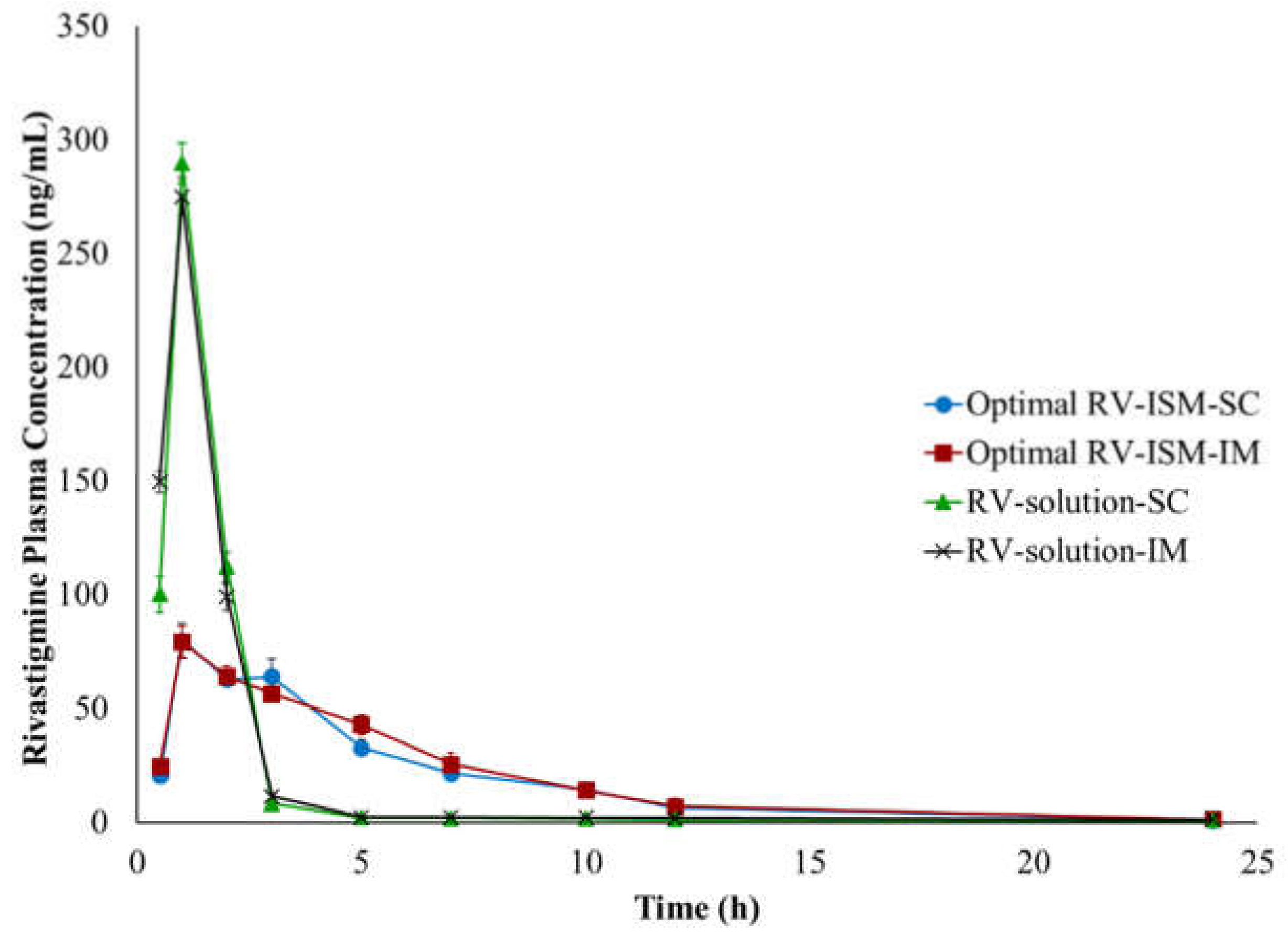
3.9. In Vivo Pharmacokinetic Studies
4. Conclusions
Author Contributions
Funding
Institutional Review Board Statement
Informed Consent Statement
Data Availability Statement
Conflicts of Interest
References
- Barker, W.W.; Luis, C.A.; Kashuba, A.; Luis, M.; Harwood, D.G.; Loewenstein, D.; Waters, C.; Jimison, P.; Shepherd, E.; Sevush, S.; et al. Relative frequencies of Alzheimer disease, Lewy body, vascular and frontotemporal dementia, and hippocampal sclerosis in the State of Florida Brain Bank. Alzheimer Dis. Assoc. Disord. 2002, 16, 203–212. [Google Scholar] [CrossRef]
- Onor, M.; Trevisiol, M.; Aguglia, E. Rivastigmine in the treatment of Alzheimer’s disease: An update. Clin. Interv. Aging 2007, 2, 17–32. [Google Scholar] [CrossRef]
- Feldman, H.H.; Lane, R. Rivastigmine: A placebo controlled trial of twice daily and three times daily regimens in patients with Alzheimer’s disease. J. Neurol. Neurosurg. Psychiatry 2007, 78, 1056–1063. [Google Scholar] [CrossRef]
- Noetzli, M.; Eap, C.B. Pharmacodynamic, pharmacokinetic and pharmacogenetic aspects of drugs used in the treatment of alzheimer’s disease. Clin. Pharmacokinet. 2013, 52, 225–241. [Google Scholar] [CrossRef]
- Adler, G.; Brassen, S.; Chwalek, K.; Dieter, B.; Teufel, M. Prediction of treatment response to rivastigmine in Alzheimer’s dementia. J. Neurol. Neurosurg. Psychiatry 2004, 75, 292–294. [Google Scholar] [CrossRef]
- Kurz, A.; Farlow, M.; Lefèvre, G. Pharmacokinetics of a novel transdermal rivastigmine patch for the treatment of Alzheimer’s disease: A review. Int. J. Clin. Pract. 2009, 63, 799–805. [Google Scholar] [CrossRef] [PubMed]
- Arumugam, K.; Subramanian, G.S.; Mallayasamy, S.R.; Averineni, R.K.; Reddy, M.S.; Udupa, N. A study of rivastigmine liposomes for delivery into the brain through intranasal route. Acta Pharm. 2008, 58, 287–297. [Google Scholar] [CrossRef] [PubMed]
- Shah, B.M.; Misra, M.; Shishoo, C.J.; Padh, H. Nose to brain microemulsion-based drug delivery system of rivastigmine: Formulation and ex-vivo characterization. Drug Deliv. 2015, 22, 918–930. [Google Scholar] [CrossRef] [PubMed]
- Craparo, E.F.; Pitarresi, G.; Bondi, M.L.; Casaletto, M.P.; Licciardi, M.; Giammona, G. A nanoparticulate drug-delivery system for rivastigmine: Physico-chemical and in vitro biological characterization. Macromol. Biosci. 2008, 8, 247–259. [Google Scholar] [CrossRef] [PubMed]
- Lefèvre, G.; Sȩdek, G.; Jhee, S.S.; Leibowitz, M.T.; Huang, H.L.A.; Enz, A.; Maton, S.; Ereshefsky, L.; Pommier, F.; Schmidli, H.; et al. Pharmacokinetics and pharmacodynamics of the novel daily rivastigmine transdermal patch compared with twice-daily capsules in Alzheimer’s disease patients. Clin. Pharmacol. Ther. 2008, 83, 106–114. [Google Scholar] [CrossRef]
- Kapil, R.; Dhawan, S.; Beg, S.; Singh, B. Buccoadhesive films for once-a-day administration of rivastigmine: Systematic formulation development and pharmacokinetic evaluation. Drug Dev. Ind. Pharm. 2013, 39, 466–480. [Google Scholar] [CrossRef] [PubMed]
- Vintiloiu, A.; Lafleur, M.; Bastiat, G.; Leroux, J.C. In situ-forming oleogel implant for rivastigmine delivery. Pharm. Res. 2008, 25, 845–852. [Google Scholar] [CrossRef] [PubMed]
- Chitkara, D.; Shikanov, A.; Kumar, N.; Domb, A.J. Biodegradable injectable in situ depot-forming drug delivery systems. Macromol. Biosci. 2006, 6, 977–990. [Google Scholar] [CrossRef] [PubMed]
- Hatefi, A.; Amsden, B. Biodegradable injectable in situ forming drug delivery systems. J. Control. Release 2002, 80, 9–28. [Google Scholar] [CrossRef]
- Packhaeuser, C.B.; Schnieders, J.; Oster, C.G.; Kissel, T. In situ forming parenteral drug delivery systems: An overview. Eur. J. Pharm. Biopharm. 2004, 58, 445–455. [Google Scholar] [CrossRef] [PubMed]
- Kranz, H.; Brazeau, G.A.; Napaporn, J.; Martin, R.L.; Millard, W.; Bodmeier, R. Myotoxicity studies of injectable biodegradable in-situ forming drug delivery systems. Int. J. Pharm. 2001, 212, 11–18. [Google Scholar] [CrossRef]
- Kranz, H.; Bodmeier, R. A novel in situ forming drug delivery system for controlled parenteral drug delivery. Int. J. Pharm. 2007, 332, 107–114. [Google Scholar] [CrossRef]
- Kranz, H.; Bodmeier, R. Structure formation and characterization of injectable drug loaded biodegradable devices: In situ implants versus in situ microparticles. Eur. J. Pharm. Sci. 2008, 34, 164–172. [Google Scholar] [CrossRef]
- Rungseevijitprapa, W.; Bodmeier, R. Injectability of biodegradable in situ forming microparticle systems (ISM). Eur. J. Pharm. Sci. 2009, 36, 524–531. [Google Scholar] [CrossRef]
- Khattab, A.; Abouhussein, D.M.N.; Mohammad, F.E. Development of injectable tenoxicam in situ forming microparticles based on sesame oil and poly-dl-lactide: Characterization, efficacy and acute toxicity. J. Drug Deliv. Sci. Technol. 2019, 51, 682–694. [Google Scholar] [CrossRef]
- Lu, Y.; Yu, Y.; Tang, X. Sucrose acetate isobutyrate as an in situ forming system for sustained risperidone release. J. Pharm. Sci. 2007, 96, 3252–3262. [Google Scholar] [CrossRef] [PubMed]
- Nally, J.E.; Artiushin, S.; Sheoran, A.S.; Burns, P.J.; Simon, B.; Gilley, R.M.; Gibson, J.; Sullivan, S.; Timoney, J.F. Induction of mucosal and systemic antibody specific for SeMF3 of Streptococcus equi by intranasal vaccination using a sucrose acetate isobutyrate based delivery system. Vaccine 2000, 19, 492–497. [Google Scholar] [CrossRef]
- Reynolds, R.C. Metabolism and pharmacokinetics of sucrose acetate isobutyrate (SAIB) and sucrose octaisobutyrate (SOIB) in rats, dogs, monkeys or humans: A review. Food Chem. Toxicol. 1998, 36, 95–99. [Google Scholar] [CrossRef]
- Okumu, F.W.; Dao, L.N.; Fielder, P.J.; Dybdal, N.; Brooks, D.; Sane, S.; Cleland, J.L. Sustained delivery of human growth hormone from a novel gel system: SABERTM. Biomaterials 2002, 23, 4353–4358. [Google Scholar] [CrossRef]
- Kempe, S.; Mäder, K. In situ forming implants—An attractive formulation principle for parenteral depot formulations. J. Control. Release 2012, 161, 668–679. [Google Scholar] [CrossRef]
- Yehia, S.A.; Halim, S.A.A.; Aziz, M.Y. Polymeric and Non Polymeric Injectable In-situ Forming Implant Systems for Sustained Delivery of Lornoxicam: In vitro and In vivo Evaluation. Curr. Drug Deliv. 2018, 15, 1193–1203. [Google Scholar] [CrossRef] [PubMed]
- Graves, R.A.; Freeman, T.; Mandal, T.K. In vitro dissolution method for evaluation of buprenorphine in situ gel formulation: A technical note. AAPS PharmSciTech 2007, 8, E88–E91. [Google Scholar] [CrossRef]
- Ammar, H.O.; Ibrahim, M.; Mahmoud, A.A.; Shamma, R.N.; El Hoffy, N.M. Non-ionic Surfactant Based In Situ Forming Vesicles as Controlled Parenteral Delivery Systems. AAPS PharmSciTech 2018, 19, 1001–1010. [Google Scholar] [CrossRef] [PubMed]
- Jain, R.A.; Rhodes, C.T.; Railkar, A.M.; Malick, A.W.; Shah, N.H. Controlled delivery of drugs from a novel injectable in situ formed biodegradable PLGA microsphere system. J. Microencapsul. 2000, 17, 343–362. [Google Scholar] [CrossRef]
- Higuchi, T. Mechanism of sustained-action medication. Theoretical analysis of rate of release of solid drugs dispersed in solid matrices. J. Pharm. Sci. 1963, 52, 1145–1149. [Google Scholar] [CrossRef]
- Gohel, M.C.; Amin, A.F. Formulation design and optimization of modified-release microspheres of diclofenac sodium. Drug Dev. Ind. Pharm. 1999, 25, 247–251. [Google Scholar] [CrossRef] [PubMed]
- Leroux, L.; Hatim, Z.; Frèche, M.; Lacout, J.L. Effects of various adjuvants (lactic acid, glycerol, and chitosan) on the injectability of a calcium phosphate cement. Bone 1999, 25, 31S–34S. [Google Scholar] [CrossRef]
- Paul, S.; Hoey, M.F.; Egbert, J.E. Pressure measurements during injection of corticosteroids: In vivo studies. Med. Biol. Eng. Comput. 1999, 37, 645–651. [Google Scholar] [CrossRef] [PubMed]
- Yehia, S.A.; Elshafeey, A.H.; Elsayed, I. Biodegradable donepezil lipospheres for depot injection: Optimization and in-vivo evaluation. J. Pharm. Pharmacol. 2012, 64, 1425–1437. [Google Scholar] [CrossRef]
- Herrero-Vanrell, R.; Ramirez, L. Biodegradable PLGA microspheres loaded with ganciclovir for intraocular administration. Enapsulation technique in vitro release profiles, and sterilization process. Pharm. Res. 2000, 17, 1323–1328. [Google Scholar] [CrossRef]
- Costa, P. An alternative method to the evaluation of similarity factor in dissolution testing. Int. J. Pharm. 2001, 220, 77–83. [Google Scholar] [CrossRef]
- Dharani, S.; Barakh Ali, S.F.; Afrooz, H.; Khan, M.A.; Rahman, Z. Development and Validation of a Discriminatory Dissolution Method for Rifaximin Products. J. Pharm. Sci. 2019, 108, 2112–2118. [Google Scholar] [CrossRef]
- Nair, A.; Jacob, S. A simple practice guide for dose conversion between animals and human. J. Basic Clin. Pharm. 2016, 7, 27–31. [Google Scholar] [CrossRef]
- Reagan-Shaw, S.; Nihal, M.; Ahmad, N. Dose translation from animal to human studies revisited. FASEB J. 2008, 22, 659–661. [Google Scholar] [CrossRef]
- Haider, M.; Elsherbeny, A.; Jagal, J.; Hubatová-Vacková, A.; Ahmed, I.S. Optimization and evaluation of poly(Lactide-co-glycolide) nanoparticles for enhanced cellular uptake and efficacy of paclitaxel in the treatment of head and neck cancer. Pharmaceutics 2020, 9, 828. [Google Scholar] [CrossRef]
- Ding, D.; Zhu, Q. Recent advances of PLGA micro/nanoparticles for the delivery of biomacromolecular therapeutics. Mater. Sci. Eng. C 2018, 92, 1041–1060. [Google Scholar] [CrossRef] [PubMed]
- Danhier, F.; Ansorena, E.; Silva, J.M.; Coco, R.; Le Breton, A.; Préat, V. PLGA-based nanoparticles: An overview of biomedical applications. J. Control. Release 2012, 161, 505–522. [Google Scholar] [CrossRef] [PubMed]
- Myers, W. Response Surface Methodology. Encycl. Biopharm. Stat. Third Ed. 2012, 2, 1171–1179. [Google Scholar] [CrossRef]
- Lin, X.; Yang, S.; Gou, J.; Zhao, M.; Zhang, Y.; Qi, N.; He, H.; Cai, C.; Tang, X.; Guo, P. A novel risperidone-loaded SAIB-PLGA mixture matrix depot with a reduced burst release: Effects of solvents and PLGA on drug release behaviors in vitro/in vivo. J. Mater. Sci. Mater. Med. 2012, 23, 443–455. [Google Scholar] [CrossRef] [PubMed]
- Lin, X.; Xu, Y.; Tang, X.; Zhang, Y.; Chen, J.; Zhang, Y.; He, H.; Yang, Z. A Uniform Ultra-Small Microsphere/SAIB Hybrid Depot with Low Burst Release for Long-Term Continuous Drug Release. Pharm. Res. 2015, 32, 3708–3721. [Google Scholar] [CrossRef]
- Thedrattanawong, C.; Manaspon, C.; Nasongkla, N. Controlling the burst release of doxorubicin from polymeric depots via adjusting hydrophobic/hydrophilic properties. J. Drug Deliv. Sci. Technol. 2018, 46, 446–451. [Google Scholar] [CrossRef]
- Wright, J.; Tamraz, W.; Leonard, J.; Gibson, J. Compositions and Methods Involving Polymer, Solvent, and High Viscosity Liquid Carrier Material. U.S. Patent number 20200085829, 19 March 2020. [Google Scholar]
- Duvvuri, S.; Janoria, K.G.; Mitra, A.K. Development of a novel formulation containing poly(d,l-lactide-co-glycolide) microspheres dispersed in PLGA–PEG–PLGA gel for sustained delivery of ganciclovir. J. Control. Release 2005, 108, 282–293. [Google Scholar] [CrossRef]
- Harloff-Helleberg, S.; Fliervoet, L.A.L.; Fanø, M.; Schmitt, M.; Antopolski, M.; Urtti, A.; Nielsen, H.M. Exploring the mucoadhesive behavior of sucrose acetate isobutyrate: A novel excipient for oral delivery of biopharmaceuticals. Drug Deliv. 2019, 26, 532–541. [Google Scholar] [CrossRef]
- Montoto, S.S. Routes of drug administration: Dosage, design, and pharmacotherapy success. In ADME Processes in Pharmaceutical Sciences; Talevi, A., Quiroga, P.A., Eds.; Springer: Cham, Switzerland, 2018; pp. 97–133. ISBN 9783319995939. [Google Scholar]
- Ducharme, M.P. Drug elimination, clearance, and renal clearance. In Applied Biopharmaceutics & Pharmacokinetics, 7th ed.; Shargel, L., Yu, A.B.C., Eds.; McGraw-Hill Education: New York, NY, USA, 2016; pp. 149–175. ISBN 978-0-07-182964-9. [Google Scholar]
- Yáñez, J.A.; Remsberg, C.M.; Sayre, C.L.; Forrest, M.L.; Davies, N.M. Flip-flop pharmacokinetics—Delivering a reversal of disposition: Challenges and opportunities during drug development. Ther. Deliv. 2011, 2, 643–672. [Google Scholar] [CrossRef]
- Dhanikula, A.; Singh, D.; Panchagnula, R. In Vivo Pharmacokinetic and Tissue Distribution Studies in Mice of Alternative Formulations for Local and Systemic Delivery of Paclitaxel: Gel, Film, Prodrug, Liposomes and Micelles. Curr. Drug Deliv. 2005, 2, 35–44. [Google Scholar] [CrossRef] [PubMed]
- Turncliff, R.Z.; Dunbar, J.L.; Dong, Q.; Silverman, B.L.; Ehrich, E.W.; Dilzer, S.C.; Lasseter, K.C. Pharmacokinetics of long-acting naltrexone in subjects with mild to moderate hepatic impairment. J. Clin. Pharmacol. 2005, 45, 1259–1267. [Google Scholar] [CrossRef] [PubMed]
- Mueller, J.; del Re, G.B.; Buerki, H.; Keller, H.-U.; Hess, M.W.; Cottier, H. Nonspecific acid esterase activity: A criterion for differentation of T and B lymphocytes in mouse lymph nodes. Eur. J. Immunol. 1975, 5, 270–274. [Google Scholar] [CrossRef] [PubMed]
- Phillips, J.C.; Kingsnorth, J.; Rowland, I.; Gangolli, S.D.; Lloyd, A.G. Studies on the metabolism of sucrose acetate isobutyrate in the rat and in man. Food Cosmet. Toxicol. 1976, 14, 375–380. [Google Scholar] [CrossRef]
- Li, H.; Xu, Y.; Tong, Y.; Dan, Y.; Zhou, T.; He, J.; Liu, S.; Zhu, Y. Sucrose Acetate Isobutyrate as an In situ Forming Implant for Sustained Release of Local Anesthetics. Curr. Drug Deliv. 2018, 16, 331–340. [Google Scholar] [CrossRef] [PubMed]
- Díaz-Torres, R.; López-Arellano, R.; Escobar-Chávez, J.J.; García-García, E.; Domínguez-Delgado, C.L.; Ramírez-Noguera, P. Effect of size and functionalization of pharmaceutical nanoparticles and their interaction with biological systems. In Handbook of Nanoparticles; Springer: Cham, Switzerland, 2015; pp. 1041–1060. [Google Scholar] [CrossRef]
- Voigt, M.; Koerber, M.; Bodmeier, R. Improved physical stability and injectability of non-aqueous in situ PLGA microparticle forming emulsions. Int. J. Pharm. 2012, 434, 251–256. [Google Scholar] [CrossRef]
- El Maghraby, G.M.; Elzayat, E.M.; Alanazi, F.K. Development of modified in situ gelling oral liquid sustained release formulation of dextromethorphan. Drug Dev. Ind. Pharm. 2012, 38, 971–978. [Google Scholar] [CrossRef]
- Park, K.; Skidmore, S.; Hadar, J.; Garner, J.; Park, H.; Otte, A.; Soh, B.K.; Yoon, G.; Yu, D.; Yun, Y.; et al. Injectable, long-acting PLGA formulations: Analyzing PLGA and understanding microparticle formation. J. Control. Release 2019, 304, 125–134. [Google Scholar] [CrossRef]
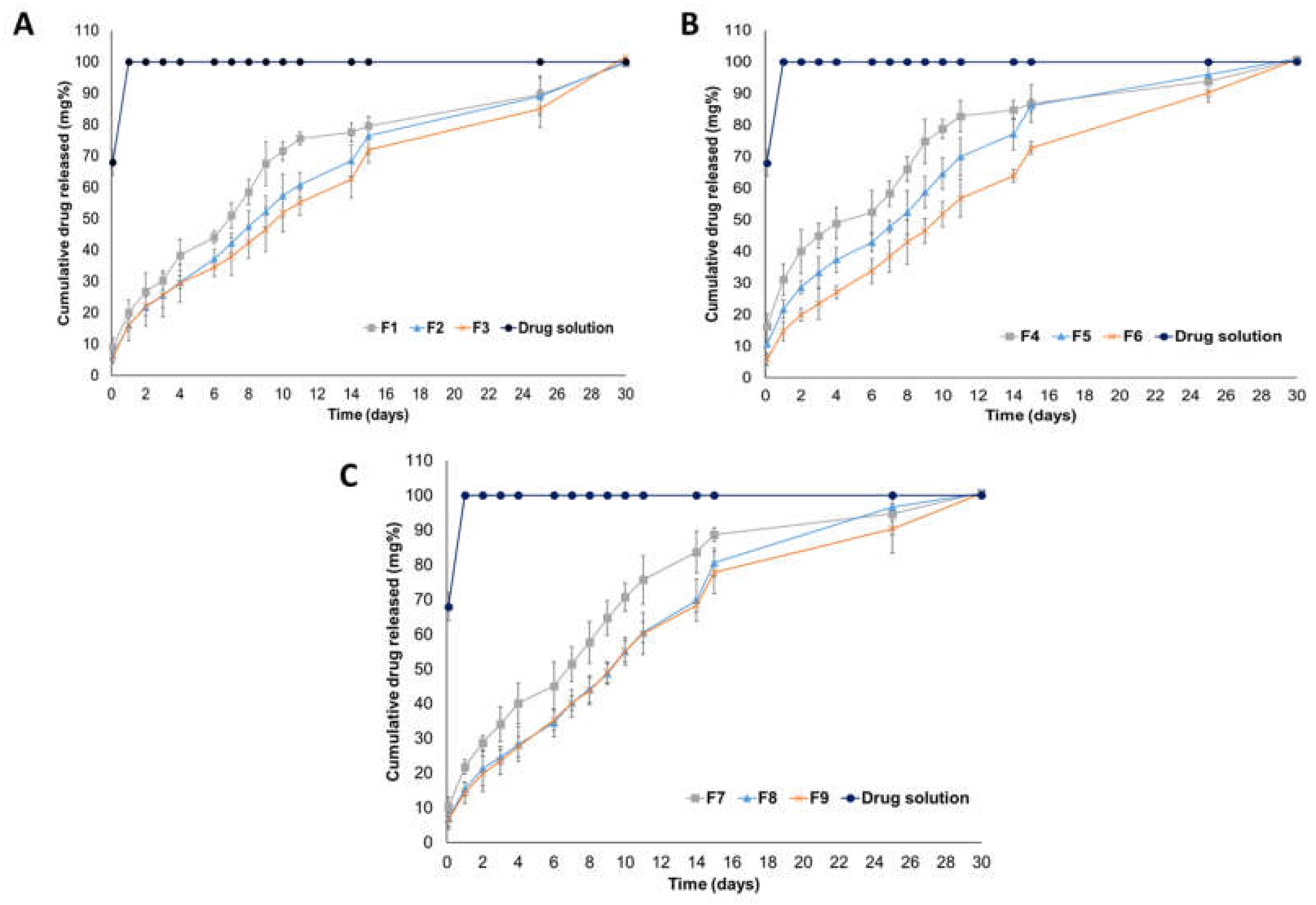

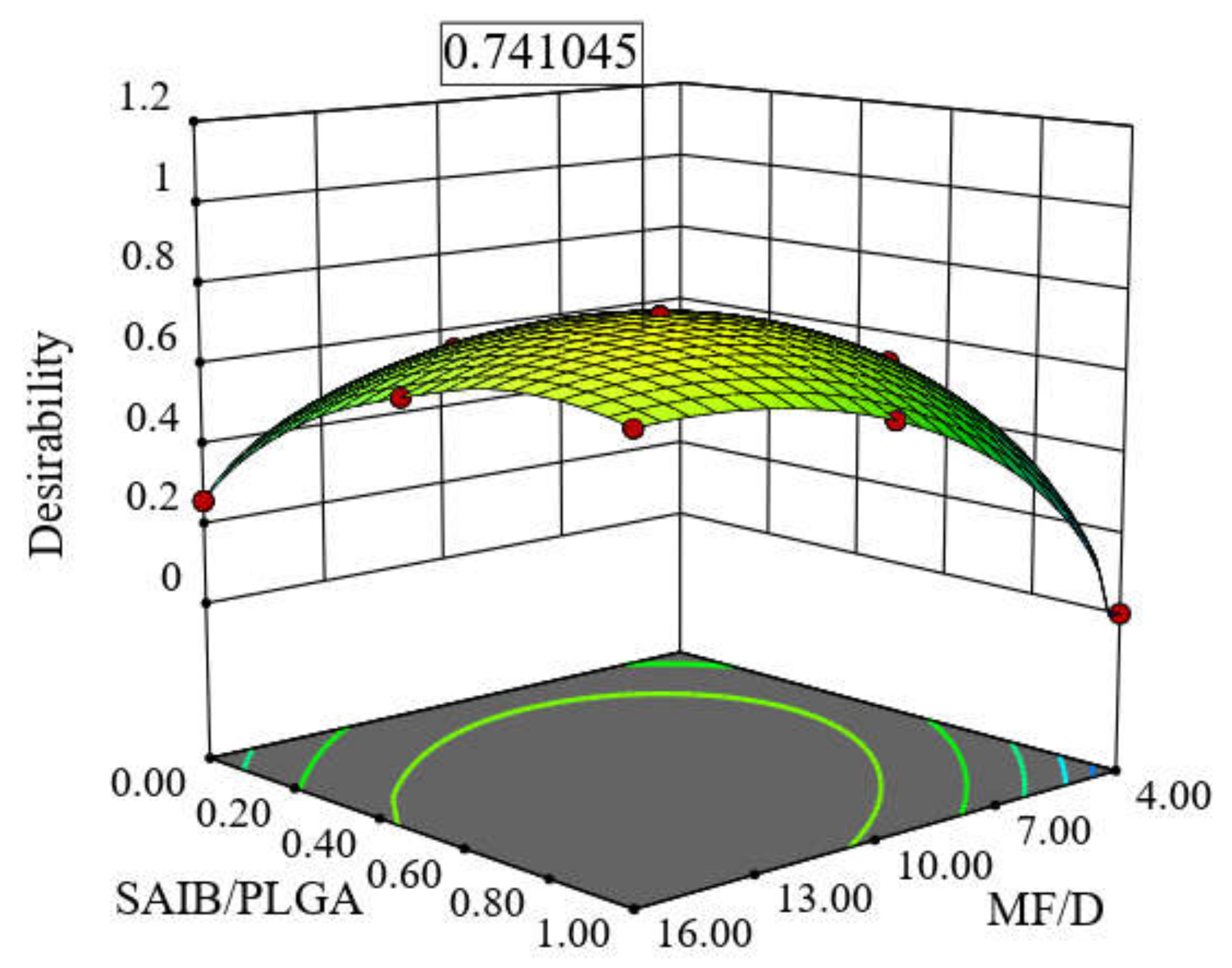
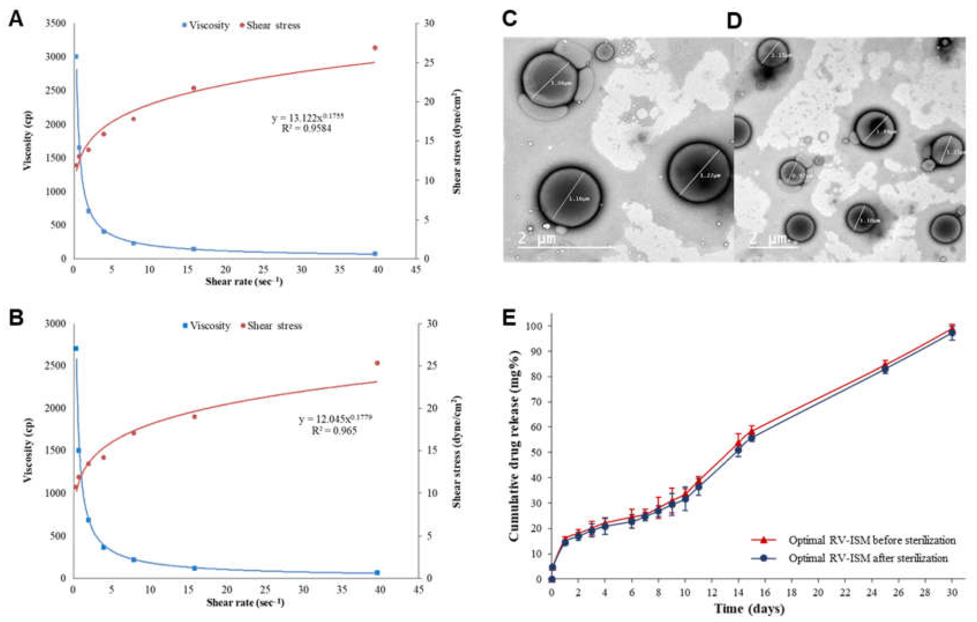
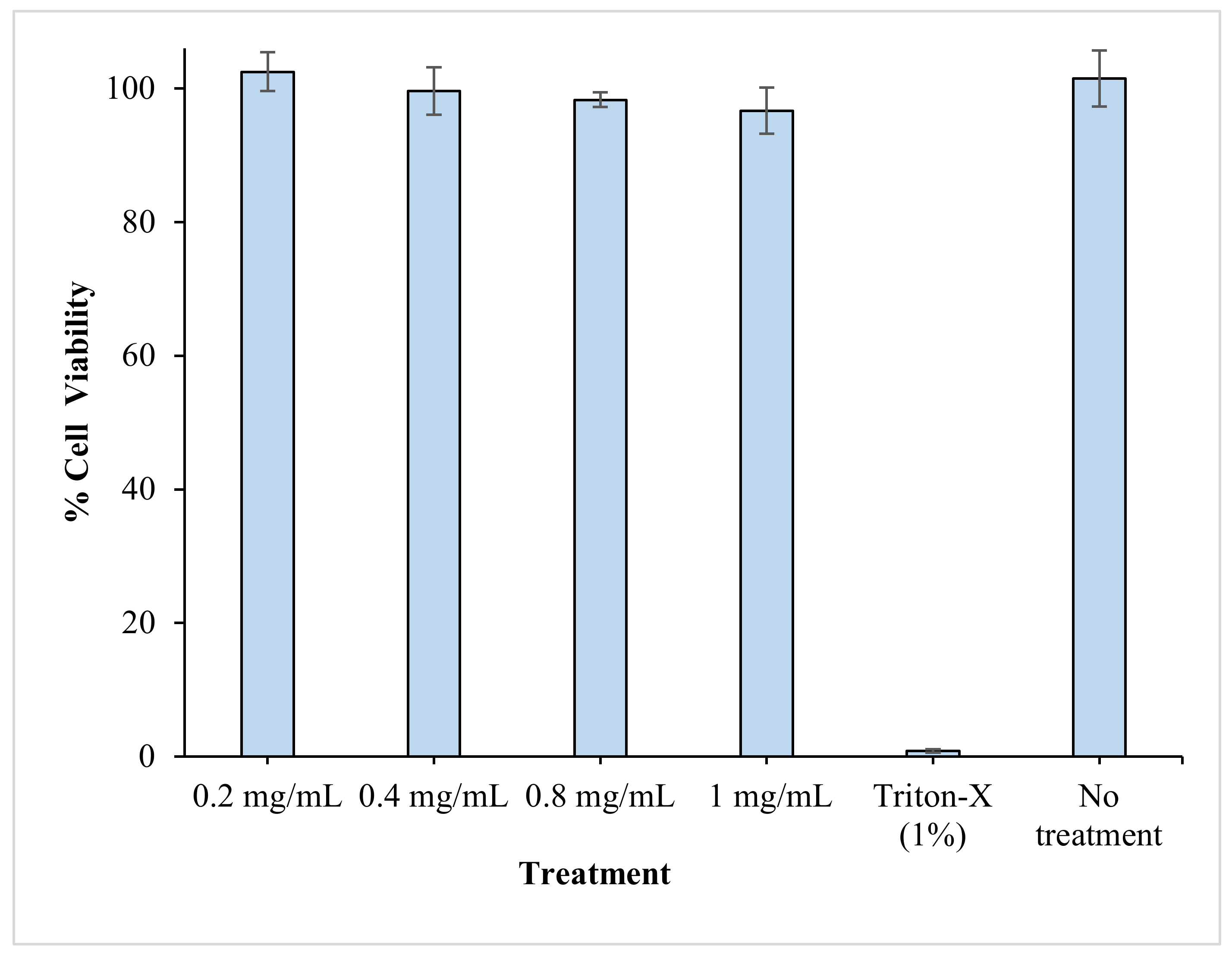
| Numerical Factors | Applied Levels | |||
|---|---|---|---|---|
| Low (−1) | Medium (0) | High (+1) | ||
| X1 | MF/D (w/w) | 4:1 | 10:1 | 16:1 |
| X2 | S/P (w/w) | 0:1 | 1:1 | 1:0 |
| Responses | Optimization Goal | |||
| Y1 | Q1 (%) | Minimize | ||
| Y2 | T50% (days) | Maximize | ||
| Y3 | Rate of injection (mL/min) | Maximize | ||
| Formulation | MF/D (X1) | S/P (X2) | Y1: Q1 (%) | Y2: T50% (Days) | Y3: Injection Rate (mL/Min) |
|---|---|---|---|---|---|
| F1 | 4:1 | 0:1 | 20.1 ± 1.5 | 8.54 ± 0.3 | 1.24 ± 0.09 |
| F2 | 10:1 | 0:1 | 16.0 ± 1.3 | 10.59 ± 0.7 | 0.60 ± 0.03 |
| F3 | 16:1 | 0:1 | 16.0 ± 2.1 | 13.24 ± 1.1 | 0.60 ± 0.04 |
| F4 | 4:1 | 1:0 | 31.1 ± 1.7 | 6.74 ± 0.4 | 1.38 ± 0.11 |
| F5 | 10:1 | 1:0 | 21.7 ± 0.9 | 9.96 ± 0.1 | 1.11 ± 0.08 |
| F6 | 16:1 | 1:0 | 14.7 ± 1.6 | 12.51 ± 1.4 | 1.06 ± 0.05 |
| F7 | 4:1 | 1:1 | 21.8 ± 0.8 | 8.64 ± 0.6 | 1.30 ± 0.02 |
| *F8 | 10:1 | 1:1 | 16.3 ± 1.4 | 13.10 ± 1.3 | 1.08 ± 0.01 |
| 15.5 ± 1.1 | 13.61 ± 0.7 | 1.03 ± 0.10 | |||
| 13.3 ± 1.0 | 13.20 ± 1.2 | 1.11 ± 0.03 | |||
| F9 | 16:1 | 1:1 | 14.3 ± 1.5 | 14.61 ± 1.0 | 0.80 ± 0.06 |
| Response | Model Equation (p-Value) | Lack of Fit (p-Value) | Adjusted R2 | Predicted R2 | Adequate Precision | Significant Terms |
|---|---|---|---|---|---|---|
| Q1 (%) | Quadratic (p = 0.011) | 0.6951 | 0.9333 | 0.8236 | 18.3 | X1 (p = 0.0004) X2 (p = 0.0053) X1×2 (p = 0.0058) |
| T50% (days) | Quadratic (p = 0.0011) | 0.0978 | 0.9334 | 0.7685 | 17.19 | X1 (p = 0.0002) |
| Rate of injection (mL/min) | Linear (p = 0.0003) | 0.1305 | 0.8421 | 0.7177 | 16.19 | X1 (p = 0.0003) X2 (p = 0.0028) |
| Variables | Values | Responses | Predicted Values | Observed Values |
|---|---|---|---|---|
| X1 | 11.71:1 (w/w) | Y1 (Q1) | 14.774% | 15.518% |
| X2 | 1.64:1 (w/w) | Y2 (T50%) | 13.436 days | 13.09 days |
| Y3 (Rate of injection) | 1.009 mL/min | 1.012 mL/min |
| PK Parameter | Optimal RV-ISM System | RV Solution | ||
|---|---|---|---|---|
| SC Injection | IM Injection | SC Injection | IM Injection | |
| Cmax (ng/mL) | 79.95 ± 7.26 | 79.25 ± 6.35 | 275.00 ± 4.26 | 289.77 ± 7.59 |
| Tmax (h) * | 1.00 | 1.00 | 1.00 | 1.00 |
| AUC0–24 (ng·h/mL) | 440.39 ± 12.70 | 465.69 ± 10.54 | 400.03 ± 6.21 | 394.65 ± 9.37 |
| AUC0–∞ (ng·h/mL) | 447.50 ± 9.33 | 473.32 ± 8.02 | 402.01 ± 8.95 | 396.37 ± 10.48 |
| AUMC0–∞ (ng·h/mL) | 2509.76 ± 25.66 | 2714.36 ± 32.33 | 517.70 ± 11.89 | 526.08 ± 3.94 |
| kel (h−1) | 0.18 ± 0.01 | 0.18 ± 0.08 | 1.22 ± 0.05 | 1.27 ± 0.04 |
| t½ (h) | 3.78 ± 0.28 | 3.82 ± 0.32 | 0.57 ± 0.03 | 0.54 ± 0.05 |
| MTT (h) | 5.61 ± 0.47 | 5.73 ± 0.16 | 1.29 ± 0.10 | 1.33 ± 0.14 |
Publisher’s Note: MDPI stays neutral with regard to jurisdictional claims in published maps and institutional affiliations. |
© 2021 by the authors. Licensee MDPI, Basel, Switzerland. This article is an open access article distributed under the terms and conditions of the Creative Commons Attribution (CC BY) license (http://creativecommons.org/licenses/by/4.0/).
Share and Cite
Haider, M.; Elsayed, I.; Ahmed, I.S.; Fares, A.R. In Situ-Forming Microparticles for Controlled Release of Rivastigmine: In Vitro Optimization and In Vivo Evaluation. Pharmaceuticals 2021, 14, 66. https://doi.org/10.3390/ph14010066
Haider M, Elsayed I, Ahmed IS, Fares AR. In Situ-Forming Microparticles for Controlled Release of Rivastigmine: In Vitro Optimization and In Vivo Evaluation. Pharmaceuticals. 2021; 14(1):66. https://doi.org/10.3390/ph14010066
Chicago/Turabian StyleHaider, Mohamed, Ibrahim Elsayed, Iman S. Ahmed, and Ahmed R. Fares. 2021. "In Situ-Forming Microparticles for Controlled Release of Rivastigmine: In Vitro Optimization and In Vivo Evaluation" Pharmaceuticals 14, no. 1: 66. https://doi.org/10.3390/ph14010066
APA StyleHaider, M., Elsayed, I., Ahmed, I. S., & Fares, A. R. (2021). In Situ-Forming Microparticles for Controlled Release of Rivastigmine: In Vitro Optimization and In Vivo Evaluation. Pharmaceuticals, 14(1), 66. https://doi.org/10.3390/ph14010066


