Central Serous Chorioretinopathy Classification


Abstract
1. Introduction
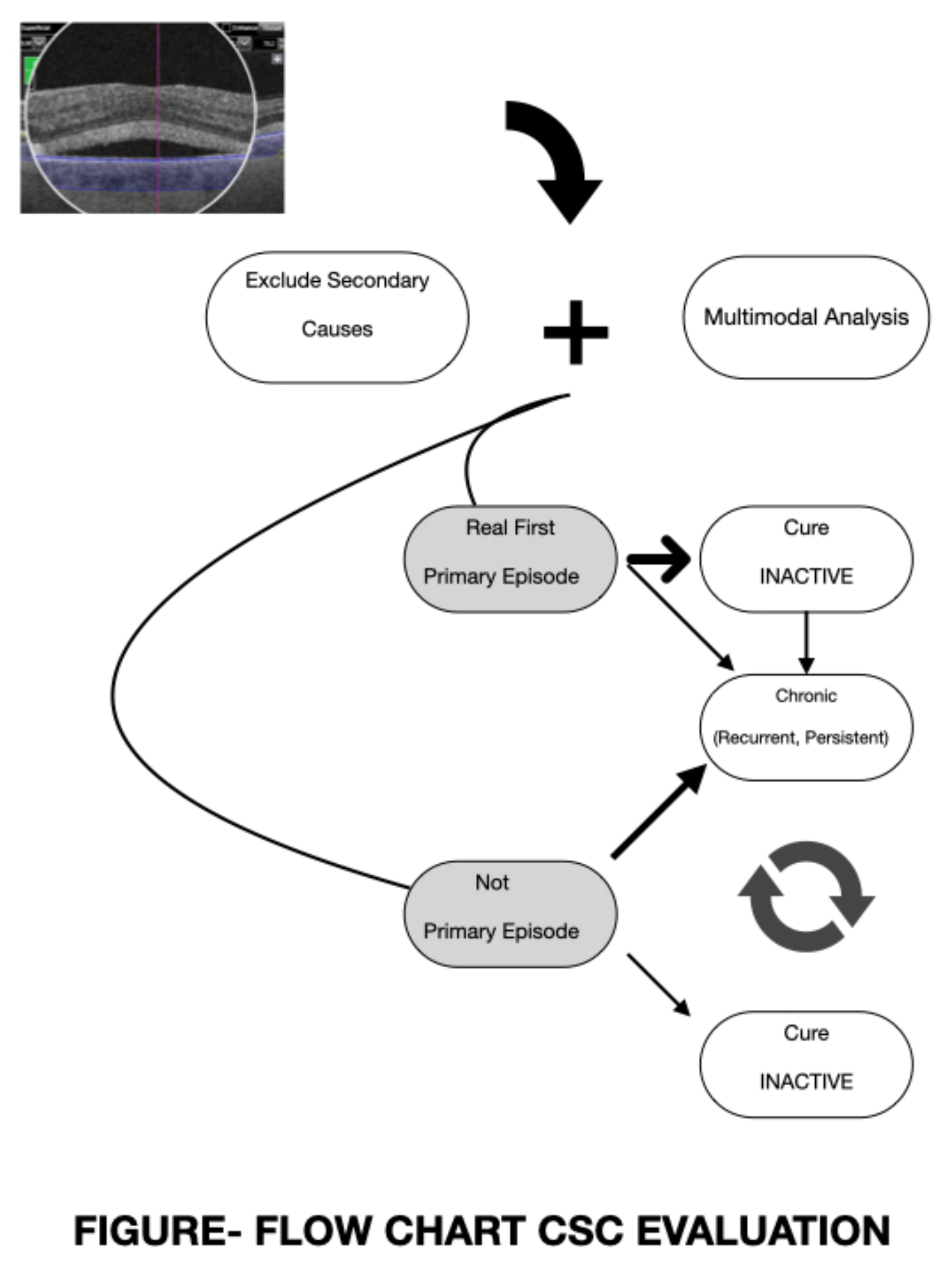
2. Classification Models
3. Complementary Analysis
4. Conclusions
Author Contributions
Funding
Conflicts of Interest
References
- Ivanisevic, M.; Stanic, R.; Ivanisevic, P.; Vukovic, A. Albrecht von Graefe (1828–1870) and his contributions to the development of ophthalmology. Int. Ophthalmol. 2020, 40, 1029–1033. [Google Scholar] [CrossRef]
- Von Graefe, A. Ueber centrale recidivirende retinitis. Arch Ophthalmol. 1866, 12, 211–215. [Google Scholar]
- Maumenee, A.E. Symposium on macular diseases: Clinical manifestations. Trans. Am. Acad. Ophthalmol. Otolaryngol. 1965, 69, 605–613. [Google Scholar] [PubMed]
- Gass, J.D. Pathogenesis of disciform detachment of the neuroepithelium. Am. J. Ophthalmol. 1967, 63, 1–139. [Google Scholar]
- Guyer, D.R.; Yannuzzi, L.A.; Slakter, J.S.; Sorenson, J.A.; Ho, A.; Orlock, D. Digital indocyanine green videoangiography of central serous chorioretinopathy. Arch. Ophthalmol. 1994, 112, 1057–1062. [Google Scholar] [CrossRef] [PubMed]
- Spaide, R.F.; Hall, L.; Haas, A.; Campeas, L.; Yannuzzi, L.A.; Fisher, Y.L.; Guyer, D.R.; Slakter, J.S.; Sorenson, J.A.; Orlock, D.A. Indocyanine green videoangiography of older patients with central serous chorioretinopathy. Retina 1996, 16, 203–213. [Google Scholar] [CrossRef]
- Central Serous Chorioretinopathy. Available online: https://pubmed.ncbi.nlm.nih.gov/?term=central+serous+retinopathy&size=200 (accessed on 7 July 2020).
- Nicholson, B.; Noble, J.; Forooghian, F.; Meyerle, C. Central serous chorioretinopathy: Update on pathophysiology and treatment. Surv. Ophthalmol. 2013, 58, 103–126. [Google Scholar] [CrossRef]
- Daruich, A.; Matet, A.; Dirani, A.; Bousquet, E.; Zhao, M.; Farman, N.; Jaisser, F.; Behar-Cohen, F. Central serous chorioretinopathy: Recent findings and new physiopathology hyphotesis. Prog. Retin. Eye Res. 2015, 48, 82–118. [Google Scholar] [CrossRef]
- van Rijssen, T.J.; van Dijk, E.H.C.; Yzer, S.; Ohno-Matsui, K.; Keunen, J.E.; Schlingemann, R.O.; Sivaprasad, S.; Querques, G.; Downes, S.M.; Fauser, S.; et al. Central serous chorioretinopathy: Towards an evidence-based treatment guideline. Prog. Retin. Eye Res. 2019, 73, 100770. [Google Scholar] [CrossRef]
- Cardillo Piccolino, F.; de la Longrais, R.R.; Ravera, G.; Eandi, C.M.; Ventre, L.; Manea, M. The foveal photoreceptor layer and visual acuity loss in central serous chorioretinopathy. Am. J. Ophthalmol. 2005, 139, 87–89. [Google Scholar] [CrossRef]
- Spaide, R.F.; Campeas, L.; Haas, A.; Yannuzzi, L.A.; Fisher, Y.L.; Guyer, D.R.; Slakter, J.S.; Sorenson, J.A.; Orlock, D.A. Central serous chorioretinopathy in younger and older adults. Ophthalmology 1996, 103, 2070–2079. [Google Scholar] [CrossRef]
- Tittl, M.K.; Spaide, R.F.; Wong, D.; Pilotto, E.; Yannuzzi, L.A.; Fisher, Y.L.; Freund, B.; Guyer, D.R.; Slakter, J.S.; Sorenson, J.A. Systemic findings associated with central serous chorioretinopathy. Am. J. Ophthalmol. 1999, 128, 63–68. [Google Scholar] [CrossRef]
- Kaye, R.; Chandra, S.; Sheth, J.; Boon, C.J.F.; Sivaprasad, S.; Lotery, A. Central serous chorioretinopathy: An update on risk factors, pathophysiology and imaging modalities. Prog. Retin. Eye Res. 2020, 11, 100865. [Google Scholar] [CrossRef] [PubMed]
- Tsai, D.-C.; Chen, S.-J.; Huang, C.-C.; Chou, P.; Chung, C.M.; Huang, P.H.; Lin, S.J.; Chen, J.W.; Chen, T.J.; Leu, H.B.; et al. Epidemiology of idiopathic central serous chorioretinopathy in Taiwan, 2001–2006: A population-based study in Taiwan. PLoS ONE 2013, 8, e66858. [Google Scholar] [CrossRef] [PubMed]
- Mrejen, S.; Balaratnasingam, C.; Kaden, T.R.; Bottini, A.; Dansingani, K.; Bhavsar, K.V.; Yannuzzi, N.A.; Patel, S.; Chen, K.C.; Yu, S. Long-term outcomes and causes of visual loss in chronic central serous chorioretinopathy. Ophthalmology 2019, 126, 576–588. [Google Scholar] [CrossRef] [PubMed]
- Ersoz, M.G.; Arf, S.; Hocaoglu, M.; Muslubas, I.S.; Karacorlu, M. Patient characteristics and risk factors for central serous chorioretinopathy: An analysis of 811 patients. Br. J. Ophthalmol. 2019, 103, 725–729. [Google Scholar] [CrossRef]
- Laatikainen, L. Diffuse chronic retinal pigment epitheliopathy and exudative retinal detachments. Acta Ophthalmol. 1994, 72, 533–536. [Google Scholar] [CrossRef] [PubMed]
- Loo, R.H.; Scott, I.U.; Flynn, H.W., Jr.; Gass, J.D.M.; Murray, T.G.; Lewis, M.L.; Rosenfeld, P.J.; Smiddy, W.E. Factors associated with reduced visual acuity during long-term follow-up of patients with idiopathic central serous chorioretinopathy. Retina 2002, 22, 19–24. [Google Scholar] [CrossRef]
- Wang, M.S.M.; Sander, B.; Larsen, M. Retinal atrophy in idiopathic central serous chorioretinopathy. Am. J. Ophthalmol. 2002, 133, 787–793. [Google Scholar] [CrossRef]
- Castro-Correia, J.; Coutinho, M.F.; Rosas, V.; Maia, J.J.D.O. Long-term follow-up of central serous chorioretinopathy in 150 patients. Doc. Ophthalmol. 1992, 81, 379–386. [Google Scholar] [CrossRef]
- Baran, N.V.; Gurlu, V.P.; Esgin, H. Long-term macular function in eyes with central serous chorioretinopathy. Clin. Exp. Ophthalmol. 2005, 33, 369–372. [Google Scholar] [CrossRef] [PubMed]
- Behnia, M.; Khabazkhoob, M.; Aliakbari, S.; Abadi, A.E.; Hashemi, H.; Pourvahidi, P. Improvement in visual acuity and contrast sensitivity in patients with central serous chorioretinopathy after macular subthreshold laser therapy. Retina 2013, 33, 324–328. [Google Scholar] [CrossRef] [PubMed]
- Hata, M.; Oishi, A.; Shimozono, M.; Mandai, M.; Nishida, A.; Kurimoto, Y. Early changes in foveal thickness in eyes with central serous chorioretinopathy. Retina 2013, 33, 296–301. [Google Scholar] [CrossRef] [PubMed]
- Daruich, A.; Matet, A.; Marchionno, L.; Azevedo, J.D.; Ambresin, A.; Mantel, I.; Behar-Cohen, F. Acute central serous chorioretinopathy: Factors influencing episode duration. Retina 2017, 37, 1905–1915. [Google Scholar] [CrossRef] [PubMed]
- Ooto, S.; Hangai, M.; Sakamoto, A.; Tsujikawa, A.; Yamashiro, K.; Ojima, Y.; Yamada, Y.; Mukai, H.; Oshima, S.; Inoue, T.; et al. High-resolution imaging of resolved central serous chorioretinopathy using adptative optics scanning laser ophthalmoscopy. Ophthalmology 2010, 117, 1800–1809. [Google Scholar] [CrossRef] [PubMed]
- Hasegawa, T.; Okamoto, M.; Masuda, N.; Ueda, T.; Ogata, N. Relationship between foveal microstructures and visual outcomes in eyes with resolved central serous chorioretinopathy. Graefes Arch. Clin. Exp. Ophthalmol. 2015, 253, 343–350. [Google Scholar] [CrossRef] [PubMed]
- Ross, B.X.; Choi, J.; Yao, J.; Hager, H.M.; Abcouwer, S.F.; Zacks, D.N. Loss of high-mobility group 1 (HMGB1) protein in rods accelerates rod photoreceptor degeneration after retinal detachment. Investig. Ophthalmol. Vis. Sci. 2020, 61, 50. [Google Scholar] [CrossRef]
- Ambiya, V.; Yogi, R.; Li, A.; Shah, S.; Sarvaiya, C.; Mehta, P.; Meyerele, C.; Wu, L.; Singh, R.; Banker, A.; et al. Subfoveal choroidal thickness as a predictor of central serous chorioretinopathy. Eye 2016, 30, 1623–1629. [Google Scholar] [CrossRef]
- Yu, J.; Jiang, C.; Xu, G. Correlations between changes in photoreceptor layer and dother clinical characteristics in central serous chorioretinopathy. Retina 2019, 39, 1110–1116. [Google Scholar] [CrossRef]
- Gawecki, M.; Jaszczuk-Maciejewska, A.; Jurska-Jasko, A.; Grzybowski, A. Functional and morphological outcome in patients with central serous chorioretinopathy treated by subthreshold micropulse laser. Graefes Arch. Clin. Exp. Ophthalmol. 2017, 255, 2299–2306. [Google Scholar] [CrossRef]
- Gemenetzi, M.; De Salvo, G.; Lotery, A.J. Central serous chorioretinopathy: An update on pathogenesis and treatment. Eye 2010, 24, 1743–1756. [Google Scholar] [CrossRef] [PubMed]
- Singh, S.R.; Matet, A.; van Dijk, E.H.C.; Daruich, A.; Fauser, S.; Yzer, S.; Peiretti, E.; Sivaprasad, S.; Lotery, A.J.; Boon, C.J.; et al. Discrepancy in current central serous chorioretinopathy classification. Br. J. Ophthalmol. 2019, 103, 737–742. [Google Scholar] [CrossRef] [PubMed]
- Sartini, F.; Menchini, M.; Posarelli, C.; Casini, G.; Figus, M. Bullous central serous chorioretinopathy: A rare and atypical form of central serous chorioretinopathy. A systematic review. Pharmarceuticals 2020, 13, 221. [Google Scholar] [CrossRef] [PubMed]
- Mohabati, D.; van Rijssen, T.J.; van Dijk, E.H.C.; Luyten, G.P.N.; Missotten, T.O.; Hoyng, C.B.; Yzer, S.; Boon, C.J. Clinical characteristics and long-term visual outcome of severe phenotypes of chronic central serous chorioretinopathy. Clin. Ophthalmol. 2018, 12, 1061–1070. [Google Scholar] [CrossRef] [PubMed]
- Murrow, E.J.; Oglesby, F.M. Acute and chronic illness: Similarities, differences and challenges. Orthop. Nurs. 1996, 15, 47–51. [Google Scholar] [CrossRef] [PubMed][Green Version]
- Goodman, R.A.; Posner, S.F.; Huang, E.S.; Parekh, A.K.; Koh, H.K. Defining and measuring chronic conditions: Imperatives for research, policy, program, and practice. Prev. Chronic Dis. 2013, 10, 120239. [Google Scholar] [CrossRef]
- Bernell, S.; Howard, S.W. Use your words carefully: What is a chronic disease? Front. Public Health 2016, 4, 159. [Google Scholar] [CrossRef]
- Gupta, P.; Gupta, V.; Dogra, M.R.; Singh, R.; Gupta, A. Morphological changes in the retinal pigment epithelium on spectral-domain OCT in the unaffected eyes with idiopathic central serous chorioretinopathy. Int. Ophthalmol. 2010, 30, 175–181. [Google Scholar] [CrossRef]
- Gackle, H.C.; Lang, G.E.; Freissler, K.A.; Lang, G.K. Central serous chorioretinopathy. Clinica, fluorescein angiography and demographic aspects. Ophthalmologe 1998, 95, 529–533. [Google Scholar] [CrossRef]
- Sahoo, N.; Singh, S.R.; Rajendran, A.; Shukla, D.; Chhablani, J. Masquaredes of central serous chorioretinopathy. Surv. Ophthalmol. 2019, 64, 30–44. [Google Scholar] [CrossRef]
- Semeraro, F.; Morescalchi, F.; Russo, A.; Gambicorti, E.; Pilotto, A.; Parmeggiani, F.; Bartollino, S.; Costagliola, C. Central serous chorioretinopathy: Pathogenesis and management. Clin. Ophthalmol. 2019, 13, 2341–2352. [Google Scholar] [CrossRef] [PubMed]
- Nicholson, B.P.; Idris, A.M.A.; Bakri, S.J. Central serous chorioretinopathy: Clinical characteristics associated with visual outcomes. Semin. Ophthalmol. 2018, 33, 804–807. [Google Scholar] [CrossRef] [PubMed]
- Jaisankar, D.; Kumar, M.; Rishi, P.; Singh, S.; Ravman, R. Correlation of retinal changes with choroidal changes in acute and recurrent central serous chorioretinopathy assessed by swept-source optical coherence tomography. Ther. Adv. Ophthalmol. 2020, 12, 1–9. [Google Scholar] [CrossRef] [PubMed]
- Gawecki, M.; Jaszczuk-Maciejewska, A.; Jurska-Jasko, A.; Kneba, M.; Grzybowski, A. Impairment of visual acuity and retinal morphology following resolved chronic central serous chorioretinopathy. BMC Ophthalmol. 2019, 19, 160. [Google Scholar] [CrossRef] [PubMed]
- Iyer, P.G.; Schwartz, S.G.; Russell, J.F.; Flynn, H.W., Jr. Central Serous Chorioretinopathy: Multimodal Imaging and Management Options. Case Rep. Ophthalmol. Med. 2020, 2020, 8890404. [Google Scholar] [CrossRef] [PubMed]
- Hagag, A.M.; Chandra, S.; Khalid, H.; Lamin, A.; Keane, P.A.; Lotery, A.J.; Sivaprasad, S. Multimodal imaging in the management of choroidal neovascularization secondary to central serous chorioretinopathy. J. Clin. Med. 2020, 9, 1934. [Google Scholar] [CrossRef]
- Iacono, P.; Toto, L.; Costanzo, E.; Varano, M.; Parravano, M.C. Pharmacotheraphy of central serous chorioretinopathy: A review of the current treatments. Curr. Pharm. Des. 2018, 24, 4864–4873. [Google Scholar] [CrossRef]
- Framme, C.; Walter, A.; Gabler, B.; Roider, J.; Sachs, H.G.; Gabel, V.P. Fundus autofluorescence in acute and chronic-recurrent central serous chorioretinopathy. Acta Ophthalmol. Scand. 2005, 83, 161–167. [Google Scholar] [CrossRef]
- Spaide, R.F.; Klancnik, J.M., Jr. Fundus autofluorescence and central serous chorioretinopathy. Ophthalmology 2005, 112, 825–833. [Google Scholar] [CrossRef]
- Iacono, P.; Parodi, M.B.; Papayannis, A.; La Spina, C.; Varano, M.; Bandello, F. Acute central serous chorioretinopathy: A correlation study between funds autofluorescence and spectral-domain OCT. Graefes Arch. Clin. Exp. Ophthalmol. 2015, 253, 1889–1897. [Google Scholar] [CrossRef]
- Lee, W.J.; Lee, J.-H.; Lee, B.R. Fundus autofluorescence imaging patterns in central serous chorioretinopathy according to chronicity. Eye 2016, 30, 1336–1342. [Google Scholar] [CrossRef] [PubMed]
- Ayata, A.; Tatlipinar, S.; Kar, T.; Unal, M.; Ersanli, D.; Bilge, A.H. Near-infrared and short-wavelength autofluorescence imaging in central serous chorioretinopathy. Br. J. Ophthalmol. 2009, 93, 79–82. [Google Scholar] [CrossRef] [PubMed]
- Lehmann, M.; Bousquet, E.; Beydoun, T.; Behar-Cohen, F. Pachychoroid: An inherited condition. Retina 2015, 35, 10–16. [Google Scholar] [CrossRef] [PubMed]
- Cheung, C.M.G.; Lee, W.K.; Koizumi, H.; Dansingani, K.; Lai, T.Y.; Freund, K.B. Pachychoroid disease. Eye 2019, 33, 14–33. [Google Scholar] [CrossRef]
- Jirarattanasopa, P.; Ooto, S.; Tsujikawa, A.; Yamashiro, K.; Hangai, M.; Hirata, M.; Matsumoto, A.; Yoshimura, N. Assessment of macular choroidal thickness by optical coherence tomography and angiographic changes in central serous chorioretinopathy. Ophthalmology 2012, 119, 1666–1678. [Google Scholar] [CrossRef]
- Kuroda, S.; Ikuno, Y.; Yasuno, Y.; Nakai, K.; Usui, S.; Sawa, M.; Tsujikawa, M.; Gomi, F.; Nishida, K. Choroidal thickness in central serous chorioretinopathy. Retina 2013, 33, 302–308. [Google Scholar] [CrossRef]
- Battista, M.; Borrelli, E.; Veronese, C.; Gelormini, F.; Sacconi, R.; Querques, L.; Prascina, F.; Vella, G.; Ciardella, A.P.; Bandello, F.; et al. Choroidal Rift: A New OCT Finding in Eyes with Central Serous Chorioretinopathy. J. Clin. Med. 2020, 9, 2260. [Google Scholar] [CrossRef]
- Nair, U.; Ganekat, S.; Soman, M.; Nair, K. Correlation of spectral domain optical coherence tomography findings in acute central serous chorioretinopathy with visual acuity. Clin. Ophthalmol. 2012, 6, 1949–1954. [Google Scholar]
- Landa, G.; Barnett, J.A.; Garcia, P.M.T.; Tai, K.W.; Rosen, R.B. Quantitative and qualitative spectral-domain optical coherence tomography analysis of subretinal deposits in patients with acute central serous retinopathy. Ophthalmologica 2013, 230, 62–68. [Google Scholar] [CrossRef]
- Matsumoto, H.; Sato, T.; Kishi, S. Outer nuclear layer thickness at the fovea determines visual outcomes in resolved central serous chorioretinopathy. Am. J. Ophthalmol. 2009, 148, 105–110e1. [Google Scholar] [CrossRef]
- Yalcinbayir, O.; Gelirken, O.; Akova-Budak, B.; Ozkaya, G.; Cevik, S.G.; Yucel, A.A. Correlation of spectral domain optical coherence tomography and visual acuity in central serous chorioretinopathy. Retina 2014, 34, 705–712. [Google Scholar] [CrossRef] [PubMed]
- Sulzbacher, F.; Schütze, C.; Burgmüller, M.; Vécsei-Marlovitz, P.; Weingessel, B. Clinical evaluation of neovascular and non-neovascular chronic central serous chorioretinopathy (CSC) diagnosed by swept-source optical coherence tomography angiography. Graefes Arch. Clin. Exp. Ophthalmol. 2019, 257, 1581–1590. [Google Scholar] [CrossRef] [PubMed]
- Demirok, G.; Kocamaz, F.; Topalak, Y.; Altay, Y.; Sengun, A. Macular ganglion cell complex thickness in acute and chronic central serous chorioretinopathy. Int. Ophthalmol. 2017, 37, 409–416. [Google Scholar] [CrossRef] [PubMed]
- Moussa, M.; Leila, M.; Khalid, H.; Lolah, M. Detection of Silent Type I Choroidal Neovascular Membrane in Chronic Central Serous Chorioretinopathy Using En Face Swept-Source Optical Coherence Tomography Angiography. J. Ophthalmol. 2017, 2017, 6913980. [Google Scholar] [CrossRef]
- Bonini Filho, M.A.; De Carlo, T.E.; Ferrara, D.; Adhi, M.; Baumal, C.R.; Witkin, A.J.; Reichel, E.; Duker, J.S.; Waheed, N.K. Association Association of Choroidal Neovascularization and Central Serous Chorioretinopathy with Optical Coherence Tomography Angiography. JAMA Ophthalmol. 2015, 133, 899–906. [Google Scholar] [CrossRef]
- Quaranta-El Maftouhi, M.; El Maftouhi, A.; Eandi, C.M. Chronic central serous chorioretinopathy imaged by optical coherence tomographic angiography. Am. J. Ophthalmol. 2015, 160, 581–587. [Google Scholar] [CrossRef]
- Gołębiewska, J.; Brydak-Godowska, J.; Moneta-Wielgos, J.; Turczyńska, M.; Kęcik, D.; Hautz, W. Correlation between Choroidal Neovascularization Shown by OCT Angiography and Choroidal Thickness in Patients with Chronic Central Serous Chorioretinopathy. J. Ophthalmol. 2017, 2017, 3048013. [Google Scholar] [CrossRef]
- Bosquet, E.; Bonnin, S.; Mrejen, S.; Krivosic, V.; Tadayoni, R.; Gaudric, A. Optical coherence tomography angiography of flat irregular pigment epithelium detachment in chronic central serous chorioretinopathy. Retina 2018, 38, 629–638. [Google Scholar] [CrossRef]
- Sahoo, N.K.; Mishra, S.B.; Iovino, C.; Singh, S.R.; Munk, M.R.; Berger, L.; Peiretti, E.; Chhablani, J. Optical coherence tomography angiography findings in cystoid macular degeneration associated with central serous chorioretinopathy. Br. J. Ophthalmol. 2019, 103, 1615–1618. [Google Scholar] [CrossRef]
- Tang, P.; Shields, R.; Silva, R.A. Optical Coherence Tomography Angiography Findings in Chronic Central Serous Chorioretinopathy After Photodynamic Therapy. Ophthalmic Surg. Lasers Imaging Retin. 2019, 50, 25–32. [Google Scholar] [CrossRef]
- Rochepeau, C.; Kodjikian, L.; Garcia, M.A.; Coulon, C.; Burillon, C.; Denis, P.; Delaunay, B.; Mathis, T. Optical Coherence Tomography Angiography Quantitative Assessment of Choriocapillaris Blood Flow in Central Serous Chorioretinopathy. Am. J. Ophthalmol. 2018, 194, 26–34. [Google Scholar] [CrossRef] [PubMed]
- Yun, C.; Huh, J.; Ahn, S.M.; Lee, B.; Kim, J.T.; Hwang, S.Y.; Kim, S.W.; Oh, J. Choriocapillaris flow features and choroidal vasculature in the fellow eyes of patients with acute central serous chorioretinopathy. Graefes Arch. Clin. Exp. Ophthalmol. 2019, 257, 57–70. [Google Scholar] [CrossRef] [PubMed]
- Wu, Z.H.; Lai, R.Y.; Yip, Y.W.; Chan, W.L.M. Improvement in multifocal electroretinography after half-dose verteporfin photodynamic therapy for central serous chorioretinopathy: A randomized placebo-controlled trial. Retina 2011, 31, 1378–1386. [Google Scholar] [CrossRef] [PubMed]
- Ojima, Y.; Tsujikawa, A.; Hangai, M.; Nakanishi, N.; Inoue, R.; Sakamoto, A.; Yoshimura, N. Retinal sensitivity measured with the micro perimeter 1 after resolution of central serous chorioretinopathy. Am. J. Ophthalmol. 2008, 146, 77–84. [Google Scholar] [CrossRef] [PubMed]
- Gulkas, S.; Sahin, O. Microperimetric changes and fixation stability status after half-dose photodynamic therapy for chronic central serous chorioretinopathy. Eur. J. Ophthalmol. 2020, 30, 1053–1060. [Google Scholar] [CrossRef]
- Nicolo, M.; Desideri, L.F.; Vagge, A.; Traverso, C.E. Current pharmacoligical treatment options for central serous chorioretinopathy: A review. Phramaceuticals 2020, 13, 264. [Google Scholar]


| Multimodal Images | Critical Signs |
|---|---|
| Retinography | RPE focal changes (mottling, atrophy, bumps, tracks) [8,12,14,21,32] |
| Fluorescein angiography | A new leak point in a different location [14] |
| Multifocal leak pinpoints [14,21] | |
| Autofluorescence | Descending or gravitational tracks progression [49,50,51,52] |
| Persistent hyperautofluorescence in the zone of serous detachment [41,42,43,44] | |
| Increase granularity after resolution [42,43,44,45] | |
| Infrared autofluorescence | Granular pattern [53] |
| ICG | Multifocal leak zones without correspondence in IVFA [5,6,17,35] |
| OCT | CT > 500 micron [25] |
| PED height > 50 micron [59] | |
| Large amount or recurrent subretinal fluid [9,14] | |
| Choroidal hyperreflective dots [9,10,14] | |
| Hyperreflective choroidal vessel walls [9,10,14] | |
| Intra- and subretinal hyperreflective dots and material [60] | |
| Thinning and atrophy of the outer nuclear layer [61] | |
| External limiting membrane discontinuity [62] | |
| Cystoid degeneration [11] | |
| Elongation of the photoreceptor outer segments [9,10,32] | |
| Double sign at external retina (irregular, flat, and dense RPE detachment) [65] | |
| Fibrinoid reaction [9,10] | |
| Pigment deposition [9,10,32] | |
| Choroidal rift [58] | |
| Reduced ganglion cell complex thickness [64] | |
| OCTA | Subretinal neovascularization [65,66,67,68,69,70,71] |
| Flow void zones [72,73] | |
| MfERG | Reduced implicit time [74] |
| MICROPERIMETRY | Reduced retinal sensitivity in areas of RPE irregularities or IS/OS disruption [75,76] |
Publisher’s Note: MDPI stays neutral with regard to jurisdictional claims in published maps and institutional affiliations. |
© 2020 by the authors. Licensee MDPI, Basel, Switzerland. This article is an open access article distributed under the terms and conditions of the Creative Commons Attribution (CC BY) license (http://creativecommons.org/licenses/by/4.0/).
Share and Cite
Vilela, M.; Mengue, C. Central Serous Chorioretinopathy Classification. Pharmaceuticals 2021, 14, 26. https://doi.org/10.3390/ph14010026
Vilela M, Mengue C. Central Serous Chorioretinopathy Classification. Pharmaceuticals. 2021; 14(1):26. https://doi.org/10.3390/ph14010026
Chicago/Turabian StyleVilela, Manuel, and Carolina Mengue. 2021. "Central Serous Chorioretinopathy Classification" Pharmaceuticals 14, no. 1: 26. https://doi.org/10.3390/ph14010026
APA StyleVilela, M., & Mengue, C. (2021). Central Serous Chorioretinopathy Classification. Pharmaceuticals, 14(1), 26. https://doi.org/10.3390/ph14010026