Article

Comparison of Chemical Composition and Biological Activities of Eight Selaginella Species

1
Department of Biochemistry and Microbiology, University of Chemistry and Technology Prague, Technická 5, 166 28 Prague, Czech Republic
2
Department of Analytical Chemistry, University of Chemistry and Technology Prague, Technická 5, 166 28 Prague, Czech Republic
3
Department of Chemistry, De La Salle University, 2401 Taft Avenue, Manila 1004, Philippines
4
Department of Biology, De La Salle University, 2401 Taft Avenue, Manila 1004, Philippines
5
Molecular Science Unit Laboratory, Center for Natural Sciences and Environmental Research, De La Salle University, 2401 Taft Avenue, Manila 1004, Philippines
*
Author to whom correspondence should be addressed.
Contributed equally to this manuscript.
Pharmaceuticals 2021, 14(1), 16; https://doi.org/10.3390/ph14010016
Submission received: 20 November 2020 / Revised: 22 December 2020 / Accepted: 23 December 2020 / Published: 26 December 2020
(This article belongs to the Special Issue Medicinal Plants 2020)

Abstract

:
Selaginella P. Beauv. is a group of vascular plants in the family Selaginellaceae Willk., found worldwide and numbering more than 700 species, with some used as foods and medicines. The aim of this paper was to compare methanolic (MeOH) and dichloromethane (DCM) extracts of eight Selaginella species on the basis of their composition and biological activities. Six of these Selaginella species are underinvestigated. Using ultra-high performance liquid chromatography–high-resolution mass spectrometry (UHPLC–HRMS) analysis, we identified a total of 193 compounds among the tested Selaginella species, with flavonoids predominating. MeOH extracts recovered more constituents that were detected, including selaginellins, the occurrence of which is only typical for this plant genus. Of all the tested species, Selaginella apoda contained the highest number of identified selaginellins. The majority of the compounds were identified in S. apoda, the fewest compounds in Selaginella cupressina. All the tested species demonstrated antioxidant activity using oxygen radical absorption capacity (ORAC) assay, which showed that MeOH extracts had higher antioxidant capacity, with the half maximal effective concentration (EC50) ranging from 12 ± 1 (Selaginella myosuroides) to 124 ± 2 (Selaginella cupressina) mg/L. The antioxidant capacity was presumed to be correlated with the content of flavonoids, (neo)lignans, and selaginellins. Inhibition of acetylcholinesterase (AChE) was mostly discerned in DCM extracts and was only exhibited in S. myosuroides, S. cupressina, Selaginella biformis, and S. apoda extracts with the half maximal inhibitory concentration (IC50) in the range of 19 ± 3 to 62 ± 1 mg/L. Substantial cytotoxicity against cancer cell lines was demonstrated by the MeOH extract of S. apoda, where the ratio of the IC50 HEK (human embryonic kidney) to IC50 HepG2 (hepatocellular carcinoma) was 7.9 ± 0.2. MeOH extracts inhibited the production of nitrate oxide and cytokines in a dose-dependent manner. Notably, S. biformis halved the production of NO, tumor necrosis factor (TNF)-α, and interleukin (IL)-6 at the following concentrations: 105 ± 9, 11 ± 1, and 10 ± 1 mg/L, respectively. Our data confirmed that extracts from Selaginella species exhibited cytotoxicity against cancer cell lines and AChE inhibition. The activity observed in S. apoda was the most promising and is worth further exploration.

Graphical Abstract

1. Introduction

Selaginellaceae Willk. (spikemoss), Lycopodiaceae P. Beauv. ex Mirb. (clubmosses), and Isoetaceae Dumort. (quillworts) collectively fall under the division Lycopodiophyta D.H.Scott, which is the oldest lineage of vascular plants surviving on earth [1,2]. Selaginella P. Beauv., which is the only surviving genus in the family Selaginellaceae, has more than 700 known species, which are mostly found in tropical and subtropical regions around the globe [1,3]. Selaginella species are mainly adapted to these xeric regions by their surface composition (the presence of waxes [4]) and by the ability of shoots to roll themselves up during a drought [4,5]. The genus Selaginella is divided into seven subgenera [6]. One of the most famous is Selaginella lepidophylla (Hook. & Grev.) Spring (commonly called the false rose of Jericho), a poikilohydric plant that has the ability to survive extreme dehydration and resurrect after a long period without water. This ability is supported by a huge concentration of trehalose, which helps to protect some of the proteins and membranes during a drought [7]. Another important example is Selaginella apoda (L.) C. Morren [8], which has all the prerequisites for being a model organism of lycophytes, mainly because of its short life cycle.
Because of their pharmacological activities, several species of Selaginella have been widely used among many cultures across the world as herbal and traditional medicines [1] for the treatment of inflammation [9], dysmenorrhea [10], chronic hepatitis [9], and hyperglycemia [2]. These activities were demonstrated mainly in Selaginella tamariscina (P. Beauv.) Spring, Selaginella moellendorffii Hieron, Selaginella pulvinata (Hook. & Grev.) Maxim., and Selaginella doederleinii Hieron, which are among the most studied plants from this genus. The essential oils of Selaginella delicatula (Desv. ex Poir.) Alston were shown to exhibit antimicrobial activity, especially against Escherichia coli, Pseudomonas aeruginosa, and Corynebacterium pyogenes [11]. Neuroprotective activity was also detected in in vivo testing of the aqueous extract of this species [12,13]. Biflavonoids isolated from S. delicatula also exhibited significant cytotoxicity against various tumor cell lines [14]. Selaginella uncinata is well known due to the blue color of the leaves caused by an anthocyanin named delphinidin [15]. An extract of this plant showed antiproliferative activity on human liver cells (Bel-7402) and human cervical carcinoma cells (HeLa) [16]. In addition, ethanolic extract of S. uncinata (Desv.) Spring demonstrated promising anti-anoxic activity [17]. Flavonoids from S. uncinata (Desv.) Spring showed the inhibition of airway inflammation in vivo [18].
Chemical investigation of Selaginella has resulted in the identification of various new compounds such as flavonoids [19,20], lignans [21,22], alkaloids [23], and terpenoids [24], which have exhibited a wide range of biological activities, including antioxidant [25,26], antidiabetic [2], anticancer [24,27], and antimicrobial [28,29] effects. A group of unique compounds named selaginellins, which possess a p-quinone methide and alkynylphenol functional groups, have also been identified from various species of Selaginella [30]. Selaginellins are predominantly colored compounds; the first selaginellin (Figure 1) was isolated as a racemic mixture from Selaginella sinensis [31]. Due to its structural complexity, selaginellin crystallization was unsuccessful until a crystalline methoxy derivative of selaginellin was synthesized and the structure was determined using X-ray crystallographic data analysis [31]. Shortly after this discovery, the second and third members of this interesting class of compounds, selaginellins A (2) and B (3), were isolated from S. tamariscina [32]. These compounds have antioxidant potential and cytotoxic activity against human cervical carcinoma cells [33]. To date, more than 60 selaginellins and selaginpulvilin analogues have been reported [34], and have been shown to exhibit various biological activities such as cytotoxicity [33,35], phosphodiesterase-4 (PDE4) inhibitory activity [36], protein tyrosine phosphatase 1B (PTP1B) inhibitory activity [37], antimicrobial activity [38], and antidiabetic activity [2]. Selaginellin derivatives exhibited antimicrobial activity against Staphylococcus aureus and antifungal activity against Candida albicans [29]. Here, we report the chemical composition and biological activities of eight underinvestigated Selaginella species.

2. Results

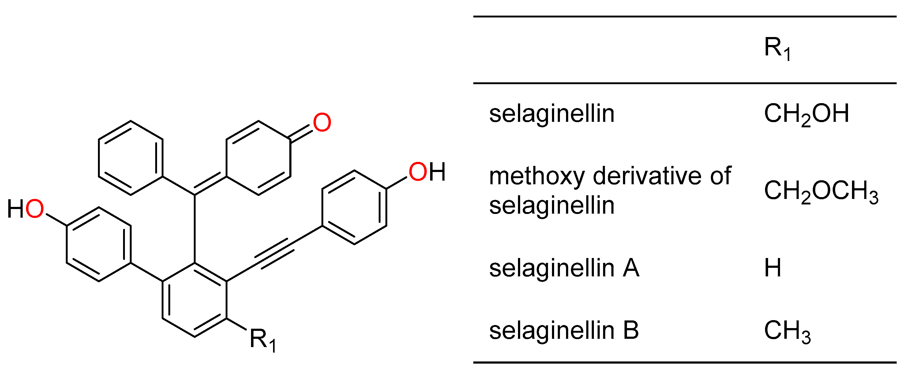
2.1. Chemical Composition

By using the ultra-high performance liquid chromatography–high-resolution mass spectrometry (UHPLC–HRMS) analysis of dichloromethane and methanol crude extracts of Selaginella, we identified 193 known secondary metabolites (Table 1 and Table 2) and found an additional 30 unidentified compounds. Approximately 60% of the unidentified compounds were present in the dichloromethane (DCM) extracts (see Table S1 for full list of compounds). In total, eight different classes of compounds were present in the dichloromethane extracts, while nine classes were found in the methanolic extracts. Due to their polar nature, no alkaloids were detected in the dichloromethane extracts. Of the known compounds in the methanolic extract of Selaginella (Table 1), flavonoids were found to be the major constituents, followed by lignans, phenolics, and selaginellins in that order. Moreover, the methanolic extract of S. apoda possessed the highest number of secondary metabolites amongst the species of Selaginella analyzed in this study. A total of 25 different selaginellins and selaginellin derivatives were also identified in the methanolic and dichloromethane extracts.

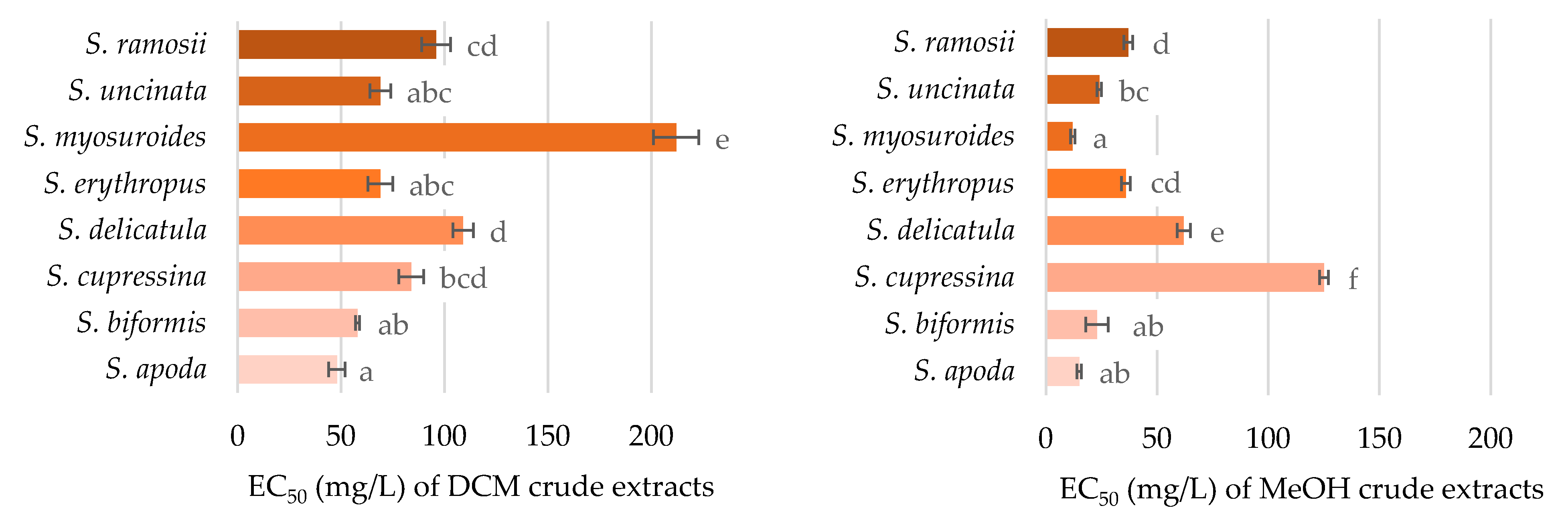
2.2. Antioxidant Activity

The antioxidant activity of the crude extracts was investigated to observe their scavenging potential of oxygen radicals. As can be seen in Figure 2, all the tested extracts had some degree of radical quenching activity. Comparisons of the extraction solvent techniques showed that methanolic extracts were stronger antioxidants than dichloromethane extracts (confirmed by one-way ANOVA with Tukey’s post hoc test with p < 0.01). This is congruent with the chemical analyses, which detected a higher concentration of flavonoids, known for their antioxidant activity, in the methanolic extracts. More pronounced antioxidant capacity was seen in Selaginella myosuroides, Selaginella biformis, and S. apoda methanolic extracts. Unlike S. myosuroides, Selaginella cupressina methanolic extract was almost 10 times less active, and was the least potent species for radical quenching. The antioxidant function of each species was in accordance with the content of flavonoids, (neo)lignans, and selaginellins, and was further substantiated by the values of Pearson correlation coefficient being equal to 0.513, 0.573, and 0.522, respectively, while the critical value was 0.497 (n = 16, df = 14, α = 0.05). However, no correlation was obtained between the antioxidant activity and the detected compound.

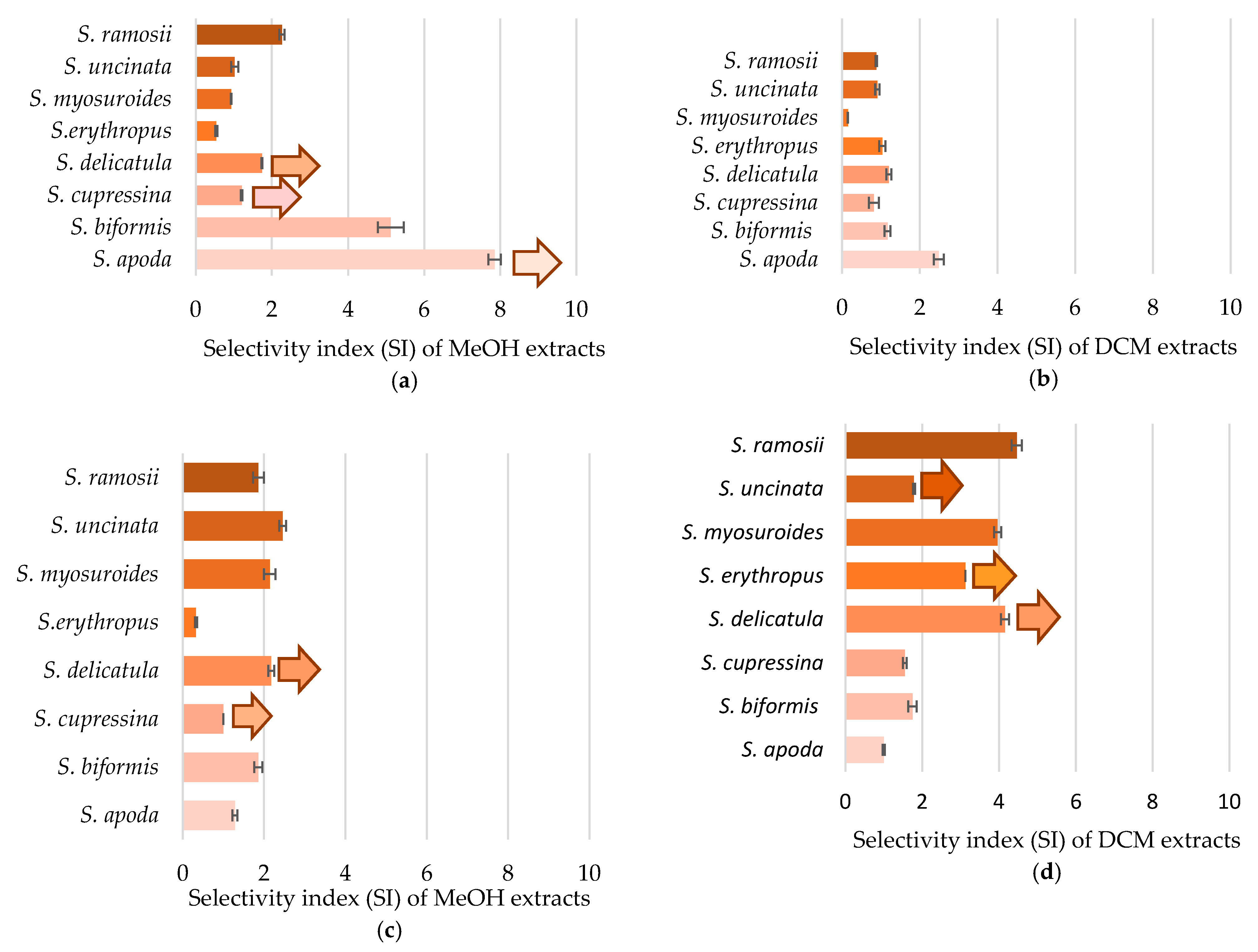
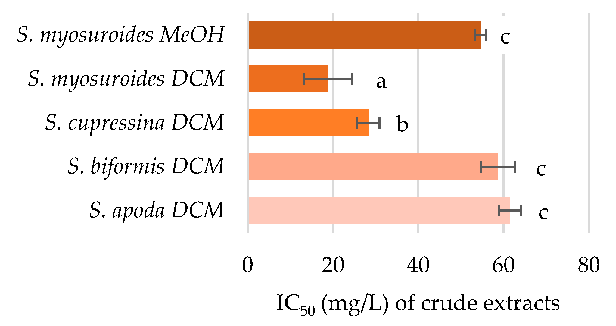
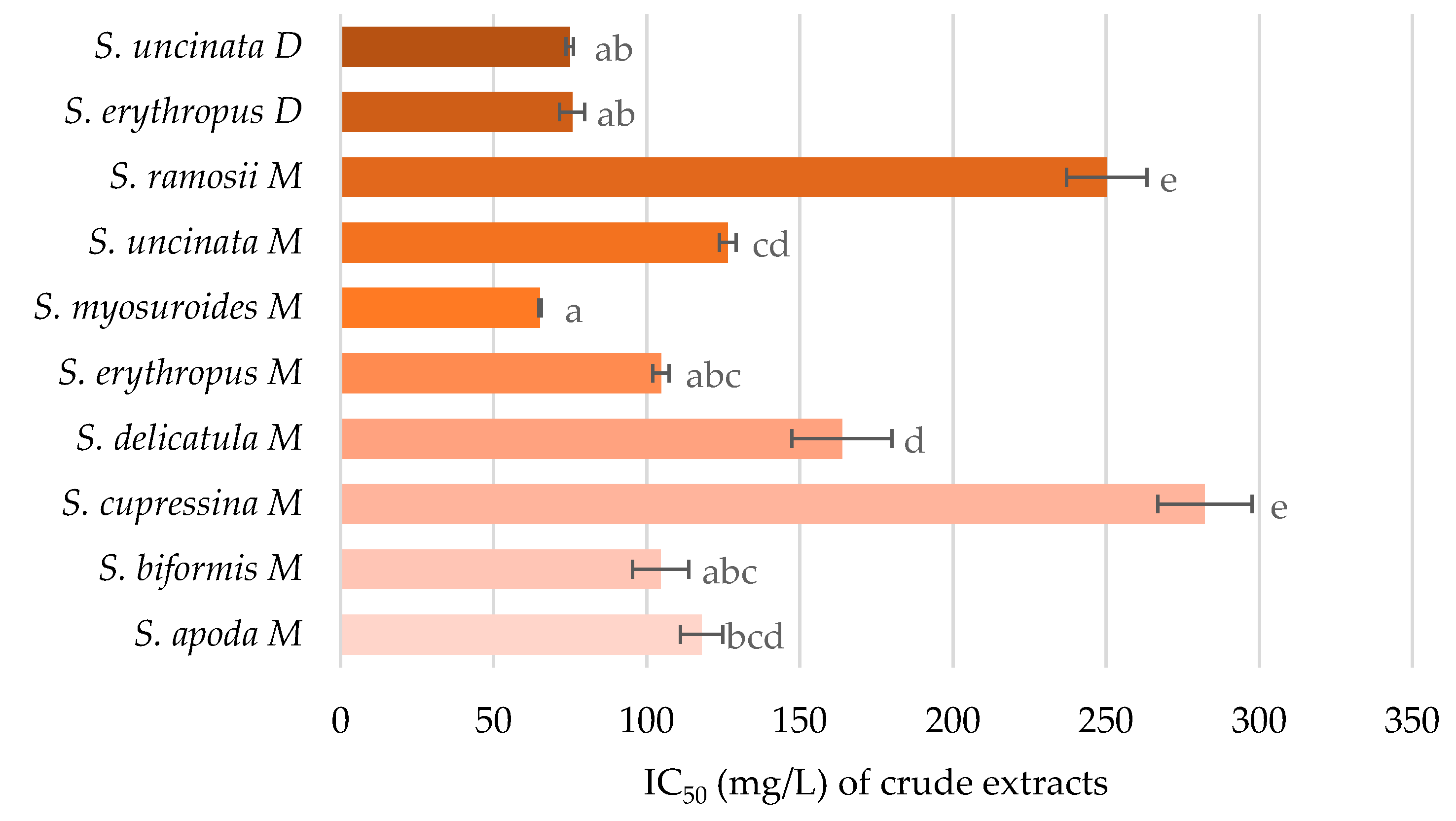
2.3. Cytotoxicity against Cancer Cell Lines

Cytotoxicity against cancer cell lines was determined as the selectivity index, which represents the differences between the cytotoxicity for normal (HEK293T, HaCat) cells and for cancer (HepG2, HeLa) cells (Figure 3). The selectivity index (SI) was calculated as the ratio of the extract’s concentration that halved the viability of the control cell line to the concentration that halved the viability of the cancer cell line. The higher the SI, the higher the selectivity of the extract to affect the viability of cancerous cells and not adversely affect normal cells. The SI was calculated for the cell lines derived from internal organs (kidney and liver) and for epithelial tissues (dermal fibroblasts and cervix). All the tested extracts possess cytotoxicity against cancer cell lines. Interestingly, methanolic extracts were more selective for the organ-derived carcinoma, while dichloromethane extracts were more selective against epithelial carcinoma. The highest selectivity index was determined for the methanolic extract of S. apoda (SI > 7.9 for HEK293T and HepG2), followed by S. biformis (SI = 5.1) and the dichloromethane extracts of Selaginella ramosii (SI = 4.5) and S. delicatula (SI > 4.2).
Both the selectivity index and IC50 values corresponding to each cell line were associated with the 193 compounds identified by chemical analyses of the respective methanolic and dichloromethane extracts. When plotting the results for all 16 extracts tested, we also determined the Pearson’s correlation coefficient. Table 3 shows the chemical compounds which correlated with the cytotoxicity against cancer cell lines with a significance level (α) either equal or lower than 0.05. The cytotoxicity of crude extracts against human cervical adenocarcinoma correlated with five compounds identified in the methanolic extract, four of them belonging to selaginellins and their derivatives. Cytotoxicity against cancer cell lines also correlated with seven flavonoids, three phenols, two lignans, and one representative of each of the following metabolites: terpenoid, fatty acid, quinone, and steroid. The amount of chalcone correlated with cytotoxicity against cancer cell lines with the highest significance (α < 0.01).
Pearson’s correlation coefficient was determined by correlating the cytotoxicity against cancer cell lines and total amount of secondary metabolite classes. The dependence of the selectivity index obtained from organ-derived cell lines and either selaginellins or quinones showed a significant correlation, with coefficients equal to 0.537 (n = 16, df = 14, crit. value = 0.497, α = 0.05) and 0.632 (n = 15, df = 13, crit. value = 0.592, α = 0.02), respectively.

2.4. Inhibition of Acetylcholinesterase Activity

In contrast to previously shown results, the inhibition of acetylcholinesterase was demonstrated mainly by dichloromethane extracts (Figure 4). The only methanolic extract capable of inhibition of this cholinergic enzyme was the extract of S. myosuroides; similarly, its dichloromethane extract was the most active among the samples. The DCM extract of this species was almost 1.5 times more active than that of S. cupressina, and three times more active than S. biformis and S. apoda DCM extracts. The remainder of the extracts exhibited no activity, even at the highest stipulated concentration (62.5 mg/L). The inhibition of acetylcholinesterase can be attributed to selaginellins on the basis of the Pearson’s correlation (value = 0.952, n = 4, df = 2, crit. value = 0.950, α = 0.05).

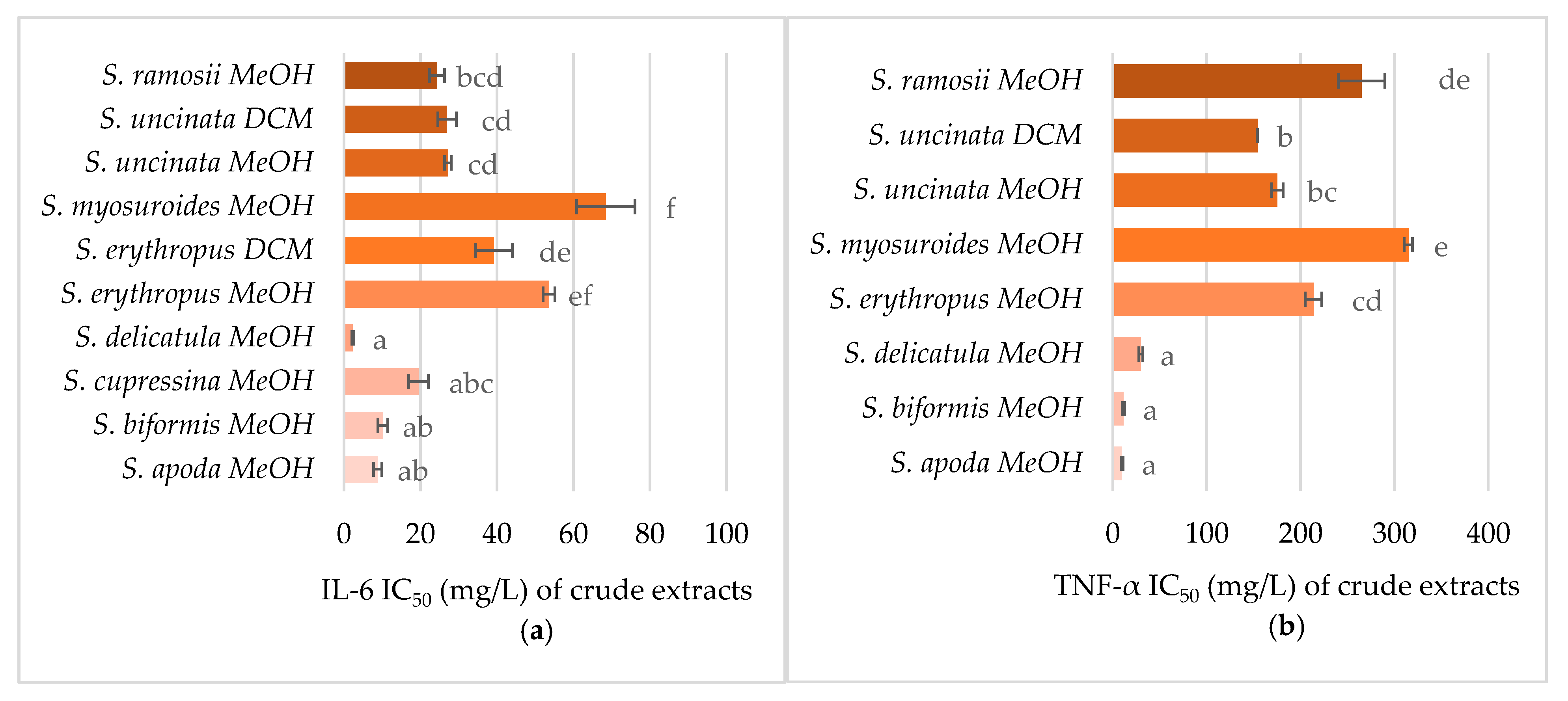
2.5. Anti-Inflammatory Activity

The anti-inflammatory activity of crude extracts was tested as the ability to reduce nitrate oxide production and the release of cytokines. Activity affecting nitrate oxide production was observed primarily in methanolic extracts of S. myosuroides, Selaginella erythropus, and S. biformis (Figure 5). Only DCM extracts of S. uncinata and S. erythropus exhibited any activity. The inhibition of nitrate oxide production was related to the total (neo)lignans content, specifically seladoeflavone E (flavonoid) and selamoellenin A (lignin). The correlation coefficients are presented in Table 4.
As several Selaginella species exhibited a promising reduction of nitrate oxide production, their ability to inhibit the release of cytokines was also tested to further validate the results. As can be seen in Figure 6, all methanolic extracts inhibited tumor necrosis factor (TNF)-α and interleukin (IL)-6 production in a dose-dependent manner; however, the production of cytokines was only modulated by two DCM extracts—S. uncinata and S. erythropus. The IL-6 production was more sensitive to the extracts than that of TNF-α. The most potent species for inhibiting cytokine release were S. apoda, S. biformis, S. delicatula, and S. cupressina. The correlations showed that the upregulation of IL-6 was affected by Selaginella-unique compounds: selagin (flavonoid), pulvinataphendiol (selaginellin), and further by daucosterol—a natural phytosterol-like compound and derivative of phenylpropanoic acid. Selagin, pulvinataphendiol, and the natural phytosterol-like compound also inhibited the expression of TNFα resembling the Selaginella-unique biflavonoid isomers (robustaflavone 4′-methyl ether, podocarpusflavone, neocryptomerin, sequoiaflavone, isocryptomerin, or sotetsuflavone).

3. Discussion

Plant bioprospecting is a process leading to the exploration of natural plant species for biologically active compounds, which could be further isolated or chemically synthetized and used in any sector of the market economy for profit. Although many of these compounds have found applications in the pharmaceutical industry, in many cases the bioprospecting of medical plants has failed. Usually, the reason for the failure lies in the fact that the isolation of pure compounds usually negates the additive or synergistic effect of the additive compounds present in the extract. Therefore, over the last decade, attention has been refocused back on the use of more complex crude or partially purified extracts.
Of all the Selaginella species, S. tamariscina (to date, 144 publications in Web of Science) is the one that is best characterized for its biological activities and composition. Herein, we focused on some lesser-known representatives of Selaginella species, including S. uncinata (31 publications), which was reported in more than half of the total publications, followed by S. delicatula (12 publications), S. apoda (8 publications), S. erythropus (7 publications), S. biformis (1 publication), and species that have not been reported before, namely, S. cupressina, S. myosuroides, and S. ramosii.
It is well documented in the literature that the genus Selaginella produces a wide range of structurally different secondary metabolites such as flavonoids [19,20] and lignans [21,22], together with their unique pigments, selaginellins [27,29,30,32,36]. Our MS data analysis (see Table S1) also confirmed that flavonoids, (neo)lignans, and selaginellins are among the most common natural products identified in both the methanolic and dichloromethane extracts of Selaginella. The majority of the identified flavonoids (90%), (neo)lignans (81%), and selaginellins (79%) were present in the methanolic extracts compared to flavonoids (10%), (neo)lignans (19%), and selaginellins (21%) in the dichloromethane extracts, respectively. Selaginellin; selaginellins D, F, O, P, Q, S, and W; selaginpulvilins A, B, E, F, I, K, L, N, O, and T; with selariscins A, and B; and selariscinins B, and D were the unique compounds identified from both the DCM and methanolic (MeOH) extracts. Selaginellin was initially isolated from S. sinensis [31], selaginellin S from S. moellendorffii Hieron [40]; selaginellin O [33], selaginpulvilin N [36], selaginpulvilin O [36], selaginpulvilin T [36], selariscin A [36], selariscin B [36], selariscinin B [3], and selariscinin D [3] from S. tamariscina; and selaginellin D [29], selaginellin F [29], selaginellin P [30], selaginellin Q [30], selaginpulvilin A [41], selaginpulvilin B [41], selaginpulvilin E [42,43], selaginpulvilin F [43], selaginpulvilin I [43], selaginpulvilin K [44], and selaginpulvilin L [44] were isolated for the first time from S. pulvinata.
Several compounds identified here have not been previously reported for any of the Selaginella plants used in this study. Interestingly, only 12 unidentified compounds were detected in the methanolic extract, while 18 unidentified compounds were observed in the dichloromethane extract, indicating a potential plethora of new bioactive compounds from the Selaginella samples tested in this study.
The biological activity of tested samples is usually compared with standard compound(s) that have been reported to either exhibit the activity being assessed or used as a quantitative tool for comparison on the test samples. However, some researchers consider such distinctions to be inappropriate for crude extracts, as many compounds without activity usually dilute the effect or interfere with biologically active compounds. In contrast to the above statements, the combinatory action observed in complex matrices such as synergism/antagonism has been observed to affect the results. In addition, the concentrations of standards are usually prepared in a molar concentration, while the crude extracts can only be presented in weight (either wet biomass or dry weight) concentration. On the other hand, calibration curves or standards are usually shown to be a good laboratory praxis representing a validated methodology. Therefore, the standards for our methods are presented in Figure S1.
All of the tested Selaginella species demonstrated antioxidant behavior, cytotoxicity against cancer cell lines, and anti-inflammatory activities. The antioxidant activity of Selaginella species was facilitated by flavonoids, (neo)lignans, and selaginellins. We evaluated the correlation with antioxidant activity and identified compounds as well as the correlation of antioxidant activity and amount of secondary metabolite classes. The first mentioned correlation showed no hit with a significance ≤ 0.05, which is in agreement with the additive/synergistic mode of action of compounds in such complex matrixes as plant crude extracts [45]. The antioxidant activity of flavonoids is well known for flavonoids isolated from Selaginella species [46], but the antioxidant activity of (neo)lignans [47] and selaginellins [33] has not been verified yet. However, it has to be mentioned that biochemical assays have their limitations—they omit such important parameters as compound availability, first-pass metabolism, ability to cross the membranes, etc. Therefore, their results should be considered for information only. However, due to their price and speed, they are the most suitable alternative for high-throughput screening and first pre-selection.
The anticancer activity of Selaginella species is often reported as the toxicity of extracts for cancer cell lines; however, such a conclusion should not be established without appropriate controls. Therefore, here we report on the cytotoxicity against cancer cell lines of Selaginella species as a selectivity index (SI), which compares the concentration that halves the viability of the control cell line to the concentration that halves the viability of the cancerous cell line. Although the selectivity index has been known for more than a decade, it is still not commonly used, which hinders the uniform interpretation of results. The SI value reflects selectivity, where a higher SI indicates more selective cytotoxicity. An SI value lower than 2 indicates either low selectivity [48] or general toxicity [49]. Values higher than 2 indicate selectivity. According to Poirier et al. [50], SI = 5.5 indicates a good selectivity. The strictest criterion was applied by Peña-Morán [51], who considered the compound to be selective if SI ≥ 10. We determined the highest SI in methanolic extracts of S. apoda (SI > 7.9 ± 0.2) and S. biformis (SI = 5.1 ± 0.3) when applied to organ-derived cell lines. In contrast to organ-derived cell lines, where the methanolic extracts were more active, dichloromethane extracts were more active against epithelial carcinoma with SI = 4.5 ± 0.1, > 4.2 ± 0.1, and = 4.0 ± 0.1 for S. ramosii, S. delicatula, and S. myosuroides, respectively. The association of cytotoxicity against cancer cell lines and chemical composition showed a clear relationship, since up to 20 compounds were found to be responsible for the cytotoxicity against cancer cell lines of Selaginella species. Only a few of them have been already reported for their cytotoxicity against cancer cell lines, these being hexahydroxybiflavone [52], selaginellin O [33] and P [30], methyl cinnamate [53], seladoeflavone E [20], sinensioside A [54], oleic acid [55,56], chalcone [57], and phellodensin F [58].
The ability of Selaginella extracts to inhibit acetylcholinesterase has been previously tested by several groups. The methanolic extract of Selaginella moellendorffii exhibited no modulation of acetylcholinesterase (AChE) activity [23], which is in agreement with our results demonstrating the activity mainly by compounds extracted by DCM. The inhibition of AChE activity by a polar extract of Selaginella delicatula has been previously reported at a concentration higher than 50 mg/L [13], which is in line with our results, where the most active methanol-extracted species—S. myosuroides—had an AChE IC50 equal to 54.5 ± 1.3 mg/L.
Modulation of the immune system by Selaginella extracts were previously detected in some species. The oral administration of S. moellendorffii extract reduced the production of nitrate oxide and anti-inflammatory cytokines in rats [59]. Our results demonstrated a significant anti-inflammatory activity of selagin, which has been also previously demonstrated in vivo [60]. Immunoregulatory activity of daucosterol was also found both in vitro and in vivo [61,62,63]. Selaginella-unique biflavonoids also exhibited anti-inflammatory activity by reversing the TNF-α signaling pathway [64].

4. Materials and Methods

4.1. Material and Reagents

2,2′-Azo-bis-(2-methylpropionamidine) dihydrochloride (AAPH, Sigma-Aldrich, St. Louis, MO, USA); 2′,7′-dichlorofluorescin diacetate (DCFH-DA, Sigma-Aldrich); acetylcholinesterase (AChE, Sigma-Aldrich); 100× antibiotic antimycotic solution (Sigma-Aldrich); dichloromethane (Penta, Prague, Czech Republic); dimethylsulfoxide (DMSO, Sigma-Aldrich); Dulbecco’s modified Eagle’s medium–high glucose (DMEM, Sigma-Aldrich); Eagle’s minimum essential medium (EMEM, Sigma-Aldrich); Eagle’s minimal essential medium no phenol red (MEM, Sigma-Aldrich); fetal bovine serum (FBS, Sigma-Aldrich); fluorescein (Sigma-Aldrich); Griess reagent modified (Sigma-Aldrich); l-glutamine solution (Sigma-Aldrich); lipopolysaccharides from Escherichia coli O111:B4 (LPS, Sigma-Aldrich); methanol (Penta, Prague, Czech Republic); resazurin sodium salt (Sigma-Aldrich; trypsin–ethylenediaminetetraacetic acid (EDTA) solution (Sigma-Aldrich).

4.2. Plant Material

Eight Selaginella species were investigated in this study: S. apoda (L.) C. Morren, S. biformis A. Braun ex Kuhn, S. cupressina (Willd.) Spring, S. delicatula (Desv. ex Poir.) Alston, S. erythropus (Mart.) Spring, S. myosuroides (Kaulf.) Spring, S. ramosii Hieron, and S. uncinata (Desv. ex Poir.) Spring. The aerial parts of S. apoda (voucher specimen no. Linis 5808–18), an introduced Filipino Selaginella species, were gathered on 9 July 2018 from pots inside a plant nursery at Burnham Park, City of Baguio, province of Benguet. Meanwhile, the aerial parts of S. biformis (voucher specimen no. Linis 5812–18) were collected on 4 August 2018 from a moist, shaded soil bank beside a trail of a lowland forest on Mount Makiling, province of Laguna. For S. cupressina (voucher specimen no. Linis 5811–18), its aerial parts were collected on 14 July 2018 from a muddy soil bank of Pilis Creek inside a disturbed lowland forest of Mount Natib in the province of Bataan. Similarly, the aerial parts of S. delicatula (voucher specimen no. Linis 5810–18) were collected on the same date as S. cupressina (14 July 2018) from another muddy soil bank near Pilis Creek also inside a disturbed lowland forest of Mount Natib in the province of Bataan. S. erythropus (voucher specimen no. Linis 5809–18) is another introduced species in the Philippines. Its aerial parts were collected on 9 July 2018 from clay pots cultivated in a plant nursery in Burnham Park, Baguio City, Benguet Province, the Philippines. Aerial parts of S. myosuroides (voucher specimen no. Linis 5813–18), on the other hand, were collected on 4 August 2018 from a heavily shaded moist rockslide within a lowland forest of Mount Makiling in the province of Laguna, while aerial parts of S. ramosii (voucher specimen no. Linis 5814–18) were collected on 4 August 2018 from a steep eroded soil bank in the montane forest of the same mountain. Like S. apoda and S. erythropus, S. uncinata (voucher specimen no. Linis 5807–18) is the third introduced Selaginella species in the Philippines investigated in this study. Aerial parts of S. uncinata were collected on 5 June 2018 from a shady area of grassy, moist, humid ground inside De La Salle University, Manila. All eight Selaginella specimens were identified by Dr. V.C. Linis, a professor and botanist at De La Salle University. Voucher specimens were prepared, preserved, and deposited in De La Salle University Herbarium (code, DLSUH), with some duplicates sent to the Philippine National Herbarium (code, PNH) for further reference.
The collected Selaginella samples were all air-dried in an air-conditioned laboratory room (room temperature set to 17 °C). The samples were laid on wide, open cardboard boxes and left to air dry until visibly dry throughout.

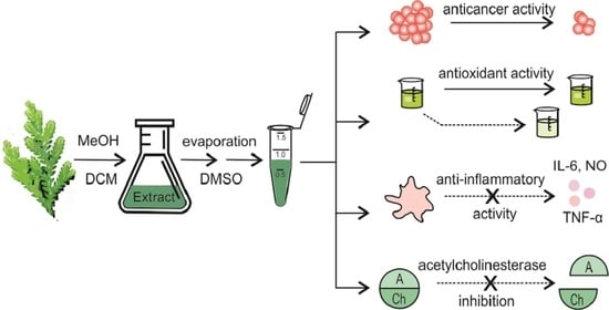
4.3. Extraction of Secondary Metabolites

The dried and finely ground plant materials were extracted with DCM (2 × 100 mL) followed by MeOH extraction (2 × 100 mL) for 24 h, after which the solvents were removed under pressure to yield dried DCM and MeOH crude extracts. A solution of 1 g/L concentration for each crude extract (16 samples) was prepared, and the extracts were analyzed using UHPLC–HRMS. Samples were diluted 10 times (v/v) and injected into UHPLC using the starting mobile phase.

4.4. Determination and Identification of Secondary Metabolites

UHPLC–HRMS analysis was performed using an Accela Open autosampler (20 µL sample loop) and an Accela 600 pump (Thermo Fisher Scientific, Walham, MA, USA) coupled to the electrospray ion (ESI) source of an LTQ Orbitrap Velos mass spectrometer (Thermo Fisher Scientific, Walham, MA, USA) in positive and negative ion modes. HPLC separations were performed in a Phenomenex Luna C18(2) column (150 × 3 mm, particle 3 µm) at a flow rate of 300 µL/min at 35 °C (column thermostat SICO-100). Mobile phases consisted of A (0.1% formic acid in water) and B (0.1% formic acid in methanol). The linear gradient mobile phase protocol was as follows: 0–0.2 min, 10% B; 0.2–6 min, 10–95% B; 6–10 min, 95% B; 10–11 min, 10% B; 11–15 min, 10% B. The MS data were processed and analyzed using Xcalibur 3.0.63. Molecular ion adducts such as [M + H]+, [M + Na]+, [2M + H]+, [2M + Na]+, [M − H], and [2M − H] were identified, and potential compounds were searched by using the Scifindern database [34].

4.5. Oxygen Radical Absorption Capacity (ORAC)

The antioxidant activity was tested using the standard ORAC assay [65]. The crude extracts were analyzed using binary dilution in the concentration range of 0.1–2500 mg/L in 3 replicates. The ability of samples to absorb the generated radicals was monitored by measuring the fluorescence (excitation/emission 485/535 nm), recorded at 5 min intervals for 2 h using a SpectraMax i3x microplate reader (Molecular Devices, San Jose, CA, USA). The EC50 values were determined according to the concentration range using AAT Bioquest EC50 calculator [66].

4.6. Cytotoxicity Assay

The cytotoxicity assay was performed according to the standard protocol [67]. We used the following human cell lines: HepG2 (hepatocellular carcinoma), HeLa (cervical adenocarcinoma), HaCat (keratinocytes), and HEK293T (embryonal kidney cells) purchased from American Type Culture Collection ATCC (VA, USA). The cells were counted using an Auto T4 Cellometer (Nexcelom Bioscience, Lawrence, MA, USA). We seeded 1 × 10⁵ cells/mL into a 96-well plate containing 100 μL of the medium. The medium was changed after 24 h, and the crude extracts were added in the concentration range of 125–500 mg/L in 4 replicates. After 72 h of incubation, a standard resazurin assay was performed. The fluorescence was measured using a SpectraMax i3x microplate reader (Molecular Devices, San Jose, CA, USA) at a wavelength of 560/590 nm excitation/emission.

4.7. Modulation of Acetylcholinesterase Activity

The Ellman colorimetric method [68] was used to measure the activity of acetylcholinesterase (AChE). The crude extracts were analyzed using binary dilution in the concentration range of 62.5–0.125 mg/L. The AChE was added to the reaction mixture and the plate was incubated (37 °C, 5% CO2) for 15 min. After the incubation with AChE (2.5 U/well), the reaction was started by the addition of 5,5′-dithiobis-2-nitrobenzoic acid (DTNB, 0.02 g/L) and acetylcholine chloride (0.01 g/L). The absorbance was recorded using a SpectraMax i3x microplate reader (Molecular Devices, San Jose, CA, USA) at a wavelength of 412 nm at 1 min intervals for 10 min.

4.8. Anti-Inflammatory Activity

Nitrate oxide was quantified in the supernatant of LPS-stimulated (100 ng/L) macrophages (RAW 264.7) cultivated in MEM medium after 24 h. The supernatant was mixed with Griess reagent (0.04 g/L) at a 1:1 ratio, and the absorbance (540 nm) was measured after 15 min of incubation at room temperature. The viability of cells was verified by the resazurin assay described above.
Both IL-6 and TNF-α were quantified by ELISA. Clear Flat-Bottom Immuno Nonsteril 96-well plates (Thermo Fisher Scientific) were coated with 100 µL/well of capture antibody (TNF-α Monoclonal Antibody (TN3-19.12) or IL-6 Monoclonal Antibody (MP5-20F3), Thermo Fisher Scientific) in phosphate-buffered saline (PBS, 2 µg/mL). After overnight incubation at 4 °C, the plates were washed 3 times using wash buffer (PBS supplemented with 0.05% Tween20). The plates were then blocked with blocking buffer (20% FBS, 0.1% Tween20, PBS). The supernatant of LPS-induced (100 ng/mL) RAW 264.7 cells were collected by centrifugation after 6 h of incubation, and 10 µL was mixed with 90 µL of blocking buffer and applied to primary antibody-coated plates. After overnight incubation at 4 °C, the plates were washed and secondary antibody was added (TNF-α Polyclonal Antibody, Biotin, eBioscience or IL-6 Monoclonal Antibody (MP5-32C11), Biotin, eBioscience, 1 µg/mL, Thermo Fisher Scientific). After 1 h of incubation at room temperature, the washed plates were loaded with streptavidin–horseradish peroxidase (HRP, 1.25 µg/mL, Thermo Fisher Scientific). After 30 min of incubation at room temperature, the washed plate was loaded with tetramethyl benzidine (TMB) solution (Sigma-Aldrich). The reaction was stopped after 15 min with 1M H3PO4, and the absorbance (450 nm) was recorded, subtracting the absorbance at 570 nm.

4.9. Statistical Analysis and Correlation

The experiments were performed with the appropriate number (n) of repetitions, which are presented in each figure. The EC50/IC50 values were obtained using the software GraphPad Prism 7 (GraphPad Software, San Diego, CA, USA). The data are presented as the averages of the repetitions with the standard error of the mean (SEM). One-way analysis of variance (ANOVA) was used, followed by Tukey’s post hoc test (p < 0.05 or p < 0.01) to show the differences between the groups. Denoting the statistically significant levels with different letters is a way to summarize the differences between the extracts. If the two extracts share at least one letter, it means that they are not significantly different. It also reflects the comparison of every extract to every other extract in the same assay (column in the figure). For ANOVA, the software Statistica version 13 was used (Tibco Software Inc., Tulse, OK, USA). The selectivity index (SI) was calculated using the formula SI = IC50 (control cells)/IC50 (cancer cells).
Pearson’s correlation coefficients were calculated using the automatic function “CORREL” in Microsoft Office Excel. The analytical standards of identified components were not available, and thus we correlated either the areas of the peaks or the sum of peak areas belonging to the respective chemical class (alkaloids, fatty acids, flavonoids, (neo)lignans, phenols, quinones, selaginellins, steroids, terpenoids), which served as matrix I. The results of the biological activity of 16 Selaginella extracts obtained from all assays investigated (ORAC assay, selectivity index, cytotoxicity, inhibition of AChE, inhibition of NO, TNF-α and IL-6 production) were utilized as matrix II. The significance of the correlation coefficient was evaluated by using a comparison of coefficients and the critical values (α = 0.05 or lower), which were determined using the degrees of freedom (df = n−2).

5. Conclusions

Selaginella is well known for its unique natural products and its wide range of biological effects, which is why numerous Selaginella species are used in traditional medicines for the treatment of various diseases. Here, we compared the antioxidant, anti-inflammatory, and acetylcholinesterase inhibition activity as well as the cytotoxicity against cancer cell lines of eight Selaginella species, which highlighted S. apoda species among all the tested activities. The correlations of biological activity and composition of extracts verified the activity of several known biologically active compounds and for the first time highlighted several compounds for their previously unknown activities. This paper is one of the first reporting the selective cytotoxicity of Selaginella extracts against cancerous cells. However, significantly deeper studies are needed to confirm the compounds responsible for the activities, to determine the selectivity against various tumors derived from different tissues, and also to determine the mechanism of action.

Supplementary Materials

The following are available online at https://www.mdpi.com/1424-8247/14/1/16/s1: Figure S1: Cytotoxicity against cancer cell lines and antioxidant, anti-inflammatory, and anti-acetylcholinesterase activity of appropriate standards: (a) doxorubicin, (b) quercetin, (c) indomethacin, and (d) eserin. Table S1: Overview of phytochemicals detected and tentatively identified in Selaginella by ultra-high performance liquid chromatography-high-resolution mass spectrometry (UHPLC–HRMS). Figure S2: Chemical structures of pharmacologically active ingredients mentioned in Table 3 and Table 4, which are unique for Selaginella species.

Author Contributions

Conceptualization J.V., T.R.; plant collection and drying, M.C.T. and G.O.; botanical determination, V.L.; extraction and analytical determinations, R.K.; MS measurements R.K. and D.S.; ORAC, B.K.; cytotoxicity, B.K.; AChE activity modulation, B.K.; anti-inflammatory activity, D.K. and S.D.; statistical analysis and correlation, B.K and K.Ř.; investigation, B.K. and J.V.; resources, J.L.; writing—original draft preparation, B.K., R.K., and J.V.; writing—review and editing, T.R. and J.L.; supervision, T.R.; project administration, J.V. and J.L.; funding acquisition, J.V. and T.R. All authors have read and agreed to the published version of the manuscript.

Funding

R.K. gratefully acknowledges the European Fund: European Structural and Investment Funds Operational Program Research, Development and Education (ChemJets UCT Prague, CZ.02.2.69/0.0/0.0/16_027/0008351). This research was funded by the Technology Agency of the Czech Republic–program Zeta (No. TJ02000372).

Data Availability Statement

The data presented in this study are available in this article or associated supplementary material.

Conflicts of Interest

The authors declare no conflict of interest.

References

  1. Weng, J.-K.; Noel, J. Chemodiversity in selaginella: A reference system for parallel and convergent metabolic evolution in terrestrial plants. Front. Plant Sci. 2013, 4, 1–17. [Google Scholar] [CrossRef] [PubMed] [Green Version]
  2. Nguyen, P.H.; Zhao, B.T.; Ali, M.Y.; Choi, J.S.; Rhyu, D.Y.; Min, B.S.; Woo, M.H. Insulin-mimetic selaginellins from selaginella tamariscina with protein tyrosine phosphatase 1B (PTP1B) inhibitory activity. J. Nat. Prod. 2015, 78, 34–42. [Google Scholar] [CrossRef] [PubMed]
  3. Nguyen, P.H.; Ji, D.J.; Han, Y.R.; Choi, J.S.; Rhyu, D.Y.; Min, B.S.; Woo, M.H. Selaginellin and biflavonoids as protein tyrosine phosphatase 1b inhibitors from Selaginella tamariscina and their glucose uptake stimulatory effects. Bioorg. Med. Chem. 2015, 23, 3730–3737. [Google Scholar] [CrossRef] [PubMed]
  4. Adame-González, A.B.; Muñíz-Dl, M.E.; Valencia, -A.S. Comparative leaf morphology and anatomy of six Selaginella species (selaginellaceae, subgen, rupestrae) with notes on xerophytic adaptations. Flora 2019, 260, 151482. [Google Scholar] [CrossRef]
  5. Uphof, J.C.T. Physiological anatomy of xerophytic Selaginellas. New Phytol. 1920, 19, 101–131. [Google Scholar] [CrossRef]
  6. Weststrand, S.; Korall, P. Phylogeny of Selaginellaceae: There is value in morphology after all! Am. J. Bot. 2016, 103, 2136–2159. [Google Scholar] [CrossRef] [Green Version]
  7. Magazù, S.; Migliardo, F.; Benedetto, A.; La Torre, R.; Hennet, L. Bio-protective effects of homologous disaccharides on biological macromolecules. Eur. Biophys. J. 2012, 41, 361–367. [Google Scholar] [CrossRef]
  8. Schulz, C.; Little, D.P.; Stevenson, D.W.; Bauer, D.; Moloney, C.; Stützel, T. An overview of the morphology, anatomy, and life cycle of a new model species: The lycophyte Selaginella apoda (l.) spring. Int. J. Plant Sci. 2010, 171, 693–712. [Google Scholar] [CrossRef]
  9. Heo, J.K.; Nguyen, P.H.; Kim, W.C.; Phuc, N.M.; Liu, K.H. Inhibitory effect of selaginellins from Selaginella tamariscina (beauv.) spring against cytochrome P450 and uridine 5’-diphosphoglucuronosyltransferase isoforms on human liver microsomes. Molecules 2017, 22, 1590. [Google Scholar] [CrossRef] [Green Version]
  10. Shim, S.Y.; Lee, S.G.; Lee, M. Biflavonoids isolated from Selaginella tamariscina and their anti-inflammatory activities via ERK 1/2 signaling. Molecules 2018, 23, 926. [Google Scholar] [CrossRef] [Green Version]
  11. Wang, G.; Ma, X.; Li, D.; Jiang, Y.; Liang, Q.; Hui, C. Chemical composition of essential volatile oils of Selaginella spp. and their antibacterial activity. Bangladesh J. Bot. 2018, 47, 719–726. [Google Scholar]
  12. Chandran, G.; Muralidhara. Neuroprotective effect of aqueous extract of Selaginella delicatula as evidenced by abrogation of rotenone-induced motor deficits, oxidative dysfunctions, and neurotoxicity in mice. Cell Mol. Neurobiol. 2013, 33, 929–942. [Google Scholar] [CrossRef] [PubMed]
  13. Girish, C.; Muralidhara. Propensity of Selaginella delicatula aqueous extract to offset rotenone-induced oxidative dysfunctions and neurotoxicity in drosophila melanogaster: Implications for parkinson’s disease. NeuroToxicology 2012, 33, 444–456. [Google Scholar] [CrossRef] [PubMed]
  14. Chen, J.J.; Duh, C.Y.; Chen, J.F. New cytotoxic biflavonoids from Selaginella delicatula. Planta medica 2005, 71, 659–665. [Google Scholar] [CrossRef] [PubMed]
  15. Li, L.; Wang, Q.; Deng, R.; Zhang, S.; Lu, Y. Transcriptome profiling of blue leaf coloration in Selaginella uncinata. Canadian J. Plant Sci. 2016, 97. [Google Scholar] [CrossRef] [Green Version]
  16. Li, J.; Lei, X.; Chen, K.L. Comparison of cytotoxic activities of extracts from Selaginella species. Pharmacogn. Mag. 2014, 10, 529–535. [Google Scholar] [CrossRef] [PubMed] [Green Version]
  17. Zheng, J.; Zheng, Y.; Zhi, H.; Dai, Y.; Wang, N.-L.; Fang, Y.-X.; Du, Z.-Y.; Zhang, K.; Wu, L.-Y.; Fan, M. γ-Lactone derivatives and terpenoids from Selaginella uncinata and their protective effect against anoxia. Chem. Nat. Compd. 2014, 50, 366–369. [Google Scholar] [CrossRef]
  18. Yu, B.; Cai, W.; Zhang, H.H.; Zhong, Y.S.; Fang, J.; Zhang, W.Y.; Mo, L.; Wang, L.C.; Yu, C.H. Selaginella uncinata flavonoids ameliorated ovalbumin-induced airway inflammation in a rat model of asthma. J. Ethnopharmacol. 2017, 195, 71–80. [Google Scholar] [CrossRef]
  19. Zou, Z.X.; Xu, P.S.; Zhang, G.G.; Cheng, F.; Chen, K.; Li, J.; Zhu, W.X.; Cao, D.S.; Xu, K.P.; Tan, G.S. Selagintriflavonoids with BACE1 inhibitory activity from the fern Selaginella doederleinii. Phytochemistry 2017, 134, 114–121. [Google Scholar] [CrossRef]
  20. Zou, Z.X.; Xu, K.P.; Xu, P.S.; Li, X.M.; Cheng, F.; Li, J.; Yu, X.; Cao, D.S.; Li, D.; Zeng, W.; et al. Seladoeflavones A–F, six novel flavonoids from Selaginella doederleinii. Fitoterapia 2017, 116, 66–71. [Google Scholar] [CrossRef]
  21. Zhu, Y.; Huang, R.Z.; Wang, C.G.; Ouyang, X.L.; Jing, X.T.; Liang, D.; Wang, H.S. New inhibitors of matrix metalloproteinases 9 (MMP-9): Lignans from Selaginella moellendorffii. Fitoterapia 2018, 130, 281–289. [Google Scholar] [CrossRef] [PubMed]
  22. Cheng, F.; Xu, K.P.; Liu, L.F.; Yao, C.P.; Xu, P.S.; Zhou, G.; Li, D.; Li, X.M.; Chen, K.; Zou, Z.X.; et al. New neolignans from Selaginella pieta and their protective effect on HT-22 cells. Fitoterapia 2018, 127, 69–73. [Google Scholar] [CrossRef] [PubMed]
  23. Wang, Y.-H.; Long, C.-L.; Yang, F.-M.; Wang, X.; Sun, Q.-Y.; Wang, H.-S.; Shi, Y.-N.; Tang, G.-H. Pyrrolidinoindoline alkaloids from Selaginella moellendorfii. J. Nat. Prod. 2009, 72, 1151–1154. [Google Scholar] [CrossRef] [PubMed]
  24. Ke, L.Y.; Zhang, Y.; Xia, M.Y.; Zhuo, J.X.; Wang, Y.H.; Long, C.L. Modified abietane diterpenoids from whole plants of Selaginella moellendorffii. J. Nat. Prod. 2018, 81, 418–422. [Google Scholar] [CrossRef] [PubMed]
  25. Li, D.; Qian, Y.; Tian, Y.J.; Yuan, S.M.; Wei, W.; Wang, G. Optimization of ionic liquid-assisted extraction of biflavonoids from Selaginella doederleinii and evaluation of its antioxidant and antitumor activity. Molecules 2017, 22, 586. [Google Scholar] [CrossRef] [PubMed] [Green Version]
  26. Jiang, Y.; Li, D.; Ma, X.; Jiang, F.; He, Q.; Qiu, S.; Li, Y.; Wang, G. Ionic liquid(-)ultrasound-based extraction of biflavonoids from Selaginella helvetica and investigation of their antioxidant activity. Molecules 2018, 23, 3284. [Google Scholar] [CrossRef] [PubMed] [Green Version]
  27. Cao, Y.; Zhao, M.; Zhu, Y.; Zhu, Z.H.; Oberer, L.; Duan, J.A. Diselaginellin b, an unusual dimeric molecule from Selaginella pulvinata, inhibited metastasis and induced apoptosis of smmc-7721 human hepatocellular carcinoma cells. J. Nat. Prod. 2017, 80, 3151–3158. [Google Scholar] [CrossRef]
  28. Zou, Z.; Xu, P.; Wu, C.; Zhu, W.; Zhu, G.; He, X.; Zhang, G.; Hu, J.; Liu, S.; Zeng, W.; et al. Carboxymethyl flavonoids and a chromone with antimicrobial activity from Selaginella moellendorffii hieron. Fitoterapia 2016, 111, 124–129. [Google Scholar] [CrossRef]
  29. Cao, Y.; Chen, J.J.; Tan, N.H.; Oberer, L.; Wagner, T.; Wu, Y.P.; Zeng, G.Z.; Yan, H.; Wang, Q. Antimicrobial selaginellin derivatives from Selaginella pulvinata. Bioorg. Med. Chem. Lett. 2010, 20, 2456–2460. [Google Scholar] [CrossRef]
  30. Cao, Y.; Yao, Y.; Huang, X.-J.; Oberer, L.; Wagner, T.; Guo, J.-M.; Gu, W.; Liu, W.-D.; Lv, G.-X.; Shen, Y.-N.; et al. Four new selaginellin derivatives from Selaginella pulvinata: Mechanism of racemization process in selaginellins with quinone methide. Tetrahedron 2015, 71, 1581–1587. [Google Scholar] [CrossRef]
  31. Zhang, L.-P.; Liang, Y.-M.; Wei, X.-C.; Cheng, D.-L. A new unusual natural pigment from Selaginella sinensis and its noticeable physicochemical properties. J. Org. Chem. 2007, 72, 3921–3924. [Google Scholar] [CrossRef] [PubMed]
  32. Cheng, X.L.; Ma, S.C.; Yu, J.D.; Yang, S.Y.; Xiao, X.Y.; Hu, J.Y.; Lu, Y.; Shaw, P.C.; But, P.P.; Lin, R.C. Selaginellin A and B, two novel natural pigments isolated from Selaginella tamariscina. Chem. Pharm. Bull. 2008, 56, 982–984. [Google Scholar] [CrossRef] [PubMed] [Green Version]
  33. Yang, C.; Shao, Y.; Li, K.; Xia, W. Bioactive selaginellins from Selaginella tamariscina (beauv.) spring. Beilstein J. Org. Chem. 2012, 8, 1884–1889. [Google Scholar] [CrossRef] [PubMed]
  34. Scifinder. Available online: https://scifinder-n.cas.org/ (accessed on 31 July 2020).
  35. Zhang, G.G.; Jing, Y.; Zhang, H.M.; Ma, E.L.; Guan, J.; Xue, F.N.; Liu, H.X.; Sun, X.Y. Isolation and cytotoxic activity of selaginellin derivatives and biflavonoids from Selaginella tamariscina. Planta Med. 2012, 78, 390–392. [Google Scholar] [CrossRef] [Green Version]
  36. Woo, S.; Kang, K.B.; Kim, J.; Sung, S.H. Molecular networking reveals the chemical diversity of selaginellin derivatives, natural phosphodiesterase-4 inhibitors from Selaginella tamariscina. J. Nat. Prod. 2019, 82, 1820–1830. [Google Scholar] [CrossRef]
  37. Le, D.D.; Nguyen, D.H.; Zhao, B.T.; Seong, S.H.; Choi, J.S.; Kim, S.K.; Kim, J.A.; Min, B.S.; Woo, M.H. Ptp1b inhibitors from selaginella tamariscina (beauv.) spring and their kinetic properties and molecular docking simulation. Bioorg. Chem. 2017, 72, 273–281. [Google Scholar] [CrossRef]
  38. Cao, Y.; Chen, J.-J.; Tan, N.-H.; Wu, Y.-P.; Yang, J.; Wang, Q. Structure determination of selaginellins G and H from Selaginella pulvinata by NMR spectroscopy. Magn. Reson. Chem. 2010, 48, 656–659. [Google Scholar] [CrossRef]
  39. Zálešák, F.; Bon, D.J.D.; Pospíšil, J. Lignans and neolignans: Plant secondary metabolites as a reservoir of biologically active substances. Pharmacol. Res. 2019, 146, 104284. [Google Scholar] [CrossRef]
  40. Zhu, B.; Wang, T.-B.; Hou, L.-J.; Lv, H.-X.; Liu, A.M.; Zeng, P.; Li, A.-H. A new selaginellin from Selaginella moellendorffii inhibits hepatitis B virus gene expression and replication. Chem. Nat. Compd. 2016, 52, 624–627. [Google Scholar] [CrossRef]
  41. Liu, X.; Luo, H.-B.; Huang, Y.-Y.; Bao, J.-M.; Tang, G.-H.; Chen, Y.-Y.; Wang, J.; Yin, S. Selaginpulvilins A–D, new phosphodiesterase-4 inhibitors with an unprecedented skeleton from Selaginella pulvinata. Org. Lett. 2014, 16, 282–285. [Google Scholar] [CrossRef]
  42. Yao, W.-N.; Huang, R.-Z.; Hua, J.; Zhang, B.; Wang, C.-G.; Liang, D.; Wang, H.-S. Selagintamarlin a: A selaginellin analogue possessing a 1H-2-benzopyran core from Selaginella tamariscina. ACS Omega 2017, 2, 2178–2183. [Google Scholar] [CrossRef] [PubMed] [Green Version]
  43. Zhang, J.-S.; Liu, X.; Weng, J.; Guo, Y.-Q.; Li, Q.-J.; Ahmed, A.; Tang, G.-H.; Yin, S. Natural diarylfluorene derivatives: Isolation, total synthesis, and phosphodiesterase-4 inhibition. Org. Chem. Front. 2017, 4, 170–177. [Google Scholar] [CrossRef]
  44. Huang, Y.; Liu, X.; Wu, D.; Tang, G.; Lai, Z.; Zheng, X.; Yin, S.; Luo, H.-B. The discovery, complex crystal structure, and recognition mechanism of a novel natural PDE4 inhibitor from Selaginella pulvinata. Biochem. Pharmacol. 2017, 130, 51–59. [Google Scholar] [CrossRef] [PubMed]
  45. Yao, Y.; Luong, T.N.; Lepik, M.; Aftab, N.; Fong, V.H.; Vieira, A. Synergism of antioxidant phytochemicals: Comparisons among purified polyphenols and dietary-plant extracts, XXVIII International Horticultural Congress, 2012; International Society for Horticultural Science (ISHS): Leuven, Belgium, 2012; pp. 121–127. [Google Scholar] [CrossRef]
  46. Macêdo, L.A.R.d.O.; Oliveira Júnior, R.G.d.; Souza, G.R.; de Oliveira, A.P.; de Lavor, É.M.; Silva, M.G.e.; Pacheco, A.G.M.; de Menezes, I.R.A.; Coutinho, H.D.M.; Pessoa, C.d.Ó.; et al. Chemical composition, antioxidant and antibacterial activities and evaluation of cytotoxicity of the fractions obtained from Selaginella convoluta (arn.) spring (Selaginellaceae). Biotechnol. Equip. 2018, 32, 506–512. [Google Scholar] [CrossRef] [Green Version]
  47. Rao, L.; You, Y.-X.; Su, Y.; Fan, Y.; Liu, Y.; He, Q.; Chen, Y.; Meng, J.; Hu, L.; Li, Y.; et al. Lignans and neolignans with antioxidant and human cancer cell proliferation inhibitory activities from Cinnamomum bejolghota confirm its functional food property. J. Agric. Food Chem. 2020, 68, 8825–8835. [Google Scholar] [CrossRef]
  48. Singh, K.; Gangrade, A.; Jana, A.; Mandal, B.B.; Das, N. Design, synthesis, characterization, and antiproliferative activity of organoplatinum compounds bearing a 1,2,3-triazole ring. ACS Omega 2019, 4, 835–841. [Google Scholar] [CrossRef]
  49. Badisa, R.B.; Darling-Reed, S.F.; Joseph, P.; Cooperwood, J.S.; Latinwo, L.M.; Goodman, C.B. Selective cytotoxic activities of two novel synthetic drugs on human breast carcinoma MCF-7 cells. Anticancer Res. 2009, 29, 2993–2996. [Google Scholar]
  50. Perreault, M.; Maltais, R.; Dutour, R.; Poirier, D. Explorative study on the anticancer activity, selectivity and metabolic stability of related analogs of aminosteroid RM-133. Steroids 2016, 115, 105–113. [Google Scholar] [CrossRef]
  51. Peña-Morán, O.A.; Villarreal, M.L.; Álvarez-Berber, L.; Meneses-Acosta, A.; Rodríguez-López, V. Cytotoxicity, post-treatment recovery, and selectivity analysis of naturally occurring podophyllotoxins from Bursera fagaroides var. fagaroides on breast cancer cell lines. Molecules 2016, 21, 1013. [Google Scholar] [CrossRef]
  52. Qian, Z.; Cai-xia, W.; Yan-ling, L.; Cui-yan, L.; Yan-hua, R. Chemical constituents from Selaginella doederleinii and their bioactivities. Zhongcaoyao 2013, 44, 3270–3275. [Google Scholar]
  53. Kathirvel, P.; Ravi, S. Chemical composition of the essential oil from basil (Ocimum basilicum linn.) and its in vitro cytotoxicity against HeLa and HEP-2 human cancer cell lines and nih 3t3 mouse embryonic fibroblasts. Nat. Prod. Res. 2012, 26, 1112–1118. [Google Scholar] [CrossRef] [PubMed]
  54. Cheng, F.; Zou, Z.X.; Xu, P.S.; Zhang, S.H.; Zhang, Y.; Yao, C.P.; Xu, K.P.; Tan, G.S. Pictalignans D–F, three new neolignan derivatives from Selaginella picta. Nat. Prod. Res. 2020, 34, 1264–1269. [Google Scholar] [CrossRef] [PubMed]
  55. Permyakov, S.E.; Knyazeva, E.L.; Khasanova, L.M.; Fadeev, R.S.; Zhadan, A.P.; Roche-Hakansson, H.; Håkansson, A.P.; Akatov, V.S.; Permyakov, E.A. Oleic acid is a key cytotoxic component of hamlet-like complexes. Biol. Chem. 2012, 393, 85. [Google Scholar] [CrossRef] [PubMed]
  56. Malhi, H.; Barreyro, F.J.; Isomoto, H.; Bronk, S.F.; Gores, G.J. Free fatty acids sensitise hepatocytes to trail mediated cytotoxicity. Gut 2007, 56, 1124–1131. [Google Scholar] [CrossRef] [PubMed] [Green Version]
  57. Mahapatra, D.K.; Bharti, S.K.; Asati, V. Anti-cancer chalcones: Structural and molecular target perspectives. Eur. J. Med. Chem. 2015, 98, 69–114. [Google Scholar] [CrossRef]
  58. Chiu, C.Y.; Li, C.Y.; Chiu, C.C.; Niwa, M.; Kitanaka, S.; Damu, A.G.; Lee, E.J.; Wu, T.S. Constituents of leaves of Phellodendron japonicum maxim. and their antioxidant activity. Chem. Pharm. Bull. 2005, 53, 1118–1121. [Google Scholar] [CrossRef] [Green Version]
  59. Paswan, S.K.; Gautam, A.; Verma, P.; Rao, C.V.; Sidhu, O.P.; Singh, A.P.; Srivastava, S. The indian magical herb ‘sanjeevni’ (Selaginella bryopteris l.)—A promising anti-inflammatory phytomedicine for the treatment of patients with inflammatory skin diseases. J. Pharmacopunct. 2017, 20, 93–99. [Google Scholar] [CrossRef]
  60. Gayathri, V.; Asha, V.V.; John, J.A.; Subramoniam, A. Protection of immunocompromised mice from fungal infection with a thymus growth-stimulatory component from Selaginella involvens, a fern. Immunopharmacol. Immunotoxicol. 2011, 33, 351–359. [Google Scholar] [CrossRef]
  61. Lee, J.-H.; Lee, J.Y.; Park, J.H.; Jung, H.S.; Kim, J.S.; Kang, S.S.; Kim, Y.S.; Han, Y. Immunoregulatory activity by daucosterol, a β-sitosterol glycoside, induces protective th1 immune response against disseminated candidiasis in mice. Vaccine 2007, 25, 3834–3840. [Google Scholar] [CrossRef]
  62. Kim, M.J.; Wang, H.S.; Lee, M.W. Anti-inflammatory effects of fermented bark of Acanthopanax sessiliflorus and its isolated compounds on lipopolysaccharide-treated RAW 264.7 macrophage cells. Evid. Based Complement Alternat. Med. 2020, 2020, 6749425. [Google Scholar] [CrossRef]
  63. Jang, J.; Kim, S.-M.; Yee, S.-M.; Kim, E.-M.; Lee, E.-H.; Choi, H.-R.; Lee, Y.-S.; Yang, W.-K.; Kim, H.-Y.; Kim, K.-H.; et al. Daucosterol suppresses dextran sulfate sodium (dss)-induced colitis in mice. Int. Immunopharmacol. 2019, 72, 124–130. [Google Scholar] [CrossRef] [PubMed]
  64. Wang, S.; Yan, Y.; Cheng, Z.; Hu, Y.; Liu, T. Sotetsuflavone suppresses invasion and metastasis in non-small-cell lung cancer a549 cells by reversing EMT via the TNF-α/NF-κB and PI3K/AKT signaling pathway. Cell Death Discov. 2018, 4, 26–36. [Google Scholar] [CrossRef] [PubMed]
  65. Viktorova, J.; Stranska-Zachariasova, M.; Fenclova, M.; Vitek, L.; Hajslova, J.; Kren, V.; Ruml, T. Complex evaluation of antioxidant capacity of milk thistle dietary supplements. Antioxidants 2019, 8, 317. [Google Scholar] [CrossRef] [PubMed] [Green Version]
  66. Aatbio EC50 calculator. Available online: https://www.aatbio.com/tools/ec50-calculator/ (accessed on 31 September 2020).
  67. Tran, V.N.; Viktorova, J.; Augustynkova, K.; Jelenova, N.; Dobiasova, S.; Rehorova, K.; Fenclova, M.; Stranska-Zachariasova, M.; Vitek, L.; Hajslova, J.; et al. In silico and in vitro studies of mycotoxins and their cocktails; their toxicity and its mitigation by silibinin pre-treatment. Toxins 2020, 12, 148. [Google Scholar] [CrossRef] [Green Version]
  68. Dobiasová, S.; Řehořová, K.; Kučerová, D.; Biedermann, D.; Káňová, K.; Petrásková, L.; Koucká, K.; Václavíková, R.; Valentová, K.; Ruml, T.; et al. Multidrug resistance modulation activity of silybin derivatives and their anti-inflammatory potential. Antioxidants 2020, 9, 455. [Google Scholar] [CrossRef]
Figure 1. Chemical structures of selaginellin, methoxy derivative of selaginellin, and selaginellins A–B.
Pharmaceuticals 14 00016 g001
Figure 2. Antioxidant activity, measured by oxygen radical absorption capacity (ORAC) assay, is presented as concentrations (mg/L) of crude extracts that halved the amounts of free oxygen radicals (EC50). Data are presented as the average of three repetitions ± standard error of the mean. Letters indicate the differences between the groups (ANOVA followed by Tukey’s post hoc test, p < 0.05) within one assay. Statistically significant levels are denoted with different letters.
Pharmaceuticals 14 00016 g002
Figure 3. Cytotoxicity against cancer cell lines of the crude extracts expressed as the selectivity index: (a) ratio of IC50 values of HEK293T and HepG2 cell lines; (b) ratio between IC50 for HEK293T and HepG2 cell lines; (c) ratio between IC50 for HaCat and HeLa cell lines; (d) ratio between IC50 for HaCat and HeLa cell lines. Data are presented as the average of three repetitions ± standard error of the mean. The arrows indicate the minimum values of the selective indexes, where the IC50 value for the control line was not reached even at the highest tested concentration (500 mg/L).
Pharmaceuticals 14 00016 g003
Figure 4. Inhibitory effect of Selaginella extracts on acetylcholinesterase activity expressed as IC50 values. Data are presented as the average of four repetitions ± standard error of the mean. Letters indicate the differences between the groups (ANOVA followed by Tukey’s post hoc test, p < 0.05) within one assay. Statistically significant levels were denoted with different letters.
Pharmaceuticals 14 00016 g004
Figure 5. Inhibition of nitrate oxide production. Data are presented as the average of three repetitions ± standard error of the mean. Letters indicate the differences between the groups (ANOVA followed by Tukey’s post hoc test, p < 0.05) within one assay. Statistically significant levels were denoted with different letters.
Pharmaceuticals 14 00016 g005
Figure 6. The effect of Selaginella extracts on (a) interleukin (IL)-6 and (b) tumor necrosis factor (TNF)-α levels in RAW 264.7 cells. Data are presented as the average of three repetitions ± standard error of the mean. Letters indicate the differences between the groups (ANOVA followed by Tukey’s post hoc test, p < 0.05) within one assay. Statistically significant levels were denoted with different letters.
Pharmaceuticals 14 00016 g006
Table 1. Number of metabolites detected in methanolic extracts of Selaginella species.
MetabolitesS. apodaS. biformisS. cupressinaS. delicatulaS. erythropusS. myosuroidesS. uncinataS. ramosii
Alkaloids 22141022
Fatty acids31233322
Flavonoids3637273335253229
Lignans/neolignans2015791610156
Phenols1915111411111112
Quinones11111101
Selaginellins13127874118
Steroids43231032
Terpenoids11222232
Unidentified32112222
Total 10289617879588166
Table 2. Numbers of metabolites detected in dichloromethane extracts of Selaginella species.
MetabolitesS. apodaS. biformisS. cupressinaS. delicatulaS. erythropusS. myosuroidesS. uncinataS. ramosii
Fatty acids21112322
Flavonoids35457777
Lignans/neolignans11556755
Phenols34346747
Quinones11111111
Selaginellins13335445
Steroids22004325
Terpenoids00000001
Unidentified262110141213
Total 1523192041463746
Table 3. Pearson’s correlation coefficient demonstrating relationship between chemical composition and cytotoxicity against cancer cell lines of crude extracts.
ValuendfCrit. Valueα
HeLa
5,5′′,7,7′′,4′,4′′′-Hexahydroxy-(2′,6′′)-biflavone or 5,5′′,7,7′′,4′,4′′′-Hexahydroxy-(2′,8′′)-biflavone0.759750.7540.05
Selaginpulvilin F or selaginpulvilin K or selaginellin Q0.759750.7540.05
Selaginellin O or selaginpulvilin B0.999310.9970.05
Selaginpulvilin E or selaginpulvilin L or selaginellin P0.999310.9970.05
Selaginpulvilin I or selaginellin W0.997310.9970.05
SI (HaCat/HeLa)
Seladoeflavone E0.982420.9800.02
Sinensioside A1.000311.0000.02
SI (HEK 293T/HepG2)
Pinocembrin-7-O-β-d-glucopyranoside0.733860.7070.05
Compound 1 *0.733860.7070.05
Methyl cinnamate0.733860.7070.05
Compound 2 *0.895640.8820.02
Oleic acid0.892530.8780.05
Chalcone0.864860.8340.01
5-Carbomethoxymethyl-4′,7-dihydroxyflavone0.814640.8110.05
Phellodensin F0.774860.7070.05
Compound 3 *0.789860.7890.02
Viburnolide C0.774860.7070.05
1-Methoxy-3-methylanthraquinone0.789860.7890.02
Compound 4 *0.998310.9970.02
HEK 293T
Compound 5 *0.999310.9970.05
* Compound 1: 6-(5-acetyl-2-methoxyphenyl)-5,7-dihydroxy-2-(4-hydroxyphenyl)-4H-chromen-4-one or (7S,8R)-4,9-dihydroxy-3,3′,5-trimethoxy-4′,7-epoxy-8,5′-neolignan-9′-oic acid methyl ester (= (2S,3R)-2,3-dihydro-2-(4-hydroxy-3,5-dimethoxyphenyl)-3-(hydroxymethyl)-7-methoxybenzofuran-5-propanoic acid methyl ester. Compound 2: 6-formyl-5-isopropyl-3-hydroxymethyl-7-methyl-1H-indene. Compound 3: (3R)-5,6,7-trihydroxy-3-isopropyl-3-methylisochroman-1-one. Compound 4: 3β,4β,23-trihydroxy-24,30-dinorolean-12,20(29)-dien-28-oic acid. Compound 5: (3R,4S)-dihydro-4-hydroxy-3,4-bis[(4-hydroxy-3-methoxyphenyl)methyl]-2(3H)-furanone or 1-[(2R,3S)-2,3-dihydro-2-(4-hydroxy-3,5-dimethoxyphenyl)-3-(hydroxymethyl)-7-methoxy-5-benzofuranyl]ethanone. Value is the value of the Pearson’s correlation coefficient, n is the number of repetitions, df is the degree of freedom calculated as n−2, Crit. Value is the value stated in the table of the Pearson’s correlation coefficient at selected significance level (α).
Table 4. Pearson’s correlation coefficient demonstrating relationship between chemical composition and anti-inflammatory activity of crude extracts.
ValuendfCrit. Valueα
NO production
Lignans or neolignans [39] 0.856860.8340.01
Seladoeflavone E0.920530.8780.05
Selamoellenin A0.920530.8780.05
TNF-α production
Robustaflavone 4′-methyl ether or podocarpusflavone or neocryptomerin or sequoiaflavoneor isocryptomerin or sotetsuflavone0.989530.9340.02
Selagin0.908640.8110.05
3-Hydroxy-4-carboxy-2-methoxyphenyl ester benzenepropanoic acid0.908640.8110.05
Pulvinataphendiol0.908640.8110.05
IL-6 production
Selagin0.943640.9170.01
3-Hydroxy-4-carboxy-2-methoxyphenyl ester benzenepropanoic acid0.943640.9170.01
Pulvinataphendiol0.943640.9170.01
Daucosterol0.813640.8110.05
The slashes between the compounds in a row reflects the fact that these are indistinguishable by the mass spectrometry. Value is the value of the Pearson’s correlation coefficient, n is the number of repetitions, df is the degree of freedom calculated as n−2, Crit. Value is the value stated in the table of the Pearson’s correlation coefficient at selected significance level (α).
Publisher’s Note: MDPI stays neutral with regard to jurisdictional claims in published maps and institutional affiliations.

