Near-Infrared Spectroscopy Used During Cardiopulmonary Resuscitation: Instrumentation, Signal Metrics, Clinical Context, and Feasibility: A Scoping Review

 , , , , , , and
Abstract
1. Introduction
- Which NIRS devices, acquisition parameters, signal types, and signal-processing approaches have been reported during conventional CPR, and how completely are these parameters captured across the literature?
- What NIRS-derived metrics, thresholds, and quantitative performance measures have been associated with ROSC, CPR quality, or hemodynamic surrogates; what functional roles were described for NIRS (predictive, confirmatory, or CPR-quality feedback), and how did these associations vary across devices, timing windows, and resuscitation contexts?
- How do reported clinical applications, outcomes, and feasibility constraints vary by arrest setting, patient population, and study model, and what evidence gaps and limitations recur across the literature?
2. Materials and Methods
2.1. Eligibility Criteria
2.2. Information Sources and Search Strategy
2.3. Selection Process
2.4. Data Extraction
2.5. Synthesis
3. Results
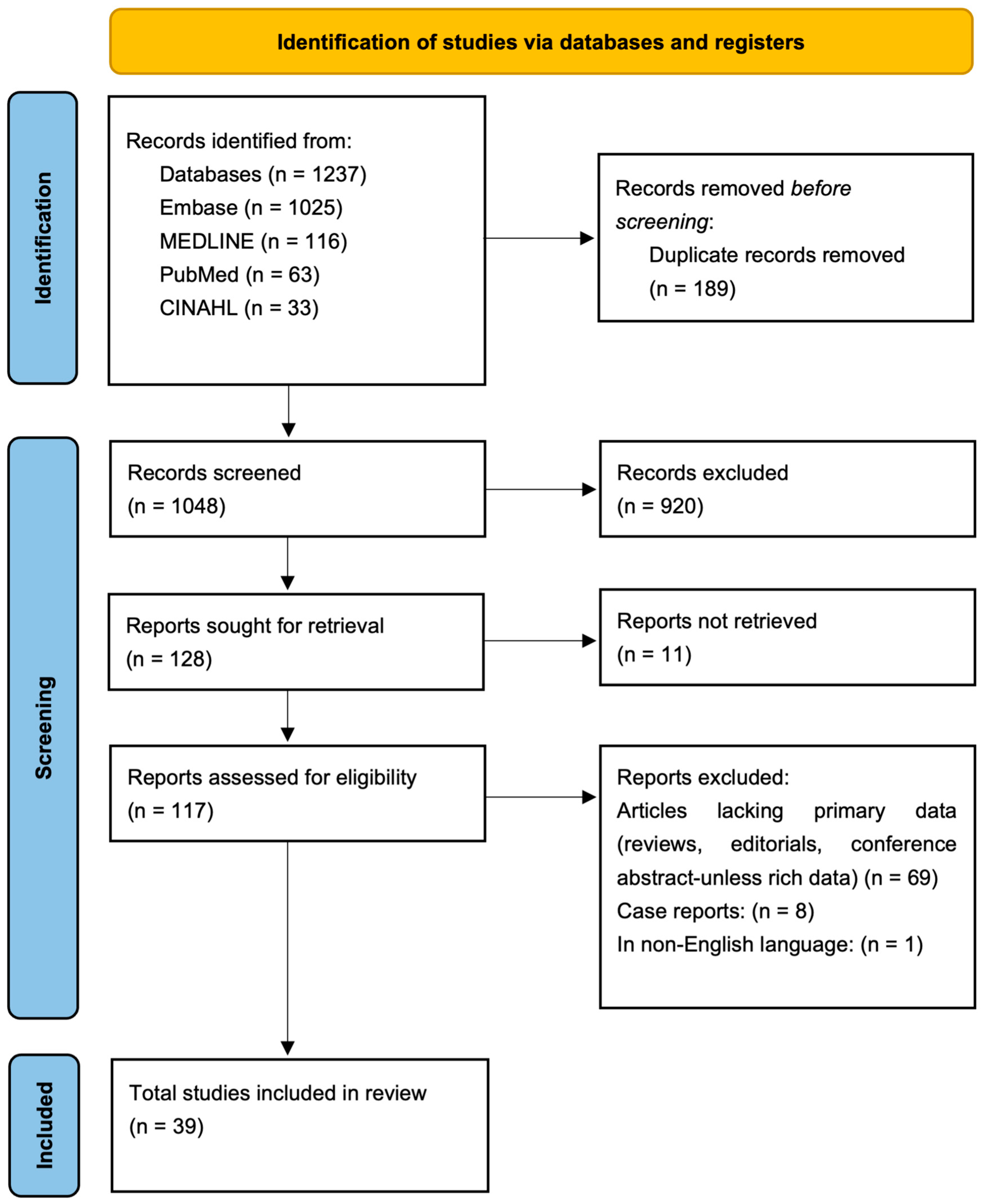
3.1. Selection of Sources of Evidence
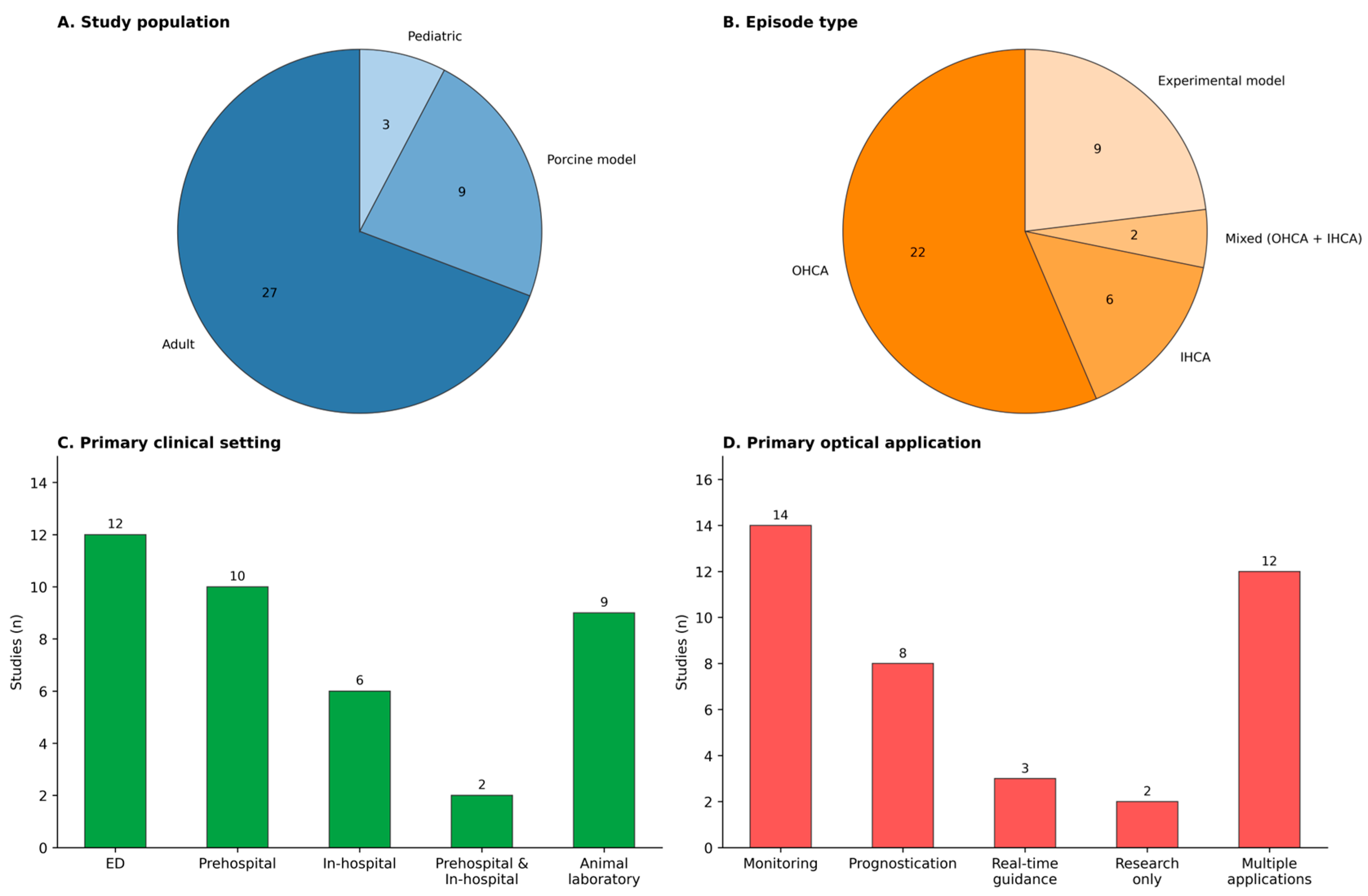
3.2. Characteristics of Sources of Evidence
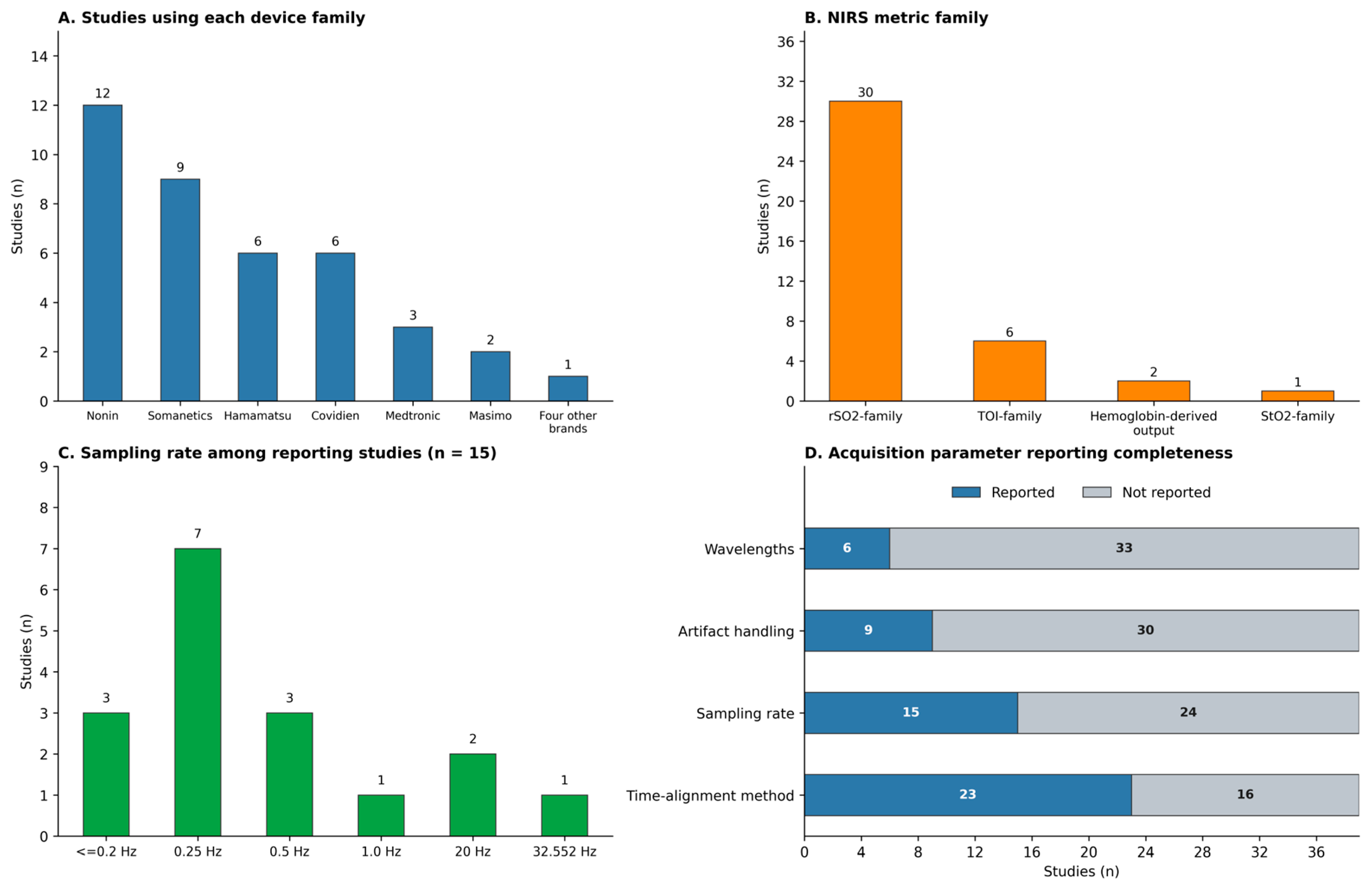
3.3. Instrumentation, Acquisition Parameters, Signal Types, and Reporting Completeness
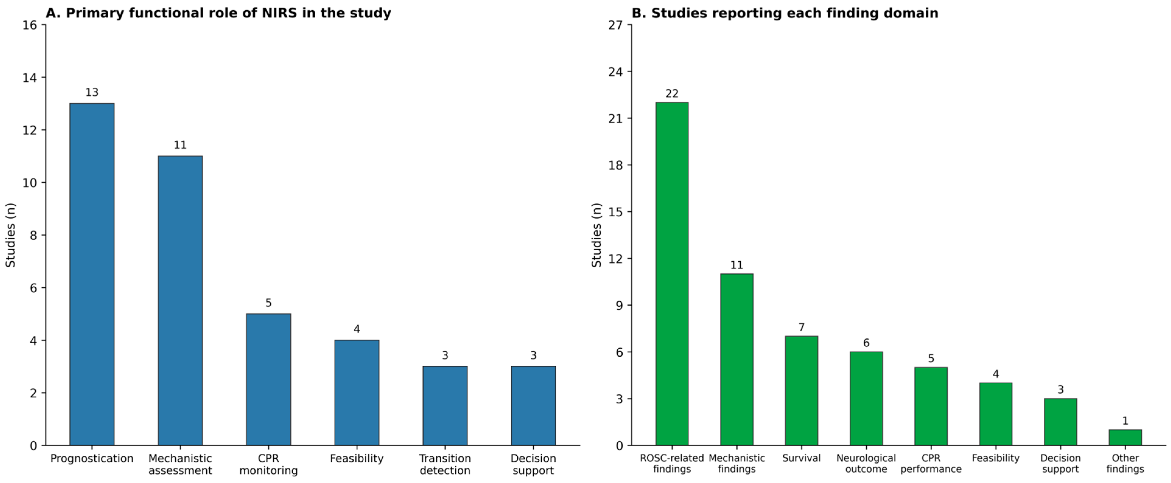
3.4. Physiologic Associations, Thresholds, Quantitative Performance, and Functional Roles
3.5. Clinical Context, Outcomes, and Feasibility Constraints
3.6. Critical Appraisal of Sources of Evidence
3.7. Integrated Synthesis of Evidence
4. Discussion
4.1. Instrumentation, Device Heterogeneity, and Acquisition Reporting
4.2. Physiologic Interpretation, Quantitative Performance, and Clinical Role of NIRS
4.3. Clinical Translation, Feasibility, and Evidence Gaps
4.4. Limitations
4.5. Future Directions
5. Conclusions
Author Contributions
Funding
Institutional Review Board Statement
Informed Consent Statement
Data Availability Statement
Conflicts of Interest
Abbreviations
| ABG | Arterial blood gas |
| ALS | Advanced life support |
| AUC | Area under the curve |
| AVP | Arginine vasopressin |
| BLS | Basic life support |
| BVI | Blood volume index |
| CICU | Cardiac intensive care unit |
| CPC | Cerebral Performance Category |
| CPP | Cerebral perfusion pressure |
| CPR | Cardiopulmonary resuscitation |
| crSo2 | Cerebral regional oxygen saturation |
| CSF | Cerebrospinal fluid |
| DBP | Diastolic blood pressure |
| ECPR | Extracorporeal cardiopulmonary resuscitation |
| ED | Emergency department |
| EMD | Electromechanical dissociation |
| EMS | Emergency medical services |
| ETCO2 | End-tidal carbon dioxide |
| FiO2 | Fraction of inspired oxygen |
| HEMS | Helicopter emergency medical services |
| HHb | Deoxygenated hemoglobin |
| HUP-CPR | Head-up cardiopulmonary resuscitation |
| ICU | Intensive care unit |
| IHCA | In-hospital cardiac arrest |
| IPTW | Inverse probability of treatment weighting |
| JBI | Joanna Briggs Institute |
| MAP | Mean arterial pressure |
| MCCD | Mechanical chest compression device |
| NIRS | Near-infrared spectroscopy |
| OHCA | Out-of-hospital cardiac arrest |
| O2Hb | Oxygenated hemoglobin |
| PaCO2 | Partial pressure of arterial carbon dioxide |
| PaO2 | Partial pressure of arterial oxygen |
| PbtO2 | Partial pressure of brain tissue oxygen |
| PCC | Population–Concept–Context |
| PEA | Pulseless electrical activity |
| PED | Pediatric emergency department |
| PICU | Pediatric intensive care unit |
| POHCA | Pediatric out-of-hospital cardiac arrest |
| ROSC | Return of spontaneous circulation |
| rSO2 | Regional cerebral oxygen saturation |
| SctO2 | Cerebral tissue oxygen saturation |
| ScvO2 | Central venous oxygen saturation |
| StO2 | Tissue oxygen saturation |
| SWT | Stationary wavelet transform |
| THb | Total hemoglobin |
| THI | Tissue hemoglobin index |
| TOI | Tissue oxygenation index |
| VF | Ventricular fibrillation |
| VT | Ventricular tachycardia |
| ΔcHb | Change in total hemoglobin |
| ΔHHb | Change in deoxygenated hemoglobin |
| ΔO2Hb | Change in oxygenated hemoglobin |
| ΔTOI | Change in tissue oxygenation index |
Appendix A

| Article First Author/Year | Study Design | Optical Application | Setting | Episode Type | Witnessed | By Stander CPR | Initial Rhythm | Population | Age (Years) | Sex Male (%) | Sample Size |
|---|---|---|---|---|---|---|---|---|---|---|---|
| Francoeur 2022 [35] | Prospective single-centre observational pilot study | Monitoring; Prognostication (research only) | In-hospital (PICU/CICU/ED) | IHCA; OHCA | Not reported | Not reported | Ventricular Fibrillation (VF), pulseless Ventricular Tachycardia (VT), Asystole, Pulseless electrical activity (PEA) | Pediatric | Median 1.67 (0.42–14) | 42.9 | 23 |
| Shin 2022 [14] | Prospective observational cohort study | Prognostication | Prehospital | OHCA | 49% | 67% | Asystole, PEA, VF/VT | Adult | Mean 64 (SD 16) | 56 | 59 |
| Nelskylä 2023 [49] | Experimental animal study | Monitoring (research only) | Animal laboratory | Not applicable (experimental model) | Not applicable | Not applicable | shockable (induced VF) | Animal | 14–18-week landrace | Not reported | 28 |
| Tsukuda 2021 [21] | Prospective observational cohort pilot study | Monitoring; Prognostication | Prehospital (ambulance transport) | OHCA | 61.5% (ROSC); 16.4 (non-ROSC) | 38.5% (ROSC); 42.9% (non-ROSC) | Shockable | Adult | Mean 72.4 (ROSC); 80.8 (non-ROSC) | 73.1% (ROSC); 57.1% (non-ROSC) | 81 |
| Yazar 2019 [46] | Observational preliminary study | Monitoring; Prognostication | In-hospital (ICU) | IHCA | Not reported | Not reported | Not reported | Adult | Mean age 72.6 ± 4.2 (survivors); 77.3 ± 6.5 (no survivors) | 40 | 20 |
| Nelskylä 2017 [48] | Randomized experimental animal study | Monitoring | Animal laboratory | Not applicable (experimental model) | Not applicable | Not applicable | Shockable | Animal | Not reported | Not reported | 19 |
| Kishihara 2022 [43] | Prospective observational cohort study | Real-time guidance (quality indicator for chest compressions) | ED | OHCA | 70.3% | 32.4% yes | VF/pulseless VT, PEA, asystole | Adult | Median 75 (69–82) | 83.8 | 37 |
| Nelskylä 2022 [19] | Prospective observational cohort study | Monitoring | Prehospital (physician-staffed helicopter service) | OHCA | Not reported | 71% | Shockable; non-shockable | Adult | Median 68 (59–73) | 73 | 75 |
| Takegawa 2021 [22] | Prospective multicentre interventional study with historical control cohort | Real-time guidance (rSO2-guided continuous chest compressions without rhythm checks) | ED | OHCA | 42% (94) | 55% (123) | Non-shockable (PEA and asystole) | Adult | Median 77 (70–83) | 57 | 225 |
| Schewe 2014 [38] | Prospective observational feasibility cohort study | Monitoring (research only) | Prehospital | OHCA | Not reported | Not reported | Shockable (VF), non-shockable (asystole) | Adult | Mean 73 ± 13 | 80 | 10 |
| Baloglu Kaya 2021 [12] | Prospective randomized clinical trial | Monitoring; Prognostication | ED | IHCA | 100% | Not reported | Asystole, PEA, VF/pulseless VT | Adult | MCCD: 71.85 ± 13.46; Manual: 71.37 ± 13.46 | MCCD: 62.5; Manual: 62.9 | 75 |
| Storm 2016 [34] | Prospective observational cohort study | Prognostication (research only) | Prehospital; in-hospital | OHCA | Not reported | Not reported | VF/Asystole/EMD | Adult | Mean 61 (non- ROSC), 66 (pre-ROSC), 68 (post ROSC) | Post ROSC 100%; Pre ROSC 80%; No ROSC 78% | 29 |
| Tsukuda 2019 [40] | Prospective observational cohort study | Prognostication; Real-time guidance | ED | OHCA | 51.3% | 40.2% yes | Shockable 11.1% | Adult | Mean 69.7 ± 17.3 | 57.3 | 117 |
| Asim 2014 [42] | Observational cohort/Not reported | Monitoring; Prognostication | ED | OHCA | No | Not reported | VF, Asystole, PEA | Adult | Mean 64.09 ± 13.66 | 47.8 | 23 |
| Singer 2015 [13] | Retrospective observational study | Monitoring; Prognostication | ED | OHCA | Not reported | Not reported | VF/VT, PEA, asystole | Adult | Mean 68.7 ± 14.9 | 84.70% | 59 |
| Kalkan 2015 [25] | observational study | Prognostication | ED | OHCA | No | Not reported | VF, PEA, asystole | Adult | Mean 63.06 ± 11.66 | 50% | 34 |
| Meex 2013 [7] | Observational feasibility study | Monitoring (research only) | Prehospital; in-hospital | OHCA; IHCA | Not reported | Not reported | Not reported | Adult | Mean 66 ± 20 | 71.40% | 14 |
| Prosen 2018 [36] | Prospective observational study | Monitoring (research only) | Prehospital | OHCA | 90% (ROSC); 90% (no ROSC) | 5% (ROSC); 13% (no ROSC) | VF/VT, PEA, asystole | Adult | Mean age 65; Median 70.5 (ROSC) vs. 67.0 (non-ROSC) | 84% (overall); 81% (ROSC), 87% (no ROSC) | 53 |
| Genbrugge 2018 [39] | Prospective non-randomized multicenter study | Monitoring; Prognostication | Prehospital | OHCA | 74% (ROSC); 51% (no ROSC) | 41% (ROSC); 39% (no ROSC) | Asystole, VF, PEA | Adult | Mean 68 ± 14 (ROSC) vs. 69 ± 15 (non-ROSC) | 65% (ROSC); 73% (no ROSC) | 329 |
| Al-Subu 2020 [23] | Prospective experimental animal study | Monitoring | Animal laboratory | Not applicable (experimental model) | Not applicable | Not applicable | Shockable (VF) | Animal | 2–3 months | Not reported | 8 swine, 28 VF arrests |
| Duvekot 2015 [58] | Prospective single-centre observational cohort study | Prognostication (research only) | ED | OHCA | Not reported | Not reported | VF 57% | Adult | Mean 64 | 63 | 46 |
| Frisch 2012 [29] | Case series | Monitoring; Real-time guidance (research only) | Prehospital | OHCA | 3 of 5 cases | 100% | Not reported | Adult | 84, 76, 81, 83, 61 | 20 | 5 |
| Deakin 2016 [51] | Prospective cohort study | Research only | In-hospital | IHCA | Yes | Not reported | Asystole, PEA, VF/VT | Adult | Median 77 | 58.30% | 36 |
| Putowski 2025 [27] | Prospective single-centre observational cohort study | Prognostication; Monitoring | In-hospital | IHCA | Not reported | Not reported | Asystole, PEA, VF/VT | Adult | Mean 68 (SD 12) | 63.5 | 104 |
| Sanz-Pescador 2024 [30] | Observational cohort study | Real-time guidance (research only) | Prehospital | OHCA | Not reported | Not reported | Not reported | Adult | Not reported | Not reported | 30 |
| Pourzand 2024 [26] | Randomized pre-clinical experimental animal study | Monitoring | Animal research laboratory | Not applicable (experimental model) | Not applicable | Not applicable | Shockable (VF) | Animal | Not reported | Not reported | 22 |
| Raymond 2024 [31] | Multicenter observational study | Prognostication | In-hospital | IHCA | Not reported | Not reported | Not reported | Pediatric | Median 0.3 (0.1–1.4) | 56 | 123 |
| Koyama 2023 [50] | Experimental animal study | Monitoring | Animal research laboratory | Not applicable (experimental model) | Not applicable | Not applicable | VF-CA; A-CA | Animal | Median 67.5 days (VF-CA group); 76.0 days (A-CA group) | 0 | 20 |
| Jang 2023 [37] | Single-center observational cohort | Prognostication | Prehospital to ED | OHCA | 84.6% (ROSC); 43.6% (non-ROSC) | 61.5% (ROSC); 48.7% (non-ROSC) | Shockable | Adult | Median 55 (ROSC); 72 (non-ROSC) | 84.6% (ROSC); 59.0% (non-ROSC) | 52 |
| Košir 2023 [24] | Single-center observational cohort | Monitoring; Prognostication | Prehospital | OHCA | 50% of cases | 70% yes | VF, asystole, PEA | Adult | Median 66.0 (60.5–79.5) | 65 | 20 |
| Koyama 2013 [5] | Non-consecutive observational case series | Monitoring; Prognostication; Real-time guidance | ED | OHCA | Not reported | Not reported | VF, PEA, Asystole | Adult | Mean 79 (55–99) | 66 | 15 |
| Parnia 2012 [10] | Feasibility (pilot study) | Prognostication | In-hospital | IHCA | Not reported | Not reported | VF, Asystole, PEA | Adult | Mean 76 ± 15 (survivors); 73 ± 11 (non-survivors) | Not reported | 19 |
| Bein 2006 [47] | Experimental animal study | Monitoring | Animal research laboratory | Not applicable (experimental model) | Not applicable | Not applicable | VF | Animal | 12 to 16 weeks | Not reported | 12 |
| Bouček 2018 [9] | Prospective experimental animal study | Monitoring | Animal research laboratory | Not applicable (experimental model) | Not applicable | Not applicable | VF | Animal | 16–20 weeks | 0% (all female) | 24 |
| Putzer 2016 [44] | Experimental animal study | Monitoring | Animal research laboratory | Not applicable (experimental model) | Not applicable | Not applicable | VF | Animal | 12 to 16 weeks | Not reported | 9 |
| Lennmyr 2010 [28] | Experimental animal study | Monitoring | Animal research laboratory | Not applicable (experimental model) | Not applicable | Not applicable | shockable (VF) | Animal | Not reported | Not reported | 17 |
| Abramo 2014 [45] | Case series | Monitoring; Prognostication; Real-time guidance; | ED (pediatric emergency department) | OHCA | Not reported | yes | Case 1: VF; Case 2: asystole, VF | Pediatric | Case 1: 15-year-old; Case 2: 14-year-old | Case 1 male; Case 2 female | 2 |
| Kim 2022 [20] | Prospective interventional pilot study | Research only | ED | OHCA | 50% of cases | 53.6 | Shockable 10.7% | Adult | 80.5 (71.5–84.0) | 57.10% | 28 |
| Ito 2014 [41] | Prospective multicenter observational cohort study | Prognostication | ED | OHCA | 53% of cases | 39 | VF/VT, Asystole, PEA | Adult | Mean 71 | 60% | 672 |
| First Author/Year | Brand/Model | Wavelengths (nm) | Number of Sensors | Sampling Rate (Hz) | Wired/Wireless | Sensor Site |
|---|---|---|---|---|---|---|
| Francoeur 2022 [35] | Equanox 7600 (Nonin Medical, Plymouth, MN, USA) | Not reported | ≥1 | 0.25 | Wired | Cerebral (forehead) |
| Shin 2022 [14] | SenSmart Model X-100 Universal Oximetry System (Nonin Medical, Inc.) | Not reported | 1 | Not reported | Wired | Cerebral (left forehead) |
| Nelskylä 2023 [49] | INVOS 5100C Cerebral Oximeter (Somanetics Inc., Troy, MI, USA) | Not reported | Not reported | Not reported | Wired | Cerebral (left forehead) |
| Tsukuda 2021 [21] | CCR-1 (Hamamatsu Photonics, Hamamatsu-City, Shizuoka, Japan) | Not reported | 1 | Not reported | Wired | Cerebral forehead left-lateral above eyebrow |
| Yazar 2019 [46] | INVOS 5100C (Somanetics, Troy, MI, USA) | Not reported | 2 | Not reported | Wired | Cerebral, bilateral forehead |
| Nelskylä 2017 [48] | INVOS 5100C (Somanetics Inc., Troy, MI, USA) | Not reported | 1 | Not reported | Wired | Cerebral left forehead |
| Kishihara 2022 [43] | INVOS 5100C (Medtronic, Boulder, CO, USA) | Not reported | 2 | Not reported | Wired | Cerebral left and right forehead |
| Nelskylä 2022 [19] | SenSmart X-100 (Nonin Inc., Plymouth, MN, USA) | Not reported | 2 | 0.25 | Wired | Cerebral, bilateral forehead |
| Takegawa 2021 [22] | TOS-QQ® brain oximeter (TOSTEC Co., Ltd., Tokyo, Japan) | Not reported | Not reported | Not reported | Not reported | Cerebral forehead |
| Schewe 2014 [38] | Equanox Model 7600 (Nonin Medical, Plymouth, MN, USA) | Not reported | 1 | 0.25 | Wired | The left frontal forehead lateral to midline and above the eyebrow |
| Baloglu Kaya 2021 [12] | Root and O3™ Regional Oximeter (Massimo, Irvine, CA, USA) | Not reported | 2 | 0.5 | Wired | Cerebral, bilateral forehead |
| Storm 2016 [34] | INVOS 5100C (Covidien; Mansfield, MA, USA) | 724, 810 | 2 | Not reported | Wired | Cerebral, bilateral forehead |
| Tsukuda 2019 [40] | NIRO-200NX (Hamamatsu Photonics, Hamamatsu-City, Shizuoka, Japan) | Not reported | 2 | Not reported | Wired | Cerebral bilateral supraorbital |
| Asim 2014 [42] | INVOS 5100C (Covidien, Boulder, CO, USA) | Not reported | 2 | Not reported | Wired | Cerebral; bilateral frontal |
| Singer 2015 [13] | Equanox Advance monitor (Nonin Medical Inc., Plymouth, MN, USA) | Not reported | 1 | 0.25 | Wired | Cerebral forehead |
| Kalkan 2015 [25] | INVOS 5100C (Covidien, Boulder, CO, USA) | Not reported | 4 | Not reported | Wired | Bilateral cerebral; bilateral abdominal |
| Meex 2013 [7] | FORE-SIGHT monitor (CAS Medical Systems, Branford, CT, USA); Equanox Advance monitor (Nonin Medical Inc., Plymouth, MN, USA) | Not reported | 2 (FORE-SIGHT); 1 (Equanox) | Not reported | Wired | FORE-SIGHT bilateral forehead; Equanox unilateral forehead (right forehead) |
| Prosen 2018 [36] | INVOS Oximeter (Somanetics Corporation, Troy, MI, USA) | Not reported | 2 | Not reported | Wired | Cerebral, bilateral forehead |
| Genbrugge 2018 [39] | Equanox 7600 or SenSmart X-100 (Nonin Medical Inc., Plymouth, MN, USA) | Not reported | 1 | 0.25 | Wired | Right forehead |
| Al-Subu 2020 [23] | Somatic INVOS 5100C (Somanetics Corpora-tion, Troy, MI, USA) | Not reported | 2 | Not reported | Wired | Left forehead (cerebral) and left flank over kidney (somatic/renal) |
| Duvekot 2015 [58] | NIRS (Covidien, The Netherlands) | Not reported | 1 | Not reported | Not reported | Cerebral (forehead) |
| Frisch 2012 [29] | InSpectra StO2 Tissue Oxygenation Monitor (Hutchinson Technology, Hutchinson, MN, USA) | Not reported | 1 | Not reported | Wired | Thenar eminence (hand) |
| Deakin 2016 [51] | Equanox Model 7600 (Nonin Medical, Plymouth, MN, USA) | Not reported | 1 | 1 per 6 s | Wired | Cerebral (forehead) |
| Putowski 2025 [27] | Masimo Open Connect (MOC-9) (Masimo, Irvine, CA, USA) | Not reported | Not reported | 0.5 | Wired | Cerebral |
| Sanz-Pescador 2024 [30] | NIRO-200NX (Hamamatsu Photonics, Hamamatsu, Japan) | Not reported | 1 | 20 | Wired | Cerebral (left frontal lobe) |
| Pourzand 2024 [26] | SenSmart X-100 (Nonin Medical Inc., Plymouth, MN, USA) | Not reported | Not reported | Not reported | Wired | Cerebral |
| Raymond 2024 [31] | INVOS 5100C (Medtronic, Minneapolis, MN, USA); Equanox 7600 (Nonin Medical, Plymouth, MN, USA) | Not reported | 1 (INVOS); 1 (Equanox) | 0.25 (INVOS); 1 (Equanox) | Wired | Cerebral (single sensor on either forehead) |
| Koyama 2023 [50] | NIRO 200NX (Hamamatsu Photonics, Hamamatsu-shi, Shizuoka, Japan) | Not reported | 2 | Not reported | Wired | Cerebral bilateral anterior to coronal suture |
| Jang 2023 [37] | INVOS 5100C (Covidien, Boulder, CO, USA) | Not reported | 2 | Not reported | Wired | Cerebral, bilateral forehead |
| Košir 2023 [24] | SenSmart X-100 (Nonin Medical, Inc., Playmouth, MN, USA) | Four wavelengths | 2 | 0.25 | Wired | Cerebral right forehead and somatic right thenar |
| Koyama 2013 [5] | NIRO (Hamamatsu Photonics, Hamamatsu-shi, Shizuoka, Japan) | 700–1300 | 2 | 0.5 recalibrated to 20 | Wired | Cerebral, bilateral forehead |
| Parnia 2012 [10] | Invos (Somanetics, Troy, MI, USA) | Not reported | 2 | Not reported | Wired | Cerebral (left or right forehead) |
| Bein 2006 [47] | NIRO 300 (Hamamatsu Photonics, Hamamatsu City, Japan) | 775, 810, 850, 910 | 2 | 0.2 | Wired | Cerebral forehead |
| Bouček 2018 [9] | INVOS 5100C (Medtronic Inc., Minneapolis, MN, USA) | 650–900 | 2 | Not reported | Wired | Two INVOS infant-neonatal probes; cerebral probe on forehead (middle of eyes, over cerebrum); peripheral probe on left thigh (over a muscle) |
| Putzer 2016 [44] | INVOSs (Somanetics Inc., Troy, MI, USA) | Not reported | 1 | Not reported | Wired | Cerebral, right forehead |
| Lennmyr 2010 [28] | INVOS (Somanetics Inc., Troy, MI, USA) | Not reported | 1 | Not reported | Wired | parietal skull |
| Abramo 2014 [45] | NIR INVOS 5100v (Somanetics Inc., Troy, MI, USA) | Not reported | 2 | 0.2 | Wired | Cerebral, bilateral forehead |
| Kim 2022 [20] | NIRSIT ON (OBELAB Inc., Seoul, Republic of Korea) | 780, 850 | 2 | 32.552 | Wireless | Cerebral, bilateral forehead |
| Ito 2014 [41] | INVOS 5100C (Covidien, Boulder, CO, USA) | 730, 805 | 2 | Not reported | Wired | Cerebral, bilateral forehead |
| First Author/Year | Metric Family | Timing Window | Compression Context | Compression Ratio | Time Alignment Method | Artifact Handling Methods | Data Loss (%) | Software/Toolbox | Exposure of Interest/NIRS Feature |
|---|---|---|---|---|---|---|---|---|---|
| Francoeur 2022 [35] | rSO2 | During CPR | Not reported | Not reported | Not reported | Manual artifact removal plus noise reduction filters. | Not reported | MATLAB custom for DBP; REDCap | Mean/median value |
| Shin 2022 [14] | rSO2 | During compressions; pre-ROSC; Early post ROSC ≤ 5 min | Not reported | Not reported | Synchronized via defibrillator time stamps | Not reported | Not reported | Not specified | Initial value; Mean/median value |
| Nelskylä 2023 [49] | rSO2 | during pre-arrest; untreated VF; 5 min of ineffective manual chest compressions; mechanical CPR; after ROSC. | Manual and Mechanical | Poor CPR: 82/min (FiO2 50%); 79/min (FiO2 100%); Mechanical CPR: 101/min | Narrative | Not reported | n = 1 (50% group); n = 1 (100% group) | MPR2 logO Dat-alogger, SPSS 27.0.1.0 | Mean/median value; Change/trajectory (Δ) |
| Tsukuda 2021 [21] | TOI; O2Hb; HHb | During pre-hospital CPR, ambulance transportation, initial TOI at probe attachment in ambulance, final TOI at arrival to ED | Manual | Not reported | Not reported | Not reported | Not reported | SPSS; R | Change/trajectory (Δ) |
| Yazar 2019 [46] | rSO2 | During compressions; Early post ROSC ≤ 10 min | Manual | Not reported | Not reported | Not reported | Not reported | SPSS (analysis) | Mean/median value; Maximum value; |
| Nelskylä 2017 [48] | rSO2 | Pre ROSC (VF period prior to CPR); during compressions (CPR); early post ROSC (20 min after ROSC) | Mechanical chest compressions (LUCAS); ventilation by manual bag valve ventilation | Manual bag valve ventilation at 10/min | Synchronized to invasive PbO2 | Not reported | Not reported | Not reported | Mean/median value; Change/trajectory (Δ) |
| Kishihara 2022 [43] | rSO2 | During compressions, 0 to 15 min after arrival | Not reported | Not reported | Narrative only (measurements recorded at predefined minute marks: 0, 3, 6, 9, 12, 15 min) | Not reported | n = 4 | EZR version 1.38; R version 3.5.2; SAS version 9.4 (analyses) | Median value; log-transformed value |
| Nelskylä 2022 [19] | rSO2 | During CPR compressions | Manual and mechanical (LUCAS) continuous with supraglottic airway | Continuous | Device and defibrillator clocks synchronized weekly | Not reported | n = 4 | Prism 9.0 (analysis) | median value; Change/trajectory (Δ) |
| Takegawa 2021 [22] | rSO2 | During compressions: Continuous CC up to 16 min or until rSO2 target achieved | Manual and mechanical | Continuous | Not reported | Not reported | Not reported | R software version 3.6.3 (statistical analysis) | Mean/median/max value; baseline value |
| Schewe 2014 [38] | rSO2 | During CPR | Manual vs. Mechanical | Not reported | Device time stamps and manual event markers for ROSC, use of mechanical compression device, and termination of CPR | Not reported | 10.2 | Microsoft Excel | Change/trajectory (Δ); waveform features |
| Baloglu Kaya 2021 [12] | rSO2 | During compressions | Manual vs. Mechanical (LUCAS2) | Not reported | Not reported | The higher one of the two rSO2 values was used for analysis. | Not reported | IBM SPSS Statistics 21.0; MedCalc 19 | Mean/median value |
| Storm 2016 [34] | rSO2 | During compressions; near ROSC (within 2 min after ROSC) | Not reported | Not reported | Narrative (during CPR or within 2 min post ROSC) | None reported | Not reported | INVOS Analytics Tool, version 1.2 | Initial value |
| Tsukuda 2019 [40] | TOI | During compressions | Manual | Not reported | Narrative | Not reported | n = 21 | SPSS (V. 25) and R statistical software (V. 1.0.143) | Initial value |
| Asim 2014 [42] | ScO2 | During compressions | Manual | Not reported | Narrative | Not reported | Not reported | SPSS 18.00 | Change/trajectory (Δ) |
| Singer 2015 [13] | rSO2 | during CPR | Manual and mechanical | Not reported | Narrative (from time sensor placed to ROSC or CPR termination) | Artifact values recognized by absent value or values at least three SDs away from mean rSO2. | Not reported | Not reported | Mean/median value |
| Kalkan 2015 [25] | ScO2; rSO2 | during CPR | Manual | Not reported | Not reported | Not reported | Not reported | SPSS 18.00 (statistical analysis) | Change/trajectory (Δ) |
| Meex 2013 [7] | SctO2 | during CPR; at/near ROSC | Not reported | Not reported | Not reported | Protective adhesive tape used to minimize external light interference. | Not reported | SPSS V19.0 | Initial/Maximum value; Change/trajectory (Δ) |
| Prosen 2018 [36] | rSO2 | during CPR; at/near ROSC; early post-ROSC | Manual | Not reported | Defibrillator timestamps | Not reported | 8.62 | SigmaPlot software (Stat Co, Version 11.0) | Initial value; Change/trajectory (Δ) |
| Genbrugge 2018 [39] | rSO2 | During compressions (prehospital ALS); pre-ROSC; at/near ROSC | manual or mechanical (LUCAS/AutoPulse) | Not reported | Event button; ROSC time noted on Utstein forms | Not reported | 12.71 | SPSS 22.00; GraphPad Prism 5.01 (data downloaded per manufacturer instructions) | Change/trajectory (Δ) |
| Al-Subu 2020 [23] | rSO2 | Pre-arrest; during CPR; after ROSC | Manual | ~100/min | Not reported | Not reported | Not reported | SAS | Change/trajectory (Δ) |
| Duvekot 2015 [58] | TOI | During compressions | Not reported | Not reported | Narrative | Not reported | Not reported | IBM SPSS 20 | Initial value |
| Frisch 2012 [29] | StO2 | During compressions; at/near ROSC; pre-ROSC; early post-ROSC during transport to hospital (exact duration not reported) | Not reported | Not reported | Narrative | Not reported | Not reported | Not reported software | Change/trajectory (Δ) |
| Deakin 2016 [51] | rSO2 | During CPR | Not reported | Not reported | Event button on oximeter | Outlier removal (rSO2 values > 3 SD from mean removed) | n = 1 | SAS version 9.3 (analysis); REDCap used for data entry | Initial value; Mean/median value; Change/trajectory (Δ) |
| Putowski 2025 [27] | rSO2 | During compressions; near ROSC; post-ROSC < 24 h | Mechanical | Not reported | Not reported | Not reported | Maximum of 10% per patient | SPSS version 29.0; R; MedCalc | Mean/median/max value; Change/trajectory (Δ) |
| Sanz-Pescador 2024 [30] | ∆cHb | During compressions (segments during CC series) | Not reported | Not reported | Thoracic impedance synchronized | Stationary wavelets transform (SWT); Kurtosis-based quality control | Not reported | MATLAB custom GUI | Waveform features |
| Pourzand 2024 [26] | rSO2 | Measurements at baseline (pre-VF), end of 15 min of untreated VF, during CPR at 10 and 24 min, and at 15 and 60 min after ROSC (during early post ROSC ≤ 1 h) | Mechanical | 105/min with 10:1 compression to ventilation ratio | Not reported | Not reported | Not reported | BioPac (acquisition only); SPSS version 26 | Mean Value/Change/trajectory (Δ) |
| Raymond 2024 [31] | crSO2 | During compressions (entire event); first 5 min; last 5 min | Not reported | Not reported | Not reported | Not reported | Not reported | R, R stats, and pROC package Version 4.2.2 | Mean/median value |
| Koyama 2023 [50] | TOI | Before, during compressions; after CPR | Mechanical (LUCAS 2) | Asynchronous ventilation 10 breaths/min | Synchronized across monitors | Not reported | Not reported | EZR 1.52 on R | Median value; Change/trajectory (Δ) |
| Jang 2023 [37] | rSO2 | During compressions; first 5 min; first 10 min; entire CPR | Manual | Not reported | Not reported | Not reported | Not reported | SPSS 18.0; Med-Calc 12.7.7.0 | Initial value; Max/Min/Mean value; Change/trajectory (Δ) |
| Košir 2023 [24] | rSO2 | During compressions | Not reported | Not reported | Narrative (paired with intervention protocol). | Spiking signals removed as artifacts. | n = 5 | SenSmart v1.0.1.0 (Nonin); MedCalc ver. 20.104 | Initial value; Maximum value; Change/trajectory (Δ); End-CPR value |
| Koyama 2013 [5] | ΔcHb; TOI | During compressions | Manual | Not reported | Not reported | Not reported | Not reported | Not reported | waveform features; Change/trajectory |
| Parnia 2012 [10] | rSO2 | During compressions | Not reported | Not reported | Not reported | Not reported | n = 4 | PRISM 6 | Mean value; Change/trajectory (Δ) |
| Bein 2006 [47] | TOI; THI | Pre ROSC (VF then CPR) and during compressions; at/near ROSC and early post ROSC (followed for 1 h after resuscitation) | Not reported | 100/min | Narrative | Values were aver-aged over 30 s. | Not reported | SPSS | Change/trajectory (Δ) |
| Bouček 2018 [9] | rSO2 | During compressions (five minutes of CPR); comparisons used mean rSO2 over 15 min baseline, 3rd minute of untreated CA, and five minutes of CPR | Mechanical compressions (LUCAS 2) | Not reported | Narrative (baseline vs. untreated CA vs. CPR periods) | Not reported | <10% | MedCalc 18 | Mean/median value; Change/trajectory (Δ) |
| Putzer 2016 [44] | rSO2 | During compressions, includes periods before and after a CPR interruption and after adrenaline administration during ongoing CPR | Mechanical chest compression (LUCAS2TM) | Not reported | Protocol-defined time points relative to CA/CPR phases and adrenaline administration | Not reported | Not reported | SPSS 20 | Mean value; Change/trajectory (Δ) |
| Lennmyr 2010 [28] | rSO2 | During resuscitation | Mechanical chest compressions (LUCAS) | 100 cpm | Narrative/structured timepoints | Not reported | Not reported | Workbench 3.0 (Strawberry Tree Inc.) for acquisition; Microsoft Excel 2007 and Prism 4.0 for statistics | Trajectory |
| Abramo 2014 [45] | rSO2; Blood Volume Index (BVI) | Case 1 during chest compressions; Case 2 postarrest in PED | Not reported | Not reported | Not reported | Not reported | Not reported | Not reported | Not reported |
| Kim 2022 [20] | O2Hb; HHb; THb; | During compressions | Mechanical | Not reported | Narrative | Removed detached/zero/spiking segments; excluded recordings with >70% poor-quality data. | 14.28 | R-package software, version 4.0.5 | Maximum value; Change/trajectory (Δ) |
| Ito 2014 [41] | rSO2 | During resuscitation upon hospital arrival | Not reported | Not reported | Not reported | Not reported | n = 1 | JMP 10.0.0, MedCalc 12.3.0, STATA 11.1 (analysis) | Initial value; minimum value at hospital arrival |
| First Author Year | Aim | Primary Finding | Main Finding | Feasibility Notes | Notes/Limitations |
|---|---|---|---|---|---|
| Francoeur 2022 [35] | To examine whether higher rSO2 values recorded during arrest were linked to achieving ROSC and surviving to hospital discharge. | ROSC; Survival | During pediatric in-hospital cardiac arrest, higher intra-arrest cerebral rSO2, particularly higher median values and a greater proportion of time with rSO2 above 50% in the final five minutes of CPR, was associated with ROSC, but these rSO2 metrics did not translate into improved survival to hospital discharge. | The NIRS device was brought in by a respiratory therapist who was not directly involved in the resuscitation; at least one cerebral probe was placed quickly on the forehead, any probe already present for clinical care was left in place, and the monitor was positioned outside the resuscitation field and out of view of the CPR team. | The study enrolled a small convenience cohort and applied NIRS only to patients who remained in arrest long enough for probe placement during CPR, which likely under-sampled brief arrests in which ROSC occurred before monitoring could be initiated. |
| Shin 2022 [14] | To characterize early and ongoing rSO2 trajectories during first-responder resuscitation and evaluate how these patterns relate to ROSC and survival with favourable functional outcome. | ROSC; Survival; Neurological outcome | rSO2 was low at the onset of resuscitation (mean 41%) and rose during first-responder CPR; within the first five minutes, rSO2 values were higher in patients who subsequently achieved ROSC, and early post-ROSC rSO2 levels were higher among survivors with favourable neurological outcome (CPC 1–2). | EMS providers received 45 min of hands-on training; the sensor was applied to the left forehead after first-responder CPR had started, the screen was covered to blind EMS providers, and the initial three-person first-responder team was able to deploy the oximeter relatively early. | EMS providers were blinded to cerebral oximetry readings, so oximetry was not used to guide treatment decisions. The modest sample size limited more advanced inferential analyses, and early application may reduce generalizability. |
| Nelskylä 2023 [49] | To compare ventilation using 100% versus 50% oxygen during ineffective manual chest compressions and assess whether the higher oxygen fraction improves cerebral oxygenation. | Physiologic comparator; Oxygen-fraction effect | Comparing oxygen fractions during ineffective manual chest compressions, ventilation with 100% FiO2 increased brain tissue oxygen tension (PbtO2) relative to 50% FiO2 but did not meaningfully alter cerebral rSO2; during mechanical CPR, PbtO2 and rSO2 were similar with FiO2 50% and 100%. | Not reported | The use of an experimental pig model under general anesthesia may not fully reflect human cardiac arrest, as the animals were a homogeneous group of young, healthy pigs, and the study was not blinded. NIRS measurements may also have been influenced by anatomical differences in skull and skin thickness, as well as by extracerebral vasoconstriction induced by adrenaline administration. |
| Tsukuda 2021 [21] | To examine the association between TOI and ROSC. | ROSC | During pre-hospital manual CPR for OHCA, larger increases in tissue oxygenation index (ΔTOI) during ambulance transport strongly predicted ROSC and were positively correlated with higher chest compression rates. ΔTOI thresholds in the range of approximately +5–8% discriminated episodes with ROSC, whereas decreases of ≤−2% were associated with non-ROSC. | Inside the ambulance, one of three trained paramedics applied the probe to the left forehead to minimize interruptions to CPR; the device was portable and battery-operated for two hours, but only five EMS teams were equipped. Apparatus dysfunction occurred in 19 of 104 patients (attachment failure or start-up delay), and 23 of 81 patients did not meet guideline chest compression rates due to the narrow ambulance space; paramedics were not educated on the meaning of NIRS values and did not adjust CPR based on TOI. | This was a single-center pilot with a small sample, and only five EMS teams were equipped with NIRS devices; numerous data errors were attributed to probe performance, and ΔTOI cut-offs were specific to this study. The NIRS device did not measure chest compression depth, and analyses were limited to mean compression rate and its correlation with ΔTOI, with limited neurological outcome data and could not evaluate the change in PaO2 during transport. |
| Yazar 2019 [46] | To assess how effectively chest compressions support cerebral oxygenation during ongoing CPR. | ROSC; Survival; Neurological outcome | Maximum rSO2 values during resuscitation were higher in patients who achieved ROSC than in non-survivors, and both the minimum and mean rSO2 during CPR were positively correlated with mean FOUR scores at one week among survivors. | Study-related procedures did not interfere with routine CPR practice. | The cohort was small, and the authors noted that technical monitoring in ICUs is complex, limiting the scope of the study’s conclusions. Additional illnesses may have influenced post-resuscitation FOUR scores, and the authors could not assess the effect of disease on FOUR. |
| Nelskylä 2017 [48] | To test whether administering 50% oxygen, compared with 100% oxygen, preserves cerebral oxygenation and reduces disruption of cardiac mitochondrial respiration during CPR. | Physiologic comparator; Oxygen-fraction effect | During CPR, FiO2 50% resulted in lower cerebral rSO2 values on NIRS than FiO2 100%, whereas brain tissue oxygen tension (PbO2) during CPR did not differ significantly between the two FiO2 groups. | The NIRS sensor was secured on the left forehead, and an invasive PbO2 probe was placed through a right-forehead burr hole and advanced to approximately 1 cm below the dura. | The study was unblinded and used young, healthy pigs under general anesthesia. Differences in forehead anatomy between pigs and humans may affect NIRS values. |
| Kishihara 2022 [43] | To analyze the relationship between mean arterial pressure and rSO2 during resuscitation and assess whether rSO2 reflects chest-compression quality. | Chest compression quality. | This analysis demonstrated a modest but statistically significant positive association between log-transformed rSO2 and log-transformed mean and systolic arterial pressures during resuscitation, leading the authors to propose cerebral rSO2 as a non-invasive indicator of chest compression quality. | Arterial pressure monitoring was described as requiring an arterial catheter and technical skill, whereas rSO2 was described as non-invasive. | The observed association was mild, which may limit clinical applicability for assessing chest compression quality. The study included only patients with poor prognosis, all of whom had a CPC of 5 at 90 days. |
| Nelskylä 2022 [19] | To estimate how frequently hyperoxia occurs during and immediately after successful CPR and to identify factors associated with intra-arrest cerebral oxygenation measured by NIRS. | Physiologic comparator; Hyperoxia association | In adult OHCA managed by a physician-staffed HEMS service, severe hyperoxia during or immediately after CPR was uncommon. Cerebral rSO2 measured by NIRS during CPR showed only a weak correlation with arterial blood pressure and no association with PaO2, PaCO2 or EtCO2, but increased after the initiation of mechanical chest compressions. | The study protocol was described as cumbersome and difficult to execute in time-critical prehospital conditions, requiring screening of many more patients than could be included. The insertion of invasive blood pressure lines and the collection of arterial blood gases during CPR were described as difficult, and the distinction between arterial and venous origin could not always be confirmed; invasive blood pressure values could not be obtained for all patients. The crew underwent theoretical and simulation training to support protocol compliance without compromising quality of care. | The sample may not reflect typical OHCA populations because delays to HEMS arrival and inclusion criteria restricted enrollment to patients still in cardiac arrest at HEMS arrival, contributing to a low secondary survival rate. Technical and logistical barriers limited invasive blood pressure and ABG measurements during CPR, and the authors noted that multiple technical issues with NIRS measurements could have influenced rSO2 values and may not have been fully captured. |
| Takegawa 2021 [22] | To assess the effectiveness of an rSO2-guided resuscitation strategy that omits rhythm checks, building on prior work. | ROSC; CPR guidance/decision support | In this pilot evaluation of TripleCPR, an rSO2-guided protocol using continuous chest compressions without routine rhythm checks, ROSC rates did not differ significantly from historical controls, and no serious adverse events were reported. The findings suggest that rSO2-guided protocols could be used to redesign the timing of rhythm checks, although this implementation did not improve ROSC. | The NIRS oximeter was attached within one minute of hospital arrival; chest compressions were briefly paused to check the tracheal tube position, ultrasonography was performed, and adrenaline was administered every four minutes. | The study was nonblinded and used a historical control cohort and did not evaluate neurological prognosis. Omitting rhythm checks every two minutes may have led to a missed conversion to a potentially shockable rhythm. |
| Schewe 2014 [38] | To evaluate whether NIRS monitoring is feasible as a surrogate measure of cerebral perfusion during physician-staffed out-of-hospital resuscitation. | ROSC; Feasibility | Prehospital NIRS monitoring during OHCA in a physician-staffed EMS setting was feasible, with rSO2 trajectories tracking clinical transitions: increases accompanied ROSC, whereas declines aligned with re-arrest. In that cohort, rSO2 values were higher during mechanical than manual chest compressions and were generally higher among patients who achieved ROSC. | Prehospital rSO2 monitoring achieved 89.8% valid recording time, with forehead optode placement in under 30 s by three briefly trained physicians and no interruption to basic life support. | This very small single-center feasibility cohort (10 OHCA patients, three with ROSC) ended monitoring at hospital arrival and used unilateral frontal sensing; ETCO2 was not stored and advanced artifact control was not implemented, limiting the strength of associations between rSO2, hemodynamics, and long-term outcomes. |
| Baloglu Kaya 2021 [12] | To compare rSO2 during manual versus mechanical chest compressions in witnessed ED cardiac arrests, and to examine how compression approach and perfusion relate to survival and neurologic outcomes. | ROSC | In the comparison of mechanical chest compression devices (MCCD) and manual CPR, mean rSO2 did not differ between groups; however, higher rSO2 during CPR was associated with ROSC and showed moderate discrimination for predicting ROSC (AUC 0.74). | rSO2 placement time was recorded and staff were trained; the rSO2 device was positioned out of the CPR performers’ line of sight, and in the MCCD group, manual compressions continued until the device was installed (15–20 s). | Neurological outcomes could not be assessed because no patients survived to hospital discharge. Cases achieving ROSC after short-duration CPR without rSO2 measurement or MCCD application were not included. |
| Storm 2016 [34] | To determine whether cerebral oxygen saturation measured during CPR has prognostic value. | ROSC; Neurological outcome | Very low initial cerebral oxygen saturation during CPR was nonetheless compatible with ROSC and a desirable neurological outcome, indicating that low starting values should not be treated as an absolute marker of poor prognosis. | An additional paramedic conducted trial-related monitoring to prevent interference, and sensors were placed during CPR or within two minutes after ROSC. | The rSO2 trajectory during resuscitation up to ROSC was not recorded. The NIRS signal was not exclusively cerebral, and extracerebral contamination could have confounded measurements. |
| Tsukuda 2019 [40] | To examine whether TOI is associated with ROSC and whether it could inform decisions to stop CPR or escalate to ECPR. | ROSC; CPR guidance/decision support | Initial TOI values were higher in ROSC than in non-ROSC patients and incorporating initial TOI improved discrimination between these outcomes. The authors framed TOI as potentially informative for prognostication and for decision-making around CPR continuation, termination, or escalation to ECPR. | Probes were applied within 30 s by the physician team leader without interrupting CPR. | This was a single-centre study with a small sample size, and the validity of the TOI cut-off values was not evaluated. Because NIRS devices use non-uniform proprietary algorithms, results may not be comparable across devices. |
| Asim 2014 [42] | To monitor cerebral oxygenation during CPR in OHCA patients using near-infrared spectrophotometry. | ROSC; Survival | Rises in cerebral saturation during CPR correlated with ROSC, supporting the premise that intra-arrest cerebral oximetry may provide a prognostic signal for ROSC and survival. | NIRS was not applied before admission due to the risk of disconnection; a nurse monitored and recorded saturation values. | The authors indicated that multicenter studies with larger patient numbers are needed. Neuroprotective agents and hypothermia were not used. |
| Singer 2015 [13] | To evaluate whether rSO2 measured during CPR is associated with ROSC and survival among cardiac arrest patients treated in the emergency department. | ROSC | Higher mean rSO2 during CPR was associated with a greater likelihood of ROSC, with ROSC rarely observed when rSO2 remained below 30% throughout resuscitation. | This approach was feasible, did not interfere with or interrupt care, and probe placement was comparable to applying a pulse oximetry pad. | Only patients presenting without ROSC were included, and analyses were limited to ED-arrival measurements rather than true arrest or CPR onset. The sample size was small with only one survivor; the device measured only the frontal cortex, and the study was single-centre. |
| Kalkan 2015 [25] | To compare initial versus end-of-resuscitation abdominal and cerebral saturation values in OHCA and assess whether increases in these measures correlate with ROSC. | ROSC | Changes in abdominal rSO2 over the course of CPR also carried prognostic information: a greater increase from start to end of resuscitation was significantly correlated with ROSC, and abdominal and cerebral saturation elevation values were themselves correlated. | NIRS was not applied before admission because of possible disconnection; values were visible to the team but not used for decision-making, and one nurse was assigned to monitor and record them. | The primary limitation was the small cohort. The duration of cardiac arrest before resuscitation was not determined, and its effect on hospital discharge remains unknown. |
| Meex 2013 [7] | To assess the practical feasibility of implementing NIRS monitoring during CPR. | Feasibility; Chest compression quality | Using FORE-SIGHT and EQUANOX systems, cerebral oxygen saturation (SctO2) monitoring was feasible during CPR after both IHCA and OHCA, and SctO2 dynamics appeared to vary with chest compression quality, suggesting sensitivity to resuscitation performance. | FORE-SIGHT required a third person to carry the device, whereas EQUANOX did not. To minimize delay, only one EQUANOX sensor was applied on the right forehead. Time to the first value was reported as ±10 s for EQUANOX and ±32 s for FORE-SIGHT. Signals remained stable for the first minute except in the two excluded patients. Protective adhesive tape was used to reduce interference from external light. | The study was a small pilot feasibility cohort, and not all patient or CPR characteristics were available. Two NIRS technologies with different proprietary algorithms were used; extremely low EQUANOX values (0%) were observed, and the authors discussed possible technical artifact interference. |
| Prosen 2018 [36] | To describe temporal changes in cerebral oximetry during OHCA resuscitation, with particular focus on the period surrounding ROSC. | ROSC | Initial rSO2 at CPR initiation was often extremely low (below 15%) but rose with ongoing resuscitation and was higher among those who achieved ROSC. A rapid, sustained rise was observed minutes before ROSC with normalization after ROSC, and no ROSC cases exhibited persistently low rSO2 punctuated only by transient spikes. | The median interval from arrival on scene to initiation of NIRS monitoring was 6 min (range 1–38 min); barriers included competing ALS priorities with limited personnel, and technical constraints such as restricted space, lack of a power source, and limited battery duration. | Convenience sampling and delayed sensor placement meant that not all eligible OHCA cases were captured, and not all patients had measurements throughout CPR. The INVOS device displays rSO2 only above 15% (values below are treated as 0), limiting analyses at very low levels. |
| Genbrugge 2018 [39] | To test whether increases in rSO2 during advanced life support in OHCA are associated with achieving ROSC. | ROSC | During prehospital ALS, higher intra-arrest rSO2 values and an absolute rSO2 increase of at least 15% were associated with ROSC, indicating that both absolute level and directional change conveyed prognostic value. | To reduce delay, a single sensor was used on the right forehead without skin preparation; the study was unblinded due to bedside confirmation of signal quality. | Not all eligible OHCA patients were enrolled. rSO2 and EtCO2 were measured on separate monitors, and clinicians were not blinded to rSO2, which could have influenced decisions to discontinue resuscitation. |
| Al-Subu 2020 [23] | To determine whether combined changes in two-site rSO2 and EtCO2 can evaluate resuscitation effectiveness and identify ROSC in a pediatric swine ventricular fibrillation model. | ROSC | Two-site rSO2 and EtCO2 tracked changes in cardiac output during CPR, and sudden increases in these signals identified ROSC without requiring interruption of resuscitation. | rSO2 and EtCO2 were measured during CPR and after ROSC using adhesive, noninvasive NIRS probes. | The open-chest model may have altered venous return and cardiac output; animals had relatively healthy lungs and received analgesic drugs, which may have confounded EtCO2 and related physiology. |
| Duvekot 2015 [58] | To identify OHCA patients at greatest risk of hyperfibrinolysis, with a specific focus on cerebral oxygenation measurements. | Hyperfibrinolysis risk stratification | Hyperfibrinolysis occurred more frequently when the initial cerebral TOI during resuscitation was 50% or lower, and this pattern was linked to higher t-PA levels. | NIRS monitoring was initiated immediately on ED arrival while CPR continued. | The study was single-center, observational, with a relatively small sample size (n = 46). |
| Frisch 2012 [29] | To assess continuous NIRS-derived StO2 monitoring during and after CPR and compare StO2 trends with ETCO2 for detecting ROSC or rearrest. | ROSC | StO2 fell prior to re-arrest or loss of pulses and rose rapidly with ROSC, supporting its potential utility for identifying ROSC during CPR and potentially reducing pauses for pulse checks; relative to EtCO2, StO2 also exhibited less variance. | The monitor was applied to patients who had already experienced ROSC those being transported to the hospital, and those expected to re-main in the resuscitation effort for a sufficient duration. physicians had no prior training and were instructed not to base decisions on the values. Time-to-placement, dislodgement, and interference were not reported. | This case series was limited by a small sample (n = 5) and by imprecise timing of pulse loss and return in the absence of an arterial line, with CPR start and stop used as surrogates for pulselessness. The StO2 monitor also produced more data points than the ETCO2 monitor, which may have influenced the apparent smoothness of the respective curves. |
| Deakin 2016 [51] | To characterize cerebral oximetry changes during in-hospital CPR and assess whether epinephrine administration improves cerebral tissue oxygenation. | Physiologic comparator; Epinephrine response | During CPR for IHCA, administration of epinephrine was associated with only a small mean increase in cerebral rSO2 over the subsequent five minutes and did not produce a meaningful change in the rSO2 slope when comparing the periods before and after dosing. Consistent with this pattern, the authors interpreted the early post-1 mg IV epinephrine interval as showing no clinically significant alteration in cerebral tissue oxygenation. | Median time to oximeter sensor placement was 5 (3, 7) minutes, and the duration of oximetry monitoring during cardiac arrest was 15.5 (8.3, 22.8) minutes; staff training and certification for data collection and REDCap entry were described. | CPR quality was not directly monitored, and the authors assumed it was similar before and after epinephrine administration. The five-minute “epinephrine-free” baseline may still have reflected circulating epinephrine, given its short half-life. |
| Putowski 2025 [27] | To compare rSO2 with ETCO2 during CPR and examine how each measure relates to ROSC and neurological outcomes. | ROSC; Survivals | rSO2 and EtCO2 measured during CPR were predictive of ROSC and survival, with rSO2 demonstrating greater predictive value than EtCO2 in the reported analyses. | Patients were excluded due to a lack of sensor connection (n = 31), and post-application exclusions included late sensor placement (n = 8), CPR discontinuation for palliative care (n = 4), electrode damage (n = 3), and device malfunction (n = 2); sensor calibration time was 10 s. | Proper CPR delivery was assumed, and brief, unrecorded interruptions may have occurred during ALS procedures. Because multiple confounders influenced rSO2, the study did not account for peri-resuscitation medications and did not examine rSO2 in relation to CPR decision-making, including termination or ECPR. |
| Sanz-Pescador 2024 [30] | To develop an approach for estimating chest compression rate in OHCA using features derived from the cerebral oximetry signal. | Chest compression rate estimation | A wavelet-based analysis of cerebral oximetry-derived ΔcHb oscillations during CPR can be used to estimate chest compression rate with low median absolute error and narrow 90% limits of agreement relative to thoracic impedance. The authors suggested that this approach could be implemented within existing cerebral oximetry monitors to provide real-time feedback on compression rate. | The method leverages existing high-temporal-resolution cerebral oximetry signals and can be implemented via software-only modifications to oximetry equipment without hardware changes. | Although overall accuracy was good, substantial errors could occur within individual 10 s windows when the spectral peak aligned with a harmonic, particularly near the 100–120 cpm guideline range or at high chest compression rates; the RA/Ra-based correction could fail or introduce miscorrections. Evaluation was limited to a single EMS dataset of 30 OHCA patients and focused on chest compression rate estimation, without clinical outcome validation or comparison to other feedback devices. |
| Pourzand 2024 [26] | To determine whether the combined strategy of head and thorax elevation, active compression–decompression CPR, and an impedance threshold device (AHUP-CPR) should begin early as BLS versus later as ALS in a severe porcine cardiac arrest model. | Mechanistic findings; Survival; Neurological outcome | In this severe porcine model of prolonged cardiac arrest, early initiation of AHUP-CPR yielded higher rSO2 and EtCO2 during CPR and in the early post-ROSC period, accompanied by improved hemodynamics, greater responsiveness to epinephrine, higher 24 h survival, and better neurological outcomes when compared with delayed transition to AHUP-CPR following an initial period of conventional CPR. | Not reported | The animals were young and otherwise healthy, differing from typical OHCA populations, and the first defibrillation shock was intentionally delayed, creating a highly severe model that may underestimate the potential for favourable neurologic outcomes with earlier treatment. Neurologic assessment was restricted to short-term (24 h) outcomes, with no evaluation of longer-term recovery, limiting inference about chronic outcomes. |
| Raymond 2024 [31] | To examine whether pediatric crSO2 measured by NIRS during CPR is associated with ROSC and survival to hospital discharge. | ROSC; Survival; Neurological outcome | In pediatric IHCA, higher intra-arrest cerebral oxygenation during CPR, along with spending more time above prespecified crSO2 thresholds (≥20–50%), was associated with higher rates of ROSC, survival to hospital discharge, and favourable neurological outcome. Notably, all patients who achieved ROSC, survival to discharge, or favourable neurological outcome maintained crSO2 above 30% throughout the resuscitation event. | NIRS monitoring followed routine clinical practice: at two sites, probes were placed on all patients at admission, whereas at one site probes were applied at the time of cardiac arrest; data were acquired using existing devices and BedMasterEX. The article describes cerebral NIRS as a practical noninvasive monitoring approach but does not report time to probe placement or quantify dislodgement or interference during CPR. | Only three centers contributed cardiac arrest events despite 56 hospitals in the collaborative, which may limit generalizability and reflect sites with a particular emphasis on CPR quality. Two NIRS devices (INVOS and Equanox) were used, and device-specific calibration and threshold differences may influence crSO2 values and limit the applicability of a single universal target. |
| Koyama 2023 [50] | To compare ScO2 patterns during ventricular fibrillation versus asphyxial cardiac arrest in porcine models. | Physiologic comparator; Etiology comparison | This study indicated that the physiological response captured by cerebral oximetry differed by arrest etiology: TOI rose substantially faster during CPR in a cardiogenic VF arrest model than in an asphyxial arrest model (16.6 vs. 1.1%/min), and TOI values from one to six minutes after CPR initiation were higher in VF arrest, paralleling higher rates of movement recovery following ROSC. | NIRS probes were placed bilaterally over each cerebral hemisphere; post-mortem dissection confirmed a scalp-to-brain distance of no more than 1.5 cm and indicated that the device penetration depth (3 cm) was sufficient for cerebral measurements. | The observation window was limited to 60 min after ROSC, and limb movement within one hour was used as a neurologic surrogate, which the authors noted was insufficient for a comprehensive neurologic assessment. The small sample size and protocol timing choices, including CPR initiation four minutes after cardiac arrest, further limit the generalizability of TOI–outcome relationships. |
| Jang 2023 [37] | To evaluate whether rSO2 in the first 5 and 10 min of CPR, relative to initial rSO2 and mean rSO2 across the full resuscitation, can help predict resuscitation futility in OHCA. | ROSC | In adult OHCA, the prognostic signal within cerebral rSO2 appeared to depend on temporal aggregation rather than single early snapshots. Highest and mean rSO2 values across the first five and ten minutes, as well as across the full resuscitation, showed moderate predictive performance and high specificity for non-ROSC, whereas initial single values were poor discriminators; persistent overall rSO2 ≤ 18% was uniformly associated with failure to achieve ROSC. | NIRS probes were applied to the forehead within one minute of ED arrival while CPR continued; rSO2 was recorded continuously until CPR termination or sustained ROSC, and values were blinded to clinicians. When the two probe readings differed, the lower value was used for prognostic analyses (except for the initial value), and no interruptions, dislodgements, or interference were described. | This was a small, single-center observational study within a specific EMS and hospital context, limiting generalizability and constraining multivariable adjustment. Only ED rSO2 during CPR was assessed, while prehospital rSO2 and long-term neurologic outcomes were not evaluated; accordingly, rSO2 thresholds should be considered within a broader multimodal framework rather than in isolation. |
| Košir 2023 [24] | To assess whether skeletal muscle rSO2 can be feasibly monitored during resuscitation and whether meaningful changes can be detected. | ROSC; Feasibility | Skeletal muscle oximetry during OHCA resuscitation was feasible, and both baseline and maximal skeletal muscle rSO2 values were higher among patients who achieved ROSC than among those who did not. The authors proposed that peripheral rSO2 may add information relevant to arrest duration and resuscitation efficiency. | NIRS probes were applied as soon as possible after ALS initiation, secured with additional tape on the right forehead and right thenar; monitoring was unblinded, but teams were instructed not to use rSO2 values for decision-making. The flowchart indicates feasibility, with 20 of 30 cases suitable for analysis after addressing signal and timing issues. | The study was limited by a small sample size and a single-center design. The small number of ROSC cases precluded analysis of temporal trends and the prognostic value of skeletal muscle rSO2 for favourable neurologic outcomes, and the study was not designed to assess rSO2 as a guide for post-resuscitation therapy. |
| Koyama 2013 [5] | To evaluate NIRS as a tool for assessing chest-compression quality in cardiac arrest and to determine its value for outcome prediction. | ROSC; Chest compression quality | During manual CPR in adult cardiac arrest patients, NIRS-derived ΔcHb waveforms tracked chest compressions in real time, and higher cerebral TOI both at emergency department admission and during ongoing CPR (using thresholds of TOI_adm ≥ 40% and TOI_CPR ≥ 50%) was significantly associated with ROSC. | NIRS provided synchronous ΔcHb waveforms and enabled real-time assessment during CPR. | The small cohort and absence of HbO2 and HHb measurements limited the evaluation of NIRS-derived metrics. All patients ultimately died despite some achieving ROSC, which the authors identified as a major limitation that may reflect procedural issues. |
| Parnia 2012 [10] | To assess the feasibility of deploying a commercially available cerebral oximeter during in-hospital cardiac arrest and to test whether cerebral oximetry predicts ROSC. | ROSC; Feasibility | Feasibility findings during IHCA supported intra-arrest cerebral oximetry acquisition, with higher rSO2 during CPR, considering both the overall resuscitation period and the final five minutes, associated with ROSC; the authors consequently suggested a potential role for cerebral oximetry in predicting ROSC. | Use was reported not to interfere with care, with placement time described as approximately 15 (±10) seconds, and CPR was not stopped during the process. | The authors noted that larger studies are needed, as findings were based on a small sample, and other CPR-quality parameters were not consistently available. |
| Bein 2006 [47] | To compare NIRS-based cerebral oxygenation with local brain tissue oxygen partial pressure during porcine CPR, and to determine whether measurements differ when optodes are placed on intact skin versus directly on the skull. | Physiologic comparator; Optode-placement effect | In the porcine VF/CPR/ROSC experiment, NIRS measurements were sensitive to optode placement, with signals acquired over the skull differing from those recorded over the skin. Skull-based NIRS correlated with ptiO2, and skin-versus-skull readings displayed transient dissociation following vasopressin administration during CPR, highlighting anatomical and pharmacologic influences on the measured oximetry signal. | Not reported | The study did not include a control condition without vasopressin, leaving the effect of AVP speculative. Interpretation of TOI was also constrained by the absence of a defined biological “zero,” and TOI values below 50 were noted to require cautious interpretation. |
| Bouček 2018 [9] | To examine whether cerebral and peripheral rSO2 measurements are associated with microcirculatory disturbances during cardiac arrest and CPR. | Physiologic comparator; CPR-quality indicator | rSO2 demonstrated greater responsiveness to the physiological effects of CPR than peripheral measurements. During mechanical CPR, brain rSO2 typically increased in a manner consistent with improved perfusion, whereas peripheral rSO2 did not show comparable changes, leading the authors to propose cerebral rSO2 as a potential indicator of CPR quality. | Application was rapid, use was simple and non-invasive, and the device was described as “easy-to-use.” | Baseline assessments showed variability in baseline values across individual animals. The study also compared intermittent, albeit frequent, rSO2 measurements with real-time microcirculatory and hemodynamic measures. |
| Putzer 2016 [44] | To characterize CPP, PbtO2, ScvO2, and rSO2 during CPR in a hypothermic porcine cardiac arrest model and quantify correlations between rSO2 and CPP, PbtO2, and ScvO2. | Physiologic comparator; Perfusion-pressure/oxygenation correlation | Physiologic coupling between perfusion pressure and oxygenation metrics appeared to depend on pharmacologic state. Prior to adrenaline administration, CPP, PbtO2, ScvO2, and rSO2 increased in parallel during chest compressions, suggesting broadly concordant changes in systemic and cerebral oxygen delivery. Following adrenaline, CPP and PbtO2 increased further while ScvO2 fell and rSO2 remained unchanged, indicating a divergence between global venous oxygenation, cerebral saturation, and perfusion pressure after vasopressor exposure. | Not reported | Absolute rSO2 values were not comparable across pigs, so analyses were performed relative to baseline. Correlations between rSO2 and CPP or ScvO2 were observed only when CPP and ScvO2 changed in parallel. |
| Lennmyr 2010 [28] | To investigate how cardiac arrest influences cerebral perfusion and oxidative stress under hyperglycemic conditions compared with normoglycemic conditions. | Physiologic comparator; Hyperglycemia effect | Post-resuscitation cerebral oxygenation was shaped by metabolic context, with cerebral rSO2 higher in hyperglycaemic patients than in normoglycaemic patients after ROSC (p < 0.05), suggesting that post-ROSC saturation values may reflect differences in systemic physiology beyond perfusion alone. | Not reported | rSO2 was described as a “summation index” with “superficial absorbance adjusted,” and as commonly reflecting deeper tissue oxygenation. Subcutaneous sensor placement may have influenced which tissues contributed to the measured signal. |
| Abramo 2014 [45] | To report two POHCA cases monitored with cerebral oximetry and BVI in the emergency department during arrest and post-arrest care, and to discuss potential prognostic implications. | CPR guidance/decision support | Cerebral oximetry incorporating a blood volume index (BVI) was described as a useful adjunct for resuscitation monitoring, particularly when conventional capnography became unobtainable. In these circumstances, cerebral oximetry was used to support decisions about whether to continue resuscitative efforts when EtCO2 was no longer recoverable. | Not reported | This study indicated a need for future research and stated that further investigation is warranted. |
| Kim 2022 [20] | To investigate the effect of the head-up position implemented during CPR on cerebral blood flow (CBF) | Physiologic comparator; Head-up positioning effect | In the emergency department, adopting a head-up position in which only the head and neck were elevated, without raising the chest, was associated with higher NIRS-derived cerebral blood flow and increased maximum cerebral blood flow velocity compared with the supine position. | NIRS data collection was challenging due to alternations between head-up and supine positioning; missing measurement values were reported, and patches were sometimes partially detached, which was addressed using processing rules. | NIRS data collection was challenging due to alternating head-up and supine positions, leading to missing values. NIRS was characterized as an indirect measure, with no clear evidence that it reflects forward flow, and scalp or CSF contributions were noted as potential influences on the measurements. |
| Ito 2014 [41] | To examine whether rSO2 measured on hospital arrival is associated with 90-day neurologic outcomes among OHCA patients. | Neurological outcome | rSO2 measured immediately after hospital arrival was associated with a good neurological outcome at 90 days and was used as a predictor in the study, with an optimal threshold reported at rSO2 > 42% across enrolled patients. | Two probes were applied immediately after hospital arrival, with a target of within three minutes; investigators were not blinded because monitoring required real-time visual confirmation. Exclusions due to inability to monitor rSO2 included insufficient personnel, probe shortages, and technical problems. | Investigators could not be blinded to rSO2 values, and rSO2 might have influenced decisions to stop resuscitation. Monitoring was limited to a short period after hospital arrival, and the lack of a portable device prevented it before hospital arrival. |
References
- Meaney, P.A.; Bobrow, B.J.; Mancini, M.E.; Christenson, J.; de Caen, A.R.; Bhanji, F.; Abella, B.S.; Kleinman, M.E.; Edelson, D.P.; Berg, R.A.; et al. Cardiopulmonary Resuscitation Quality: Improving Cardiac Resuscitation Outcomes Both Inside and Outside the Hospital. Circulation 2013, 128, 417–435. [Google Scholar] [CrossRef]
- Christenson, J.; Andrusiek, D.; Everson-Stewart, S.; Kudenchuk, P.; Hostler, D.; Powell, J.; Callaway, C.W.; Bishop, D.; Vaillancourt, C.; Davis, D.; et al. Chest Compression Fraction Determines Survival in Patients With Out-of-Hospital Ventricular Fibrillation. Circulation 2009, 120, 1241–1247. [Google Scholar] [CrossRef]
- Stiell, I.G.; Brown, S.P.; Nichol, G.; Cheskes, S.; Vaillancourt, C.; Callaway, C.W.; Morrison, L.J.; Christenson, J.; Aufderheide, T.P.; Davis, D.P.; et al. What Is the Optimal Chest Compression Depth During Out-of-Hospital Cardiac Arrest Resuscitation of Adult Patients? Circulation 2014, 130, 1962–1970. [Google Scholar] [CrossRef]
- Cheskes, S.; Schmicker, R.H.; Christenson, J.; Salcido, D.D.; Rea, T.; Powell, J.; Edelson, D.P.; Sell, R.; May, S.; Menegazzi, J.J.; et al. Perishock Pause. Circulation 2011, 124, 58–66. [Google Scholar] [CrossRef]
- Koyama, Y.; Wada, T.; Lohman, B.D.; Takamatsu, Y.; Matsumoto, J.; Fujitani, S.; Taira, Y. A New Method to Detect Cerebral Blood Flow Waveform in Synchrony with Chest Compression by Near-Infrared Spectroscopy during CPR. Am. J. Emerg. Med. 2013, 31, 1504–1508. [Google Scholar] [CrossRef] [PubMed]
- Nagdyman, N.; Fleck, T.P.K.; Ewert, P.; Abdul-Khaliq, H.; Redlin, M.; Lange, P.E. Cerebral Oxygenation Measured by Near-infrared Spectroscopy during Circulatory Arrest and Cardiopulmonary Resuscitation. BJA Br. J. Anaesth. 2003, 91, 438–442. [Google Scholar] [CrossRef]
- Meex, I.; De Deyne, C.; Dens, J.; Scheyltjens, S.; Lathouwers, K.; Boer, W.; Vundelinckx, G.; Heylen, R.; Jans, F. Feasibility of Absolute Cerebral Tissue Oxygen Saturation during Cardiopulmonary Resuscitation. Crit. Care 2013, 17, R36. [Google Scholar] [CrossRef]
- Jöbsis, F.F. Noninvasive, Infrared Monitoring of Cerebral and Myocardial Oxygen Sufficiency and Circulatory Parameters. Science 1977, 198, 1264–1267. [Google Scholar] [CrossRef]
- Bouček, T.; Mlček, M.; Krupičková, P.; Huptych, M.; Belza, T.; Kittnar, O.; Linhart, A.; Bělohlávek, J. Brain Perfusion Evaluated by Regional Tissue Oxygenation as a Possible Quality Indicator of Ongoing Cardiopulmonary Resuscitation. An Experimental Porcine Cardiac Arrest Study. Perfusion 2018, 33, 65–70. [Google Scholar] [CrossRef] [PubMed]
- Parnia, S.; Nasir, A.; Shah, C.; Patel, R.; Mani, A.; Richman, P. A Feasibility Study Evaluating the Role of Cerebral Oximetry in Predicting Return of Spontaneous Circulation in Cardiac Arrest. Resuscitation 2012, 83, 982–985. [Google Scholar] [CrossRef]
- Sanfilippo, F.; Murabito, P.; Messina, A.; Dezio, V.; Busalacchi, D.; Ristagno, G.; Cecconi, M.; Astuto, M. Cerebral Regional Oxygen Saturation during Cardiopulmonary Resuscitation and Return of Spontaneous Circulation: A Systematic Review and Meta-Analysis. Resuscitation 2021, 159, 19–27. [Google Scholar] [CrossRef]
- Baloglu Kaya, F.; Acar, N.; Ozakin, E.; Canakci, M.E.; Kuas, C.; Bilgin, M. Comparison of Manual and Mechanical Chest Compression Techniques Using Cerebral Oximetry in Witnessed Cardiac Arrests at the Emergency Department: A Prospective, Randomized Clinical Study. Am. J. Emerg. Med. 2021, 41, 163–169. [Google Scholar] [CrossRef]
- Singer, A.J.; Ahn, A.; Inigo-Santiago, L.A.; Thode, H.C.; Henry, M.C.; Parnia, S. Cerebral Oximetry Levels during CPR Are Associated with Return of Spontaneous Circulation Following Cardiac Arrest: An Observational Study. Emerg. Med. J. 2015, 32, 353–356. [Google Scholar] [CrossRef]
- Shin, J.; Walker, R.; Blackwood, J.; Chapman, F.; Crackel, J.; Kudenchuk, P.; Rea, T. Cerebral Oximetry during Out-of-Hospital Resuscitation: Pilot Study of First Responder Implementation. Prehosp. Emerg. Care 2022, 26, 519–523. [Google Scholar] [CrossRef] [PubMed]
- Kool, M.; Atkins, D.L.; Van de Voorde, P.; Maconochie, I.K.; Scholefield, B.R.; Aickin, R.; Hazinski, M.F.; Bingham, R.; Bittencourt Couto, T.; Guerguerian, A.-M.; et al. Focused Echocardiography, End-Tidal Carbon Dioxide, Arterial Blood Pressure or near-Infrared Spectroscopy Monitoring during Paediatric Cardiopulmonary Resuscitation: A Scoping Review. Resusc. Plus 2021, 6, 100109. [Google Scholar] [CrossRef]
- Tricco, A.C.; Lillie, E.; Zarin, W.; O’Brien, K.K.; Colquhoun, H.; Levac, D.; Moher, D.; Peters, M.D.J.; Horsley, T.; Weeks, L.; et al. PRISMA Extension for Scoping Reviews (PRISMA-ScR): Checklist and Explanation. Ann. Intern. Med. 2018, 169, 467–473. [Google Scholar] [CrossRef] [PubMed]
- 2017 Guidance for the Conduct of JBI Scoping Reviews. Available online: https://www.simplypsychology.org/wp-content/uploads/Chapter11_ScopingReviewsOnline_2017.pdf (accessed on 17 February 2026).
- Bono, R.; Blanca, M.J. Tools and Resources for Conducting Systematic Reviews and Meta-Analyses. Cir. Esp. Engl. Ed. 2024, 102, 444–445. [Google Scholar] [CrossRef] [PubMed]
- Nelskylä, A.; Skrifvars, M.B.; Ångerman, S.; Nurmi, J. Incidence of Hyperoxia and Factors Associated with Cerebral Oxygenation during Cardiopulmonary Resuscitation. Resuscitation 2022, 170, 276–282. [Google Scholar] [CrossRef]
- Kim, D.W.; Choi, J.K.; Won, S.H.; Yun, Y.J.; Jo, Y.H.; Park, S.M.; Lee, D.K.; Jang, D.-H. A New Variant Position of Head-up CPR May Be Associated with Improvement in the Measurements of Cranial near-Infrared Spectroscopy Suggestive of an Increase in Cerebral Blood Flow in Non-Traumatic out-of-Hospital Cardiac Arrest Patients: A Prospective Interventional Pilot Study. Resuscitation 2022, 175, 159–166. [Google Scholar] [CrossRef]
- Tsukuda, J.; Fujitani, S.; Rahman, M.; Morisawa, K.; Kawaguchi, T.; Taira, Y. Monitoring Tissue Oxygenation Index Using Near-infrared Spectroscopy during Pre-hospital Resuscitation among Out-of-hospital Cardiac Arrest Patients: A Pilot Study. Scand. J. Trauma Resusc. Emerg. Med. 2021, 29, 42. [Google Scholar] [CrossRef]
- Takegawa, R.; Taniuchi, S.; Ohnishi, M.; Muroya, T.; Hayakawa, K.; Tachino, J.; Hirose, T.; Nakao, S.; Muratsu, A.; Sakai, T.; et al. Effectiveness of Near-Infrared Spectroscopy-Guided Continuous Chest Compression Resuscitation without Rhythm Check in Patients with out-of-Hospital Cardiac Arrest: The Prospective Multicenter TripleCPR 16 Study. Resuscitation 2021, 169, 146–153. [Google Scholar] [CrossRef]
- Al-Subu, A.M.; Hacker, T.A.; Eickhoff, J.C.; Ofori-Amanfo, G.; Eldridge, M.W. Two-Site Regional Oxygen Saturation and Capnography Monitoring during Resuscitation after Cardiac Arrest in a Swine Pediatric Ventricular Fibrillatory Arrest Model. J. Clin. Monit. Comput. 2020, 34, 63–70. [Google Scholar] [CrossRef]
- Košir, M.; Možina, H.; Podbregar, M. Correction: Skeletal Muscle Oxygenation during Cardiopulmonary Resuscitation as a Predictor of Return of Spontaneous Circulation: A Pilot Study. Eur. J. Med. Res. 2023, 28, 522. [Google Scholar] [CrossRef]
- Kalkan, A.; Bilir, O.; Ersunan, G.; Ozel, D.; Tas, M.; Memetoglu, M.E. Abdominal Oxygen Saturation for Monitoring Return of Spontaneous Circulation in Out-of-Hospital Cardiac Arrest Using near Infrared Spectrophometry. Am. J. Emerg. Med. 2015, 33, 344–348. [Google Scholar] [CrossRef]
- Pourzand, P.; Moore, J.; Metzger, A.; Salverda, B.; Suresh, M.; Arango, S.; Rosenhagen, H.; Kaizer, A.; Duval, S.; Debaty, G.; et al. Hemodynamics, Survival and Neurological Function with Early versus Delayed Automated Head-up CPR in a Porcine Model of Prolonged Cardiac Arrest. Resuscitation 2024, 194, 110067. [Google Scholar] [CrossRef] [PubMed]
- Putowski, M.; Dudzikowska, M.; Wieczorek, W.; Pruc, M.; Szarpak, L.; Siudak, Z. Correlation Between End-Tidal Carbon Dioxide and Regional Cerebral Oxygen Saturation During Cardiopulmonary Resuscitation. J. Clin. Med. 2025, 14, 3747. [Google Scholar] [CrossRef]
- Lennmyr, F.; Molnar, M.; Basu, S.; Wiklund, L. Cerebral Effects of Hyperglycemia in Experimental Cardiac Arrest. Crit. Care Med. 2010, 38, 1726. [Google Scholar] [CrossRef]
- Frisch, A.; Suffoletto, B.P.; Frank, R.; Martin-Gill, C.; Menegazzi, J.J. Potential Utility of Near-Infrared Spectroscopy in Out-of-Hospital Cardiac Arrest: An Illustrative Case Series. Prehosp. Emerg. Care 2012, 16, 564–570. [Google Scholar] [CrossRef][Green Version]
- Sanz-Pescador, A.; Alonso, E.; Isasi, I.; Larrea, A.; Salaberria, R.; Aramendi, E. Monitoring Chest Compression Rate in Cerebral Oximetry Signals during Cardiopulmonary Resuscitation Using Wavelet Analysis. In Proceedings of the 2024 46th Annual International Conference of the IEEE Engineering in Medicine and Biology Society (EMBC), Orlando, FL, USA, 15–19 July 2024; pp. 1–4. [Google Scholar]
- Raymond, T.T.; Esangbedo, I.D.; Rajapreyar, P.; Je, S.; Zhang, X.; Griffis, H.M.; Wakeham, M.K.; Petersen, T.L.; Kirschen, M.P.; Topjian, A.A.; et al. Cerebral Oximetry During Pediatric In-Hospital Cardiac Arrest: A Multicenter Study of Survival and Neurologic Outcome. Crit. Care Med. 2024, 52, 775. [Google Scholar] [CrossRef]
- Kester, W. What the Nyquist Criterion Means to Your Sampled Data System Design. Available online: https://www.analog.com/media/en/training-seminars/tutorials/MT-002.pdf (accessed on 17 February 2026).
- Panchal, A.R.; Bartos, J.A.; Cabañas, J.G.; Donnino, M.W.; Drennan, I.R.; Hirsch, K.G.; Kudenchuk, P.J.; Kurz, M.C.; Lavonas, E.J.; Morley, P.T.; et al. Part 3: Adult Basic and Advanced Life Support: 2020 American Heart Association Guidelines for Cardiopulmonary Resuscitation and Emergency Cardiovascular Care. Circulation 2020, 142, S366–S468. [Google Scholar] [CrossRef]
- Storm, C.; Wutzler, A.; Trenkmann, L.; Krannich, A.; von Rheinbarben, S.; Luckenbach, F.; Nee, J.; Otto, N.; Schroeder, T.; Leithner, C. Good Neurological Outcome despite Very Low Regional Cerebral Oxygen Saturation during Resuscitation—A Prospective Preclinical Trial in 29 Patients. Scand. J. Trauma Resusc. Emerg. Med. 2016, 24, 43. [Google Scholar] [CrossRef] [PubMed]
- Francoeur, C.; Landis, W.P.; Winters, M.; Naim, M.Y.; Donoghue, A.; Dominick, C.L.; Huh, J.W.; MacDonald, J.M.; Lang, S.S.; Yuan, I.; et al. Near-Infrared Spectroscopy during Cardiopulmonary Resuscitation for Pediatric Cardiac Arrest: A Prospective, Observational Study. Resuscitation 2022, 174, 35–41. [Google Scholar] [CrossRef]
- Prosen, G.; Strnad, M.; Doniger, S.J.; Markota, A.; Stožer, A.; Borovnik-Lesjak, V.; Mekiš, D. Cerebral Tissue Oximetry Levels during Prehospital Management of Cardiac Arrest—A Prospective Observational Study. Resuscitation 2018, 129, 141–145. [Google Scholar] [CrossRef]
- Jang, J.H.; Lim, Y.S.; Choi, W.S.; Choi, J.Y.; Cho, J.S.; Woo, J.-H.; Yang, H.J. Initial Five and Ten-Minute Regional Cerebral Oxygen Saturation as a Predictor of the Futility of Resuscitation for Out-of-Hospital Cardiac Arrest. Available online: https://www.signavitae.com/articles/10.22514/sv.2022.027 (accessed on 17 February 2026).
- Schewe, J.-C.; Thudium, M.O.; Kappler, J.; Steinhagen, F.; Eichhorn, L.; Erdfelder, F.; Heister, U.; Ellerkmann, R. Monitoring of Cerebral Oxygen Saturation during Resuscitation in Out-of-Hospital Cardiac Arrest: A Feasibility Study in a Physician Staffed Emergency Medical System. Scand. J. Trauma Resusc. Emerg. Med. 2014, 22, 58. [Google Scholar] [CrossRef] [PubMed]
- Genbrugge, C.; De Deyne, C.; Eertmans, W.; Anseeuw, K.; Voet, D.; Mertens, I.; Sabbe, M.; Stroobants, J.; Bruckers, L.; Mesotten, D.; et al. Cerebral Saturation in Cardiac Arrest Patients Measured with Near-Infrared Technology during Pre-Hospital Advanced Life Support. Results from Copernicus I Cohort Study. Resuscitation 2018, 129, 107–113. [Google Scholar] [CrossRef]
- Tsukuda, J.; Fujitani, S.; Morisawa, K.; Shimozawa, N.; Lohman, B.D.; Okamoto, K.; Kawaguchi, T.; Takamatsu, Y.; Fujii, S.; Ozaki, M.; et al. Near-Infrared Spectroscopy Monitoring during out-of-Hospital Cardiac Arrest: Can the Initial Cerebral Tissue Oxygenation Index Predict ROSC? Emerg. Med. J. 2019, 36, 33–38. [Google Scholar] [CrossRef]
- Ito, N.; Nishiyama, K.; Callaway, C.W.; Orita, T.; Hayashida, K.; Arimoto, H.; Abe, M.; Endo, T.; Murai, A.; Ishikura, K.; et al. Noninvasive Regional Cerebral Oxygen Saturation for Neurological Prognostication of Patients with Out-of-Hospital Cardiac Arrest: A Prospective Multicenter Observational Study. Resuscitation 2014, 85, 778–784. [Google Scholar] [CrossRef]
- Asim, K.; Gokhan, E.; Ozlem, B.; Ozcan, Y.; Deniz, O.; Kamil, K.; Murat, Z.; Aydın, C.; Selman, Y. Near Infrared Spectrophotometry (Cerebral Oximetry) in Predicting the Return of Spontaneous Circulation in out-of-Hospital Cardiac Arrest. Am. J. Emerg. Med. 2014, 32, 14–17. [Google Scholar] [CrossRef]
- Kishihara, Y.; Yasuda, H.; Kashiura, M.; Harada, N.; Moriya, T. Can Cerebral Regional Oxygen Saturation (rSO2) Be Used as an Indicator of the Quality of Chest Compressions in Patients With Cardiopulmonary Arrest? A Study Evaluating the Association Between rSO2 and Mean Arterial Pressure: The PRESS Study. Front. Med. 2022, 9, 810449. [Google Scholar] [CrossRef]
- Putzer, G.; Braun, P.; Strapazzon, G.; Toferer, M.; Mulino, M.; Glodny, B.; Falk, M.; Brugger, H.; Paal, P.; Helbok, R.; et al. Monitoring of Brain Oxygenation during Hypothermic CPR—A Prospective Porcine Study. Resuscitation 2016, 104, 1–5. [Google Scholar] [CrossRef] [PubMed]
- Abramo, T.; Aggarwal, N.; Kane, I.; Crossman, K.; Meredith, M. Cerebral Oximetry and Cerebral Blood Flow Monitoring in 2 Pediatric Survivors with Out-of-Hospital Cardiac Arrest. Am. J. Emerg. Med. 2014, 32, 394.e5–394.e10. [Google Scholar] [CrossRef]
- Yazar, M.; Açikgöz, M.; Bayram, A. Does Chest Compression during Cardiopulmonary Resuscitation Provide Sufficient Cerebral Oxygenation? Turk. J. Med. Sci. 2019, 49, 311–317. [Google Scholar] [CrossRef]
- Bein, B.; Cavus, E.; Stadlbauer, K.H.; Tonner, P.H.; Steinfath, M.; Scholz, J.; Dörges, V. Monitoring of Cerebral Oxygenation with near Infrared Spectroscopy and Tissue Oxygen Partial Pressure during Cardiopulmonary Resuscitation in Pigs. Eur. J. Anaesthesiol. 2006, 23, 501–509. [Google Scholar] [CrossRef]
- Nelskylä, A.; Nurmi, J.; Jousi, M.; Schramko, A.; Mervaala, E.; Ristagno, G.; Skrifvars, M.B. The Effect of 50% Compared to 100% Inspired Oxygen Fraction on Brain Oxygenation and Post Cardiac Arrest Mitochondrial Function in Experimental Cardiac Arrest. Resuscitation 2017, 116, 1–7. [Google Scholar] [CrossRef]
- Nelskylä, A.; Humaloja, J.; Litonius, E.; Pekkarinen, P.; Babini, G.; Mäki-Aho, T.P.; Heinonen, J.A.; Skrifvars, M.B. The Use of 100% Compared to 50% Oxygen during Ineffective Experimental Cardiopulmonary Resuscitation Improves Brain Oxygenation. Resuscitation 2023, 182, 109656. [Google Scholar] [CrossRef]
- Koyama, Y.; Ouchi, A.; Shimojo, N.; Inoue, Y. Differences in Cerebral Oxygenation in Cardiogenic and Respiratory Cardiac Arrest Before, During, and After Cardiopulmonary Resuscitation. J. Clin. Med. 2023, 12, 2923. [Google Scholar] [CrossRef]
- Deakin, C.D.; Yang, J.; Nguyen, R.; Zhu, J.; Brett, S.J.; Nolan, J.P.; Perkins, G.D.; Pogson, D.G.; Parnia, S. Effects of Epinephrine on Cerebral Oxygenation during Cardiopulmonary Resuscitation: A Prospective Cohort Study. Resuscitation 2016, 109, 138–144. [Google Scholar] [CrossRef] [PubMed]
- Watzman, H.M.; Kurth, C.D.; Montenegro, L.M.; Rome, J.; Steven, J.M.; Nicolson, S.C. Arterial and Venous Contributions to Near-Infrared Cerebral Oximetry. Anesthesiology 2000, 93, 947–953. [Google Scholar] [CrossRef]
- Bickler, P.E.; Feiner, J.R.; Rollins, M.D. Factors Affecting the Performance of 5 Cerebral Oximeters During Hypoxia in Healthy Volunteers. Anesth. Analg. 2013, 117, 813–823. [Google Scholar] [CrossRef] [PubMed]
- Wong, F.Y.; Alexiou, T.; Samarasinghe, T.; Brodecky, V.; Walker, A.M. Cerebral Arterial and Venous Contributions to Tissue Oxygenation Index Measured Using Spatially Resolved Spectroscopy in Newborn Lambs. Anesthesiology 2010, 113, 1385. [Google Scholar] [CrossRef]
- Davie, S.N.; Grocott, H.P. Impact of Extracranial Contamination on Regional Cerebral Oxygen Saturation: A Comparison of Three Cerebral Oximetry Technologies. Anesthesiology 2012, 116, 834. [Google Scholar] [CrossRef]
- Picton, P.; Vlisides, P.E.; Teig, M.K.; Heth, J.A.; Orringer, D.; Brooks, J.; McKinney, A.; Mentz, G.; Mashour, G.A. Correlation between Brain Tissue Oxygen Tension and Regional Cerebral Oximetry in Uninjured Human Brain under Conditions of Changing Ventilation Strategy. J. Clin. Monit. Comput. 2022, 36, 1227–1232. [Google Scholar] [CrossRef] [PubMed]
- Hinkelbein, J.; Genzwuerker, H.V.; Fiedler, F. Detection of a Systolic Pressure Threshold for Reliable Readings in Pulse Oximetry. Resuscitation 2005, 64, 315–319. [Google Scholar] [CrossRef]
- Duvekot, A.; Viersen, V.A.; Dekker, S.E.; Geeraedts, L.M.G.; Schwarte, L.A.; Spoelstra-Man, A.M.E.; van de Ven, P.M.; van den Brom, C.E.; de Waard, M.C.; Loer, S.A.; et al. Low Cerebral Oxygenation Levels during Resuscitation in Out-of-Hospital Cardiac Arrest Are Associated with Hyperfibrinolysis. Anesthesiology 2015, 123, 820–829. [Google Scholar] [CrossRef] [PubMed]


Disclaimer/Publisher’s Note: The statements, opinions and data contained in all publications are solely those of the individual author(s) and contributor(s) and not of MDPI and/or the editor(s). MDPI and/or the editor(s) disclaim responsibility for any injury to people or property resulting from any ideas, methods, instructions or products referred to in the content. |
© 2026 by the authors. Licensee MDPI, Basel, Switzerland. This article is an open access article distributed under the terms and conditions of the Creative Commons Attribution (CC BY) license.
Share and Cite
Askari, Z.; Nourizadeh, M.; Hutton, J.; Hossain, S.; Kuo, C.; Christenson, J.; Grunau, B.; Shadgan, B. Near-Infrared Spectroscopy Used During Cardiopulmonary Resuscitation: Instrumentation, Signal Metrics, Clinical Context, and Feasibility: A Scoping Review. Sensors 2026, 26, 2136. https://doi.org/10.3390/s26072136
Askari Z, Nourizadeh M, Hutton J, Hossain S, Kuo C, Christenson J, Grunau B, Shadgan B. Near-Infrared Spectroscopy Used During Cardiopulmonary Resuscitation: Instrumentation, Signal Metrics, Clinical Context, and Feasibility: A Scoping Review. Sensors. 2026; 26(7):2136. https://doi.org/10.3390/s26072136
Chicago/Turabian StyleAskari, Zahra, Mehdi Nourizadeh, Jacob Hutton, Sumaiya Hossain, Calvin Kuo, Jim Christenson, Brian Grunau, and Babak Shadgan. 2026. "Near-Infrared Spectroscopy Used During Cardiopulmonary Resuscitation: Instrumentation, Signal Metrics, Clinical Context, and Feasibility: A Scoping Review" Sensors 26, no. 7: 2136. https://doi.org/10.3390/s26072136
APA StyleAskari, Z., Nourizadeh, M., Hutton, J., Hossain, S., Kuo, C., Christenson, J., Grunau, B., & Shadgan, B. (2026). Near-Infrared Spectroscopy Used During Cardiopulmonary Resuscitation: Instrumentation, Signal Metrics, Clinical Context, and Feasibility: A Scoping Review. Sensors, 26(7), 2136. https://doi.org/10.3390/s26072136