Deduction of Back Pain Patients Using EMG Technology and Inertial Sensors During Functional Tests

 , and
, and 
Abstract
1. Introduction
2. Materials and Methods
2.1. Participants
2.2. Study Design
2.3. Experimental Procedures and Measures
2.3.1. Fatigue Resistance/Strength Endurance—Gait Analysis
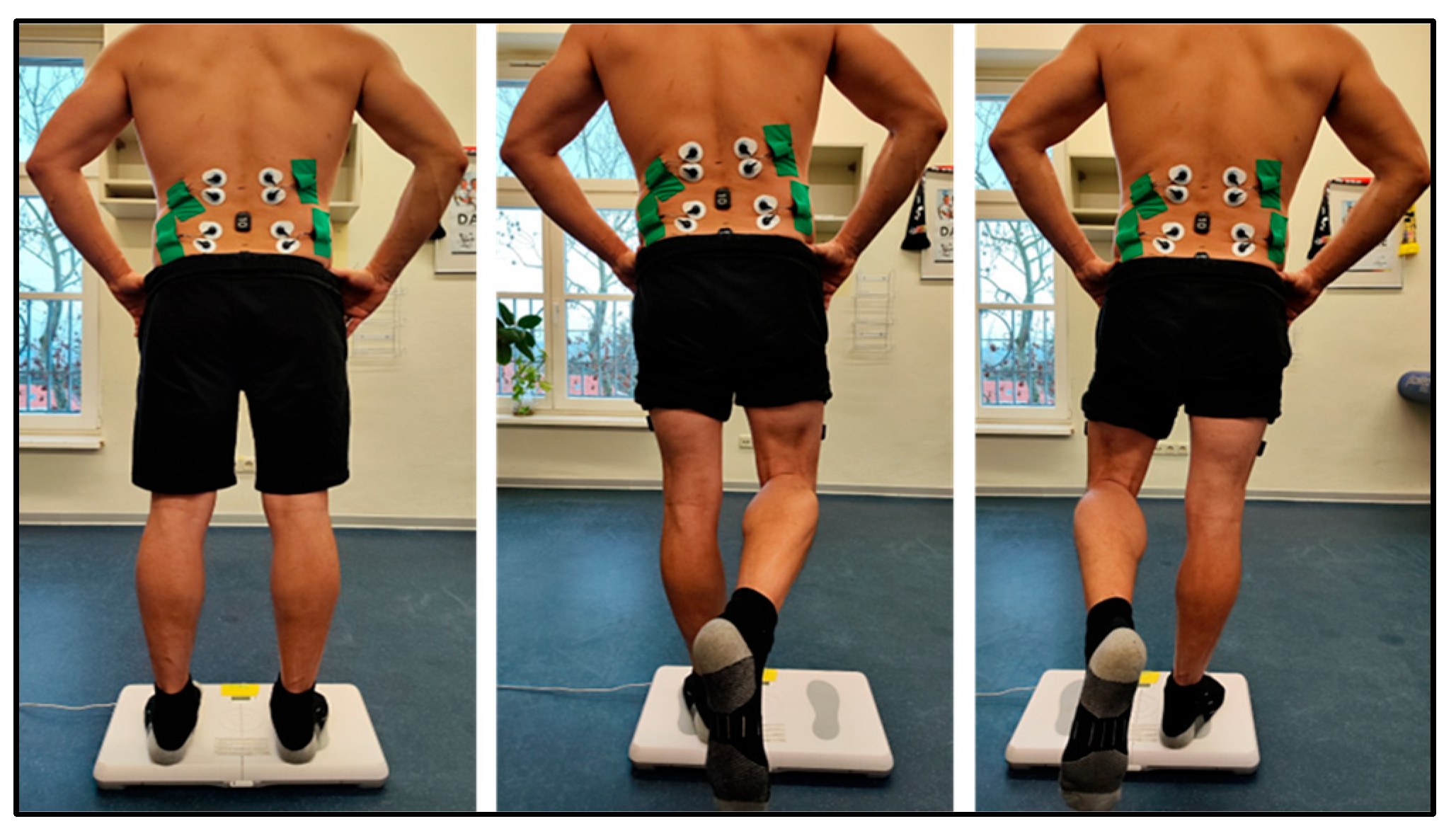
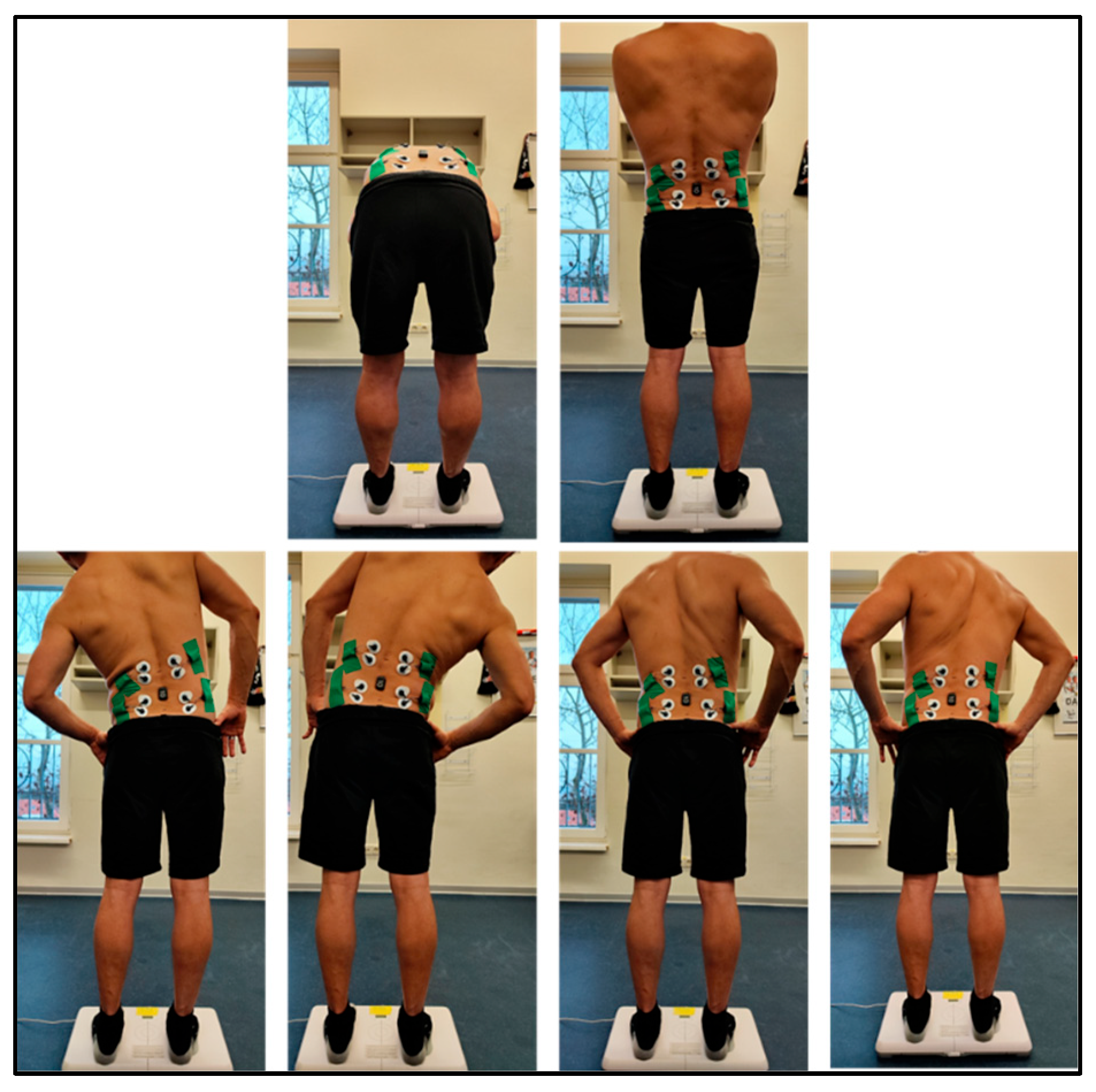
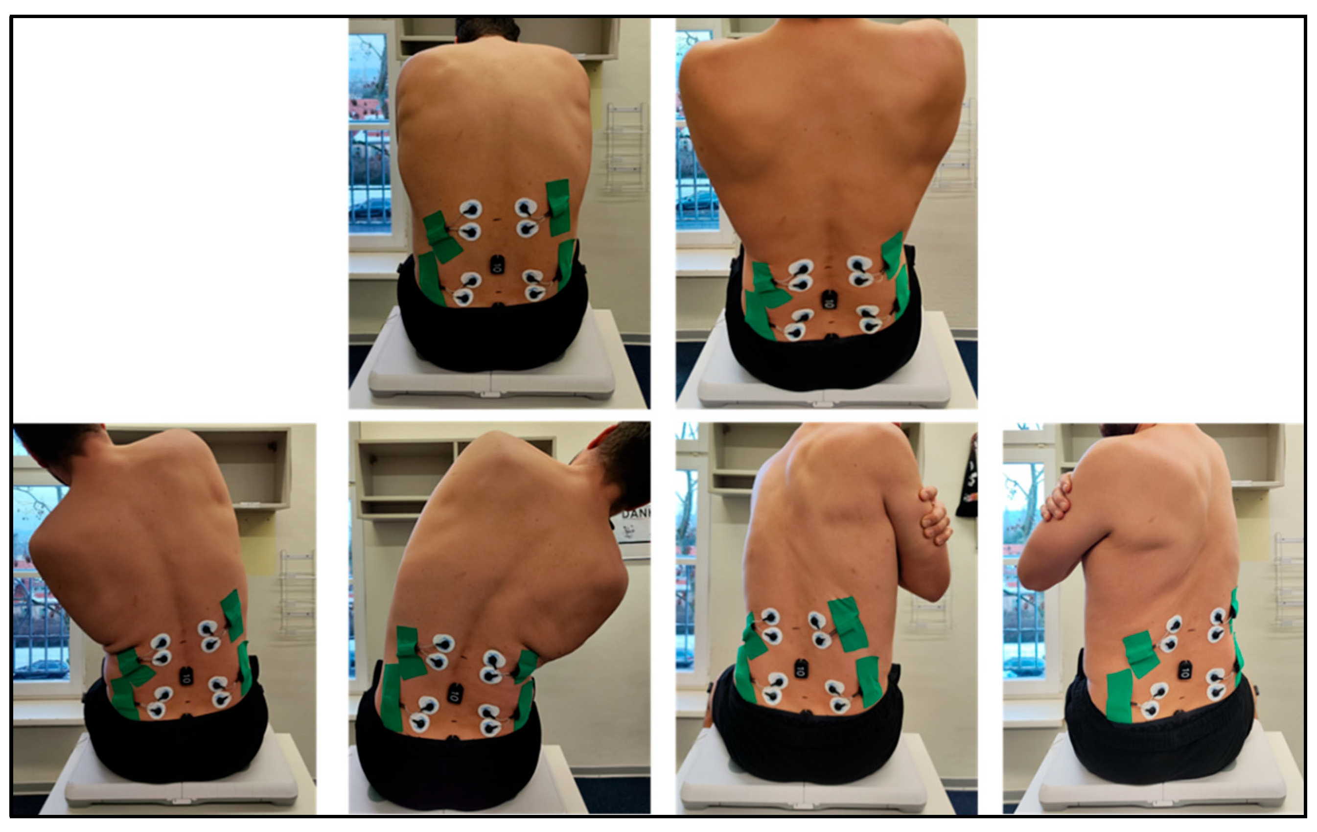
2.3.2. Neuromuscular Control and Range of Motion (ROM)
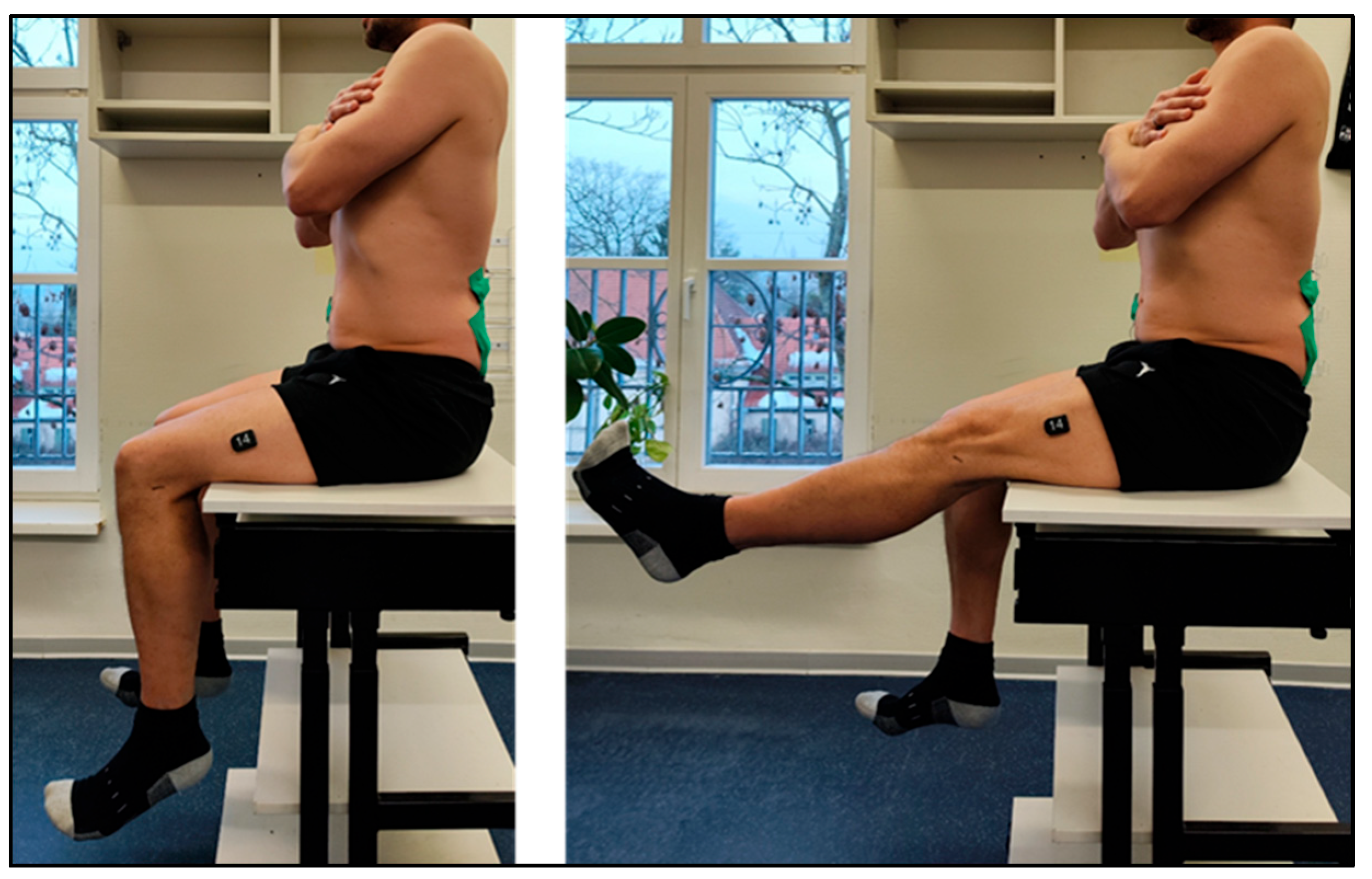
2.3.3. Lumbopelvic Stability
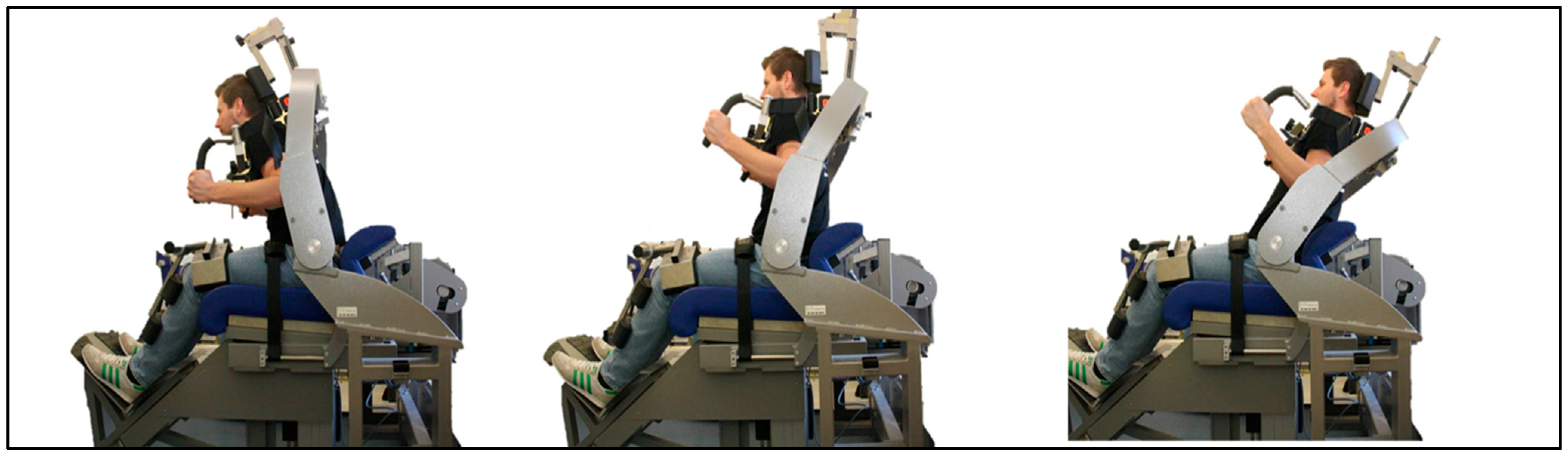
2.3.4. Global Trunk Musculature
2.4. Data Acquisition and Statistical Analysis
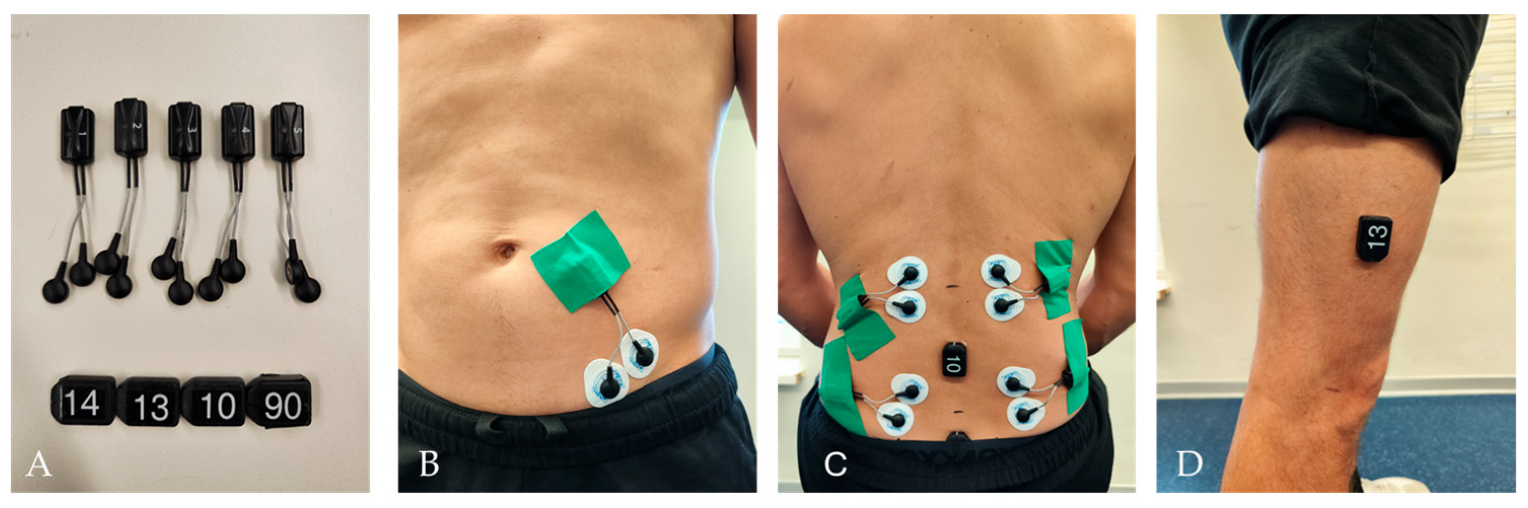
2.4.1. EMG and IMU System “Cometa”
2.4.2. CoP Measuring System
2.4.3. Statistics
2.5. Ethic
3. Results
3.1. Fatigue Resistance/Strength Endurance—Gait Analysis
3.2. Neuromuscular Control
3.3. Mobility/Bending
3.4. Lumbopelvic Stability
3.5. Core Strength
4. Discussion
4.1. Functional Assessment
4.1.1. Fatigue Resistance/Strength Endurance—Gait Analysis
4.1.2. Neuromuscular Control
4.1.3. Mobility Bending
4.1.4. Lumbar–Pelvic Stability
4.1.5. Core Strength
4.2. Limitations and Future Directions
5. Conclusions
Author Contributions
Funding
Institutional Review Board Statement
Informed Consent Statement
Data Availability Statement
Conflicts of Interest
Appendix A
Appendix A.1. Search Strings
Search Strategy
| Identification | ||
| Database | Search Query | Hits |
| PubMed | ((((“Low Back Pain” OR “LBP”) AND (“functional movement screen*” OR “functional assessment” OR “assessment instruments” OR assessment* OR FMS OR “EMG” OR “NRS”)) AND (diagn* OR prevent* OR control)) NOT (therapy OR reha* OR fractur* OR pregnancy OR imag* OR prevalence OR EEG OR effect* OR stress OR question* OR biomark* OR inflam*)) | n = 296 |
| CINAHL | n = 40 | |
| Screening | ||
| Results reviewed for duplicates | n = 312 | |
| Excluded after title abstract screening | n = 213 | |
| Full texts checked for suitability | n = 99 | |
| Full texts checked for suitability | n = 99 | |
| Excluded | Reason | Case number |
| No access to full text/no full text (too old) | n = 29 | |
| Population | Different search terms (no LBP) | n = 35 |
| Intervention | Different intervention | n = 12 |
| Outcome | Different outcome | n = 13 |
| Studies included in review | n = 10 |
Appendix B
Appendix B.1. Detailed Description of IMU-Based Kinematic Features Gait Analysis
| Muscle/Sensor | Metric | Meaning | Calculation | Interpretation |
| Pelvis_Acc_X | max_slopeR | Maximum change (slope) of the acceleration signal between two consecutive samples | Difference of the time series × sampling frequency (first time derivative) | High values indicate abrupt, jerky pelvic movements → suggestive of instability or pain-related protective reactions |
| R_Multifidii (EMG) | max_slopeL | Maximum rate of change of the EMG envelope per unit time | Difference of the EMG envelope × fs | Reflects the speed of changes in muscle activation; high values = rapid recruitment increases |
| Pelvis_Acc_X | max_steigungR | Maximum change (slope) of the acceleration signal between two consecutive samples | Same as above | Same as above |
| Left_Thigh_Acc_Z | varL | Variance of the Z-axis signal | Variance of the time series | Higher variance = more irregular, less controlled thigh movement |
| Left_Thigh_Acc_Z | max_slopeR | Maximum slope of the Z-axis acceleration signal | First derivative (diff × fs) | High peaks = abrupt vertical acceleration changes during gait or bending |
| Left_Thigh_Acc_Z | max_steigungR | Identical to max_slopeR | Same as above | Same as above |
| L_Multifidii (EMG) | meanR | Mean value of the EMG envelope over the analysis window | Averaging the EMG envelope | Elevated values = increased tonic activation; reduced values may indicate inhibition or fatigue |
| R_Multifidii (EMG) | aucR | Area under the EMG envelope (integral activation) | Trapezoidal integration of the EMG envelope | High AUC = greater overall activation load; low AUC = reduced recruitment or inhibition |
Appendix B.2. Detailed Overview of CoP, Frequency-Band Energy, and Trembling-Related Metrics
| Measurement | Meaning | Calculation (Description) | Interpretation |
| FreqBandEnergy_L | Energy in the low-frequency band (<0.3 Hz) | Sum of spectral magnitudes in the low-frequency band (freq < 0.3 Hz) | Higher values indicate a greater contribution of slow sway components, typically reflecting reduced postural stability due to larger, slower corrective movements |
| AreaTrembling | Area under the trembling curve (high-frequency CoP component) | Numerical integration of the trembling component using the trapezoidal rule | Larger area reflects stronger micro-sway, indicating reduced postural control and less efficient neuromuscular regulation |
| SwayArea_Ellipse | Ellipse covering the CoP sway (95% confidence ellipse) | Calculation of the 95% confidence ellipse around the CoP trajectory | Larger ellipse area indicates greater sway amplitude, suggestive of poorer overall postural stability |
| MeanFreq_X | Weighted mean frequency of the CoP signal in X-direction | mean_freq_x = Σ(f × Pxx)/Σ(Pxx) | Higher values indicate stronger involvement of higher frequencies → stiffer, faster corrective adjustments by the neuromuscular system |
| Power_Y | Maximum spectral power contribution in the Y-direction | Dominant peak in the Y-axis power spectrum | Higher values reflect dominant lateral sway activity, potentially associated with compensatory strategies or side-specific instability |
| Power_X | Maximum spectral power contribution in the X-direction | Dominant peak in the X-axis power spectrum | Lower values indicate reduced anterior–posterior sway, often associated with pain-related stiffness strategies |
| DistanceTrembling | Total path length of trembling (micro-sway trajectory) | Sum of Euclidean distances between consecutive trembling points | Higher distance indicates increased neuromuscular effort, often inefficient or compensatory in individuals with pain |
| FreqBandEnergy_H | Energy in the high-frequency band (1–3 Hz) | Sum of spectral magnitudes in the high-frequency band (1 ≤ freq ≤ 3 Hz) | Higher values indicate more high-frequency compensatory movements, reflecting stiffer, faster but less efficient postural control |
| VelocityTrembling | Mean trembling velocity | Mean Euclidean point-to-point velocity × sampling frequency | Higher values indicate accelerated micro-corrections, suggesting over-compensation or instability |
| DominantFreq_Y | Frequency of the highest spectral peak in Y-direction | Frequency location of the dominant peak in the Y-axis signal | Higher values indicate dominant high-frequency lateral corrections, typical in conditions of pain, stiffness, or compensatory behavior |
Appendix B.3. EMG-Based Trunk Muscle Parameters: Definitions, Computation, and Interpretation During CoP Measurement
| Muscle/Sensor | Metric | Meaning | Calculation | Interpretation |
| R_Int_Oblique | minENV | Minimum value of the EMG envelope | min(env) | High (relative): elevated baseline activation, potential excessive muscle tension. Low: adequate relaxation phases, energy-efficient control. |
| L_Multifidii | minENV | Minimum value of the EMG envelope | min(env) | High (relative): increased tonic activity, possible protective overactivation. Low: sufficient relaxation, physiologically efficient. |
| R_ThoracicEs | minRAW | Minimum value of the raw EMG signal | min(raw) | Very low values may indicate artefacts; moderately low values fall within a plausible physiological range. |
| R_ThoracicEs | maxRAW | Maximum value of the raw EMG signal | max(raw) | High peaks: strong activation spikes, possibly reflecting neuromuscular compensation or signal distortion. Low values: typical physiological activity. |
| L_ThoracicEs | minRAW | Minimum value of the raw EMG signal | min(raw) | Extremely low values may reflect noise or artefacts; moderately low values are physiologically plausible. |
| R_Multifidii | minENV | Minimum value of the EMG envelope | min(env) | High (relative): increased tonic baseline activation, potential muscle guarding. Low: appropriate relaxation capability and efficient neuromuscular control. |
Appendix B.4. Definitions and Interpretation of IMU and EMG Features Extracted During Seated Flexion and Extension Movements
| Muscle/Sensor | Metric | Meaning | Calculation | Interpretation |
| LWS | Acc_X_max | Maximum lumbar acceleration in X-direction | max(LWS_Acc_X) | High values: abrupt movements or compensatory evasive strategies; low values: controlled or reduced lumbar motion |
| LWS | Acc_X_range | Acceleration range (max–min) of the lumbar signal | max(LWS_Acc_X)–min(LWS_Acc_X) | High: large movement amplitude, potential overload; low: restricted mobility |
| LWS | roll_max | Maximum roll angle of the lumbar spine | Maximum of the roll signal | High: strong lateral motion; low: restricted or pain-related “stiffness” |
| LWS | roll_range | Roll angle range | max–min of the roll angle | High: large lateral mobility; low: stabilized or pain-limited movement |
| Pelvis | Acc_X_max | Maximum pelvic acceleration in X-direction | Maximum of pelvic_acc_x | High peaks: jerky pelvic motion, instability |
| Pelvis | Acc_X_range | Acceleration range of the pelvis (X) | max–min | Large: high mobility; small: restricted pelvic tilt |
| Pelvis | roll_max | Maximum roll angle of the pelvis | Maximum of pelvic roll | Larger values indicate lateral compensation; high = reduced stability |
| R. Multifidus (EMG) | env_min | Minimum value of the EMG envelope (right side) | min(envelope) | High: minimal relaxation, tonic overactivation; low: normal relaxation |
| L. Multifidus (EMG) | env_min | Minimum value of the EMG envelope (left side) | min(envelope) | High: compensatory baseline tension; low: good relaxation capability |
| LWS | area | Integrated lumbar acceleration over time | Area under the curve (trapezoidal integration) | High: large overall movement; low: restricted mobility |
| LWS/Pelvis | pitch_min | Minimum pitch angle between lumbar spine and pelvis | min(θ(t)) | Near zero: strongly reduced flexion → pain or guarding; more negative values = full flexion |
| Thoracic ES (Right) | env_mean | Mean EMG envelope of the thoracic erector spinae | mean(envelope) | High: compensatory activation/load transfer; low: efficient posture control |
| Thoracic ES (Right) | env_RMS | RMS of the EMG envelope (energy-related activation measure) | RMS(envelope) | High: elevated muscular energy → fatigue risk |
| L. Multifidus (EMG) | DutyCycle | Proportion of time the MF is active | Share of samples > 20% of maximal envelope | High: static load, less relaxation; low: efficient intermittent activation |
| LWS | Acc_Y_min | Minimum lumbar acceleration in Y-direction | min(Acc_Y) | Low values: downward/backward motion → possible pain-avoidance behavior |
| LWS | Acc_Y_area | Integrated pitch-related lumbar motion | Area under the pitch curve | Low: reduced lumbar/pelvic motion, increased stiffness |
| Pelvis | pelvis_pitch_area | Total pitch motion between lumbar spine and pelvis | Trapezoidal integration | Large: high movement range; low: pain-related movement restriction |
| LWS | pitch_area | Integrated lumbar pitch motion | Trapezoidal integration | High: greater movement amplitude; low: restricted motion |
| LWS | roll_range | Roll movement range of the lumbar spine | max–min of roll | High: potential instability; low: restricted mobility |
| Pelvis | Acc_X_area | Integrated pelvic acceleration in X-direction | Area under the curve | High: dynamic pelvic behavior; low: stiff/restricted pelvis |
| Pelvis | thigh_roll_area | Integrated roll movement between pelvis and thigh | Trapezoidal integration | High: increased movement magnitude; low: restricted movement |
| R. Multifidus (EMG) | env_min | Minimum EMG envelope (again) | min(envelope) | High: tonic overactivation; low: good relaxation ability |
| Thoracic ES (Right) | raw_mean | Mean raw EMG value | mean(raw EMG) | High: increased muscle activity; low: efficient baseline activation |
References
- Duhautois, R.; Erhel, C.; Guergoat-Larivière, M.; Mofakhami, M. More and Better Jobs, But Not for Everyone: Effects of Innovation in French Firms. ILR Rev. 2022, 75, 90–116. [Google Scholar] [CrossRef]
- Rose, S.J. The Growth of the Office Economy: Underrecognized Sector in High-Income Economies. Crit. Sociol. 2021, 47, 795–805. [Google Scholar] [CrossRef]
- Parry, S.; Straker, L. The contribution of office work to sedentary behaviour associated risk. BMC Public Health 2013, 13, 296. [Google Scholar] [CrossRef] [PubMed] [PubMed Central]
- Ferreira, M.L.; Luca, K.d.; Haile, L.M.; Steinmetz, J.D.; Culbreth, G.T.; Cross, M.; Kopec, J.A.; Ferreira, P.H.; Blyth, F.M.; Buchbinder, R.; et al. Global, regional, and national burden of low back pain, 1990–2020, its attributable risk factors, and projections to 2050: A systematic analysis of the Global Burden of Disease Study 2021. Lancet Rheumatol. 2023, 5, e316–e329. [Google Scholar] [CrossRef] [PubMed]
- Violante, F.S.; Mattioli, S.; Bonfiglioli, R. Low-back pain. In Handbook of Clinical Neurology; Elsevier: Amsterdam, The Netherlands, 2015; Volume 131, pp. 397–410. [Google Scholar] [CrossRef]
- Hoy, D.; March, L.; Brooks, P.; Blyth, F.; Woolf, A.; Bain, C.; Williams, G.; Smith, E.; Vos, T.; Barendregt, J.; et al. The global burden of low back pain: Estimates from the Global Burden of Disease 2010 study. Ann. Rheum. Dis. 2014, 73, 968–974. [Google Scholar] [CrossRef]
- Schmidt, C.O.; Raspe, H.; Pfingsten, M.; Hasenbring, M.; Basler, H.D.; Eich, W.; Kohlmann, T. Back Pain in the German Adult Population: Prevalence, Severity, and Sociodemographic Correlates in a Multiregional Survey. Spine 2007, 32, 2005–2011. [Google Scholar] [CrossRef] [PubMed]
- Brinjikji, W.; Luetmer, P.H.; Comstock, B.; Bresnahan, B.W.; Chen, L.E.; Deyo, R.A.; Halabi, S.; Turner, J.A.; Avins, A.L.; James, K.; et al. Systematic literature review of imaging features of spinal degeneration in asymptomatic populations. AJNR Am. J. Neuroradiol. 2015, 36, 811–816. [Google Scholar] [CrossRef] [PubMed] [PubMed Central]
- Webster, B.S.; Bauer, A.Z.; Choi, Y.; Cifuentes, M.; Pransky, G.S. Iatrogenic consequences of early magnetic resonance imaging in acute, work-related, disabling low back pain. Spine 2013, 38, 1939–1946. [Google Scholar] [CrossRef] [PubMed] [PubMed Central]
- Chou, R.; Qaseem, A.; Owens, D.K.; Shekelle, P. for the Clinical Guidelines Committee of the American College of Physicians. Diagnostic imaging for low back pain: Advice for high-value health care from the American College of Physicians. Ann. Intern. Med. 2011, 154, 181–189. [Google Scholar] [CrossRef] [PubMed]
- Hall, A.M.; Aubrey-Bassler, K.; Thorne, B.; Maher, C.G. Do not routinely offer imaging for uncomplicated low back pain. BMJ 2021, 372, n291. [Google Scholar] [CrossRef] [PubMed] [PubMed Central]
- Downie, A.; Hancock, M.; Jenkins, H.; Buchbinder, R.; Harris, I.; Underwood, M.; Goergen, S.; Maher, C.G. How common is imaging for low back pain in primary and emergency care? Systematic review and meta-analysis of over 4 million imaging requests across 21 years. Br. J. Sports Med. 2020, 54, 642–651. [Google Scholar] [CrossRef] [PubMed]
- Jenkins, H.J.; Downie, A.S.; Maher, C.G.; Moloney, N.A.; Magnussen, J.S.; Hancock, M.J. Imaging for low back pain: Is clinical use consistent with guidelines? A systematic review and meta-analysis. Spine J. Off. J. North Am. Spine Soc. 2018, 18, 2266–2277. [Google Scholar] [CrossRef] [PubMed]
- Sánchez-Zuriaga, D.; López-Pascual, J.; Garrido-Jaén, D.; de Moya, M.F.P.; Prat-Pastor, J. Reliability and validity of a new objective tool for low back pain functional assessment. Spine 2011, 36, 1279–1288. [Google Scholar] [CrossRef] [PubMed]
- Rosenberg, N. Objective Measurement of Musculoskeletal Pain: A Comprehensive Review. Diagnostics 2025, 15, 1581. [Google Scholar] [CrossRef] [PubMed] [PubMed Central]
- Chu, Y.; Zhao, X.; Han, J.; Su, Y. Physiological Signal-Based Method for Measurement of Pain Intensity. Front. Neurosci. 2017, 11, 279. [Google Scholar] [CrossRef]
- Wang, X.; Jia, R.; Li, J.; Zhu, Y.; Liu, H.; Wang, W.; Sun, Y.; Zhang, F.; Guo, L.; Zhang, W. Research Progress on the Mechanism of Lumbarmultifidus Injury and Degeneration. Oxidative Med. Cell. Longev. 2021, 2021, 6629037. [Google Scholar] [CrossRef]
- Abd-Elsayed, A.; Kurt, E.; Kollenburg, L.; Hasoon, J.; Wahezi, S.E.; Storlie, N.R. Lumbar Multifidus Dysfunction and Chronic Low Back Pain: Overview, Therapies, and an Update on the Evidence. Pain Pract. Off. J. World Inst. Pain 2025, 25, e70044. [Google Scholar] [CrossRef] [PubMed]
- Foster, N.E.; Anema, J.R.; Cherkin, D.; Chou, R.; Cohen, S.P.; Gross, D.P.; Ferreira, P.H.; Fritz, J.M.; Koes, B.W.; Peul, W.; et al. Prevention and treatment of low back pain: Evidence, challenges, and promising directions. Lancet 2018, 391, 2368–2383. [Google Scholar] [CrossRef]
- da Silva, R.A.; Vieira, E.R.; Cabrera, M.; Altimari, L.R.; Aguiar, A.F.; Nowotny, A.H.; Carvalho, A.F.; Oliveira, M.R. Back muscle fatigue of younger and older adults with and without chronic low back pain using two protocols: A case-control study. J. Electromyogr. Kinesiol. Off. J. Int. Soc. Electrophysiol. Kinesiol. 2015, 25, 928–936. [Google Scholar] [CrossRef] [PubMed]
- Abdelaty, E.M.; Shendy, S.; Lotfy, O.; Hassan, K.A. The difference in multifidus muscle morphology and motor control in non-specific low back pain with clinical lumbar instability and healthy subjects: A case-control study. Physiother. Res. Int. J. Res. Clin. Phys. Ther. 2024, 29, e2047. [Google Scholar] [CrossRef] [PubMed]
- Jung, S.-H.; Hwang, U.-J.; Ahn, S.-H.; Kim, H.-A.; Kim, J.-H.; Kwon, O.-Y. Lumbopelvic motor control function between patients with chronic low back pain and healthy controls: A useful distinguishing tool: The STROBE study. Medicine 2020, 99, e19621. [Google Scholar] [CrossRef] [PubMed] [PubMed Central]
- Behennah, J.; Conway, R.; Fisher, J.; Osborne, N.; Steele, J. The relationship between balance performance, lumbar extension strength, trunk extension endurance, and pain in participants with chronic low back pain, and those without. Clin. Biomech. 2018, 53, 22–30. [Google Scholar] [CrossRef]
- van Dieën, J.H.; Reeves, N.P.; Kawchuk, G.; van Dillen, L.R.; Hodges, P.W. Motor Control Changes in Low Back Pain: Divergence in Presentations and Mechanisms. J. Orthop. Sports Phys. Ther. 2019, 49, 370–379. [Google Scholar] [CrossRef] [PubMed] [PubMed Central]
- O’Sullivan, P.B.; Caneiro, J.P.; O’Keeffe, M.; Smith, A.; Dankaerts, W.; Fersum, K.; O’Sullivan, K. Cognitive Functional Therapy: An Integrated Behavioral Approach for the Targeted Management of Disabling Low Back Pain. Phys. Ther. 2018, 98, 408–423. [Google Scholar] [CrossRef] [PubMed] [PubMed Central]
- O’Sullivan, P.; Caneiro, J.P.; O’Keeffe, M.; O’Sullivan, K. Unraveling the Complexity of Low Back Pain. J. Orthop. Sports Phys. Ther. 2016, 46, 932–937. [Google Scholar] [CrossRef]
- Shakourisalim, M.; Martinez, K.B.; Golabchi, A.; Tavakoli, M.; Rouhani, H. Estimation of lower back muscle force in a lifting task using wearable IMUs. J. Biomech. 2024, 167, 112077. [Google Scholar] [CrossRef]
- Wattananon, P.; Ibrahim, A.A.; Rujirek, N.; Kongoun, S.; Klahan, K.; Richards, J. Motor unit behavior of lumbar multifidus during a forward trunk bending task performed under different speeds and loads in asymptomatic participants. Sci. Rep. 2025, 15, 44822. [Google Scholar] [CrossRef] [PubMed] [PubMed Central]
- Varrassi, G.; Leoni, M.L.G.; Al-Alwany, A.A.; Puttini, P.S.; Farì, G. Bioengineering Support in the Assessment and Rehabilitation of Low Back Pain. Bioengineering 2025, 12, 900. [Google Scholar] [CrossRef]
- Hodges, P.W.; Danneels, L. Changes in Structure and Function of the Back Muscles in Low Back Pain: Different Time Points, Observations, and Mechanisms. J. Orthop. Sports Phys. Ther. 2019, 49, 464–476. [Google Scholar] [CrossRef] [PubMed]
- Prieske, O.; Muehlbauer, T.; Granacher, U. The Role of Trunk Muscle Strength for Physical Fitness and Athletic Performance in Trained Individuals: A Systematic Review and Meta-Analysis. Sports Med. 2016, 46, 401–419. [Google Scholar] [CrossRef] [PubMed]
- Sheeran, L.; Al-Amri, M.; Sparkes, V.; Davies, J.L.; Sheeran, L.; Al-Amri, M.; Sparkes, V.; Davies, J.L. Assessment of Spinal and Pelvic Kinematics Using Inertial Measurement Units in Clinical Subgroups of Persistent Non-Specific Low Back Pain. Sensors 2024, 24, 2127. [Google Scholar] [CrossRef]
- Klasen, B.W.; Hallner, D.; Schaub, C.; Willburger, R.; Hasenbring, M. Validation and reliability of the German version of the Chronic Pain Grade questionnaire in primary care back pain patients. Psycho-Soc. Med. 2004, 1, Doc07. [Google Scholar] [PubMed] [PubMed Central]
- Ekstrom, R.A.; Osborn, R.W.; Hauer, P.L. Surface electromyographic analysis of the low back muscles during rehabilitation exercises. J. Orthop. Sports Phys. Ther. 2008, 38, 736–745. [Google Scholar] [CrossRef] [PubMed]
- Hermens, H.J.; Freriks, B.; Disselhorst-Klug, C.; Rau, G. Development of recommendations for SEMG sensors and sensor placement procedures. J. Electromyogr. Kinesiol. Off. J. Int. Soc. Electrophysiol. Kinesiol. 2000, 10, 361–374. [Google Scholar] [CrossRef] [PubMed]
- Phothirook, P.; Amatachaya, S.; Peungsuwan, P.; Phothirook, P.; Amatachaya, S.; Peungsuwan, P. Muscle Activity and Co-Activation of Gait Cycle during Walking in Water and on Land in People with Spastic Cerebral Palsy. Int. J. Environ. Res. Public Health 2023, 20, 1854. [Google Scholar] [CrossRef]
- Koltermann, J.J.; Gerber, M. Quantification of the Dependence of the Measurement Error on the Quantization of the A/D Converter for Center of Pressure Measurements. Biomechanics 2022, 2, 309–318. [Google Scholar] [CrossRef]
- Clark, R.A.; Bryant, A.L.; Pua, Y.; McCrory, P.; Bennell, K.; Hunt, M. Validity and reliability of the Nintendo Wii Balance Board for assessment of standing balance. Gait Posture 2010, 31, 307–310. [Google Scholar] [CrossRef] [PubMed]
- Koltermann, J.J.; Beck, H.; Beck, M.; Beck, H.; Beck, M. Investigation of the Correlation between Factors Influencing the Spectrum of Center of Pressure Measurements Using Dynamic Controlled Models of the Upright Stand and Subject Measurements. Appl. Sci. 2020, 10, 3741. [Google Scholar] [CrossRef]
- Koltermann, J.J.; Gerber, M.; Beck, H.; Beck, M. Validation of Different Filters for Center of Pressure Measurements by a Cross-Section Study. Technologies 2019, 7, 68. [Google Scholar] [CrossRef]
- Kongsted, A.; Kent, P.; Quicke, J.G.; Skou, S.T.; Hill, J.C. Risk-stratified and stepped models of care for back pain and osteoarthritis: Are we heading towards a common model? Pain Rep. 2020, 5, e843. [Google Scholar] [CrossRef] [PubMed] [PubMed Central]
- Tagliaferri, S.D.; Miller, C.T.; Owen, P.J.; Mitchell, U.H.; Brisby, H.; Fitzgibbon, B.; Masse-Alarie, H.; Van Oosterwijck, J.; Belavy, D.L. Domains of Chronic Low Back Pain and Assessing Treatment Effectiveness: A Clinical Perspective. Pain Pract. Off. J. World Inst. Pain 2020, 20, 211–225. [Google Scholar] [CrossRef] [PubMed]
- Xu, X.; Sekiguchi, Y.; Honda, K.; Izumi, S.-I. Motion analysis of 3D multi-segmental spine during gait in symptom remission people with low back pain: A pilot study. BMC Musculoskelet. Disord. 2025, 26, 269. [Google Scholar] [CrossRef] [PubMed] [PubMed Central]
- Smith, J.A.; Kulig, K. Altered Multifidus Recruitment During Walking in Young Asymptomatic Individuals with a History of Low Back Pain. J. Orthop. Sports Phys. Ther. 2016, 46, 365–374. [Google Scholar] [CrossRef] [PubMed]
- Al-Eisa, E.; Egan, D.; Deluzio, K.; Wassersug, R. Effects of pelvic asymmetry and low back pain on trunk kinematics during sitting: A comparison with standing. Spine 2006, 31, E135–E143. [Google Scholar] [CrossRef] [PubMed]
- Yu, Q.; Huang, H.; Zhang, Z.; Hu, X.; Li, W.; Li, L.; Chen, M.; Liang, Z.; Lo, W.L.A.; Wang, C. The association between pelvic asymmetry and non-specific chronic low back pain as assessed by the global postural system. BMC Musculoskelet. Disord. 2020, 21, 596. [Google Scholar] [CrossRef] [PubMed] [PubMed Central]
- Bryndal, A.; Nawos-Wysocki, W.; Grochulska, A.; Łosiński, K.; Glowinski, S. Effects of gait speed on paraspinal muscle activation: An sEMG analysis of the multifidus and erector spinae. PeerJ 2025, 13, e19244. [Google Scholar] [CrossRef] [PubMed] [PubMed Central]
- Danneels, L.A.; Coorevits, P.L.; Cools, A.M.; Vanderstraeten, G.G.; Cambier, D.C.; Witvrouw, E.E.; De, C.H.J. Differences in electromyographic activity in the multifidus muscle and the iliocostalis lumborum between healthy subjects and patients with sub-acute and chronic low back pain. Eur. Spine J. Off. Publ. Eur. Spine Soc. Eur. Spinal Deform. Soc. Eur. Sect. Cerv. Spine Res. Soc. 2002, 11, 13–19. [Google Scholar] [CrossRef] [PubMed] [PubMed Central]
- Zhang, S.; Xu, Y.; Han, X.; Wu, W.; Tang, Y.; Wang, C. Functional and Morphological Changes in the Deep Lumbar Multifidus Using Electromyography and Ultrasound. Sci. Rep. 2018, 8, 6539. [Google Scholar] [CrossRef] [PubMed] [PubMed Central]
- Ruhe, A.; Fejer, R.; Walker, B. Center of pressure excursion as a measure of balance performance in patients with non-specific low back pain compared to healthy controls: A systematic review of the literature. Eur. Spine J. Off. Publ. Eur. Spine Soc. Eur. Spinal Deform. Soc. Eur. Sect. Cerv. Spine Res. Soc. 2011, 20, 358–368. [Google Scholar] [CrossRef] [PubMed] [PubMed Central]
- Koltermann, J.J.; Floessel, P.; Hammerschmidt, F.; Disch, A.C.; Koltermann, J.J.; Floessel, P.; Hammerschmidt, F.; Disch, A.C. A Statistical and AI Analysis of the Frequency Spectrum in the Measurement of the Center of Pressure Track in the Seated Position in Healthy Subjects and Subjects with Low Back Pain. Sensors 2024, 24, 3011. [Google Scholar] [CrossRef]
- Hodges, P.W.; Richardson, C.A. Inefficient muscular stabilization of the lumbar spine associated with low back pain. A motor control evaluation of transversus abdominis. Spine 1996, 21, 2640–2650. [Google Scholar] [CrossRef] [PubMed]
- van Dieën, J.H.; Cholewicki, J.; Radebold, A. Trunk muscle recruitment patterns in patients with low back pain enhance the stability of the lumbar spine. Spine 2003, 28, 834–841. [Google Scholar] [CrossRef] [PubMed]
- Naqvi, W.M.; Vaidya, L.; Kumar, K. Impact of low back pain on fear of movement and functional activities. Int. J. Res. Pharm. Sci. 2020, 11, 4830–4835. [Google Scholar] [CrossRef]
- Zhou, T.; Salman, D.; McGregor, A.H. Recent clinical practice guidelines for the management of low back pain: A global comparison. BMC Musculoskelet. Disord. 2024, 25, 344. [Google Scholar] [CrossRef] [PubMed] [PubMed Central]
- Paoletti, M.; Belli, A.; Palma, L.; Paniccia, M.; Tombolini, F.; Ruggiero, A.; Vallasciani, M.; Pierleoni, P. Data acquired by wearable sensors for the evaluation of the flexion-relaxation phenomenon. Data Brief 2020, 31, 105957. [Google Scholar] [CrossRef]
- Bednar, D.A.; Bednar, E.D. Internal lumbar disc derangement with instability catch from monosegmental discopathy. The forgotten mechanical and kinetic surgical back pain syndrome. Clin. Neurol. Neurosurg. 2022, 212, 107033. [Google Scholar] [CrossRef] [PubMed]
- Sorensen, C.J.; Johnson, M.B.; Norton, B.J.; Callaghan, J.P.; Van Dillen, L.R. Asymmetry of lumbopelvic movement patterns during active hip abduction is a risk factor for low back pain development during standing. Hum. Mov. Sci. 2016, 50, 38–46. [Google Scholar] [CrossRef] [PubMed] [PubMed Central]
- Faur, C.; Patrascu, J.M.; Haragus, H.; Anglitoiu, B. Correlation between multifidus fatty atrophy and lumbar disc degeneration in low back pain. BMC Musculoskelet. Disord. 2019, 20, 414. [Google Scholar] [CrossRef] [PubMed] [PubMed Central]
- Lee, J.H.; Hoshino, Y.; Nakamura, K.; Kariya, Y.; Saita, K.; Ito, K. Trunk muscle weakness as a risk factor for low back pain. A 5-year prospective study. Spine 1999, 24, 54–57. [Google Scholar] [CrossRef] [PubMed]
- Dvir, Z.; Keating, J.L. Trunk Extension Effort in Patients with Chronic Low Back Dysfunction. Spine 2003, 28, 685–692. [Google Scholar] [CrossRef]
- Reyes-Ferrada, W.; Chirosa-Rios, L.; Rodriguez-Perea, A.; Jerez-Mayorga, D.; Chirosa-Rios, I. Isokinetic Trunk Strength in Acute Low Back Pain Patients Compared to Healthy Subjects: A Systematic Review. Int. J. Environ. Res. Public Health 2021, 18, 2576. [Google Scholar] [CrossRef] [PubMed] [PubMed Central]
- Kumar, R.; Marla, K.; Sporn, K.; Paladugu, P.; Khanna, A.; Gowda, C.; Ngo, A.; Waisberg, E.; Jagadeesan, R.; Tavakkoli, A. Emerging Diagnostic Approaches for Musculoskeletal Disorders: Advances in Imaging, Biomarkers, and Clinical Assessment. Diagnostics 2025, 15, 1648. [Google Scholar] [CrossRef]
- O’Keeffe, M.; Ferreira, G.E.; Harris, I.A.; Darlow, B.; Buchbinder, R.; Traeger, A.C.; Zadro, J.R.; Herbert, R.D.; Thomas, R.; Belton, J.; et al. Effect of diagnostic labelling on management intentions for non-specific low back pain: A randomized scenario-based experiment. Eur. J. Pain 2022, 26, 1532–1545. [Google Scholar] [CrossRef] [PubMed] [PubMed Central]
- Wirth, B.; Schweinhardt, P. Personalized assessment and management of non-specific low back pain. Eur. J. Pain 2024, 28, 181–198. [Google Scholar] [CrossRef] [PubMed]




| Group | Male [n] | Height [cm] | Weight [kg] | Age [years] |
| No pain | 2 | 1760 (2.8) | 82.5 (13.4) | 34.0 (7.1) |
| Low-intensity pain | 6 | 181.3 (5.9) | 89.6 (19.8) | 40.1 (8.3) |
| High-intensity pain | 5 | 182.8 (7.8) | 87.0 (13.2) | 40.2 (7.2) |
| Group | Female [n] | Height [cm] | Weight [kg] | Age [years] |
| No pain | 7 | 170.5 (5.2) | 66.9 (8.0) | 44.7 (11.1) |
| Low-intensity pain | 8 | 169.6 (6.8) | 69.2 (11.6) | 46.8 (16.1) |
| High-intensity pain | 10 | 167.4 (4.1) | 70.06 (13.2) | 45.7 (10.4) |
| Functional Level | Assessment | Technical Equipment |
|---|---|---|
| Fatigue resistance/strength endurance | 100 m gait test; | EMG and IMU |
| Mobility and neuromuscular control (CoP) | 60 s bipedal and monopedal (left and right) stance with eyes open; Spinal ROM tests while standing and sitting, every movement twice: flexion/extension, lateral bending and rotation; 60 s upright sitting with eyes open | CoP-force plate EMG and IMU |
| Lumbopelvic stability | Modified slump test | EMG and IMU |
| Global trunk musculature | times for 6 s isometric trunk strength measurements in the sagittal plane in different flexion angular positions of the hip: 90°, 80°, 105° and 120° | IsoMed 2000 (D. & R. Ferstl GmbH, Hemau, Germany) |
| Position | Measurement | p-Value | Power | Mean No Pain | Mean Low-Intensity Pain | Mean High-Intensity Pain |
|---|---|---|---|---|---|---|
| Pelvis_Acc_X | max_slopeR | 0.043 | 0.558 | 0.02 | 0.02 | 0.035 |
| R_Multifidii | max_slopeL | 0.049 | 0.488 | 0.02 | 0.03 | 0.03 |
| Pelvis_Acc_X | max_steigungR | 0.043 | 0.558 | 0.02 | 0.02 | 0.035 |
| Left_Thigh_Acc_Z | varL | 0.042 | 0.562 | 0.03 | 0.04 | 0.063 |
| Left_Thigh_Acc_Z | max_slopeR | 0.012 | 0.759 | 0.01 | 0.02 | 0.028 |
| Left_Thigh_Acc_Z | max_steigungR | 0.012 | 0.759 | 0.01 | 0.02 | 0.02 |
| L_Multifidii | meanR | 0.05 | 0.505 | 0.0009 | 0.0007 | 0.0005 |
| R_Multifidii | aucR | 0.046 | 0.509 | 0.07 | 0.07 | 0.05 |
| Measurement | p-Value | Power | Mean No Pain | Mean Low-Intensity Pain | Mean High-Intensity Pain |
|---|---|---|---|---|---|
| FreqBandEnergy_L | 0.023 | 0.661 | 2.54 × 10¹⁴ | 2.7 × 10¹⁴ | 3.98 × 10¹⁴ |
| AreaTrembling | 0.033 | 0.599 | 1.53 × 10¹⁴ | 4.46 × 10¹⁴ | 4.2 × 10¹⁴ |
| SwayArea_Ellipse | 0.024 | 0.654 | 1.99 × 10¹⁴ | 4.3 × 10¹⁴ | 4.61 × 10¹⁴ |
| MeanFreq_X | 0.048 | 0.505 | 2.30 × 10¹⁴ | 3.34 × 10¹⁴ | 3.82 × 10¹⁴ |
| Power_Y | 0.031 | 0.608 | 1.99 × 10¹⁴ | 2.3 × 10¹⁴ | 2.67 × 10¹⁴ |
| Power_X | 0.027 | 0.634 | 3.6 × 10¹⁴ | 2.94 × 10¹⁴ | 1.72 × 10¹⁴ |
| DistanceTrembling | 0.05 | 0.523 | 5.0 × 10¹⁴ | 4.58 × 10¹⁴ | 3.87 × 10¹⁴ |
| FreqBandEnergy_H | 0.019 | 0.691 | 3.06 × 10¹⁴ | 3.89 × 10¹⁴ | 5.72 × 10¹⁴ |
| VelocityTrembling | 0.04 | 0.57 | 3.88 × 10¹⁴ | 3.07 × 10¹⁴ | 1.65 × 10¹⁴ |
| DominantFreq_Y | 0.049 | 0.497 | 152,587,890.6 | 3.33 × 10⁸ | 6.1 × 10⁸ |
| Position | Measurement | p-Value | Power | Mean No Pain | Mean Low-Intensity Pain | Mean High-Intensity Pain |
|---|---|---|---|---|---|---|
| R_Int_Oblique | minENV | 0.033 | 0.60 | −7.48 | −0.05 | 0.54 |
| L_Multifidii | minENV | 0.001 | 0.93 | 2.12 | 0.81 | −0.47 |
| R_ThoracicEs | minRAW | 0.055 | 0.47 | −81.22 | −102.67 | −166.51 |
| R_ThoracicEs | maxRAW | 0.052 | 0.48 | 76.29 | 109.66 | 181.36 |
| L_ThoracicEs | minRAW | 0.047 | 0.53 | −96.84 | −83.80 | −163.481 |
| R_Multifidii | minENV | 0.012 | 0.75 | −2.07 | −0.45 | 1.60 |
| Measurement | p-Value | Power | Mean No Pain | Mean Low-Intensity Pain | Mean High-Intensity Pain |
|---|---|---|---|---|---|
| LWS_Acc_X_max | 0.024 | 0.658 | 1.185 | 0.92 | 0.84 |
| LWS_Acc_X_range | 0.03 | 0.623 | 1.18 | 0.94 | 0.85 |
| LWS_roll_max | 0.019 | 0.69 | 78.44 | 68.40 | 60.99 |
| LWS_roll_range | 0.023 | 0.665 | 81.93 | 65.67 | 58.26 |
| Pelvis_Acc_X_max | 0.03 | 0.62 | 0.940 | 0.68 | 0.64 |
| Pelvis_Acc_X_range | 0.03 | 0.623 | 0.93 | 0.68 | 0.64 |
| Pelvis_roll_max | 0.028 | 0.632 | 58.19 | 47.66 | 42.95 |
| R_Multifidii_env_min | 0.049 | 0.512 | 3.14 | 3.20 | 6.66 |
| L_Multifidii_env_min | 0.034 | 0.598 | 2.79 | 3.38 | 7.99 |
| LWS_Acc_X_area | 0.037 | 0.582 | 3.02 | 2.34 | 1.92 |
| lws_pelvis_pitch_min | 0.003 | 0.905 | 4.47 | 0.43 | 0.06 |
| Measurement | p-Value | Power | Mean No Pain | Mean Low-Intensity Pain | Mean High-Intensity Pain |
|---|---|---|---|---|---|
| LWS_Acc_X_area | 0.012 | 0.753 | 4.03 | 2.66 | 2.37 |
| lws_pelvis_pitch_min | 0.009 | 0.798 | 6.5 | 1.99 | 0.09 |
| R_ThoracicEs_env_mean | 0.035 | 0.594 | 13.46 | 18.02 | 23.08 |
| R_ThoracicEs_env_RMS | 0.039 | 0.576 | 15.51 | 21.55 | 26.43 |
| L_Multifidii_DutyCycle | 0.042 | 0.563 | 0.71 | 0.77 | 0.88 |
| LWS_Acc_X_area | 0.005 | 0.845 | 3.90 | 2.82 | 2.24 |
| LWS_Acc_Y_min | 0.029 | 0.626 | −0.12 | −0.06 | −0.041 |
| lws_pelvis_pitch_area | 0.021 | 0.681 | 216.87 | 123.89 | 125.03 |
| LWS_pitch_area | 0.031 | 0.613 | 426.20 | 342.91 | 286.77 |
| LWS_roll_range | 0.031 | 0.617 | 102.96 | 65.04 | 62.29 |
| Pelvis_Acc_X_area | 0.017 | 0.711 | 2.91 | 2.18 | 1.68 |
| pelvis_thigh_roll_area | 0.018 | 0.705 | 175.35 | 125.86 | 117.24 |
| R_Multifidii_env_min | 0.016 | 0.719 | 2.33 | 2.48 | 6.02 |
| R_ThoracicEs_raw_mean | 0.036 | 0.587 | 0.00 | 0.007 | 0.016 |
| Measurement | Term | p-Value | Power | Mean No Pain | Mean Low-Intensity Pain | Mean High-Intensity Pain |
|---|---|---|---|---|---|---|
| L_Multifidii | minENV | 0.033 | 0.6 | 1.8 | 0.43 | −0.83 |
| R_Int_Oblique | meanRAW | 0.053 | 0.43 | −0.00018 | 0.0004 | 0.000721 |
| L_ThoracicEs | meanRAW | 0.050 | 0.41 | −0.00049 | 3.94 × 10⁻⁵ | 0.000743 |
| Measurement | p-Value | Power | Mean No Pain Nm/kg | Mean Low-Intensity Pain Nm/kg | Mean High-Intensity Pain Nm/kg |
|---|---|---|---|---|---|
| flex_max_0 | 0.033 | 0.607 | 1.28 | 1.31 | 1.67 |
| flex_max_10 | 0.702 | 0.067 | 1.24 | 1.22 | 1.58 |
| ext_max_0 | 0.029 | 0.634 | 3.01 | 2.68 | 2.62 |
| ext_max_15 | 0.016 | 0.724 | 3.19 | 2.78 | 2.73 |
| ext_max_30 | 0.879 | 0.053 | 3.13 | 2.71 | 2.82 |
Disclaimer/Publisher’s Note: The statements, opinions and data contained in all publications are solely those of the individual author(s) and contributor(s) and not of MDPI and/or the editor(s). MDPI and/or the editor(s) disclaim responsibility for any injury to people or property resulting from any ideas, methods, instructions or products referred to in the content. |
© 2026 by the authors. Licensee MDPI, Basel, Switzerland. This article is an open access article distributed under the terms and conditions of the Creative Commons Attribution (CC BY) license.
Share and Cite
Floessel, P.; Wunderlich, F.C.; Funke, J.-J.; Kaplick, H.; Koltermann, J.J.; Disch, A.C. Deduction of Back Pain Patients Using EMG Technology and Inertial Sensors During Functional Tests. Sensors 2026, 26, 1882. https://doi.org/10.3390/s26061882
Floessel P, Wunderlich FC, Funke J-J, Kaplick H, Koltermann JJ, Disch AC. Deduction of Back Pain Patients Using EMG Technology and Inertial Sensors During Functional Tests. Sensors. 2026; 26(6):1882. https://doi.org/10.3390/s26061882
Chicago/Turabian StyleFloessel, Philipp, Freya Charlotte Wunderlich, Jil-Justin Funke, Hannes Kaplick, Jan Jens Koltermann, and Alexander C. Disch. 2026. "Deduction of Back Pain Patients Using EMG Technology and Inertial Sensors During Functional Tests" Sensors 26, no. 6: 1882. https://doi.org/10.3390/s26061882
APA StyleFloessel, P., Wunderlich, F. C., Funke, J.-J., Kaplick, H., Koltermann, J. J., & Disch, A. C. (2026). Deduction of Back Pain Patients Using EMG Technology and Inertial Sensors During Functional Tests. Sensors, 26(6), 1882. https://doi.org/10.3390/s26061882