Optimizing OPM-MEG Sensor Layouts Using the Sequential Selection Algorithm with Simulated Sources and Individual Anatomy


Abstract
1. Introduction
2. Materials and Methods
2.1. Construction of a Hypothetical Unitary Sensor Holder
2.2. Measurements of Auditory Evoked Fields (AEFs)
2.3. Simulating the Magnetic Field Maps
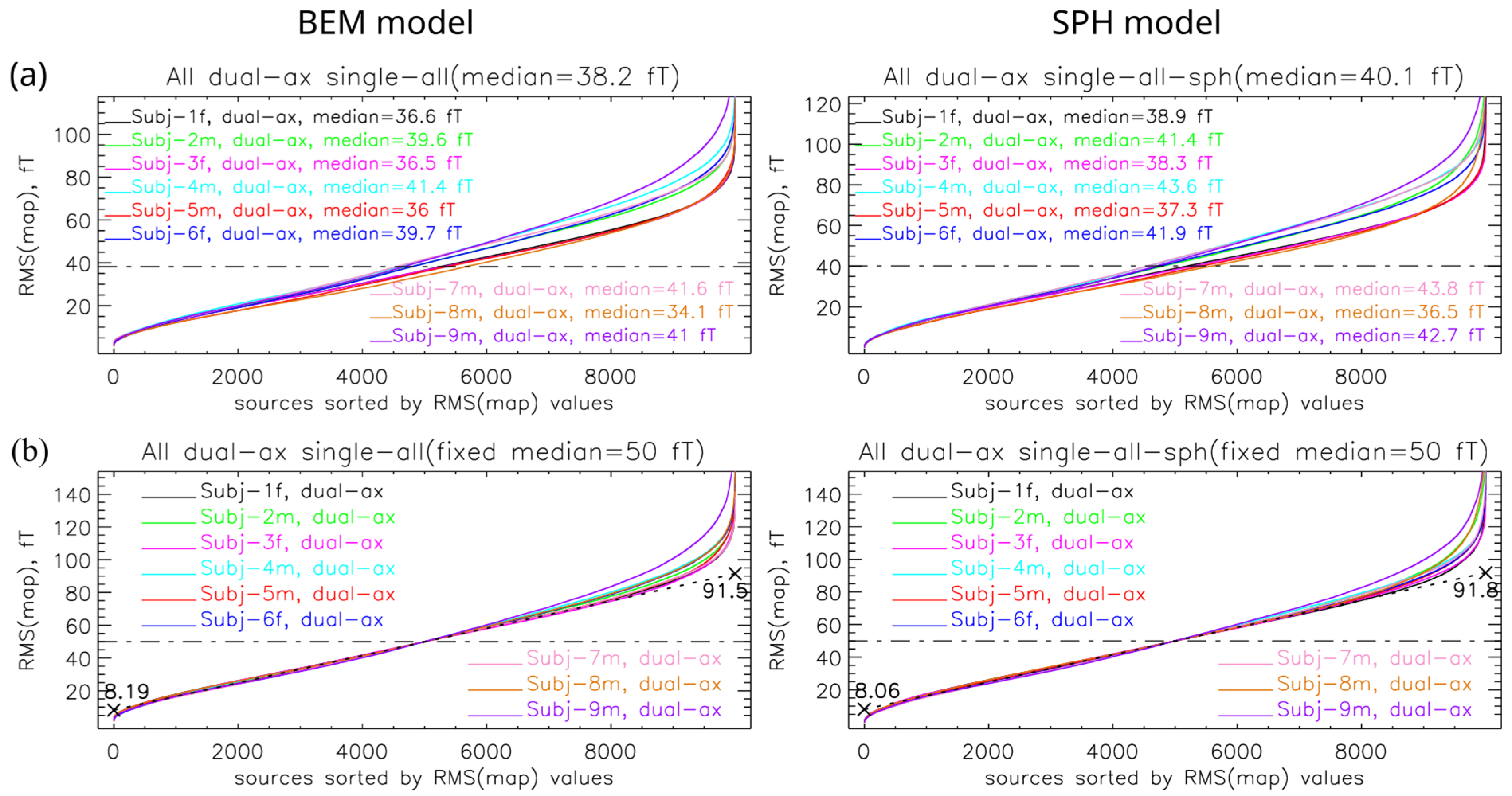
- Protocol 1 (single–all): For each sample, one ECD was selected at random from the full cortical source space.
- Protocol 2 (single–3cm): Identical to Protocol 1 but restricted to ECDs with a cortical depth of <3 cm, where depth was defined as the minimal Euclidean distance from the dipole location to the outer scalp surface.
- Protocol 3 (double–3cm): For each sample, two ECDs were selected: one from the left hemisphere and one from the right hemisphere. Only dipoles with a depth < 3 cm were allowed.
- Protocol 4 (double–auditory): For each sample, two ECDs were simulated: one in the left auditory cortex and one in the right auditory cortex. These regions were defined using the Destrieux atlas [45], specifically the labels G_temp_sup-G_T_transv-lh and G_temp_sup-G_T_transv-rh.
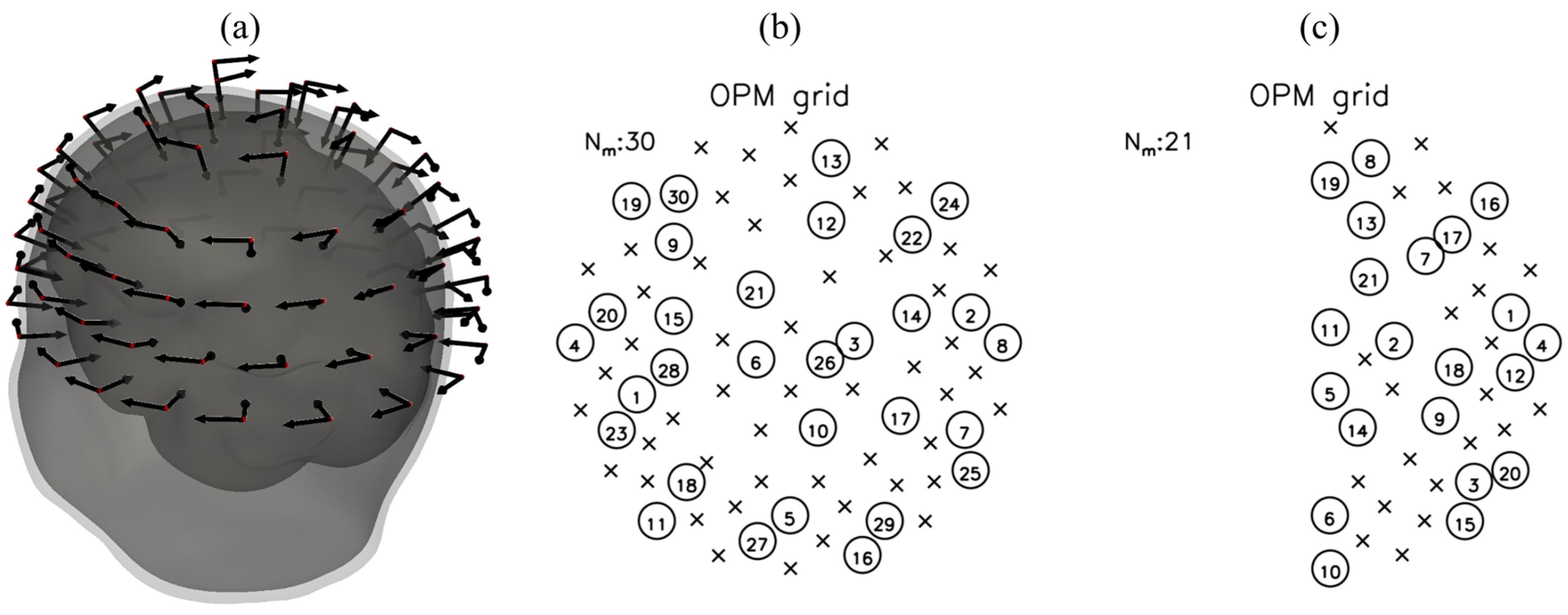
- A dense grid corresponding to a hypothetical unitary OPM sensor holder (Section 2.1);
- The geometry of the SQUID-MEG system used for empirical data recording (Section 2.2).
2.4. Estimating the Optimal Layout Using the SSA
Overview of the SSA Sensor Selection Procedure
2.5. Applying the SSA to the Simulated Data
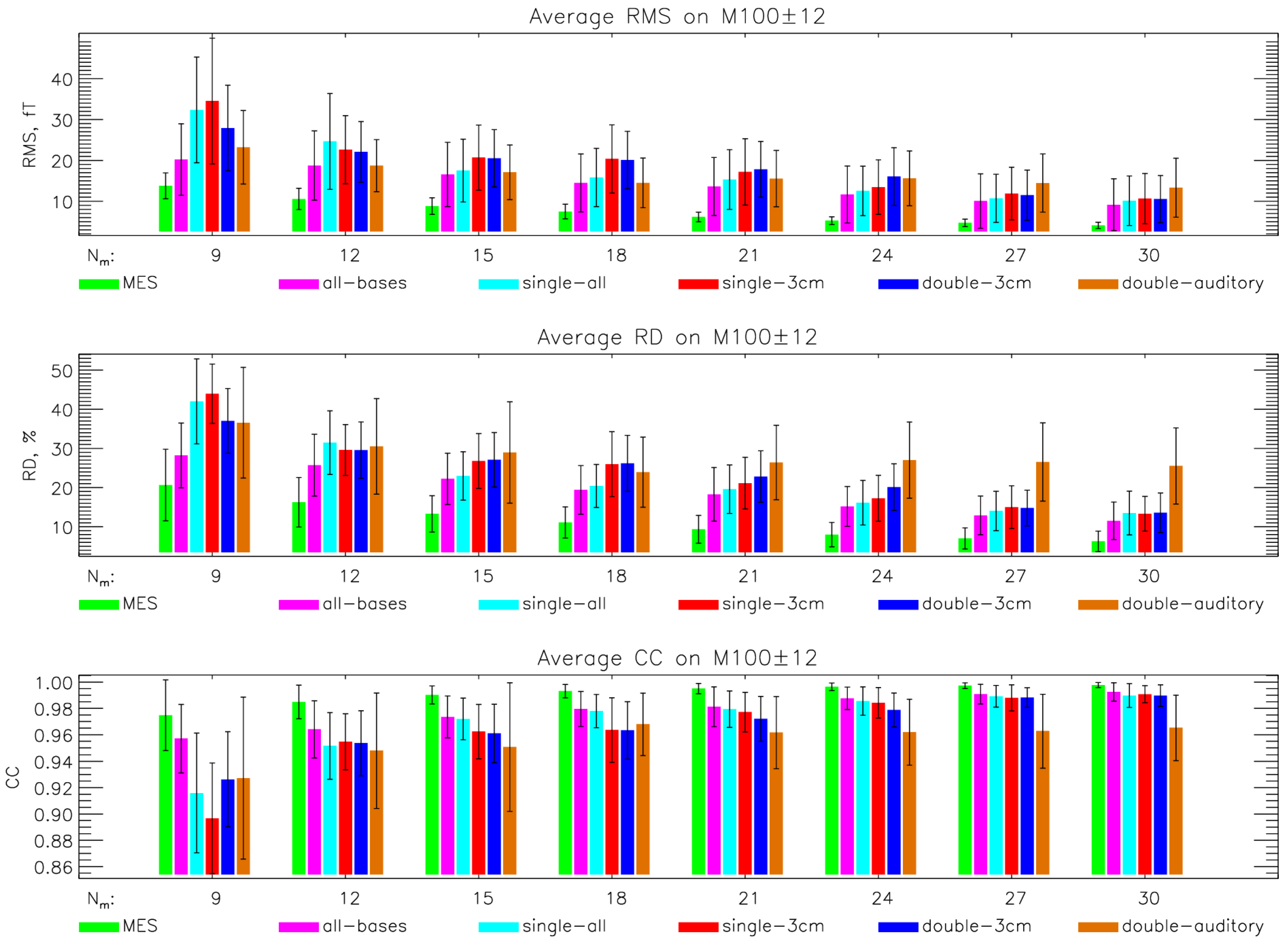
- Combining simulated data from all 9 subjects and all 4 protocols, i.e., 100 MFMs per subject and protocol (all–bases);
- Combining simulated data from all subjects and one protocol, i.e., 400 MFMs per subject;
- Combining simulated data from a single subject and all 4 protocols, i.e., 900 MFMs per protocol;
- Using 3600 simulated MFMs for a single subject and one protocol.
2.6. Evaluation Metrics
2.6.1. Root Mean Square (RMS) of a Single Map
2.6.2. Average RMS, Average Relative Difference (RD), and Average Correlation Coefficient (CC) to Assess the SSA Optimized Layout
2.6.3. ECD Fit and the Localization Error
3. Results
3.1. Overview of the Simulations
3.2. Different Simulation Protocols for the SSA Selection
3.3. Effect of Individualized Geometry on Simulation-Driven SSA
3.4. Localization Error for the Simulation-Driven SSA
4. Discussion
5. Conclusions
Supplementary Materials
Author Contributions
Funding
Institutional Review Board Statement
Informed Consent Statement
Data Availability Statement
Conflicts of Interest
Appendix A

| N | Recording 1 | Date | Age |
|---|---|---|---|
| 1 | Subject-1f1 | 8 October 2018 | 30 |
| 2 | Subject-1f2 | 7 May 2019 | 31 |
| 3 | Subject-2m1 | 16 October 2019 | 26 |
| 4 | Subject-3f1 | 27 June 2019 | 31 |
| 5 | Subject-3f2 | 10 April 2019 | 31 |
| 6 | Subject-4m1 | 9 October 2018 | 36 |
| 7 | Subject-4m2 | 10 May 2019 | 37 |
| 8 | Subject-5m1 | 8 January 2020 | 31 |
| 9 | Subject-6f1 | 5 July 2018 | 33 |
| 10 | Subject-6f2 | 5 April 2019 | 34 |
| 11 | Subject-7m1 | 7 May 2019 | 54 |
| 12 | Subject-7m2 | 17 October 2019 | 54 |
| 13 | Subject-8m1 | 11 October 2019 | 26 |
| 14 | Subject-8m2 | 18 October 2019 | 26 |
| 15 | Subject-9m1 | 18 June 2018 | 57 |
| 16 | Subject-9m2 | 12 June 2019 | 58 |
References
- Hämäläinen, M.; Hari, R.; Ilmoniemi, R.J.; Knuutila, J.; Lounasmaa, O.V. Magnetoencephalography—Theory, Instrumentation, and Applications to Noninvasive Studies of the Working Human Brain. Rev. Mod. Phys. 1993, 65, 413–497. [Google Scholar] [CrossRef]
- Jaklevic, R.C.; Lambe, J.; Silver, A.H.; Mercereau, J.E. Quantum Interference Effects in Josephson Tunneling. Phys. Rev. Lett. 1964, 12, 159–160. [Google Scholar] [CrossRef]
- Shah, V.; Osborne, J.; Orton, J.; Alem, O. Fully Integrated, Standalone Zero Field Optically Pumped Magnetometer for Biomagnetism. In Proceedings of the Steep Dispersion Engineering and Opto-Atomic Precision Metrology XI; Shahriar, S.M., Scheuer, J., Eds.; SPIE: San Francisco, CA, USA, 2018; p. 51. [Google Scholar]
- Boto, E.; Bowtell, R.; Krüger, P.; Fromhold, T.M.; Morris, P.G.; Meyer, S.S.; Barnes, G.R.; Brookes, M.J. On the Potential of a New Generation of Magnetometers for MEG: A Beamformer Simulation Study. PLoS ONE 2016, 11, e0157655. [Google Scholar] [CrossRef]
- Marhl, U.; Sander, T.; Jazbinšek, V. Simulation Study of Different OPM-MEG Measurement Components. Sensors 2022, 22, 3184. [Google Scholar] [CrossRef]
- Iivanainen, J.; Stenroos, M.; Parkkonen, L. Measuring MEG Closer to the Brain: Performance of on-Scalp Sensor Arrays. NeuroImage 2017, 147, 542–553. [Google Scholar] [CrossRef]
- Kim, K.; Begus, S.; Xia, H.; Lee, S.-K.; Jazbinsek, V.; Trontelj, Z.; Romalis, M.V. Multi-Channel Atomic Magnetometer for Magnetoencephalography: A Configuration Study. NeuroImage 2014, 89, 143–151. [Google Scholar] [CrossRef]
- Beltrachini, L.; von Ellenrieder, N.; Eichardt, R.; Haueisen, J. Optimal Design of On-scalp Electromagnetic Sensor Arrays for Brain Source Localisation. Hum. Brain Mapp. 2021, 42, 4869–4879. [Google Scholar] [CrossRef]
- Budker, D.; Romalis, M. Optical Magnetometry. Nat. Phys. 2007, 3, 227–234. [Google Scholar] [CrossRef]
- Boto, E.; Holmes, N.; Leggett, J.; Roberts, G.; Shah, V.; Meyer, S.S.; Muñoz, L.D.; Mullinger, K.J.; Tierney, T.M.; Bestmann, S.; et al. Moving Magnetoencephalography towards Real-World Applications with a Wearable System. Nature 2018, 555, 657–661. [Google Scholar] [CrossRef]
- Zhang, X.; Chen, C.; Zhang, M.; Ma, C.; Zhang, Y.; Wang, H.; Guo, Q.; Hu, T.; Liu, Z.; Chang, Y.; et al. Detection and Analysis of MEG Signals in Occipital Region with Double-Channel OPM Sensors. J. Neurosci. Methods 2020, 346, 108948. [Google Scholar] [CrossRef]
- De Lange, P.; Boto, E.; Holmes, N.; Hill, R.M.; Bowtell, R.; Wens, V.; De Tiège, X.; Brookes, M.J.; Bourguignon, M. Measuring the Cortical Tracking of Speech with Optically-Pumped Magnetometers. NeuroImage 2021, 233, 117969. [Google Scholar] [CrossRef]
- Boto, E.; Hill, R.M.; Rea, M.; Holmes, N.; Seedat, Z.A.; Leggett, J.; Shah, V.; Osborne, J.; Bowtell, R.; Brookes, M.J. Measuring Functional Connectivity with Wearable MEG. NeuroImage 2021, 230, 117815. [Google Scholar] [CrossRef]
- Wittevrongel, B.; Holmes, N.; Boto, E.; Hill, R.; Rea, M.; Libert, A.; Khachatryan, E.; Van Hulle, M.M.; Bowtell, R.; Brookes, M.J. Practical Real-Time MEG-Based Neural Interfacing with Optically Pumped Magnetometers. BMC Biol. 2021, 19, 158. [Google Scholar] [CrossRef]
- Chen, C.; Teng, P.; Meng, Q.; Jiang, Y.; Li, R.; Wang, J.; Wu, J.; Guan, Y.; Wang, M.; Zhou, J.; et al. The Potential of OPM-Based Magnetoencephalography in Pre-Surgical Evaluation of Drug-Resistant Epilepsy. Neurophysiol. Clin. 2025, 55, 103087. [Google Scholar] [CrossRef]
- Feys, O.; Corvilain, P.; Labyt, E.; Mahmoudzadeh, M.; Routier, L.; Sculier, C.; Holmes, N.; Brookes, M.; Goldman, S.; Romain, R.; et al. Tri-Axial Rubidium and Helium Optically Pumped Magnetometers for on-Scalp Magnetoencephalography Recording of Interictal Epileptiform Discharges: A Case Study. Front. Neurosci. 2023, 17, 1284262. [Google Scholar] [CrossRef]
- Hillebrand, A.; Holmes, N.; Sijsma, N.; O’Neill, G.C.; Tierney, T.M.; Liberton, N.; Stam, A.H.; Van Klink, N.; Stam, C.J.; Bowtell, R.; et al. Non-Invasive Measurements of Ictal and Interictal Epileptiform Activity Using Optically Pumped Magnetometers. Sci. Rep. 2023, 13, 4623. [Google Scholar] [CrossRef]
- Li, W.; Cao, F.; An, N.; Wang, W.; Wang, C.; Xu, W.; Gao, Y.; Ning, X. Source Extent Estimation in OPM-MEG: A Two-Stage Champagne Approach. IEEE Trans. Med. Imaging 2025, 44, 828–840. [Google Scholar] [CrossRef]
- Mellor, S.; Timms, R.C.; O’Neill, G.C.; Tierney, T.M.; Spedden, M.E.; The MELD Project Consortium; Spitzer, H.; Ripart, M.; Whitaker, K.; Napolitano, A.; et al. Combining OPM and Lesion Mapping Data for Epilepsy Surgery Planning: A Simulation Study. Sci. Rep. 2024, 14, 2882. [Google Scholar] [CrossRef]
- Nugent, A.C.; Benitez Andonegui, A.; Holroyd, T.; Robinson, S.E. On-Scalp Magnetocorticography with Optically Pumped Magnetometers: Simulated Performance in Resolving Simultaneous Sources. Neuroimage Rep. 2022, 2, 100093. [Google Scholar] [CrossRef]
- Power, L.; Bardouille, T.; Ikeda, K.M.; Omisade, A. Validation of On-Head OPM MEG for Language Laterality Assessment. Brain Topogr. 2025, 38, 8. [Google Scholar] [CrossRef]
- Ren, J.; Ding, M.; Peng, Y.; Sun, C.; Yang, C.; Zhou, S.; Tian, J.; Wang, Q.; Li, Z. A Comparative Study on the Detection and Localization of Interictal Epileptiform Discharges in Magnetoencephalography Using Optically Pumped Magnetometers versus Superconducting Quantum Interference Devices. NeuroImage 2025, 312, 121232. [Google Scholar] [CrossRef]
- Seedat, Z.A.; Pier, K.S.; Holmes, N.; Rea, M.; Al-Hilaly, L.; Tierney, T.M.; Embury, C.M.; Pardington, R.; Mullinger, K.J.; Cross, J.H.; et al. Simultaneous Whole-Head Electrophysiological Recordings Using EEG and OPM-MEG. Imaging Neurosci. 2024, 2, imag-2-00179. [Google Scholar] [CrossRef]
- Vivekananda, U.; Mellor, S.; Tierney, T.M.; Holmes, N.; Boto, E.; Leggett, J.; Roberts, G.; Hill, R.M.; Litvak, V.; Brookes, M.J.; et al. Optically Pumped Magnetoencephalography in Epilepsy. Ann. Clin. Transl. Neurol. 2020, 7, 397–401. [Google Scholar] [CrossRef]
- Pedersen, M.; Abbott, D.F.; Jackson, G.D. Wearable OPM-MEG: A Changing Landscape for Epilepsy. Epilepsia 2022, 63, 2745–2753. [Google Scholar] [CrossRef]
- Carreno, C.A.; Evans, M.E.; Lockhart, B.K.; Chinaka, O.; Katz, B.; Bell, M.A.; Howell, B.R. Optimizing Infant Neuroimaging Methods to Understand the Neurodevelopmental Impacts of Early Nutrition and Feeding. Dev. Cogn. Neurosci. 2025, 71, 101481. [Google Scholar] [CrossRef]
- Rhodes, N.; Rier, L.; Singh, K.D.; Sato, J.; Vandewouw, M.M.; Holmes, N.; Boto, E.; Hill, R.M.; Rea, M.; Taylor, M.J.; et al. Measuring the Neurodevelopmental Trajectory of Excitatory-Inhibitory Balance via Visual Gamma Oscillations. Imaging Neurosci. 2025, 3, imag_a_00527. [Google Scholar] [CrossRef]
- Rhodes, N.; Sato, J.; Safar, K.; Amorim, K.; Taylor, M.J.; Brookes, M.J. Paediatric Magnetoencephalography and Its Role in Neurodevelopmental Disorders. Br. J. Radiol. 2024, 97, 1591–1601. [Google Scholar] [CrossRef]
- Rier, L.; Rhodes, N.; Pakenham, D.O.; Boto, E.; Holmes, N.; Hill, R.M.; Reina Rivero, G.; Shah, V.; Doyle, C.; Osborne, J.; et al. Tracking the Neurodevelopmental Trajectory of Beta Band Oscillations with Optically Pumped Magnetometer-Based Magnetoencephalography. eLife 2024, 13, RP94561. [Google Scholar] [CrossRef]
- Sanders, B.J.; Gilmartin, C.G.S.; Rier, L.; Gascoyne, L.; McCann, E.; Cabrera, J.; Leggett, J.; Holmes, N.; Hill, R.M.; Boto, E.; et al. OPM-MEG in Multiple Sclerosis: Proof of Principle, and the Effect of Naturalistic Posture. NeuroImage Clin. 2025, 48, 103888. [Google Scholar] [CrossRef]
- Brickwedde, M.; Anders, P.; Kühn, A.A.; Lofredi, R.; Holtkamp, M.; Kaindl, A.M.; Grent-‘t-Jong, T.; Krüger, P.; Sander, T.; Uhlhaas, P.J. Applications of OPM-MEG for Translational Neuroscience: A Perspective. Transl. Psychiatry 2024, 14, 341. [Google Scholar] [CrossRef]
- Wang, F.; Cao, F.; An, N.; Yang, J.; Wang, Y.; Xiang, M.; Wu, Q.; Li, W. Artifact Suppression in OPM-MEG for Parkinson’s Disease Patients with DBS Implants Using Oblique Projection-Based Extended Homogeneous Field Correction. IEEE J. Biomed. Health Inform. 2025, 1–14. [Google Scholar] [CrossRef] [PubMed]
- Lux, R.L.; Smith, C.R.; Wyatt, R.F.; Abildskov, J.A. Limited Lead Selection for Estimation of Body Surface Potential Maps in Electrocardiography. IEEE Trans. Biomed. Eng. 1978, 25, 270–276. [Google Scholar] [CrossRef] [PubMed]
- Jazbinsek, V.; Hren, R.; Trontelj, Z. High Resolution ECG and MCG Mapping: Simulation Study of Single and Dual Accessory Pathways and Influence of Lead Displacement and Limited Lead Selection on Localisation Results. Bull. Pol. Acad. Sci. Tech. Sci. 2005, 53, 195–205. [Google Scholar]
- Jazbinšek, V.; Hren, R.; Trontelj, Z. Influence of Limited Lead Selection on Source Localization in Magnetocardiography and Electrocardiography. Int. Congr. Ser. 2007, 1300, 492–495. [Google Scholar] [CrossRef]
- Jazbinšek, V.; Burghoff, M.; Kosch, O.; Steinhoff, U.; Hren, R.; Trahms, L.; Trontelj, Z. Selection of Optimal Recording Sites in Electrocardiography and Magnetocardiography. Biomed. Eng. Biomed. Tech. 2004, 48, 174–177. [Google Scholar]
- Marhl, U.; Hren, R.; Sander, T.; Jazbinšek, V. Optimization of OPM-MEG Layouts with a Limited Number of Sensors. Sensors 2025, 25, 2706. [Google Scholar] [CrossRef]
- Marhl, U.; Sander, T.; Jazbinšek, V. Using a Limited Number of Sensors in MEG or the Feasibility of Partial Head Coverage OPM MEG. In Proceedings of the 2024 47th MIPRO ICT and Electronics Convention (MIPRO); IEEE: Opatija, Croatia, 2024; pp. 1134–1138. [Google Scholar]
- Marhl, U.; Jodko-Władzińska, A.; Brühl, R.; Sander, T.; Jazbinšek, V. Transforming and Comparing Data between Standard SQUID and OPM-MEG Systems. PLoS ONE 2022, 17, e0262669. [Google Scholar] [CrossRef]
- Fischl, B.; Sereno, M.I.; Dale, A.M. Cortical Surface-Based Analysis. NeuroImage 1999, 9, 195–207. [Google Scholar] [CrossRef]
- Fischl, B. FreeSurfer. NeuroImage 2012, 62, 774–781. [Google Scholar] [CrossRef]
- Shimogawara, M.; Tanaka, H.; Kazumi, K.; Haruta, Y. MEGvision Magnetoencephalograph System and Its Applications. Yokogawa Tech. Rep. 2004, 38, 23–27. [Google Scholar]
- Gramfort, A. MEG and EEG Data Analysis with MNE-Python. Front. Neurosci. 2013, 7, 267. [Google Scholar] [CrossRef]
- Gramfort, A.; Luessi, M.; Larson, E.; Engemann, D.A.; Strohmeier, D.; Brodbeck, C.; Parkkonen, L.; Hämäläinen, M.S. MNE Software for Processing MEG and EEG Data. NeuroImage 2014, 86, 446–460. [Google Scholar] [CrossRef]
- Destrieux, C.; Fischl, B.; Dale, A.; Halgren, E. Automatic Parcellation of Human Cortical Gyri and Sulci Using Standard Anatomical Nomenclature. NeuroImage 2010, 53, 1–15. [Google Scholar] [CrossRef] [PubMed]
- Ségonne, F.; Dale, A.M.; Busa, E.; Glessner, M.; Salat, D.; Hahn, H.K.; Fischl, B. A Hybrid Approach to the Skull Stripping Problem in MRI. NeuroImage 2004, 22, 1060–1075. [Google Scholar] [CrossRef] [PubMed]
- Lux, R.L. Karhunen-Loeve Representation of ECG Data. J. Electrocardiol. 1992, 25, 195–198. [Google Scholar] [CrossRef]
- Lux, R.L.; Burgess, M.J.; Wyatt, R.F.; Evans, A.K.; Vincent, G.M.; Abildskov, J.A. Clinically Practical Lead Systems for Improved Electrocardiography: Comparison with Precordial Grids and Conventional Lead Systems. Circulation 1979, 59, 356–363. [Google Scholar] [CrossRef] [PubMed]
- Lux, R.L.; Evans, A.K.; Burgess, M.J.; Wyatt, R.F.; Abildskov, J.A. Redundancy Reduction for Improved Display and Analysis of Body Surface Potential Maps. I. Spatial Compression. Circ. Res. 1981, 49, 186–196. [Google Scholar] [CrossRef]
- Levenberg, K. A Method for the Solution of Certain Non-Linear Problems in Least Squares. Q. Appl. Math. 1944, 2, 164–168. [Google Scholar] [CrossRef]
- Press, W.H.; Teukolsky, S.A.; Vetterling, W.T.; Flannery, B.P. Numerical Recipes 3rd Edition: The Art of Scientific Computing, 3rd ed.; Cambridge University Press: Cambridge, UK; New York, NY, USA, 2007; ISBN 978-0-521-88068-8. [Google Scholar]
- Hren, R.; Stroink, G. Application of the Surface Harmonic Expansions for Modeling the Human Torso. IEEE Trans. Biomed. Eng. 1995, 42, 521–524. [Google Scholar] [CrossRef]
- Sarvas, J. Basic Mathematical and Electromagnetic Concepts of the Biomagnetic Inverse Problem. Phys. Med. Biol. 1987, 32, 11–22. [Google Scholar] [CrossRef]
- Gage, N.M.; Siegel, B.; Callen, M.; Roberts, T.P.L. Cortical Sound Processing in Children with Autism Disorder: An MEG Investigation. NeuroReport 2003, 14, 2047–2051. [Google Scholar] [CrossRef]
- Roberts, T.P.L.; Matsuzaki, J.; Blaskey, L.; Bloy, L.; Edgar, J.C.; Kim, M.; Ku, M.; Kuschner, E.S.; Embick, D. Delayed M50/M100 Evoked Response Component Latency in Minimally Verbal/Nonverbal Children Who Have Autism Spectrum Disorder. Mol. Autism 2019, 10, 34. [Google Scholar] [CrossRef] [PubMed]
- Demopoulos, C.; Yu, N.; Tripp, J.; Mota, N.; Brandes-Aitken, A.N.; Desai, S.S.; Hill, S.S.; Antovich, A.D.; Harris, J.; Honma, S.; et al. Magnetoencephalographic Imaging of Auditory and Somatosensory Cortical Responses in Children with Autism and Sensory Processing Dysfunction. Front. Hum. Neurosci. 2017, 11, 259. [Google Scholar] [CrossRef]
- Demopoulos, C.; Kopald, B.E.; Bangera, N.; Paulson, K.; David Lewine, J. Rapid Auditory Processing of Puretones Is Associated with Basic Components of Language in Individuals with Autism Spectrum Disorders. Brain Lang. 2023, 238, 105229. [Google Scholar] [CrossRef]
- Kikuchi, M.; Yoshimura, Y.; Mutou, K.; Minabe, Y. Magnetoencephalography in the Study of Children with Autism Spectrum Disorder. Psychiatry Clin. Neurosci. 2016, 70, 74–88. [Google Scholar] [CrossRef]
- Port, R.G.; Edgar, J.C.; Ku, M.; Bloy, L.; Murray, R.; Blaskey, L.; Levy, S.E.; Roberts, T.P.L. Maturation of Auditory Neural Processes in Autism Spectrum Disorder—A Longitudinal MEG Study. NeuroImage Clin. 2016, 11, 566–577. [Google Scholar] [CrossRef]
- Knosche, T.R. Transformation of Whole-Head MEG Recordings Between Different Sensor Position. Biomed. Tech. 2002, 47, 59–62. [Google Scholar] [CrossRef]
- Hämäläinen, M.S.; Ilmoniemi, R.J. Interpreting Magnetic Fields of the Brain: Minimum Norm Estimates. Med. Biol. Eng. Comput. 1994, 32, 35–42. [Google Scholar] [CrossRef]
- Van Veen, B.D.; Van Drongelen, W.; Yuchtman, M.; Suzuki, A. Localization of Brain Electrical Activity via Linearly Constrained Minimum Variance Spatial Filtering. IEEE Trans. Biomed. Eng. 1997, 44, 867–880. [Google Scholar] [CrossRef] [PubMed]
- Holmes, N.; Bowtell, R.; Brookes, M.J.; Taulu, S. An Iterative Implementation of the Signal Space Separation Method for Magnetoencephalography Systems with Low Channel Counts. Sensors 2023, 23, 6537. [Google Scholar] [CrossRef] [PubMed]
- Tierney, T.M.; Alexander, N.; Mellor, S.; Holmes, N.; Seymour, R.; O’Neill, G.C.; Maguire, E.A.; Barnes, G.R. Modelling Optically Pumped Magnetometer Interference in MEG as a Spatially Homogeneous Magnetic Field. NeuroImage 2021, 244, 118484. [Google Scholar] [CrossRef]
- Bergquist, J.; Rupp, L.; Zenger, B.; Brundage, J.; Busatto, A.; MacLeod, R.S. Body Surface Potential Mapping: Contemporary Applications and Future Perspectives. Hearts 2021, 2, 514–542. [Google Scholar] [CrossRef]
- Hubley-Kozey, C.L.; Mitchell, L.B.; Gardner, M.J.; Warren, J.W.; Penney, C.J.; Smith, E.R.; Horácek, B.M. Spatial Features in Body-Surface Potential Maps Can Identify Patients with a History of Sustained Ventricular Tachycardia. Circulation 1995, 92, 1825–1838. [Google Scholar] [CrossRef]
- Hren, R.; Horácek, B.M. Value of Simulated Body Surface Potential Maps as Templates in Localizing Sites of Ectopic Activation for Radiofrequency Ablation. Physiol. Meas. 1997, 18, 373–400. [Google Scholar] [CrossRef]
- SippensGroenewegen, A.; Spekhorst, H.; Van Hemel, N.M.; Kingma, J.H.; Hauer, R.N.; De Bakker, J.M.; Grimbergen, C.A.; Janse, M.J.; Dunning, A.J. Localization of the Site of Origin of Postinfarction Ventricular Tachycardia by Endocardial Pace Mapping. Body Surface Mapping Compared with the 12-Lead Electrocardiogram. Circulation 1993, 88, 2290–2306. [Google Scholar] [CrossRef] [PubMed]
- SippensGroenewegen, A.; Spekhorst, H.; Van Hemel, N.M.; Kingma, J.H.; Hauer, R.N.; Janse, M.J.; Dunning, A.J. Body Surface Mapping of Ectopic Left Ventricular Activation. QRS Spectrum in Patients with Prior Myocardial Infarction. Circ. Res. 1992, 71, 1361–1378. [Google Scholar] [CrossRef]
- Spekhorst, H.; SippensGroenewegen, A.; David, G.K.; Janse, M.J.; Dunning, A.J. Body Surface Mapping during Percutaneous Transluminal Coronary Angioplasty. QRS Changes Indicating Regional Myocardial Conduction Delay. Circulation 1990, 81, 840–849. [Google Scholar] [CrossRef]
- SippensGroenewegen, A.; Spekhorst, H.; Van Hemel, N.M.; Kingma, J.H.; Hauer, R.N.; Janse, M.J.; Dunning, A.J. Body Surface Mapping of Ectopic Left and Right Ventricular Activation. QRS Spectrum in Patients without Structural Heart Disease. Circulation 1990, 82, 879–896. [Google Scholar] [CrossRef]
- Dambrink, J.-H.E.; SippensGroenewegen, A.; Van Gilst, W.H.; Peels, K.H.; Grimbergen, C.A.; Kingma, J.H. Association of Left Ventricular Remodeling and Nonuniform Electrical Recovery Expressed by Nondipolar QRST Integral Map Patterns in Survivors of a First Anterior Myocardial Infarction. Circulation 1995, 92, 300–310. [Google Scholar] [CrossRef] [PubMed]
- SippensGroenewegen, A.; Spekhorst, H.; Van Hemel, N.M.; Kingma, J.H.; Hauer, R.N.W.; De Barker, J.M.T.; Grimbergen, C.A.; Janse, M.J.; Dunning, A.J. Value of Body Surface Mapping in Localizing the Site of Origin of Ventricular Tachycardia in Patients with Previous Myocardial Infarction. J. Am. Coll. Cardiol. 1994, 24, 1708–1724. [Google Scholar] [CrossRef] [PubMed][Green Version]
- Mitchell, L.B.; Hubley-Kozey, C.L.; Smith, E.R.; Wyse, D.G.; Duff, H.J.; Gillis, A.M.; Horacek, B.M. Electrocardiographic Body Surface Mapping in Patients with Ventricular Tachycardia. Assessment of Utility in the Identification of Effective Pharmacological Therapy. Circulation 1992, 86, 383–393. [Google Scholar] [CrossRef]
- Tsunakawa, H.; Nishiyama, G.; Kusahana, Y.; Harumi, K. Identification of Susceptibility to Ventricular Tachycardia after Myocardial Infarction by Nondipolarity of QRST Area Maps. J. Am. Coll. Cardiol. 1989, 14, 1530–1536. [Google Scholar] [CrossRef]
- Stellbrink, C.; Stegemann, E.; Killmann, R.; Mischke, K.; Schütt, H.; Hanrath, P. Analyse von QRST-Integral und QT-Dispersion mittels “Body surface potential mapping” bei Patienten mit malignen ventrikulären Arrhythmien. Herzschr. Elektrophys. 1997, 8, 107–112. [Google Scholar] [CrossRef]
- Green, L.S.; Lux, R.L.; Stilli, D.; Haws, C.W.; Taccardi, B. Fine Detail in Body Surface Potential Maps: Accuracy of Maps Using a Limited Lead Array and Spatial and Temporal Data Representation. J. Electrocardiol. 1987, 20, 21–26. [Google Scholar] [CrossRef] [PubMed]
- Peeters, H.A.P.; SippensGroenewegen, A.; Schoonderwoerd, B.A.; Wever, E.F.D.; Grimbergen, C.A.; Hauer, R.N.W.; Robles De Medina, E.O. Body-Surface QRST Integral Mapping: Arrhythmogenic Right Ventricular Dysplasia Versus Idiopathic Right Ventricular Tachycardia. Circulation 1997, 95, 2668–2676. [Google Scholar] [CrossRef]
- Hren, R.; Steinhoff, U.; Gessner, C.; Endt, P.; Goedde, P.; Agrawal, R.; Oeff, M.; Lux, R.L.; Trahms, L. Value of Magnetocardiographic QRST Integral Maps in the Identification of Patients at Risk of Ventricular Arrhythmias. Pacing Clin. Electrophysiol. 1999, 22, 1292–1304. [Google Scholar] [CrossRef] [PubMed]
- Göudde, P.; Agrawal, R.; Müuller, H.-P.; Czerski, K.; Endt, P.; Steinhoff, U.; Oeff, M.; Schultheiss, H.-P.; Behrens, S. Magnetocardiographic Mapping of QRS Fragmentation in Patients with a History of Malignant Tachyarrhythmias. Clin. Cardiol. 2001, 24, 682–688. [Google Scholar] [CrossRef]
- Roth, B.J. The Magnetocardiogram. Biophys. Rev. 2024, 5, 021305. [Google Scholar] [CrossRef]
- Endt, P.; Montonen, J.; Mäkijärvi, M.; Nenonen, J.; Steinhoff, U.; Trahms, L.; Katila, T. Identification of Post-Myocardial Infarction Patients with Ventricular Tachycardia by Time-Domain Intra-QRS Analysis of Signal-Averaged Electrocardiogram and Magnetocardiogram. Med. Biol. Eng. Comput. 2000, 38, 659–665. [Google Scholar] [CrossRef] [PubMed]
- Korhonen, P.; Mointonen, J.; Mäkinärvi, M.; Katila, T.; Nieminen, M.S.; Toivonen, L. Late Fields of the Magnetocardiographic QRS Complex as Indicators of Propensity to Sustained Ventricular Tachycardia after Myocardial Infarction. Cardiovasc. Electrophysiol. 2000, 11, 413–420. [Google Scholar] [CrossRef]
- Stroink, G.; Lant, J.; Elliott, P.; Charlebois, P.; Gardner, M.J. Discrimination between Myocardial Infarct and Ventricular Tachycardia Patients Using Magnetocardiographic Trajectory Plots and Iso-Integral Maps. J. Electrocardiol. 1992, 25, 129–142. [Google Scholar] [CrossRef] [PubMed]
- Hänninen, H.; Takala, P.; Korhonen, P.; Oikarinen, L.; Mäkijärvi, M.; Nenonen, J.; Katila, T.; Toivonen, L. Features of ST Segment and T-Wave in Exercise-Induced Myocardial Ischemia Evaluated with Multichannel Magnetocardiography. Ann. Med. 2002, 34, 120–129. [Google Scholar] [CrossRef]
- Takala, P.; Hänninen, H.; Montonen, J.; Mäkijärvi, M.; Nenonen, J.; Oikarinen, L.; Simelius, K.; Toivonen, L.; Katila, T. Magnetocardiographic and Electrocardiographic Exercise Mapping in Healthy Subjects. Ann. Biomed. Eng. 2001, 29, 501–509. [Google Scholar] [CrossRef] [PubMed]
- Hänninen, H.; Takala, P.; Mäkijärvi, M.; Montonen, J.; Korhonen, P.; Oikarinen, L.; Simelius, K.; Nenonen, J.; Katila, T.; Toivonen, L. Recording Locations in Multichannel Magnetocardiography and Body Surface Potential Mapping Sensitive for Regional Exercise-Induced Myocardial Ischemia. Basic Res. Cardiol. 2001, 96, 405–414. [Google Scholar] [CrossRef] [PubMed]
- Hänninen, H.; Holmström, M.; Vesterinen, P.; Karvonen, M.; Väänänen, H.; Oikarinen, L.; Mäkijärvi, M.; Nenonen, J.; Lauerma, K.; Katila, T.; et al. Magnetocardiographic Assessment of Healed Myocardial Infarction. Noninvasive Electrocardiol. 2006, 11, 211–221. [Google Scholar] [CrossRef]
- Ikefuji, H.; Nomura, M.; Nakaya, Y.; Mori, T.; Kondo, N.; Ieishi, K.; Fujimoto, S.; Ito, S. Visualization of Cardiac Dipole Using a Current Density Map: Detection of Cardiac Current Undetectable by Electrocardiography Using Magnetocardiography. J. Med. Investig. 2007, 54, 116–123. [Google Scholar] [CrossRef][Green Version]
- Leeuwen, P.V.; Hailer, B.; Lange, S.; Grönemeyer, D.H.W. Identification of Patients with Coronary Artery Disease Using Magnetocardiographic Signal Analysis. Biomed. Eng. 2006, 51, 83–88. [Google Scholar] [CrossRef]
- Leeuwen, P.V.; Hailer, B.; Lange, S.; Grönemeyer, D. Spatial Distribution of Repolarization Times in Patients with Coronary Artery Disease. Pacing Clin. Electrophysiol. 2003, 26, 1706–1714. [Google Scholar] [CrossRef]
- Li, J.; Shen, Y.; Shen, C.; Ning, X.; Xiang, M. Advances of Magnetocardiography in Application of Adult and Fetal Cardiac Diseases. Front. Cardiovasc. Med. 2025, 12, 1522467. [Google Scholar] [CrossRef]
- Strand, S.; Lutter, W.; Strasburger, J.F.; Shah, V.; Baffa, O.; Wakai, R.T. Low-Cost Fetal Magnetocardiography: A Comparison of Superconducting Quantum Interference Device and Optically Pumped Magnetometers. J. Am. Heart Assoc. 2019, 8, e013436. [Google Scholar] [CrossRef]
- Wurm, D.; Ewert, P.; Fierlinger, P.; Wakai, R.; Wallner, V.; Wunderl, L.; Wacker-Gußmann, A. A Small Scale Optically Pumped Fetal Magnetocardiography System. J. Clin. Med. 2023, 12, 3380. [Google Scholar] [CrossRef]
- Strasburger, J.F. Fetal Magnetocardiography: Using Quantum Technologies to Define Fetal Rhythm, Conduction, and Repolarization Prior to Birth. Am. Heart J. Plus Cardiol. Res. Pract. 2025, 59, 100593. [Google Scholar] [CrossRef]
- Escalona-Vargas, D.; Ramirez, A.; Eswaran, H. Fetal Movement Assessment Using Optically Pumped Magnetometers from Multisensor Magnetocardiographic Recordings. In Proceedings of the 2025 47th Annual International Conference of the IEEE Engineering in Medicine and Biology Society (EMBC); IEEE: Copenhagen, Denmark, 2025; pp. 1–4. [Google Scholar]
- Escalona-Vargas, D.; Ramirez, A.; Siegel, E.R.; Bolin, E.H.; Eswaran, H. A Customized Bed Based Stand Alone Array of Optically Pumped Magnetometers for Fetal Magnetocardiography Measurements. Sci. Rep. 2025, 15, 7236. [Google Scholar] [CrossRef] [PubMed]
- Escalona-Vargas, D.; Siegel, E.R.; Bolin, E.H.; Eswaran, H. Fetal Magnetocardiographic Recordings with a Prototype Bed-Based Array System of Optically-Pumped Magnetometers. Med. Eng. Phys. 2024, 128, 104175. [Google Scholar] [CrossRef] [PubMed]
- Escalona-Vargas, D.; Bolin, E.H.; Lowery, C.L.; Siegel, E.R.; Eswaran, H. Recording and Quantifying Fetal Magnetocardiography Signals Using a Flexible Array of Optically-Pumped Magnetometers. Physiol. Meas. 2020, 41, 125003. [Google Scholar] [CrossRef] [PubMed]
- Escalona-Vargas, D.; Eswaran, H. Adaptable Sensor Arrays for Fetal Magnetocardiographic Measurements Using Optically-Pumped Magnetometers: A Pilot Study. In Proceedings of the 2020 42nd Annual International Conference of the IEEE Engineering in Medicine & Biology Society (EMBC); IEEE: Montreal, QC, Canada, 2020; pp. 1803–1806. [Google Scholar]
- Eswaran, H.; Escalona-Vargas, D.; Bolin, E.H.; Wilson, J.D.; Lowery, C.L. Fetal Magnetocardiography Using Optically Pumped Magnetometers: A More Adaptable and Less Expensive Alternative? Prenat. Diagn. 2017, 37, 193–196. [Google Scholar] [CrossRef]
- Wyllie, R.; Kauer, M.; Wakai, R.T.; Walker, T.G. Optical Magnetometer Array for Fetal Magnetocardiography. Opt. Lett. 2012, 37, 2247. [Google Scholar] [CrossRef]
- Jakab, I.; Dimitrova, M.; Houÿez, F.; Bereczky, T.; Fövényes, M.; Maravic, Z.; Belina, I.; Andriciuc, C.; Tóth, K.; Piniazhko, O.; et al. Recommendations for Patient Involvement in Health Technology Assessment in Central and Eastern European Countries. Front. Public Health 2023, 11, 1176200. [Google Scholar] [CrossRef]
- MacCormac, O.; Elliot, M.; Whittaker, L.; Bahl, A.; Ségaud, S.; Plowright, A.J.; Winslade, S.; Taylor-Gee, A.; Spencer, B.; Vercauteren, T.; et al. Science for Tomorrow’s Neurosurgery: Insights on Establishing a Neurosurgery Patient Group Focused on Developing Novel Intra-Operative Imaging Techniques. Res. Involv. Engagem. 2024, 10, 112. [Google Scholar] [CrossRef]


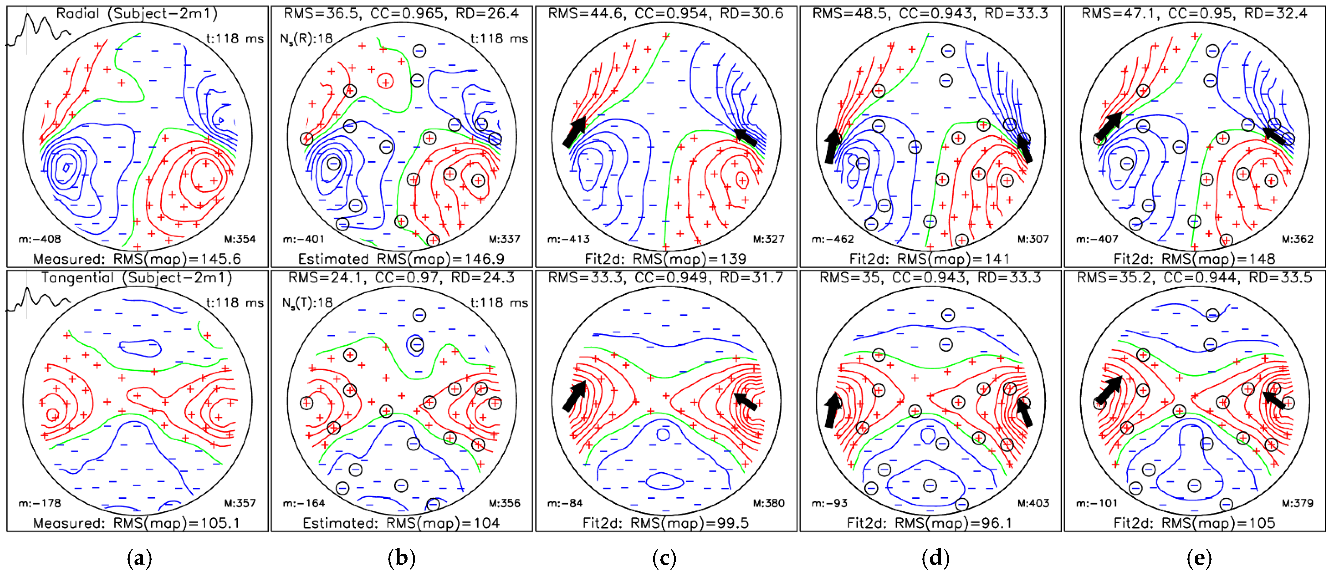


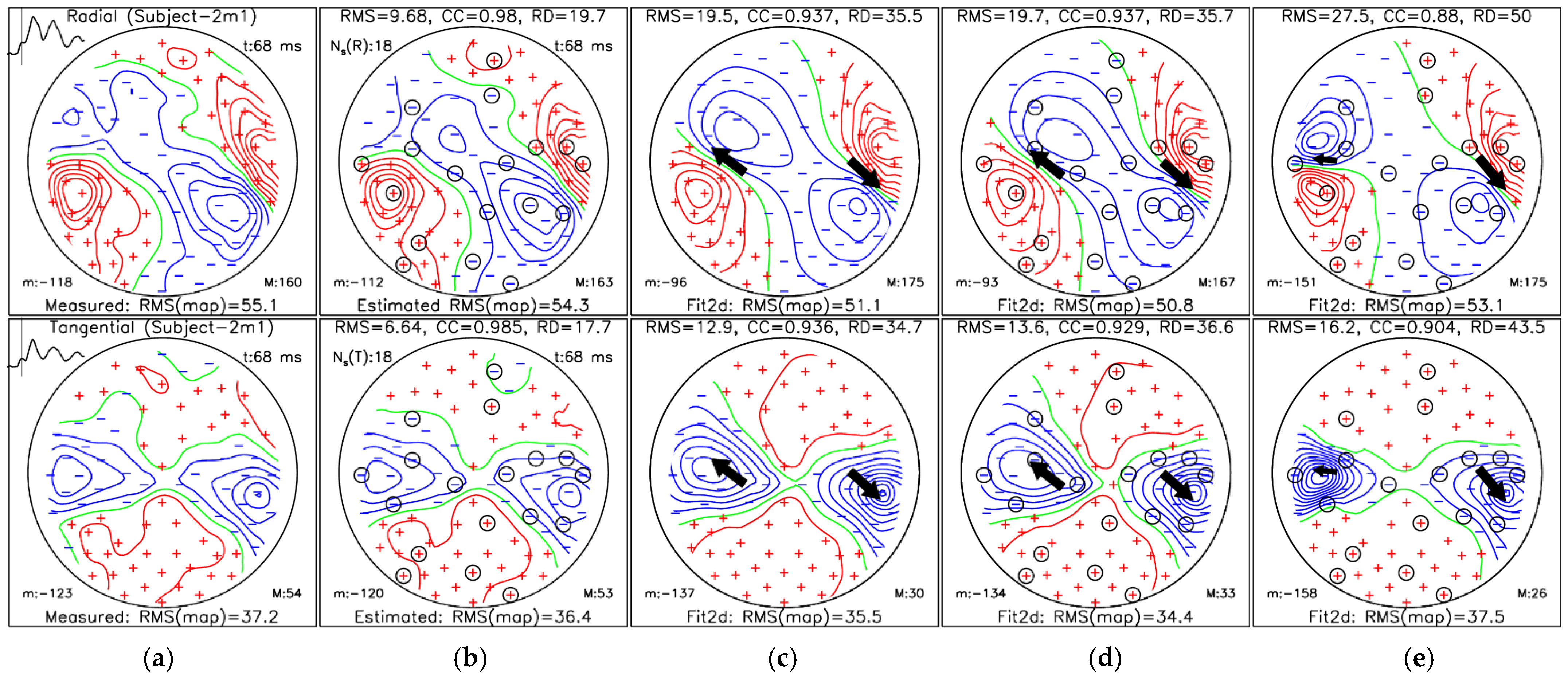
| Measures | Measured Map Fit (c) | Estimated Map Fit (d) | Selected Chan. Fit (e) |
|---|---|---|---|
| [mm] | (44.6, 0.9, 15.2) | (44.4, −4.7, 1.4) | (39.3, 1.5, 13.6) |
| [mm] | (−37.8, 3.1, 6.2) | (−37.8, −2.9, 0.6) | (−32.5, 5.3, 8.7) |
| [µAm] | (10.5, −12.0, −30.0) | (−1.3, −23.1, −36.7) | (15.4, −18.8, −42.6) |
| [µAm] | (−8.1, −26.2, −36.6) | (2.2, −37.0, −39.0) | (−19.8, −37.0, −51.9) |
| [mm] | / | 14.9 | 5.6 |
| [mm] | / | 8.2 | 6.2 |
| [mm] | / | 17.0 | 8.3 |
| [°] | / | 22.3 | 1.9 |
| [°] | / | 14.8 | 7.1 |
| Measures | Measured Map Fit (c) | Estimated Map Fit (d) | Selected Chan. Fit (e) |
|---|---|---|---|
| [mm] | (49.7, −6.4, 27.3) | (48.9, −6.9, 25.5) | (52.4, −6.0, 24.7) |
| [mm] | (−34.0, 2.1, 41.7) | (−29.6, 0.2, 43.8) | (−57.9, 2.8, 34.5) |
| [µAm] | (−3.2, 2.9, 6.5) | (−3.3, 2.8, 7.0) | (−2.1, 3.3, 5.2) |
| [µAm] | (5.5, −3.3, 4.6) | (6.1, −4.4, 4.1) | (1.8, −0.1, 3.1) |
| [mm] | / | 2.0 | 3.7 |
| [mm] | / | 5.3 | 25.0 |
| [mm] | / | 5.6 | 25.2 |
| [°] | / | 1.8 | 9.9 |
| [°] | / | 8.0 | 29.3 |
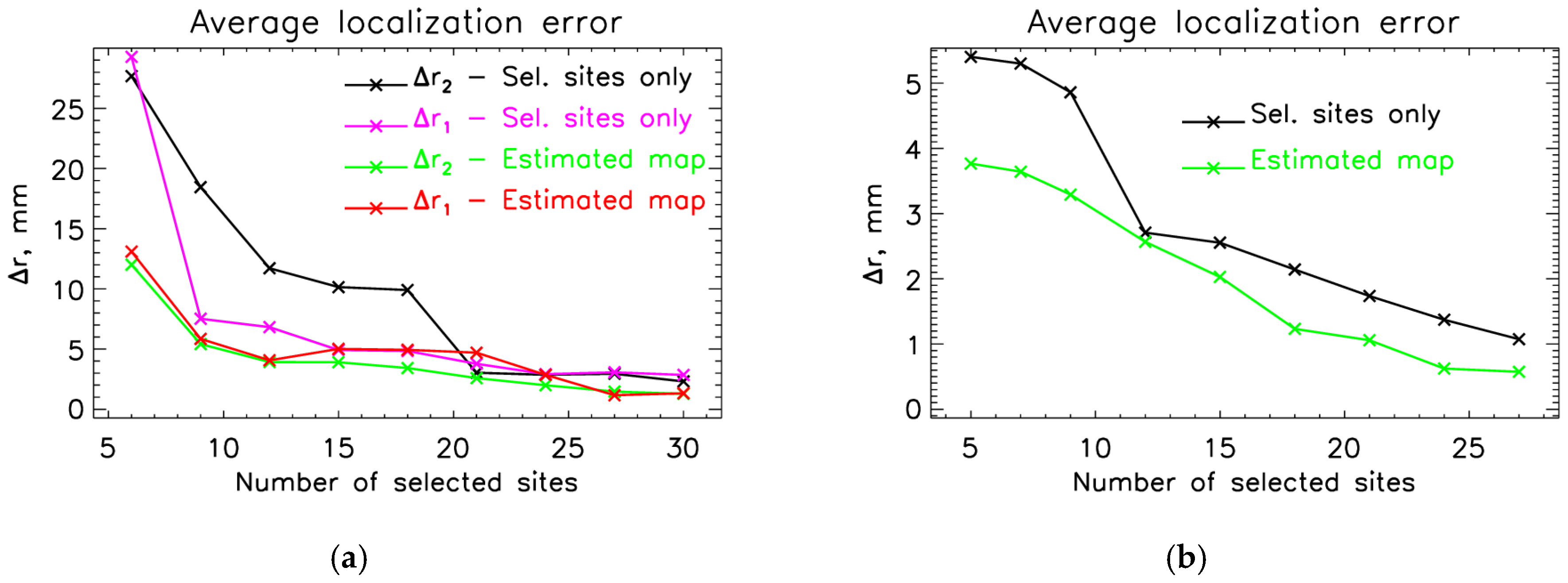
| Evaluation of Estimated M100 | Localization—Estimated | Localization—Sel. Sites Only | |||||
|---|---|---|---|---|---|---|---|
| Nm | [fT] | [%] | [mm] | [mm] | [mm] | [mm] | |
| 6 | 34.9 ± 19.2 | 37.8 ± 7.5 | 0.926 ± 0.034 | 13.1 ± 5.3 | 12 ± 8.2 | 29.3 ± 11.9 | 27.7 ± 16.8 |
| 9 | 21.5 ± 11.7 | 24.2 ± 4.6 | 0.97 ± 0.01 | 5.8 ± 3.8 | 5.4 ± 2.7 | 7.5 ± 6.1 | 18.5 ± 18.8 |
| 12 | 20.5 ± 10.9 | 22.9 ± 4.4 | 0.973 ± 0.01 | 4.1 ± 1.5 | 3.9 ± 2.5 | 6.8 ± 4.3 | 11.7 ± 16.8 |
| 15 | 19.1 ± 10.6 | 21.1 ± 5.2 | 0.977 ± 0.011 | 5.0 ± 3.8 | 3.9 ± 1.7 | 4.9 ± 2.4 | 10.1 ± 16.2 |
| 18 | 17.5 ± 9.5 | 19.3 ± 5.5 | 0.98 ± 0.011 | 4.9 ± 4.4 | 3.4 ± 2.2 | 4.8 ± 3.1 | 9.9 ± 16.9 |
| 21 | 16.8 ± 9.5 | 18.8 ± 6.3 | 0.981 ± 0.013 | 4.7 ± 4.1 | 2.6 ± 1.7 | 3.8 ± 2.9 | 3.0 ± 1.2 |
| 24 | 14.2 ± 9.7 | 15.3 ± 5.3 | 0.988 ± 0.009 | 2.9 ± 3.8 | 2 ± 1.4 | 2.9 ± 2.9 | 2.9 ± 1.5 |
| 27 | 12.7 ± 9.0 | 13.4 ± 5.0 | 0.99 ± 0.007 | 1.2 ± 0.8 | 1.5 ± 1.1 | 3.1 ± 2.6 | 2.9 ± 1.6 |
| 30 | 11.7 ± 8.5 | 12.1 ± 4.9 | 0.992 ± 0.007 | 1.3 ± 1.0 | 1.3 ± 0.9 | 2.9 ± 2.7 | 2.3 ± 1.0 |
| Evaluation of Estimated M100 | Localization of M100 | ||||
|---|---|---|---|---|---|
| Estimated Map | Selected Sites Only | ||||
| Nm | [fT] | [%] | [mm] | [mm] | |
| 5 | 32.2 ± 13.1 | 32.7 ± 12.1 | 0.944 ± 0.037 | 3.8 ± 1.8 | 5.4 ± 2.5 |
| 7 | 26.1 ± 11.4 | 25.7 ± 9.4 | 0.966 ± 0.021 | 3.6 ± 1.6 | 5.3 ± 3.2 |
| 9 | 22.5 ± 9.9 | 22.4 ± 10.1 | 0.975 ± 0.024 | 3.3 ± 2.5 | 4.9 ± 3.3 |
| 12 | 17.6 ± 8.5 | 18.2 ± 8.3 | 0.982 ± 0.018 | 2.6 ± 1.6 | 2.7 ± 0.8 |
| 15 | 16.1 ± 8.3 | 16.0 ± 7.5 | 0.985 ± 0.015 | 2.0 ± 1.3 | 2.6 ± 1.0 |
| 18 | 13.4 ± 9.8 | 12.3 ± 6.3 | 0.991 ± 0.010 | 1.2 ± 0.8 | 2.1 ± 0.9 |
| 21 | 11.2 ± 8.6 | 9.8 ± 5.5 | 0.994 ± 0.007 | 1.1 ± 1.1 | 1.7 ± 1.2 |
| 24 | 9.4 ± 8.2 | 8.3 ± 5.4 | 0.995 ± 0.006 | 0.6 ± 0.7 | 1.4 ± 0.9 |
| 27 | 9.3 ± 8.9 | 7.9 ± 5.8 | 0.995 ± 0.007 | 0.6 ± 0.6 | 1.1 ± 0.9 |
Disclaimer/Publisher’s Note: The statements, opinions and data contained in all publications are solely those of the individual author(s) and contributor(s) and not of MDPI and/or the editor(s). MDPI and/or the editor(s) disclaim responsibility for any injury to people or property resulting from any ideas, methods, instructions or products referred to in the content. |
© 2026 by the authors. Licensee MDPI, Basel, Switzerland. This article is an open access article distributed under the terms and conditions of the Creative Commons Attribution (CC BY) license.
Share and Cite
Marhl, U.; Hren, R.; Sander, T.; Jazbinšek, V. Optimizing OPM-MEG Sensor Layouts Using the Sequential Selection Algorithm with Simulated Sources and Individual Anatomy. Sensors 2026, 26, 1292. https://doi.org/10.3390/s26041292
Marhl U, Hren R, Sander T, Jazbinšek V. Optimizing OPM-MEG Sensor Layouts Using the Sequential Selection Algorithm with Simulated Sources and Individual Anatomy. Sensors. 2026; 26(4):1292. https://doi.org/10.3390/s26041292
Chicago/Turabian StyleMarhl, Urban, Rok Hren, Tilmann Sander, and Vojko Jazbinšek. 2026. "Optimizing OPM-MEG Sensor Layouts Using the Sequential Selection Algorithm with Simulated Sources and Individual Anatomy" Sensors 26, no. 4: 1292. https://doi.org/10.3390/s26041292
APA StyleMarhl, U., Hren, R., Sander, T., & Jazbinšek, V. (2026). Optimizing OPM-MEG Sensor Layouts Using the Sequential Selection Algorithm with Simulated Sources and Individual Anatomy. Sensors, 26(4), 1292. https://doi.org/10.3390/s26041292