Estimation of Respiratory Volumes During Tidal Breathing Using Two Depth Cameras


Highlights
- A system of two facing depth cameras can accurately estimate breathing volumes.
- Depth cameras could be used instead of a laboratory motion capture system for optoelectronic plethysmography applications.
- Depth cameras offer the potential for clinical implementation of respiratory monitoring.
Abstract
1. Introduction
2. Methods
2.1. Participants
2.2. Set up
2.3. Defining a Global Coordinate System
2.4. Experimental Data Collection
2.5. Volume Algorithm
2.6. Spirometry Comparison and Statistical Analysis
3. Results
4. Discussion
5. Conclusions
Author Contributions
Funding
Institutional Review Board Statement
Informed Consent Statement
Data Availability Statement
Acknowledgments
Conflicts of Interest
Abbreviations
| RMSE | Root Mean Square Error |
| TV | Tidal Volume |
| OEP | Optoelectronic Plethysmography |
| SD | Standard Deviation |
Appendix A. Marker Interpolation


| Label | Marker Type | Equation/Placement | |
|---|---|---|---|
| Full Set | Reduced Set | ||
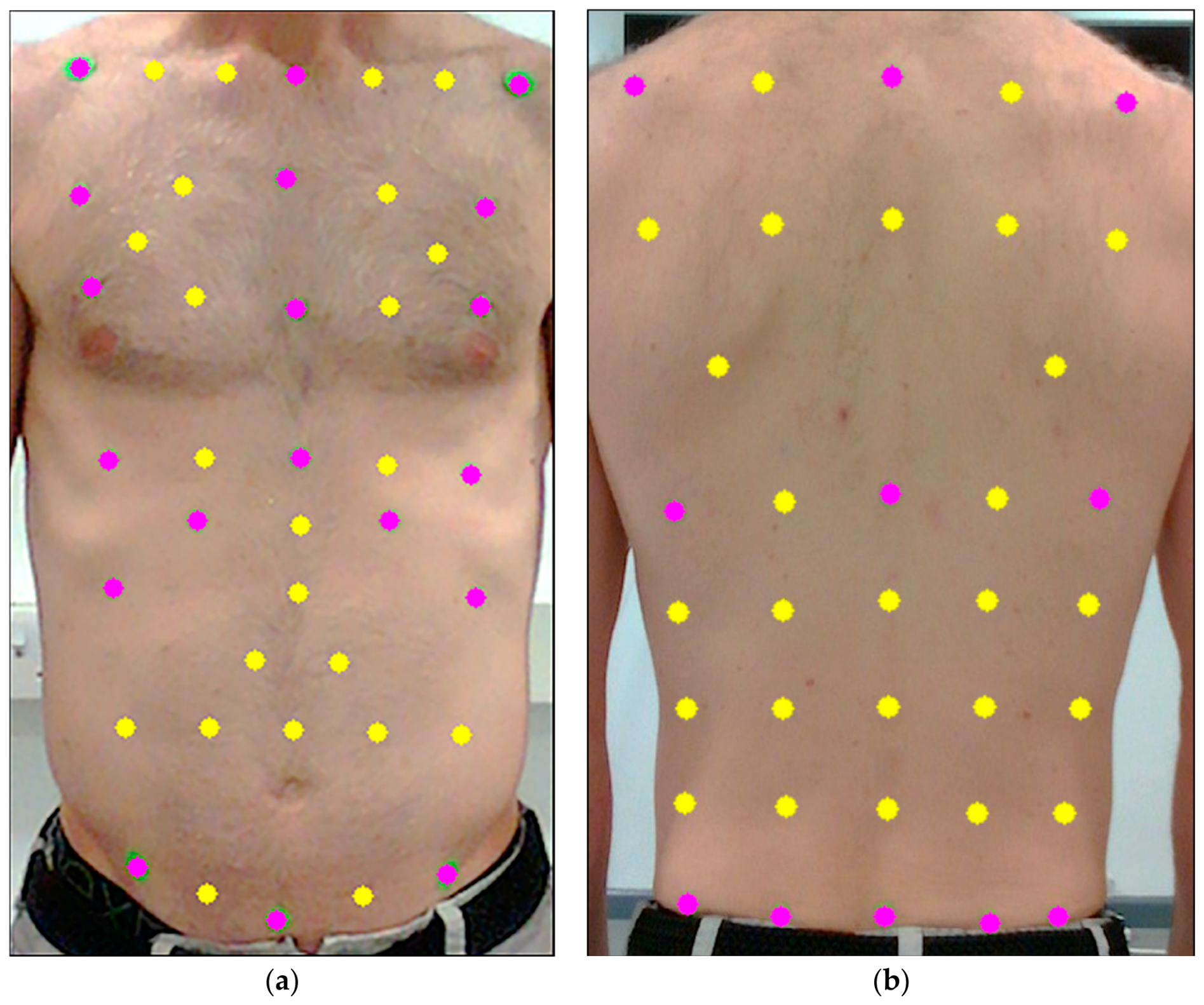
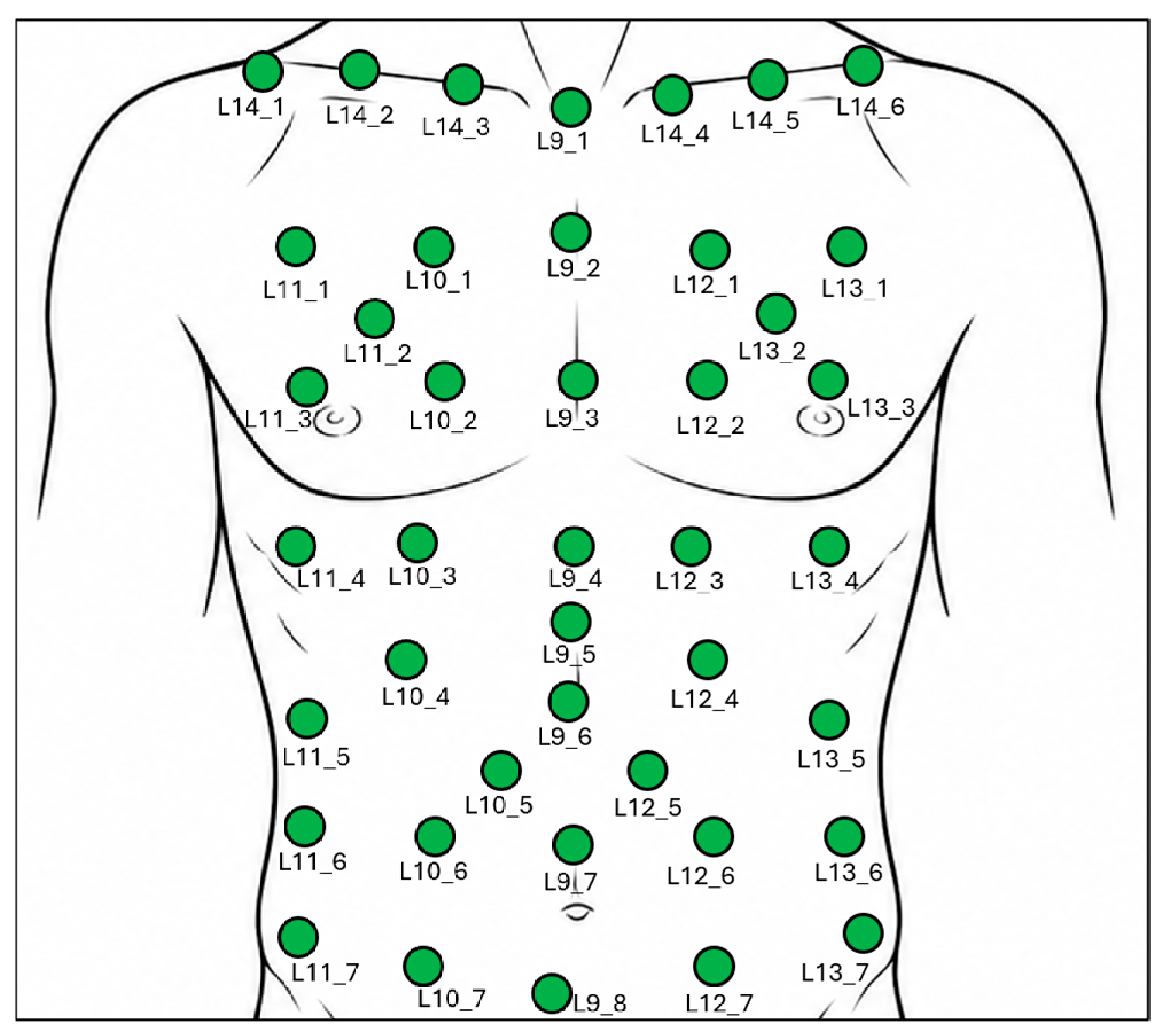
| L9_1 | Physical | Physical | Intrajugular notch |
| L14_1 | Physical | Physical | End of right clavicle |
| L14_6 | Physical | Physical | End of left clavicle |
| L11_7 | Physical | Physical | Right ASIS |
| L9_8 | Physical | Physical | Midline of the body on the lowest point of the abdomen |
| L13_7 | Physical | Physical | Left ASIS |
| L9_4 | Physical | Physical | Xiphoid Process |
| L11_4 | Physical | Physical | Level with the Xiphoid Process and in line with right ASIS |
| L11_5 | Physical | Physical | Costal Margin in line with the right ASIS |
| L13_4 | Physical | Physical | Level with the Xiphoid Process and in line with left ASIS |
| L13_5 | Physical | Physical | Costal Margin in line with the left ASIS |
| L10_4 | Physical | Physical | Costal margin at the right mid-clavicular line |
| L12_4 | Physical | Physical | Costal margin at the left mid-clavicular line |
| L9_2 | Physical | Virtual | |
| L9_3 | Physical | Virtual | |
| L11_1 | Physical | Virtual | |
| L11_3 | Physical | Virtual | |
| L13_1 | Physical | Virtual | |
| L13_3 | Physical | Virtual | |
| L14_2 | Virtual | Virtual | |
| L14_3 | Virtual | Virtual | |
| L14_4 | Virtual | Virtual | |
| L14_5 | Virtual | Virtual | |
| L10_1 | Virtual | Virtual | |
| L12_1 | Virtual | Virtual | |
| L10_2 | Virtual | Virtual | |
| L12_2 | Virtual | Virtual | |
| L10_3 | Virtual | Virtual | |
| L12_3 | Virtual | Virtual | |
| L11_2 | Virtual | Virtual | |
| L13_2 | Virtual | Virtual | |
| L10_7 | Virtual | Virtual | |
| L12_7 | Virtual | Virtual | |
| L11_6 | Virtual | Virtual | |
| L13_6 | Virtual | Virtual | |
| L10_6 | Virtual | Virtual | |
| L9_7 | Virtual | Virtual | |
| L12_6 | Virtual | Virtual | |
| L9_5 | Virtual | Virtual | |
| L9_6 | Virtual | Virtual | |
| L10_5 | Virtual | Virtual | |
| L12_5 | Virtual | Virtual | |

| Label | Marker Type | Placement/Equation | |
|---|---|---|---|
| Full Set | Reduced Set | ||
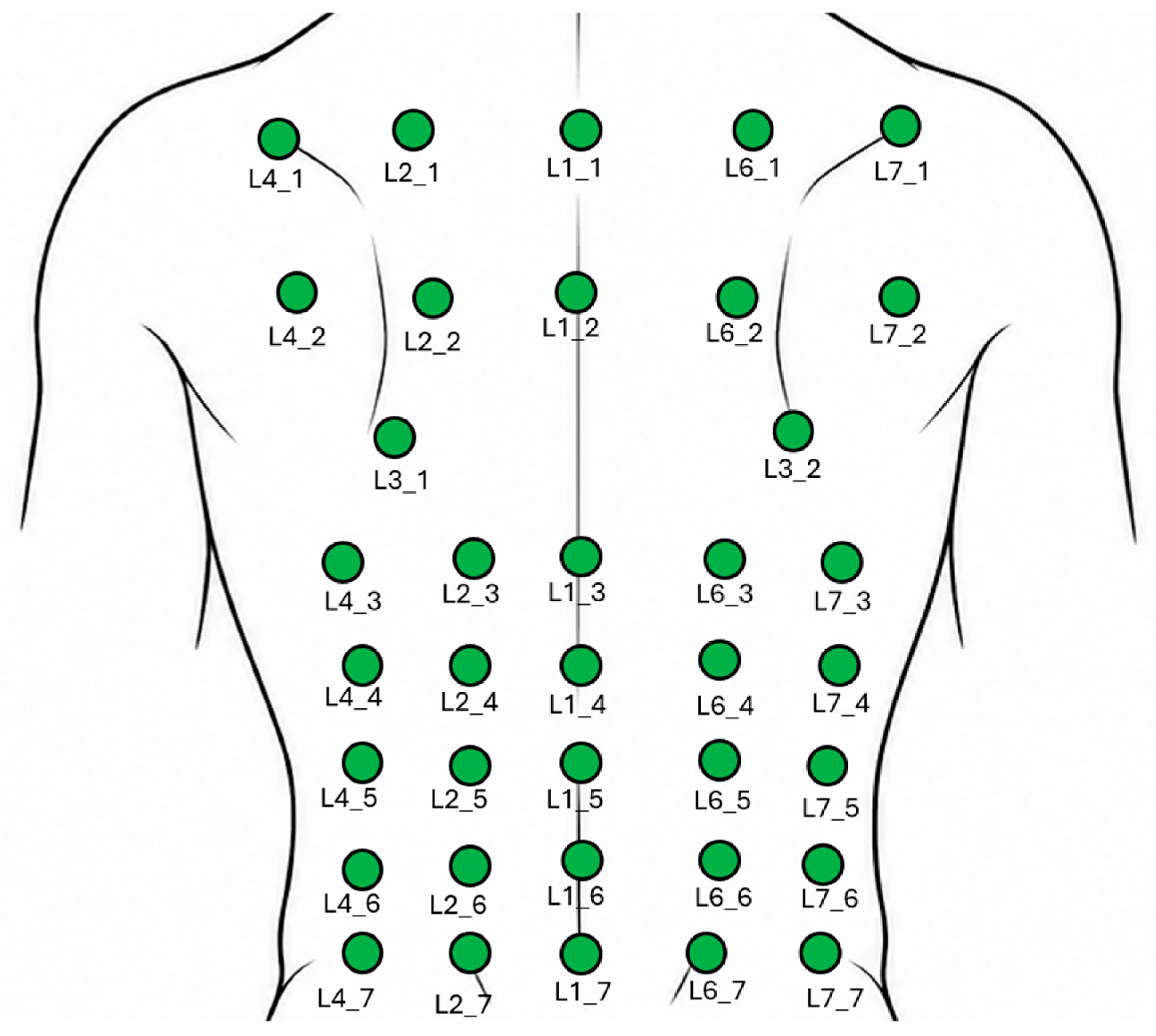
| L4_1 | Physical | Physical | Directly posterior to the end of the left clavicle |
| L7_1 | Physical | Physical | Directly posterior to the end of the right clavicle |
| L4_7 | Physical | Physical | At the level of the PSIS on the left posterior auxillary line |
| L7_7 | Physical | Physical | At the level of the PSIS on the line of the |
| L1_1 | Physical | Physical | |
| L1_7 | Physical | Virtual | |
| L2_7 | Physical | Virtual | Left PSIS/ |
| L6_7 | Physical | Virtual | Right PSIS/ |
| L4_3 | Physical | Virtual | |
| L1_3 | Physical | Virtual | |
| L7_3 | Physical | Virtual | |
| L2_1 | Virtual | Virtual | |
| L6_1 | Virtual | Virtual | |
| L2_3 | Virtual | Virtual | |
| L6_3 | Virtual | Virtual | |
| L4_2 | Virtual | Virtual | |
| L2_2 | Virtual | Virtual | |
| L1_2 | Virtual | Virtual | |
| L6_2 | Virtual | Virtual | |
| L7_2 | Virtual | Virtual | |
| L3_1 | Virtual | Virtual | |
| L3_2 | Virtual | Virtual | |
| L4_4 | Virtual | Virtual | |
| L2_4 | Virtual | Virtual | |
| L1_4 | Virtual | Virtual | |
| L6_4 | Virtual | Virtual | |
| L7_4 | Virtual | Virtual | |
| L4_5 | Virtual | Virtual | |
| L2_5 | Virtual | Virtual | |
| L1_5 | Virtual | Virtual | |
| L6_5 | Virtual | Virtual | |
| L7_5 | Virtual | Virtual | |
| L4_6 | Virtual | Virtual | |
| L2_6 | Virtual | Virtual | |
| L1_6 | Virtual | Virtual | |
| L6_6 | Virtual | Virtual | |
| L7_6 | Virtual | Virtual | |
Appendix B
| Participant | Sex | BMI | Mean Euclidean Distance (mm) | Total Volume Bias (L) | Mean Breath Difference (L) | Mean Spirometer TV (L) | Discrepancy (%) |
|---|---|---|---|---|---|---|---|
| Spiro17 | M | 18.8 | 37.6 | −1.726 | 0.029 | 1.345 | −2.17% |
| Spiro19 | M | 20.6 | 38.6 | −1.786 | 0.031 | 0.875 | −3.56% |
| Spiro9 | M | 28.7 | 40.5 | −2.854 | 0.035 | 1.127 | −3.07% |
| Spiro16 | F | 23.7 | 35.9 | −1.614 | 0.021 | 0.489 | −4.37% |
| Spiro3 | F | 20.4 | 35.9 | −1.716 | 0.041 | 1.107 | −3.66% |
| Spiro24 | F | 26.2 | 39.6 | −2.006 | 0.031 | 0.748 | −4.13% |
| Mean | 23.1 | 38.0 | −1.951 | 0.031 | 0.949 | −3.49% |
Appendix C. Linear Mixed-Effects Model
Appendix C.1. Linear Mixed Model Method
Appendix C.2. Linear Mixed Model Results
Appendix D

References
- Graham, B.L.; Steenbruggen, I.; Miller, M.R.; Barjaktarevic, I.Z.; Cooper, B.G.; Hall, G.L.; Hallstrand, T.S.; Kaminsky, D.A.; McCarthy, K.; McCormack, M.C.; et al. Standardization of spirometry 2019 update. An official American thoracic society and European respiratory society technical statement. Am. J. Respir. Crit. Care Med. 2019, 200, e70–e88. [Google Scholar] [CrossRef] [Scilit]
- Wasserman, K. Diagnosing cardiovascular and lung pathophysiology from exercise gas exchange. Chest 1997, 112, 1091–1101. [Google Scholar] [CrossRef] [Scilit]
- Massaroni, C.; Carraro, E.; Vianello, A.; Miccinilli, S.; Morrone, M.; Levai, I.K.; Schena, E.; Saccomandi, P.; Sterzi, S.; Dickinson, J.W.; et al. Optoelectronic Plethysmography in Clinical Practice and Research: A Review. Respiration 2017, 93, 339–354. [Google Scholar] [CrossRef] [Scilit]
- Massaroni, C.; Senesi, G.; Schena, E.; Silvestri, S. Analysis of breathing via optoelectronic systems: Comparison of four methods for computing breathing volumes and thoraco-abdominal motion pattern. Comput. Methods Biomech. Biomed. Eng. 2017, 20, 1678–1689. [Google Scholar] [CrossRef] [Scilit]
- Adhikari, N.G.; Hunsicker, E.; Pain, M.T.G.; Dickinson, J.W.; Winter, S.L. Agreement Between a Pre-Markered T-Shirt and Manual Marker Placement for Opto-Electronic Plethysmography (OEP) Measures. Sensors 2025, 25, 4464. [Google Scholar] [CrossRef] [Scilit]
- Smyth, C.M.E.; Winter, S.L.; Dickinson, J.W. Novel Real-Time OEP Phase Angle Feedback System for Dysfunctional Breathing Pattern Training—An Acute Intervention Study. Sensors 2021, 21, 3714. [Google Scholar] [CrossRef] [Scilit]
- Wichum, F.; Wiede, C.; Seidl, K. Depth-Based Measurement of Respiratory Volumes: A Review. Sensors 2022, 22, 9680. [Google Scholar] [CrossRef] [Scilit]
- Addison, A.P.; Addison, P.S.; Smit, P.; Jacquel, D.; Borg, U.R. Noncontact Respiratory Monitoring Using Depth Sensing Cameras: A Review of Current Literature. Sensors 2021, 21, 1135. [Google Scholar] [CrossRef] [Scilit]
- Kempfle, J.; Van Laerhoven, K. Breathing In-Depth: A Parametrization Study on RGB-D Respiration Extraction Methods. Front. Comput. Sci. 2021, 3, 757277. [Google Scholar] [CrossRef] [Scilit]
- Kojima, Y.; Higaki, T.; Inoue, H.; Raytchev, B.; Gu, Y.; Nakamura, Y. Contactless Respiratory Waveform Estimation Using a Depth Camera and AI-Based Body Detection. Sensors 2026, 26, 3476. [Google Scholar] [CrossRef] [Scilit]
- Romano, C.; Schena, E.; Silvestri, S.; Massaroni, C. Non-Contact Respiratory Monitoring Using an RGB Camera for Real-World Applications. Sensors 2021, 21, 5126. [Google Scholar] [CrossRef] [Scilit]
- Sun, C.; Li, W.; Chen, C.; Wang, Z.; Chen, W. An Unobtrusive and Non-Contact Method for Respiratory Measurement with Respiratory Region Detecting Algorithm Based on Depth Images. IEEE Access 2019, 7, 8300–8315. [Google Scholar] [CrossRef] [Scilit]
- Valenzuela, A.; Sibuet, N.; Hornero, G.; Casas, O. Non-Contact Video-Based Assessment of the Respiratory Function Using a RGB-D Camera. Sensors 2021, 21, 5605. [Google Scholar] [CrossRef] [Scilit]
- Van Hove, O.; Andrianopoulos, V.; Dabach, A.; Debeir, O.; Van Muylem, A.; Leduc, D.; Legrand, A.; Ercek, R.; Feipel, V.; Bonnechère, B. The use of time-of-flight camera to assess respiratory rates and thoracoabdominal depths in patients with chronic respiratory disease. Clin. Respir. J. 2023, 17, 176–186. [Google Scholar] [CrossRef] [Scilit]
- Oh, K.; Shin, C.S.; Kim, J.; Yoo, S.K. Level-Set Segmentation-Based Respiratory Volume Estimation Using a Depth Camera. IEEE J. Biomed. Health Inform. 2019, 23, 1674–1682. [Google Scholar] [CrossRef] [Scilit]
- Aoki, H.; Miyazaki, M.; Nakamura, H.; Furukawa, R.; Sagawa, R.; Kawasaki, H. Non-contact respiration measurement using structured light 3-D sensor. In Proceedings of the 2012 Proceedings of SICE Annual Conference (SICE), Akita, Japan, 20–23 August 2012. [Google Scholar]
- L’hEr, E.; Nazir, S.; Pateau, V.; Visvikis, D. Accuracy of noncontact surface imaging for tidal volume and respiratory rate measurements in the ICU. J. Clin. Monit. Comput. 2022, 36, 775–783. [Google Scholar] [CrossRef] [Scilit]
- Wijenayake, U.; Park, S.-Y. Real-Time External Respiratory Motion Measuring Technique Using an RGB-D Camera and Principal Component Analysis. Sensors 2017, 17, 1840. [Google Scholar] [CrossRef] [Scilit]
- Imano, W.; Kameyama, K.; Hollingdal, M.; Refsgaard, J.; Larsen, K.; Topp, C.; Kronborg, S.H.; Gade, J.D.; Dinesen, B. Non-Contact Respiratory Measurement Using a Depth Camera for Elderly People. Sensors 2020, 20, 6901. [Google Scholar] [CrossRef] [Scilit]
- Addison, P.S.; Smit, P.; Jacquel, D.; Addison, A.P.; Miller, C.; Kimm, G. Continuous non-contact respiratory rate and tidal volume monitoring using a Depth Sensing Camera. J. Clin. Monit. Comput. 2022, 36, 657–665. [Google Scholar] [CrossRef] [Scilit]
- Bachir, W. Time-of-flight abdominal wall displacement for non-invasive longitudinal monitoring of pulmonary function. Physiol. Meas. 2026, 47, 035009. [Google Scholar] [CrossRef] [Scilit]
- Takamoto, H.; Nishine, H.; Sato, S.; Sun, G.; Watanabe, S.; Seokjin, K.; Asai, M.; Mineshita, M.; Matsui, T. Development and Clinical Application of a Novel Non-contact Early Airflow Limitation Screening System Using an Infrared Time-of-Flight Depth Image Sensor. Front. Physiol. 2020, 11, 552942. [Google Scholar] [CrossRef] [Scilit]
- Soleimani, V.; Mirmehdi, M.; Damen, D.; Camplani, M.; Hannuna, S.; Sharp, C.; Dodd, J. Depth-Based Whole Body Photoplethysmography in Remote Pulmonary Function Testing. IEEE Trans. Biomed. Eng. 2018, 65, 1421–1431. [Google Scholar] [CrossRef] [Scilit]
- Sharp, C.; Soleimani, V.; Hannuna, S.; Camplani, M.; Damen, D.; Viner, J.; Mirmehdi, M.; Dodd, J.W. Toward Respiratory Assessment Using Depth Measurements from a Time-of-Flight Sensor. Front. Physiol. 2017, 8, 65. [Google Scholar] [CrossRef] [Scilit]
- RealSense. Intel RealSense LiDAR Camera L515 Datasheet. Available online: https://dev.realsenseai.com/docs/lidar-camera-l515-datasheet/ (accessed on 22 July 2026).
- RealSense. librealsense. v2.50.0. Available online: https://github.com/realsenseai/librealsense (accessed on 22 July 2026).
- Massaroni, C.; Schena, E.; Bastianini, F.; Scorza, A.; Saccomandi, P.; Lupi, G.; Botta, F.; Sciuto, S.A.; Silvestri, S. Development Of A Bio-Inspired Mechatronic Chest Wall Simulator for Evaluating the Performances of Opto-Electronic Plethysmography. Open Biomed. Eng. J. 2014, 8, 120–130. [Google Scholar] [CrossRef] [Scilit]
- Nakagawa, S.H. Schielzeth, A general and simple method for obtaining R2 from generalized linear mixed-effects models. Methods Ecol. Evol. 2013, 4, 133–142. [Google Scholar] [CrossRef] [Scilit]
- Bland, J.M.; Altman, D.G. Agreement Between Methods of Measurement with Multiple Observations Per Individual. J. Biopharm. Stat. 2007, 17, 571–582. [Google Scholar] [CrossRef] [Scilit]
- Konno, K.; Mead, J. Measurement of the separate volume changes of rib cage and abdomen during breathing. J. Appl. Physiol. 1967, 22, 407–422. [Google Scholar] [CrossRef] [Scilit]




| Full Marker Set | Reduced Marker Set | |||||||||
|---|---|---|---|---|---|---|---|---|---|---|
| ID | Sex | n | Abs Difference (mL) | Abs Discrepancy (%) | Norm. Euclidean d | RMSE (mL) | Abs Difference (mL) | Abs Discrepancy (%) | Norm. Euclidean d | RMSE (mL) |
| Spiro2 | M | 32 | 58.0 | 4.1 | 0.003 | 74.0 | 133.3 | 9.1 | 0.013 | 154.2 |
| Spiro5 | M | 21 | 54.1 | 5.6 | 0.005 | 65.9 | 150.0 | 15.2 | 0.031 | 159.1 |
| Spiro7 | M | 31 | 42.6 | 6.6 | 0.008 | 52.9 | 67.2 | 10.2 | 0.090 | 111.0 |
| Spiro9 | M | 13 | 80.2 | 8.1 | 0.008 | 90.4 | 76.7 | 8.0 | 0.009 | 86.8 |
| Spiro10 | M | 33 | 56.4 | 13.2 | 0.031 | 67.5 | 74.1 | 17.1 | 0.048 | 84.1 |
| Spiro11 | M | 30 | 94.3 | 14.5 | 0.036 | 109.2 | 128.1 | 19.7 | 0.067 | 144.4 |
| Spiro12 | M | 20 | 65.9 | 4.4 | 0.003 | 74.9 | 132.7 | 8.7 | 0.010 | 146.1 |
| Spiro13 | M | 39 | 57.5 | 10.5 | 0.017 | 71.5 | 64.8 | 11.4 | 0.022 | 78.7 |
| Spiro14 | M | 27 | 64.5 | 5.6 | 0.005 | 82.2 | 216.8 | 18.9 | 0.049 | 227.3 |
| Spiro15 | M | 26 | 84.6 | 13.1 | 0.021 | 96.6 | 70.9 | 10.4 | 0.016 | 84.0 |
| Spiro17 | M | 32 | 110.5 | 7.6 | 0.008 | 127.4 | 225.0 | 15.5 | 0.031 | 237.0 |
| Spiro19 | M | 30 | 47.0 | 6.0 | 0.005 | 58.7 | 26.0 | 3.3 | 0.002 | 34.0 |
| Spiro20 | M | 22 | 68.8 | 5.4 | 0.004 | 82.1 | 127.1 | 10.2 | 0.015 | 143.0 |
| Mean (M) | 27.4 | 68.0 | 8.1 | 0.012 | 81.0 | 114.8 | 12.1 | 0.031 | 130.0 | |
| SD (M) | 6.9 | 19.5 | 3.6 | 0.011 | 20.7 | 59.5 | 4.8 | 0.026 | 58.7 | |
| Spiro3 | F | 12 | 146.6 | 11.6 | 0.013 | 155.4 | 67.1 | 5.4 | 0.003 | 76.7 |
| Spiro4 | F | 9 | 94.3 | 5.9 | 0.005 | 107.4 | 226.8 | 13.9 | 0.026 | 247.4 |
| Spiro6 | F | 17 | 143.1 | 12.4 | 0.016 | 155.9 | 94.3 | 7.9 | 0.007 | 111.5 |
| Spiro8 | F | 11 | 74.9 | 10.4 | 0.014 | 86.4 | 73.9 | 10.3 | 0.021 | 87.1 |
| Spiro16 | F | 34 | 38.6 | 9.7 | 0.017 | 47.9 | 43.3 | 11.1 | 0.024 | 52.8 |
| Spiro21 | F | 35 | 39.6 | 6.6 | 0.008 | 49.6 | 57.5 | 9.0 | 0.024 | 87.2 |
| Spiro22 | F | 11 | 154.4 | 21.9 | 0.040 | 160.5 | 81.9 | 11.6 | 0.014 | 93.6 |
| Spiro24 | F | 30 | 63.0 | 9.9 | 0.017 | 77.4 | 118.8 | 18.7 | 0.055 | 133.1 |
| Mean (F) | 19.9 | 94.3 | 11.0 | 0.016 | 105.1 | 95.4 | 11.0 | 0.022 | 111.2 | |
| SD (F) | 11.2 | 48.1 | 4.9 | 0.011 | 47.3 | 57.8 | 4.0 | 0.016 | 59.9 | |
| Mean SD | 78.0 | 9.2 | 0.014 | 90.2 | 107.4 | 11.7 | 0.027 | 122.8 | ||
| 34.8 | 4.3 | 0.011 | 34.4 | 58.2 | 4.4 | 0.023 | 58.4 | |||
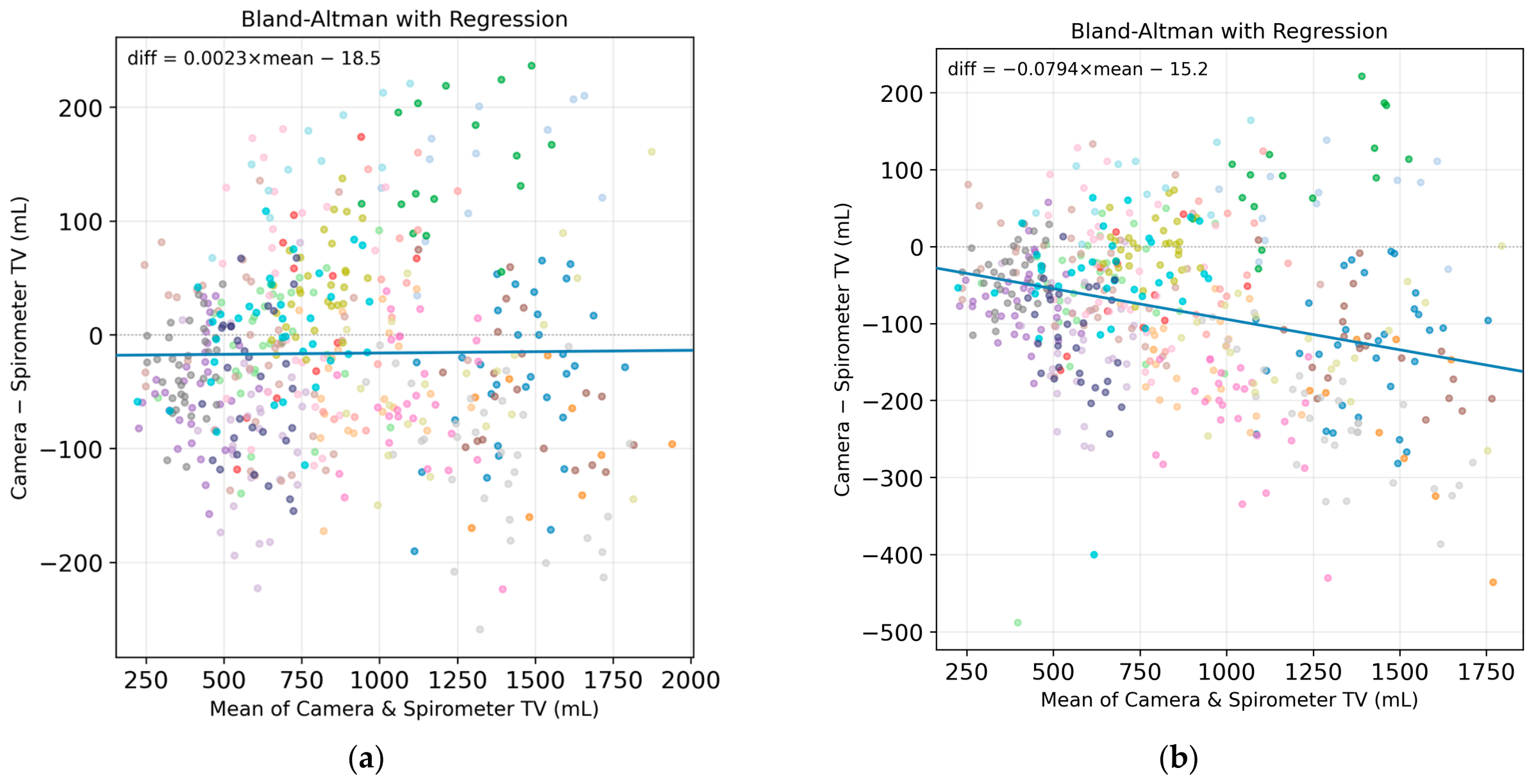
| Marker Set | Term | b | SE | t | p | 95% CI |
|---|---|---|---|---|---|---|
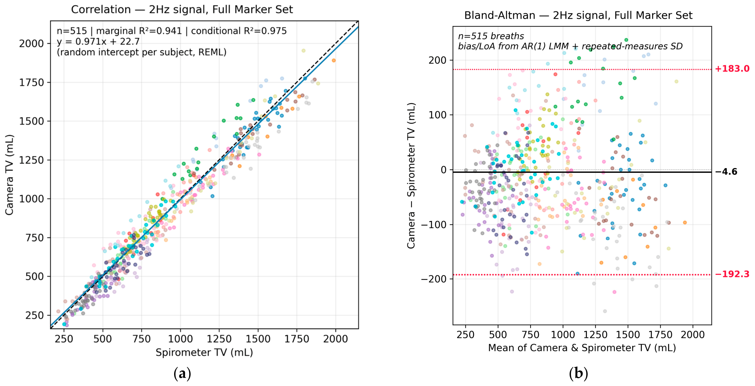
| Full | Intercept (bias, mL) | −4.62 | 16.16 | −0.29 | 0.775 | [−36.36, 27.13] |
| Full | Slope (spiro TV) | 0.971 | 0.0158 | 61.53 | <0.001 | [0.940, 1.002] |
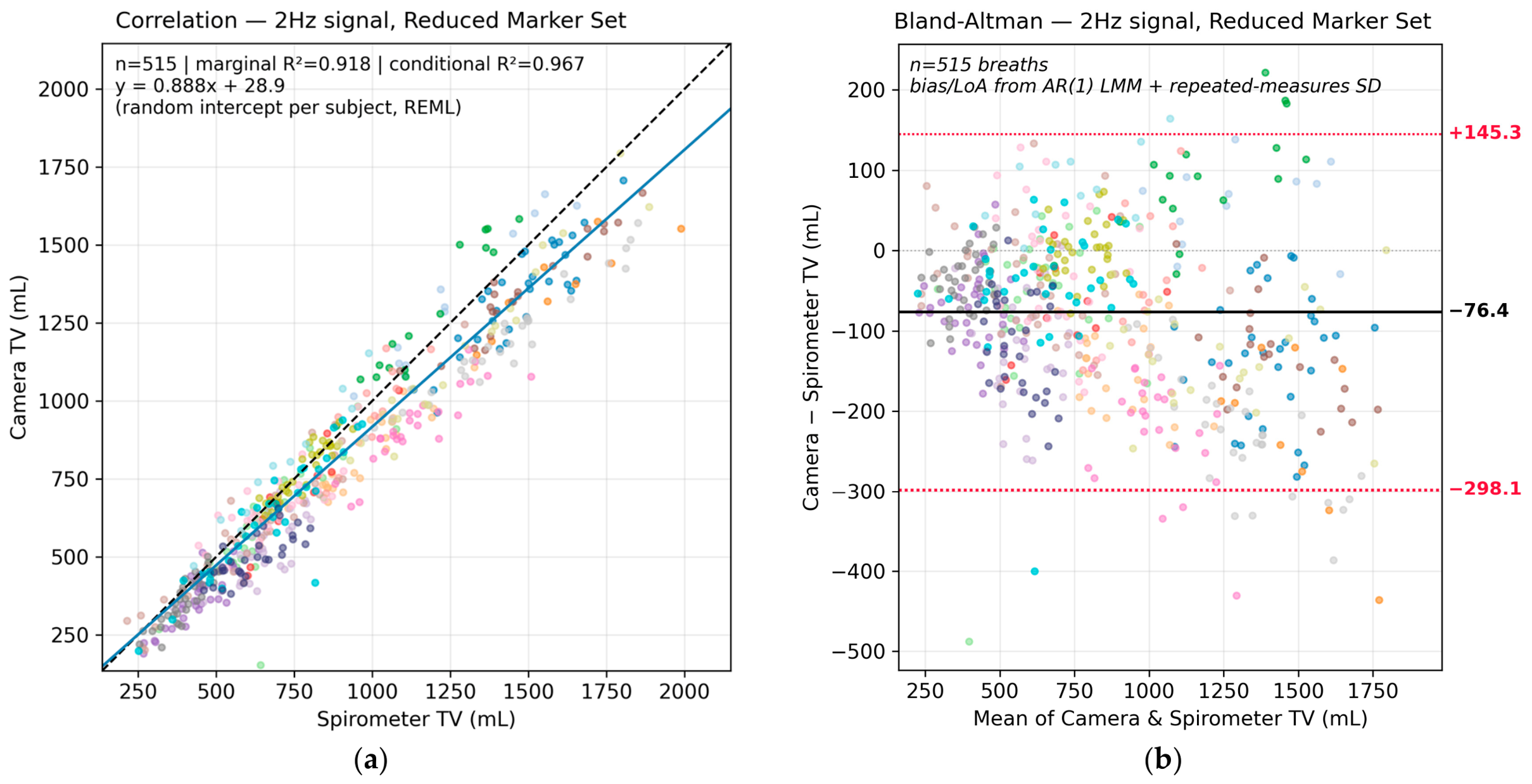
| Reduced | Intercept (bias, mL) | −76.43 | 19.93 | −3.84 | <0.001 | [−115.58, −37.27] |
| Reduced | Slope (spiro TV) | 0.888 | 0.0169 | 52.57 | <0.001 | [0.855, 0.921] |
Disclaimer/Publisher’s Note: The statements, opinions and data contained in all publications are solely those of the individual author(s) and contributor(s) and not of MDPI and/or the editor(s). MDPI and/or the editor(s) disclaim responsibility for any injury to people or property resulting from any ideas, methods, instructions or products referred to in the content. |
© 2026 by the authors. Licensee MDPI, Basel, Switzerland. This article is an open access article distributed under the terms and conditions of the Creative Commons Attribution (CC BY) license.
Share and Cite
Handley, A.; Preece, S.; Tresadern, P. Estimation of Respiratory Volumes During Tidal Breathing Using Two Depth Cameras. Sensors 2026, 26, 5793. https://doi.org/10.3390/s26185793
Handley A, Preece S, Tresadern P. Estimation of Respiratory Volumes During Tidal Breathing Using Two Depth Cameras. Sensors. 2026; 26(18):5793. https://doi.org/10.3390/s26185793
Chicago/Turabian StyleHandley, Adam, Stephen Preece, and Phil Tresadern. 2026. "Estimation of Respiratory Volumes During Tidal Breathing Using Two Depth Cameras" Sensors 26, no. 18: 5793. https://doi.org/10.3390/s26185793
APA StyleHandley, A., Preece, S., & Tresadern, P. (2026). Estimation of Respiratory Volumes During Tidal Breathing Using Two Depth Cameras. Sensors, 26(18), 5793. https://doi.org/10.3390/s26185793