Enhancing Cerebral Blood Flow Quantification: A Comprehensive Review of Denoising, Artifact Correction, and Simulation in Arterial Spin Labeling MRI
 , , , , , , and
Abstract
1. Introduction
2. ASL Image Enhancement
2.1. Traditional Methods for Improving the Quality of ASL Images
Noise Reduction—Traditional, Machine Learning and Deep Learning Techniques
3. Artifacts and Outlier Removal
4. ASL Dataset Simulation Procedure
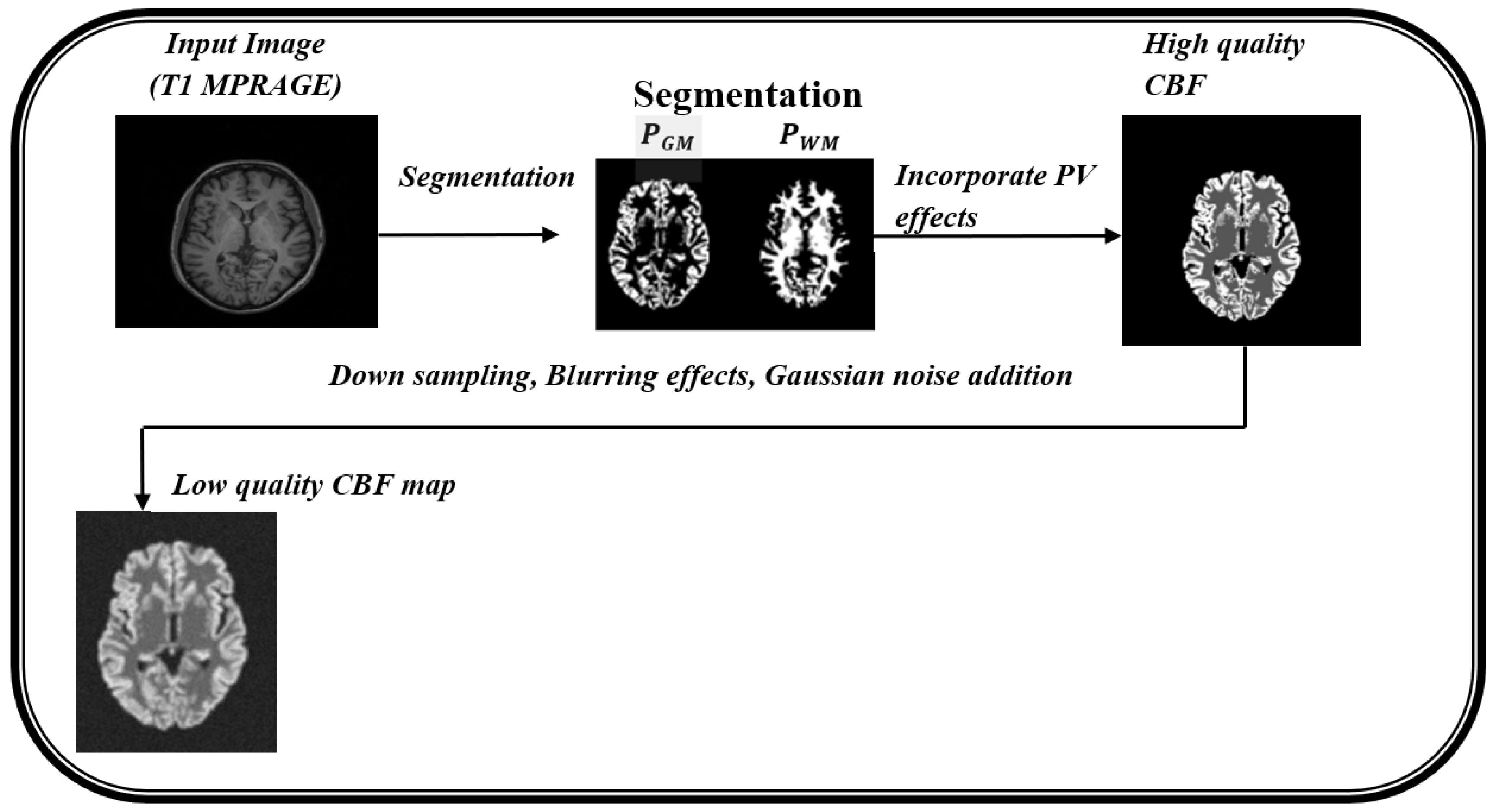
| Algorithm 1. Simulation Procedure for creating synthetic ASL CBF map |
| Input: Structural image (T1 MPRAGE) Output: ASL CBF map (high resolution and low resolution)
|
| Algorithm 2. Simulation Procedure for creating synthetic ASL CBF map and ASL L-C pairs |
| Input: Structural image (T1 MPRAGE), M0 image (PD) Output: Synthetic ASL CBF map, Label and Control images
|
5. Challenges in Clinical Translation and Future Research Directions
5.1. Challenges in Clinical Translation
- Standardization of ASL Protocols:
- Generalizability of AI Models:
- Availability of Reliable Reference Data:
- Clinical Validation
- Integration into Clinical Workflows:
- Interpretability of AI Models:
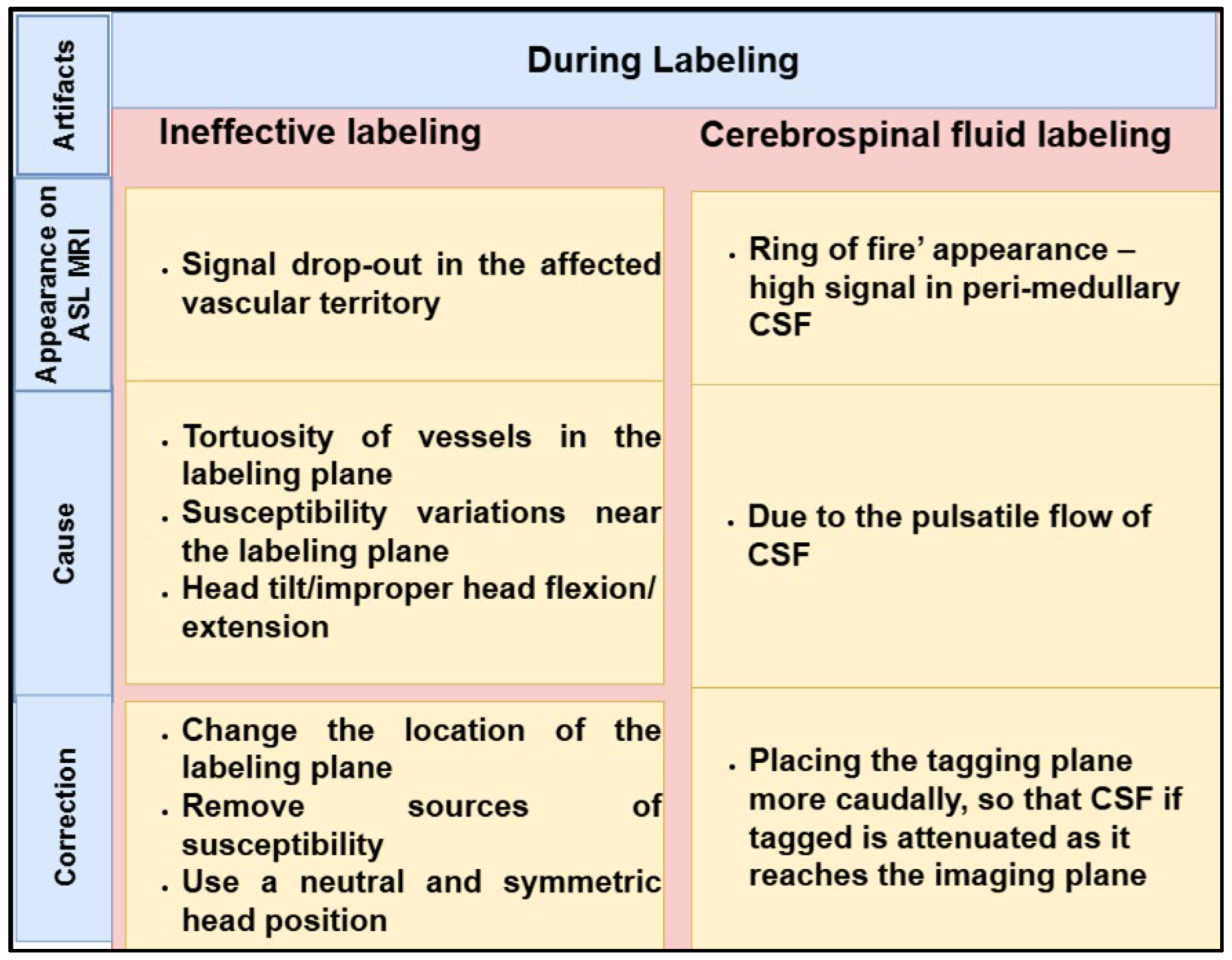
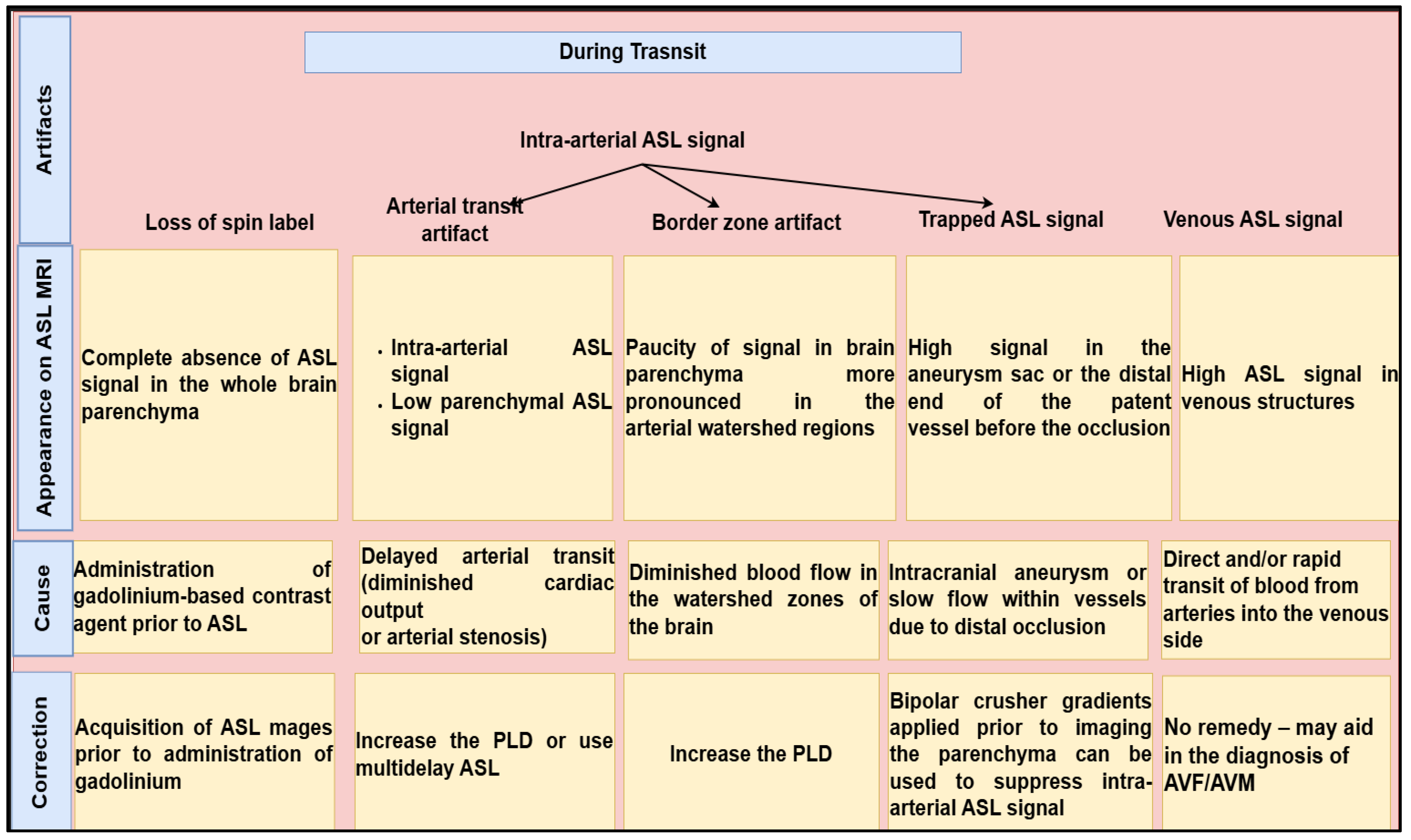
- Integrated Artifact and Outlier Correction:
5.2. Future Research Directions
- Enhancing Preprocessing for CBF Quantification
- Improving Deep Learning Architectures
- ➢
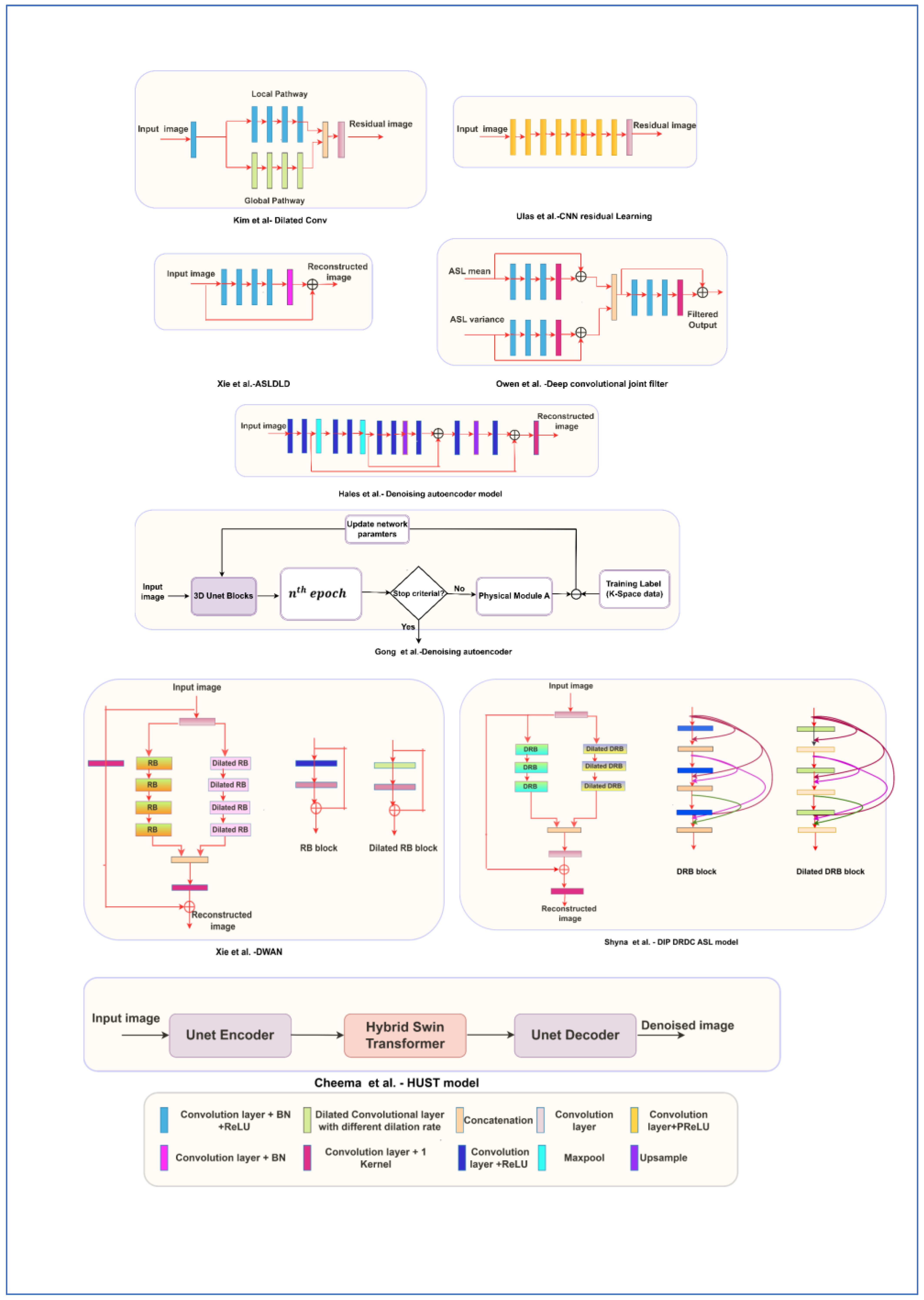
- Existing models, such as the Dual Independent Pathway–Densely Connected Residual Network with Dilated Convolution (DIP-DRDC), can be further enhanced through improvements in network architecture, training strategies, and validation methodologies. Training these models using better datasets tailored towards ASL imaging can produce favorable results.
- ➢
- Generative models, particularly Generative Adversarial Networks (GANs) [74,75], offer a promising approach for generating synthetic L–C pairs, helping to address the limited availability of training datasets while also reducing acquisition time. However, despite these advantages, it is essential to verify the accuracy of the generated images. If the training dataset is not sufficiently representative, the resulting synthetic images may contain unrealistic perfusion characteristics, which could introduce bias into CBF measurements.
- ➢
- The use of attention mechanisms [76] in models such as DIP-DRDC may allow the network to assign greater importance to relevant features while reducing the influence of irrelevant or noisy features. Moreover, incorporating TV regularization into the loss function may help ensure an appropriate balance between data fidelity and regularization in the generated images.
- Fine-Tuning and Longitudinal Studies
- Functional Applications and ASL fMRI Integration
- Optimization Techniques for Multi-PLD Approaches
- Domain Adaptation and Multi-center Generalization
- AI-based techniques for artifacts and Outlier detection and removal
6. Conclusions
Author Contributions
Funding
Institutional Review Board Statement
Informed Consent Statement
Data Availability Statement
Acknowledgments
Conflicts of Interest
Abbreviations
| AD | Alzheimer’s Disease |
| ADNI | Alzheimer’s Disease Neuroimaging Initiative |
| AI | Artificial Intelligence |
| AOC | Adaptive Outlier Cleaning |
| ANTs | Advanced Normalization Tools |
| ASL | Arterial Spin labeling |
| ASLDLD | Arterial Spin Labeling Deep Learning Denoising |
| ATT | Arterial Transit Time |
| CCC | Concordance Correlation Coefficient |
| CASL | Continuous ASL |
| CBF | Cerebral Blood Flow |
| CNN | Convolutional Neural Networks |
| CNR | Contrast to Noise Ratio |
| CompCor | Component-Based Noise Correction Method |
| DIP-DRDC | Dual Independent Pathway–Densely Connected Residual Network with Dilated Convolution |
| DL | Deep Learning |
| DT-CWT | Dual-tree complex wavelet transform |
| DWAN | Deep Weighted Averaging Network |
| EPISTAR | Echo Planar Imaging and Signal Targeting with Alternating Radio Frequency |
| FAIR | Flow-sensitive Alternating Inversion Recovery |
| fMRI | Functional MRI |
| FSL | Functional Magnetic Resonance Imaging of the Brain Software Library |
| GAN | Generative Adversarial Networks |
| GLM | General Linear Model |
| GM | Gray Matter |
| GRASE | Gradient and Spin Echo |
| ICA | Independent Component Analysis |
| ISMRM | International Society for Magnetic Resonance in Medicine |
| L-C | Label–Control |
| ML | Machine Learning |
| MPRAGE | Magnetization-Prepared Rapid Gradient Echo |
| MRI | Magnetic Resonance Imaging |
| MSE | Mean Squared Error |
| NESMA | Non-local Estimation of Multispectral Magnitudes |
| NLM | Non-Local Means |
| PAOC | Priors-guided Slice-wise Adaptive Outlier Cleaning |
| PASL | Pulsed Arterial Spin Labeling |
| PCA | Principal Component Analysis |
| PCASL | Pseudo-Continuous Arterial Spin Labeling |
| PD | Proton Density |
| PICORE | Proximal Inversion with a Control for Off-Resonance Effects |
| PLD | Post-Labeling Delay |
| PV | Partial Volume |
| PVE | Partial Volume Effect |
| PSNR | Peak SNR |
| PWI | Perfusion-Weighted Image |
| Q2TIPS | QUIPSS II with Thin-slice TI1 Periodic Saturation |
| QUIPSS | Quantitative Imaging of Perfusion using a Single Subtraction, Second Version |
| RF | Radio Frequency |
| RETROICOR | RETROspective Image CORrection |
| RMSE | Root Mean Square Error |
| ROC | Receiver Operating Characteristic |
| RPCA | Robust Principal Component Analysis |
| SAR | Specific Absorption Rate |
| SCORE | Structural Correlation-based Outlier Rejection |
| SNR | Signal to Noise Ratio |
| SPM | Statistical Parametric Mapping |
| STLRTV | Spatio-Temporal Low-Rank Total Variation |
| SVM | Support Vector Machine |
| SVMASLQ | Support Vector Machine-based Arterial Spin Labeling Quantification |
| TGV | Total Generalized Variation |
| TI | Inversion Time |
| TV | Total Variation |
| tSNR (TSNR) | Temporal Signal-to-Noise Ratio |
| WM | White Matter |
| XAI | Explainable Artificial Intelligence |
References
- Detre, J.A.; Leigh, J.S.; Williams, D.S.; Koretsky, A.P. Perfusion imaging. Magn. Reson. Med. 1992, 23, 37–45. [Google Scholar] [CrossRef] [Scilit] [PubMed]
- Williams, D.S.; Detre, J.A.; Leigh, J.S.; Koretsky, A.P. Magnetic resonance imaging of perfusion using spin inversion of arterial water. Proc. Natl. Acad. Sci. USA 1992, 89, 212–216. [Google Scholar] [CrossRef] [Scilit] [PubMed]
- Chikui, T.; Obara, M.; Simonetti, A.W.; Ohga, M.; Koga, S.; Kawano, S.; Matsuo, Y.; Kamintani, T.; Shiraishi, T.; Kitamoto, E.; et al. The principal of dynamic contrast enhanced MRI, the method of pharmacokinetic analysis, and its application in the head and neck region. Int. J. Dent. 2012, 2012, 480659. [Google Scholar] [CrossRef] [Scilit] [PubMed]
- Shiroishi, M.S.; Castellazzi, G.; Boxerman, J.L.; D’Amore, F.; Essig, M.; Nguyen, T.B.; Provenzale, J.M.; Enterline, D.S.; Anzalone, N.; Dörfler, A.; et al. Principles of t2*-weighted dynamic susceptibility contrast MRI technique in brain tumor imaging. J. Magn. Reson. Imaging 2015, 41, 296–313. [Google Scholar] [CrossRef] [Scilit] [PubMed]
- Gordon, Y.; Partovi, S.; Müller-Eschner, M.; Amarteifio, E.; Bäuerle, T.; Weber, M.-A.; Kauczor, H.-U.; Rengier, F. Dynamic contrast-enhanced magnetic resonance imaging: Fundamentals and application to the evaluation of the peripheral perfusion. Cardiovasc. Diagn. Ther. 2014, 4, 147. [Google Scholar] [CrossRef] [Scilit] [PubMed]
- Wong, E. Potential and Pitfalls of Arterial Spin Labeling Based Perfusion Imaging Techniques for MRI. In Functional MRI; Springer: Berlin/Heidelberg, Germany, 1999; pp. 63–69. [Google Scholar]
- Edelman, R.R.; Siewert, B.; Adamis, M.; Gaa, J.; Laub, G.; Wielopolski, P. Signal targeting with alternating radiofrequency (STAR) sequences: Application to MR angiography. Magn. Reson. Med. 1994, 31, 233–238. [Google Scholar] [CrossRef] [Scilit] [PubMed]
- Kim, S.-G. Quantification of relative cerebral blood flow change by flowsensitive alternating inversion recovery (FAIR) technique: Application to functional mapping. Magn. Reson. Med. 1995, 34, 293–301. [Google Scholar] [CrossRef] [Scilit] [PubMed]
- Wong, E.C.; Buxton, R.B.; Frank, L.R. Quantitative imaging of perfusion using a single subtraction (QUIPSS and QUIPSS II). Magn. Reson. Med. 1998, 39, 702–708. [Google Scholar] [CrossRef] [Scilit] [PubMed]
- Tanaka, F.; Umino, M.; Maeda, M.; Nakayama, R.; Inoue, K.; Kogue, R.; Obara, M.; Sakuma, H. Pseudocontinuous arterial spin labeling: Clinical applications and usefulness in head and neck entities. Cancers 2022, 14, 3872. [Google Scholar] [CrossRef] [Scilit] [PubMed]
- Woods, J.G.; Chappell, M.A.; Okell, T.W. A general framework for optimizing arterial spin labeling MRI experiments. Magn. Reson. Med. 2019, 81, 2474–2488. [Google Scholar] [CrossRef] [Scilit] [PubMed]
- Buxton, R.B.; Frank, L.R.; Wong, E.C.; Siewert, B.; Warach, S.; Edelman, R.R. A general kinetic model for quantitative perfusion imaging with arterial spin labeling. Magn. Reson. Med. 1998, 40, 383–396. [Google Scholar] [CrossRef] [Scilit] [PubMed]
- Chappell, M.A.; Groves, A.R.; MacIntosh, B.J.; Donahue, M.J.; Jezzard, P.; Woolrich, M.W. Partial volume correction of multiple inversion time arterial spin labeling MRI data. Magn. Reson. Med. 2011, 65, 1173–1183. [Google Scholar] [CrossRef] [Scilit] [PubMed]
- Petcharunpaisan, S.; Ramalho, J.; Castillo, M. Arterial spin labeling in neuroimaging. World J. Radiol. 2010, 2, 384. [Google Scholar] [CrossRef] [Scilit] [PubMed]
- Sollmann, N.; Hoffmann, G.; Schramm, S.; Reichert, M.; Petzsche, M.H.; Strobel, J.; Nigris, L.; Kloth, C.; Rosskopf, J.; Börner, C.; et al. Arterial spin labeling (ASL) in neuroradiological diagnostics–methodological overview and use cases. In RöFo-Fortschritte auf dem Gebiet der Röntgenstrahlen und der Bildgebenden Verfahren; Georg Thieme Verlag KG: Stuttgart, Germany, 2024; Volume 196, pp. 36–51. [Google Scholar]
- Chappell, M.A.; McConnell, F.A.K.; Golay, X.; Günther, M.; Hernandez-Tamames, J.A.; van Osch, M.J.; Asllani, I. Partial volume correction in arterial spin labeling perfusion MRI: A method to disentangle anatomy from physiology or an analysis step too far? Neuroimage 2021, 238, 118236. [Google Scholar] [CrossRef] [Scilit] [PubMed]
- Clement, P.; Petr, J.; Dijsselhof, M.B.; Padrela, B.; Pasternak, M.; Dolui, S.; Jarutyte, L.; Pinter, N.; Hernandez-Garcia, L.; Jahn, A.; et al. A beginner’s guide to arterial spin labeling (asl) image processing. Front. Radiol. 2022, 2, 929533. [Google Scholar] [CrossRef] [Scilit] [PubMed]
- Lindner, T.; Bolar, D.S.; Achten, E.; Barkhof, F.; Bastos-Leite, A.J.; Detre, J.A.; Golay, X.; Günther, M.; Wang, D.J.; Haller, S.; et al. Current state and guidance on arterial spin labeling perfusion MRI in clinical neuroimaging. Magn. Reson. Med. 2023, 89, 2024–2047. [Google Scholar] [CrossRef] [Scilit] [PubMed]
- Grade, M.; Hernandez Tamames, J.A.; Pizzini, F.B.; Achten, E.; Golay, X.; Smits, M. A neuroradiologist’s guide to arterial spin labeling MRI in clinical practice. Neuroradiology 2015, 57, 1181–1202. [Google Scholar] [CrossRef] [Scilit] [PubMed]
- Woods, J.G.; Achten, E.; Asllani, I.; Bolar, D.S.; Dai, W.; Detre, J.A.; Fan, A.P.; Fernández-Seara, M.A.; Golay, X.; Günther, M.; et al. Recommendations for quantitative cerebral perfusion MRI using multi-timepoint arterial spin labeling: Acquisition, quantification, and clinical applications. Magn. Reson. Med. 2024, 92, 469–495. [Google Scholar] [CrossRef] [Scilit] [PubMed]
- Telischak, N.A.; Detre, J.A.; Zaharchuk, G. Arterial spin labeling MRI: Clinical applications in the brain. J. Magn. Reson. Imaging 2015, 41, 1165–1180. [Google Scholar] [CrossRef] [Scilit] [PubMed]
- Iutaka, T.; de Freitas, M.B.; Omar, S.S.; Scortegagna, F.A.; Nael, K.; Nunes, R.H.; Pacheco, F.T.; Maia Júnior, A.C.M.; do Amaral, L.L.F.; da Rocha, A.J. Arterial spin labeling: Techniques, clinical applications, and interpretation. Radiographics 2022, 43, e220088. [Google Scholar] [CrossRef] [Scilit] [PubMed]
- Jaafar, N.; Alsop, D.C. Arterial spin labeling: Key concepts and progress towards use as a clinical tool. Magn. Reson. Med. Sci. 2024, 23, 352–366. [Google Scholar] [CrossRef] [Scilit] [PubMed]
- Togao, O.; Obara, M.; Yamashita, K.; Kikuchi, K.; Wada, T.; Murazaki, H.; Arimura, K.; Nishimura, A.; Horie, N.; van de Ven, K.; et al. Arterial Spin Labeling-Based MR Angiography for Cerebrovascular Diseases: Principles and Clinical Applications. J. Magn. Reson. Imaging 2024, 60, 1305–1324. [Google Scholar] [CrossRef] [Scilit] [PubMed]
- Alsaedi, A.; Thomas, D.; Bisdas, S.; Golay, X. Overview and critical appraisal of arterial spin labelling technique in brain perfusion imaging. Contrast Media Mol. Imaging 2018, 2018, 5360375. [Google Scholar] [CrossRef] [Scilit] [PubMed]
- Jaganmohan, D.; Pan, S.; Kesavadas, C.; Thomas, B. A pictorial review of brain arterial spin labelling artefacts and their potential remedies in clinical studies. Neuroradiol. J. 2021, 34, 154–168. [Google Scholar] [CrossRef] [Scilit] [PubMed]
- Amukotuwa, S.A.; Yu, C.; Zaharchuk, G. 3D Pseudocontinuous arterial spin labeling in routine clinical practice: A review of clinically significant artifacts. J. Magn. Reson. Imaging 2016, 43, 11–27. [Google Scholar] [CrossRef] [Scilit] [PubMed]
- Hu, H.H.; Rusin, J.A.; Peng, R.; Shao, X.; Smith, M.; Krishnamurthy, R.; Selvaraj, B.; Wang, D.J. Multi-phase 3D arterial spin labeling brain MRI in assessing cerebral blood perfusion and arterial transit times in children at 3T. Clin. Imaging 2019, 53, 210–220. [Google Scholar] [CrossRef] [Scilit] [PubMed]
- Bause, J.; Ehses, P.; Mirkes, C.; Shajan, G.; Scheffler, K.; Pohmann, R. Quantitative and functional pulsed arterial spin labeling in the human brain at 9.4 T. Magn. Reson. Med. 2016, 75, 1054–1063. [Google Scholar] [CrossRef] [Scilit] [PubMed]
- Gardener, A.; Gowland, P.; Francis, S. Implementation of quantitative perfusion imaging using pulsed arterial spin labeling at ultra-high field. Magn. Reson. Med. Off. J. Int. Soc. Magn. Reson. Med. 2009, 61, 874–882. [Google Scholar] [CrossRef] [Scilit] [PubMed]
- Pfeuffer, J.; Adriany, G.; Shmuel, A.; Yacoub, E.; Van De Moortele, P.-F.; Hu, X.; Ugurbil, K. Perfusion-based high-resolution functional imaging in the human brain at 7 Tesla. Magn. Reson. Med. Off. J. Int. Soc. Magn. Reson. Med. 2002, 47, 903–911. [Google Scholar] [CrossRef] [Scilit] [PubMed]
- Hernandez-Garcia, L.; Aramendía-Vidaurreta, V.; Bolar, D.S.; Dai, W.; Fernández-Seara, M.A.; Guo, J.; Madhuranthakam, A.J.; Mutsaerts, H.; Petr, J.; Qin, Q.; et al. Recent Technical Developments in ASL: A Review of the State of the Art. Magn. Reson. Med. 2022, 88, 2021–2042. [Google Scholar] [CrossRef] [Scilit] [PubMed]
- Avants, B.; Lakshmikanth, S.; Duda, J.; Detre, J.; Grossman, M. Robust cerebral blood flow reconstruction from perfusion imaging with an open-source, multiplatform toolkit. In Proceedings of the Perfusion MRI: Standardization, Beyond CBF and Everyday Clinical Applications, Amsterdam, The Netherlands, 11–14 October 2012; Volume 21, p. 21. [Google Scholar]
- Maumet, C.; Maurel, P.; Ferré, J.-C.; Barillot, C. Robust estimation of the cerebral blood flow in arterial spin labelling. Magn. Reson. Imaging 2014, 32, 497–504. [Google Scholar] [CrossRef] [Scilit] [PubMed]
- Wang, Z. Priors-guided adaptive outlier cleaning for arterial spin labeling perfusion MRI. In Proceedings of the ISMRM 24th Annual Meeting & Exhibition, Singapore, 7–13 May 2016; p. 1490. [Google Scholar]
- Dolui, S.; Wang, Z.; Shinohara, R.T.; Wolk, D.A.; Detre, J.A. Alzheimer’s Disease Neuroimaging Initiative. Structural Correlation-based Outlier Rejection (SCORE) algorithm for arterial spin labeling time series. J. Magn. Reson. Imaging 2017, 45, 1786–1797. [Google Scholar] [PubMed]
- Li, Y.; Dolui, S.; Xie, D.F.; Wang, Z. Alzheimer’s Disease Neuroimaging Initiative. Priors-guided slice-wise adaptive outlier cleaning for arterial spin labeling perfusion MRI. J. Neurosci. Methods 2018, 307, 248–253. [Google Scholar] [PubMed]
- Restom, K.; Behzadi, Y.; Liu, T.T. Physiological noise reduction for arterial spin labeling functional MRI. Neuroimage 2006, 31, 1104–1115. [Google Scholar] [CrossRef] [Scilit] [PubMed]
- Behzadi, Y.; Restom, K.; Liau, J.; Liu, T.T. A component based noise correction method (CompCor) for BOLD and perfusion based fMRI. Neuroimage 2007, 37, 90–101. [Google Scholar] [CrossRef] [Scilit] [PubMed]
- Hassanpour, M.S.; Luo, Q.; Simmons, W.K.; Feinstein, J.S.; Paulus, M.P.; Luh, W.-M.; Bodurka, J.; Khalsa, S.S. Cardiorespiratory noise correction improves the ASL signal. Hum. Brain Mapp. 2018, 39, 2353–2367. [Google Scholar] [CrossRef] [Scilit] [PubMed]
- Bibic, A.; Knutsson, L.; Ståhlberg, F.; Wirestam, R. Denoising of arterial spin labeling data: Wavelet-domain filtering compared with Gaussian smoothing. Magn. Reson. Mater. Phys. Biol. Med. 2010, 23, 125–137. [Google Scholar] [CrossRef] [Scilit] [PubMed]
- Wells, J.A.; Thomas, D.L.; King, M.D.; Connelly, A.; Lythgoe, M.F.; Calamante, F. Reduction of errors in ASL cerebral perfusion and arterial transit time maps using image de-noising. Magn. Reson. Med. 2010, 64, 715–724. [Google Scholar] [CrossRef] [Scilit] [PubMed]
- Petr, J.; Ferre, J.-C.; Gauvrit, J.-Y.; Barillot, C. Improving arterial spin labeling data by temporal filtering. Med. Imaging 2010 Image Process. 2010, 7623, 1011–1019. [Google Scholar]
- Liang, X.; Connelly, A.; Calamante, F. Voxel-wise functional connectomics using arterial spin labeling functional magnetic resonance imaging: The role of denoising. Brain Connect. 2015, 5, 543–553. [Google Scholar] [CrossRef] [Scilit] [PubMed]
- Fang, R.; Huang, J.; Luh, W.-M. A spatio-temporal low-rank total variation approach for denoising arterial spin labeling MRI data. In Proceedings of the 2015 IEEE 12th International Symposium on Biomedical Imaging (ISBI), New York, NY, USA, 16–19 April 2015; pp. 498–502. [Google Scholar]
- Ulas, C.; Kaczmarz, S.; Preibisch, C.; Sperl, J.I.; Menzel, M.I.; Haase, A.; Menze, B.H. A Spatio-temporal Denoising Approach based on Total Variation Regularization for Arterial Spin Labeling. In Proceedings of the International Society for Magnetic Resonance in Medicine (ISMRM), Honolulu, HI, USA, 22–27 April 2017. [Google Scholar]
- Spann, S.M.; Kazimierski, K.S.; Aigner, C.S.; Kraiger, M.; Bredies, K.; Stollberger, R. Spatio-temporal TGV denoising for ASL perfusion imaging. Neuroimage 2017, 157, 81–96. [Google Scholar] [CrossRef] [Scilit] [PubMed]
- Zhu, H.; Zhang, J.; Wang, Z. Arterial spin labeling perfusion MRI signal denoising using robust principal component analysis. J. Neurosci. Methods 2018, 295, 10–19. [Google Scholar] [CrossRef] [Scilit] [PubMed]
- Bouhrara, M.; Lee, D.Y.; Rejimon, A.C.; Bergeron, C.M.; Spencer, R.G. Spatially adaptive unsupervised multispectral nonlocal filtering for improved cerebral blood flow mapping using arterial spin labeling magnetic resonance imaging. J. Neurosci. Methods 2018, 309, 121–131. [Google Scholar] [CrossRef] [Scilit] [PubMed]
- Wang, Z.; Childress, A.R.; Wang, J.; Detre, J.A. Support vector machine learning-based fMRI data group analysis. NeuroImage 2007, 36, 1139–1151. [Google Scholar] [CrossRef] [Scilit] [PubMed]
- Zhu, H.; He, G.; Wang, Z. Patch-based local learning method for cerebral blood flow quantification with arterial spin-labeling MRI. Med. Biol. Eng. Comput. 2018, 56, 951–956. [Google Scholar] [CrossRef] [Scilit] [PubMed]
- Kim, K.H.; Choi, S.H.; Park, S.-H. Improving arterial spin labeling by using deep learning. Radiology 2018, 287, 658–666. [Google Scholar] [CrossRef] [Scilit] [PubMed]
- Ulas, C.; Tetteh, G.; Kaczmarz, S.; Preibisch, C.; Menze, B.H. DeepASL: Kinetic model incorporated loss for denoising arterial spin labeled MRI via deep residual learning. In Proceedings of the Medical Image Computing and Computer Assisted Intervention–MICCAI, Granada, Spain, 16–20 September 2018; pp. 30–38. [Google Scholar]
- Xie, D.; Bai, L.; Wang, Z. Denoising arterial spin labeling cerebral blood flow images using deep learning. arXiv 2018, arXiv:1801.09672. [Google Scholar]
- Owen, D.; Melbourne, A.; Eaton-Rosen, Z.; Thomas, D.L.; Marlow, N.; Rohrer, J.; Ourselin, S. Deep convolutional filtering for spatio-temporal denoising and artifact removal in arterial spin labelling MRI. In Proceedings of the Medical Image Computing and Computer Assisted Intervention–MICCAI, Granada, Spain, 16–20 September 2018; pp. 21–29. [Google Scholar]
- Hales, P.W.; Pfeuffer, J.; Clark, C.A. Combined denoising and suppression of transient artifacts in arterial spin labeling MRI using deep learning. J. Magn. Reson. Imaging 2020, 52, 1413–1426. [Google Scholar] [CrossRef] [Scilit] [PubMed]
- Gong, K.; Han, P.; El Fakhri, G.; Ma, C.; Li, Q. Arterial spin labeling MR image denoising and reconstruction using unsupervised deep learning. NMR Biomed. 2022, 35, e4224. [Google Scholar] [CrossRef] [Scilit] [PubMed]
- Xie, D.; Li, Y.; Yang, H.; Bai, L.; Wang, T.; Zhou, F.; Zhang, L.; Wang, Z. Denoising arterial spin labeling perfusion MRI with deep machine learning. Magn. Reson. Imaging 2020, 68, 95–105. [Google Scholar] [CrossRef] [Scilit] [PubMed]
- Shyna, A.; Ushadevi Amma, C.; John, A.; Kesavadas, C.; Thomas, B. Dual independent pathway-densely connected residual network with dilated convolution-based arterial spin labeling MRI image reconstruction with minimum label-control pairs. Int. J. Imaging Syst. Technol. 2024, 34, e23040. [Google Scholar] [CrossRef] [Scilit]
- Cheema, M.N.; Zhang, L.; Nazir, A.; Li, Y.; Detre, J.A.; Wang, Z. Transformer-based arterial spin labeling perfusion MRI denoising. Vis. Comput. 2025, 41, 10663–10673. [Google Scholar] [CrossRef] [Scilit] [PubMed]
- Asllani, I.; Borogovac, A.; Brown, T.R. Regression algorithm correcting for partial volume effects in arterial spin labeling MRI. Magn. Reson. Med. 2008, 60, 1362–1371. [Google Scholar] [CrossRef] [Scilit] [PubMed]
- Xu, Y.; Zheng, J.; Lin, R.; Shi, L.; Li, J.; Dai, W.; Alsop, D.C.; Zhang, H.; Zhao, L. Alzheimer’s Disease Neuroimaging Initiative Anatomy guided truncated conditional diffusion model for super-resolution arterial spin labeling imaging. NeuroImage 2026, 336, 122000. [Google Scholar] [PubMed]
- Gudbjartsson, H.; Patz, S. The Rician distribution of noisy MRI data. Magn. Reson. Med. 1995, 34, 910–914. [Google Scholar] [CrossRef] [Scilit] [PubMed]
- Coupé, P.; Manjón, J.V.; Gedamu, E.; Arnold, D.; Robles, M.; Collins, D.L. Robust Rician noise estimation for MR images. Med. Image Anal. 2010, 14, 483–493. [Google Scholar] [CrossRef] [Scilit] [PubMed]
- Shyna, A.; Ushadevi, A.C.; Ansamma, J.; Kesavadas, C.; Bejoy, T.; Anagha, T.J. A mixed loss joint approach-based deep learning strategy to enhance SNR and resolution of arterial spin labelling MRI. Int. J. Biomed. Eng. Technol. 2024, 44, 324–347. [Google Scholar] [CrossRef] [Scilit]
- Shyna, A.; John, A.; Kesavadas, C.; Thomas, B. Deep-ASL enhancement technique in arterial spin labeling MRI–A novel approach for the error reduction of partial volume correction technique with linear regression algorithm. J. Comput. Sci. 2022, 58, 101546. [Google Scholar] [CrossRef] [Scilit]
- Deibler, A.; Pollock, J.; Kraft, R.; Tan, H.; Burdette, J.H.; Maldjian, J.A. Arterial spin-labeling in routine clinical practice, part 1: Technique and artifacts. Am. J. Neuroradiol. 2008, 29, 1228–1234. [Google Scholar] [CrossRef] [Scilit] [PubMed]
- Shyna, A.; Amma, C.U.D.; John, A.; Athira, B. Effects of preprocessing on the quantification of cerebral blood flow from arterial spin labeling MRI. In Proceedings of the 2020 Advanced Computing and Communication Technologies for High Performance Applications (ACCTHPA), Cochin, India, 2–4 July 2020; pp. 242–248. [Google Scholar]
- Miranda, M.J.; Olofsson, K.; Sidaros, K. Noninvasive measurements of regional cerebral perfusion in preterm and term neonates by magnetic resonance arterial spin labeling. Pediatr. Res. 2006, 60, 359–363. [Google Scholar] [CrossRef] [Scilit] [PubMed]
- Wang, Z.; Aguirre, G.K.; Rao, H.; Wang, J.; Fernández-Seara, M.A.; Childress, A.R.; Detre, J.A. Empirical optimization of ASL data analysis using an ASL data processing toolbox: ASLtbx. Magn. Reson. Imaging 2008, 26, 261–269. [Google Scholar] [CrossRef] [Scilit] [PubMed]
- Tan, H.; Maldjian, J.A.; Pollock, J.M.; Burdette, J.H.; Yang, L.Y.; Deibler, A.R.; Kraft, R.A. A fast, effective filtering method for improving clinical pulsed arterial spin labeling MRI. J. Magn. Reson. Imaging 2009, 29, 1134–1139. [Google Scholar] [CrossRef] [Scilit] [PubMed]
- Alzheimer’s Disease Neuroimaging Initiative (ADNI). 2017. Available online: http://adni.loni.usc.edu/ (accessed on 31 March 2024).
- The FIL Methods Group. Statistical Parametric Mapping (SPM12) [Computer Software]. Welcome Trust Centre for Neuroimaging. Available online: http://www.fil.ion.ucl.ac.uk/spm/ (accessed on 31 March 2024).
- Goodfellow, I.; Pouget-Abadie, J.; Mirza, M.; Xu, B.; Warde-Farley, D.; Ozair, S.; Courville, A.; Bengio, Y. Generative adversarial networks. Commun. ACM 2020, 63, 139–144. [Google Scholar] [CrossRef] [Scilit]
- Li, F.; Huang, W.; Luo, M.; Zhang, P.; Zha, Y. A new VAE-GAN model to synthesize arterial spin labeling images from structural MRI. Displays 2021, 70, 102079. [Google Scholar] [CrossRef] [Scilit]
- Niu, Z.; Zhong, G.; Yu, H. A review on the attention mechanism of deep learning. Neurocomputing 2021, 452, 48–62. [Google Scholar] [CrossRef] [Scilit]
- Woods, J.G.; Chappell, M.A.; Okell, T.W. Designing and comparing optimized pseudo-continuous arterial spin labeling protocols for measurement of cerebral blood flow. Neuroimage 2020, 223, 117246. [Google Scholar] [CrossRef] [Scilit] [PubMed]






| Reference | Primary Focus | AI-Based Denoising | Artifact & Outlier Correction | Simulation Frameworks | Clinical Translation | Unique Contribution |
|---|---|---|---|---|---|---|
| Petcharunpaisan et al. [14] | Fundamentals of ASL and neuroimaging applications | ✗ | ✗ | ✗ | Partial | Introduced ASL principles and neuroimaging applications. |
| Clement et al. [17] | ASL image processing pipeline for beginners | ✗ | Partial | ✗ | ✗ | Focused on preprocessing and image processing workflow. |
| Lindner et al. [18] | ISMRM clinical guidance for ASL MRI | ✗ | ✗ | ✗ | ✓ | Standardization, acquisition recommendations, and clinical implementation. |
| Woods et al. [20] | Multi-timepoint ASL acquisition and quantitative perfusion | ✗ | ✗ | ✗ | ✓ | Recommendations for multi-PLD ASL acquisition and quantification. |
| Sollmann et al. [15] | Methodological overview and clinical applications | ✗ | ✗ | ✗ | ✓ | Overview of ASL methodology and clinical use cases. |
| Chappell et al. [16] | Partial volume correction | ✗ | ✗ | ✗ | Partial | Comprehensive review of PVE correction techniques. |
| Tanaka et al.[10], Telischak et al. [21], Iutaka et al. [22], Jaafar et al. [23], Togao et al. [24] | Clinical applications of ASL MRI | ✗ | ✗ | ✗ | ✓ | Disease-specific and clinical applications. |
| Jaganmohan et al. [26] | ASL artifacts and remedies | ✗ | ✓ | ✗ | Partial | Comprehensive review of ASL artifacts and their correction. |
| Amukotuwa et al. [27] | Clinically significant ASL artifacts | ✗ | ✓ | ✗ | Partial | Review of clinically significant ASL artifacts. |
| Alsaedi et al. [25] | General overview and critical appraisal of ASL | ✗ | Partial | ✗ | Partial | General overview of ASL techniques and challenges. |
| Proposed Review | AI-enhanced ASL image reconstruction for CBF quantification | ✗ | ✓ | ✓ | ✓ | Comprehensive review of denoising, artifact/outlier correction, simulation frameworks, and clinical translation organized around RQ1–RQ4. |
| Category | Method/Principle | Advantages | Limitations |
|---|---|---|---|
| Physiological noise correction | GLM [38], CompCor [39], RETROICOR [40] | Model or regress physiological fluctuations, improve perfusion reliability | Assumptions may not hold; residual noise may remain |
| Spatial filtering | Gaussian [41], Wavelet [42], ICA [42], Anisotropic diffusion [42] | Improve SNR, reduce random/systematic noise | May smooth edges, alter quantitative CBF |
| Temporal filtering | Adaptive NLM [43], DT-CWT [44] | Utilize temporal redundancy, preserve connectivity in ASL-fMRI | Can introduce temporal blurring, loss of high-frequency info |
| Spatio-temporal methods | TV regularization [45], STLRTV [46], TGV [47], PCA [48], NESMA [49] | Balance denoising with edge preservation, robust to artifacts | Require larger datasets, computationally intensive |
| Machine Learning | SVMASLQ [50], Patch-wise SVM [51] | Exploit spatial correlations, improve local detail | Computationally expensive, sensitive to parameter choice |
| Deep Learning | Dilated Conv [52], Residual CNN [53], ASLDLD [54], Deep convolutional joint filter [55], Denoising Autoencoder [56], Unsupervised network [57], DWAN [58], DIP-DRDC ASL [59], HUST [60] | Learn complex spatio-temporal patterns, strong denoising with fewer L–C pairs, superior image quality | Data-hungry, needs reference images or augmentation, generalizability remains a challenge |
| Category | Technique Used | Labeling Technique | Single/Multiple (PLD/TI) | Number of L–C Pairs | Dataset | Performance Evaluation Metric | Key Observations/Limitations |
|---|---|---|---|---|---|---|---|
| Physiological noise reduction | General linear model (GLM) [38] | PICORE-QUIPSS II | Single TI | 130 | In vivo, 7 subjects (Visual cortex), 7 subjects (Hippocampus) | p values, F-statistics, multiple correlation coefficients |
|
| CompCor using PCA [39] | PICORE-QUIPPS II | Single TI | NA | In vivo, 10 subjects | Paired t-tests, Spectral analysis, Band-averaged coherence, Cohxy (fband), ROC curve, tSTD |
| |
| RETROICOR [40] | PASL FAIR and CASL | Single TI | 60 | In vivo, 10 subjects | Two-tailed paired Student’s t-test, Temporal SNR |
| |
| Spatial noise reduction | Wavelet-based filtering [41] | PICORE Q2TIPS | Single TI | 300, 40 | Simulated, CBF from structural images (GM = 65, WM = 20 mL/100 g/min), 3 subjects | Std deviation, Coefficient of variation, SNR |
|
| Wiener, Anisotropic diffusion, Gaussian, Wavelet decomposition, ICA [42] | CASL (Animal), PCASL GRASE (Human) | Multiple PLD, Single PLD | 5, 64 | Simulated, CBF = 250 mL/min/100 g for 12 PLDs; in vivo 3 male rats, In vivo, single subject | CNR, absolute difference from gold standard |
| |
| Temporal noise reduction | Non-Local Means (NLM) [43] | PASL EPISTAR with Look-Locker | Multi-TI | 150 | Simulated (fitted from healthy volunteer) | SNR |
|
| Dual-tree complex wavelet transform (DT-CWT) [44] | 3D-GRASE pCASL | Single PLD | 59 | Simulated/in vivo, 10 subjects | SNR, Sensitivity, Accuracy |
| |
| Spatio-temporal noise reduction | Spatio-Temporal Low Rank Total Variation [45] | PCASL | Single PLD | 128 | In vivo, 2 subjects | PSNR |
|
| Two-step 3D Total Variation regularization [46] | PCASL | Single PLD | NA | In vivo, 5 subjects | CNR |
| |
| Total Generalized Variation (TGV) [47] | PICORE-Q2TIPS | Single TI | 500, 100 | In vivo, 10 subjects | PSNR, SSIM |
| |
| Robust PCA [48] | PCASL, 2D EPI | Single PLD | 40 | In vivo, 43 subjects | TSNR |
| |
| NESMA [49] | PCASL | Single PLD | 3, 5, 10, 20, 30 | Simulated (noise-free control, labeled, PD images) + in vivo, 10 subjects | Bias, Dispersion |
| |
| Machine Learning | SVMASLQ [50] | CASL | Single PLD | 50 | Simulated, synthetic baseline; In vivo, 13 healthy, 2 patients (moyamoya & Alzheimer’s) | SNR |
|
| Patch-wise SVM [51] | PCASL | Single PLD | 40 | In vivo, 30 subjects | SNR |
| |
| Deep Learning | Dilated Conv [52] | PCASL (Conventional & Hadamard) | Single PLD | 6 | In vivo, 7 healthy, 114 patients (tumor, infarction, Moyamoya) | MSE, Bland–Altman, Wilcoxon test, Radiologist eval |
|
| CNN residual learning [53] | PCASL | Single PLD | 30 | Simulated, 5 subjects | PSNR, RMSE, Lin’s CCC, Bland–Altman |
| |
| ASLDLD [54] | PCASL | Single PLD | 40 | In vivo, 240 subjects | SNR |
| |
| Deep convolutional joint filter [55] | PCASL | Single PLD | 30 | In vivo, 35 subjects | PSNR |
| |
| Unsupervised learning framework [56] | PCASL | Single PLD | NA | In vivo, 3 subjects | PSNR, SSIM, Wilcoxon test |
| |
| Denoising autoencoder [57] | PCASL | Single & Multi-PLD | 10 | In vivo, 135 patients | PSNR, SSIM |
| |
| DWAN [58] | PCASL | Single PLD | 40 | In vivo, 280 subjects | PSNR, SSIM, Lin’s CCC, Bland–Altman |
| |
| DIP-DRDC ASL [59] | PASL | Single PLD | 52 | In vivo, 72 healthy, 30 patients (AD) | PSNR, SSIM, Lin’s CCC, Bland–Altman |
| |
| HUST [60] | PCASL | Single PLD | NA | 2D, Healthy, 277 3D, 110 | PSNR, SSIM |
|
| Method | Working Principle | Advantages | Limitations |
|---|---|---|---|
| Robust Fitting [33] | Reduces the influence of voxel-wise outliers during CBF estimation. | Simple implementation and effective in suppressing isolated outliers. | Does not consider spatial relationships between neighboring voxels, limiting performance in low-SNR images. |
| Motion Exclusion [69] | Discards scans affected by excessive head motion. | Effectively removes severe motion-corrupted images. | Reduces the number of available L–C pairs, which may decrease SNR. |
| M-estimator [34] | Reduces the influence of extreme voxel values before averaging. | Improves robustness of CBF estimation in the presence of outliers. | Performance depends on empirically selected thresholds and may be less reliable for very noisy ASL data. |
| Mean/Standard Deviation-based Detection [70,71] | Detects abnormal CBF volumes using statistical thresholds. | Fast and computationally efficient. | Reference statistics may themselves be affected by outliers, leading to misclassification. |
| AOC [35] | Identifies outlier volumes using correlation with the mean CBF image. | More adaptive than simple statistical thresholding methods. | Performance depends on the quality of the reference image and selected correlation thresholds. |
| SCORE [36] | Combines outlier detection with structural MRI information. | Improves removal of motion-related artifacts while preserving anatomical information. | Requires accurate image registration and reliable structural segmentation. |
| SCORE+ [36] | Similarity-based enhancement of SCORE. | More robust against outliers and improves CBF quality with minimal data loss. | Validation across different scanners and disease populations remains limited. |
| PAOC [37] | Improved adaptive outlier cleaning approach. | Better preserves useful perfusion information while removing corrupted volumes. | Computationally more demanding and requires broader clinical validation. |
Disclaimer/Publisher’s Note: The statements, opinions and data contained in all publications are solely those of the individual author(s) and contributor(s) and not of MDPI and/or the editor(s). MDPI and/or the editor(s) disclaim responsibility for any injury to people or property resulting from any ideas, methods, instructions or products referred to in the content. |
© 2026 by the authors. Licensee MDPI, Basel, Switzerland. This article is an open access article distributed under the terms and conditions of the Creative Commons Attribution (CC BY) license.
Share and Cite
A, S.; Raju, J.; John, A.; Kesavadas, C.; Ajith, A.; Pillai, M.J.; Ansar, S.; Rajan, G. Enhancing Cerebral Blood Flow Quantification: A Comprehensive Review of Denoising, Artifact Correction, and Simulation in Arterial Spin Labeling MRI. Sensors 2026, 26, 5202. https://doi.org/10.3390/s26165202
A S, Raju J, John A, Kesavadas C, Ajith A, Pillai MJ, Ansar S, Rajan G. Enhancing Cerebral Blood Flow Quantification: A Comprehensive Review of Denoising, Artifact Correction, and Simulation in Arterial Spin Labeling MRI. Sensors. 2026; 26(16):5202. https://doi.org/10.3390/s26165202
Chicago/Turabian StyleA, Shyna, Jini Raju, Ansamma John, Chandrasekharan Kesavadas, Aditya Ajith, Manu J. Pillai, Shameem Ansar, and Ginu Rajan. 2026. "Enhancing Cerebral Blood Flow Quantification: A Comprehensive Review of Denoising, Artifact Correction, and Simulation in Arterial Spin Labeling MRI" Sensors 26, no. 16: 5202. https://doi.org/10.3390/s26165202
APA StyleA, S., Raju, J., John, A., Kesavadas, C., Ajith, A., Pillai, M. J., Ansar, S., & Rajan, G. (2026). Enhancing Cerebral Blood Flow Quantification: A Comprehensive Review of Denoising, Artifact Correction, and Simulation in Arterial Spin Labeling MRI. Sensors, 26(16), 5202. https://doi.org/10.3390/s26165202