Article

High-Sensitivity SWIR Photodetector Based on PbS Quantum Dots via Solution-Phase MAPI Ligand Exchange


1
Advanced Mobility System Group, Korea Institute of Industrial Technology (KITECH), Daegu 42994, Republic of Korea
2
School of Electronic and Electrical Engineering, Kyungpook National University, Daegu 41566, Republic of Korea
3
Department of Semiconductor Engineering, Kyungwoon University, 730, Gangdong-ro, Sandong-eup, Gumi 39160, Republic of Korea
4
Advanced Semiconductor Research Center, IT Materials & Components Research Division, Gumi Electronics & Information Technology Research Institute (GERI), Gumi 39171, Republic of Korea
5
Department of Nanomechatronics Engineering, College of Nanoscience & Nanotechnology, Pusan National University, 2 Busandaehak-ro, Busan 46241, Republic of Korea
*
Author to whom correspondence should be addressed.
Sensors 2026, 26(11), 3391; https://doi.org/10.3390/s26113391
Submission received: 17 March 2026 / Revised: 20 May 2026 / Accepted: 23 May 2026 / Published: 27 May 2026
(This article belongs to the Special Issue Feature Papers in Optical Sensors 2026)

Abstract

The use of LIDAR sensors is rapidly increasing across various fields, including autonomous transportation, robotics, security systems, and biosensing. Among the core components of LIDAR systems, short-wave infrared (SWIR) sensors play a crucial role in detecting infrared light reflected from objects to recognize the surrounding environment and humans. Various types of SWIR sensors have been reported, with growing demand for those capable of detecting eye-safe infrared wavelengths above 1400 nm. In particular, quantum dot (QD)-based SWIR sensors are attracting attention due to their tunable wavelength range within the eye-safe region, narrow full width at half maximum (FWHM), and selective detection with minimal interference from other infrared wavelengths. Moreover, QD-based SWIR photodetectors can be synthesized and fabricated via solution-based methods, offering advantages such as low cost and ease of fabrication. However, the long organic ligands typically present on QDs exhibit insulating properties, limiting the sensitivity and stability of the photodetectors. To address this issue, organic ligands can be replaced with short inorganic ligands possessing superior electrical conductivity. In this study, the organic ligands of synthesized PbS QDs were replaced with the inorganic ligand methylammonium lead iodide (MAPI) in solution, and a SWIR photodetector was fabricated. The MAPI-capped PbS QD-based photodetector exhibited remarkable external quantum efficiency (EQE) of 62%, a responsivity of 0.73 A/W, and a detectivity of 2.26 × 10¹¹ Jones within the 1400–1500 nm wavelength range.

1. Introduction

Short-wave infrared (SWIR) photodetectors have attracted significant attention as key sensing components for a wide range of applications, including light detection and ranging (LIDAR), optical communication, industrial monitoring, and autonomous systems. In particular, SWIR detection in the wavelength region beyond 1400 nm is considered eye-safe, making it highly suitable for human-interactive sensing platforms such as automotive and robotic LIDAR systems. As a result, the development of sensitive, stable, and cost-effective SWIR photodetectors has become an important research topic in sensor engineering [1,2]. Conventional SWIR photodetectors based on III–V compound semiconductors, such as InGaAs, InSb, HgCdTe, and HgTe, exhibit excellent sensitivity and low noise characteristics. However, their widespread adoption is hindered by high fabrication costs, complex epitaxial growth processes, and limited scalability. These limitations motivate the exploration of alternative material systems that enable low-cost, solution-processable SWIR sensors while maintaining competitive optoelectronic performance [3,4,5,6,7]. Colloidal quantum dots (QDs), particularly lead sulfide (PbS) QDs, have emerged as promising candidates for SWIR photodetectors due to their size-tunable absorption in the 1000–2800 nm range, narrow spectral bandwidth, and compatibility with solution-based fabrication processes. These characteristics allow PbS QDs to be integrated into flexible and large-area sensor platforms. Despite these advantages, the performance of PbS QD-based SWIR photodetectors is often limited by the presence of long-chain organic ligands, such as oleylamine, which act as insulating barriers and restrict charge transport within the QD solid. This results in reduced sensitivity, slow response speed, and increased noise in photoconductive sensor devices [8,9,10,11,12,13,14,15,16,17,18,19,20,21,22,23,24,25,26,27,28,29,30,31,32,33,34,35]. Ligand exchange has been widely investigated as an effective strategy to overcome these limitations. Early approaches employed short organic molecules such as ethanedithiol and mercaptopropionic acid, followed by halide-based inorganic ligands that further improved carrier mobility and stability. Among various exchange methods, solution-phase ligand exchange offers the advantage of increasing quantum dot packing density prior to film formation, thereby enabling improved carrier transport compared to solid-state exchange methods. More recently, perovskite-related inorganic ligands have gained attention due to their favorable electronic properties and lattice compatibility with PbS QDs, which can further enhance charge transfer and interparticle coupling [36,37]. In this work, we report a high-sensitivity SWIR photodetector based on PbS QDs employing methylammonium lead iodide (MAPI) as an inorganic ligand introduced via solution-phase ligand exchange, where the effect of MAPI concentration on device performance is systematically investigated. To the best of our knowledge, a systematic investigation of MAPI ligand concentration effects in PbS QD-based SWIR photodetectors employing P3HT/ZnO transport layers has not yet been reported. Compared to conventional halide-based inorganic ligands, MAPI offers additional advantages owing to its perovskite crystal structure, which provides more complete surface passivation of PbS QDs and favorable band alignment for efficient charge extraction. Furthermore, its small lattice mismatch with PbS QDs and relatively high electrical conductivity enable the formation of a compact and electrically well-coupled QD thin film, making MAPI a particularly appropriate ligand choice for high-performance SWIR photodetectors. To further optimize charge extraction, poly(3-hexylthiophene-2,5-diyl) (P3HT) was incorporated as the hole-transport layer due to its well-matched valence band with PbS QDs, facilitating efficient hole extraction, while ZnO nanoparticles were employed as the electron-transport layer owing to their favorable conduction band alignment that promotes electron transport [38]. The resulting device structure exhibits significantly enhanced sensitivity, faster response speed, and improved signal-to-noise characteristics compared to devices based on organic-ligand-capped PbS QDs. These results demonstrate that MAPI-capped PbS QDs constitute a viable material platform for solution-processed, eye-safe SWIR photodetectors suitable for next-generation sensing applications.

2. Methods

2.1. Synthesis of Colloidal PbS QDs

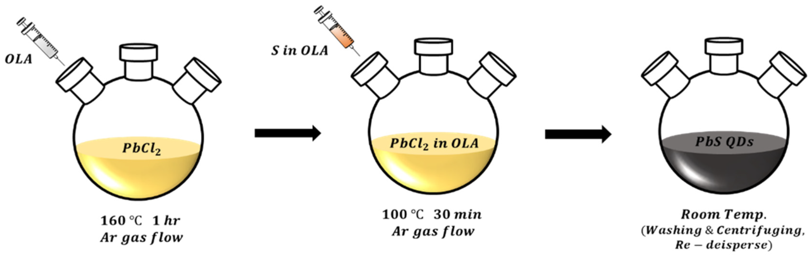
Colloidal PbS quantum dots were prepared using a modified synthetic route adapted from previously reported procedures [39]. Initially, 0.1155 g of sulfur (S, Sigma-Aldrich, St. Louis, MO, USA) powder was combined with 2.4 mL of oleylamine (OLA, Sigma-Aldrich, St. Louis, MO, USA) in a three-neck flask and stirred for 30 min at room temperature under an argon atmosphere. Separately, 0.2781 g (1 mmol) of lead chloride (PbCl2, Sigma-Aldrich, St. Louis, MO, USA) was dissolved in 5 mL of OLA, stirred for 30 min at room temperature under Ar, and subsequently heated to 160 °C for 1 h. To eliminate impurities, the solution was degassed under vacuum at 120 °C for 15 min. Then, 225 μL of the pre-prepared S–OLA solution was swiftly injected into the PbCl2–OLA mixture and maintained at 100 °C under Ar to initiate QD formation. After a 30-min reaction, the mixture was cooled to room temperature. Methanol and toluene were then added at a 4:1:1 volume ratio for purification. The product was centrifuged at 4000 rpm for 5 min to collect the PbS QDs. Finally, the purified QDs were redispersed in hexane (20 mg/mL), yielding a stable PbS QD solution, as illustrated in Figure 1

2.2. Ligand Exchange of PbS QDs

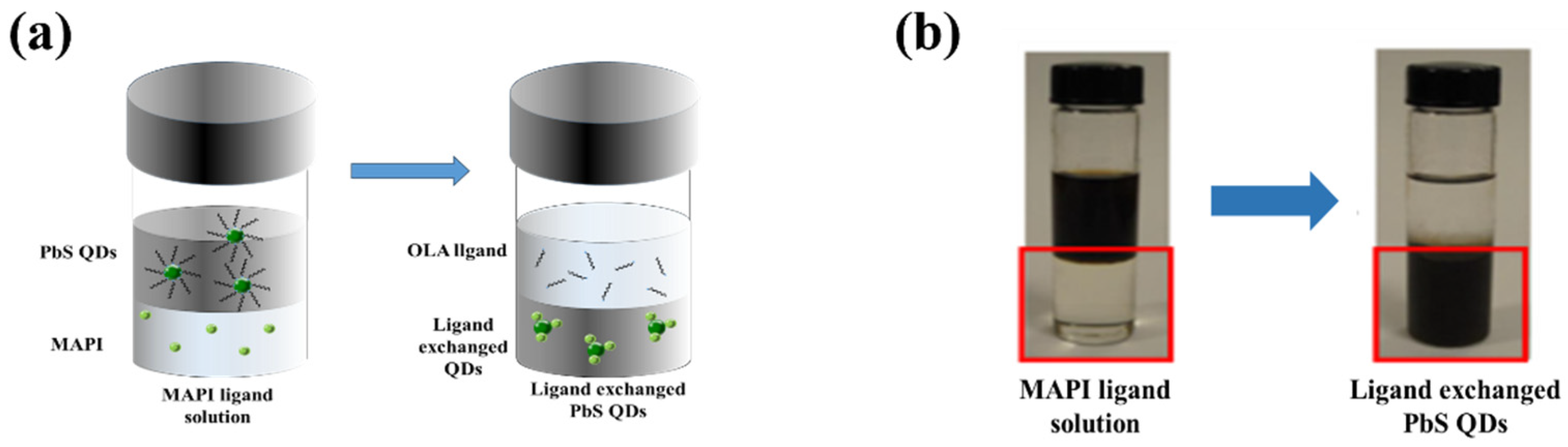
Lead iodide (PbI2, Sigma-Aldrich, St. Louis, MO, USA) and methylammonium iodide (MAI, Sigma-Aldrich, St. Louis, MO, USA) were utilized as precursors for the methylammonium lead iodide (MAPI, Sigma-Aldrich, St. Louis, MO, USA) ligand exchange process. Initially, PbI2 and MAI were dissolved in N,N-dimethylformamide (DMF, Sigma-Aldrich, St. Louis, MO, USA) solvent at concentrations of 0.025, 0.05, and 0.1 mmol, respectively, to prepare the ligand exchange solution. Subsequently, the synthesized PbS QD solution was added to the ligand solution at a 1:1 volume ratio and stirred for 5 min to initiate the organic–inorganic ligand exchange reaction, followed by precipitation for 1 h. To eliminate unreacted or residual species, the resulting solution was washed with a mixture of hexane and the exchange solution in a 2:1 ratio, and this washing procedure was repeated three times to obtain high-purity QDs. Finally, toluene was added at a 1:1 ratio, and the MAPI-ligand-exchanged PbS QDs were collected by centrifugation. The purified MAPI-capped PbS QDs were then redispersed in butylamine, a polar solvent, at a concentration of 20 mg/mL to yield the final MAPI-capped PbS QD solution (Figure 2).

2.3. Device Fabrication

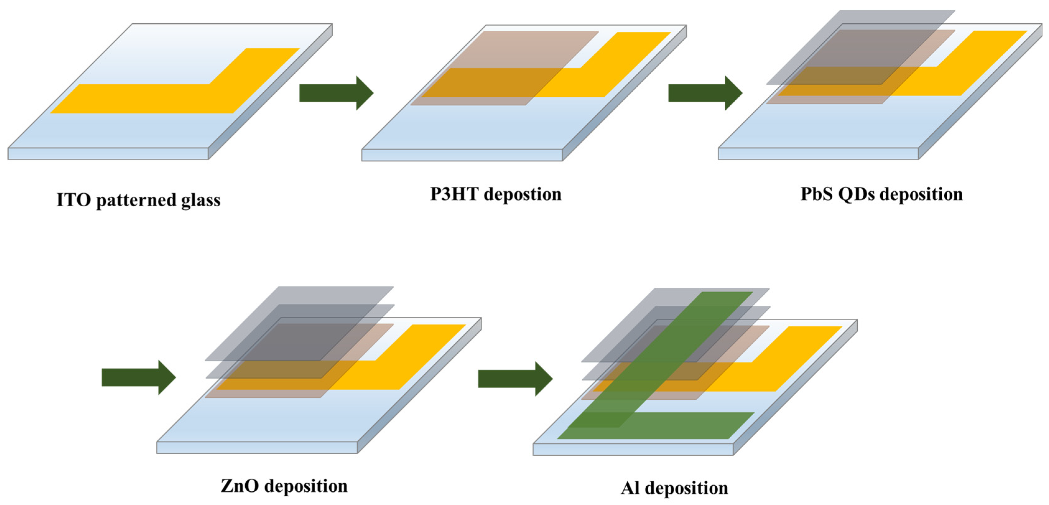
A SWIR photodetector utilizing PbS QDs was fabricated by spin-coating a thin film onto an indium tin oxide (ITO)-patterned glass substrate. Before fabrication, the ITO-coated glass was sequentially cleaned with acetone, methanol, and isopropyl alcohol (IPA) to remove surface contaminants. As the hole transport layer, a conductive polymer solution of P3HT (20 mg/mL in chloroform) was prepared and spin-coated onto the cleaned substrate at 1500 rpm for 15 s, followed by annealing at 95 °C for 30 min to eliminate residual solvent. Subsequently, depending on the type of QD ligand, OLA-capped PbS QDs and MAPI-capped PbS QDs were separately spin-coated under identical conditions (1500 rpm, 15 s) to form the active sensing layer and then annealed at 95 °C for 30 min. A ZnO nanoparticle layer (20 mg/mL in ethanol) was then deposited by spin-coating at 1500 rpm for 30 s to serve as the electron transport layer, followed by annealing at 80 °C for 30 min. Finally, an aluminum electrode with a thickness of 150 nm was deposited by thermal evaporation, completing the fabrication of a photodetector with an active sensing area of 9 mm2 (Figure 3).

2.4. Sensor Measurement System

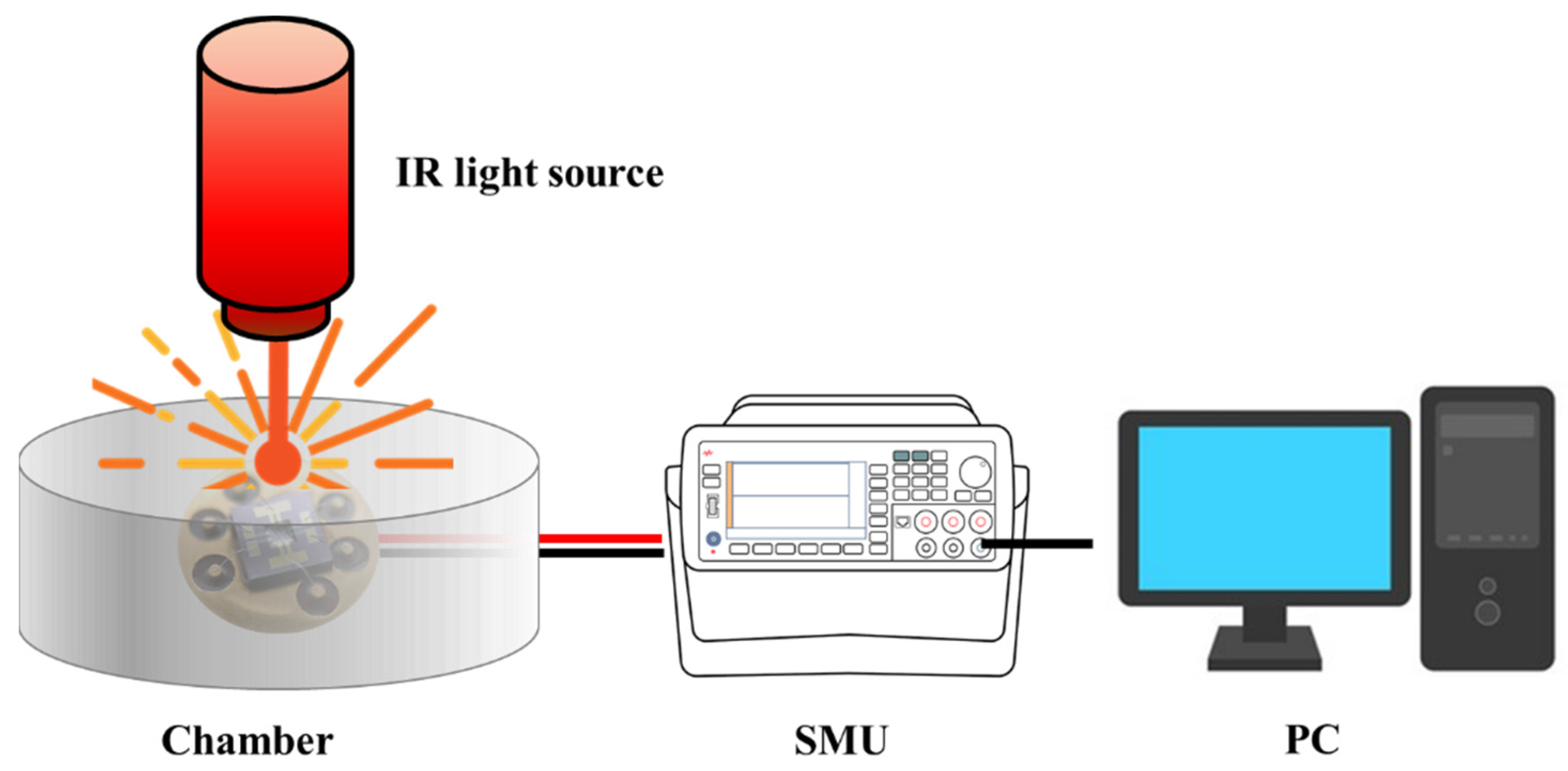
To evaluate the electrical and optical performance of the fabricated SWIR photodetector, the variation in current upon IR light irradiation was measured. All experiments were carried out in a dark chamber to eliminate the influence of external light sources. The fabricated device was secured with a probe tip, and voltage was applied using a source meter unit (SMU, B2902A, Keysight Technologies, Santa Rosa, CA, USA) to measure current characteristics. An IR light source (SLS 202L/M, Thorlabs, Inc., Newton, NJ, USA) was positioned on the top side of the chamber to provide controlled illumination, as shown in Figure 4. The dark current (Idark), defined as the current measured without illumination, and the photocurrent (Ilight), defined as the current measured under illumination, were confirmed. By comparing Idark and Ilight, the photocurrent-to-dark-current ratio (PDCR) of the SWIR photodetector was analyzed to confirm its light response characteristics. To exclude the contribution of visible wavelength light to the photoresponse, a longpass optical filter (FELH0700, cutoff wavelength: 700 nm, Thorlabs, Inc., Newton, NJ, USA) was placed between the light source and the device during all measurements. The incident optical power on the device active area was measured using a calibrated power meter (Thorlabs S401C) and determined to be approximately 1.067 μW.

3. Results

3.1. Characteristics of the Synthesized PbS QDs

During the solution-phase ligand exchange, the iodide anions (I) from MAPI coordinate to undercoordinated Pb2+ sites on the PbS QD surface, replacing the long-chain oleylamine ligands. The methylammonium cations (MA+) do not directly passivate the surface but contribute to charge balance and colloidal stability of the exchanged QDs. This results in reduced interparticle spacing and elimination of insulating organic barriers, thereby enhancing carrier mobility within the QD film.
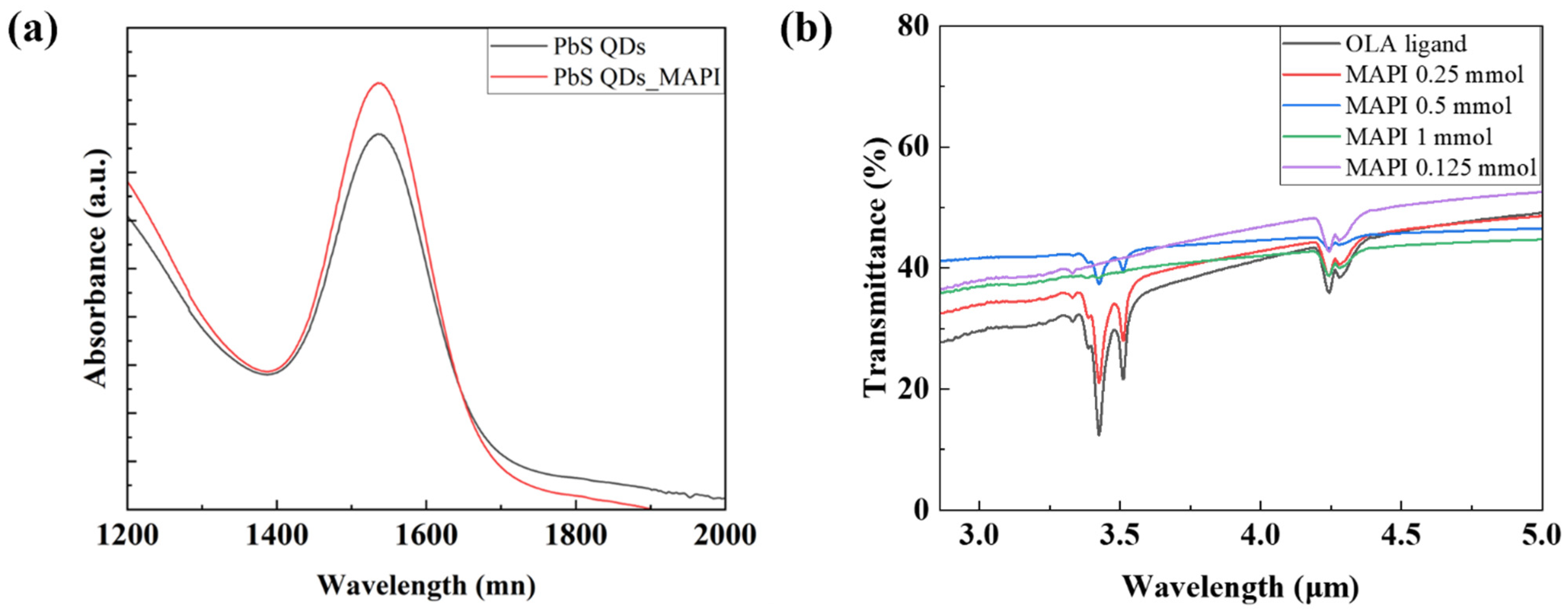
To verify the optical and chemical characteristics of the synthesized QDs, absorbance spectra and FT-IR analysis were conducted. As shown in Figure 5a, the absorption peak of the MAPI-capped PbS QDs appeared at 1550 nm, identical to that of the OLA-capped PbS QDs, with a full width at half maximum (FWHM) of approximately 92 nm. These results indicate that the absorption wavelength and FWHM of the PbS QDs were unaffected by the ligand exchange to the inorganic MAPI ligand, confirming that the MAPI-capped PbS QDs maintain stable optical properties and can selectively detect IR light around 1550 nm. This wavelength band is known to be eye-safe, suggesting that photodetectors fabricated using MAPI-capped PbS QDs can exhibit both high selectivity and responsivity while ensuring safety for human vision.
As shown in Figure 5b, FT-IR analysis was performed to verify successful ligand exchange through functional group characterization. A PbS QD solution (20 mg/mL) was spin-coated at 1000 rpm for 15 s onto a 10 × 10 mm2 glass substrate to form a thin film and then annealed at 95 °C for 30 min to prepare the measurement sample. The FT-IR spectra revealed that the characteristic C–H stretching peak in the 2800–3000 cm−1 region decreased as the MAPI concentration increased and completely disappeared when the MAPI concentration reached 1 mmol. This result confirms that the long-chain C–H bonds of the OLA organic ligand were replaced by the MAPI inorganic ligand, indicating a successful ligand exchange process. Additionally, a weak absorption peak observed near 2350 cm−1 was attributed to trace amounts of unremoved PbCl2 precursor remaining from the synthesis stage.
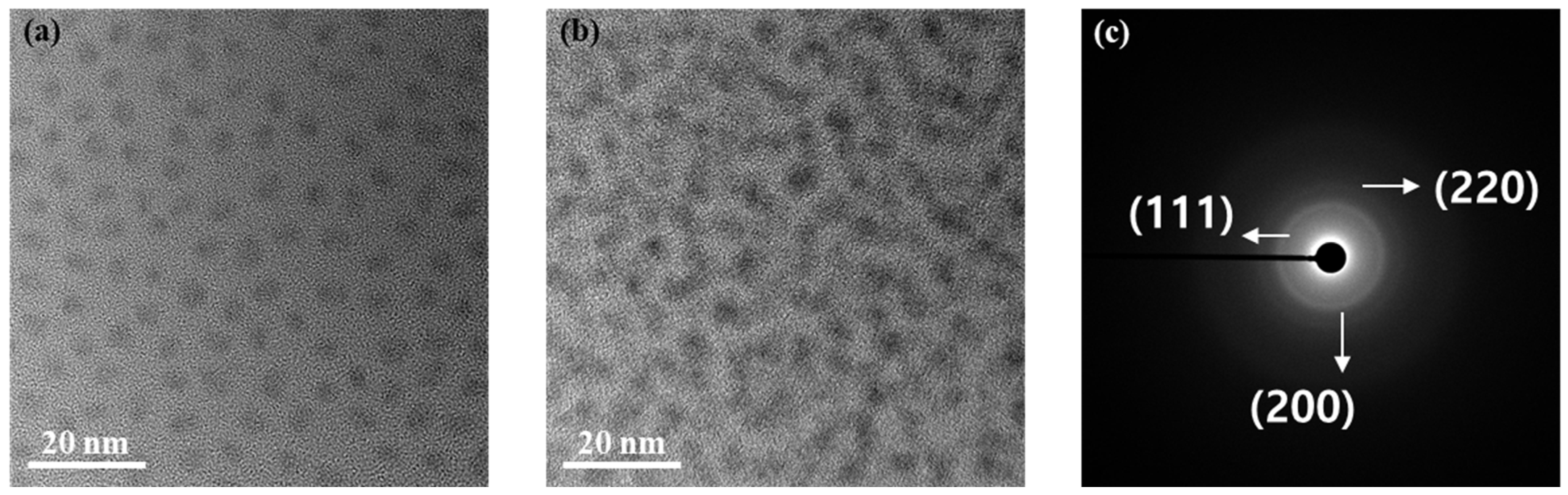
In addition, transmission electron microscopy (TEM) analysis was carried out to compare the surface density of the synthesized PbS QDs within the thin films (Figure 6). The TEM specimens were prepared by forming a thin layer of the synthesized PbS QDs on a mesh grid. The TEM observations demonstrated that the areal number density of the MAPI-exchanged PbS QDs was notably higher under identical imaging conditions. This increase is attributed to the substitution of the long-chain organic OLA ligands with shorter inorganic MAPI ligands, which reduced the interdot spacing between neighboring PbS QDs. As a result, a highly compact and uniform PbS QD thin film was formed, contributing to enhanced carrier transport and improved optoelectronic properties of the photodetector. Furthermore, the lattice fringe spacing of the quantum dots was analyzed using selected area electron diffraction (SAED), and both OLA- and MAPI-capped PbS QDs exhibited diffraction patterns consistent with XRD results, confirming that the crystallographic structure and phase purity of the PbS nanocrystals were maintained after ligand exchange.

3.2. Performance of the SWIR Photodetector

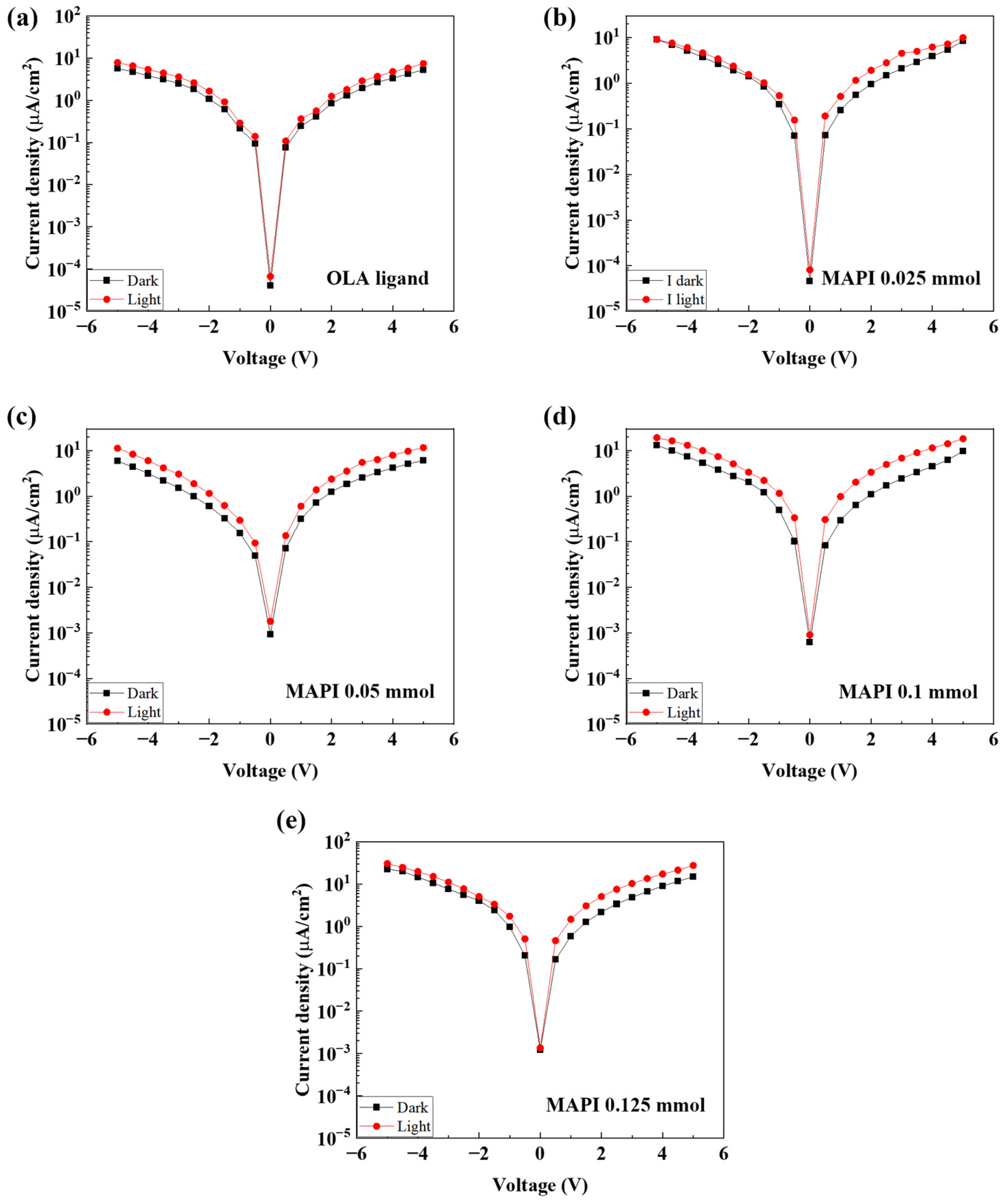
To investigate the performance characteristics of the fabricated SWIR photodetector based on PbS QDs, the device was mounted inside a dark chamber, and its photo response to the incident light source was evaluated. Initially, the current density–voltage (J–V) characteristics were measured to determine the on/off ratio of the SWIR photodetectors fabricated under different ligand conditions. The dark current density was obtained in the absence of illumination, while the photocurrent density was measured under light irradiation as the applied voltage was swept from −5 V to 5 V. As shown in Figure 7, the results revealed that the device exhibited the highest on/off ratio at an applied voltage of 3 V, indicating the maximum PDCR under this condition. At voltages exceeding 3 V, the on/off ratio decreased due to a disproportionate increase in dark current, which also resulted in higher power consumption. Therefore, 3 V was determined to be the optimal bias for subsequent measurements. Accordingly, all subsequent measurements and analyses were performed under the optimized operating voltage of 3 V to ensure consistent evaluation of the photodetector’s performance.
Next, the current response characteristics of the fabricated SWIR photodetector were analyzed under conditions with and without real-time light irradiation. The PbS QD-based photodetector operates on the photoconductivity principle, in which light absorption generates electron–hole pairs within the QD layer, leading to an increase in current. In the absence of illumination, the device exhibits dark current, representing its intrinsic electrical behavior, whereas under illumination, it exhibits photocurrent, which reflects the contribution of photo-generated carriers.
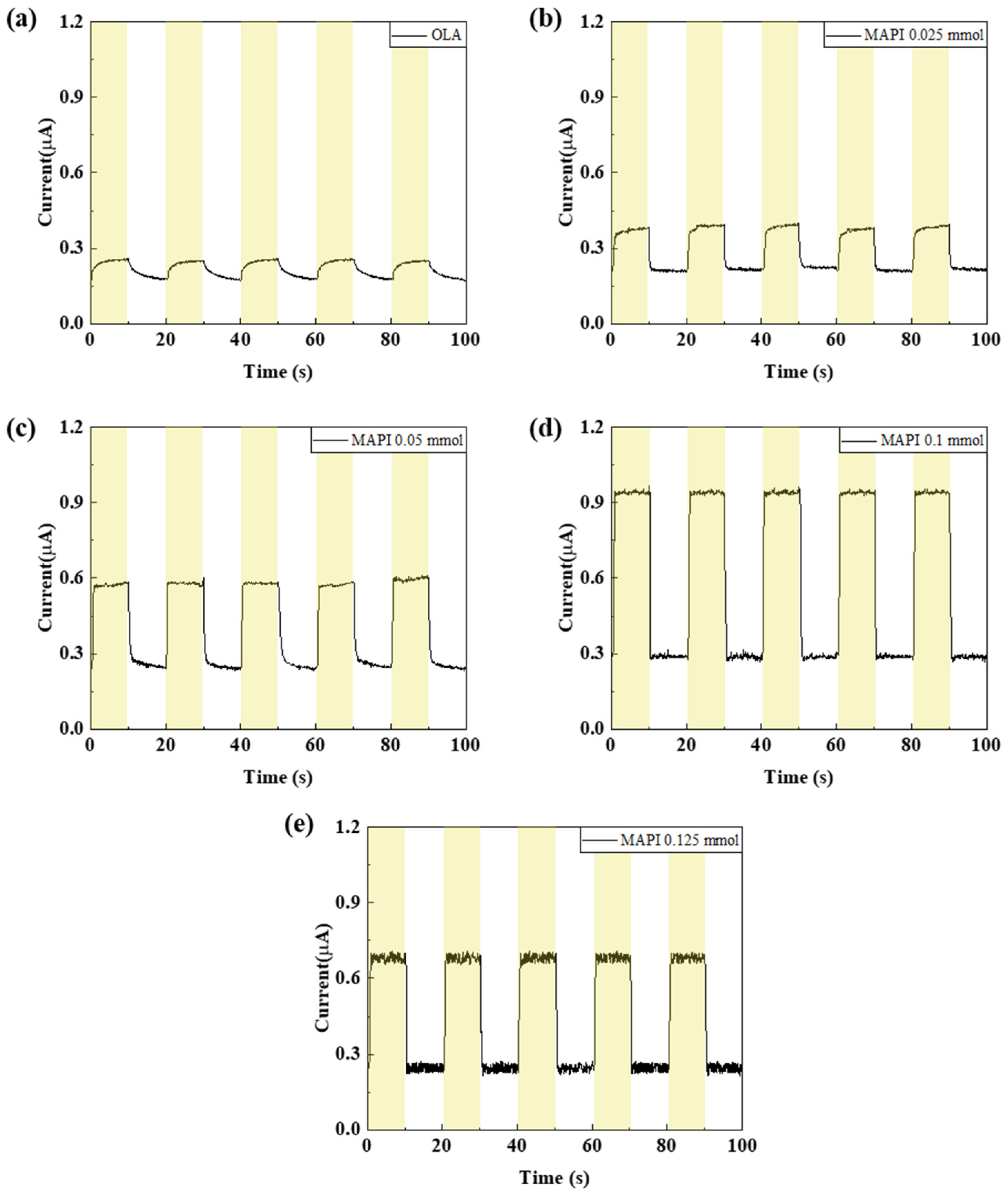
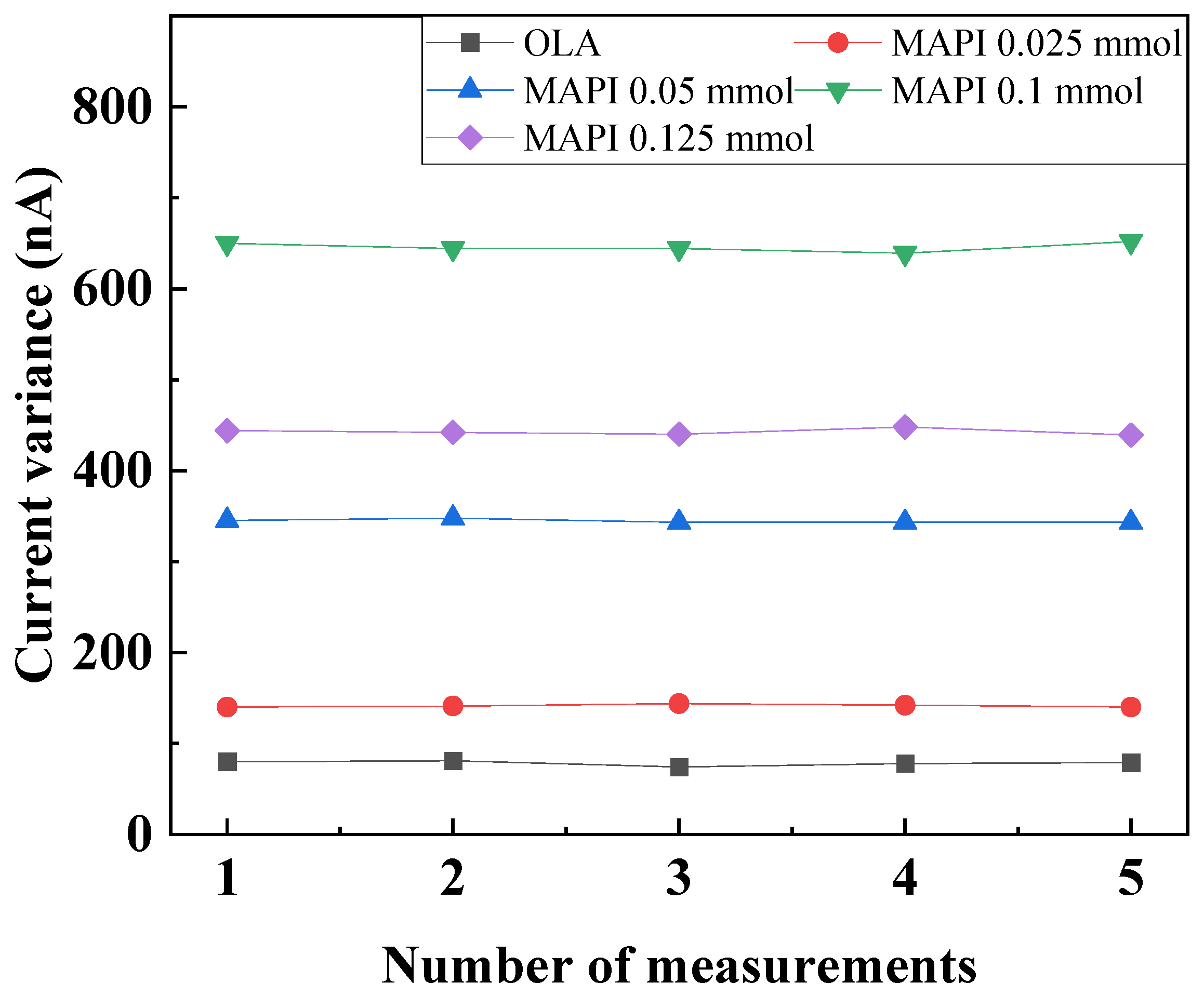
As shown in Figure 8, the SWIR photodetector employing MAPI-capped PbS QDs exhibited a significantly higher current variation compared to that based on OLA-capped PbS QDs, with the maximum change observed at a MAPI concentration of 0.1 mmol. The corresponding current variance is summarized in Figure 9. For the OLA-capped PbS QD device, the current difference between dark and illuminated states reached approximately 60 nA, and the current response was unstable due to noticeable electrical noise. In contrast, the MAPI-capped PbS QD devices demonstrated dark-to-light current differences of 140 nA, 345 nA, 650 nA, and 442 nA, depending on MAPI concentration—corresponding to an enhancement of up to about 2.67 times compared to the OLA-capped device. Furthermore, the current response of the MAPI-based device was notably more stable, with reduced noise. This improvement can be attributed to the replacement of long insulating organic ligands (OLAs) with short inorganic MAPI ligands, which enhanced charge transport by reducing interparticle barriers and increasing QD film density per unit area. However, at an excessive MAPI concentration of 0.125 mmol, increased noise was observed, likely due to the presence of unreacted MAPI residues acting as impurities. To quantitatively compare the photoresponse, the PDCR of the SWIR photodetector was calculated using the following relation under an incident optical power of approximately 1.067 μW.
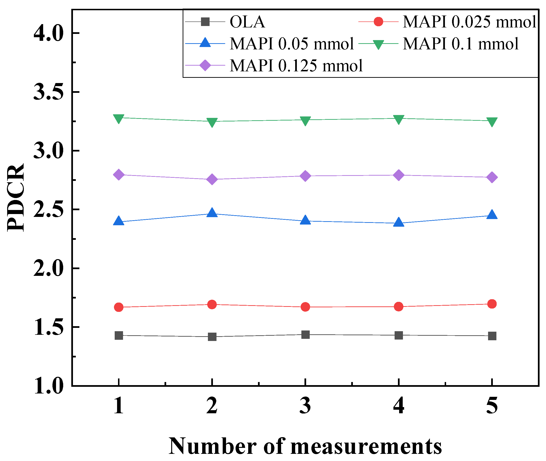
PDCR = Ilight/Idark,
As shown in Figure 10, the conventional OLA-capped PbS QD device exhibited an average PDCR of approximately 41%, whereas the MAPI-capped PbS QD device achieved an average PDCR of about 225%, representing an improvement of up to 5.48 times.
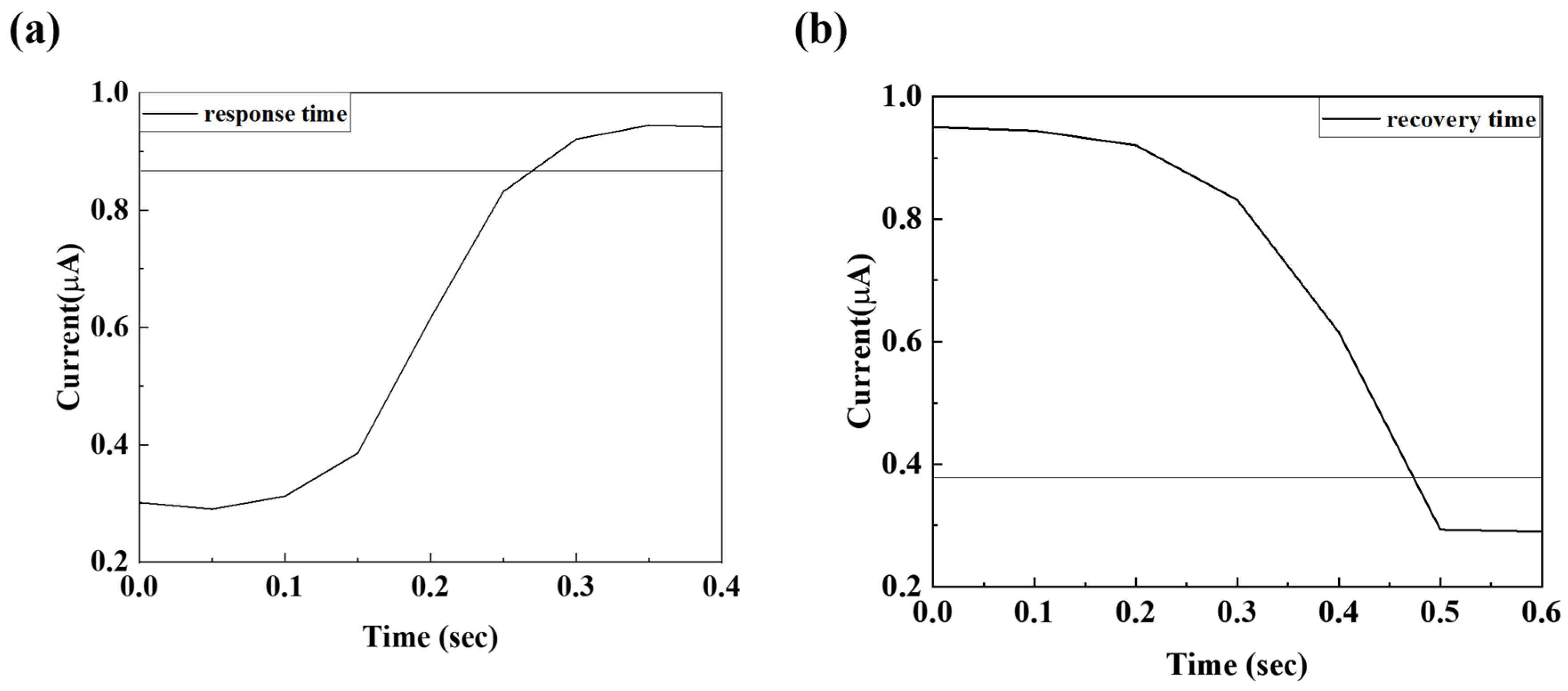
The rise time (τr) and fall time (τf) are defined as the time required for the photocurrent to increase from 10% to 90% and decrease from 90% to 10% of its maximum value, respectively. As shown in Figure 11, the rise time improved markedly—from 4.15 s for the OLA-capped device to 0.26 s for the MAPI-capped device—corresponding to an enhancement of approximately 11.5 times. Similarly, the fall time improved from 4.15 s for the OLA-capped device to 0.45 s for the MAPI 0.1 mmol device, confirming the superior optoelectronic response enabled by MAPI ligand exchange.
The resistance and photoresponse time characteristics of all fabricated devices are summarized in Table 1. As the MAPI concentration increased from OLA to 0.1 mmol, Rdark decreased from 17.4 MΩ to 4.6 MΩ and both rise and fall times improved, which is attributed to reduced interdot spacing and enhanced carrier mobility enabled by the shorter MAPI ligands, as well as changes in the RC constant of the device. At 0.125 mmol, Rdark increased to 6.9 MΩ and the fall time slightly increased compared to the 0.1 mmol device, which is attributed to excess MAPI ligands disrupting the film uniformity and increasing interdot spacing, consistent with the increased noise observed in Figure 8e.
The external quantum efficiency (EQE) and responsivity [R(λ)] of the SWIR photodetector with the device structure ITO/P3HT/PbS QDs/ZnO/Al were characterized using a quantum yield spectrometer (Quantaurus-QY Plus, Hamamatsu Photonics, Hamamatsu, Japan). The EQE represents the ratio of the number of charge carriers (electrons and holes) successfully collected by the external circuit to the total number of incident photons on the device, thus reflecting how effectively the photogenerated carriers are extracted without undergoing recombination losses. Meanwhile, responsivity [R(λ)] indicates the magnitude of the photocurrent produced per unit optical power at a specific wavelength, essentially quantifying how sensitively the detector converts incoming light into electrical signals. Both EQE and R(λ) are key performance indicators that determine the overall efficiency and spectral selectivity of a photodetector. A high EQE value suggests efficient charge separation and transport within the active layer, while high responsivity demonstrates strong photoconversion capability, particularly important for low-intensity or broadband light detection. In this study, EQE measurements were carried out across the SWIR spectral range, and the corresponding responsivity was derived from the EQE spectrum using the following equation:
R(λ) = EQE × qλ/hc
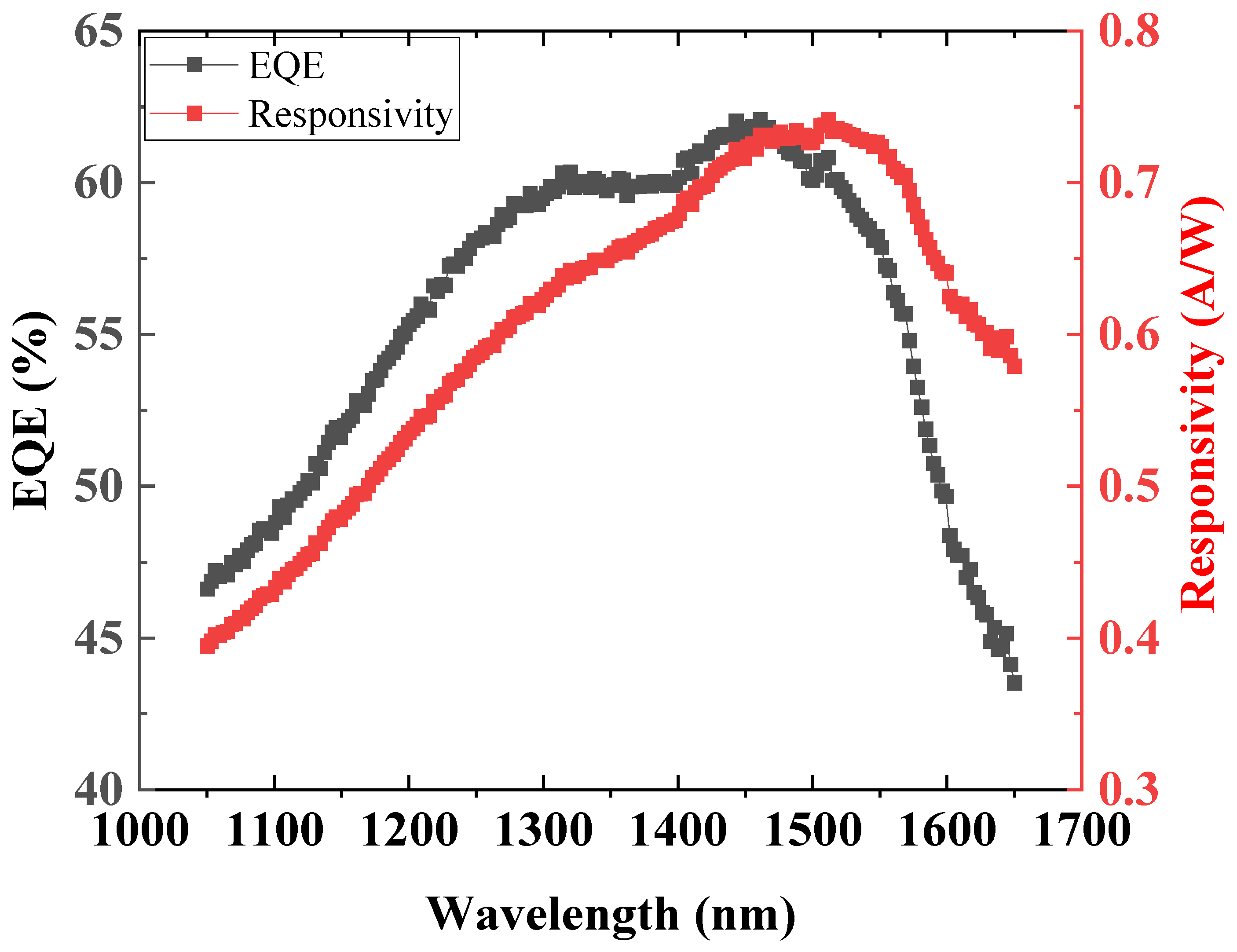
The parameters h, c, and λ denote Planck’s constant, the speed of light in vacuum, and the wavelength of the incident light, respectively, with the unit of hc/λ expressed in electron volts (eV). The EQE of the fabricated SWIR photodetector was measured across the spectral range of 1000–1650 nm, and the corresponding responsivity [R(λ)] was derived. As illustrated in Figure 12, the device exhibited outstanding photoresponse characteristics, achieving an EQE of approximately 62% within the 1400–1500 nm range. In the same spectral region, the responsivity reached a maximum value of 0.73 A/W, confirming the high photon-to-electron conversion efficiency and excellent optoelectronic performance of the fabricated SWIR photodetector.
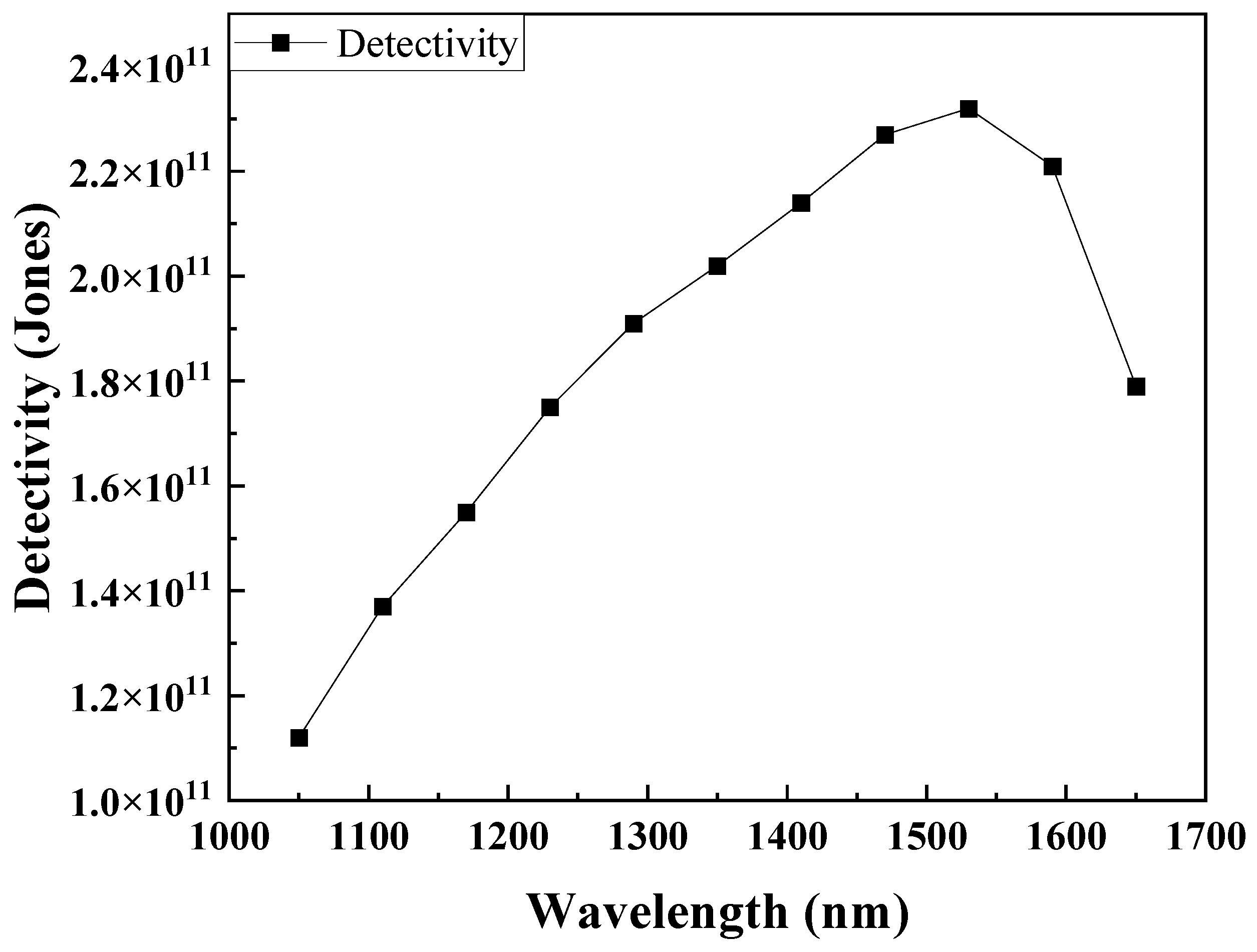
The specific detectivity (D*) was subsequently calculated using the measured R(λ) and the corresponding dark current under varying bias voltages. Detectivity is a critical figure of merit (FOM) for photodetectors, representing the inverse of the noise-equivalent power (NEP), which defines the minimum detectable optical power limited by intrinsic device noise. A higher D* value indicates superior PDCR, particularly advantageous for detecting weak optical signals near the noise threshold. In this analysis, it was assumed that the dominant noise component originated from the dark current shot noise under biased operating conditions. Accordingly, the noise-limited detectivity (D*) can be expressed by the following relation:
D = R λ A 1 / 2 2 q I d a r k 1 / 2
where R(λ) denotes the wavelength-dependent photoresponsivity, q is the elementary charge, A represents the active area of the device, and Idark corresponds to the dark current of the photodetector under reverse bias conditions. As shown in Figure 13, the fabricated SWIR photodetector exhibited relatively stable detectivity characteristics within the reverse bias range of several hundred millivolts, indicating that D* is not strongly bias-dependent in this regime. Moreover, the device demonstrated a high detectivity exceeding 2.26 × 10¹¹ Jones across the long-wavelength spectral region of 1000–1400 nm, confirming its excellent PDCR and capability to detect weak infrared signals with minimal noise contribution. This result validates the effectiveness of the MAPI-capped PbS QDs structure in enhancing both carrier transport and signal-to-noise performance in SWIR photodetection applications. Table 2 reveals the performance comparison of different photodetectors within 5 years. The results show that the performance of the photodetector from this work had higher responsivity and detectivity.

4. Conclusions

In this study, a solution-processed SWIR photodetector based on PbS quantum dots with inorganic MAPI ligands was successfully developed and systematically investigated. The conventional long-chain organic ligands of PbS QDs were replaced by methylammonium lead iodide through a solution-phase ligand exchange process, enabling the formation of a compact QD thin film with enhanced electrical coupling and charge transport properties. The MAPI-capped PbS QDs maintained stable optical absorption characteristics in the eye-safe SWIR region around 1550 nm, while exhibiting markedly improved photoconductive performance compared to their organic-ligand-capped counterparts. The fabricated photodetector demonstrated a high external quantum efficiency of 62%, a responsivity of 0.73 A/W, and a detectivity of 2.26 × 10¹¹ Jones in the 1400–1500 nm wavelength range. In addition, the device showed significantly enhanced PDCR and a faster rise time, highlighting the effectiveness of inorganic ligand engineering for improving SWIR sensor performance. The improved optoelectronic characteristics are attributed to reduced interparticle spacing, increased quantum dot packing density, and more efficient carrier transport enabled by the MAPI ligand, as well as optimized charge extraction through the P3HT and ZnO transport layers. Systematic linear dynamic range (LDR) characterization was beyond the scope of the present experimental setup and will be investigated in future work using a dedicated 1550 nm monochromatic laser source with controlled optical power modulation. Owing to its simple device architecture, solution-based fabrication, and competitive sensing performance, the proposed MAPI-capped PbS QD photodetector offers strong potential for practical implementation in eye-safe LIDAR systems, optical sensing, and emerging autonomous and industrial monitoring technologies.

Author Contributions

Conceptualization, D.J. and J.K.; methodology, S.K. and J.K.; formal analysis, Y.H.; investigation, Y.H.; resources, J.K.; data curation, S.K.; writing—original draft preparation, J.K. and Y.H.; writing—review and editing, D.G.J.; visualization, S.K.; project administration, D.G.J.; funding acquisition, D.G.J. and D.J. All authors have read and agreed to the published version of the manuscript.

Funding

This study has been conducted with the support of the Korea Institute of Industrial Technology as “Development and commercialization for clean hydrogen production/storage and CO2 monitoring system in the field of industrial complex (Kitech EH-26-0008)”. This study is a 2024 SME Technology Innovation Development Export-Oriented (Tech-Bridge) R&D project of the Small and Medium Business Technology Information Promotion Agency (SME Technology Innovation Development Institute), and is part of the study “Development of 8-Channel Total Organic Carbon (TOC) Measuring Device for Regular Monitoring of Water Quality of Waste Water and Process Water” (RS-2024-00469087). This work was supported by the Technological Innovation R&D Program (RS-2024-00487867) funded by the Ministry of SMEs and Startups (MSS, Korea).

Institutional Review Board Statement

Not applicable.

Informed Consent Statement

Not applicable.

Data Availability Statement

The original contributions presented in this study are included in the article. Further inquiries can be directed to the corresponding author.

Conflicts of Interest

The authors declare no conflicts of interest.

References

  1. Cao, F.; Liu, L.; Li, L. Short-wave infrared photodetector. Mater. Today 2023, 62, 327–349. [Google Scholar] [CrossRef]
  2. Swasey, S.M.; Copp, S.M.; Nicholson, H.C.; Gorovits, A.; Bogdanov, P.; Gwinn, E.G. High throughput near infrared screening discovers DNA-templated silver clusters with peak fluorescence beyond 950 nm. Nanoscale 2018, 10, 19701–19705. [Google Scholar] [CrossRef]
  3. Klem, J.F.; Kim, J.K.; Cich, M.J.; Keeler, G.A.; Hawkins, S.D.; Fortune, T.R. Mesa-isolated InGaAs photodetectors with low dark current. Appl. Phys. Lett. 2009, 95, 341–345. [Google Scholar] [CrossRef]
  4. Michel, E.; Singh, G.; Slivken, S.; Besikci, C.; Bove, P.; Ferguson, I.; Razeghi, M. MBE growth of high quality InSb optimized using RHEED oscillations. Appl. Phys. Lett. 1994, 65, 3338–3340. [Google Scholar] [CrossRef]
  5. Gunapala, S.D.; Bandara, S.V. GaAs/AlGaAs multi-quantum well based infrared focal plane arrays for infrared imaging applications. Int. J. High Speed Electron. Syst. 2002, 12, 99–121. [Google Scholar] [CrossRef]
  6. Takano, Y.; Masuda, M.; Kobayashi, K.; Kuwahara, K.; Fuke, S.; Shirakata, S. Epitaxial growth of InGaAs on misoriented GaAs(100) substrate by metal-organic vapor phase epitaxy. J. Cryst. Growth 2002, 236, 31–36. [Google Scholar] [CrossRef]
  7. Hyder, S.B. Observation of Vapor-phase epitaxial growth of InGaAs lattice matched to (100) InP for photodiode application Available to Purchase. Appl. Phys. Lett. 1979, 35, 787–789. [Google Scholar] [CrossRef]
  8. Chen, W.; Tang, H.; Chen, Y.; Heger, J.E.; Li, N.; Kreuzer, L.P.; Xie, Y.; Li, D.; Anthony, C.; Pikramenou, Z.; et al. Spray-deposited PbS colloidal quantum dot solid for near-infrared photodetectors. Nano Energy 2020, 78, 105254–105366. [Google Scholar] [CrossRef]
  9. Xu, K.; Zhou, W.; Ning, Z. Integrated structure and device engineering for high performance and scalable quantum dot infrared photodetectors. Small 2020, 16, 2003397–2003408. [Google Scholar] [CrossRef]
  10. Fang, Y.; Guo, F.; Xiao, Z.; Huang, J. Large gain, low noise nanocomposite ultraviolet photodetectors with a linear dynamic range of 120 dB. Adv. Opt. Mater. 2014, 2, 348–353. [Google Scholar] [CrossRef]
  11. De Iacovo, A.; Venettacci, C.; Colace, L.; Scopa, L.; Foglia, S. PbS colloidal quantum dot photodetectors operating in the near infrared. Sci. Rep. 2016, 6, 37913. [Google Scholar] [CrossRef]
  12. Choi, H.T.; Kang, J.H.; Ahn, J.; Jin, J.; Kim, J.; Park, S.; Kim, Y.H.; Kim, H.; Song, J.D.; Hwang, G.W.; et al. Zero-dimensional PbS quantum dot-InGaZnO film heterostructure for short-wave infrared flat-panel imager. ACS Photon. 2020, 7, 1932–1941. [Google Scholar] [CrossRef]
  13. Ankah, G.N.; Büchele, P.; Poulsen, K.; Rauch, T.; Tedde, S.F.; Gimmler, C.; Schmidt, O.; Kraus, T. PbS quantum dot-based hybrid-organic photodetectors for X-ray sensing. Org. Electron. 2016, 33, 201–206. [Google Scholar] [CrossRef]
  14. de Arquer, F.P.G.; Armin, A.; Meredith, P.; Sargent, E.H. Solution-processed semiconductors for next-generation photodetectors. Nat. Rev. Mater. 2017, 2, 16100. [Google Scholar] [CrossRef]
  15. Konstantatos, G.; Howard, I.; Fischer, A.; Hoogland, S.; Clifford, J.; Klem, E.; Levina, L.; Sargent, E.H. Ultrasensitive solution-cast quantum dot photodetectors. Nature 2006, 442, 180–183. [Google Scholar] [CrossRef]
  16. Graf, M.; Scalari, G.; Hofstetter, D.; Faist, J. Terahertz range quantum well infrared photodetector. Appl. Phys. Lett. 2004, 84, 475–477. [Google Scholar] [CrossRef]
  17. Wu, W.; Han, B.; Shao, J. Plasmonic enhanced quantum well infrared photodetector with high detectivity. Appl. Phys. Lett. 2010, 96, 161107. [Google Scholar] [CrossRef]
  18. Levine, B.F.; Bethea, C.G.; Hasnain, G.; Shen, V.O.; Pelve, E.; Abbott, R.R.; Hsieh, S.J. High sensitivity low dark current 10 µm GaAs quantum well infrared photodetectors. Appl. Phys. Lett. 1990, 56, 851–853. [Google Scholar] [CrossRef]
  19. Rauch, T.; Böberl, M.; Tedde, S.F.; Fürst, J.; Kovalenko, M.V.; Hesser, G.; Lemmer, U.; Heiss, W.; Hayden, O. Near-infrared imaging with quantum-dot-sensitized organic photodiodes. Nat. Photon. 2009, 3, 332–336. [Google Scholar] [CrossRef]
  20. Lee, J.W.; Kim, D.Y.; Baek, S.; Yu, H.; So, F. Inorganic UV–visible–SWIR broadband photodetector based on monodisperse PbS nanocrystals. Small 2016, 12, 1328–1333. [Google Scholar] [CrossRef]
  21. Hines, M.A.; Scholes, G.D. Colloidal PbS nanocrystals with size-tunable near-infrared emission: Observation of post-synthesis self-narrowing of the particle size distribution. Adv. Mater. 2003, 15, 1844–1849. [Google Scholar] [CrossRef]
  22. Fu, H.Y.; Tsang, S.-W.; Zhang, Y.; Ouyang, J.; Lu, J.; Yu, K.; Tao, Y. Impact of the growth conditions of colloidal PbS nanocrystals on photovoltaic device performance. Chem. Mater. 2011, 23, 1805–1810. [Google Scholar] [CrossRef]
  23. Kwon, J.-B.; Kim, S.-W.; Kang, B.-H.; Yeom, S.-H.; Lee, W.-H.; Kwon, D.-H.; Lee, J.-S.; Kang, S.-W. Air-stable and ultrasensitive solution-cast SWIR photodetectors utilizing modified core/shell colloidal quantum dots. Nano Converg. 2020, 7, 28. [Google Scholar] [CrossRef]
  24. Kwon, J.; Ha, Y.; Choi, S.; Jung, D.G.; An, H.K.; Kong, S.H.; Jung, D. Solution-processed NO2 gas sensor based on poly(3-hexylthiophene)-doped PbS quantum dots operable at room temperature. Sci. Rep. 2024, 14, 20600. [Google Scholar] [CrossRef] [PubMed]
  25. McDonald, S.A.; Cyr, P.W.; Levina, L.; Sargent, E.H. Photoconductivity from PbS-nanocrystal/semiconducting polymer composites for solution-processible, quantum-size tunable infrared photodetectors. Appl. Phys. Lett. 2004, 85, 2089–2091. [Google Scholar] [CrossRef]
  26. Sulaman, M.; Shafique, S.; Tahir, M. High quantum yield and pH sensitive fluorescence dyes based on coumarin derivatives: Fluorescence characteristics and theoretical study. RSC Adv. 2016, 6, 87730–87737. [Google Scholar] [CrossRef]
  27. Hevesa, E.; Gurbuz, Y. PbS colloidal quantum dot photodiodes for SWIR detection. Procedia Eng. 2012, 47, 1426–1429. [Google Scholar] [CrossRef]
  28. Hechster, E.; Amgar, D.; Arad-Vosk, N.; Binyamin, T.; Sa, A.; Etgar, L.; Sarusi, G. Electrical and optical characterization of quantum dots PbS/TiO2 based heterojunction as a SWIR detector and a proposed design of PbS/TiO2-PeLED as a SWIR to visible upconversion device. Mater. Res. Express 2019, 6, 066210. [Google Scholar] [CrossRef]
  29. Bothra, U.; Albaladejo-Siguan, M.; Vaynzof, Y.; Kabra, D. Impact of ligands on the performance of PbS quantum dot visible–near-infrared photodetectors. Adv. Opt. Mater. 2023, 11, 2201897. [Google Scholar] [CrossRef]
  30. Tang, H.; Zhong, J.; Chen, W.; Shi, K.; Mei, G.; Zhang, Y.; Wen, Z.; Müller-Buschbaum, P.; Wu, D.; Wang, K.; et al. Lead sulfide quantum dot photodetector with enhanced responsivity through a two-step ligand-exchange method. ACS Appl. Nano Mater. 2019, 2, 6135–6143. [Google Scholar] [CrossRef]
  31. Kufer, D.; Nikitskiy, I.; Lasanta, T.; Navickaite, G.; Koppens, F.H.L.; Konstantatos, G. Hybrid 2D–0D MoS2–PbS quantum dot photodetectors. Adv. Mater. 2015, 27, 176–180. [Google Scholar] [CrossRef]
  32. Wei, Y.; Ren, Z.; Zhang, A.; Mao, P.; Li, H.; Zhong, X.; Li, W.; Yang, S.; Wang, J. Hybrid organic/PbS quantum dot bilayer photodetector with low dark current and high detectivity. Adv. Funct. Mater. 2018, 28, 1706690. [Google Scholar] [CrossRef]
  33. Jung, B.K.; Woo, H.K.; Shin, C.; Park, T.; Li, N.; Lee, K.J.; Kim, W.; Bae, J.H.; Ahn, J.P.; Ng, T.N.; et al. Suppressing the dark current in quantum dot infrared photodetectors by controlling carrier statistics. Adv. Opt. Mater. 2022, 10, 2101611. [Google Scholar] [CrossRef]
  34. Blachowicz, T.; Ehrmann, A. Recent developments of solar cells from PbS colloidal quantum dots. Appl. Sci. 2020, 10, 1743. [Google Scholar] [CrossRef]
  35. Im, S.H. Light enhanced moisture degradation of perovskite solar cell using first principles calculations. J. Mater. Chem. A 2019, 7, 23126–23134. [Google Scholar]
  36. Shirzadi, E.; Jin, Q.; Zeraati, A.S.; Dorakhan, R.; Goncalves, T.J.; Abed, J.; Lee, B.-H.; Rasouli, A.S.; Wicks, J.; Zhang, J.; et al. Ligand-modified nanoparticle surfaces influence CO electroreduction selectivity. Nat. Commun. 2024, 15, 2995. [Google Scholar] [CrossRef]
  37. Li, B.; Duan, Z.; Zeng, W.; Bu, Y.; Yuan, Z.; Jiang, Y.; Tai, H. Advances in PbX (X = S, Se, Te) Quantum-Dot Photodetectors. Laser Photon. Rev. 2026, 20, e01759. [Google Scholar] [CrossRef]
  38. Kwon, J.B.; Han, M.; Jung, D.G.; Kong, S.H.; Jung, D. High sensitivity shortwave infrared photodetector based on PbS QDs using P3HT. Nanomaterials 2021, 11, 2683. [Google Scholar] [CrossRef]
  39. Zhang, J.-Y.; Xu, J.-L.; Chen, T.; Gao, X.; Wang, S.-D. Toward broadband imaging: Surface-engineered PbS quantum dot/perovskite composite integrated ultrasensitive photodetectors. ACS Appl. Mater. Interfaces 2019, 11, 44041–44048. [Google Scholar] [CrossRef]
  40. Dong, C.; Liu, S.; Barange, N.; Lee, J.; Pardue, T.; Yi, X.; Yin, S.; So, F. Long-wavelength lead sulfide quantum dots sensing up to 2600 nm for short-wavelength infrared photodetectors. ACS Appl. Mater. Interfaces 2019, 11, 44451–44457. [Google Scholar] [CrossRef]
  41. Xu, K.; Xiao, X.; Zhou, W.; Jiang, X.; Wei, Q.; Chen, H.; Deng, Z.; Huang, J.; Chen, B.; Ning, Z. Inverted Si:PbS colloidal quantum dot heterojunction-based infrared photodetector. ACS Appl. Mater. Interfaces 2020, 12, 15414–15421. [Google Scholar] [CrossRef]
  42. Chen, H.; Chen, J. High performance near-infrared photodetector based on PbS quantum dots and graphene. Sens. Actuators A Phys. 2022, 339, 113508. [Google Scholar] [CrossRef]
  43. Wang, X.; Song, Z.; Tang, H.; Li, Y.; Zhong, H.; Wu, J.; Wang, W.; Chen, S.; Zhang, W.; Fang, F.; et al. Synergic Surface Modifications of PbS Quantum Dots by Sodium Acetate in Solid-State Ligand Exchange toward Short-Wave Infrared Photodetectors. ACS Appl. Mater. Interfaces 2024, 16, 44164–44173. [Google Scholar] [CrossRef]
Figure 1. Schematic of the synthesis of PbS QDs.
Sensors 26 03391 g001
Figure 2. (a) Schematic, (b) experiments of MAPI ligand exchange process. Red squares indicate the transition of the PbS QD layer from the upper to the lower phase after ligand exchange.
Sensors 26 03391 g002
Figure 3. Schematic diagram of fabrication of SWIR photodetector based on PbS QDs.
Sensors 26 03391 g003
Figure 4. Schematic diagram of the measurement system.
Sensors 26 03391 g004
Figure 5. (a) Absorption spectra and (b) FT-IR analysis according to ligand exchange.
Sensors 26 03391 g005
Figure 6. TEM image of PbS QDs according to (a) OLA ligand, (b) MAPI ligand, and (c) SAED characterization.
Sensors 26 03391 g006
Figure 7. J–V characteristics according to light source irradiation. (a) OLA ligand, (b) MAPI 0.025 mmol, (c) MAPI 0.05 mmol, (d) MAPI 0.1 mmol, and (e) MAPI 0.125 mmol.
Sensors 26 03391 g007
Figure 8. Real-time current characteristics according to light source irradiation. (a) OLA ligand, (b) MAPI 0.025 mmol, (c) MAPI 0.05 mmol, (d) MAPI 0.1 mmol, and (e) MAPI 0.125 mmol.
Sensors 26 03391 g008
Figure 9. Current characteristics according to MAPI concentration.
Sensors 26 03391 g009
Figure 10. Photocurrent-to-dark-current ratio according to MAPI concentration.
Sensors 26 03391 g010
Figure 11. (a) Rise time and (b) fall time of the MAPI-ligand-exchanged SWIR photodetector.
Sensors 26 03391 g011
Figure 12. External quantum efficiency and responsivity of the MAPI-ligand-exchanged SWIR photodetector.
Sensors 26 03391 g012
Figure 13. Detectivity of the MAPI-ligand-exchanged SWIR photodetector.
Sensors 26 03391 g013
Table 1. Resistance and photoresponse time characteristics of SWIR photodetectors with different MAPI ligand concentrations.
LigandRdark (MΩ)Rlight (MΩ)Rise Time (s)Fall Time (s)
OLA17.411.74.154.15
MAPI 0.025 mmol7.93.70.300.40
MAPI 0.05 mmol5.22.40.300.85
MAPI 0.1 mmol4.61.60.260.45
MAPI 0.125 mmol6.93.20.280.55
Table 2. Comparison of SWIR photodetector performance.
Wavelength (nm)Responsivity (A/W)Detectivity (Jones)References
15400.2641.47 × 1011[40]
21000.3851.5 × 1011[41]
15500.972.21 × 1010[42]
13100.261.1 × 1011[43]
15500.732.26 × 1011This work
Disclaimer/Publisher’s Note: The statements, opinions and data contained in all publications are solely those of the individual author(s) and contributor(s) and not of MDPI and/or the editor(s). MDPI and/or the editor(s) disclaim responsibility for any injury to people or property resulting from any ideas, methods, instructions or products referred to in the content.