
Three-Dimensional Focusing Measurement Method for Confocal Microscopy Based on Liquid Crystal Spatial Light Modulator


Abstract
1. Introduction
2. Experimental Materials and Fabrication Methods
2.1. Study of LC-SLM Modulation Characteristics
2.2. Binary Fresnel Lens Construction Method
2.3. Analysis of Wavelength Effect of Incident Light
3. Experimental Methods
4. Results
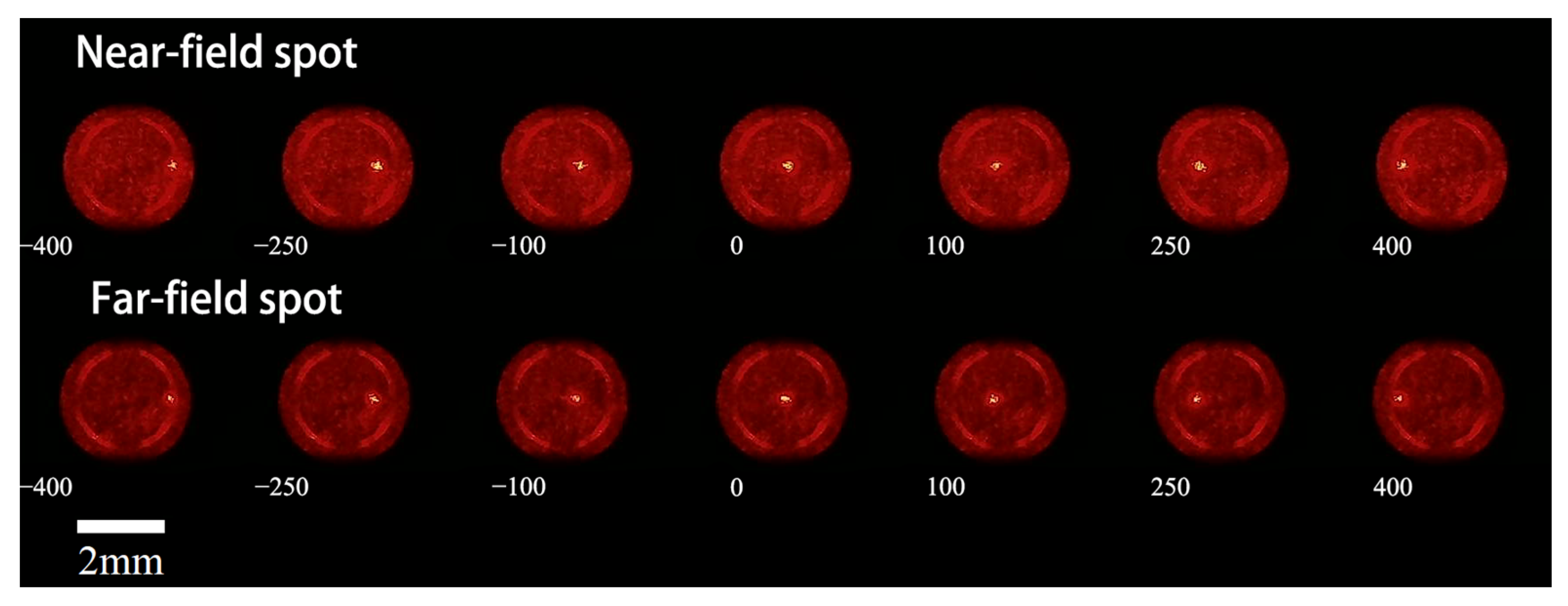
4.1. Lateral Focusing Experiment
4.1.1. Experimental Data Acquisition
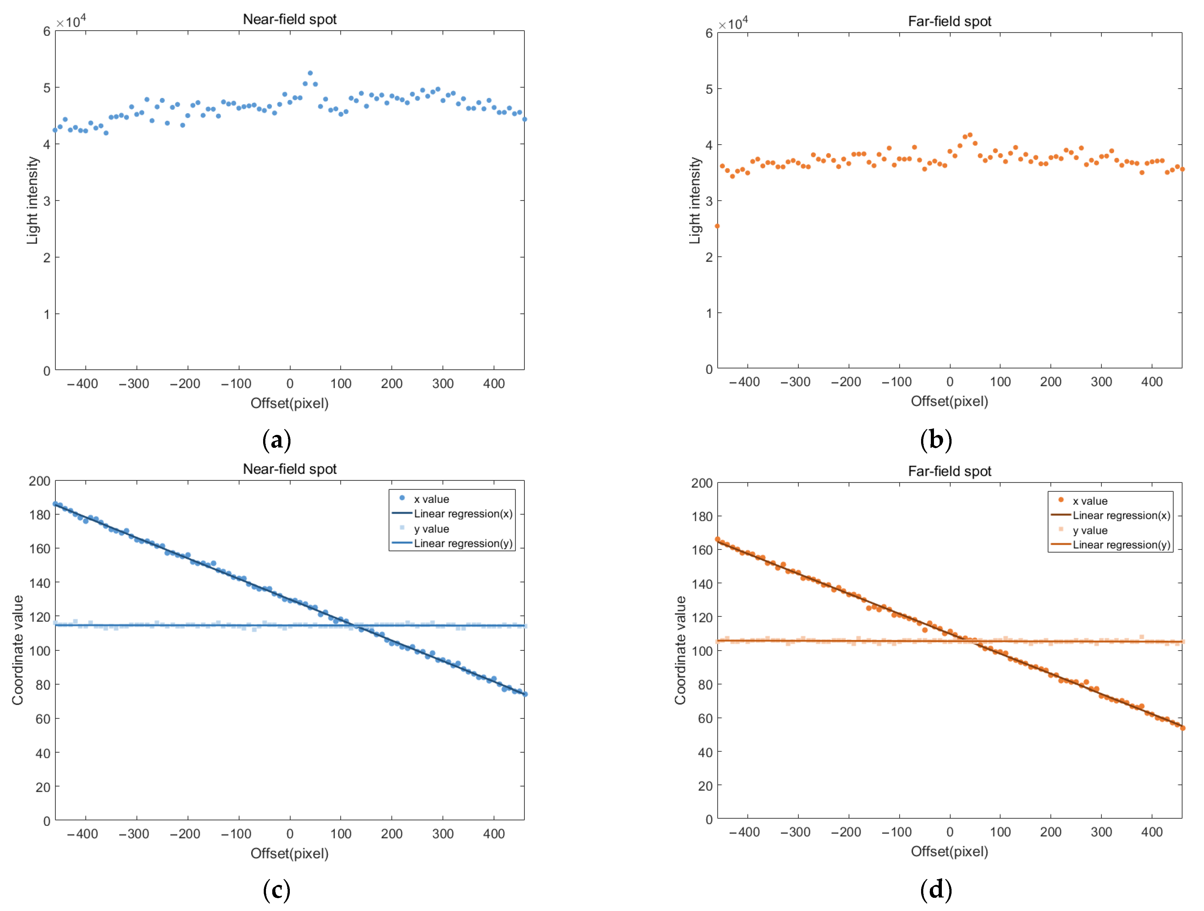
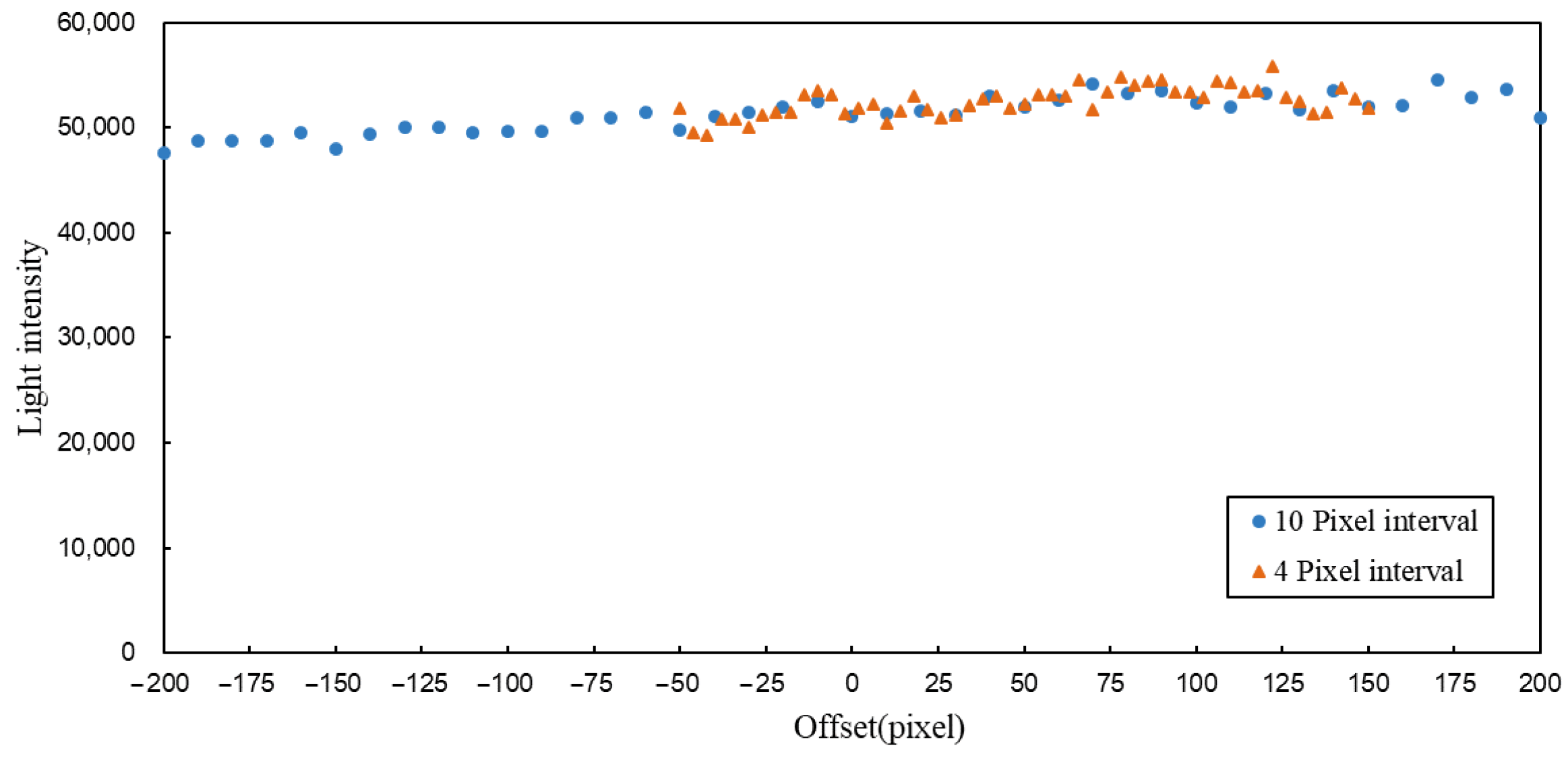
4.1.2. Experimental Data Analysis
4.1.3. Analysis of Abnormal Peaks
4.1.4. Experimental Error Analysis
- Laser Source: The laser used in this experiment is a small LED laser. Compared to the helium-neon lasers commonly used in laboratories, its light source is a square LED, which is focused by a lens at the laser head to form a quasi-point-like beam at a distant point. The beam quality is relatively poor, and its stability is slightly weaker. During prolonged operation, fluctuations in output power and frequency may occur, affecting the shape and brightness of the final spot;
- Optical Path Structure: The laser emits a Gaussian beam, where the center brightness is higher than the edges. Due to limitations in the experimental setup, no beam shaping was performed on the Gaussian beam. As a result, the light received by the LC-SLM has non-uniform brightness across its surface. During lateral modulation, the modulation region on the liquid crystal surface shifts, causing changes in the brightness of the focused spot, which, in turn, affects the final light intensity calculations;
- Measured Surface: Due to the influence of the polarizing beam splitter, a mirror could not be used as the measured surface. Instead, a white plastic block with high surface reflectivity was used as the measured object. While the surface appears smooth and flat to the naked eye, at the micro-nano scale, it may be relatively rough, affecting the light intensity values at each lateral modulation position. Additionally, if the measured surface is not perfectly perpendicular to the optical axis during installation, it can also lead to variations in the final measured light intensity values.
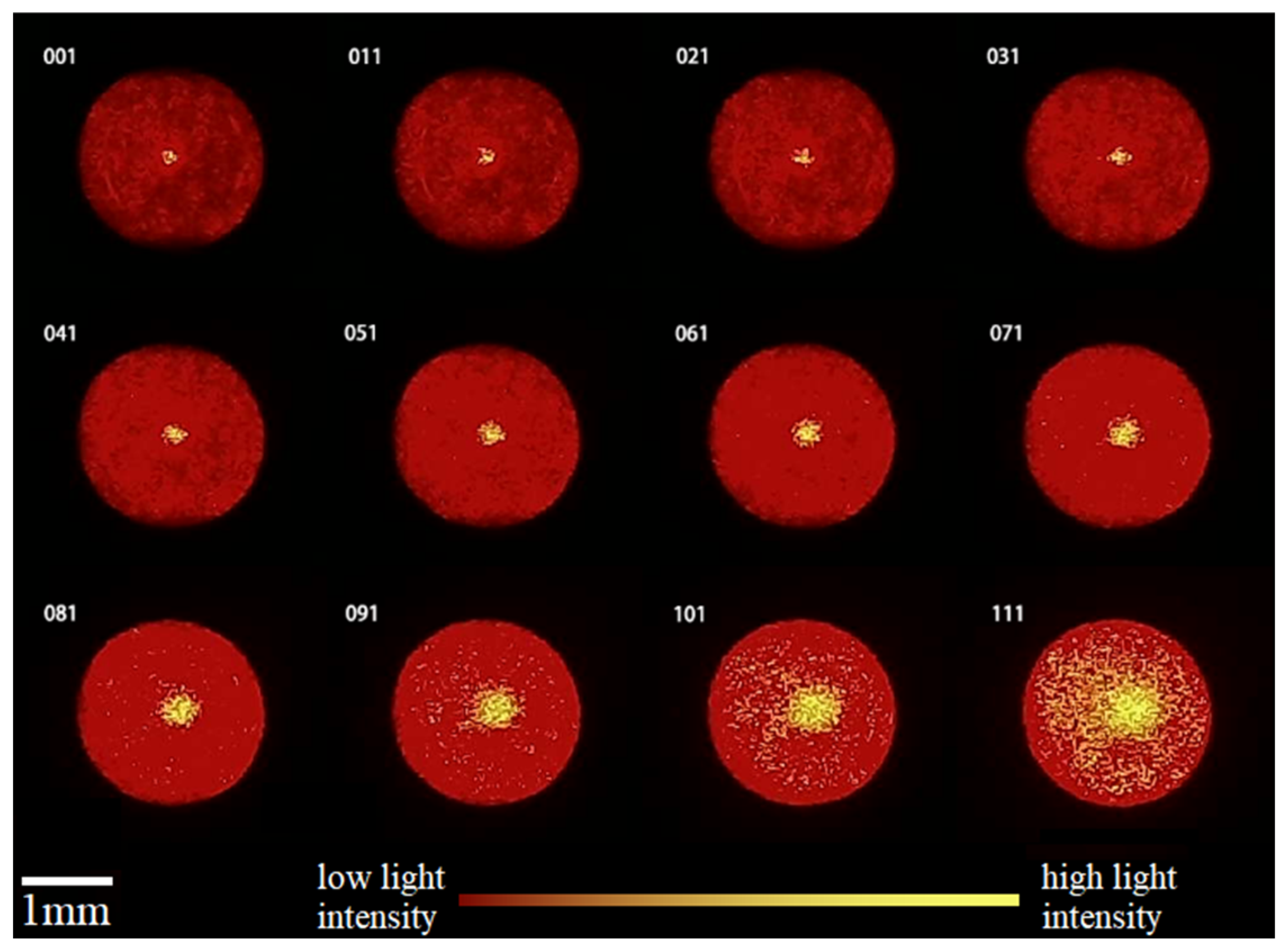
4.2. Axial Focusing Experiment
4.2.1. Experimental Data Acquisition
4.2.2. Experimental Data Analysis
4.2.3. Experimental Error Analysis
- Optical Path Structure: The laser emits a Gaussian beam, where the center brightness is higher than the edges. After passing through the collimating beam expander, it can be approximated as a plane wave; however, in reality, the light intensity distribution is non-uniform. Therefore, the light received by the LC-SLM also has non-uniform brightness across its surface. During axial modulation, the quasi-collimated beam obtained through the microscope objective has components with certain deflection angles. When the axial distance changes, the positions and intensities of these components also change, causing the captured spot images to vary irregularly with the defocus amount, showing only an overall trend in size changes;
- Alignment Precision: When setting up and aligning the optical system, the installation angles and heights of numerous components, such as the LC-SLM, PBS, microscope objective, and industrial camera, require manual adjustment, which affects the alignment of all subsequent components and devices. The final impact is that the components are not perfectly collimated or perpendicular, which affects the uniformity of the modulated beam in the focusing range, leading to deviations in the spot light intensity values during axial focusing;
- Ambient Light Interference: All images in this experiment were captured under dark conditions; however, the lights from electronic equipment around the laboratory are unavoidable. Additionally, the LC-SLM modulation, camera capture, and air-bearing platform control during the experiment are all operated via a computer; therefore, the confocal microscopy 3D focusing system is exposed to interference light from the display. The display screen changes during the experiment, causing the brightness of the interference light to vary accordingly. Although changes in pixel grayscale values are not visible to the naked eye, they may still interfere with the data acquisition process.
5. Conclusions
Author Contributions
Funding
Institutional Review Board Statement
Informed Consent Statement
Data Availability Statement
Acknowledgments
Conflicts of Interest
Abbreviations
| LC-SLM | Liquid crystal spatial light modulator |
| DMD | Digital micromirror device |
| SLM | Spatial light modulator |
| FLC-SLM | Ferroelectric liquid crystal spatial light modulator |
| R2 | Goodness of fit |
References
- Gao, W.; Lei, Z.; Wu, K.; Chen, Y. Reconfigurable and Renewable Nano-Micro-Structured Plastics for Radiative Cooling. Adv. Funct. Mater. 2021, 31, 2100535. [Google Scholar] [CrossRef]
- Xiang, T.; Hou, J.; Xie, H.; Liu, X.; Gong, T.; Zhou, S. Biomimetic micro/nano structures for biomedical applications. Nano Today 2020, 35, 100980. [Google Scholar] [CrossRef]
- Gao, Y.; Lee, H.; Jiao, J.; Chun, B.J.; Kim, S.; Kim, D.-H.; Kim, Y.-J. Surface third and fifth harmonic generation at crystalline Si for non-invasive inspection of Si wafer’s inter-layer defects. Opt. Express 2018, 26, 32812–32823. [Google Scholar] [CrossRef]
- Podulka, P. The Effect of Surface Topography Feature Size Density and Distribution on the Results of a Data Processing and Parameters Calculation with a Comparison of Regular Methods. Materials 2021, 14, 4077. [Google Scholar] [CrossRef] [PubMed]
- Kim, C.-S.; Yoo, H. Three-dimensional confocal reflectance microscopy for surface metrology. Meas. Sci. Technol. 2021, 32, 102002. [Google Scholar] [CrossRef]
- Xiao, G.Q.; Corle, T.R.; Kino, G.S. Real-time confocal scanning optical microscope. Appl. Phys. Lett. 1988, 53, 716–718. [Google Scholar] [CrossRef]
- Petráň, M.; Hadravský, M.; Boyde, A. Tandem-scanning reflected-light microscope. Scanning 1985, 7, 97–108. [Google Scholar] [CrossRef]
- Grant, D.M.; McGinty, J.; McGhee, E.J.; Bunney, T.D.; Owen, D.M.; Talbot, C.B.; Zhang, W.; Kumar, S.; Munro, I.; Lanigan, P.M.; et al. High speed optically sectioned fluorescence lifetime imaging permits study of live cell signaling events. Opt. Express 2007, 15, 15656–15673. [Google Scholar] [CrossRef]
- Miyajima, H.; Asaoka, N.; Isokawa, T.; Ogata, M.; Aoki, Y.; Imai, M.; Fujimori, O.; Katashiro, M.; Matsumoto, K. A MEMS electromagnetic optical scanner for a commercial confocal laser scanning microscope. J. Microelectromechan. Syst. 2003, 12, 243–251. [Google Scholar] [CrossRef]
- Chen, H.-R.; Chen, L.-C. Full-field chromatic confocal microscopy for surface profilometry with sub-micrometer accuracy. Opt. Lasers Eng. 2023, 161, 107384. [Google Scholar] [CrossRef]
- Du, C.; Chen, B.; Qiu, C.; Bai, L.; Deng, Q.; Zhou, C.; Zhou, L. Microlens Array and Application Systems; SPIE: Chengdu, China, 2000; pp. 153–157. [Google Scholar] [CrossRef]
- Eisner, M.; Lindlein, N.; Schwider, J. Confocal microscopy with a refractive microlens-pinhole array. Opt. Lett. 1998, 23, 748–749. [Google Scholar] [CrossRef] [PubMed]
- Gong, W.; Si, K.; Chen, N.; Sheppard, C.J.R. Focal modulation microscopy with annular apertures: A numerical study. J. Biophotonics 2010, 3, 476–484. [Google Scholar] [CrossRef]
- Chen, C.; Yang, W.; Wang, J.; Lu, W.; Liu, X.; Jiang, X. Accurate and efficient height extraction in chromatic confocal microscopy using corrected fitting of the differential signal. Precis. Eng.-J. Int. Soc. Precis. Eng. Nanotechnol. 2019, 56, 447–454. [Google Scholar] [CrossRef]
- Nayar, S.K.; Branzoi, V.; Boult, T.E. Programmable Imaging: Towards a Flexible Camera. Int. J. Comput. Vis. 2006, 70, 7–22. [Google Scholar] [CrossRef]
- Nayar, S.K.; Branzoi, V.; Boult, T.E. Programmable imaging using a digital micromirror array. In Proceedings of the 2004 IEEE Computer Society Conference on Computer Vision and Pattern Recognition, Washington, DC, USA, 27 June–2 July 2004; pp. I-I. [Google Scholar] [CrossRef]
- Ri, S.; Fujigaki, M.; Morimoto, Y. Intensity range extension method for three-dimensional shape measurement in phase-measuring profilometry using a digital micromirror device camera. Appl. Opt. 2008, 47, 5400–5407. [Google Scholar] [CrossRef]
- Mohan, A.; Raskar, R.; Tumblin, J. Agile spectrum imaging: Programmable wavelength modulation for cameras and projectors. Comput. Graph. Forum 2008, 27, 709–717. [Google Scholar] [CrossRef]
- Chen, H.-M.P.; Yang, J.-P.; Yen, H.-T.; Hsu, Z.-N.; Huang, Y.; Wu, S.-T. Pursuing High Quality Phase-Only Liquid Crystal on Silicon (LCoS) Devices. Appl. Sci. 2018, 8, 2323. [Google Scholar] [CrossRef]
- Reichelt, S. Spatially resolved phase-response calibration of liquid-crystal-based spatial light modulators. Appl. Opt. 2013, 52, 2610–2618. [Google Scholar] [CrossRef]
- Lindle, J.R.; Watnik, A.T.; Cassella, V.A. Efficient multibeam large-angle nonmechanical laser beam steering from computer-generated holograms rendered on a liquid crystal spatial light modulator. Appl. Opt. 2016, 55, 4336–4341. [Google Scholar] [CrossRef]
- Das, A.; Boruah, B.R. Optical sectioning microscope with a binary hologram based beam scanning. Rev. Sci. Instrum. 2011, 82, 043702. [Google Scholar] [CrossRef]
- Das, A.; Boruah, B.R. Point Scanning Microscope with Adaptive Illumination Beam Intensity. In Proceedings of the International Conference on Light—Optics-Phenomena, Materials, Devices, and Characterization, Calicut, India, 23–25 May 2011. [Google Scholar]
- Park, C.; Lee, K.; Baek, Y.; Park, Y. Low-coherence optical diffraction tomography using a ferroelectric liquid crystal spatial light modulator. Opt. Express 2020, 28, 39649–39659. [Google Scholar] [CrossRef] [PubMed]
- McManamon, P.F.; Dorschner, T.A.; Corkum, D.L.; Friedman, L.J.; Hobbs, D.S.; Holz, M.; Liberman, S.; Nguyen, H.Q.; Resler, D.P.; Sharp, R.C.; et al. Optical phased array technology. Proc. IEEE 1996, 84, 268–298. [Google Scholar] [CrossRef]
- He, Z.; Gou, F.; Chen, R.; Yin, K.; Zhan, T.; Wu, S.-T. Liquid Crystal Beam Steering Devices: Principles, Recent Advances, and Future Developments. Crystals 2019, 9, 292. [Google Scholar] [CrossRef]
- Beeckman, J.; Neyts, K.; Vanbrabant, P.J.M. Liquid-crystal photonic applications. Opt. Eng. 2011, 50, 081202. [Google Scholar] [CrossRef]
- Engstrom, D.; Bengtsson, J.; Eriksson, E.; Goksor, M. Improved beam steering accuracy of a single beam with a 1D phase-only spatial light modulator. Opt. Express 2008, 16, 18275–18287. [Google Scholar] [CrossRef] [PubMed]
- Tholl, H. Novel laser beam steering techniques. Proc. SPIE-Int. Soc. Opt. Eng. 2006, 6397, 51–64. [Google Scholar] [CrossRef]
- Fan, Y.-H.; Ren, H.; Wu, S.-T. Switchable Fresnel lens using polymer-stabilized liquid crystals. Opt. Express 2003, 11, 3080–3086. [Google Scholar] [CrossRef]

Disclaimer/Publisher’s Note: The statements, opinions and data contained in all publications are solely those of the individual author(s) and contributor(s) and not of MDPI and/or the editor(s). MDPI and/or the editor(s) disclaim responsibility for any injury to people or property resulting from any ideas, methods, instructions or products referred to in the content. |
© 2025 by the authors. Licensee MDPI, Basel, Switzerland. This article is an open access article distributed under the terms and conditions of the Creative Commons Attribution (CC BY) license (https://creativecommons.org/licenses/by/4.0/).
Share and Cite
Li, Y.; Li, Y. Three-Dimensional Focusing Measurement Method for Confocal Microscopy Based on Liquid Crystal Spatial Light Modulator. Sensors 2025, 25, 2620. https://doi.org/10.3390/s25082620
Li Y, Li Y. Three-Dimensional Focusing Measurement Method for Confocal Microscopy Based on Liquid Crystal Spatial Light Modulator. Sensors. 2025; 25(8):2620. https://doi.org/10.3390/s25082620
Chicago/Turabian StyleLi, Yupeng, and Yifan Li. 2025. "Three-Dimensional Focusing Measurement Method for Confocal Microscopy Based on Liquid Crystal Spatial Light Modulator" Sensors 25, no. 8: 2620. https://doi.org/10.3390/s25082620
APA StyleLi, Y., & Li, Y. (2025). Three-Dimensional Focusing Measurement Method for Confocal Microscopy Based on Liquid Crystal Spatial Light Modulator. Sensors, 25(8), 2620. https://doi.org/10.3390/s25082620