Review

Coelenterazine Analogs for Bioassays and Molecular Imaging

1
Environmental Management Research Institute (EMRI), National Institute of Advanced Industrial Science and Technology (AIST), 16-1 Onogawa, Tsukuba 305-8569, Japan
2
Department of Engineering Science, Graduate School of Informatics and Engineering, The University of Electro-Communications, Chofu 182-8585, Japan
3
School of Life Science and Technology, Institute of Science Tokyo, B-62 4259 Nagatsuta-cho, Midori-ku, Yokohama 226-8501, Japan
*
Author to whom correspondence should be addressed.
Sensors 2025, 25(6), 1651; https://doi.org/10.3390/s25061651
Submission received: 12 February 2025 / Revised: 28 February 2025 / Accepted: 5 March 2025 / Published: 7 March 2025
(This article belongs to the Special Issue Feature Papers in Biosensors Section 2025)

Abstract

:
Coelenterazine (CTZ) is a common substrate of marine luciferases upon emission of bioluminescence (BL) in living organisms. Because CTZ works as a “luminophore” in the process of BL emission, the chemical modification has been centered for improving the optical properties of BL. In this review, we showcase recent advances in CTZ designs with unique functionalities. We first elucidate the light-emitting mechanisms of CTZ, and then focus on how the rational modification of CTZ analogs developed in recent years are connected to the development of unique functionalities even without luciferases, which include color tunability covering the visible region, specificity to various proteins (e.g., luciferase, albumin, and virus protein), and activatability to ions or reactive oxygen species (ROS) and anticancer drugs. This review provides new insights into the broad utilities of CTZ analogs with designed functionalities in bioassays and molecular imaging.

1. Introduction

A myriad number of light-emitting species exist in nature and are especially common in the oceans. This bioluminescence (BL) from living organisms is a cold light-emitting event caused by the oxidation of a small molecular weight substrate called luciferin, catalyzed by an enzyme named luciferase inside living organisms [1].
As the key components for BL, both luciferases and luciferins are equally important, but recent studies have focused on the genetic engineering of luciferases rather than the chemical modification of luciferins. This genetic engineering has contributed to the creation of new mutants with excellent optical properties or the development of molecular imaging probes, such as bioluminescence resonance energy transfer (BRET) [2,3] and protein-fragment complementation assay (PCA) probes [4,5]. In parallel, the authors note the benefits of chemical modification of luciferins, although they have attracted relatively less attention to date.
Luciferin is an essential component for BL as the energy source and is supplied by the biosynthesis in living organisms. However, most biosynthesis mechanisms are unknown, although those of fungal luciferin [6] and bacterial luciferin [7] have been partly elucidated. In contrast to the variety of light-emitting species in nature, their luciferin repertoires are extremely small, because few luciferins are shared by many luciferases. The representative ones include (i) beetle luciferin (d-luciferin) for beetle luciferases, (ii) coelenterazine (CTZ) for marine luciferases, (iii) tetrapyrrole luciferin, (iv) fungal luciferin, (v) bacterial luciferin, and (vi) Cypridina luciferin [8].
In this review, the authors focus on CTZ and its analogs developed in recent years, and showcase the light-emitting mechanisms, recent advances in the chemical designs, and the corresponding versatility in bioassays and molecular imaging.

2. Coelenterazine as a Common Substrate of Marine Luciferases

Luciferin is a key component for developing BL with luciferases. The role of luciferins has been explained as “bioluminophore”, because it determines the colors and works as the energy source through its oxidization catalyzed by luciferases.
The luciferin in the aqua phase is diffused into the active site of the specific luciferase to work as a “bioluminophore”, whereas the “fluorophore” of fluorescent proteins is derived from an intrinsic part of the FP. The most common luciferin of marine luciferases, CTZ, chemically resembles the fluorophore of fluorescent proteins (most often a derivative of the tripeptide sequence 65XYG67), sharing the same imidazolone ring structure [9]. Because of its uniqueness as a potential optical platform, CTZ has been centered on organic modification by appending unique functional groups to the backbone of CTZ.
This section mainly focuses on the chemical basis of CTZ and its recent studies on the analogs that are connected to the applications to bioassays and molecular imaging.

2.1. Chemical Structure of Coelenterazine

In the early 1960s, Dr. Johnson’s group at Princeton University isolated the Ca2+-sensitive photoprotein aequorin from jellyfish [10]. Later, they isolated two compounds, now called coelenteramine (CTM) and coelenteramide (CTMD), from aequorin [11]. CTZ was originally isolated as an actual substance from the liver of the luminous squid Watasenia scintillans, and was chemically synthesized by Inoue et al. for the first time [12]. The occurrence of CTZ is extremely widespread among marine bioluminescent organisms. However, CTZ has not been found in terrestrial creatures [1].
Marine luciferins such as Cypridina luciferin (Vargulin) and CTZ are biosynthesized in living organisms, and their chemical structures are derived from amino acids, that is, actually modified tripeptides that are made from arginine, isoleucine, and tryptophan (RIW) in the case of Cypridina luciferin [13,14]; and one phenylalanine and two tyrosine residues (FYY) in the case of CTZ [1,15], respectively.

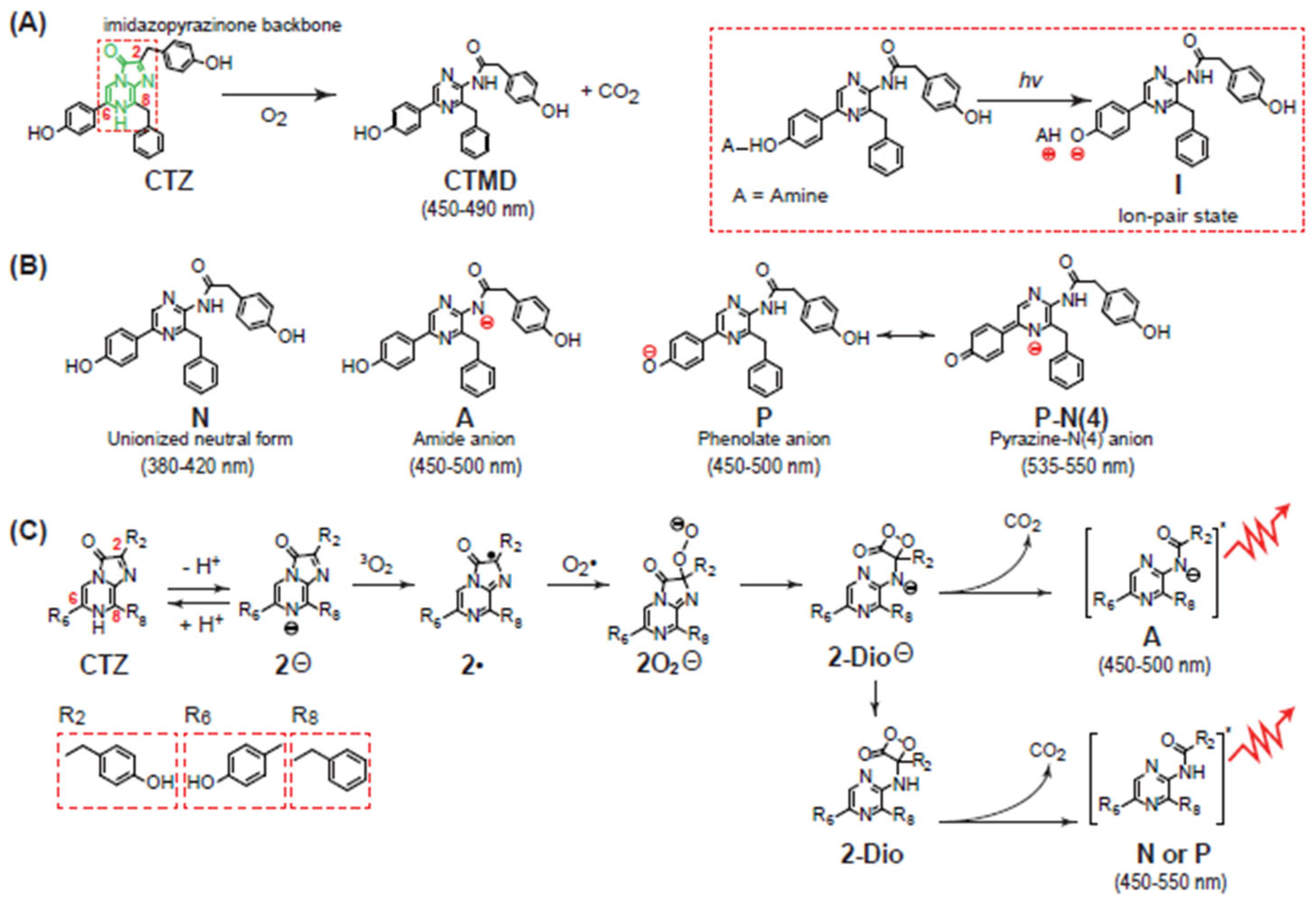
2.2. Chemiluminescence-Emitting Mechanism of CTZ

CTZ produces luminescence in the process of an oxidative decarboxylation reaction of its imidazopyrazinone structure. Briefly, CTZ is peroxidized by molecular oxygen (O2). The peroxide forms an amide anion of coelenteramide (CTMD) in its excited state, resulting in the emission of blue luminescence (typically λmax 450~490 nm) (Figure 1A) [1]. In the process, it is known that CTZ forms five potential intermediate light emitters: unionized neutral form (N), phenolate anion (P), ion-pair state (I), amide anion (A), and pyrazine-N(4) anion (P-N(4)).
Among the intermediates, the most common light emitter is the amide anion (A). Other light emitters such as phenolate anion (P) and its resonance structure called pyrazine-N(4) anion (P-N(4)) can be generated according to the experimental conditions and luciferases. The neutral form (N) light emitter can be made in the case that the OH group at the C-6 position is replaced with other functional groups. This neutral form (N) emits the shortest wavelength luminescence (generally λmax 380–420 nm). The phenolate anion (P) and amide anion (A) generate slightly longer wavelength luminescence than that of the neutral form (N), typically λmax 450–500 nm. The longest wavelength luminescence is created by the pyrazine-N(4) anion (P-N(4)), generally λmax 535–550 nm [16]. The phenolate anion temporarily formed from the ion-pair state (I) can be made only by light excitation; thus, it is very exceptional and can be excluded from the list of the emitter forms. The phenolate anion (P) and its resonance structure pyrazine-N(4) anion (P-N(4)) are practically the same intermediate (Figure 1B).
The first rational studies on the light-emitting mechanism of marine organisms began with Cypridina luciferin [17], which was the first marine luciferin isolated and shares the same imidazopyrazinone backbone as that of CTZ.
Cypridina luciferin can emit light in two different ways: chemiluminescence (CL) induced by aprotic solvents; and BL catalyzed by luciferases, whose mechanisms are known to be similar to each other [18]. The mechanism of the CL emission is explained below.
As illustrated in Figure 1C, the light-emitting reaction of CTZ in aprotic solvent starts via deprotonation of the N-7 nitrogen to form a CTZ anion (2) that reacts directly with O2 at the C-2 carbon to form a CTZ peroxide ion (2O2). Following the oxygen addition, the resulting nucleophilic peroxide moiety performs an intramolecular attack on the C-3 carbonyl to form a cyclic dioxetanone intermediate (2-Dio). Subsequent ring decomposition via decarboxylation leads to the loss of CO2 and the formation of an anionic excited-state aminopyrazine product (coelenteramide ion) in a singlet excited state (A). This unstable anionic species then decomposes with the emission of light, resulting in CL. Alternatively, the cyclic dioxetanone intermediate (2-Dio) can be protonated (2-Dio) and form the neutral (N) or phenolate anion intermediates (P) after losing CO2. These unstable anionic species then decompose with the emission of light [19].

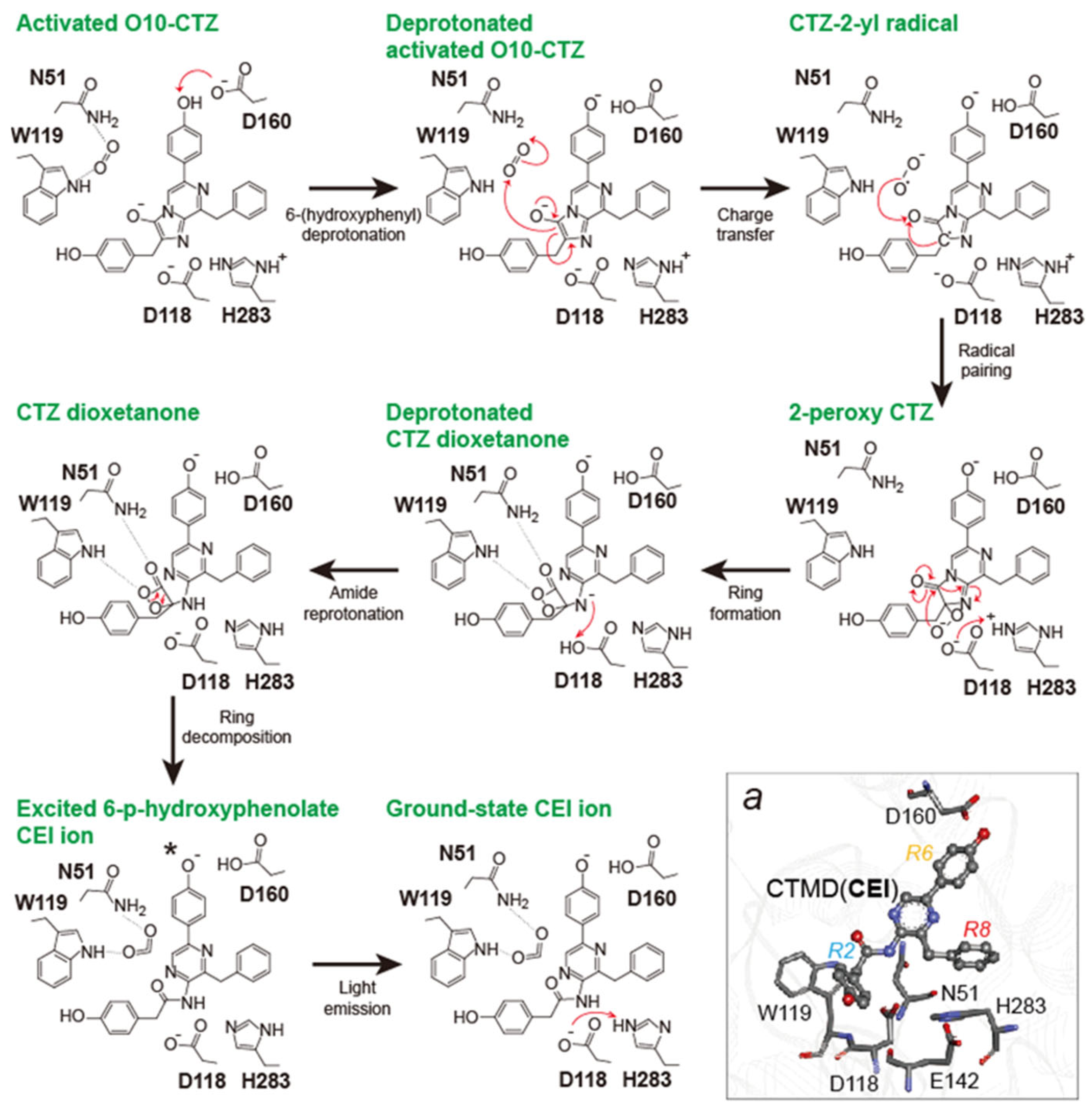
2.3. Bioluminescence-Emitting Mechanism of Coelenterazine

This section reviews the light-emitting mechanism of BL. Recently, Schenkmayerova et al. presented a unique approach for investigating the light-emitting mechanism of CTZ using a unique non-oxidative CTZ analog called azaCTZ and X-ray crystallographic information of Renilla luciferase 8 (RLuc8) and its ancestor enzyme AncFT [20]. The mechanism basically follows the same process as that of the CL as described above.
The proposed BL-emitting mechanism is as follows (Figure 2): CTZ enters the enzyme active site with a deprotonated imidazopyrazinone core because the pKa of the core (7.55) is very close to the physiological pH. Upon binding, the -OH group of the C-6-(p-hydroxyphenyl) substituent is deprotonated by D160 to form the activated dianion O10-CTZ, which affects the emission maximum (λmax). In the ternary Michaelis complex, the side chains of N51 and W119 position a co-substrate molecule (dioxygen) such that it can be directly attacked by the C-2 carbon of O10-CTZ. Their initial interaction occurs via a charge-transfer radical mechanism. The next step involves radical pairing and termination to form the 2-peroxy-CTZ anion, which then cyclizes via a nucleophilic addition-elimination mechanism. This step yields a highly unstable and energetic dioxetanone structure of CTZ with a deprotonated amide group. At this point, the amide group is protonated by D118 to avoid significant attenuation of the BL signal due to the formation of the deprotonated CTMD product. D118 also prevents hydrolysis of the amide bond by a water molecule, which yields the side-product coelenteramine (CTM). After reprotonation, the unstable dioxetanone ring decomposes and the released energy excites the newly formed CTMD (CEI) ion. As it returns to the ground state, the excited molecule releases a photon, representing the BL signal, together with the final products, ground-state CTMD ion and carbon dioxide (CO2). The residue H283 is reprotonated via an interaction with E142.

3. Creation of Coelenterazine Analogs

Although CTZ is the common substrate for marine luciferases, it practically has intrinsic limitations in its application to bioassays and molecular imaging.
For example, (i) CTZ has relatively poor solubility and is chemically unstable in physiological samples [21]; (ii) the optical intensity of CTZ is still not strong enough for challenging molecular imaging missions including animal imaging; (iii) the color variation in CTZ is limited to blue and green that is easily attenuated by physiological samples and tissues [22]; and (iv) the conventional role of native CTZ is limited to luminesce with marine luciferases, and does not address the necessities of additional functionalities such as analyte sensing and conditional signal reporting.
To date, CTZ has been chemically modified to employ additional optical properties or functionalities, besides the conventional role. Upon synthesis of CTZ analogs, the imidazopyrazinone backbone of CTZ itself has not been modified because it serves as the scaffold of the oxidation reaction. Instead, the functional groups at the C-2, C-5, C-6, and C-8 positions of the backbone have been chemically engineered as a way to improve the optical properties [23]. The CTZ analogs were briefly categorized into six groups according to the chemical structural similarity as illustrated in Figure 3 (named Groups A–F).

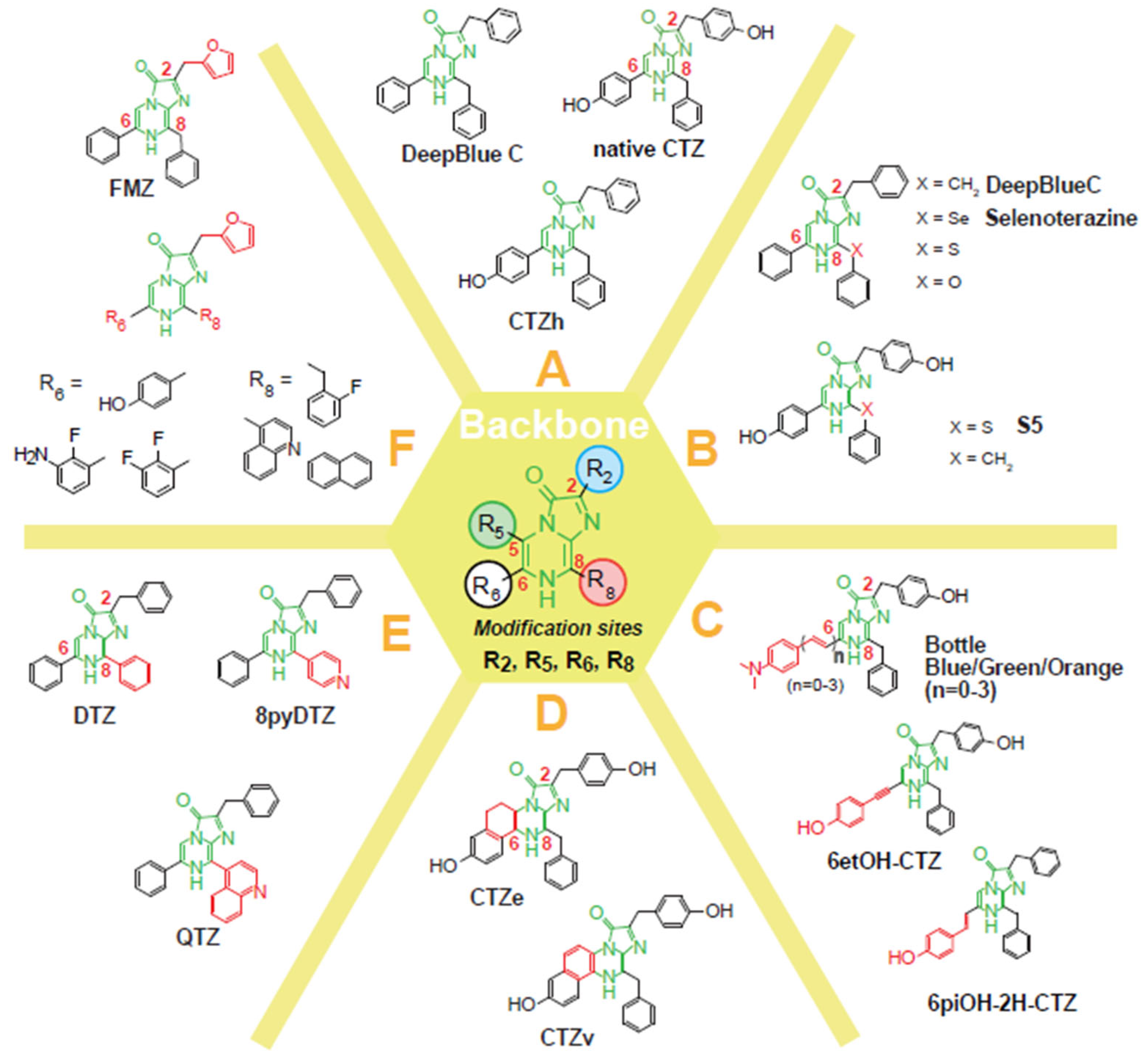
3.1. Categorization of Coelenterazine Analogs

Among many CTZ analogs, Group A represents historically old and structurally simple chemicals (Group A in Figure 3): (i) Coelenterazine h (CTZh) was originally reported by Hori et al. in 1973 [24], where the absence of the OH group at the C-2 position is the different feature from CTZ. It is known that CTZh generates strong BL intensities with NanoLuc, RLuc derivatives, and ALuc. CTZh does not show a significant luciferase specificity, but is the most widely used among CTZ analogs; and (ii) DeepBlueC (DBC) is a well-known CTZ analog that is characteristic in the absence of the OH group at both the C-2 and C-6 positions, compared to CTZ [25]. It is known that DBC luminesces in blue with NanoLuc and RLuc. The far blue-shifted peak advantageously works for transferring the resonance energy to fluorescent proteins in BRET systems.
Figure 3 briefly illustrates the overall modification of the C-2, C-6, and C-8 positions of the imidazopyrazinone backbone of CTZ: Group B represents the elbow-modified CTZ analogs at the C-8 position. Group C shows the π-electron conjugation prolonged CTZ analogs at the C-6 position. Group D is characteristic of CTZ analogs with a bridged form at the C-5 and C-6 positions. Groups E and F were commonly designed for NanoLuc and its derivatives, where Group E represents CTZ analogs with a benzyl group at the C-2 position and phenyl group or pyridyl group at the C-6 position, whereas Group F is unique in having a furfuryl group at the C-2 position.
It is known that the designs of Groups B–F contribute to red-shifted BL and those of Groups C–F are effective for luciferase specificity. The designs of Groups E–F are focused on enhancing both BL intensities and spectral redshifts of NanoLuc. The distinctively designed CTZ analogs are surprisingly tolerated by marine luciferases. Each case is explained in detail in the following subsections.

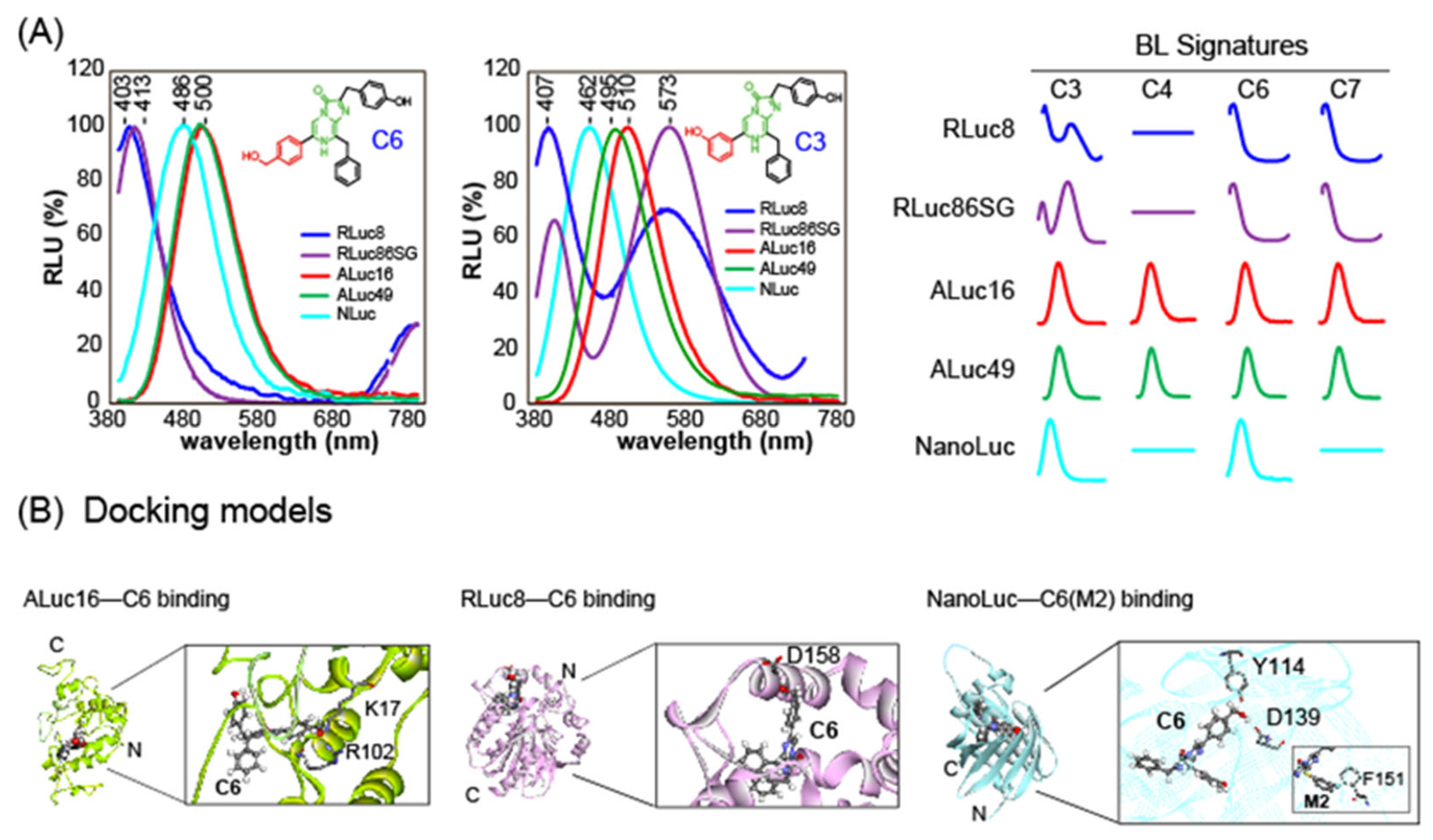
3.2. Color-Tunable Coelenterazine Analogs

CTZ and its analogs generate unique colors according to which of the five intermediates are predominately made during the oxidative reaction catalyzed by marine luciferases as explained above. For example, a red-shifted BL is expected to be with the pyrazine-N(4) anion (P-N(4)) intermediate, whereas a blue-shifted BL is with the neutral form (N) intermediate.
Technically speaking, a significantly blue-shifted BL can be made by modifying the OH group at the C-6 position of CTZ, because this modification creates the neutral form (N) intermediate during the reaction with RLuc derivatives [26].
This mechanism of blue-shifted BL spectrum fits well with RLuc derivatives, but not necessarily with the other marine luciferases. For example, a C-6 position-modified CTZ analog named C6 (R6 = Bnz-p-CH2OH) shows a significantly blue-shifted BL spectrum especially with RLuc8 and RLuc86SG (λmax ca. 403–413 nm), whereas the same CTZ analog generates moderate blue (λmax ca. 465 nm) and greenish blue (λmax ca. 500 nm) BL spectra with the other marine luciferases NanoLuc and ALuc16, respectively [27]. This result indicates that the intermediates of the CTZ analog C6 are differently formed according to marine luciferases (Figure 4A).
Another CTZ analog C3 (R6 = Bnz-m-OH) shows much more diverse shapes of BL spectra according to marine luciferases. The CTZ analog interestingly develops two separated peaks (blue and orange) in the BL spectrum with RLuc8 and RLuc86SG, although it generates single-peaked BL spectra (bluish green) with the other marine luciferases. This feature indicates that C3 generated two major intermediates during the reaction with RLuc derivatives, whose ratios are various according to RLuc derivatives. Because of the uniqueness in the BL spectral shapes of C3, one can easily distinguish which luciferase is applied to BL imaging. Kamiya et al. previously described the “spectral signature” for identifying each reporter luciferase and these substrates were named C-series CTZ analogs [27].
This spectral uniqueness of CTZ analogs is also supported by the distinctive binding modes of the substrate C6 with marine luciferases, suggesting that the oxidative reaction environments forming the intermediates inside luciferases should be distinctive from each other (Figure 4B).
BL colors covering the visible region can be intentionally created in a way to combine specific CTZ analogs with marine luciferases. Figure 5A shows the typical combinations of the CTZ analog and marine luciferase, where S-series CTZ analogs emit relatively red-shifted BL spectra compared to C-series CTZ analogs. This occurs because the carbon ankle of the benzyl group at the C-8 position is replaced with sulfur (S) for extending the π-electron conjugation [28].
The most common and straightforward way to redshift a BL spectrum is to extend the π-electron conjugation of the imidazopyrazinone backbone (Group C in Figure 3). Based on this idea, Tamaki et al. introduced a series of CTZ analogs in a way to increase the number of double bonds for extending the π-electron conjugation at the C-6 position of the imidazopyrazinone backbone [29]. This approach was commonly effective for exerting the red shifts in the BL spectra of any marine luciferases (Figure 5B).
Furimazine (FMZ) is characteristic with respect to the absence of the OH group at the C-6 position and the substitution of the phenol group with a furan group at the C-2 position upon comparison with CTZ (Group F in Figure 3) [30]. FMZ is specific to NanoLuc and has proved its excellent optical intensities in various molecular imaging modalities to date. However, FMZ is known to be toxic in cells and animal models and is poorly soluble, thus, limiting the doses in physiological systems [31]. To address the limitations, various FMZ analogs have been reported to date, e.g., fluorofurimazine (FFz) that is brighter than FMZ with improved solubility [32] and cephalofurimazine (CFz) for bright imaging of molecular events in the brain [33].
Extending π-electron conjugation at the C-8 position of the backbone of FMZ is also known to be effective for red shifts in BL spectra especially with NanoLuc and its derivatives. For example, Shakhmin et al. reported a series of FMZ analogs. Among them, installation of a 4-quinoline group to the C-8 position of FMZ analogs exerted red-shift emission by ca. 100 nm while maintaining reasonable light output [34]. Similarly, pyridyl and 4-quinoline groups can be introduced to the C-8 position of a FMZ analog, diphenylterazine (DTZ), to create 8pyDTZ and QTZ, respectively [35].
Another effective method to create red-shifted BL with CTZ analogs is conjugating fluorescent dyes to the C-2 or C-6 position of the backbone of native CTZ (Group C in Figure 3) [36]. Nishihara et al. synthesized a series of dye-conjugated CTZ analogs: ones conjugated by Fluorescein isothiocyanate (FITC) (6-FITC-CTZ), Nile red (6-Nile-R-CTZ), and Cyanine-5 (Cy5) at the C-6 position (Cy5-CTZ). As mentioned above, C-6 position-modified CTZ analogs generally show blue-shifted BL spectral peaks at around 400 nm with RLuc derivatives. A C-6-modified analog, 6-FITC-CTZ, showed a unique spectrum peak at 522 nm with RLuc8.6-535, in addition to that at 400 nm. Among the synthesized dye-conjugated CTZ analogs, 6-Nile-R-CTZ showed a characteristic resonance energy transfer peak at 650 nm, where ca. 11% of the total photons were emitted in the red region for longer than 600 nm, which is highly tissue-permeable and commonly referred to as the “optical window” (Figure 5C) [37].
In addition, Cy5-CTZ was developed by attaching Cy5 dye to CTZ through an acetylene linker. This linker enables through-bond energy transfer (TBET) between the energy donor CTZ and the energy acceptor Cy5. This Cy5-fused CTZ analog is intrinsically fluorescent and emits near-infrared (NIR)-shifted luminescence through Cy5 upon reacting with appropriate luciferases, RLuc and ALuc (Figure 5D) [38]. The authors demonstrated that Cy5-CTZ is optically stable in physiological samples, easily passes through the plasma membrane, and emits NIR BL in animal cells.

3.3. Luciferase-Specific Coelenterazine Analogs

Although BLs have diverse colors ranging from blue to red according to luciferin–luciferase combinations, most of the peak emissions populate the blue-yellow region [39,40]. Furthermore, the spectra have broad bandwidths and, thus, overlap with each other. This feature causes optical cross-leakage that severely impairs multiplex imaging. To tackle this issue, BL systems with two or more multiple luciferase reporters conventionally make use of quenchers, optical filters, and/or deconvolution schemes so as to discriminate the multiple BL signals [41,42]. However, this cannot be considered as the fundamental solution to the issue.
In order to address the intrinsic drawback of BL, some researchers redesigned CTZ analogs so that they have luciferase specificities in multi-reporter assay systems.
Nishihara et al. previously synthesized 20 kinds of CTZ analogs with modifications to the C-2 and C-6 functional groups (Group C in Figure 3 and Figure 6A), some of which developed luciferase-specific BL signals [43].
For example, ethynylation at the C-6 position of the imidazopyrazinone backbone (i.e., 6etOH-CTZ) is specific only to a marine luciferase ALuc16, whereas an introduction of a double bond (olefin) at the C-6 position (e.g., 6piOH-CTZ) exerts its preferable binding to both RLuc8.6-535 and ALucs. It is considered that the double bond addition and orientation at the C-6 position determines the structural preference for marine luciferases in addition to the red shifts in BL. CTZ analogs without the OH group at the C-2 position (e.g., 6piOH-2H-CTZ) specifically, luminesced only with a marine luciferase RLuc8.6-535, but ones having the OH group at the C-2 position (e.g., 6piOH-CTZ) did not. This specificity indicates that the OH group at the C-2 position is a key functional group in binding to ALucs; however, it is not essential for interaction with RLuc8.6-535. It is explained that the C-2 position is located at the loose moiety of RLuc8.6-535 and, thus, is less influential, compared to the case of ALuc [43].
Kamiya et al. also reported a new series of luciferase-specific CTZ analogs (named K1K6) with modification of the C-6 and C-8 positions [44] (Figure 6B): (i) by modification of the π-electron conjugation through replacement of the benzyl group with a phenyl group at the C-8 position of the imidazopyrazinone backbone and (ii) by introduction of a dimethylaminophenyl group or a bulky benzodioxane group to the C-6 or C-8 positions of the backbone. The functional group-deficient CTZ analogs at the C-8 position, named K5 and K6, are effective for the RLuc specificity, whereas a CTZ analog carrying a bulky group at the C-8 position, named K2, is specific to ALucs.
In parallel, Kamiya et al. reported a series of regiospecific CTZ analogs, where all the functional groups at the C-2, C-6, and C-8 positions of the imidazopyrazinone backbone were regiochemically modified, and found that some of the analogs are highly specific to NanoLuc. For example, M2 is a regiospecific CTZ analog carrying a m-fluorobenzenethiol group at the C-8 position and without the OH group at the C-2 position, and specific to NanoLuc [45] (Figure 6C). The authors’ computational modeling explained the NanoLuc specificity of M2 with the following reasons: (i) the benzyl group at the C-2 position is positioned at the hydrophobic region of NanoLuc and (ii) the m-fluorobenzyl group at the C-8 position of M2 is in a suitable position to form a CF–π interaction with the benzyl group of F151 of NanoLuc (Figure 4B, right inset).
Some CTZ analogs with unique functional groups can be specific to serum albumins from different animal species (Figure 6D). Based on this concept, Nishihara et al. reported a CTZ analog for imaging human serum albumin (HSA) [46]. They reported only an HSA-specific CTZ analog emitting luminescence in blue. Independently, Kim et al. reported such unique CTZ analogs that specifically luminesce with bovine and mouse serum albumins (BSA and MSA) as well as HSA as pseudo-luciferases emitting red-shifted BL [47]. The kinetic constants of serum albumins as pseudo-luciferases are reported in references using conventional methods of enzyme kinetics. However, the specific mechanism of the substrate oxidation and light emission has not yet been elucidated. In the CTZ analog design, Kim et al. paid attention that the functional group at the C-8 position of CTZ was less investigated. They finally found that the C-8 position of CTZ is the key for the specificity with albumins and red shifts in the spectra. In their design on the CTZ analogs, the original carbon elbow of the benzyl group at the C-8 position was replaced with sulfur (S) for extending the π-electron conjugation. In addition, the para-position of the benzyl structure at the C-8 position was optionally modified with fluorine (F). It was found that the CTZ indicators immediately luminesce bluish green and yellowish green with HSA and BSA, respectively. This study shows that CTZ analogs work as simple and less-invasive optical indicators for serum albumins, which can recognize specific albumins and report the red-shifted luminescence signals.
In parallel, it was also reported that Cypridina luciferin, sharing the same imidazopyrazinone backbone, is reactive with the spike proteins of COVID-19 (SARS-CoV-2) acting as a pseudo-luciferase [48].

3.4. Activatable Coelenterazine Analogs

The CTZ analogs described above passively work as substrates of conventional luciferases or pseudo-luciferases like albumins. However, some researchers studied potential active use of CTZ analogs that have additional abilities such as responding to a specific stimulator (ligand or specific wavelength light) or recognizing a target protein and quantitatively reporting optical signal(s), in addition to their conventional role as luciferase substrates. Prototypically activatable CTZ analogs are the ones that embed a conceptional “on-off switch”. For this purpose, a chemical modification of the functional groups of the imidazopyrazinone backbone of CTZ with a cage was attempted (Figure 7).
Caged luciferins are typical activatable means, where the substrate itself is protected by a bulky cage and released by a specific signal or molecular event [49]. These caged luciferins commonly carry a bulky group, which blocks the interaction of the core imidazopyrazinone backbone with luciferases. The cages were designed to be removed by specific wavelengths of UV irradiation in animal models [50], a stimulator (analyte) including oxygen species [51], copper ions [49,52], calcium ion [53], and labile iron levels [54]. Similarly, a caged CTZ analog was reported to quantitatively image nitroreductase (NTR) activity, where the C-3 position of the imidazopyrazinone structure of the CTZ analog was modified with a nitrobenzyl moiety. Because NTR reduces the functional group with the nitrobenzyl moiety to the aniline group, net CTZ analog is released and catalyzed by marine luciferase to emit BL [55].
FMZ has also been targeted for the caging of the functional groups, because it carries the common imidazopyrazinon backbone. A series of caged FMZ analogs were developed by introducing a protective group at the C-3 position and an OH group at the C-6 position of the backbone. The protective groups are cleaved by esterase inside living animal cells to generate the deprotected forms, which work as the substrates of the specific luciferase, NanoLuc [56]. Among the series, the analogs with the bulkiest protecting group (adamantanecarbonyl group) and a hydroxy substituent (named Ad-FMZ-OH) showed significantly prolonged and constant BL signal in animal cells expressing NanoLuc, compared to the native FMZ substrate.
Protection of the C-3 position of a FMZ analog DTZ with a carboxylate functional group (named ETZ) is known to enhance both water solubility and the blood–brain barrier (BBB) permeability, and reduce BBB efflux [53]. When ETZ is delivered in the brain, nonspecific esterase hydrolyzes the linker ester bind of the protective group, releasing free DTZ for BL reaction.
Recently, a unique ion-selective CTZ analog named “Potassiorin” was developed for imaging potassium ion (K+) levels in mammalian cells [57]. The CTZ analog was made by replacing the OH group at the C-2 position with a K+-binding moiety known as a “crown ether” structure. This analog significantly reduces the BL intensity after K+ binding.
These activatable CTZ analogs have unique advantages compared to conventional passive ones: One can expect minimized autoluminescence with such activatable CTZ analogs. This is because the embedded on–off switch has the effect of reducing the background signals in basal conditions. For example, caged luciferin remains protected until an enzymatic cleavage event occurs and, thus, it minimizes autoluminescence.

3.5. Coelenterazine Analogs as Reactive Oxygen Species Scavenger and Anticancer Drugs

Aerobic organisms oxidize energy substrates with O2 and generate ATP. In the process, reactive oxygen species (ROS), superoxide radical anion, or hydrogen peroxide are inevitably generated. It is known that the physiological levels of ROS can be quantitatively imaged with CTZ analogs such as Cypridina luciferin analog (CLA) and Cypridina luciferin methoxy analog (MCLA). The working mechanism basically follows an oxidation process of the CTZ analogs for producing CL [58], i.e., MCLA reacts with ROS leading to the formation of the intermediate in the excited state. The intermediate relaxes to the ground state with the emission of CL (Figure 8).
Recently, CTZ analogs have been focused on anticancer activities. For example, brominated CTZ analogs were developed for self-activating photodynamic therapy of cancer (PDT) [59,60]. The CTZ analogs were further modified not to be activated by light irradiation but by superoxide anion [61], which is overproduced in cancer cells.
The anticancer effect of brominated CTZ analogs is explained as the bromine heteroatoms increase chemiexcitation to triplet states, not singlet states, due to the heavy-atom effect [62]. The chemiexcited triplet CTZ analogs generate highly cytotoxic singlet oxygen species to kill cancer cells after reaction with O2. Moreover, a brominated CTZ analog named Br-Clz is specifically cytotoxic to cancer cell lines such as prostate, breast, lung, gastric, and neuroblastoma cell lines, but not to normal breast cell lines [59]. The CTZ analog was further modified by fluorine at the C-2 position (named FBr-Cla) and found to maintain anticancer activity [63]. The same authors also developed a brominated analog of coelenteramine named Br-Clm, which has an aminopyrazine core instead of an imidazopyrazinon one but possesses higher anticancer activity toward gastric and lung cancer lines [64]. The working mechanism of Br-Clm was not elucidated, but the same author suggested that the anticancer effect of Br-Clm is due to significant changes in cellular membrane lipids in gastric cancer cells, which could be attributed to its anticancer mode of action [65]. Because CTZ is derived from tripeptides, it is basically nontoxic in living subjects. However, the above CTZ analogs are exceptionally toxic to a cancer cell line through specifically generating cytotoxic singlet oxygen species in the process of their chemiexcitation [66] or causing significant changes in cellular membrane lipids.
As exemplified above, expanding the role of CTZ analogs to anticancer drugs shows the versatility of CTZ analogs carrying an imidazopyrazinone structure. The examples show that CTZ analogs can provide a breakthrough in cancer and cell-signaling research, considering that ROS commonly causes pathogenic cellular damage events and physiological cellular redox signaling and regulation [58].

4. Conclusions

The recent innovative research on BL has greatly diversified the repertories of luciferases and luciferins in the toolbox. While both luciferases and luciferins are equally important as the key components for BL, the authors have observed that the major portion of recent studies have been focused on luciferases rather than luciferins and the versatility of luciferins have been underestimated, compared to that of luciferases.
This review showcased the diverse utilities of the common marine luciferin, CTZ, and its analogs, which range from the roles of conventional substrates of luciferases to activatable BL indicators, ROS scavenger, and even anticancer drugs. Considering the versatility of CTZ and its analogs to date, future studies should be devoted to expanding the utilities from developing the basic materials to advancing the functionalities. Future studies may include expanding the color palettes and developing NIR BL systems. The direct merits of these developments could be that one can construct advanced bioassay systems that can determine ligands or molecular events that have not been assayed before, while the use of red and NIR BL can enhance imaging applications in physiological samples and in living subjects.
In light of these efforts, BL should be defined by being truly quantitative, highly sensitive, and working as versatile optical readouts. Such breakthroughs in BL will be achieved from ideas inspired by the understanding of nature, artificial intelligence (AI)-assisted computer modeling, and even bold imagination.

Author Contributions

Conceptualization, S.-B.K. and S.A.M.; methodology, S.-B.K. and G.K.; software, T.F.; validation, S.-B.K., G.K. and T.F.; formal analysis, S.-B.K., G.K., T.F. and S.A.M.; investigation, S.-B.K., G.K., T.F. and S.A.M.; resources, S.-B.K. and G.K.; data curation, S.-B.K., G.K. and T.F.; writing—original draft preparation, S.-B.K., G.K. and T.F.; writing—review and editing, S.-B.K., G.K. and T.F.; visualization, S.-B.K. and T.F.; supervision, S.-B.K. and S.A.M.; project administration, S.-B.K. and S.A.M.; funding acquisition, S.-B.K. and S.A.M. All authors have read and agreed to the published version of the manuscript.

Funding

This research was partly funded by JSPS KAKENHI and JST SPRING grants: Numbers 20K21851, 21H04948, 23KK0101, 23K17389, and JPMJSP2131.

Institutional Review Board Statement

Not applicable.

Informed Consent Statement

Not applicable.

Data Availability Statement

No new data were created in this review article.

Conflicts of Interest

The authors declare no conflict of interest.

References

  1. Shimomura, O. Bioluminescence—Chemical Principles and Methods; World Scientific Pub Co Pte Ltd.: Singapore, 2006; ISBN 978-981-256-801-4. [Google Scholar]
  2. Dale, N.C.; Johnstone, E.K.M.; White, C.W.; Pfleger, K.D.G. NanoBRET: The Bright Future of Proximity-Based Assays. Front. Bioeng. Biotechnol. 2019, 7, 56. [Google Scholar] [CrossRef] [PubMed]
  3. Lu, L.; Li, B.; Ding, S.; Fan, Y.; Wang, S.; Sun, C.; Zhao, M.; Zhao, C.-X.; Zhang, F. NIR-II bioluminescence for in vivo high contrast imaging and in situ ATP-mediated metastases tracing. Nat. Commun. 2020, 11, 1–11. [Google Scholar] [CrossRef] [PubMed]
  4. Dixon, A.S.; Schwinn, M.K.; Hall, M.P.; Zimmerman, K.; Otto, P.; Lubben, T.H.; Butler, B.L.; Binkowski, B.F.; Machleidt, T.; Kirkland, T.A.; et al. NanoLuc Complementation Reporter Optimized for Accurate Measurement of Protein Interactions in Cells. ACS Chem. Biol. 2015, 11, 400–408. [Google Scholar] [CrossRef]
  5. Li, P.; Wang, L.; Di, L.-J. Applications of Protein Fragment Complementation Assays for Analyzing Biomolecular Interactions and Biochemical Networks in Living Cells. J. Proteome Res. 2019, 18, 2987–2998. [Google Scholar] [CrossRef] [PubMed]
  6. Kotlobay, A.A.; Sarkisyan, K.S.; Mokrushina, Y.A.; Marcet-Houben, M.; Serebrovskaya, E.O.; Markina, N.M.; Somermeyer, L.G.; Gorokhovatsky, A.Y.; Vvedensky, A.; Purtov, K.V.; et al. Genetically encodable bioluminescent system from fungi. Proc. Natl. Acad. Sci. USA 2018, 115, 12728–12732. [Google Scholar] [CrossRef]
  7. Meighen, E.A. Bacterial bioluminescence—Organization, regulation, and application of the lux genes. FASEB J. 1993, 7, 1016–1022. [Google Scholar] [CrossRef]
  8. Fleiss, A.; Sarkisyan, K.S. A brief review of bioluminescent systems (2019). Curr. Genet. 2019, 65, 877–882. [Google Scholar] [CrossRef]
  9. Pakhomov, A.A.; Martynov, V.I. GFP Family: Structural Insights into Spectral Tuning. Chem. Biol. 2008, 15, 755–764. [Google Scholar] [CrossRef]
  10. Shimomura, O.; Johnson, F.H.; Saiga, Y. Extraction, Purification and Properties of Aequorin, a Bioluminescent Protein from the Luminous Hydromedusan, Aequorea. J. Cell. Comp. Physiol. 1962, 59, 223–239. [Google Scholar] [CrossRef]
  11. Shimomura, O.; Johnson, F.H. Structure of the light-emitting moiety of aequorin. Biochemistry 1972, 11, 1602–1608. [Google Scholar] [CrossRef]
  12. Inoue, S.; Sugiura, S.; Kakoi, H.; Hasizume, K.; Goto, T.; Iio, H. Squid Bioluminescence. 2. Isolation from Watasenia scintillans and Synthesis of 2-(Para-hydroxybenzyl)-6-(Para-hydroxyphenyl)-3,7-dihydroimidazo[1,2-a]pyrazin-3-one. Chem. Lett. 1975, 2, 141–144. [Google Scholar] [CrossRef]
  13. Oba, Y.; Kato, S.-I.; Ojika, M.; Inouye, S. Biosynthesis of luciferin in the sea firefly, Cypridina hilgendorfii: l-tryptophan is a component in Cypridina luciferin. Tetrahedron Lett. 2002, 43, 2389–2392. [Google Scholar] [CrossRef]
  14. Bessho-Uehara, M.; Huang, W.; Patry, W.L.; Browne, W.E.; Weng, J.-K.; Haddock, S.H. Evidence for de novo Biosynthesis of the Luminous Substrate Coelenterazine in Ctenophores. iScience 2020, 23, 101859. [Google Scholar] [CrossRef]
  15. Coutant, E.P.; Janin, Y.L. Synthetic Routes to Coelenterazine and Other Imidazo[1,2-a]pyrazin-3-one Luciferins: Essential Tools for Bioluminescence-Based Investigations. Chem.–A Eur. J. 2015, 21, 17158–17171. [Google Scholar] [CrossRef]
  16. Shimomura, O.; Teranishi, K. Light-emitters involved in the luminescence of coelenterazine. Luminescence 2000, 15, 51–58. [Google Scholar] [CrossRef]
  17. Shimomura, O.; Goto, T.; Hirata, Y. Crystalline Cypridina Luciferin. Bull. Chem. Soc. Jpn. 1957, 30, 929–933. [Google Scholar] [CrossRef]
  18. Goto, T. Chemistry of bioluminescence. Pure and Applied Chemistry 1968, 17, 421–442. [Google Scholar] [CrossRef]
  19. Schenkmayerova, A.; Toul, M.; Pluskal, D.; Baatallah, R.; Gagnot, G.; Pinto, G.P.; Santana, V.T.; Stuchla, M.; Neugebauer, P.; Chaiyen, P.; et al. A catalytic mechanism for Renilla-type bioluminescence. bioRxiv 2022. bioRxiv:2022.02.09.479090. [Google Scholar]
  20. Schenkmayerova, A.; Toul, M.; Pluskal, D.; Baatallah, R.; Gagnot, G.; Pinto, G.P.; Santana, V.T.; Stuchla, M.; Neugebauer, P.; Chaiyen, P.; et al. Catalytic mechanism for Renilla-type luciferases. Nat. Catal. 2023, 6, 23–38. [Google Scholar] [CrossRef]
  21. Kim, S.-B.; Furuta, T. Bioluminescence from the bright and dark sides. Front. Chem. Biol. 2024, 3, 1459397. [Google Scholar] [CrossRef]
  22. Loening, A.M.; Dragulescu-Andrasi, A.; Gambhir, S.S. A red-shifted Renilla luciferase for transient reporter-gene expression. Nat. Methods 2010, 7, 5–6. [Google Scholar] [CrossRef] [PubMed]
  23. Shimomura, O.; Musicki, B.; Kishi, Y. Semi-synthetic aequorin. An improved tool for the measurement of calcium ion concentration. Biochem. J. 1988, 251, 405–410. [Google Scholar] [CrossRef] [PubMed]
  24. Hori, K.; Cormier, M.J. Structure and Chemical Synthesis of a Biologically Active Form of Renilla (Sea Pansy) Luciferin. Proc. Natl. Acad. Sci. 1973, 70, 120–123. [Google Scholar] [CrossRef] [PubMed]
  25. Hori, K.; Charbonneau, H.; Hart, R.C.; Cormier, M.J. Structure of native Renilla reinformis luciferin. Proc. Natl. Acad. Sci. USA 1977, 74, 4285–4287. [Google Scholar] [CrossRef]
  26. Nishihara, R.; Suzuki, H.; Hoshino, E.; Suganuma, S.; Sato, M.; Saitoh, T.; Nishiyama, S.; Iwasawa, N.; Citterio, D.; Suzuki, K. Bioluminescent coelenterazine derivatives with imidazopyrazinone C-6 extended substitution. Chem. Commun. 2014, 51, 391–394. [Google Scholar] [CrossRef]
  27. Kamiya, G.; Kitada, N.; Furuta, T.; Hirano, T.; Maki, S.; Kim, S.B. C-Series Coelenterazine-Driven Bioluminescence Signature Imaging. Int. J. Mol. Sci. 2022, 23, 13047. [Google Scholar] [CrossRef]
  28. Kamiya, G.; Kitada, N.; Furuta, T.; Hirano, T.; Maki, S.A.; Kim, S.-B. S-Series Coelenterazine-Driven Combinatorial Bioluminescence Imaging Systems for Mammalian Cells. Int. J. Mol. Sci. 2023, 24, 1420. [Google Scholar] [CrossRef]
  29. Tamaki, S.; Kitada, N.; Kiyama, M.; Fujii, R.; Hirano, T.; Kim, S.B.; Maki, S. Color-tunable bioluminescence imaging portfolio for cell imaging. Sci. Rep. 2021, 11, 1–10. [Google Scholar] [CrossRef]
  30. Hall, M.P.; Unch, J.; Binkowski, B.F.; Valley, M.P.; Butler, B.L.; Wood, M.G.; Otto, P.; Zimmerman, K.; Vidugiris, G.; Machleidt, T.; et al. Engineered Luciferase Reporter from a Deep Sea Shrimp Utilizing a Novel Imidazopyrazinone Substrate. ACS Chem. Biol. 2012, 7, 1848–1857. [Google Scholar] [CrossRef]
  31. Shipunova, V.O.; Shilova, O.N.; Shramova, E.I.; Deyev, S.M.; Proshkina, G.M. A Highly Specific Substrate for NanoLUC Luciferase Furimazine Is Toxic in vitro and in vivo. Russ. J. Bioorganic Chem. 2018, 44, 225–228. [Google Scholar] [CrossRef]
  32. Su, Y.; Walker, J.R.; Park, Y.; Smith, T.P.; Liu, L.X.; Hall, M.P.; Labanieh, L.; Hurst, R.; Wang, D.C.; Encell, L.P.; et al. Novel NanoLuc substrates enable bright two-population bioluminescence imaging in animals. Nat. Methods 2020, 17, 852–860. [Google Scholar] [CrossRef] [PubMed]
  33. Su, Y.; Walker, J.R.; Hall, M.P.; Klein, M.A.; Wu, X.; Encell, L.P.; Casey, K.M.; Liu, L.X.; Hong, G.; Lin, M.Z.; et al. An optimized bioluminescent substrate for non-invasive imaging in the brain. Nat. Chem. Biol. 2023, 19, 731–739. [Google Scholar] [CrossRef] [PubMed]
  34. Shakhmin, A.; Hall, M.P.; Machleidt, T.; Walker, J.R.; Wood, K.V.; Kirkland, T.A. Coelenterazine analogues emit red-shifted bioluminescence with NanoLuc. Org. Biomol. Chem. 2017, 15, 8559–8567. [Google Scholar] [CrossRef]
  35. Xiong, Y.; Zhang, Y.; Li, Z.; Reza, S.; Li, X.; Tian, X.; Ai, H.-W. Engineered Amber-Emitting Nano Luciferase and Its Use for Immunobioluminescence Imaging In Vivo. J. Am. Chem. Soc. 2022, 144, 14101–14111. [Google Scholar] [CrossRef]
  36. Nishihara, R.; Hoshino, E.; Kakudate, Y.; Kishigami, S.; Iwasawa, N.; Sasaki, S.-I.; Nakajima, T.; Sato, M.; Nishiyama, S.; Citterio, D.; et al. Azide- and Dye-Conjugated Coelenterazine Analogues for a Multiplex Molecular Imaging Platform. Bioconjugate Chem. 2018, 29, 1922–1931. [Google Scholar] [CrossRef]
  37. Abe, M.; Nishihara, R.; Ikeda, Y.; Nakajima, T.; Sato, M.; Iwasawa, N.; Nishiyama, S.; Paulmurugan, R.; Citterio, D.; Kim, S.B.; et al. Near-Infrared Bioluminescence Imaging with a through-Bond Energy Transfer Cassette. ChemBioChem 2019, 20, 1919–1923. [Google Scholar] [CrossRef]
  38. Kim, S.-B.; Furuta, T.; Ohmuro-Matsuyama, Y.; Kitada, N.; Nishihara, R.; Maki, S.A. Bioluminescent imaging systems boosting near-infrared signals in mammalian cells. Photochem. Photobiol. Sci. 2023, 22, 1267–1278. [Google Scholar] [CrossRef]
  39. Widder, E.A.; Latz, M.I.; Case, J.F. Marine Bioluminescence Spectra Measured with an Optical Multichannel Detection System. Biol. Bull. 1983, 165, 791–810. [Google Scholar] [CrossRef]
  40. Jiang, T.; Song, J.; Zhang, Y. Coelenterazine-Type Bioluminescence-Induced Optical Probes for Sensing and Controlling Biological Processes. Int. J. Mol. Sci. 2023, 24, 5074. [Google Scholar] [CrossRef]
  41. Sarrion-Perdigones, A.; Chang, L.; Gonzalez, Y.; Gallego-Flores, T.; Young, D.W.; Venken, K.J.T. Examining multiple cellular pathways at once using multiplex hextuple luciferase assaying. Nat. Commun. 2019, 10, 1–16. [Google Scholar] [CrossRef]
  42. Gammon, S.T.; Leevy, W.M.; Gross, S.; Gokel, G.W.; Piwnica-Worms, D. Spectral Unmixing of Multicolored Bioluminescence Emitted from Heterogeneous Biological Sources. Anal. Chem. 2006, 78, 1520–1527. [Google Scholar] [CrossRef]
  43. Nishihara, R.; Abe, M.; Nishiyama, S.; Citterio, D.; Suzuki, K.; Kim, S.B. Luciferase-Specific Coelenterazine Analogues for Optical Contamination-Free Bioassays. Sci. Rep. 2017, 7, 908. [Google Scholar] [CrossRef] [PubMed]
  44. Kamiya, G.; Kitada, N.; Maki, S.; Kim, S.B. Multiplex quadruple bioluminescent assay system. Sci. Rep. 2022, 12, 1–12. [Google Scholar] [CrossRef] [PubMed]
  45. Kamiya, G.; Kitada, N.; Furuta, T.; Thangudu, S.; Natarajan, A.; Paulmurugan, R.; Kim, S.-B.; Maki, S.A. Regiospecific Coelenterazine Analogs for Bioassays and Molecular Imaging. Bioconjugate Chem. 2024, 35, 1391–1401. [Google Scholar] [CrossRef] [PubMed]
  46. Nishihara, R.; Niwa, K.; Tomita, T.; Kurita, R. Coelenterazine Analogue with Human Serum Albumin-Specific Bioluminescence. Bioconjugate Chem. 2020, 31, 2679–2684. [Google Scholar] [CrossRef]
  47. Kim, S.-B.; Kamiya, G.; Furuta, T.; Kitada, N.; Maki, S.A. Coelenterazine Indicators for the Specific Imaging of Human and Bovine Serum Albumins. Sensors 2023, 23, 6020. [Google Scholar] [CrossRef]
  48. Nishihara, R.; Dokainish, H.M.; Kihara, Y.; Ashiba, H.; Sugita, Y.; Kurita, R. Pseudo-Luciferase Activity of the SARS-CoV-2 Spike Protein for Luciferin. ACS Central Sci. 2024, 10, 283–290. [Google Scholar] [CrossRef]
  49. O’Sullivan, J.J.; Medici, V.; Heffern, M.C. A caged imidazopyrazinone for selective bioluminescence detection of labile extracellular copper(ii). Chem. Sci. 2022, 13, 4352–4363. [Google Scholar] [CrossRef]
  50. Zhang, C.C.; Cheng, L.; Dong, G.P.; Han, G.X.; Yang, X.Y.; Tang, C.C.; Li, X.; Zhou, Y.B.; Du, L.P.; Li, M.Y. Novel photoactivatable substrates for Renilla luciferase imaging in vitro and in vivo. Org. Biomol. Chem. 2018, 16, 4789–4792. [Google Scholar] [CrossRef]
  51. Kojima, R.; Takakura, H.; Kamiya, M.; Kobayashi, E.; Komatsu, T.; Ueno, T.; Terai, T.; Hanaoka, K.; Nagano, T.; Urano, Y. Development of a Sensitive Bioluminogenic Probe for Imaging Highly Reactive Oxygen Species in Living Rats. Angew. Chem. Int. Ed. 2015, 54, 14768–14771. [Google Scholar] [CrossRef]
  52. Heffern, M.C.; Park, H.M.; Au-Yeung, H.Y.; Van de Bittner, G.C.; Ackerman, C.M.; Stahl, A.; Chang, C.J. In vivo bioluminescence imaging reveals copper deficiency in a murine model of nonalcoholic fatty liver disease. Proc. Natl. Acad. Sci. USA 2016, 113, 14219–14224. [Google Scholar] [CrossRef] [PubMed]
  53. Tian, X.; Zhang, Y.; Li, X.; Xiong, Y.; Wu, T.; Ai, H.-W. A luciferase prosubstrate and a red bioluminescent calcium indicator for imaging neuronal activity in mice. Nat. Commun. 2022, 13, 1–13. [Google Scholar] [CrossRef] [PubMed]
  54. Aron, A.T.; Heffern, M.C.; Lonergan, Z.R.; Wal, M.N.V.; Blank, B.R.; Spangler, B.; Zhang, Y.; Park, H.M.; Stahl, A.; Renslo, A.R.; et al. In vivo bioluminescence imaging of labile iron accumulation in a murine model of Acinetobacter baumannii infection. Proc. Natl. Acad. Sci. USA 2017, 114, 12669–12674. [Google Scholar] [CrossRef]
  55. Yang, X.; Li, Z.; Jiang, T.; Du, L.; Li, M. A coelenterazine-type bioluminescent probe for nitroreductase imaging. Org. Biomol. Chem. 2017, 16, 146–151. [Google Scholar] [CrossRef]
  56. Orioka, M.; Eguchi, M.; Mizui, Y.; Ikeda, Y.; Sakama, A.; Li, Q.; Yoshimura, H.; Ozawa, T.; Citterio, D.; Hiruta, Y. A Series of Furimazine Derivatives for Sustained Live-Cell Bioluminescence Imaging and Application to the Monitoring of Myogenesis at the Single-Cell Level. Bioconjugate Chem. 2022, 33, 496–504. [Google Scholar] [CrossRef]
  57. Zhao, S.; Xiong, Y.; Sunnapu, R.; Zhang, Y.; Tian, X.; Ai, H.-W. Bioluminescence Imaging of Potassium Ion Using a Sensory Luciferin and an Engineered Luciferase. J. Am. Chem. Soc. 2024, 146, 13406–13416. [Google Scholar] [CrossRef]
  58. Zielonka, J.; Kalyanaraman, B. Small-molecule luminescent probes for the detection of cellular oxidizing and nitrating species. Free. Radic. Biol. Med. 2018, 128, 3–22. [Google Scholar] [CrossRef]
  59. da Silva, L.P.; Núnez-Montenegro, A.; Magalhães, C.M.; Ferreira, P.J.; Duarte, D.; González-Berdullas, P.; Rodríguez-Borges, J.E.; Vale, N.; da Silva, J.C.E. Single-molecule chemiluminescent photosensitizer for a self-activating and tumor-selective photodynamic therapy of cancer. Eur. J. Med. Chem. 2019, 183, 111683. [Google Scholar] [CrossRef]
  60. Shramova, E.I.; Chumakov, S.P.; Shipunova, V.O.; Ryabova, A.V.; Telegin, G.B.; Kabashin, A.V.; Deyev, S.M.; Proshkina, G.M. Genetically encoded BRET-activated photodynamic therapy for the treatment of deep-seated tumors. Light. Sci. Appl. 2022, 11, 1–13. [Google Scholar] [CrossRef]
  61. Bronsart, L.L.; Stokes, C.; Contag, C.H. Multimodality Imaging of Cancer Superoxide Anion Using the Small Molecule Coelenterazine. Mol. Imaging Biol. 2015, 18, 166–171. [Google Scholar] [CrossRef]
  62. Gan, S.; Hu, S.; Li, X.-L.; Zeng, J.; Zhang, D.; Huang, T.; Luo, W.; Zhao, Z.; Duan, L.; Su, S.-J.; et al. Heavy Atom Effect of Bromine Significantly Enhances Exciton Utilization of Delayed Fluorescence Luminogens. ACS Appl. Mater. Interfaces 2018, 10, 17327–17334. [Google Scholar] [CrossRef] [PubMed]
  63. Silva, J.P.; González-Berdullas, P.; Pereira, M.; Duarte, D.; Rodríguez-Borges, J.E.; Vale, N.; da Silva, J.C.E.; da Silva, L.P. Evaluation of the anticancer activity and chemiluminescence of a halogenated coelenterazine analog. J. Photochem. Photobiol. A: Chem. 2022, 434. [Google Scholar] [CrossRef]
  64. González-Berdullas, P.; Pereira, R.B.; Teixeira, C.; Silva, J.P.; Magalhães, C.M.; Rodríguez-Borges, J.E.; Pereira, D.M.; da Silva, J.C.G.E.; da Silva, L.P. Discovery of the Anticancer Activity for Lung and Gastric Cancer of a Brominated Coelenteramine Analog. Int. J. Mol. Sci. 2022, 23, 8271. [Google Scholar] [CrossRef] [PubMed]
  65. Magalhaes, C.M.; Dučić, T.; Pereira, R.B.; González-Berdullas, P.; Rodríguez-Borges, J.E.; Pereira, D.M.; da Silva, J.C.E.; Algarra, M.; da Silva, L.P. Synchrotron-based FTIR evaluation of biochemical changes in cancer and noncancer cells induced by brominated marine coelenteramine. Arch. Biochem. Biophys. 2023, 743, 109660. [Google Scholar] [CrossRef] [PubMed]
  66. Magalhães, C.M.; Pereira, R.B.; Erbiai, E.H.; González-Berdullas, P.; da Silva, J.C.E.; Pereira, D.M.; da Silva, L.P. Comparative investigation into the anticancer activity of analogs of marine coelenterazine and coelenteramine. Bioorganic Chem. 2024, 144, 107083. [Google Scholar] [CrossRef]
Figure 1. (A) The imidazopyrazinone structure of CTZ and its oxidative reaction to generate luminescence, giving coelenteramide (CTMD). (B) The intermediate forms of CTZ in the process of the oxidative reaction with molecular oxygen. (C) The detailed mechanism of chemiluminescence emission of CTZ through its oxidative reaction.
Sensors 25 01651 g001
Figure 2. A proposed catalytic mechanism of CTZ-powered Renilla-type bioluminescence. This figure was modified from a reference [19]. Inset a shows the binding mode between Renilla-type luciferase (AncFT) and CTMD (CEI) based on the PDB structure, 7QXQ. This tertiary structure shows involvement of D160 besides the catalytic pentad, N51, D118, W119, E142, and H283.
Sensors 25 01651 g002
Figure 3. Overview of the CTZ analogs with typical modification at the imidazopyrazinone backbone. The C-2, C-5, C-6, and C-8 positions of the backbone were modified for better optical properties. Group A represents the historically old and structurally simple chemical CTZ analogs; Group B shows CTZ analogs with a specific elbow at the C-8 position; Group C categorizes CTZ analogs with a π-electron extension at the C-6 position; Group D represents CTZ analogs with a bridge connecting the C-5 and C-6 positions; and Groups E and F are designed for FMZ analogs.
Sensors 25 01651 g003
Figure 4. (A) BL spectra of CTZ analogs, C3 and C6, according to marine luciferases. The right panel showcases the spectral signatures of representative C-series CTZ analogs that represent each marine luciferase. (B) Binding of selected CTZ analogs with marine luciferases, explaining the spectral signatures.
Sensors 25 01651 g004
Figure 5. (A) BL spectra of C- and S-series CTZ analogs according to marine luciferases. (B) Diverse BL spectra of bottle-series CTZ analogs with RLuc8.6-535. (C) Dye-conjugated CTZ analogs. The red colored frames represent the fluorescent dyes. (D) The relative TBET spectra of Cy5-CTZ with ALuc30, where the peak heights were normalized to the peak height contributed by the CTZ part (474 nm) in the green region. The legend indicates the relative ratios of the luciferase (left) to the substrate (right). The numbers on the peaks denote the maximal wavelengths (λmax) of the spectra.
Sensors 25 01651 g005
Figure 6. (A) Relative luciferase specificity of representative CTZ analogs, 6et-OH-CTZ, 6pi-OH-2H-CTZ, and 6pi-OH-CTZ. The bottom panel shows the corresponding chemical structures of the substrates. The imidazopyrazinone backbone was highlighted in green and the characteristic functional groups were marked in red. (B) Relative luciferase specificity of K-series CTZ analogs according to marine luciferases. The overall BL intensities were depicted in relative values compared to that of the substrate K6 with RLuc86SG. (C) Comparison of the luciferase specificity of M2 with those of nCTZ and CTZh. (D) Relative luminescence intensities of S-series CTZ analogs in response to various serum albumins. Abbreviations: HSA, human serum albumin; BSA, bovine serum albumin; OVA, ovalbumin.
Sensors 25 01651 g006
Figure 7. (A) BL-emitting mechanism of a caged CTZ analog A5 for determining nitroreductase activity. (B) BL-emitting mechanism of a caged CTZ analog, Piv-FMZ-OH and Ad-FMZ-OH, derived from FMZ-OH (Hydrofurimazine; HFz) for determining esterase activity. (C) Design of an analog named Potassiorin that is derived from the diphenylterazine (DTZ) structure. The C-2 position of DTZ is modified with a K+-binding crown ether ring to derive Potassiorin. (D) Modification of the C-3 position of the DTZ backbone for improving the BBB permeability and minimizing BBB efflux.
Sensors 25 01651 g007
Figure 8. Chemical structures of CTZ analogs working as ROS scavengers and anticancer drugs. Native CTZ (left end in the upper panel) is modified to generate ROS scavengers (MCLA) and cancer drugs (Br-Cla-1 and FBr-Cla). The corresponding reactions produce oxidated compounds in the bottom panel.
Sensors 25 01651 g008
Disclaimer/Publisher’s Note: The statements, opinions and data contained in all publications are solely those of the individual author(s) and contributor(s) and not of MDPI and/or the editor(s). MDPI and/or the editor(s) disclaim responsibility for any injury to people or property resulting from any ideas, methods, instructions or products referred to in the content.

