Article

Sensitive Competitive Electrochemical Immunosensor for Hg (II) Based on Molybdenum Disulfide/Reduced Graphene Oxide/Gold Nanocomposites

1
College of Chemistry and Chemical Engineering, Shanxi Provincial Key Laboratory of Chemical Biosensing, Shanxi Datong University, Datong 037009, China
2
Engineering Research Center of Coal-Based Ecological Carbon Sequestration Technology of the Ministry of Education, Shanxi Datong University, Datong 037009, China
*
Authors to whom correspondence should be addressed.
Sensors 2025, 25(3), 623; https://doi.org/10.3390/s25030623
Submission received: 2 January 2025 / Revised: 17 January 2025 / Accepted: 20 January 2025 / Published: 22 January 2025
(This article belongs to the Section Chemical Sensors)

Abstract

:
A sensitive and specific competitive electrochemical immunosensor for the detection of Hg (II) using a modified electrode based on molybdenum disulfide/reduced graphene oxide/gold (MoS2/rGO/Au) nanocomposites was developed in this study. The nanocomposites were characterized and assembled with an antibody against Hg (II) for the immunosensor, demonstrating good electrical activity, high affinity and high specificity. Free Hg (II) in a solution can be measured by the competitive reaction of the Hg element in the sample and the antigen with the antibody fixed on the electrode. A differential pulse voltammetry (DPV) method was used, and the competitive current changed in accordance with the concentration of Hg (II). Under optimal conditions, the sensor showed a linear relationship from 0.1 to 600 ng/mL, and the limit of detection (LOD) was 63 pg/mL. The proposed immunosensor showed an acceptable recovery from 98.4% to 100.3% in spiked samples. Satisfactory stability and reproducibility were obtained. Competitive species, including Zn (II), Mg (II), Al (III), Cu (II), Pb (II), Ba (II), Cd (II), Ag (I), MNA, CH3Hg (I) and CH3Hg-MNA, were selected and applied according to the procedure of the assay, and their significantly different response compared to Hg (II) indicated that the assay displayed not only high sensitivity but also high selectivity. This immunosensor offers a useful model for the detection of Hg (II).

1. Introduction

As is well known, mercury is toxic and stable, and it is one of the most toxic elements among all the heavy metals. When released into the environment, it can damage human health [1]. Therefore, it is necessary to develop mercury detectors for both environmental and biological samples [2]. In the past few years, various methodologies for detecting mercury have been reported, such as cold-vapor atomic absorption spectrometry (CV-AAS) [3], cold-vapor atomic fluorescence spectrometry (CV-AFS) [4], inductively coupled plasma mass spectrometry (ICP-MS) [5], enzyme-linked immunosorbent assay (ELISA) [6] and electrochemical methods [7], etc. Recently, several sensors based on antibodies [8,9], DNA [10,11], urease [12], carbon nanodots [13] and covalent organic frameworks (COFs) [14] for detecting metal ions have also been reported.
As used in numerous methods, electrochemical biosensors are a method based on biological components, such as antigens/antibodies, enzymes, aptamers, proteins, whole cells, etc. Because of the strong specificity of biological components, these electrochemical biosensors have high selectivity. With the advantages of delivering convenient, rapid, sensitive, and simple operation, electrochemical biosensors have been widely applied to the detection of heavy metal ions [15,16,17,18,19,20,21]. The modification of the electrodes is particularly crucial for these sensors [22]. In the last decade, because of its attractive properties, such as a large surface area, high conductivity and excellent stability, reduced graphene oxide (rGO) has been extensively applied in electrochemical sensors [23,24]. On the other hand, graphene-based materials have been also widely used in electrochemistry due to their enhanced electrochemical characteristics, which could improve the activity of the electrode. Recently, the integration of molybdenum disulfide (MoS2) with graphene by different methods has been reported and applied in electrochemical sensors [25,26,27,28,29], exhibiting excellent electron conductivity. Here, rGO can not only be used as the carrier of molybdenum disulfide and gold nanoparticles, but it also has excellent electrochemical properties.
In our previous studies, we successfully prepared an antigen (methylmercury–6-mercaptonicotinic acid–bovine serum albumin, CH3Hg-MNA-BSA) and used the hybridoma technique to produce monoclonal antibodies against Hg (II), which were then characterized by an indirect competitive enzyme-linked immunosorbent assay (ELISA) [6]. Herein, we propose a sensitive and facile competitive electrochemical immunosensor for detecting Hg (II) based on MoS2/rGO/Au nanocomposites and a monoclonal antibody against Hg (II). MoS2 and MoS2/rGO were synthesized via a hydrothermal method and combined with Au nanoparticles, which showed excellent electrochemical properties due to their synergistic effects. The structures of the nanocomposites were analyzed by scanning electron microscopy (SEM), X-ray photoelectron spectroscopy (XPS) and X-ray diffraction (XRD). When the electrode was modified with the MoS2/rGO/Au nanocomposites, an immunoreaction occurred on the surface. The detection mechanism is based on the competition of the Hg element in the sample and the antigen with the antibody fixed on the electrode.

2. Materials and Methods

2.1. Materials and Apparatus

Na2MoO4·2H2O and L-Cysteine (L-Cys) were purchased from Aladdin Chemical Reagent Company (Shanghai, China). HAuCl4 and K3[Fe(CN)6] were purchased from Sinopharm Chemical Reagent Co., Ltd. (Shanghai, China). Thiourea (NH2CSNH2) was purchased from Tianjing Damao Chemical Factory (Tianjing, China). Bovine serum albumin (BSA) was purchased from Sigma-Aldrich (St. Louis, MA, USA). The antigen (CH3Hg-MNA-BSA) and antibody against Hg (II) were self-made in this experiment. Graphene oxide (GO) was obtained from the Engineering Research Center of Coal-based Ecological Carbon Sequestration Technology of the Ministry of Education of Shanxi Datong University.
Cyclic voltammetry (CV), electrochemical impedance spectra (EIS) and differential pulse voltammetry (DPV) were performed on a CHI660E electrochemical workstation (Electrochemical workstation, CHI660E, Shanghai Chenhua Instrument (Shanghai, China). The experiments were performed on a three-electrode system, with a glassy carbon electrode (GCE) as the working electrode, a platinum wire acting as the auxiliary electrode and a saturated calomel electrode as the reference electrode. X-ray diffraction (XRD, Smart Lab SE, Rigaku, Akishima, Japan) was used to study the crystal structures of the nanomaterials. Characterization of the surface morphology of the nanomaterials was conducted using scanning electron microscopy (SEM, Inspect F50, FEI, Hillsbor, OR, USA). Characterization of the elemental composition of the materials was conducted using X-ray photoelectron spectroscopy (XPS, ESCALAB Xi+, Thermo Fisher, Waltham, MA, USA).

2.2. Preparation of MoS2, MoS2/rGO and MoS2/rGO/Au Nanocomposites

MoS2 nanoflowers were prepared according to a previous study [30] through a hydrothermal method using Na2MoO4·2H2O and L-Cysteine as precursors. In brief, 0.25 g of Na2MoO4·2H2O was dissolved in 25 mL of ultrapure water, the solution was adjusted to pH 6.5, and after ultrasonication for 5 min, 0.5 g of L-Cysteine and 50 mL of water were added to the solution followed by ultrasonication for 10 min. The mixture was then transferred into a 100 mL Teflon-lined stainless-steel autoclave and reacted at 200 °C for 36 h to obtain MoS2 nanomaterials. The product was collected and centrifuged for 30 min at a speed of 10,000 rpm. The resulting black precipitate was washed with ultrapure water and ethanol several times successively and then dried at 60 °C under a vacuum.
MoS2/rGO was synthesized as described in the literature [31]. In short, 0.4 g of Na2MoO4·2H2O and 30 mg of GO were dispersed in 30 mL of ultrapure water under ultrasonication. After that, 0.63 g of thiourea was dissolved in the above mixture. The resulting mixture was sonicated for 30 min and then transferred to a 50 mL Teflon-lined stainless-steel autoclave, which was heated to 200 °C for 24 h. The resulting black precipitates were collected by centrifugation and washed with water and ethanol several times and then dried in a vacuum at 60 °C.
MoS2/rGO/Au nanocomposites were synthesized via the following method [32]. A total of 10 mg of MoS2/rGO nanocomposites was mixed in 10 mL of deionized water, and then 1 mL of 5 mM L-Cysteine was added to be the above solution, which was ultrasonicated for 30 min. After that, 1 mL of a 1 wt% HAuCl4 aqueous solution was added into the solution mixture and vigorously stirred for 3 h. After that, the sample was washed with water and ethanol several times, and the precipitates were lyophilized for use.

2.3. Preparation of the Modified Electrode

Before preparing the immunosensor, the GCE (3mm in diameter) was first polished with 1 and 0.05 μm of alumina powder and then cleaned thoroughly by sonication before use. Then, 6 μL of a 1 mg/mL MoS2/rGO/Au solution was dropped on the surface of the bare GCE electrode and dried, and this was used as the working electrode. For comparison, pure MoS2- and MoS2/rGO-modified GCE electrodes were prepared using the same procedure as above.

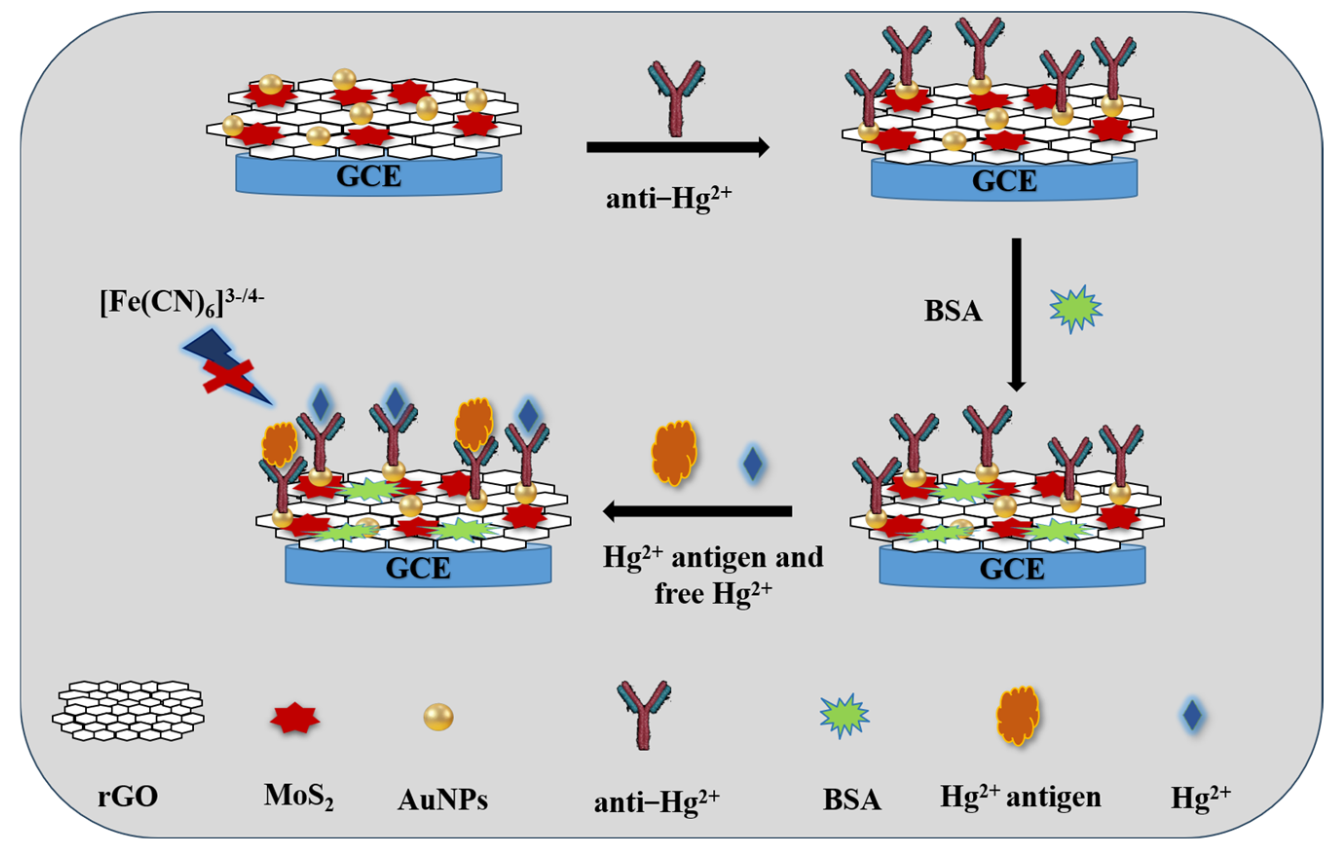
2.4. Electrochemical Competitive Immunosensor Measurement

Scheme 1 shows a diagram of the competitive electrochemical immunosensor. Firstly, 6 μL of Hg (II) antibody dispersion in PBS buffer was dropped onto the modified electrode for 1h. After washing, the electrode was incubated with 1% BSA solution to block the nonspecific binding sites for 30 min. Subsequently, the competitive detection of Hg (II) was performed by incubating the electrode in a 100 μL solution mixture (20 μg/mL antigen + Hg (II) solution with a series of concentrations) for 1h, where the antigen competed for the limited binding sites of the antibody with the Hg (II) in the solution.
The electrochemical immunosensor was measured in 0.01 M PBS (pH 7.4) and 0.1 M KCl containing a 5.0 mM [Fe(CN)6]3−/4− solution. The response was calculated with differential pulse voltammetry (DPV) measurement.

2.5. Preparation of the Spiked Samples

Lake water samples collected from Wenying lake in Datong city were filtrated using a nylon membrane filter, and the filtrates were adjusted to pH 7.4. Then, the proposed immunosensor was applied to detect the filtrates, which were spiked with Hg (II) at concentrations of 10, 20, 50 and 100 ng/mL.

3. Results and Discussion

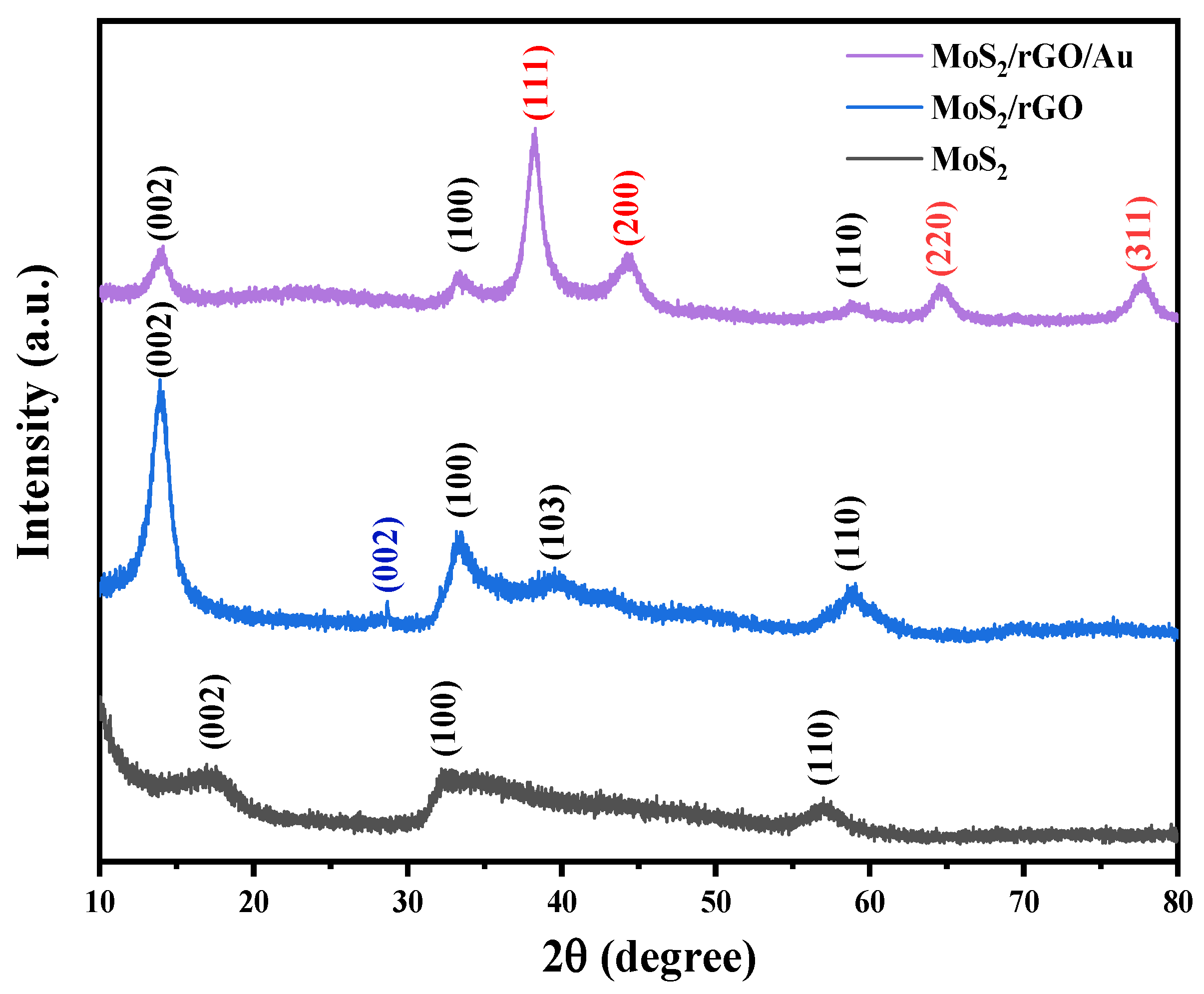
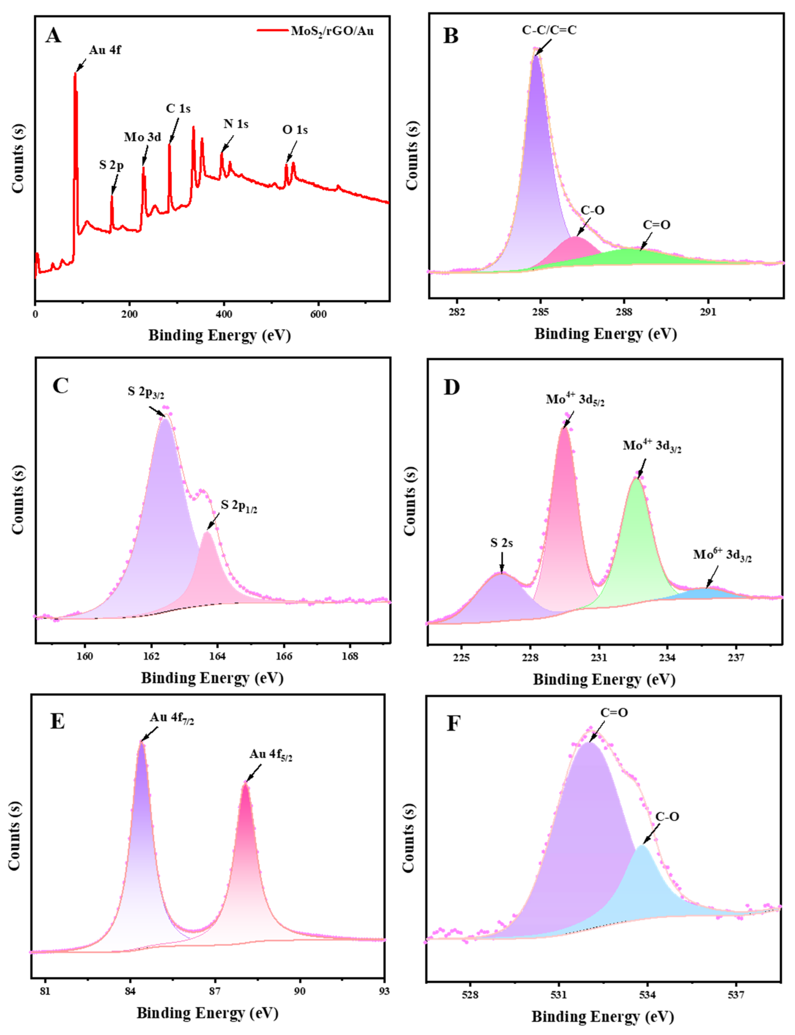
3.1. Characterization of Nanocomposites

The morphologies and structures of the MoS2, MoS2/rGO and MoS2/rGO/Au nanocomposites were characterized by XRD, SEM and XPS.
Figure 1 shows the crystal structures of the nanocomposites measured by XRD. The peaks at approximately 2θ = 17.1°, 32.8° and 57.4° corresponded to the (002), (100) and (110) planes of the crystalline MoS2, respectively (JCPDS 37-1492) [32]. At the same time, there was a peak at 2θ = 28.7°, which was characteristic of the rGO in the MoS2/rGO nanocomposite. The relative intensity of rGO was smaller than that of MoS2, and a small red shift was observed, which was because of the interaction between the MoS2 nanoflowers and the oxygen functional groups on the rGO [28], and the integration of the MoS2/rGO was dominated by the MoS2 during the synthetic process [33]. The MoS2/rGO/Au nanocomposite shows diffraction peaks at 2θ = 38.2°, 44.2°, 64.7° and 77.8°, which corresponded to the (111), (200), (220) and (311) planes of the Au (JCPDS 04-0874). Therefore, the results suggested that the above nanocomposites were successfully synthesized.
SEM was studied to characterize the morphologies of the MoS2, MoS2/rGO and MoS2/rGO/Au nanocomposites, and the results are shown in Figure 2. As shown in Figure 2A, the thin sheet formed a flower-like nanostructure, indicating the successful synthesis of the MoS2. Figure 2B,C show SEM images of the MoS2/rGO. It can be observed that large numbers of MoS2 nanoflowers were loaded on the rGO’s surface, which was transparent and lamellar and showed a loose porous structure. From Figure 2D, we can see that the small-sized AuNPs obviously decorated the surface of the MoS2 and rGO uniformly, which means that the MoS2/rGO/Au nanocomposites were successfully synthesized.
Figure 3 shows the XPS curves of the MoS2/rGO/Au nanocomposites. From the spectrum Figure 3A, we can confirm the presence of Au, S, Mo, C and O in the MoS2/rGO/Au. The deconvoluted spectrum of C 1s (Figure 3B) and O 1s (Figure 3F) had peaks located at 284.8 eV, 286.3 eV, 288.6 eV, 531.9 eV and 533.8 eV, corresponding to C-C/C=C, C-O and C=O bonds [34]. The decreased peaks of C-O and C=O indicates the loss of oxygen-containing groups during the reduced procedure of GO. The peaks at 162.4 eV and 163.7 eV in the S 2p region (Figure 3C) imply S 2p3/2 and S 2p1/2. Figure 3D shows the spectrum for Mo 3d. The peaks at 226.7 eV, 229.5 eV, 232.7 eV and 235.7 eV were related to S 2s, Mo4+ 3d5/2, Mo4+ 3d3/2 and Mo6+ 3d3/2, respectively. The two characteristic peaks at 84.4 eV and 88.1 eV (Figure 3E) corresponded to Au 4f7/2 and Au 4f5/2, which were loaded on the MoS2 and rGO.

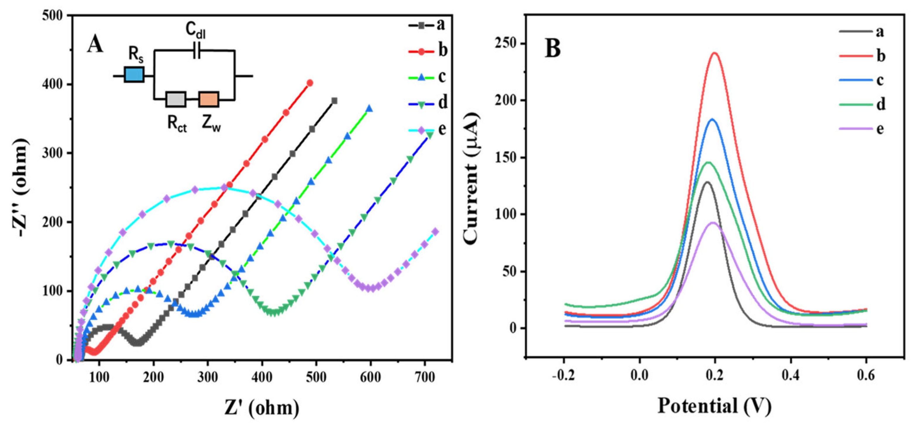
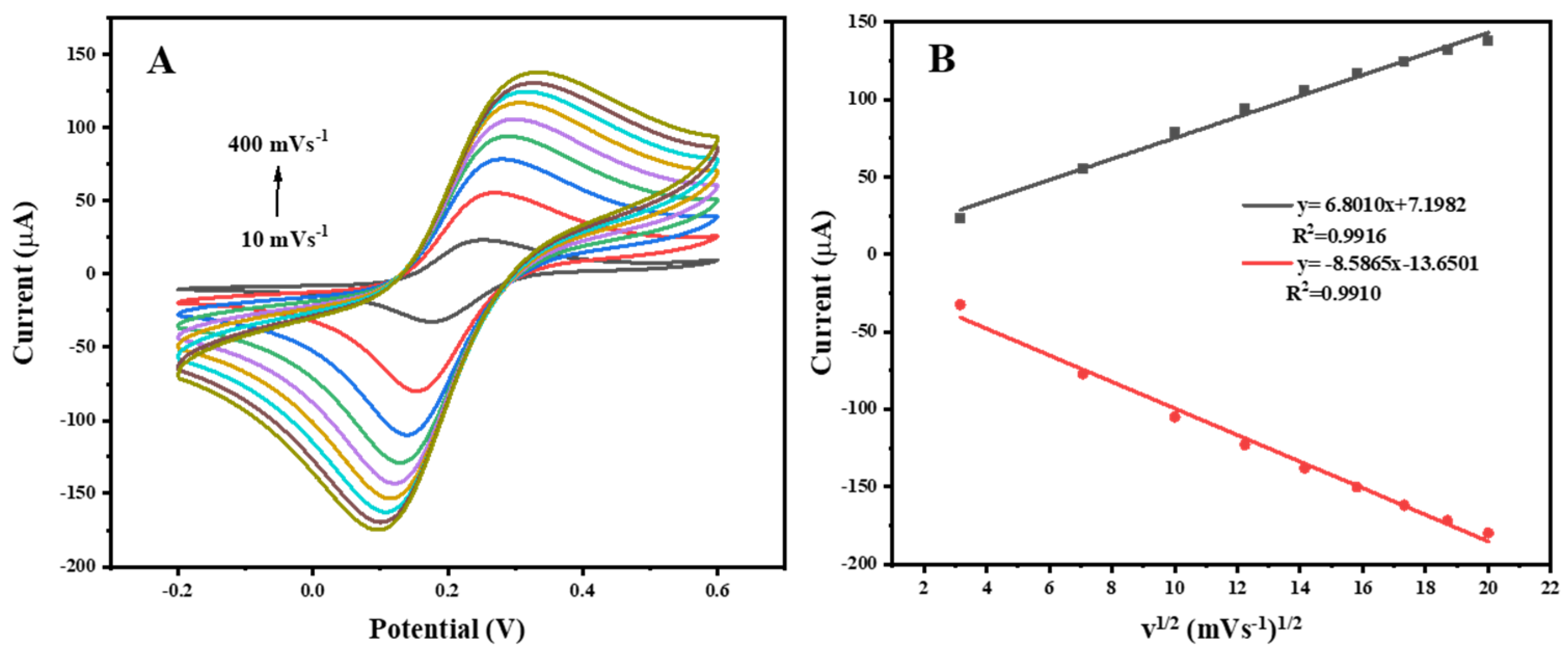
3.2. Electrochemical Properties of Modified Electrodes

The electrochemical properties of the bare GCE and MoS2-, MoS2/rGO- and MoS2/rGO/Au-modified GCE electrodes were analyzed using cyclic voltammetry (CV). As shown in Figure 4, compared with the bare GCE, the redox peak currents of the MoS2-, MoS2/rGO- and MoS2/rGO/Au-modified electrodes increased gradually, which was attributed to the synergistic electrocatalytic activity of the MoS2, rGO and Au [35]. Therefore, the MoS2/rGO/Au-modified electrode was used for the following study.

3.3. Characterization of Immunosensor

Electrochemical impedance spectroscopy (EIS) is often used to characterize the assembly of each step of the working electrode for immunosensors. As shown in Figure 5A, the impedance spectra included a semicircle at a higher frequency and a line part at a lower frequency; the diameter of the semicircle indicates the electron transfer resistance (Rct) [36]. When the MoS2/rGO/Au was coated on the GCE, the semicircle was smaller than the bare GCE, indicating that the MoS2/rGO/Au nanocomposites could enhance the electron transfer on the modified GCE. Subsequently, after the Hg (II) antibody, BSA and antigen were immobilized on the modified GCE, the Rct values were increased, respectively, demonstrating that the step-by-step preparation of the immunosensor on the modified electrode was successful.
Meanwhile, the differential pulse voltammetry (DPV) results of the bare GCE and modified GCE were consistent with the EIS results (Figure 5B). The peak current of the MoS2/rGO/Au-modified GCE was obviously higher than that of the bare GCE. With the gradual modification by the antibody, BSA and antigen of the MoS2/rGO/Au/GCE, the peak current gradually decreased due to the protein layers on the electrode surface significantly hindering the electron transfer.
Moreover, the kinetic process of the electrode reactions was studied. As shown in Figure 6A, the peak current increased with the increasing in the scan rate, and a linear relationship with a good correlation coefficient between the peak currents and the square root of the scan rates was observed (Figure 6B), suggesting that the process was diffusion-controlled.

3.4. Optimization of Assay Conditions

To establish an optimal electrochemical immunosensor, some parameters, including the pH value, the amount of nanocomposite, the incubation time of the antigen–antibody reaction and the concentration of the antigen, were determined. As shown in Figure 7, the optimal pH = 7.4, the optimal amount of the MoS2/rGO/Au nanocomposite was 6 μL (1 mg/mL), the optimal incubation time was 60 min and the optimal concentration of antigen was 20 μg/mL.

3.5. Sensitivity of the Immunosensor

For Hg (II) determination, a competitive assay was performed by the incubation of the immunosensor with mixtures of a Hg (II) standard solution (with different concentration) and an antigen solution at the optimal conditions. Figure 8A describes the DPV curves of the immunosensor before and after the immunoreaction. The DPV peak current increased with the increase in Hg (II) in the incubation solution, since the more free Hg (II) in the sample, the less the antigen coul bind to the antibody immobilized on the electrode, and thus the larger DPV current obtained.
The immunosensor response was calculated as the change in the DPV current (io − ip) [37], which was defined as follows: io is the current before the immunoreaction, and ip is the current after incubating the immunosensor with a mixture of antigen with different concentrations of Hg (II).
Under optimal conditions, the sensor for Hg (II) detection was constructed in the concentration range of 0.1–600 ng/mL. As shown in Figure 8A, the DPV responses increased with the increase in Hg (II). Moreover, there was a good linear relationship between io − ip and the concentration of Hg (II) (Figure 8B). The limit of detection (LOD) of the immunosensor was 63 pg/mL, which demonstrates the high sensitivity of the proposed method.

3.6. Selectivity, Stability and Reproducibility of the Immunosensor

The immunosensors were kept at 4 °C over 4 weeks and then tested under optimal conditions. As shown in Figure 9A, compared with the signal before storage, the peak current could still be maintained at 85.76%. The results indicate the good stability of the sensor.
In order to evaluate the specificity of the proposed immunosensor, twelve species, including Zn (II), Mg (II), Al (III), Cu (II), Pb (II), Ba (II), Cd (II), Ag (I), MNA, CH3Hg (I) and CH3Hg-MNA, were selected as the interference species and applied according to the procedure of the assay. The concentrations of the standard solutions of Hg (II) and interference species were 400 and 1000 ng/mL, respectively. As shown in Figure 9B, the response of Hg (II) was significantly different from the other interference species. This clearly indicates that the electrochemical immunosensor exhibited high specificity in terms of detecting Hg (II).
The reproducibility of the immunosensor was assessed by testing four identical immunosensors for Hg (II) at a concentration of 100 ng/mL, respectively. The relative standard deviation (RSD) was 4.78%, indicating the good reproducibility of the immunosensor.

3.7. Spiked Samples Analysis

Recovery experiments in lake water samples were carried out to evaluate the reliability of the sensor. Table 1 shows the results of the recovery and RSD. It was observed that the recovery of the spiked samples ranged from 98.4% to 100.3%, and low RSD values were obtained. These results indicate that the proposed method could be used for the detection of real samples.

4. Conclusions

In summary, a sensitive and specific MoS2/rGO/Au-based competitive electrochemical immunosensor for the detection of Hg (II) was developed successfully. The MoS2/rGO/Au nanocomposites displayed excellent electrochemical properties due to their synergistic effects, and the antibody could be adsorbed by the Au in the nanocomposites. The sensor has not only good selectivity, stability and reproducibility for Hg (II) but also has a low LOD value of 63 pg/mL. The proposed method was used for the detection of Hg (II) in spiked water samples with an acceptable recovery rate. Therefore, this approach provides a sensitive and specific analytical method for the detection of Hg (II), and it has good prospects as a useful model for the detection of other small-molecular compounds in the food safety and environmental areas. Even so, it is relatively important to screen the immune reaction conditions of the electrode, such as the pH, the amount of nanocomposite, the incubation time and other conditions. Otherwise, they will affect the occurrence of the incubation reaction, which is also the focus of our future work.

Author Contributions

Y.W. (Yuzhen Wang), writing—review and editing, supervision, methodology and conceptualization; N.S., writing—original draft; X.K., formal analysis; Q.P., investigation; M.T., visualization, validation; Y.W. (Yanfeng Wang), software and formal analysis; Y.B., supervision and conceptualization. All authors have read and agreed to the published version of the manuscript.

Funding

This research was funded by the Natural Science Foundation of Shanxi (No. 201901D211436), Scientific and Technological Innovation Programs of Higher Education Institutions in Shanxi (No. 2019L0758), Graduate Education Reform Project of Shanxi (No. 2021YJJG305), Scientific Research Projects of Shanxi Datong University (No. 2022K5) and Basic Research Project Fund of Shanxi Province (No. 202103021223335).

Institutional Review Board Statement

Not applicable.

Informed Consent Statement

Not applicable.

Data Availability Statement

Data are contained within the article.

Conflicts of Interest

The authors declare no conflict of interest.

References

  1. Rebello, S.; Sivaprasad, M.S.; Anoopkumar, A.N.; Jayakrishnan, L.; Aneesh, E.M.; Narisetty, V.; Sindhu, R.; Binod, P.; Pugazhendhi, A.; Pandey, A. Cleaner technologies to combat heavy metal toxicity. J. Environ. Manag. 2021, 296, 113231. [Google Scholar] [CrossRef] [PubMed]
  2. Lee, J.; Han, M.S.; Mirkin, C.A. Colorimetric Detection of Mercuric Ion (Hg2+) in Aqueous Media using DNA-Functionalized Gold Nanoparticle. Angew. Chem. Int. Ed. 2007, 46, 4093–4096. [Google Scholar] [CrossRef] [PubMed]
  3. Almeida, I.L.S.; Coelho, N.M.M. Direct Determination of Inorganic Mercury in Ethanol Fuel by Cold Vapor Atomic Absorption Spectrometry. Energy Fuels 2012, 26, 6003–6007. [Google Scholar] [CrossRef]
  4. Jiang, X.J.; Gan, W.; Wan, L.Z.; Zhang, H.C.; He, Y.Z. Determination of mercury by electrochemical cold vapor generation atomic fluorescence spectrometry using polyaniline modified graphite electrode as cathode. Spectrochim. Acta Part B At. Spectrosc. 2010, 65, 171–175. [Google Scholar] [CrossRef]
  5. Yao, C.H.; Jiang, S.J.; Sahayam, A.C.; Huang, Y.L. Speciation of mercury in fish oils using liquid chromatography inductively coupled plasma mass spectrometry. Microchem. J. 2017, 133, 556–560. [Google Scholar] [CrossRef]
  6. Wang, Y.Z.; Yang, H.; Pschenitza, M.; Niessner, R.; Li, Y.; Knopp, D.; Deng, A.P. Highly sensitive and specific determination of mercury in water, food and cosmetic samples with an ELISA based on a novel monoclonal antibody. Anal. Bioanal. Chem. 2012, 403, 2519–2528. [Google Scholar] [CrossRef]
  7. Aroui, F.E.; Lahrich, S.; Farahi, A.; Achak, M.; Gaini, L.E.; Bakasse, M.; Bouzidi, A.; Mhammedi, M.A.E. Palladium Particles-Impregnated Natural Phosphate Electrodes for Electrochemical Determination of Mercury in Ambient Water Samples. Electroanalysis 2014, 26, 1751–1760. [Google Scholar] [CrossRef]
  8. Wang, Y.Z.; Chen, S.; Wei, C.; Xu, M.M.; Yao, J.L.; Li, Y.; Deng, A.P.; Gu, R.A. A femtogram level competitive immunoassay of mercury (II) based on surface-enhanced Raman spectroscopy. Chem. Commnications 2014, 50, 9112–9114. [Google Scholar] [CrossRef]
  9. Kang, G.F.; Wang, Y.Z.; Bai, Y.F.; Chen, Z.Z.; Feng, F. Surface plasmon resonance based competitive immunoassay for Cd2+. RSC Adv. 2017, 7, 44054–44058. [Google Scholar] [CrossRef]
  10. Huang, S.; Wang, W.J.; Cheng, F.F.; Yao, H.Q.; Zhu, J.J. Highly Sensitive Detection of Mercury Ion Based on T-rich DNA Machine using Portable Glucose Meter. Sens. Actuators B Chem. 2017, 242, 347–354. [Google Scholar] [CrossRef]
  11. Pal, C.; Majumder, S. Label-free ultra-low level electrochemical detection of mercury(II) ions in aqueous medium via direct covalent functionalization of single-stranded DNA with MoS2 nanoflakes. J. Mater. Sci. Mater. Electron. 2022, 33, 7023–7030. [Google Scholar] [CrossRef]
  12. Li, L.; Dong, X.G.; Liu, Z.F.; Wei, J.; Li, J.R.; Zhou, H.; Zhu, J.; Shi, X.W. Visual and ultrasensitive detection of mercury ions based on urease catalysis and responsive photonic crystals. Dye. Pigment. 2021, 195, 109676. [Google Scholar] [CrossRef]
  13. Omer, K.M.; Hama Aziza, K.H.; Mohammad, S.J. Improvement of selectivity via surface modification of carbon nanodots towards quantitative detection of Mercury ions. New J. Chem. 2019, 43, 12979–12986. [Google Scholar] [CrossRef]
  14. Tang, C.M.; Qin, Y.C.; Ni, C.L.; Zou, J.P. Detection and Removal of Mercury Ions in Water by a Covalent Organic Framework Rich in Sulfur and Nitrogen. ACS Appl. Polym. Mater. 2022, 4, 849–858. [Google Scholar] [CrossRef]
  15. Jin, H.L.; Zhang, D.; Liu, Y.; Wei, M. An electrochemical aptasensor for lead ion detection based on catalytic hairpin assembly and porous carbon supported platinum as signal amplification. RSC Adv. 2020, 10, 6647–6653. [Google Scholar] [CrossRef]
  16. Zhu, N.X.; Liu, X.N.; Peng, K.M.; Cao, H.; Yuan, M.; Ye, T.; Wu, X.X.; Yin, F.Q.; Yu, J.S.; Hao, L.L.; et al. A Novel Aptamer-Imprinted Polymer-Based Electrochemical Biosensor for the Detection of Lead in Aquatic Products. Molecules 2023, 28, 196. [Google Scholar] [CrossRef]
  17. Renedo, O.D.; Alonso-Lomillo, M.A.; Ferreira-Gonçalves, L.; ArcosMartinez, M.J. Development of urease based on amperometric biosensors for the inhibitive determination of Hg (II). Talanta 2009, 79, 1306–1310. [Google Scholar] [CrossRef]
  18. Sabah, M.; Fethi, A. Electrochemical Detection of Lead in Real Samples Using Carbon Nanostructure and Inactivated E coli as Low-Cost Sensitive Biosensor with High Electrocatalytic Performance. Electrocatalysis 2022, 13, 773–783. [Google Scholar] [CrossRef]
  19. Sciuto, E.L.; Petralia, S.; van der Meer, J.R.; Conoci, S. Miniaturized Electrochemical Biosensor based on Whole-Cell for Heavy Metal Ions Detection in Water. Biotechnol. Bioeng. 2021, 118, 1456–1465. [Google Scholar] [CrossRef]
  20. Irvine, G.W.; Tan, S.N.; Stillman, M.J. A Simple Metallothionein-Based Biosensor for Enhanced Detection of Arsenic and Mercury. Biosensors 2017, 7, 14. [Google Scholar] [CrossRef]
  21. Trnkova, L.; Krizkova, S.; Adam, V.; Hubalek, J.; Kizek, R. Immobilization of metallothionein to carbon paste electrode surface via anti-MT antibodies and its use for biosensing of silver. Biosens. Bioelectron. 2011, 26, 2201–2207. [Google Scholar] [CrossRef] [PubMed]
  22. Qi, S.P.; Zhao, B.; Zhou, B.; Jiang, X.Q. An electrochemical immunosensor based on pristine graphene for rapid determination of ractopamine. Chem. Phys. Lett. 2017, 685, 146–150. [Google Scholar] [CrossRef]
  23. Hou, J.B.; Shao, Y.Y.; Ellis, M.W.; Moore, R.B.; Yi, B.L. Graphene-baed electrochemical energy conversion and storage: Fuel cells, supercapacitors and lithium ion batteries. Phys. Chem. Chem. Phys. 2011, 13, 15384–15402. [Google Scholar] [CrossRef] [PubMed]
  24. Shao, Y.Y.; Wang, J.; Wu, H.; Liu, J.; Aksay, I.A.; Lin, Y.H. Graphene Based Electrochemical Sensors and Biosensors: A Review. Electroanalysis 2010, 22, 1027–1036. [Google Scholar] [CrossRef]
  25. Lee, C.S.; Kim, T.H. Large-Scale Preparation of MoS2/Graphene Composites for Electrochemical Detection of Morin. ACS Appl. Nano Mater. 2021, 4, 6668–6677. [Google Scholar] [CrossRef]
  26. Peng, Y.; Tang, Z.R.; Dong, Y.P.; Che, G.; Xin, Z.F. Electrochemical detection of hydroquinone based on MoS2/reduced graphene oxide nanocomposites. J. Electroanal. Chem. 2018, 816, 38–44. [Google Scholar] [CrossRef]
  27. Li, X.T.; Wang, Y.C.; Zhang, X.L.; Gao, Y.; Sun, C.M.; Ding, Y.H.; Feng, F.; Jin, W.J.; Yang, G.J. An impedimetric immunosensor for determination of porcine epidemic diarrhea virus based on the nanocomposite consisting of molybdenum disulfide/reduced graphene oxide decorated with gold nanoparticles. Microchim. Acta 2020, 187, 217. [Google Scholar] [CrossRef]
  28. Wu, Y.J.; Liu, D.D.; Guo, J.H.; Wang, F. A molybdenum disulfide-reduced graphene oxide nanocomposite as an electrochemical sensing platform for detecting cyproterone acetate. New J. Chem. 2022, 46, 5385–5392. [Google Scholar] [CrossRef]
  29. Yadav, S.; Sadique, M.A.; Ranjan, P.; Khan, R. Synergistically functionalized molybdenum disulfide-reduced graphene oxide nanohybrid based ultrasensitive electrochemical immunosensor for real sample analysis of COVID-19. Anal. Chim. Acta 2023, 1265, 341326. [Google Scholar] [CrossRef]
  30. Wang, Y.; Ni, Y. Molybdenum disulfide quantum dots as a photoluminescence sensing platform for 2,4,6-trinitrophenol detection. Anal. Chem. 2014, 86, 7463–7470. [Google Scholar] [CrossRef]
  31. Wang, Y.G.; Wang, Y.L.; Wu, D.; Ma, H.M.; Zhang, Y.; Fan, D.W.; Pang, X.H.; Du, B.; Wei, Q. Label-free electrochemical immunosensor based on flower-like Ag/MoS2/rGO nanocomposites for ultrasensitive detection of carcinoembryonic antigen. Sens. Actuators B Chem. 2018, 255, 125–132. [Google Scholar] [CrossRef]
  32. Zhao, Y.N.; Zhou, J.; Jia, Z.M.; Huo, D.Q.; Liu, Q.Y.; Zhong, D.Q.; Hu, Y.; Yang, M.; Bian, M.H.; Hou, C.J. In-situ growth of gold nanoparticles on a 3D-network consisting of a MoS2/rGO nanocomposite for simultaneous voltametric determination of ascorbic acid, dopamine and uric acid. Microchim. Acta 2019, 186, 92. [Google Scholar]
  33. Rana, D.S.; Kalia, S.; Thakur, N.; Singh, R.K.; Kumar, R.; Singh, D. Synthesis of reduced graphene oxide-molybdenum disulfide nanocomposite as potential scaffold for fabrication of efficient hydrazine sensor. Mater. Chem. Phys. 2023, 294, 127048. [Google Scholar] [CrossRef]
  34. Han, Y.J.; Zhang, R.; Dong, C.; Cheng, F.Q.; Guo, Y.J. Sensitive electrochemical sensor for nitrite ions based on rose-like AuNPs/MoS2/graphene composite. Biosens. Bioelectron. 2019, 142, 111529. [Google Scholar] [CrossRef]
  35. Alarfaj, N.A.; El-Tohamy, M.F.; Hesham, O. New label-free ultrasensitive electrochemical immunosensor-based Au/MoS2/rGO nanocomposites for CA 27-29 breast cancer antigen detection. New J. Chem. 2018, 42, 11046–11053. [Google Scholar] [CrossRef]
  36. Xu, X.H.; Guo, Y.N.; Wang, L.; He, K.Y.; Guo, Y.R.; Wang, X.Q.; Gunasekaran, S. Hapten-Grafted Programmed Probe as a Corecognition Element for a Competitive Immunosensor to Detect Acetamiprid Residue in Agricultural Products. J. Agric. Food Chem. 2018, 66, 7815–7821. [Google Scholar] [CrossRef]
  37. Eissa, S.; Zouro, M. Competitive voltammetric morphine immunosensor using a gold nanoparticle decorated graphene electrode. Microchim. Acta 2017, 184, 2281–2289. [Google Scholar] [CrossRef]
Scheme 1. Schematic illustration of the competitive electrochemical immunosensor for Hg (II) based on MoS2/rGO/Au nanocomposites.
Sensors 25 00623 sch001
Figure 1. XRD patterns of MoS2/rGO/Au, MoS2/rGO and MoS2 nanocomposites.
Sensors 25 00623 g001
Figure 2. SEM images of (A) MoS2, (B,C) MoS2/rGO and (D) MoS2/rGO/Au nanocomposites.
Sensors 25 00623 g002
Figure 3. XPS spectra of (A) MoS2/rGO/Au, (B) C 1s region, (C) S 2p region, (D) Mo 3d region, (E) Au 4f region and (F) O 1s region.
Sensors 25 00623 g003
Figure 4. Cyclic voltammograms of (a) bare GCE, (b) MoS2/GCE, (c) MoS2/rGO/GCE and (d) MoS2/rGO/Au/GCE in 0.01 M PBS (pH 7.4) and 0.1 M KCl containing a 5.0 mM ([Fe(CN)6]3−/4− solution.
Sensors 25 00623 g004
Figure 5. (A) Electrochemical impedance spectra and (B) differential pulse voltammograms of (a) GCE, (b) MoS2/rGO/Au/GCE, (c) anti-Hg2+/MoS2/rGO/Au/GCE, (d) BSA/anti-Hg2+/MoS2/rGO/Au/GCE and (e) antigen/BSA/anti-Hg2+/MoS2/rGO/Au/GCE.
Sensors 25 00623 g005
Figure 6. (A) Cyclic voltammograms of the BSA/anti-Hg2+/MoS2/rGO/Au/GCE in 0.01 M PBS (pH 7.4) and 0.1 M KCl containing a 5.0 mM ([Fe(CN)6]3−/4− solution at different scan rates ranging from 10 to 400 mV s−1. (B) The linear relationship between the square root of the scan rate and the peak current.
Sensors 25 00623 g006
Figure 7. Effects of (A) pH, (B) amount of MoS2/rGO/Au nanocomposite, (C) incubation reaction time and (D) concentration of antigen.
Sensors 25 00623 g007
Figure 8. (A) Differential pulse voltammetry of the immunosensor before binding and after incubation with different concentrations of Hg (II) mixed with 20 μg/mL of antigen. (B) The calibration curve of Hg (II) detection.
Sensors 25 00623 g008
Figure 9. (A) The stability and (B) selectivity of the competitive electrochemical immunosensor for Hg (II) detection.
Sensors 25 00623 g009
Table 1. Results of recovery and relative standard deviation (RSD) for Hg (II) determination from fortified samples.
Fortified (ng/mL)Measured (ng/mL)Recovery (%)RSD (%) (n = 3)
109.88 ± 0.3498.83.44
2019.89 ± 0.5199.52.56
5049.22 ± 2.6498.45.36
100100.34 ± 4.59100.34.57
Disclaimer/Publisher’s Note: The statements, opinions and data contained in all publications are solely those of the individual author(s) and contributor(s) and not of MDPI and/or the editor(s). MDPI and/or the editor(s) disclaim responsibility for any injury to people or property resulting from any ideas, methods, instructions or products referred to in the content.

