Assessing Trunk Cross-Section Geometry and Spinal Postures with Noninvasive 3D Surface Topography: A Study of 108 Healthy Young Adults
Highlights
- Established normative values for trunk cross-sectional geometry (sagittal/coronal diameters, areas) in healthy young adults, with clear sex-specific differences.
- Identified modest but significant correlations between thoracic geometry (TorsoScan) and spinal posture parameters (DIERS Formetric).
- Provides a reference framework for future clinical studies on scoliosis, hyperkypho-sis, and chest wall deformities using noninvasive 3D surface topography.
- Supports the integrated application of TorsoScan and DIERS Formetric as comple-mentary tools for radiation-free assessment of trunk morphology and spinal alignment.
Abstract
1. Introduction
2. Materials and Methods
2.1. Study Design and Ethical Approval
2.2. Participants
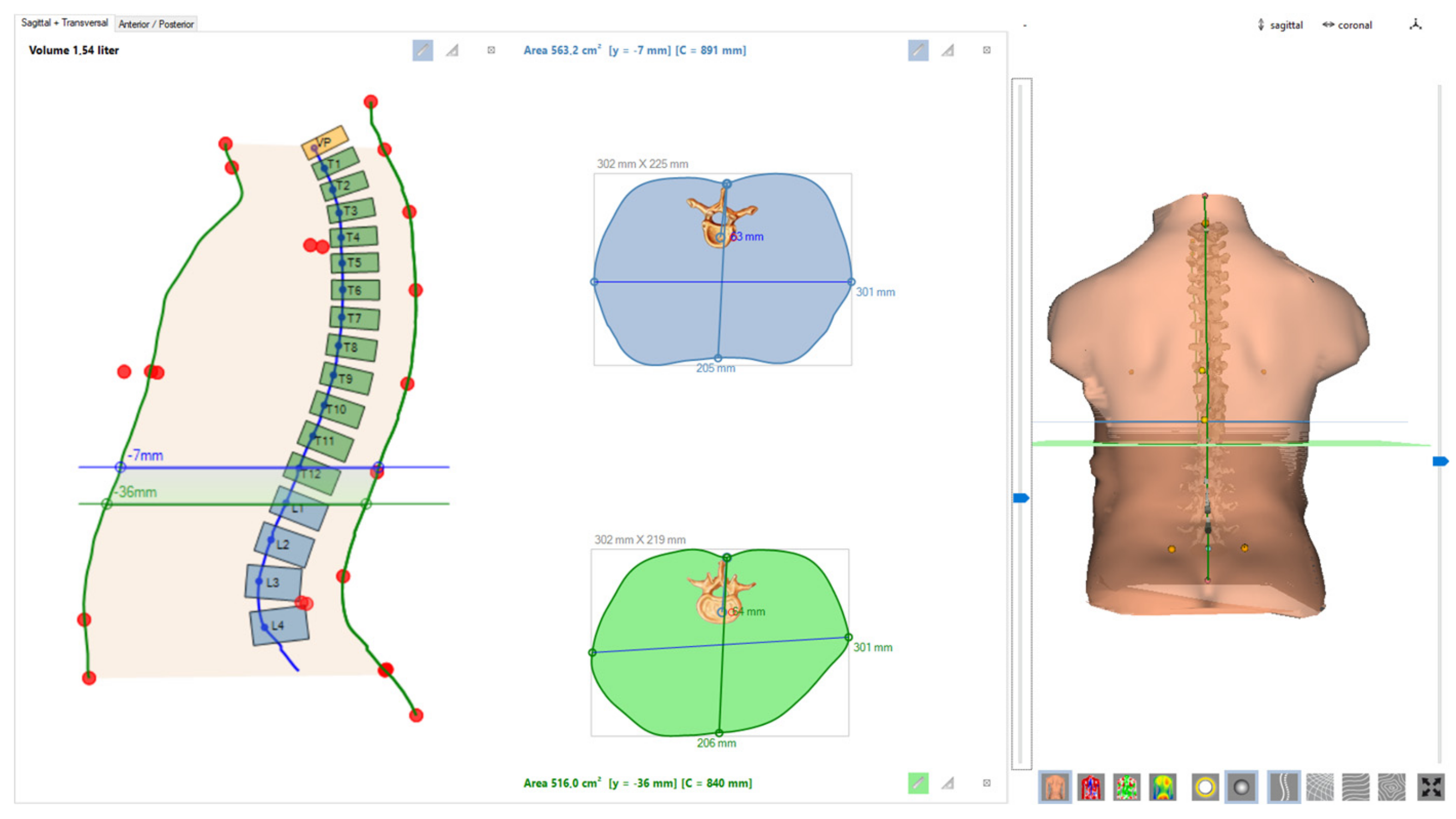
2.3. TorsoScan Acquisition (3D Surface Topography; 360° Cross-Sections)
- sagittal diameter (mm);
- coronal diameter (mm);
- cross-sectional area (cm2).
2.4. DIERS Formetric Acquisition (Rasterstereography; Posture Parameters)
- Thoracic kyphosis angle VP–T12 (°);
- Lumbar lordosis angle T12–DM (°);
- Maximal lateral deviation VP–DM (mm);
- Maximal surface rotation (°);
- Trunk length VP–DM (mm).
2.5. Standardization and Quality Control
2.6. Outcomes
2.7. Technical Details of Measurement Systems
2.8. Statistical Analysis
2.9. Sample Size and Power
3. Results
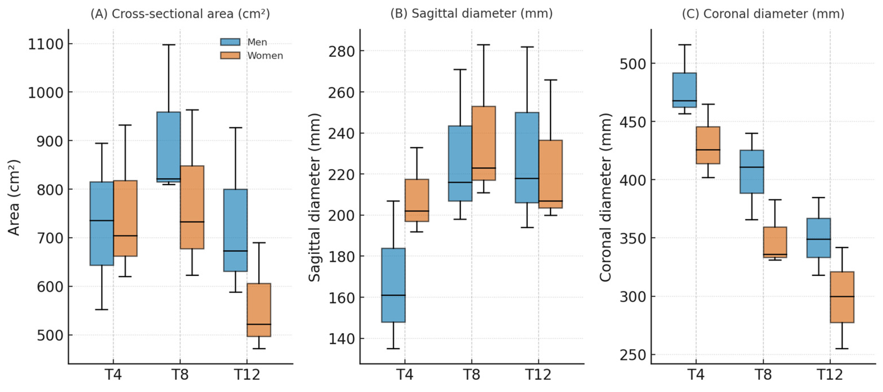
3.1. Normative TorsoScan Values
3.2. Sex Differences
3.3. Associations Between TorsoScan and DIERS Formetric
3.4. Predictive Models for TorsoScan Dimensions
4. Discussion
Clinical Implications
5. Conclusions
Supplementary Materials
Author Contributions
Funding
Institutional Review Board Statement
Informed Consent Statement
Data Availability Statement
Conflicts of Interest
References
- Michalik, R.; Siebers, H.; Eschweiler, J.; Quack, V.; Gatz, M.; Dirrichs, T.; Betsch, M. Development of a new 360-degree surface topography application. Gait Posture 2019, 73, 39–44. [Google Scholar] [CrossRef]
- Asher, M.A.; Burton, D.C. Adolescent idiopathic scoliosis: Natural history and long term treatment effects. Scoliosis 2006, 1, 2. [Google Scholar] [CrossRef] [PubMed]
- Levy, A.R.; Goldberg, M.S.; Mayo, N.E.; Hanley, J.A.; Poitras, B. Reducing the lifetime risk of cancer from spinal radiographs among people with adolescent idiopathic scoliosis. Spine 1996, 21, 1540–1547. [Google Scholar] [CrossRef] [PubMed]
- Degenhardt, B.F.; Starks, Z.; Bhatia, S. Reliability of the DIERS formetric 4D spine shape parameters in adults without postural deformities. BioMed Res. Int. 2020, 2020, 1796247. [Google Scholar] [CrossRef]
- Tabard-Fougère, A.; de Bodman, C.; Dhouib, A.; Bonnefoy-Mazure, A.; Armand, S.; Dayer, R. Three-dimensional spinal evaluation using rasterstereography in patients with adolescent idiopathic scoliosis: Is it closer to three-dimensional or two-dimensional radiography? Diagnostics 2023, 13, 2431. [Google Scholar] [CrossRef] [PubMed]
- Swaminathan, N.; Cyriac, A.M.; E Lobo, M. Rasterstereography a reliable tool in scoliosis measurement—a critical review of literature. Scoliosis 2014, 9, O5. [Google Scholar] [CrossRef]
- Pearce, M.S.; Salotti, J.A.; Little, M.P.; McHugh, K.; Lee, C.; Kim, K.P.; Howe, N.L.; Ronckers, C.M.; Rajaraman, P.; Craft, A.W.; et al. Radiation exposure from CT scans in childhood and subsequent risk of leukaemia and brain tumours: A retrospective cohort study. Lancet 2012, 380, 499–505. [Google Scholar] [CrossRef]
- Miglioretti, D.L.; Johnson, E.; Williams, A.; Greenlee, R.T.; Weinmann, S.; Solberg, L.I.; Feigelson, H.S.; Roblin, D.; Flynn, M.J.; Vanneman, N.; et al. The use of computed tomography in pediatrics and the associated radiation exposure and estimated cancer risk. JAMA Pediatr. 2013, 167, 700–707. [Google Scholar] [CrossRef]
- Arnold, T.C.; Freeman, C.W.; Litt, B.; Stein, J.M. Low-field MRI: Clinical promise and challenges. J. Magn. Reson. Imaging 2022, 57, 25–44. [Google Scholar] [CrossRef]
- Alyas, F.; Connell, D.; Saifuddin, A. Upright positional MRI of the lumbar spine. Clin. Radiol. 2008, 63, 1035–1048. [Google Scholar] [CrossRef]
- Tarantino, U.; Fanucci, E.; Iundusi, R.; Celi, M.; Altobelli, S.; Gasbarra, E.; Simonetti, G.; Manenti, G. Lumbar spine MRI in upright position for diagnosing acute and chronic low back pain: Statistical analysis of morphological changes. J. Orthop. Traumatol. 2012, 14, 15–22. [Google Scholar] [CrossRef]
- Doktor, K.; Christensen, H.W.; Jensen, T.S.; Hancock, M.J.; Vach, W.; Hartvigsen, J. Upright versus recumbent lumbar spine MRI: Do findings differ systematically, and which correlates better with pain? A systematic review. Spine J. 2025, 25, 1719–1750. [Google Scholar] [CrossRef]
- Guidetti, L.; Bonavolontà, V.; Tito, A.; Reis, V.M.; Gallotta, M.C.; Baldari, C. Intra- and interday reliability of spine rasterstereography. BioMed Res. Int. 2013, 2013, 1–5. [Google Scholar] [CrossRef]
- Mohokum, M.; Mendoza, S.; Udo, W.; Sitter, H.; Paletta, J.R.; Skwara, A. Reproducibility of rasterstereography for kyphotic and lordotic angles, trunk length, and trunk inclination: A reliability study. Spine 2010, 35, 1353–1358. [Google Scholar] [CrossRef] [PubMed]
- Betsch, M.; Wild, M.; Jungbluth, P.; Hakimi, M.; Windolf, J.; Haex, B.; Horstmann, T.; Rapp, W. Reliability and validity of 4D rasterstereography under dynamic conditions. Comput. Biol. Med. 2011, 41, 308–312. [Google Scholar] [CrossRef] [PubMed]
- Vendeuvre, T.; Tabard-Fougère, A.; Armand, S.; Dayer, R. Test characteristics of rasterstereography for the early diagnosis of adolescent idiopathic scoliosis. Bone Jt. J. 2023, 105-B, 431–438. [Google Scholar] [CrossRef]
- Knott, P.; Sturm, P.; Lonner, B.; Cahill, P.; Betsch, M.; McCarthy, R.; Kelly, M.; Lenke, L.; Betz, R. Multicenter comparison of 3D spinal measurements using surface topography with those from conventional radiography. Spine Deform. 2016, 4, 98–103. [Google Scholar] [CrossRef]
- Weaver, A.A.; Schoell, S.L.; Stitzel, J.D. Morphometric analysis of variation in the ribs with age and sex. Am. J. Anat. 2014, 225, 246–261. [Google Scholar] [CrossRef] [PubMed]
- Ng, B.K.; Hinton, B.J.; Fan, B.; Kanaya, A.M.; A Shepherd, J. Clinical anthropometrics and body composition from 3D whole-body surface scans. Eur. J. Clin. Nutr. 2016, 70, 1265–1270. [Google Scholar] [CrossRef]
- Guarnieri Lopez, M.; Matthes, K.L.; Sob, C.; Bender, N.; Staub, K. Associations between 3D surface scanner derived anthropometric measurements and body composition in a cross-sectional study. Eur. J. Clin. Nutr. 2023, 77, 972–981. [Google Scholar] [CrossRef]
- Mocini, E.; Cammarota, C.; Frigerio, F.; Muzzioli, L.; Piciocchi, C.; Lacalaprice, D.; Buccolini, F.; Donini, L.M.; Pinto, A. Digital anthropometry: A systematic review on precision, reliability and accuracy of most popular existing technologies. Nutrients 2023, 15, 302. [Google Scholar] [CrossRef] [PubMed]
- Tsiligiannis, T.; Grivas, T. Pulmonary function in children with idiopathic scoliosis. Scoliosis 2012, 7, 7. [Google Scholar] [CrossRef] [PubMed]
- Wang, Y.; Yang, F.; Wang, D.; Zhao, H.; Ma, Z.; Ma, P.; Hu, X.; Wang, S.; Kang, X.; Gao, B. Correlation analysis between the pulmonary function test and the radiological parameters of the main right thoracic curve in adolescent idiopathic scoliosis. J. Orthop. Surg. Res. 2019, 14, 1–9. [Google Scholar] [CrossRef]
- Lee, J.-Y.; Choi, J.-W.; Kim, H. Determination of body surface area and formulas to estimate body surface area using the alginate method. J. Physiol. Anthr. 2008, 27, 71–82. [Google Scholar] [CrossRef]
- Wilczyński, J. Own typology of body posture based on research using the diers formetric III 4D system. J. Clin. Med. 2025, 14, 501. [Google Scholar] [CrossRef] [PubMed]


| Parameters | N | Mean ± SD | Median | Range (Min–Max) |
|---|---|---|---|---|
| T1 Sagittal | 108 | 129.34 ± 22.47 | 128.00 | 89.00–194.00 |
| T1 Coronal | 108 | 286.06 ± 60.11 | 280.00 | 173.00–418.00 |
| T1 Area | 108 | 268.31 ± 101.65 | 247.45 | 123.70–613.00 |
| T4 Sagittal | 108 | 169.30 ± 25.77 | 167.00 | 122.00–238.00 |
| T4 Coronal | 108 | 426.81 ± 48.87 | 428.50 | 162.00–537.00 |
| T4 Area | 108 | 615.62 ± 151.32 | 594.60 | 305.20–1130.50 |
| T8 Sagittal | 108 | 216.78 ± 32.64 | 212.50 | 157.00–336.00 |
| T8 Coronal | 108 | 372.92 ± 54.99 | 363.50 | 258.00–532.00 |
| T8 Area | 108 | 748.58 ± 188.50 | 720.15 | 404.10–1530.30 |
| T12 Sagittal | 108 | 215.14 ± 38.92 | 208.50 | 107.00–370.00 |
| T12 Coronal | 108 | 310.74 ± 45.34 | 308.50 | 223.00–459.00 |
| T12 Area | 108 | 580.95 ± 172.10 | 545.50 | 274.60–1403.10 |
| Parameters | Men Mean ± SD | Women Mean ± SD | Difference (m–w) | Cohen’s d (95% CI) | p | q (FDR) | Effect |
|---|---|---|---|---|---|---|---|
| T1 Sagittal | 136.33 ± 22.62 | 124.16 ± 21.07 | +12.17 mm | 0.56 (0.17–0.95) | 0.0055 | 0.007 | medium |
| T1 Coronal | 297.09 ± 59.02 | 277.89 ± 60.07 | +19.20 mm | 0.32 (−0.06–0.70) | 0.1003 | 0.100 | small (ns) |
| T1 Area | 293.01 ± 102.71 | 249.98 ± 97.68 | +43.03 cm2 | 0.43 (0.04–0.82) | 0.0304 | 0.034 | small |
| T4 Sagittal | 175.61 ± 27.15 | 164.61 ± 23.85 | +11.00 mm | 0.43 (0.04–0.82) | 0.0311 | 0.034 | small |
| T4 Coronal | 451.20 ± 53.19 | 408.71 ± 36.35 | +42.49 mm | 0.96 (0.56–1.36) | 1.3 × 10⁻⁵ | <0.001 | large |
| T4 Area | 697.39 ± 148.23 | 554.94 ± 123.16 | +142.45 cm2 | 1.06 (0.65–1.47) | 8.8 × 10⁻⁷ | <0.001 | large |
| T8 Sagittal | 229.37 ± 35.11 | 207.44 ± 27.41 | +21.93 mm | 0.71 (0.32–1.10) | 0.0007 | 0.001 | medium |
| T8 Coronal | 409.13 ± 46.45 | 346.05 ± 44.61 | +63.08 mm | 1.39 (0.97–1.82) | 2.3 × 10⁻¹⁰ | <0.001 | very large |
| T8 Area | 864.28 ± 189.97 | 662.73 ± 134.61 | +201.55 cm2 | 1.26 (0.84–1.68) | 3.3 × 10⁻⁸ | <0.001 | very large |
| T12 Sagittal | 231.22 ± 41.65 | 203.21 ± 32.21 | +28.01 mm | 0.77 (0.38–1.17) | 0.00028 | 0.001 | medium |
| T12 Coronal | 327.96 ± 39.39 | 297.97 ± 45.53 | +29.99 mm | 0.70 (0.31–1.09) | 0.00040 | 0.001 | medium |
| T12 Area | 664.98 ± 189.54 | 518.61 ± 127.19 | +146.38 cm2 | 0.93 (0.53–1.33) | 2.2 × 10⁻⁵ | <0.001 | large |
| TorsoScan Param. | Formetric Param. | N | r (95% CI) | p | q (FDR) |
|---|---|---|---|---|---|
| T4 Coronal | lumbar lordosis T12–DM [°] | 108 | −0.32 (−0.48; −0.14) | 0.0008 | 0.027 |
| T8 Coronal | lumbar lordosis T12–DM [°] | 108 | −0.32 (−0.48; −0.13) | 0.0009 | 0.027 |
| T8 Area | lumbar lordosis T12–DM [°] | 108 | −0.29 (−0.45; −0.11) | 0.0023 | 0.034 |
| T1 Sagittal | thoracic kyphosis VP–T12 [°] | 108 | +0.30 (0.11; 0.46) | 0.0019 | 0.034 |
| Outcome (TorsoScan) | N | R2 (10 × 5) | RMSE | MAE | Significant Predictors (β, p < 0.05) |
|---|---|---|---|---|---|
| T8 Coronal | 108 | 0.185 | 44.9 | 36.0 | Sex (+50.8 mm); Pelvic tilt (−2.3 mm/°); T12–DM lordosis angle (+4.5 mm/°) |
| T8 Area | 108 | 0.068 | 154.7 | 119.7 | Sex (+164.4 cm2); T8 flexion/extension (+48.2); Flèche cervicale (+7.0 mm) |
| T12 Coronal | 108 | 0.068 | 40.1 | 32.5 | Flèche cervicale (+1.0 mm); T11 rotation (+3.7 mm) |
Disclaimer/Publisher’s Note: The statements, opinions and data contained in all publications are solely those of the individual author(s) and contributor(s) and not of MDPI and/or the editor(s). MDPI and/or the editor(s) disclaim responsibility for any injury to people or property resulting from any ideas, methods, instructions or products referred to in the content. |
© 2025 by the authors. Licensee MDPI, Basel, Switzerland. This article is an open access article distributed under the terms and conditions of the Creative Commons Attribution (CC BY) license (https://creativecommons.org/licenses/by/4.0/).
Share and Cite
Żurawski, A.Ł.; Friebe, D.; Zaleska, S.; Wojtas, K.; Gawlik, M.; Wilczyński, J. Assessing Trunk Cross-Section Geometry and Spinal Postures with Noninvasive 3D Surface Topography: A Study of 108 Healthy Young Adults. Sensors 2025, 25, 6626. https://doi.org/10.3390/s25216626
Żurawski AŁ, Friebe D, Zaleska S, Wojtas K, Gawlik M, Wilczyński J. Assessing Trunk Cross-Section Geometry and Spinal Postures with Noninvasive 3D Surface Topography: A Study of 108 Healthy Young Adults. Sensors. 2025; 25(21):6626. https://doi.org/10.3390/s25216626
Chicago/Turabian StyleŻurawski, Arkadiusz Łukasz, David Friebe, Sandra Zaleska, Karolina Wojtas, Małgorzata Gawlik, and Jacek Wilczyński. 2025. "Assessing Trunk Cross-Section Geometry and Spinal Postures with Noninvasive 3D Surface Topography: A Study of 108 Healthy Young Adults" Sensors 25, no. 21: 6626. https://doi.org/10.3390/s25216626
APA StyleŻurawski, A. Ł., Friebe, D., Zaleska, S., Wojtas, K., Gawlik, M., & Wilczyński, J. (2025). Assessing Trunk Cross-Section Geometry and Spinal Postures with Noninvasive 3D Surface Topography: A Study of 108 Healthy Young Adults. Sensors, 25(21), 6626. https://doi.org/10.3390/s25216626


