
Recent Advancement in Fluorescent Probes for Peroxynitrite (ONOO−)

 , , , and
Abstract
:1. Introduction
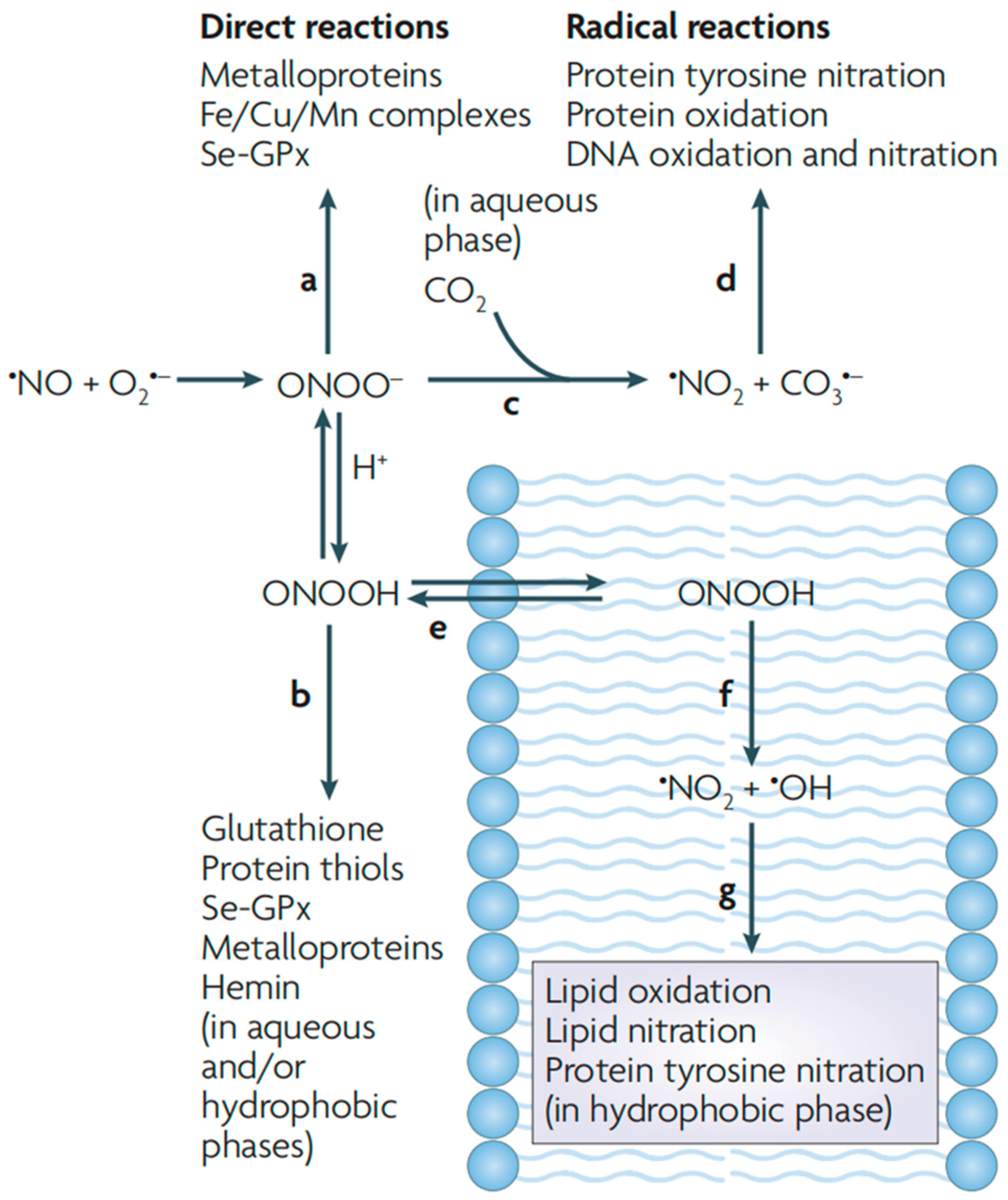
2. ONOO−
2.1. Generation of ONOO−
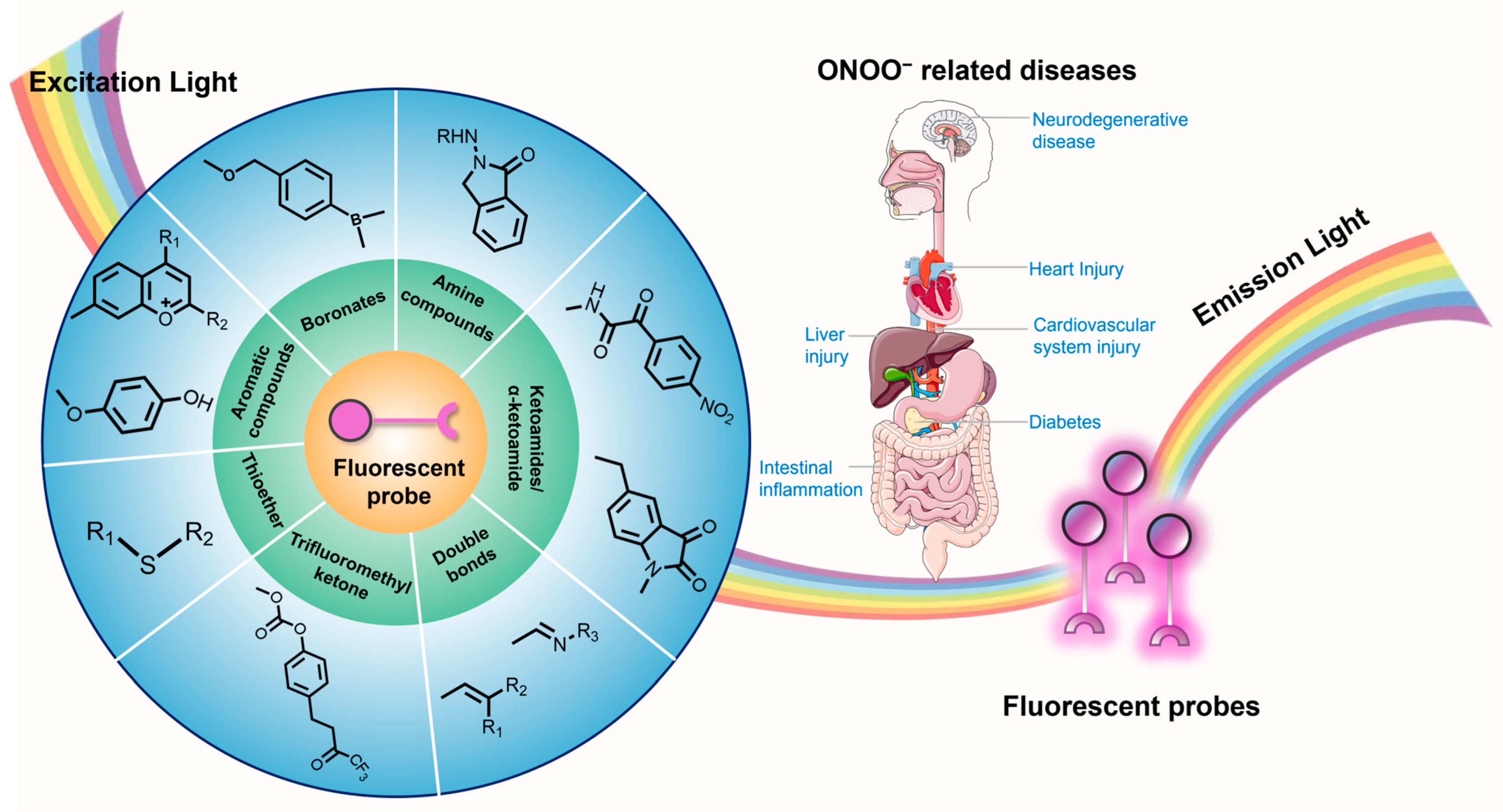
2.2. ONOO− and Diseases
2.3. ONOO− Detection
3. Fluorescent Probes for Detection of ONOO−
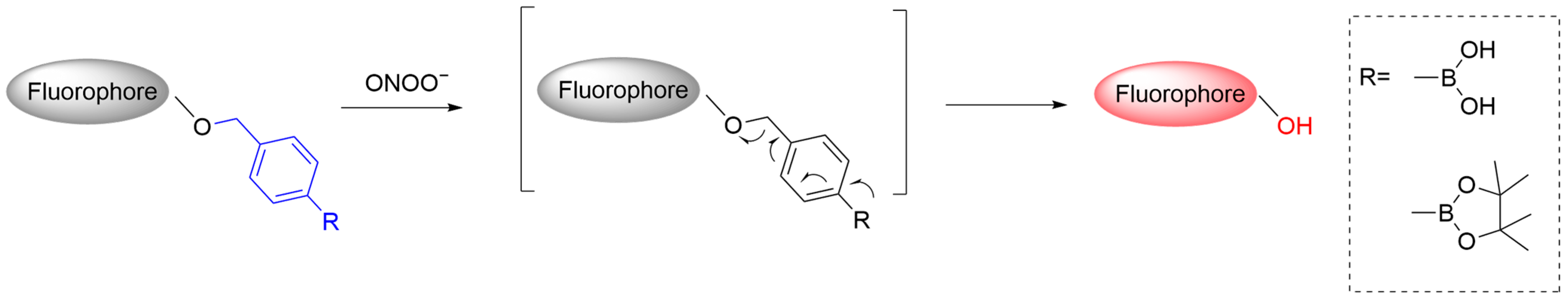
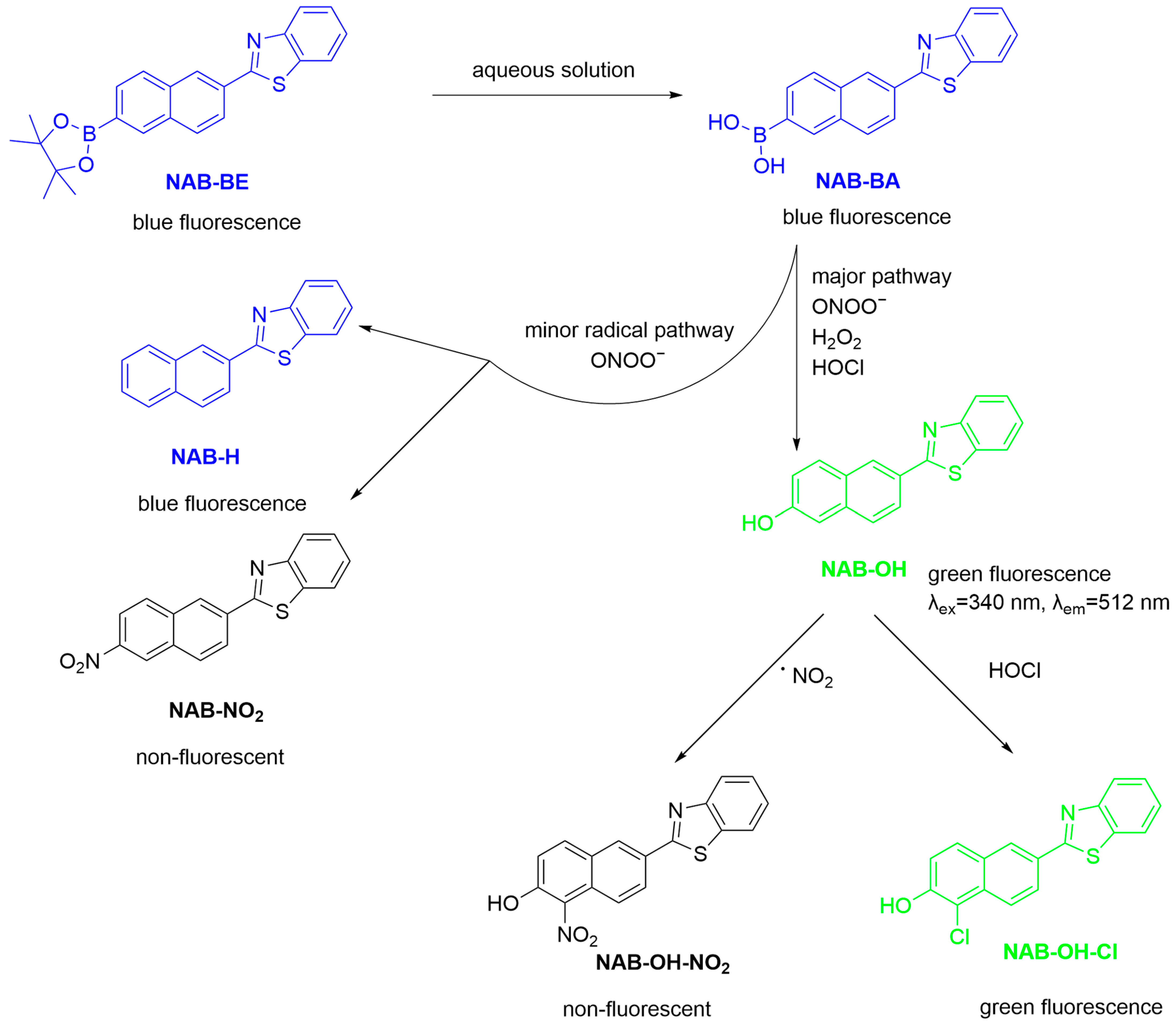
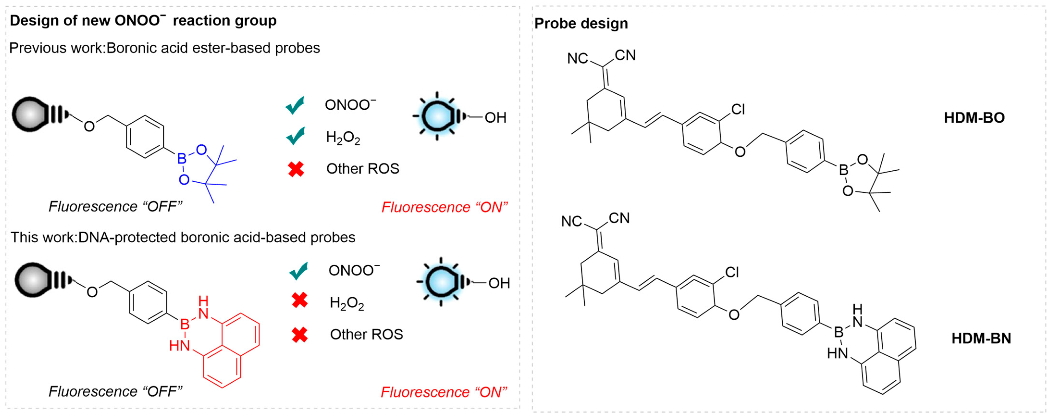
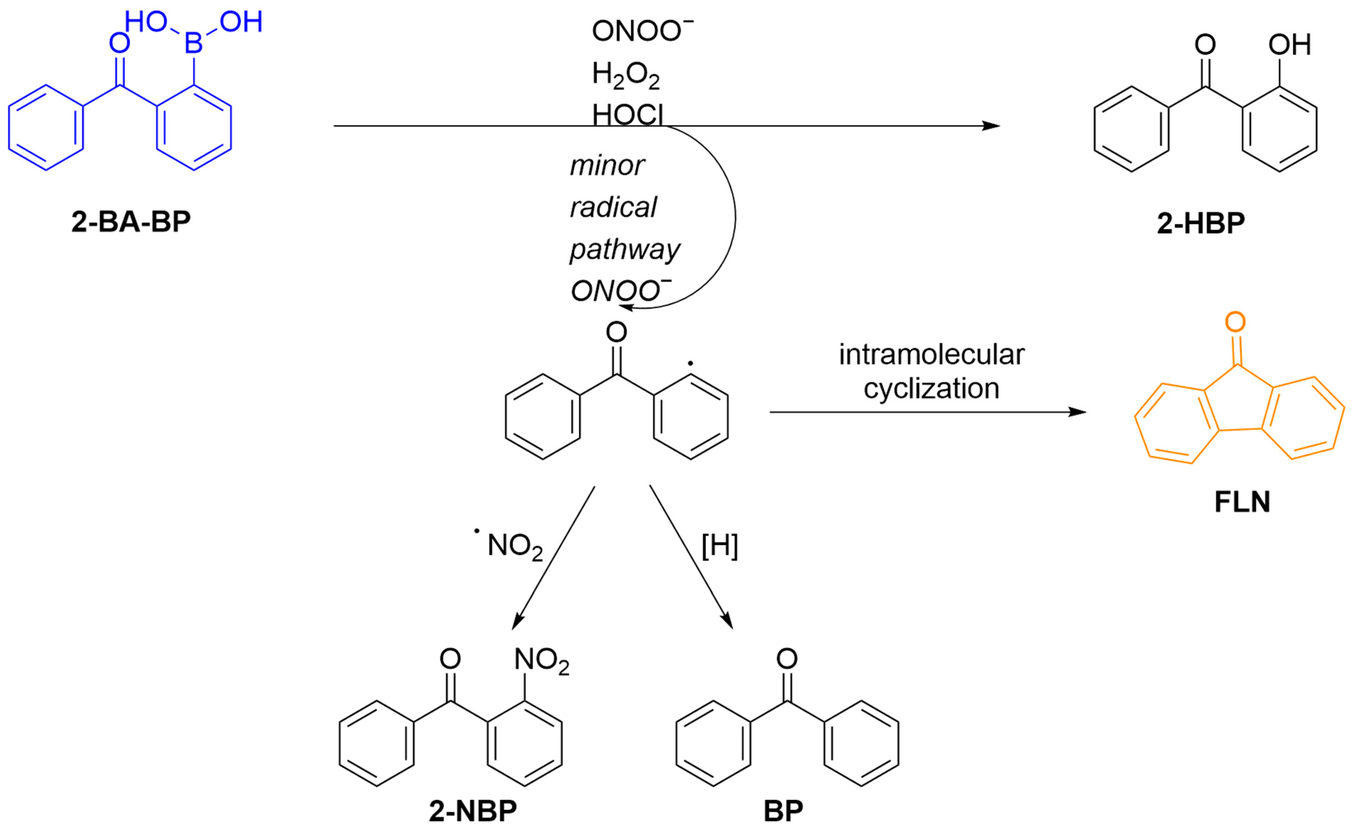
3.1. Fluorescent Probes Based on Boronates (Boronic Acids/Boronic Esters)
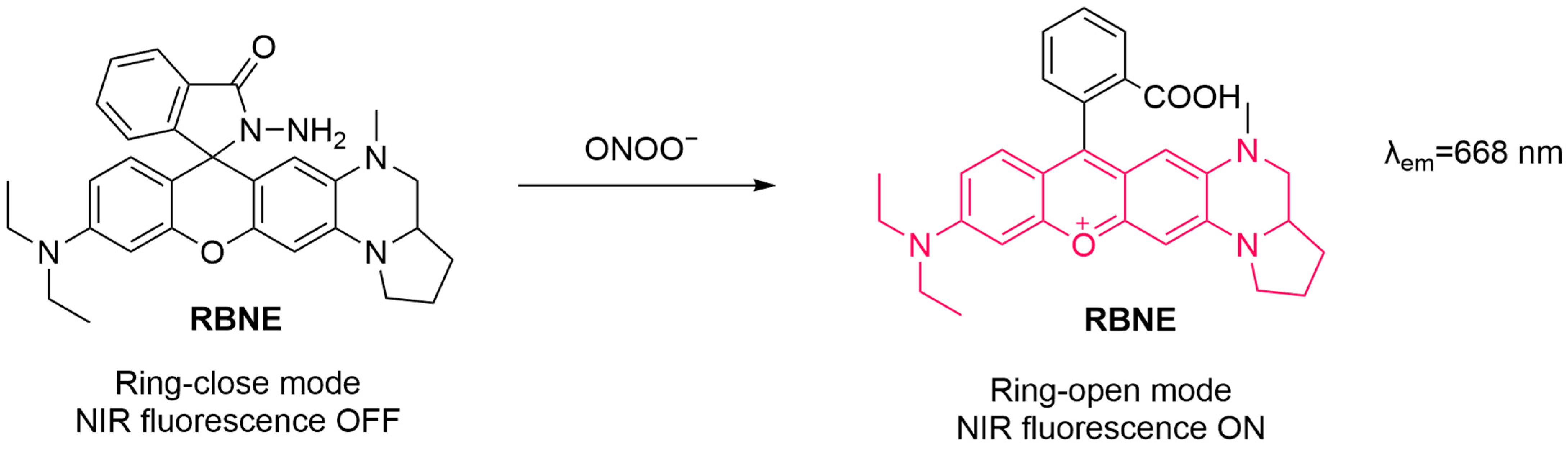
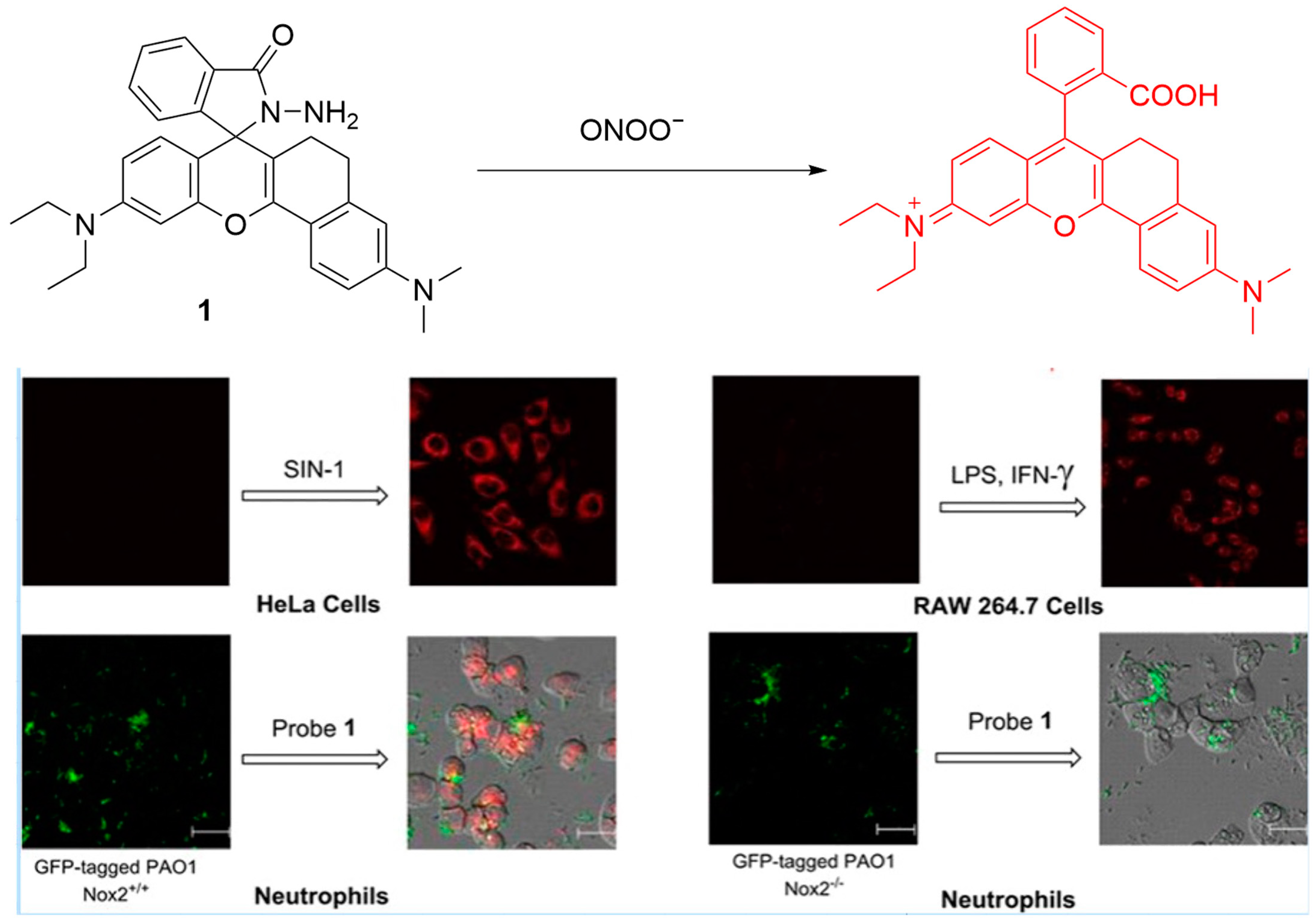
3.2. Fluorescent Probes Based on Hydrazides
3.3. Fluorescent Probes Based on Ketoamides
3.4. Fluorescent Probes Based on Thioethers
3.5. Fluorescent Probes Based on Conjugated Double Bonds (C=C/C=N)
3.5.1. Fluorescent Probes Based on C=C Bonds
3.5.2. Fluorescent Probes Based on C=N Bonds
3.6. Fluorescent Probes Based on Trifluoromethyl Ketone
3.7. Fluorescent Probes Based on Aromatic Compounds (Hydroquinone, Benzopyrylium/Benzopyran)
3.7.1. Fluorescent Probes Based on Hydroquinone
3.7.2. Fluorescent Probes Based on Benzopyrylium/Benzopyran
4. Discussion
5. Summary and Outlook
Author Contributions
Funding
Institutional Review Board Statement
Informed Consent Statement
Data Availability Statement
Acknowledgments
Conflicts of Interest
References
- Denicola, A.; Radi, R. Peroxynitrite and drug-dependent toxicity. Toxicology 2005, 208, 273–288. [Google Scholar] [CrossRef] [PubMed]
- Prolo, C.; Piacenza, L.; Radi, R. Peroxynitrite: A multifaceted oxidizing and nitrating metabolite. Curr. Opin. Chem. Biol. 2024, 80, 102459. [Google Scholar] [CrossRef]
- Radi, R. Oxygen radicals, nitric oxide, and peroxynitrite: Redox pathways in molecular medicine. Proc. Natl. Acad. Sci. USA 2018, 115, 5839–5848. [Google Scholar] [CrossRef] [PubMed]
- Ahmad, R.; Hussain, A.; Ahsan, H. Peroxynitrite: Cellular pathology and implications in autoimmunity. J. Immunoass. Immunochem. 2019, 40, 123–138. [Google Scholar] [CrossRef]
- Capietto, A.H.; Delamarre, L. Peroxynitrite promotes immune evasion by reducing tumor antigenicity. Cell Rep. Med. 2022, 3, 100787. [Google Scholar] [CrossRef]
- Ramdial, K.; Franco, M.C.; Estevez, A.G. Cellular mechanisms of peroxynitrite-induced neuronal death. Brain Res. Bull. 2017, 133, 4–11. [Google Scholar] [CrossRef]
- Prolo, C.; Rios, N.; Piacenza, L.; Alvarez, M.N.; Radi, R. Fluorescence and chemiluminescence approaches for peroxynitrite detection. Free Radic. Biol. Med. 2018, 128, 59–68. [Google Scholar] [CrossRef] [PubMed]
- Szabo, C.; Ischiropoulos, H.; Radi, R. Peroxynitrite: Biochemistry, pathophysiology and development of therapeutics. Nat. Rev. Drug Discov. 2007, 6, 662–680. [Google Scholar] [CrossRef]
- Su, H.; Wang, N.; Wang, J.; Wang, H.; Zhang, J.; Zhao, W. A resorufin-based red-emitting fluorescent probe with high selectivity for tracking endogenous peroxynitrite in living cells and inflammatory mice. Spectrochim. Acta Part A Mol. Biomol. Spectrosc. 2021, 252, 119502. [Google Scholar] [CrossRef]
- Liu, Z.; Mo, S.; Hao, Z.; Hu, L. Recent Progress of Spectroscopic Probes for Peroxynitrite and Their Potential Medical Diagnostic Applications. Int. J. Mol. Sci. 2023, 24, 12821. [Google Scholar] [CrossRef]
- Li, Z.; Lu, J.; Pang, Q.; You, J. Construction of a near-infrared fluorescent probe for ratiometric imaging of peroxynitrite during tumor progression. Analyst 2021, 146, 5204–5211. [Google Scholar] [CrossRef]
- Daiber, A.; Oelze, M.; August, M.; Wendt, M.; Sydow, K.; Wieboldt, H.; Kleschyov, A.L.; Munzel, T. Detection of superoxide and peroxynitrite in model systems and mitochondria by the luminol analogue L-012. Free Radic. Res. 2004, 38, 259–269. [Google Scholar] [CrossRef] [PubMed]
- Amatore, C.; Arbault, S.; Bruce, D.; de Oliveira, P.; Erard, L.M.; Vuillaume, M. Characterization of the electrochemical oxidation of peroxynitrite: Relevance to oxidative stress bursts measured at the single cell level. Chemistry 2001, 7, 4171–4179. [Google Scholar] [CrossRef]
- Pislaru, S.V.; Ni, Y.; Pislaru, C.; Bosmans, H.; Miao, Y.; Bogaert, J.; Dymarkowski, S.; Semmler, W.; Marchal, G.; Van de Werf, F.J. Noninvasive measurements of infarct size after thrombolysis with a necrosis-avid MRI contrast agent. Circulation 1999, 99, 690–696. [Google Scholar] [CrossRef]
- Bruemmer, K.J.; Merrikhihaghi, S.; Lollar, C.T.; Morris, S.N.; Bauer, J.H.; Lippert, A.R. 19F magnetic resonance probes for live-cell detection of peroxynitrite using an oxidative decarbonylation reaction. Chem. Commun. 2014, 50, 12311–12314. [Google Scholar] [CrossRef]
- Li, L.; Wang, C.; Hu, J.; Chen, W.H. Recent progress in organelle-targeting fluorescent probes for the detection of peroxynitrite. Chem. Commun. 2024, 60, 13629–13640. [Google Scholar] [CrossRef]
- Xu, M.; Ma, Y.; Zhao, Y.; Xu, L.; Lin, W. Peroxynitrite activatable NIR-II probe for tumor diagnosis and photothermal therapy in vivo. Anal. Chim. Acta 2025, 1335, 343443. [Google Scholar] [CrossRef]
- Li, M.; Gong, X.; Li, H.W.; Han, H.; Shuang, S.; Song, S.; Dong, C. A fast detection of peroxynitrite in living cells. Anal. Chim. Acta 2020, 1106, 96–102. [Google Scholar] [CrossRef]
- Ma, Q.; Xu, S.; Zhai, Z.; Wang, K.; Liu, X.; Xiao, H.; Zhuo, S.; Liu, Y. Recent Progress of Small-Molecule Ratiometric Fluorescent Probes for Peroxynitrite in Biological Systems. Chem.—A Eur. J. 2022, 28, e202200828. [Google Scholar] [CrossRef]
- Yang, Y.; Jinting, S.; Yiyuan, X.; Gui, Y. Fluorescent probes for sensing peroxynitrite: Biological applications. Redox Rep. 2024, 29, 2430157. [Google Scholar] [CrossRef] [PubMed]
- Grzelakowska, A.; Kalyanaraman, B.; Zielonka, J. Small molecule probes for peroxynitrite detection. Redox Biochem. Chem. 2024, 10, 100034. [Google Scholar] [CrossRef] [PubMed]
- Li, M.; Lei, P.; Shuang, S.; Dong, C.; Zhang, L. Recent advances in fluorescent probes for dual-detecting ONOO− and analytes. Spectrochim. Acta Part A Mol. Biomol. Spectrosc. 2023, 303, 123179. [Google Scholar] [CrossRef]
- Zhou, Y.; Kuang, X.; Yang, X.; Li, J.; Wei, X.; Jang, W.J.; Zhang, S.-S.; Yan, M.; Yoon, J. Recent progress in small-molecule fluorescent probes for the detection of superoxide anion, nitric oxide, and peroxynitrite anion in biological systems. Chem. Sci. 2024, 15, 19669–19697. [Google Scholar] [CrossRef]
- Radi, R.; Cassina, A.; Hodara, R.; Quijano, C.; Castro, L. Peroxynitrite reactions and formation in mitochondria. Free Radic. Biol. Med. 2002, 33, 1451–1464. [Google Scholar] [CrossRef]
- Violi, F.; Marino, R.; Milite, M.T.; Loffredo, L. Nitric oxide and its role in lipid peroxidation. Diabetes/Metab. Res. Rev. 1999, 15, 283–288. [Google Scholar] [CrossRef]
- Epe, B.; Ballmaier, D.; Roussyn, I.; Briviba, K.; Sies, H. DNA damage by peroxynitrite characterized with DNA repair enzymes. Nucleic Acids Res. 1996, 24, 4105–4110. [Google Scholar] [CrossRef]
- Ferrer-Sueta, G.; Campolo, N.; Trujillo, M.; Bartesaghi, S.; Carballal, S.; Romero, N.; Alvarez, B.; Radi, R. Biochemistry of Peroxynitrite and Protein Tyrosine Nitration. Chem. Rev. 2018, 118, 1338–1408. [Google Scholar] [CrossRef]
- Lush, C.W.; Cepinskas, G.; Kvietys, P.R. Regulation of intestinal nuclear factor-kappaB activity and E-selectin expression during sepsis: A role for peroxynitrite. Gastroenterology 2003, 124, 118–128. [Google Scholar] [CrossRef]
- Piacenza, L.; Zeida, A.; Trujillo, M.; Radi, R. The superoxide radical switch in the biology of nitric oxide and peroxynitrite. Physiol. Rev. 2022, 102, 1881–1906. [Google Scholar] [CrossRef] [PubMed]
- Pacher, P.; Beckman, J.S.; Liaudet, L. Nitric oxide and peroxynitrite in health and disease. Physiol. Rev. 2007, 87, 315–424. [Google Scholar] [CrossRef]
- Liu, S.; Horowitz, J. N-Acetylcysteine Reverses Peroxynitrite and Hydrogen Peroxide-Induced Apoptosis of Human Myocardial Cells. J. Am. Coll. Cardiol. 2019, 73, 1022. [Google Scholar] [CrossRef]
- Nossuli, T.O.; Hayward, R.; Scalia, R.; Lefer, A.M. Peroxynitrite reduces myocardial infarct size and preserves coronary endothelium after ischemia and reperfusion in cats. Circulation 1997, 96, 2317–2324. [Google Scholar] [CrossRef]
- Lancel, S.; Tissier, S.; Mordon, S.; Marechal, X.; Depontieu, F.; Scherpereel, A.; Chopin, C.; Neviere, R. Peroxynitrite decomposition catalysts prevent myocardial dysfunction and inflammation in endotoxemic rats. J. Am. Coll. Cardiol. 2004, 43, 2348–2358. [Google Scholar] [CrossRef]
- Rabinovitch, A.; Suarez-Pinzon, W.L. Cytokines and their roles in pancreatic islet beta-cell destruction and insulin-dependent diabetes mellitus. Biochem. Pharmacol. 1998, 55, 1139–1149. [Google Scholar] [CrossRef]
- McCafferty, D.M. Peroxynitrite and inflammatory bowel disease. Gut 2000, 46, 436–439. [Google Scholar] [CrossRef]
- Tcyganov, E.N.; Sanseviero, E.; Marvel, D.; Beer, T.; Tang, H.Y.; Hembach, P.; Speicher, D.W.; Zhang, Q.; Donthireddy, L.R.; Mostafa, A.; et al. Peroxynitrite in the tumor microenvironment changes the profile of antigens allowing escape from cancer immunotherapy. Cancer Cell 2022, 40, 1173–1189.e6. [Google Scholar] [CrossRef]
- Torreilles, F.; Salman-Tabcheh, S.; Guerin, M.; Torreilles, J. Neurodegenerative disorders: The role of peroxynitrite. Brain Res. Rev. 1999, 30, 153–163. [Google Scholar] [CrossRef]
- Han, H.; Desert, R.; Das, S.; Song, Z.; Athavale, D.; Ge, X.; Nieto, N. Danger signals in liver injury and restoration of homeostasis. J. Hepatol. 2020, 73, 933–951. [Google Scholar] [CrossRef]
- Frankenfeld, C.N.; Rosenbaugh, M.R.; Fogarty, B.A.; Lunte, S.M. Separation and detection of peroxynitrite and its metabolites by capillary electrophoresis with UV detection. J. Chromatogr. A 2006, 1111, 147–152. [Google Scholar] [CrossRef]
- Tsikas, D. GC-MS and HPLC methods for peroxynitrite (ONOO− and O15NOO−) analysis: A study on stability, decomposition to nitrite and nitrate, laboratory synthesis, and formation of peroxynitrite from S-nitrosoglutathione (GSNO) and KO2. Analyst 2011, 136, 979–987. [Google Scholar] [CrossRef]
- Jorgensen, S.M.; Lorentzen, L.G.; Chuang, C.Y.; Davies, M.J. Peroxynitrous acid-modified extracellular matrix alters gene and protein expression in human coronary artery smooth muscle cells and induces a pro-inflammatory phenotype. Free Radic. Biol. Med. 2022, 186, 43–52. [Google Scholar] [CrossRef]
- Li, H.; Li, X.; Wu, X.; Shi, W.; Ma, H. Observation of the Generation of ONOO− in Mitochondria under Various Stimuli with a Sensitive Fluorescence Probe. Anal. Chem. 2017, 89, 5519–5525. [Google Scholar] [CrossRef]
- Wu, G.; Li, Z.; Huang, P.; Lin, W. Shedding light on ONOO− detection: The emergence of a fast-response fluorescent probe for biological systems. J. Mater. Chem. B 2024, 12, 3436–3444. [Google Scholar] [CrossRef]
- DeFrancesco, H.; Dudley, J.; Coca, A. Boron Chemistry: An Overview. In Boron Chemistry: An Overview; ACS Symposium Series; American Chemical Society: Washington, DC, USA, 2016; Volume 1236, pp. 1–25. [Google Scholar]
- Sikora, A.; Zielonka, J.; Debowska, K.; Michalski, R.; Smulik-Izydorczyk, R.; Pieta, J.; Podsiadly, R.; Artelska, A.; Pierzchala, K.; Kalyanaraman, B. Boronate-Based Probes for Biological Oxidants: A Novel Class of Molecular Tools for Redox Biology. Front. Chem. 2020, 8, 580899. [Google Scholar] [CrossRef]
- Sikora, A.; Zielonka, J.; Lopez, M.; Joseph, J.; Kalyanaraman, B. Direct oxidation of boronates by peroxynitrite: Mechanism and implications in fluorescence imaging of peroxynitrite. Free Radic. Biol. Med. 2009, 47, 1401–1407. [Google Scholar] [CrossRef]
- Guo, B.; Shu, W.; Liu, W.; Wang, H.; Xing, S.; Chen, J.; Zhang, X. Mitochondria-specific ultrasensitive ratiometric AIE probe for imaging endogenous peroxynitrite. Sens. Actuators B Chem. 2021, 344, 130206. [Google Scholar] [CrossRef]
- Kang, H.; Shu, W.; Yu, J.; Gao, M.; Han, R.; Jing, J.; Zhang, R.; Zhang, X. A near-infrared fluorescent probe for ratiometric imaging peroxynitrite in Parkinson’s disease model. Sens. Actuators B Chem. 2022, 359, 131393. [Google Scholar] [CrossRef]
- Wu, L.; Tian, X.; Han, H.H.; Wang, J.; Groleau, R.R.; Tosuwan, P.; Wannalerse, B.; Sedgwick, A.C.; Bull, S.D.; He, X.P.; et al. A Simple Near-Infrared Fluorescent Probe for the Detection of Peroxynitrite. ChemistryOpen 2019, 8, 1407–1409. [Google Scholar] [CrossRef]
- Wu, L.; Han, H.H.; Liu, L.; Gardiner, J.E.; Sedgwick, A.C.; Huang, C.; Bull, S.D.; He, X.P.; James, T.D. ESIPT-based fluorescence probe for the rapid detection of peroxynitrite ‘AND’ biological thiols. Chem. Commun. 2018, 54, 11336–11339. [Google Scholar] [CrossRef] [PubMed]
- Sonawane, P.M.; Yudhistira, T.; Halle, M.B.; Roychaudhury, A.; Kim, Y.; Surwase, S.S.; Bhosale, V.K.; Kim, J.; Park, H.-S.; Kim, Y.-c.; et al. A water-soluble boronate masked benzoindocyanin fluorescent probe for the detection of endogenous mitochondrial peroxynitrite in live cells and zebrafish as inflammation models. Dye. Pigment. 2021, 191, 109371. [Google Scholar] [CrossRef]
- Ma, L.; Yang, Q.; Zan, Q.; Tian, H.; Zhang, X.; Dong, C.; Fan, L. A benzothiazole-based fluorescence probe for imaging of peroxynitrite during ferroptosis and diagnosis of tumor tissues. Anal. Bioanal. Chem. 2022, 414, 7753–7762. [Google Scholar] [CrossRef]
- Liu, C.; Mei, Y.; Yang, H.; Zhang, Q.; Zheng, K.; Zhang, P.; Ding, C. Ratiometric Fluorescent Probe for Real-Time Detection of beta-Galactosidase Activity in Lysosomes and Its Application in Drug-Induced Senescence Imaging. Anal. Chem. 2024, 96, 3223–3232. [Google Scholar] [CrossRef]
- Zheng, B.; Wang, S.; Xu, J.; Huang, L.; Zhao, S. A ratiometric fluorescent probe with dual near infrared emission for in vivo ratio imaging of cysteine. Talanta 2025, 286, 127564. [Google Scholar] [CrossRef]
- Palanisamy, S.; Wu, P.Y.; Wu, S.C.; Chen, Y.J.; Tzou, S.C.; Wang, C.H.; Chen, C.Y.; Wang, Y.M. In vitro and in vivo imaging of peroxynitrite by a ratiometric boronate-based fluorescent probe. Biosens. Bioelectron. 2017, 91, 849–856. [Google Scholar] [CrossRef]
- Kim, H.M.; Cho, B.R. Small-molecule two-photon probes for bioimaging applications. Chem. Rev. 2015, 115, 5014–5055. [Google Scholar] [CrossRef]
- Duan, D.C.; Pan, G.; Liu, J.; Chen, H.; Xie, T.; Long, Y.; Dai, F.; Zhang, S.; Zhou, B. Cellular and Intravital Nucleus Imaging by a D-π-A Type of Red-Emitting Two-Photon Fluorescent Probe. Anal. Chem. 2024, 96, 20425–20434. [Google Scholar] [CrossRef]
- Grzelakowska, A.; Zielonka, M.; Debowska, K.; Modrzejewska, J.; Szala, M.; Sikora, A.; Zielonka, J.; Podsiadly, R. Two-photon fluorescent probe for cellular peroxynitrite: Fluorescence detection, imaging, and identification of peroxynitrite-specific products. Free Radic. Biol. Med. 2021, 169, 24–35. [Google Scholar] [CrossRef]
- Sedgwick, A.C.; Han, H.H.; Gardiner, J.E.; Bull, S.D.; He, X.P.; James, T.D. Long-wavelength fluorescent boronate probes for the detection and intracellular imaging of peroxynitrite. Chem. Commun. 2017, 53, 12822–12825. [Google Scholar] [CrossRef]
- Tang, J.; Li, Z.; Qiang, C.; Han, Y.; Yang, L.; Zhu, L.; Dang, T.; Chen, G.; Ye, Y. A long-wavelength mitochondria-targeted fluorescent probe for imaging of peroxynitrite during dexamethasone treatment. Spectrochim. Acta Part A Mol. Biomol. Spectrosc. 2023, 292, 122429. [Google Scholar] [CrossRef] [PubMed]
- Qu, W.; Tian, R.; Yang, B.; Guo, T.; Wu, Z.; Li, Y.; Geng, Z.; Wang, Z. Dual-Channel/Localization Single-Molecule Fluorescence Probe for Monitoring ATP and HOCl in Early Diagnosis and Therapy of Rheumatoid Arthritis. Anal. Chem. 2024, 96, 5428–5436. [Google Scholar] [CrossRef]
- Wu, L.; Liu, J.; Tian, X.; Groleau, R.R.; Feng, B.; Yang, Y.; Sedgwick, A.C.; Han, H.H.; Wang, Y.; Wang, H.M.; et al. Dual-Channel Fluorescent Probe for the Simultaneous Monitoring of Peroxynitrite and Adenosine-5′-triphosphate in Cellular Applications. J. Am. Chem. Soc. 2022, 144, 174–183. [Google Scholar] [CrossRef] [PubMed]
- Gong, J.; Wang, X.; Wu, J.; Yoon, C.; Kim, Y.; Zou, J.; Mao, Z.; Kim, J.S. Diaminonaphthalene Boronic Acid (DANBA): New Approach for Peroxynitrite Sensing Site. Angew. Chem. Int. Ed. Engl. 2024, 63, e202409295. [Google Scholar] [CrossRef]
- Grzelakowska, A.; Podsiadły, R.; Zielonka, J. Phenyl Radical-Mediated Fluorogenic Cyclization for Specific Detection of Peroxynitrite. Anal. Chem. 2025, 97, 7299–7306. [Google Scholar] [CrossRef]
- Zhang, K.; Dai, Y.; Li, Q.; Su, Y.; Lv, Y. Unimolecular chemo-fluoro-luminescent probe for simultaneous detection and imaging of peroxynitrite and hypochlorite in vitro and in vivo. Sens. Actuators B Chem. 2021, 347, 130609. [Google Scholar] [CrossRef]
- Mao, G.J.; Gao, G.Q.; Dong, W.P.; Wang, Q.Q.; Wang, Y.Y.; Li, Y.; Su, L.; Zhang, G. A two-photon excited near-infrared fluorescent probe for imaging peroxynitrite during drug-induced hepatotoxicity and its remediation. Talanta 2021, 221, 121607. [Google Scholar] [CrossRef]
- Li, Z.; Liu, C.; Yu, C.; Chen, Y.; Jia, P.; Zhu, H.; Zhang, X.; Yu, Y.; Zhu, B.; Sheng, W. A highly selective and sensitive red-emitting fluorescent probe for visualization of endogenous peroxynitrite in living cells and zebrafish. Analyst 2019, 144, 3442–3449. [Google Scholar] [CrossRef]
- Zhang, Y.; Hu, J.; Rong, X.; Jiang, J.; Wang, Y.; Zhang, X.; Xu, Z.; Xu, K.; Wu, M.; Fang, M. Development of a hybrid rhodamine-hydrazine NIR fluorescent probe for sensitive detection and imaging of peroxynitrite in necrotizing enterocolitis model. Bioorg. Chem. 2024, 152, 107729. [Google Scholar] [CrossRef]
- Wu, D.; Ryu, J.C.; Chung, Y.W.; Lee, D.; Ryu, J.H.; Yoon, J.H.; Yoon, J. A Far-Red-Emitting Fluorescence Probe for Sensitive and Selective Detection of Peroxynitrite in Live Cells and Tissues. Anal. Chem. 2017, 89, 10924–10931. [Google Scholar] [CrossRef]
- Huang, W.; Du, X.; Zhang, C.; Zhang, S.; Zhang, J.; Yang, X.F. Rational Design of a Dual-Channel Fluorescent Probe for the Simultaneous Imaging of Hypochlorous Acid and Peroxynitrite in Living Organisms. Anal. Chem. 2022, 94, 17485–17493. [Google Scholar] [CrossRef]
- Yang, C.; Wang, X. Lysosome biogenesis: Regulation and functions. J. Cell Biol. 2021, 220, e202102001. [Google Scholar] [CrossRef]
- Ebner, M.; Puchkov, D.; Lopez-Ortega, O.; Muthukottiappan, P.; Su, Y.; Schmied, C.; Zillmann, S.; Nikonenko, I.; Koddebusch, J.; Dornan, G.L.; et al. Nutrient-regulated control of lysosome function by signaling lipid conversion. Cell 2023, 186, 5328–5346.e26. [Google Scholar] [CrossRef]
- Settembre, C.; Fraldi, A.; Medina, D.L.; Ballabio, A. Signals from the lysosome: A control centre for cellular clearance and energy metabolism. Nat. Rev. Mol. Cell Biol. 2013, 14, 283–296. [Google Scholar] [CrossRef] [PubMed]
- Sun, S.G.; Ding, H.; Yuan, G.; Zhou, L. An efficient TP-FRET-based lysosome-targetable fluorescent probe for imaging peroxynitrite with two well-resolved emission channels in living cells, tissues and zebrafish. Anal. Chim. Acta 2020, 1100, 200–207. [Google Scholar] [CrossRef]
- Shang, J.; Yang, Y.; Sun, Y.; Gao, W.; Ma, K.; Wang, C.; Yu, X.; Li, L.; Zheng, J.; Zhao, N.; et al. Real-time monitoring of ONOO− in cerebral ischemia-reperfusion injury mouse models using a hydrazine-based NIR fluorescent probe. Redox Biol. 2025, 80, 103494. [Google Scholar] [CrossRef] [PubMed]
- Yan, Z.; Tang, Z.; Wang, X.; Zheng, Z.; Tian, Z.; Geng, X.; Li, Y.; Jiang, H. A novel Golgi-targetable fluorescent probe for imaging peroxynitrite in Golgi stress and sepsis-induced acute lung injury. Sens. Actuators B Chem. 2022, 369, 132352. [Google Scholar] [CrossRef]
- Qin, S.; Lu, H.; Zhang, J.; Ji, X.; Wang, N.; Liu, J.; Zhao, W.; Wang, J. An activatable reporter for fluorescence imaging drug-induced liver injury in diverse cell lines and in vivo. Dye. Pigment. 2022, 203, 110345. [Google Scholar] [CrossRef]
- Qu, W.; Niu, C.; Zhang, X.; Chen, W.; Yu, F.; Liu, H.; Zhang, X.; Wang, S. Construction of a novel far-red fluorescence light-up probe for visualizing intracellular peroxynitrite. Talanta 2019, 197, 431–435. [Google Scholar] [CrossRef]
- Li, X.; Zou, X.; Xu, P.; Pang, M.; Zhao, L.; Chen, S.; Peng, Y.; Liang, S.; Deng, Z. A robust NIR fluorescence-activated probe for peroxynitrite imaging in cells and mice osteoarthritis models. Anal. Biochem. 2023, 682, 115338. [Google Scholar] [CrossRef]
- Xie, X.; Tang, F.; Liu, G.; Li, Y.; Su, X.; Jiao, X.; Wang, X.; Tang, B. Mitochondrial Peroxynitrite Mediation of Anthracycline-Induced Cardiotoxicity as Visualized by a Two-Photon Near-Infrared Fluorescent Probe. Anal. Chem. 2018, 90, 11629–11635. [Google Scholar] [CrossRef]
- Li, Y.; Xie, X.; Yang, X.; Li, M.; Jiao, X.; Sun, Y.; Wang, X.; Tang, B. Two-photon fluorescent probe for revealing drug-induced hepatotoxicity via mapping fluctuation of peroxynitrite. Chem. Sci. 2017, 8, 4006–4011. [Google Scholar] [CrossRef]
- Yan, M.; Yu, M.; Wang, Z.; Yu, F.; Fang, W. An α-ketoamide-based fluorescent probe via aggregation induced emission for ONOO− detection in vivo. Talanta 2025, 293, 128064. [Google Scholar] [CrossRef] [PubMed]
- Zhang, K.; Wang, Z.; Hu, X.; Meng, J.; Bao, W.; Wang, X.; Ding, W.; Tian, Z. A long-wavelength turn-on fluorescent probe for intracellular nanomolar level peroxynitrite sensing with second-level response. Talanta 2020, 219, 121354. [Google Scholar] [CrossRef] [PubMed]
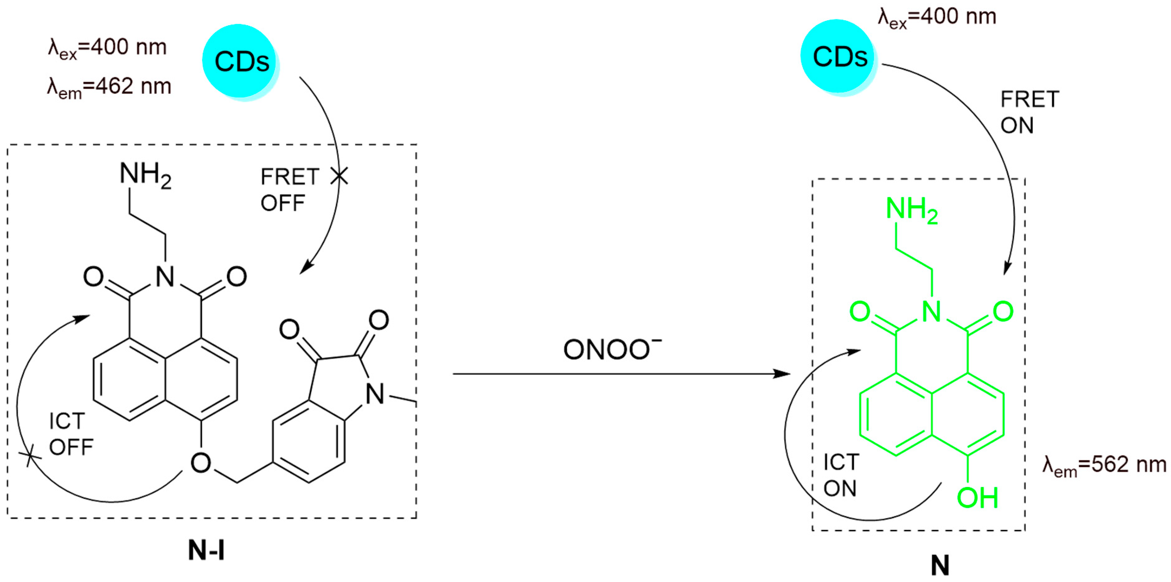
- Wu, Y.; Sun, L.L.; Han, H.H.; He, X.P.; Cao, W.; James, T.D. Selective FRET nano probe based on carbon dots and naphthalimide-isatin for the ratiometric detection of peroxynitrite in drug-induced liver injury. Chem. Sci. 2024, 15, 757–764. [Google Scholar] [CrossRef] [PubMed]
- Zhang, J.; Fu, Y.; Han, H.-H.; Zang, Y.; Li, J.; He, X.-P.; Feringa, B.L.; Tian, H. Remote light-controlled intracellular target recognition by photochromic fluorescent glycoprobes. Nat. Commun. 2017, 8, 987. [Google Scholar] [CrossRef]
- Zhang, H.L.; Wei, X.L.; Zang, Y.; Cao, J.Y.; Liu, S.; He, X.P.; Chen, Q.; Long, Y.T.; Li, J.; Chen, G.R.; et al. Fluorogenic Probing of Specific Recognitions between Sugar Ligands and Glycoprotein Receptors on Cancer Cells by an Economic Graphene Nanocomposite. Adv. Mater. 2013, 25, 4097–4101. [Google Scholar] [CrossRef]
- Chai, X.; Han, H.-H.; Sedgwick, A.C.; Li, N.; Zang, Y.; James, T.D.; Zhang, J.; Hu, X.-L.; Yu, Y.; Li, Y.; et al. Photochromic Fluorescent Probe Strategy for the Super-resolution Imaging of Biologically Important Biomarkers. J. Am. Chem. Soc. 2020, 142, 18005–18013. [Google Scholar] [CrossRef]
- Fu, Y.; Han, H.-H.; Zhang, J.; He, X.-P.; Feringa, B.L.; Tian, H. Photocontrolled Fluorescence “Double-Check” Bioimaging Enabled by a Glycoprobe–Protein Hybrid. J. Am. Chem. Soc. 2018, 140, 8671–8674. [Google Scholar] [CrossRef]
- Ji, D.K.; Zhang, Y.; Zang, Y.; Li, J.; Chen, G.R.; He, X.P.; Tian, H. Targeted Intracellular Production of Reactive Oxygen Species by a 2D Molybdenum Disulfide Glycosheet. Adv. Mater. 2016, 28, 9356–9363. [Google Scholar] [CrossRef]
- Jiang, W.; Liu, L.; Li, W.; Liu, H.; Yang, J.; Wang, P. A lysosomal-targeted switchable fluorescent probe for the detection of peroxynitrite in living tumor cells and in vivo. Talanta 2025, 291, 127866. [Google Scholar] [CrossRef]
- Hou, J.T.; Wang, B.; Zhang, Y.; Cui, B.; Cao, X.; Zhang, M.; Ye, Y.; Wang, S. Observation of peroxynitrite overproduction in cells during 5-fluorouracil treatment via a ratiometric fluorescent probe. Chem. Commun. 2020, 56, 2759–2762. [Google Scholar] [CrossRef]
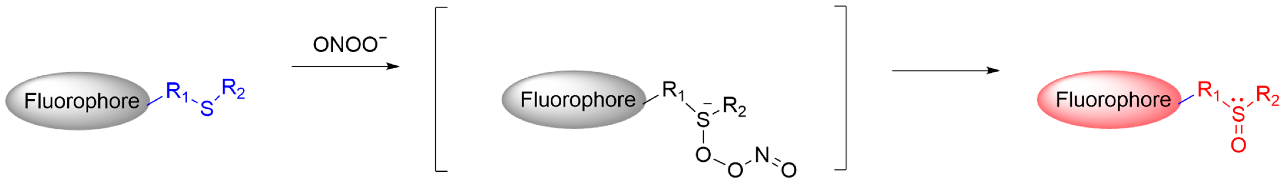
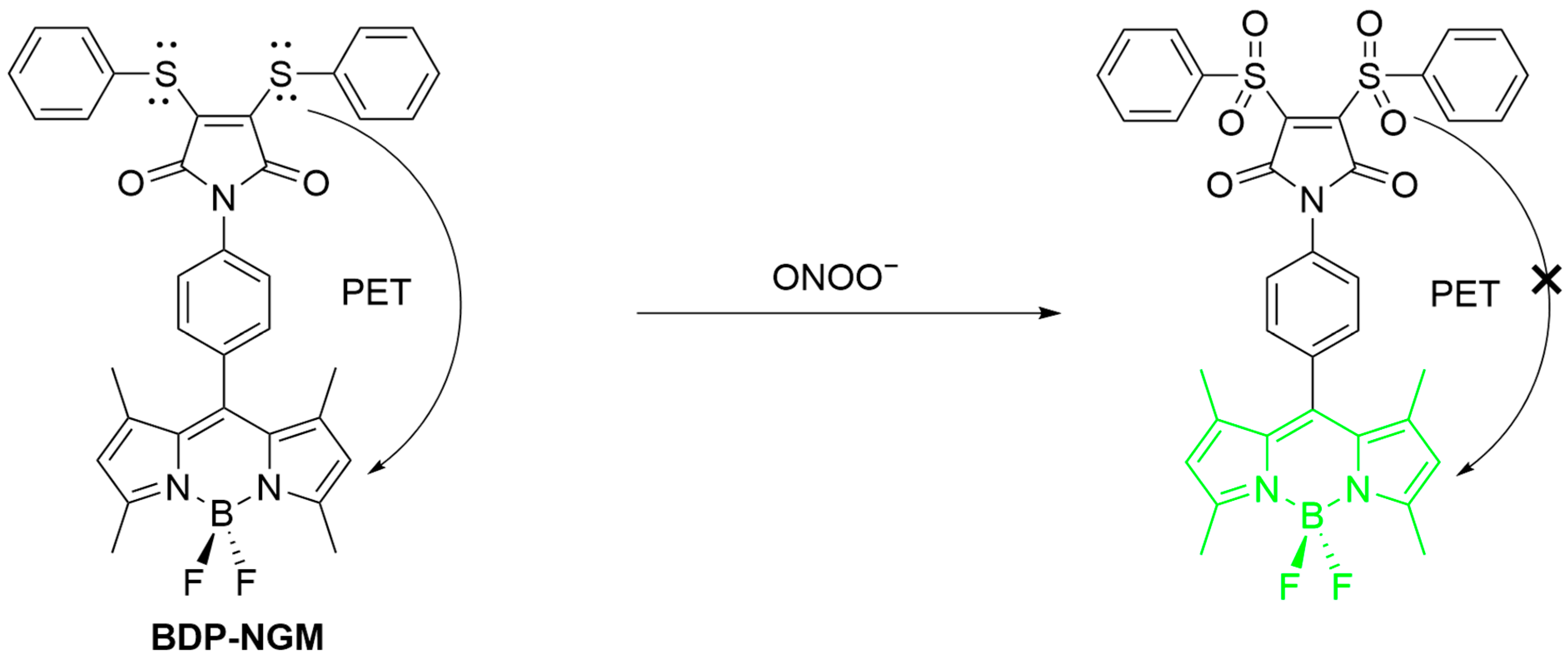
- Yudhistira, T.; Mulay, S.V.; Lee, K.J.; Kim, Y.; Park, H.S.; Churchill, D.G. Thiomaleimide Functionalization for Selective Biological Fluorescence Detection of Peroxynitrite as Tested in HeLa and RAW 264.7 Cells. Chem. Asian J. 2017, 12, 1927–1934. [Google Scholar] [CrossRef] [PubMed]
- Lin, W.; Long, L.; Chen, B.; Tan, W. A ratiometric fluorescent probe for hypochlorite based on a deoximation reaction. Chemistry 2009, 15, 2305–2309. [Google Scholar] [CrossRef] [PubMed]
- Li, J.; Peng, S.; Li, Z.; Zhao, F.; Han, X.; Liu, J.; Cao, W.; Ye, Y. Visualization of peroxynitrite in cyclophosphamide-induced oxidative stress by an activatable probe. Talanta 2022, 238, 123007. [Google Scholar] [CrossRef]
- Cobbs, C.S.; Samanta, M.; Harkins, L.E.; Gillespie, G.Y.; Merrick, B.A.; MacMillan-Crow, L.A. Evidence for Peroxynitrite-Mediated Modifications to p53 in Human Gliomas: Possible Functional Consequences. Arch. Biochem. Biophys. 2001, 394, 167–172. [Google Scholar] [CrossRef] [PubMed]
- Zhang, Q.; Zeng, S.-M.-Z.; Yuan, L.-C.; Wu, C.-J.; Wu, S.-Y.; Ren, S.-Z.; Zhao, M.-D.; Wang, X.-M.; Zhu, H.-L.; Wang, Z.-C. Real-time diagnosis of glioblastoma by sensing peroxynitrite with a highly selective and rapid-responding fluorescent probe. Sens. Actuators B Chem. 2023, 395, 134502. [Google Scholar] [CrossRef]
- Luo, X.; Zhang, C.; Yuan, F.; Cheng, S.; Zhu, Y.; Xiang, M.; Hu, X.; Xian, Y. Dual-Channel Fluorescent Probe for the Detection of Peroxynitrite and Glutathione in Mitochondria: Accurate Discrimination of Inflammatory and Progressing Tumor Cells. Anal. Chem. 2022, 94, 15790–15800. [Google Scholar] [CrossRef]
- Li, M.; Lei, P.; Shuang, S.; Dong, C.; Zhang, L. Three-channel fluorescent probe for real-time monitoring mitochondrial viscosity during mitophagy and visualizing ONOO−/SO2 in rheumatoid arthritis mice. Sens. Actuators B Chem. 2024, 413, 135909. [Google Scholar] [CrossRef]
- Li, T.; Wang, Z.-Q.; Yang, Z.-C.; Long, J.-Q.; Mao, G.-J.; Xu, F.; Li, Y.; Li, C.-Y. A hepatocyte-targeting ratiometric fluorescence probe for monitoring peroxynitrite in liver injury. Microchem. J. 2025, 208, 112566. [Google Scholar] [CrossRef]
- Wei, D.; Dai, Y.; Yan, X.; Lan, D.; Liao, J.; Qin, Z.; Fu, N. A Novel “Double-Responsive” and “Dual-Targeted” Multifunctional Fluorescent Probe Monitors the Level Changes of ONOO− in Mitochondria during Cell Pyroptosis. ACS Sens. 2025, 10, 2542–2553. [Google Scholar] [CrossRef]
- Zou, J.; Song, B.; Liu, Q.; Dong, Z.; Yuan, J. An activatable β-diketonate europium(III) complex-based probe for time-gated luminescence detection and imaging of peroxynitrite in vitro and in vivo. Talanta 2025, 292, 127928. [Google Scholar] [CrossRef]
- Wang, C.; Shu, W.; Chen, Q.; Yang, C.; Su, S.; Gao, M.; Zhang, R.; Jing, J.; Zhang, X. A simple dual-response fluorescent probe for imaging of viscosity and ONOO− through different fluorescence signals in living cells and zebrafish. Spectrochim. Acta Part A Mol. Biomol. Spectrosc. 2021, 260, 119990. [Google Scholar] [CrossRef] [PubMed]
- Liang, C.; Shu, W.; Han, R.; Kang, H.; Zhang, X.; Jing, J.; Zhang, R.; Zhang, X. A xanthene-based fluorescent probe for detection of peroxynitrite in living cells and zebrafish. Spectrochim. Acta Part A Mol. Biomol. Spectrosc. 2022, 277, 121264. [Google Scholar] [CrossRef]
- Farrar, J.M.; Patel, M.K.; Kaszynski, P.; Young, V.G., Jr. A new thiatriazine isomer: Synthesis, tautomerism, and molecular structure of 3,6-diphenyl-4H-1,2,4,5-thiatriazine as a precursor to the 1,2,4,5-thiatriazinyl radical. J. Org. Chem. 2000, 65, 931–940. [Google Scholar] [CrossRef] [PubMed]
- Yang, D.; Wang, H.L.; Sun, Z.N.; Chung, N.W.; Shen, J.G. A highly selective fluorescent probe for the detection and imaging of peroxynitrite in living cells. J. Am. Chem. Soc. 2006, 128, 6004–6005. [Google Scholar] [CrossRef]
- Zhang, J.; Zhen, X.; Zeng, J.; Pu, K. A Dual-Modal Molecular Probe for Near-Infrared Fluorescence and Photoacoustic Imaging of Peroxynitrite. Anal. Chem. 2018, 90, 9301–9307. [Google Scholar] [CrossRef]
- Cheng, P.; Zhang, J.; Huang, J.; Miao, Q.; Xu, C.; Pu, K. Near-infrared fluorescence probes to detect reactive oxygen species for keloid diagnosis. Chem. Sci. 2018, 9, 6340–6347. [Google Scholar] [CrossRef] [PubMed]
- Huang, Y.; Yu, L.; Fu, L.; Hou, J.; Wang, L.; Sun, M.; Wang, X.; Chen, L. Molecular fluorescent probes for imaging and evaluation of peroxynitrite fluctuations in living cells and in vivo under hypoxic stress. Sens. Actuators B Chem. 2022, 370, 132410. [Google Scholar] [CrossRef]
- Peng, T.; Yang, D. HKGreen-3: A rhodol-based fluorescent probe for peroxynitrite. Org. Lett. 2010, 12, 4932–4935. [Google Scholar] [CrossRef]
- Peng, T.; Wong, N.K.; Chen, X.; Chan, Y.K.; Sun, Z.; Hu, J.J.; Shen, J.; El-Nezami, H.; Yang, D. Molecular imaging of peroxynitrite with HKGreen-4 in live cells and tissues. J. Am. Chem. Soc. 2014, 136, 11728–11734. [Google Scholar] [CrossRef]
- Zhu, B.; Wang, Z.; Zhao, Z.; Shu, W.; Zhang, M.; Wu, L.; Liu, C.; Duan, Q.; Jia, P. A simple highly selective and sensitive hydroquinone-based two-photon fluorescent probe for imaging peroxynitrite in live cells. Sens. Actuators B Chem. 2018, 262, 380–385. [Google Scholar] [CrossRef]
- Liu, C.; Duan, Q.; Zhang, X.; Li, Z.; Jia, P.; Zhu, H.; Yu, Y.; Wang, Z.; Zhu, B.; Sheng, W. A novel hepatoma-specific fluorescent probe for imaging endogenous peroxynitrite in live HepG2 cells. Sens. Actuators B Chem. 2019, 289, 124–130. [Google Scholar] [CrossRef]
- Liu, C.; Rong, X.; Wang, Y.; Yan, T.; Fu, T.; Sheng, W.; Zhu, B. Rational construction of a novel fluorescent probe for imaging peroxynitrite in the endoplasmic reticulum during drug-induced liver injury. Sens. Actuators B Chem. 2024, 417, 136114. [Google Scholar] [CrossRef]
- Liu, C.; Yan, T.; Cai, X.; Zhu, H.; Zhang, P.; Liu, X.; Rong, X.; Wang, K.; Wang, Y.; Shu, W.; et al. A sequence-activatable dual-locked fluorescent probe for simultaneous detection of hypochlorous acid and peroxynitrite during drug-induced liver injury. Talanta 2025, 285, 127408. [Google Scholar] [CrossRef] [PubMed]
- Wei, L.; Zhang, P.; Yu, T.; Ren, N.; Chen, J.; Yang, F.; Shu, W. A highly sensitive and specific fluorescent probe for detecting ONOO− in liver-injured cells and tissues. Microchem. J. 2024, 205, 111209. [Google Scholar] [CrossRef]
- Cheng, D.; Pan, Y.; Wang, L.; Zeng, Z.; Yuan, L.; Zhang, X.; Chang, Y.T. Selective Visualization of the Endogenous Peroxynitrite in an Inflamed Mouse Model by a Mitochondria-Targetable Two-Photon Ratiometric Fluorescent Probe. J. Am. Chem. Soc. 2017, 139, 285–292. [Google Scholar] [CrossRef]
- Gong, X.; Cheng, D.; Li, W.; Shen, Y.; Peng, R.; Shi, L.; He, L.; Yuan, L. A highly selective ratiometric molecular probe for imaging peroxynitrite during drug-induced acute liver injury. J. Mater. Chem. B 2021, 9, 8246–8252. [Google Scholar] [CrossRef]
- Li, M.; Huang, Y.; Song, S.; Shuang, S.; Dong, C. Piperazine-Based Mitochondria-Immobilized pH Fluorescent Probe for Imaging Endogenous ONOO− and Real-Time Tracking of Mitophagy. ACS Appl. Bio. Mater. 2022, 5, 2777–2785. [Google Scholar] [CrossRef]
- Sun, N.; Cai, Y.; Yan, H.; Yang, W.; Hu, Y. Development of a ratiometric fluorescent probe for the detection of peroxynitrite. Spectrochim. Acta Part A Mol. Biomol. Spectrosc. 2024, 317, 124404. [Google Scholar] [CrossRef]
- Yang, X.-F.; Guo, X.-Q.; Zhao, Y.-B. Development of a novel rhodamine-type fluorescent probe to determine peroxynitrite. Talanta 2002, 57, 883–890. [Google Scholar] [CrossRef]
- Cheng, D.; Xu, W.; Yuan, L.; Zhang, X. Investigation of Drug-Induced Hepatotoxicity and Its Remediation Pathway with Reaction-Based Fluorescent Probes. Anal. Chem. 2017, 89, 7693–7700. [Google Scholar] [CrossRef]
- Li, J.-B.; Chen, L.; Wang, Q.; Liu, H.-W.; Hu, X.-X.; Yuan, L.; Zhang, X.-B. A Bioluminescent Probe for Imaging Endogenous Peroxynitrite in Living Cells and Mice. Anal. Chem. 2018, 90, 4167–4173. [Google Scholar] [CrossRef] [PubMed]
- Guo, B.; Jing, J.; Nie, L.; Xin, F.; Gao, C.; Yang, W.; Zhang, X. A lysosome targetable versatile fluorescent probe for imaging viscosity and peroxynitrite with different fluorescence signals in living cells. J. Mater. Chem. B 2018, 6, 580–585. [Google Scholar] [CrossRef]
- Lee, D.; Lim, C.S.; Ko, G.; Kim, D.; Cho, M.K.; Nam, S.-J.; Kim, H.M.; Yoon, J. A Two-Photon Fluorescent Probe for Imaging Endogenous ONOO− near NMDA Receptors in Neuronal Cells and Hippocampal Tissues. Anal. Chem. 2018, 90, 9347–9352. [Google Scholar] [CrossRef]
- Dai, X.j.; Li, W.J.; Xie, D.D.; Liu, B.x.; Gong, L.; Han, H.H. Stimuli-Responsive Nano Drug Delivery Systems for the Treatment of Neurological Diseases. Small 2025, 21, 2410030. [Google Scholar] [CrossRef]
- Han, H.-H.; Liu, M.-J.; Zhang, W.; Sun, L.-L.; Ma, X.; Qiao, H.; Sun, S.; Yang, J.; Chai, X.; Wu, Z.; et al. The development of logic gate-based fluorescent probes that respond to intracellular hydrogen peroxide and pH in tandem. Talanta 2024, 270, 125526. [Google Scholar] [CrossRef] [PubMed]
- Chai, X.; Li, B.; Chen, C.; Zhang, W.; Sun, L.; Han, H.-H.; Zhang, Y.; Sun, S.; Yang, J.; Zhang, J.; et al. A Highly Sensitive and Selective Near-Infrared Fluorescent Probe for Imaging Peroxynitrite in Living Cells and Drug-Induced Liver Injury Mice. Anal. Chem. 2023, 95, 5747–5753. [Google Scholar] [CrossRef]
- Han, H.-H.; Tian, H.; Zang, Y.; Sedgwick, A.C.; Li, J.; Sessler, J.L.; He, X.-P.; James, T.D. Small-molecule fluorescence-based probes for interrogating major organ diseases. Chem. Soc. Rev. 2021, 50, 9391–9429. [Google Scholar] [CrossRef] [PubMed]
- Chai, X.; Ma, X.; Sun, L.-L.; Hu, Y.; Zhang, W.; Zhang, S.; Zhou, J.; Zhu, L.; Han, H.-H.; He, X.-P. A Mitochondria-Targeting and Peroxynitrite-Activatable Ratiometric Fluorescent Probe for Precise Tracking of Oxidative Stress-Induced Mitophagy. Anal. Chem. 2024, 96, 20161–20168. [Google Scholar] [CrossRef]
- Han, H.-H.; Wang, H.-M.; Jangili, P.; Li, M.; Wu, L.; Zang, Y.; Sedgwick, A.C.; Li, J.; He, X.-P.; James, T.D.; et al. The design of small-molecule prodrugs and activatable phototherapeutics for cancer therapy. Chem. Soc. Rev. 2023, 52, 879–920. [Google Scholar] [CrossRef]


Disclaimer/Publisher’s Note: The statements, opinions and data contained in all publications are solely those of the individual author(s) and contributor(s) and not of MDPI and/or the editor(s). MDPI and/or the editor(s) disclaim responsibility for any injury to people or property resulting from any ideas, methods, instructions or products referred to in the content. |
© 2025 by the authors. Licensee MDPI, Basel, Switzerland. This article is an open access article distributed under the terms and conditions of the Creative Commons Attribution (CC BY) license (https://creativecommons.org/licenses/by/4.0/).
Share and Cite
Han, H.-H.; Ge, P.-X.; Li, W.-J.; Hu, X.-L.; He, X.-P. Recent Advancement in Fluorescent Probes for Peroxynitrite (ONOO−). Sensors 2025, 25, 3018. https://doi.org/10.3390/s25103018
Han H-H, Ge P-X, Li W-J, Hu X-L, He X-P. Recent Advancement in Fluorescent Probes for Peroxynitrite (ONOO−). Sensors. 2025; 25(10):3018. https://doi.org/10.3390/s25103018
Chicago/Turabian StyleHan, Hai-Hao, Pan-Xin Ge, Wen-Jia Li, Xi-Le Hu, and Xiao-Peng He. 2025. "Recent Advancement in Fluorescent Probes for Peroxynitrite (ONOO−)" Sensors 25, no. 10: 3018. https://doi.org/10.3390/s25103018
APA StyleHan, H.-H., Ge, P.-X., Li, W.-J., Hu, X.-L., & He, X.-P. (2025). Recent Advancement in Fluorescent Probes for Peroxynitrite (ONOO−). Sensors, 25(10), 3018. https://doi.org/10.3390/s25103018