
Exploring Convolutional Neural Network Architectures for EEG Feature Extraction


Abstract
1. Introduction
1.1. Research Problem Statement
- -
- The standard implementation of CNNs
- -
- RCNN-based architectures
- -
- Decoder-based architectures
- -
- Cascade Architecture.
1.2. Machine Learning for EEG. Why CNN?
2. Signal Processing
2.1. Signal Processing with Machine Learning
2.2. Frequency and Spatial Components in EEG Signals
3. Feature Selection and Feature Extraction
4. Datasets and Transfer Learning in EEG
4.1. Analysis of Datasets
4.2. Overfitting in EEG Data
4.3. Dimension Reduction of EEG Data
4.4. Data Representation in Different Dimensions
5. CNNs for EEG
5.1. Hyperparameters
5.2. Kernel Size
6. Popular CNN Architectures for EEG
6.1. Architectures with Encoders and Decoders
6.2. Recurrent Neural Networks
6.3. Cascaded Architecture
7. Details of CNNs in the Context of EEG Signals
8. Progress in Hardware
9. Conclusions
Author Contributions
Funding
Acknowledgments
Conflicts of Interest
Appendix A

| № | Tasks | Dataset | CNN | Learning Type | Steps | Structure | Optimization | Activation Function | Function Loss | Evaluation Metrics | Framework | Ref. | |
|---|---|---|---|---|---|---|---|---|---|---|---|---|---|
| 1 | Classification task | Sleep stage annotations | Physionet Sleep-EDF dataset | SleepEEGNet | Supervised | Decomposition of data into frequency components and subsequent classification | 2DCNN and BiRNN | RMSProp optimizer | ReLU | lMFE | k-fold cross validation. overall accuracy, precision, recall (sensitivity), specificity, and F1-score. | Python 3.7–3.10, TensorFlow 2.8 | [256] |
| 2 | Emotion recognition | DEAP dataset 39 | EEG-Based Emotion Recognition Using a 2D CNN | Supervised | Decomposition of data into frequency components and subsequent classification | 2D CNN | Particle Swarm Optimization | LeakyReLU Outpit—Softmax | Cross Entropy | 85% | Python 3.7–3.10 | [214] | |
| 3 | Motor Imagery Signals Classification | BCI Competition IV 2a (BCI-IV2a), High Gamma (HGD) | MBEEGSE | Supervised | MBEEGSE architecture. Divided into three branches, each with EEGNet and SE block | EEGNet and Squeeze-and-Excitation (SE) Block | Adam optimizer | Softmax | Cross Entropy | 70% | Keras 3.0.4, Python 3.6, 3.7, 3.8, 3.9 | [202] | |
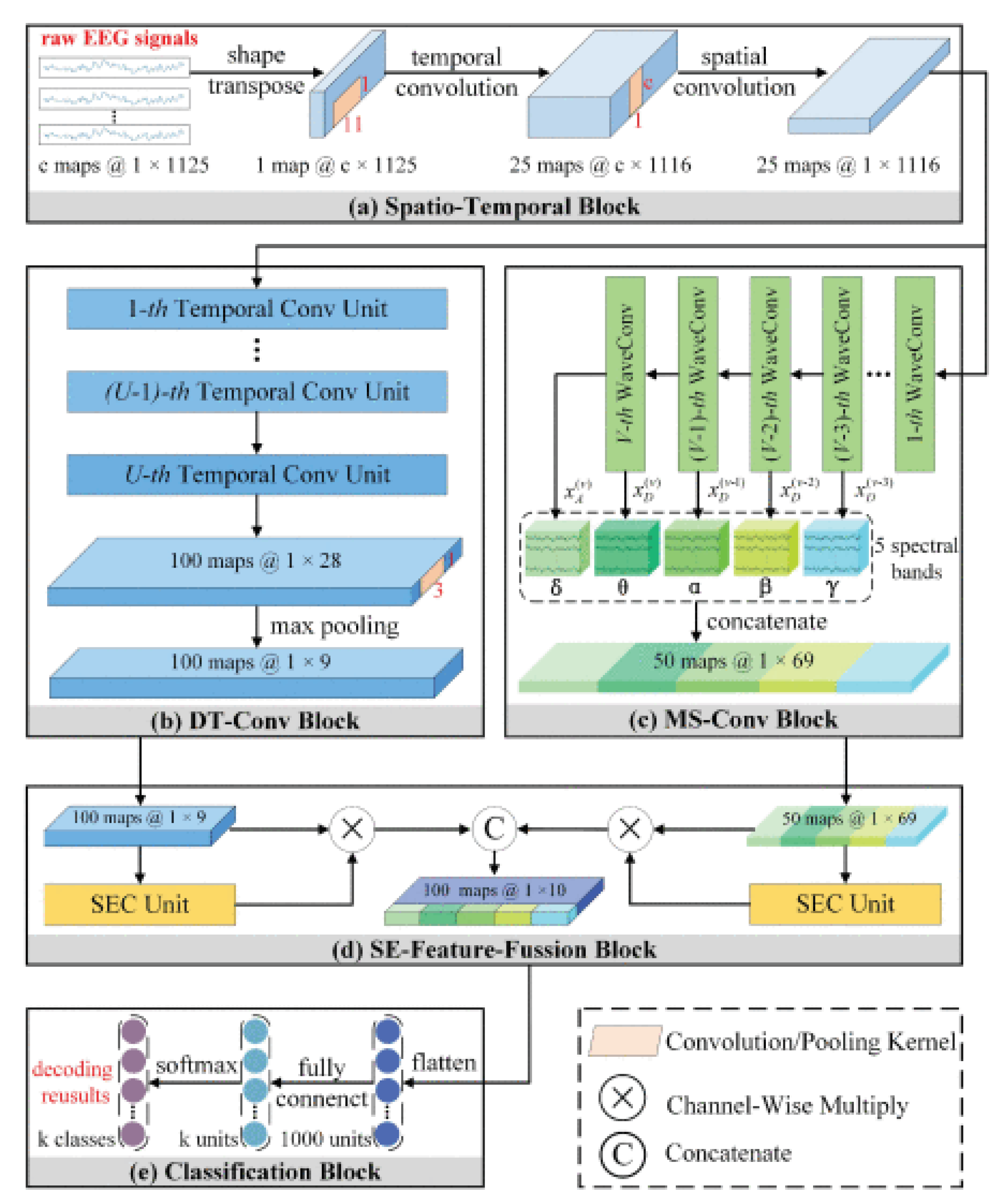
| 4 | Motor Imagery EEG Decoding | BCI Competition 2008 IV 2a Dataset High Gamma Dataset: (HGD) | TS-SEFFNet | Supervised | First, the deep temporal convolution block (DT-Conv block). Second, multispectral convolution block (MS-Conv block) is then run in parallel using multilevel wavelet convolutions. Finally, block (SE-Feature-Fusion block) displays the depth-time and multispectral features into complex pooled feature maps that extract the feature responses across channels. | DT-Conv block, MS-Conv block, SE-Feature-Fusion block | The Optimization Steps of the Proposed TS- SEFFNet Method | Softmax | Custom loss function | 93.25% | Torch 1.4, Python 3.8 | [217] | |
| 5 | Sleep stage annotations | Physionet Challenge dataset | Self-supervised learning (SSL) | Unsupervised | The first step is a sampling process by which examples are extracted from the time series S (EEG recording). The following describes a learning process where sample examples are used to train the feature extractor end-to-end. | Relative positioning (RP). Temporal shuffing (TS), Contrastive predective coding (CPC) | Adam optimizer | Rectified Linear Unit (ReLU) | Cross-entropy loss function | 72.3% | Torch 1.4, Python 3.9 | [218] | |
| 6 | Prediction task | EEG Imaginary Speech Recognition | Kara One database | - | Supervised | a CNN containing two convolutional layers with 64 and 128 filters connected to a dense layer with 64 neurons for input signal spectrum of a 0.25 s window | 2D CNN | Adam optimizer | Linear | Categorical cross-entropy | 37% | - | [257] |
| 7 | EEG-Speech Recognition | Custom dataset (not available) | - | Supervised | ResNet18/50/101 with 2 layers of managed recurrent units—Gated Recurrent Unit (GRU). And after that ResNet18 operation are fed to the input of a recurrent neural network containing 1024 hidden GRUs. | CNN and RNN | Adam optimizer | Softmax | - | 85% | - | [258] | |
| 8 | EEG speech recognition | Custom dataset (not available) | - | Supervised | The architecture used includes the already trained VGG Net CNN design and the target CNN design, while the already trained VGG Net CNN design extracts global features for general image classification work, and the target CNN design aims at efficient and accurate categorization of EEG signals using already trained Model VGG Net CNN. | Deep Residual–encoder–based VGG net CNN | - | Softmax | Softmax cross-entropy | 95% | - | [259] | |
| 9 | Seizure prediction | CHB-MIT and Kaggle | - | Supervised | a hybrid network that can combine the additional benefits of CNN and Transformer. The CNN is used to extract local information that contains two 3 × 3 convolutions with stride 1 and another 3 × 3 convolution with stride 2 to reduce the size of the input features. Each convolutional layer is followed by a GELU activation and a batch normalization (BN) layer. The model has two stages for extracting multiscale features from the EEG spectrum. Each stage consists of a set of Transformer blocks applied to extract long-term dependencies. | CNN and transformer | Adam | Softmax | Cross-entropy | 95% | Torch 1.4, Python 3.8 | [260] | |
| 10 | Predicting Human Intention-Behavior | BCI competition IV Dataset 2b | - | Supervised | The multi-scale CNN model has seven layers, which are one input layer, two convolutional layers, one max-pooling layer, one multi-scale layer, one full connection layer and one softmax output layer. The input layer in the multi-scale CNN model is fed with a time-frequency image with the size of 40 × 32 × 3 after EEG signals are preprocessed by STFT | Multi-Scale CNN Model | - | Linear | Cross-entropy | 73.9% | Python 3.8, Keras 3 | [261] | |
| 11 | Artifact Removal | EEG Artifact Detection and Correction | Costum dataset, not available | - | Unsupervised | modification of a feed-forward neural network that uses weight sharing and exhibits translation invariance. Learning in the CNNs operates on the same principle as a traditional feed-forward neural network where an error from output layer is back-propagated through the network and weights of the network are proportionally updated to the gradient of error. | CNN | Adam | - | Cross-entropy | - | Python 3.9, Keras 3 | [262] |
| 12 | Remove Muscle Artifacts from EEG | EEGdenoiseNet | - | Supervised | CNN for myogenic artifact reduction contains seven similar blocks. In each of the first six blocks, two 1D-convolution layers with small 1*3 kernels, 1 stride, and a ReLU activation function are followed by a 1D-Average pooling layer with pool size equal to two. In the seventh block, two 1D-convolution layers are followed by a flatten layer.The network gradually reduce the EEG signal sampling rate by the 1D-Average pooling layer. | CNN | RMSprop | ReLU | mean squared error (MSE) | - | Python 3.10, Tensorflow 2.8 | [263] | |
| 13 | Denoise EEG signal from artifacts | EEGdenoiseNet | MultiResUNet3+ | Supervised | Net3+ consists of full-blown pass-through connections that aggregate connections between encoders and decoders and internal connections between decoder subnets. Instead of directly combining the encoder and decoder functions, the encoder functions go through several convolutional levels with residual connections | CNN, encoders | Adam | Rely | mean squared error (MSE) | - | - | [152] | |
References
- Blanc, Y.; Dimanico, U. History of the Study of Skeletal Muscle Function with Emphasis on Kinesiological Electromyography. Open Rehabil. J. 2010, 3, 84–93. [Google Scholar] [CrossRef]
- Britton, J.W.; Frey, L.C.; Hopp, J.L.; Korb, P.; Koubeissi, M.Z.; Lievens, W.E.; Pestana-Knight, E.M.; St Louis, E.K. Electroencephalography (EEG): An Introductory Text and Atlas of Normal and Abnormal Findings in Adults, Children, and Infants. Am. Epilepsy Soc. 2016, 4. [Google Scholar]
- Nidal, K.; Malik, A. EEG/ERP Analysis: Methods and Applications; CRC Press: Boca Raton, FL, USA, 2014. [Google Scholar]
- Winkler, I.; Debener, S.; Muller, K.-R.; Tangermann, M. On the influence of high-pass filtering on ICA-based artifact reduction in EEG-ERP. In Proceedings of the 2015 37th Annual International Conference of the IEEE Engineering in Medicine and Biology Society (EMBC), Milan, Italy, 25–29 August 2015; pp. 4101–4105. [Google Scholar]
- Wang, Y.; Wang, Z.; Clifford, W.; Markham, C.; Ward, T.E.; Deegan, C. Validation of low-cost wireless EEG system for measuring event-related potentials. In Proceedings of the 2018 29th Irish Signals and Systems Conference (ISSC), Belfast, UK, 21–22 June 2018; pp. 1–6. [Google Scholar]
- Thompson, T.; Steffert, T.; Ros, T.; Leach, J.; Gruzelier, J. EEG applications for sport and performance. Methods 2008, 45, 279–288. [Google Scholar] [CrossRef]
- Armitage, R.; Hoffmann, R.F. Sleep EEG, depression and gender. Sleep Med. Rev. 2001, 5, 237–246. [Google Scholar] [CrossRef] [PubMed]
- Masood, N.; Farooq, H. Investigating EEG Patterns for Dual-Stimuli Induced Human Fear Emotional State. Sensors 2019, 19, 522. [Google Scholar] [CrossRef] [PubMed]
- Al-Quraishi, M.S.; Elamvazuthi, I.; Daud, S.A.; Parasuraman, S.; Borboni, A. EEG-Based Control for Upper and Lower Limb Exoskeletons and Prostheses: A Systematic Review. Sensors 2018, 18, 3342. [Google Scholar] [CrossRef] [PubMed]
- Vasiljevic, G.A.M.; de Miranda, L.C. Brain–Computer Interface Games Based on Consumer-Grade EEG Devices: A Systematic Literature Review. Int. J. Hum. Comput. Interact. 2019, 36, 105–142. [Google Scholar] [CrossRef]
- Zhang, Q.; Guo, B.; Kong, W.; Xi, X.; Zhou, Y.; Gao, F. Tensor-based dynamic brain functional network for motor imagery classification. Biomed. Signal Process. Control 2021, 69, 102940. [Google Scholar] [CrossRef]
- Sanchez-Reyes, L.-M.; Rodriguez-Resendiz, J.; Avecilla-Ramirez, G.N.; Garcia-Gomar, M.-L.; Robles-Ocampo, J.-B. Impact of EEG Parameters Detecting Dementia Diseases: A Systematic Review. IEEE Access 2021, 9, 78060–78074. [Google Scholar] [CrossRef]
- Tao, D.; Tan, H.; Wang, H.; Zhang, X.; Qu, X.; Zhang, T. A Systematic Review of Physiological Measures of Mental Workload. Int. J. Environ. Res. Public Health. 2019, 16, 2716. [Google Scholar] [CrossRef]
- Stocker, R.A. Intensive Care in Traumatic Brain Injury Including Multi-Modal Monitoring and Neuroprotection. Med. Sci. 2019, 7, 37. [Google Scholar] [CrossRef]
- Ang, K.K.; Guan, C. EEG-Based Strategies to Detect Motor Imagery for Control and Rehabilitation. IEEE Trans. Neural Syst. Rehabil. Eng. 2016, 25, 392–401. [Google Scholar] [CrossRef]
- Lotte, F.; Congedo, M.; Lécuyer, A.; Lamarche, F.; Arnaldi, B. A review of classification algorithms for EEG-based brain–computer interfaces. J. Neural Eng. 2007, 4, R1–R13. [Google Scholar] [CrossRef]
- Hori, T.; Wolpert, M.D. A Manual of Standardized Terminology, Techniques and Scoring System for Sleep Stages of Human Subjects’, the Rechtschaffen & Kales (1968) standard. Psychiatry Clin. Neurosci. 2001, 55, 305–310. [Google Scholar] [CrossRef]
- Carley, D.W.; Farabi, S.S. Physiology of Sleep. Diabetes Spectr. 2016, 29, 5–9. [Google Scholar] [CrossRef]
- Miah, O. Prediction of Motor Imagery Tasks from Multi-Channel EEG Data for Brain-Computer Interface Applications. bioRxiv 2020. [Google Scholar] [CrossRef]
- Pereda, E.; Gamundi, A.; Rial, R.; González, J. Non-linear behaviour of human EEG: Fractal exponent versus correlation dimension in awake and sleep stages. Neurosci. Lett. 1998, 250, 91–94. [Google Scholar] [CrossRef] [PubMed]
- Elger, C.E.; Widman, G.; Andrzejak, R.; Arnhold, J.; David, P.; Lehnertz, K. Nonlinear EEG Analysis and Its Potential Role in Epileptology. Epilepsia 2000, 41, S34–S38. [Google Scholar] [CrossRef] [PubMed]
- He, B.; Ding, L. Electrophysiological Mapping and Neuroimaging. In Neural Engineering; He, B., Ed.; Springer: Boston, MA, USA, 2013. [Google Scholar] [CrossRef]
- Riera, J.J.; Ogawa, T.; Goto, T.; Sumiyoshi, A.; Nonaka, H.; Evans, A.; Miyakawa, H.; Kawashima, R.; Halnes, G.; Mäki-Marttunen, T.; et al. Pitfalls in the dipolar model for the neocortical EEG sources. J. Neurophysiol. 2012, 108, 956–975. [Google Scholar] [CrossRef] [PubMed]
- Rai, P.; Oh, S.; Shyamkumar, P.; Ramasamy, M.; Harbaugh, R.E.; Varadan, V.K. Nano- Bio- Textile Sensors with Mobile Wireless Platform for Wearable Health Monitoring of Neurological and Cardiovascular Disorders. J. Electrochem. Soc. 2013, 161, B3116–B3150. [Google Scholar] [CrossRef]
- Sridhar, C.; Lih, O.S.; Jahmunah, V.; Koh, J.E.W.; Ciaccio, E.J.; San, T.R.; Arunkumar, N.; Kadry, S.; Acharya, U.R. Accurate detection of myocardial infarction using non linear features with ECG signals. J. Ambient. Intell. Humaniz. Comput. 2021, 12, 3227–3244. [Google Scholar] [CrossRef]
- Correia-Silva, J.R.; Berriel, R.F.; Badue, C.; De Souza, A.F.; Oliveira-Santos, T. Copycat CNN: Are random non-Labeled data enough to steal knowledge from black-box models? Pattern Recognit. 2021, 113, 107830. [Google Scholar] [CrossRef]
- Wang, B.; Ma, R.; Kuang, J.; Zhang, Y. How Decisions Are Made in Brains: Unpack “Black Box” of CNN With Ms. Pac-Man Video Game. IEEE Access 2020, 8, 142446–142458. [Google Scholar] [CrossRef]
- Ferrone, E.; Araneo, R.; Notargiacomo, A.; Pea, M.; Rinaldi, A. ZnO Nanostructures and Electrospun ZnO-Polymeric Hybrid Nanomaterials in Biomedical, Health, and Sustainability Applications. Nanomaterials 2019, 9, 1449. [Google Scholar] [CrossRef]
- Duan, L.; Ge, H.; Ma, W.; Miao, J. EEG feature selection method based on decision tree. Bio-Med. Mater. Eng. 2015, 26, S1019–S1025. [Google Scholar] [CrossRef] [PubMed]
- Li, Z.; Liu, F.; Yang, W.; Peng, S.; Zhou, J. A Survey of Convolutional Neural Networks: Analysis, Applications, and Prospects. IEEE Trans. Neural Networks Learn. Syst. 2021, 33, 6999–7019. [Google Scholar] [CrossRef]
- Tirumala, S.S.; Ali, S.; Ramesh, C.P. Evolving deep neural networks: A new prospect. In Proceedings of the 2016 12th International Conference on Natural Computation and 13th Fuzzy Systems and Knowledge Discovery (ICNC-FSKD), Changsha, China, 13–15 August 2016; pp. 69–74. [Google Scholar]
- Jordan, M.I.; Mitchell, T.M. Machine learning: Trends, perspectives, and prospects. Science 2015, 349, 255–260. [Google Scholar] [CrossRef]
- Guler, I.; Ubeyli, E.D. Multiclass Support Vector Machines for EEG-Signals Classification. IEEE Trans. Inf. Technol. Biomed. 2007, 11, 117–126. [Google Scholar] [CrossRef]
- Panda, R.; Khobragade, P.S.; Jambhule, P.D.; Jengthe, S.N.; Pal, P.; Gandhi, T.K. Classification of EEG signal using wavelet transform and support vector machine for epileptic seizure diction. In Proceedings of the 2010 International Conference on Systems in Medicine and Biology, Kharagpur, India, 16–18 December 2010; pp. 405–408. [Google Scholar] [CrossRef]
- Valueva, M.; Nagornov, N.; Lyakhov, P.; Valuev, G.; Chervyakov, N. Application of the residue number system to reduce hardware costs of the convolutional neural network. Math. Comput. Simul. 2020, 177, 232–243. [Google Scholar] [CrossRef]
- Albawi, S.; Mohammed, T.A.; Al-Zawi, S. Understanding of a convolutional neural network. In Proceedings of the International Conference on Engineering and Technology (ICET), Antalya, Turkey, 21–23 August 2017; pp. 1–6. [Google Scholar] [CrossRef]
- Indolia, S.; Goswami, A.K.; Mishra, S.; Asopa, P. Conceptual Understanding of Convolutional Neural Network- A Deep Learning Approach. Procedia Comput. Sci. 2018, 132, 679–688. [Google Scholar] [CrossRef]
- Sarvamangala, D.R.; Kulkarni, R.V. Convolutional neural networks in medical image understanding: A survey. Evol. Intell. 2022, 15, 1–22. [Google Scholar] [CrossRef] [PubMed]
- Islam, J.; Zhang, Y. Understanding 3D CNN Behavior for Alzheimer’s Disease Diagnosis from Brain PET Scan. arXiv 2019, arXiv:1912.04563. [Google Scholar] [CrossRef]
- Li, Y.-J.; Fan, F.-Y. Classification of Schizophrenia and Depression by EEG with ANNs. In Proceedings of the 2005 IEEE Engineering in Medicine and Biology 27th Annual Conference, Shanghai, China, 17–18 January 2006; pp. 2679–2682. [Google Scholar] [CrossRef]
- Srinivasan, V.; Eswaran, C.; Sriraam, N. Approximate Entropy-Based Epileptic EEG Detection Using Artificial Neural Networks. IEEE Trans. Inf. Technol. Biomed. 2007, 11, 288–295. [Google Scholar] [CrossRef] [PubMed]
- Jeong, J.-H.; Yu, B.-W.; Lee, D.-H.; Lee, S.-W. Classification of Drowsiness Levels Based on a Deep Spatio-Temporal Convolutional Bidirectional LSTM Network Using Electroencephalography Signals. Brain Sci. 2019, 9, 348. [Google Scholar] [CrossRef] [PubMed]
- Sharma, M.; Tiwari, J.; Acharya, U.R. Automatic Sleep-Stage Scoring in Healthy and Sleep Disorder Patients Using Optimal Wavelet Filter Bank Technique with EEG Signals. Int. J. Environ. Res. Public Heal. 2021, 18, 3087. [Google Scholar] [CrossRef] [PubMed]
- Lotte, F.; Bougrain, L.; Cichocki, A.; Clerc, M.; Congedo, M.; Rakotomamonjy, A.; Yger, F. A review of classification algorithms for EEG-based brain–computer interfaces: A 10 year update. J. Neural Eng. 2018, 15, 031005. [Google Scholar] [CrossRef]
- Maitin, A.M.; Muñoz, J.P.R.; García-Tejedor, A.J. Survey of Machine Learning Techniques in the Analysis of EEG Signals for Parkinson’s Disease: A Systematic Review. Appl. Sci. 2022, 12, 6967. [Google Scholar] [CrossRef]
- Rodrigues, J.d.C.; Filho, P.P.R.; Peixoto, E.; Arun, K.N.; de Albuquerque, V.H.C. Classification of EEG signals to detect alcoholism using machine learning techniques. Pattern Recognit. Lett. 2019, 125, 140–149. [Google Scholar] [CrossRef]
- Rasheed, K.; Qayyum, A.; Qadir, J.; Sivathamboo, S.; Kwan, P.; Kuhlmann, L.; O’Brien, T.; Razi, A. Machine Learning for Predicting Epileptic Seizures Using EEG Signals: A Review. IEEE Rev. Biomed. Eng. 2021, 14, 139–155. [Google Scholar] [CrossRef]
- Gemein, L.A.; Schirrmeister, R.T.; Chrabąszcz, P.; Wilson, D.; Boedecker, J.; Schulze-Bonhage, A.; Hutter, F.; Ball, T. Machine-learning-based diagnostics of EEG pathology. NeuroImage 2020, 220, 117021. [Google Scholar] [CrossRef]
- Bazgir, O.; Mohammadi, Z.; Habibi, S.A.H. Emotion Recognition with Machine Learning Using EEG Signals. In Proceedings of the 2018 25th National and 3rd International Iranian Conference on Biomedical Engineering (ICBME), Qom, Iran, 29–30 November 2018; pp. 1–5. [Google Scholar] [CrossRef]
- Wang, X.-W.; Nie, D.; Lu, B.-L. Emotional state classification from EEG data using machine learning approach. Neurocomputing 2013, 129, 94–106. [Google Scholar] [CrossRef]
- Nedelcu, E.; Portase, R.; Tolas, R.; Muresan, R.; Dinsoreanu, M.; Potolea, R. Artifact detection in EEG using machine learning. In Proceedings of the 13th IEEE International Conference on Intelligent Computer Communication and Processing (ICCP), Cluj-Napoca, Romania, 7–9 September 2017; pp. 77–83. [Google Scholar]
- Aggarwal, S.; Chugh, N. Review of Machine Learning Techniques for EEG Based Brain Computer Interface. Arch. Comput. Methods Eng. 2022, 29, 3001–3020. [Google Scholar] [CrossRef]
- Müller, K.-R.; Tangermann, M.; Dornhege, G.; Krauledat, M.; Curio, G.; Blankertz, B. Machine learning for real-time single-trial EEG-analysis: From brain–computer interfacing to mental state monitoring. J. Neurosci. Methods 2008, 167, 82–90. [Google Scholar] [CrossRef]
- Hosseini, M.-P.; Hosseini, A.; Ahi, K. A Review on Machine Learning for EEG Signal Processing in Bioengineering. IEEE Rev. Biomed. Eng. 2021, 14, 204–218. [Google Scholar] [CrossRef]
- Wu, W.; Nagarajan, S.; Chen, Z. Bayesian Machine Learning: EEG\/MEG signal processing measurements. IEEE Signal Process. Mag. 2015, 33, 14–36. [Google Scholar] [CrossRef]
- Fernández-Varela, I.; Hernández-Pereira, E.; Álvarez-Estévez, D.; Moret-Bonillo, V. Combining machine learning models for the automatic detection of EEG arousals. Neurocomputing 2017, 268, 100–108. [Google Scholar] [CrossRef]
- Saeidi, M.; Karwowski, W.; Farahani, F.V.; Fiok, K.; Taiar, R.; Hancock, P.A.; Al-Juaid, A. Neural Decoding of EEG Signals with Machine Learning: A Systematic Review. Brain Sci. 2021, 11, 1525. [Google Scholar] [CrossRef] [PubMed]
- Roman-Gonzalez, A. EEG Signal Processing for BCI Applications. Human-Computer Systems Interaction: Backgrounds and Applications 2. In Advances in Intelligent and Soft Computing; Hippe, Z.S., Kulikowski, J.L., Mroczek, T., Eds.; Springer: Berlin/Heidelberg, Germany, 2012; Volume 98, pp. 571–591. [Google Scholar] [CrossRef]
- Shedeed, H.A.; Issa, M.F.; El-Sayed, S.M. Brain EEG signal processing for controlling a robotic arm. In Proceedings of the 2013 8th International Conference on Computer Engineering & Systems (ICCES), Cairo, Egypt, 26–28 November 2013; pp. 152–157. [Google Scholar] [CrossRef]
- Kawala-Sterniuk, A.; Browarska, N.; Al-Bakri, A.; Pelc, M.; Zygarlicki, J.; Sidikova, M.; Martinek, R.; Gorzelanczyk, E.J. Summary of over Fifty Years with Brain-Computer Interfaces—A Review. Brain Sci. 2021, 11, 43. [Google Scholar] [CrossRef] [PubMed]
- Rakhmatulin, I.; Parfenov, A.; Traylor, Z.; Nam, C.S.; Lebedev, M. Low-cost brain computer interface for everyday use. Exp. Brain Res. 2021, 239, 3573–3583. [Google Scholar] [CrossRef] [PubMed]
- Baek, H. Ergonomic Issues in Brain-Computer Interface Technologies: Current Status, Challenges, and Future Direction. Comput. Intell. Neurosci. 2019, 2020, 5427154. [Google Scholar] [CrossRef] [PubMed]
- Occhipinti, E.; Davies, H.J.; Hammour, G.; Mandic, D.P. Hearables: Artefact removal in Ear-EEG for continuous 24/7 monitoring. In Proceedings of the 2022 International Joint Conference on Neural Networks (IJCNN), Padua, Italy, 18–23 July 2022; pp. 1–6. [Google Scholar] [CrossRef]
- Rakhmatulin, I. The electronic board to replace the reference voltage on the earlobe for EEG measurement. Measurement 2021, 173, 108673. [Google Scholar] [CrossRef]
- Pijn, J.P.; Van Neerven, J.; Noest, A.; da Silva, F.H.L. Chaos or noise in EEG signals; dependence on state and brain site. Electroencephalogr. Clin. Neurophysiol. 1991, 79, 371–381. [Google Scholar] [CrossRef] [PubMed]
- Cohen, M.X. Where Does EEG Come from and What Does It Mean? Trends Neurosci. 2017, 40, 208–218. [Google Scholar] [CrossRef] [PubMed]
- Bell, G.B.; Marino, A.A.; Chesson, A.L. Frequency-specific responses in the human brain caused by electromagnetic fields. J. Neurol. Sci. 1994, 123, 26–32. [Google Scholar] [CrossRef] [PubMed]
- Kottaimalai, R.; Rajasekaran, M.P.; Selvam, V.; Kannapiran, B. EEG signal classification using Principal Component Analysis with Neural Network in Brain Computer Interface applications. In Proceedings of the 2013 International Conference on Emerging Trends in Computing, Communication and Nanotechnology (ICE-CCN), Tirunelveli, India, 25–26 March 2013; pp. 227–231. [Google Scholar] [CrossRef]
- Rivet, B.; Souloumiac, A.; Attina, V.; Gibert, G. xDAWN Algorithm to Enhance Evoked Potentials: Application to Brain–Computer Interface. IEEE Trans. Biomed. Eng. 2009, 56, 2035–2043. [Google Scholar] [CrossRef]
- Mumtaz, W.; Rasheed, S.; Irfan, A. Review of challenges associated with the EEG artifact removal methods. Biomed. Signal Process. Control. 2021, 68, 102741. [Google Scholar] [CrossRef]
- Yasoda, K.; Ponmagal, R.S.; Bhuvaneshwari, K.S.; Venkatachalam, K. Automatic detection and classification of EEG artifacts using fuzzy kernel SVM and wavelet ICA (WICA). Soft Comput. 2020, 24, 16011–16019. [Google Scholar] [CrossRef]
- Shao, S.-Y.; Shen, K.-Q.; Ong, C.J.; Wilder-Smith, E.P.V.; Li, X.-P. Automatic EEG Artifact Removal: A Weighted Support Vector Machine Approach with Error Correction. IEEE Trans. Biomed. Eng. 2009, 56, 336–344. [Google Scholar] [CrossRef]
- Kaczorowska, M.; Plechawska-Wojcik, M.; Tokovarov, M.; Dmytruk, R. Comparison of the ICA and PCA methods in correction of EEG signal artefacts. In Proceedings of the 2017 10th International Symposium on Advanced Topics in Electrical Engineering (ATEE), Bucharest, Romania, 23–25 March 2017; pp. 262–267. [Google Scholar] [CrossRef]
- Hamaneh, M.B.; Chitravas, N.; Kaiboriboon, K.; Lhatoo, S.D.; Loparo, K.A. Automated Removal of EKG Artifact from EEG Data Using Independent Component Analysis and Continuous Wavelet Transformation. IEEE Trans. Biomed. Eng. 2014, 61, 1634–1641. [Google Scholar] [CrossRef]
- Chang, C.-Y.; Hsu, S.-H.; Pion-Tonachini, L.; Jung, T.-P. Evaluation of Artifact Subspace Reconstruction for Automatic EEG Artifact Removal. In Proceedings of the 2018 40th Annual International Conference of the IEEE Engineering in Medicine and Biology Society (EMBC), Honolulu, HI, USA, 18–21 July 2018; pp. 1242–1245. [Google Scholar] [CrossRef]
- Jiang, X.; Bian, G.-B.; Tian, Z. Removal of Artifacts from EEG Signals: A Review. Sensors 2019, 19, 987. [Google Scholar] [CrossRef]
- Kang, G.; Jin, S.-H.; Kim, D.K.; Kang, S.W. EEG artifacts removal using machine learning algorithms and independent component analysis. Clin. Neurophysiol. 2018, 129, e24. [Google Scholar] [CrossRef]
- Stalin, S.; Roy, V.; Shukla, P.K.; Zaguia, A.; Khan, M.M.; Shukla, P.K.; Jain, A. A Machine Learning-Based Big EEG Data Artifact Detection and Wavelet-Based Removal: An Empirical Approach. Math. Probl. Eng. 2021, 2021, 2942808. [Google Scholar] [CrossRef]
- Sun, W.; Su, Y.; Wu, X.; Wu, X. A novel end-to-end 1D-ResCNN model to remove artifact from EEG signals. Neurocomputing 2020, 404, 108–121. [Google Scholar] [CrossRef]
- Yang, B.; Duan, K.; Fan, C.; Hu, C.; Wang, J. Automatic ocular artifacts removal in EEG using deep learning. Biomed. Signal Process. Control 2018, 43, 148–158. [Google Scholar] [CrossRef]
- Zhang, H.; Zhao, M.; Wei, C.; Mantini, D.; Li, Z.; Liu, Q. EEGdenoiseNet: A benchmark dataset for deep learning solutions of EEG denoising. J. Neural Eng. 2021, 18, 056057. [Google Scholar] [CrossRef]
- Mashhadi, N.; Khuzani, A.Z.; Heidari, M.; Khaledyan, D. Deep learning denoising for EOG artifacts removal from EEG signals. In Proceedings of the 2020 IEEE Global Humanitarian Technology Conference (GHTC), Seattle, WA, USA, 29 October–1 November 2020; pp. 1–6. [Google Scholar] [CrossRef]
- Ronneberger, O.; Fischer, P.; Brox, T. U-Net: Convolutional Networks for Biomedical Image Segmentation. In Medical Image Computing and Computer-Assisted Intervention—MICCAI 2015. MICCAI 2015. Lecture Notes in Computer Science; Navab, N., Hornegger, J., Wells, W., Frangi, A., Eds.; Springer: Cham, Switzerland, 2015; Volume 9351. [Google Scholar] [CrossRef]
- Jaiswal, A.; Nenonen, J.; Parkkonen, L. On electromagnetic head digitization in MEG and EEG. Sci. Rep. 2023, 13, 3801. [Google Scholar] [CrossRef]
- Goyal, K.; Borkholder, D.A.; Day, S.W. Dependence of Skin-Electrode Contact Impedance on Material and Skin Hydration. Sensors 2022, 22, 8510. [Google Scholar] [CrossRef]
- Grobbelaar, M.; Phadikar, S.; Ghaderpour, E.; Struck, A.F.; Sinha, N.; Ghosh, R.; Ahmed, M.Z.I. A Survey on Denoising Techniques of Electroencephalogram Signals Using Wavelet Transform. Signals 2022, 3, 577–586. [Google Scholar] [CrossRef]
- Brouwer, A.-M.; Hogervorst, M.A.; van Erp, J.B.F.; Heffelaar, T.; Zimmerman, P.H.; Oostenveld, R. Estimating workload using EEG spectral power and ERPs in the n-back task. J. Neural Eng. 2012, 9, 045008. [Google Scholar] [CrossRef] [PubMed]
- Vanegas, M.I.; Ghilardi, M.F.; Kelly, S.P.; Blangero, A. Machine learning for EEG-based biomarkers in Parkinson’s disease. In Proceedings of the 2018 IEEE International Conference on Bioinformatics and Biomedicine (BIBM), Madrid, Spain, 3–6 December 2018; pp. 2661–2665. [Google Scholar] [CrossRef]
- Orban, M.; Elsamanty, M.; Guo, K.; Zhang, S.; Yang, H. A Review of Brain Activity and EEG-Based Brain–Computer Interfaces for Rehabilitation Application. Bioengineering 2022, 9, 768. [Google Scholar] [CrossRef]
- Sun, J.; Hong, X.; Tong, S. Phase Synchronization Analysis of EEG Signals: An Evaluation Based on Surrogate Tests. IEEE Trans. Biomed. Eng. 2012, 59, 2254–2263. [Google Scholar] [CrossRef]
- Roach, B.J.; Mathalon, D.H. Event-Related EEG Time-Frequency Analysis: An Overview of Measures and An Analysis of Early Gamma Band Phase Locking in Schizophrenia. Schizophr. Bull. 2008, 34, 907–926. [Google Scholar] [CrossRef] [PubMed]
- Shaw, J.C. An introduction to the coherence function and its use in EEG signal analysis. J. Med Eng. Technol. 1981, 5, 279–288. [Google Scholar] [CrossRef] [PubMed]
- Wang, D.; Ren, D.; Li, K.; Feng, Y.; Ma, D.; Yan, X.; Wang, G. Epileptic Seizure Detection in Long-Term EEG Recordings by Using Wavelet-Based Directed Transfer Function. IEEE Trans. Biomed. Eng. 2018, 65, 2591–2599. [Google Scholar] [CrossRef]
- La Rocca, D.; Campisi, P.; Vegso, B.; Cserti, P.; Kozmann, G.; Babiloni, F.; Fallani, F.D.V. Human Brain Distinctiveness Based on EEG Spectral Coherence Connectivity. IEEE Trans. Biomed. Eng. 2014, 61, 2406–2412. [Google Scholar] [CrossRef]
- Babiloni, F.; Cincotti, F.; Babiloni, C.; Carducci, F.; Mattia, D.; Astolfi, L.; Basilisco, A.; Rossini, P.; Ding, L.; Ni, Y.; et al. Estimation of the cortical functional connectivity with the multimodal integration of high-resolution EEG and fMRI data by directed transfer function. NeuroImage 2005, 24, 118–131. [Google Scholar] [CrossRef]
- Olias, J.; Martin-Clemente, R.; Sarmiento-Vega, M.A.; Cruces, S. EEG Signal Processing in MI-BCI Applications with Improved Covariance Matrix Estimators. IEEE Trans. Neural Syst. Rehabil. Eng. 2019, 27, 895–904. [Google Scholar] [CrossRef]
- Wang, F.; Wang, H.; Fu, R. Real-Time ECG-Based Detection of Fatigue Driving Using Sample Entropy. Entropy 2018, 20, 196. [Google Scholar] [CrossRef]
- Guan, S.; Zhao, K.; Yang, S. Motor Imagery EEG Classification Based on Decision Tree Framework and Riemannian Geometry. Brain-Inspired Intell. Syst. Dly. Assist. 2019, 2019, 5627156. [Google Scholar] [CrossRef]
- Yger, F.; Lotte, F.; Sugiyama, M. Averaging covariance matrices for EEG signal classification based on the CSP: An empirical study. In Proceedings of the 2015 23rd European Signal Processing Conference (EUSIPCO), Nice, France, 31 August–4 September 2015; pp. 2721–2725. [Google Scholar] [CrossRef]
- Van Vliet, M.; Salmelin, R. Post-hoc modification of linear models: Combining machine learning with domain information to make solid inferences from noisy data. NeuroImage 2019, 204, 116221. [Google Scholar] [CrossRef]
- Srinivasan, R.; Nunez, P.; Silberstein, R. Spatial filtering and neocortical dynamics: Estimates of EEG coherence. IEEE Trans. Biomed. Eng. 1998, 45, 814–826. [Google Scholar] [CrossRef]
- Kumar, S.; Sharma, A.; Mamun, K.; Tsunoda, T. A Deep Learning Approach for Motor Imagery EEG Signal Classification. In Proceedings of the 2016 3rd Asia-Pacific World Congress on Computer Science and Engineering (APWC on CSE), Nadi, Fiji, 5–6 December 2016; pp. 34–39. [Google Scholar] [CrossRef]
- Blankertz, B.; Muller, K.-R.; Krusienski, D.; Schalk, G.; Wolpaw, J.; Schlogl, A.; Pfurtscheller, G.; Millan, J.; Schroder, M.; Birbaumer, N. The BCI competition III: Validating alternative approaches to actual BCI problems. IEEE Trans. Neural Syst. Rehabil. Eng. 2006, 14, 153–159. [Google Scholar] [CrossRef]
- Tangermann, M.; Müller, K.-R.; Aertsen, A.; Birbaumer, N.; Braun, C.; Brunner, C.; Leeb, R.; Mehring, C.; Miller, K.J.; Müller-Putz, G.R.; et al. Review of the BCI Competition IV. Front. Neurosci. 2012, 6, 55. [Google Scholar] [CrossRef]
- Delorme, A.; Makeig, S. EEGLAB: An Open Source Toolbox for Analysis of Single-Trial EEG Dynamics Including Independent Component Analysis. J. Neurosci. Methods 2004, 134, 9–21. [Google Scholar] [CrossRef]
- Wu, D.; King, J.-T.; Chuang, C.-H.; Lin, C.-T.; Jung, T.-P. Spatial Filtering for EEG-Based Regression Problems in Brain–Computer Interface (BCI). arXiv 2017, arXiv:1702.02914. [Google Scholar] [CrossRef]
- Wang, Y.; Wang, Y.-T.; Jung, T.-P. Translation of EEG Spatial Filters from Resting to Motor Imagery Using Independent Component Analysis. PLoS ONE 2012, 7, e37665. [Google Scholar] [CrossRef]
- Ghasemzadeh, P.; Kalbkhani, H.; Shayesteh, M.G. Sleep stages classification from EEG signal based on Stockwell transform. IET Signal Process. 2019, 13, 242–252. [Google Scholar] [CrossRef]
- Yıldırım, Ö.; Baloglu, U.B.; Acharya, U.R. A deep convolutional neural network model for automated identification of abnormal EEG signals. Neural Comput. Appl. 2018, 32, 15857–15868. [Google Scholar] [CrossRef]
- Williamson, J.R.; Bliss, D.W.; Browne, D.W.; Narayanan, J.T. Seizure prediction using EEG spatiotemporal correlation structure. Epilepsy Behav. 2012, 25, 230–238. [Google Scholar] [CrossRef] [PubMed]
- Zhang, X.; Yao, L.; Zhang, D.; Wang, X.; Sheng, Q.Z.; Gu, T. Multi-Person Brain Activity Recognition via Comprehensive EEG Signal Analysis. In Proceedings of the 14th EAI International Conference on Mobile and Ubiquitous Systems: Computing, Networking and Services, Melbourne, Australia, 7–10 November 2017; pp. 28–37. [Google Scholar] [CrossRef]
- Amin, H.U.; Mumtaz, W.; Subhani, A.R.; Saad, M.N.M.; Malik, A.S. Classification of EEG Signals Based on Pattern Recognition Approach. METHODS article. Front. Comput. Neurosci. 2017, 11, 103. [Google Scholar] [CrossRef] [PubMed]
- Farzan, F.; Atluri, S.; Frehlich, M.; Dhami, P.; Kleffner, K.; Price, R.; Lam, R.W.; Frey, B.N.; Milev, R.; Ravindran, A.; et al. Standardization of electroencephalography for multi-site, multi-platform and multi-investigator studies: Insights from the canadian biomarker integration network in depression. Sci. Rep. 2017, 7, 7473. [Google Scholar] [CrossRef]
- Wang, J.; Wang, M. Review of the emotional feature extraction and classification using EEG signals. Cogn. Robot. 2021, 1, 29–40. [Google Scholar] [CrossRef]
- Fdez, J.; Guttenberg, N.; Witkowski, O.; Pasquali, A. Cross-Subject EEG-Based Emotion Recognition Through Neural Networks with Stratified Normalization. Front. Neurosci. Sec. Brain Imaging Methods 2021, 15, 626277. [Google Scholar] [CrossRef]
- Liu, M.; Wu, W.; Gu, Z.; Yu, Z.; Qi, F.; Li, Y. Deep learning based on Batch Normalization for P300 signal detection. Neurocomputing 2017, 275, 288–297. [Google Scholar] [CrossRef]
- Shih, M.-T.; Doctor, F.; Fan, S.-Z.; Jen, K.-K.; Shieh, J.-S. Instantaneous 3D EEG Signal Analysis Based on Empirical Mode Decomposition and the Hilbert–Huang Transform Applied to Depth of Anaesthesia. Entropy 2015, 17, 928–949. [Google Scholar] [CrossRef]
- Rakhmatulin, I. Review of EEG Feature Selection by Neural Networks. Int. J. Sci. Bus. 2020, 4, 101–112. [Google Scholar] [CrossRef]
- Alotaiby, T.; El-Samie, F.E.A.; A Alshebeili, S.; Ahmad, I. A review of channel selection algorithms for EEG signal processing. EURASIP J. Adv. Signal Process. 2015, 2015, 66. [Google Scholar] [CrossRef]
- Molla, M.K.I.; Ahamed, S.; Almassri, A.M.M.; Wagatsuma, H. Classification of Motor Imagery Using Trial Extension in Spatial Domain with Rhythmic Components of EEG. Mathematics 2023, 11, 3801. [Google Scholar] [CrossRef]
- Riyadi, M.A.; Setiawan, I.; Amir, A. EEG Multiclass Signal Classification Based on Subtractive Clustering-ANFIS and Wavelet Packet Decomposition. In Proceedings of the 2021 International Conference on Electrical and Information Technology (IEIT), Malang, Indonesia, 14–15 September 2021; pp. 81–86. [Google Scholar] [CrossRef]
- Boonyakitanont, P.; Lek-Uthai, A.; Chomtho, K.; Songsiri, J. A review of feature extraction and performance evaluation in epileptic seizure detection using EEG. Biomed. Signal Process. Control 2019, 57, 101702. [Google Scholar] [CrossRef]
- Svetlakov, M.; Kovalev, I.; Konev, A.; Kostyuchenko, E.; Mitsel, A. Representation Learning for EEG-Based Biometrics Using Hilbert–Huang Transform. Computers 2022, 11, 47. [Google Scholar] [CrossRef]
- Van Hal, B.; Rhodes, S.; Dunne, B.; Bossemeyer, R. Low-cost EEG-based sleep detection. In Proceedings of the 2014 36th Annual International Conference of the IEEE Engineering in Medicine and Biology Society (EMBC), Chicago, IL, USA, 26–30 August 2014; pp. 4571–4574. [Google Scholar] [CrossRef]
- Azarbad, M. A Time-Frequency approach for EEG signal segmentation. J. Artif. Intell. Data Min. 2014, 2, 63–71. [Google Scholar] [CrossRef]
- Birvinskas, D.; Jusas, V.; Martisius, V.; Damasevicius, E. EEG Dataset Reduction and Feature Extraction Using Discrete Cosine Transform. In Proceedings of the Sixth UKSim/AMSS European Symposium on Computer Modeling and Simulation, Malta, Malta, 14–16 November 2012; pp. 199–204. [Google Scholar] [CrossRef]
- Lan, Z.; Liu, Y.; Sourina, O.; Wang, L.; Scherer, R.; Müller-Putz, G. SAFE: An EEG dataset for stable affective feature selection. Adv. Eng. Inform. 2020, 44, 101047. [Google Scholar] [CrossRef]
- Iancu, B.; Soloviev, V.; Zelioli, L.; Lilius, J. ABOships—An Inshore and Offshore Maritime Vessel Detection Dataset with Precise Annotations. Remote Sens. 2021, 13, 988. [Google Scholar] [CrossRef]
- Blankertz, B.; Curio, G.; Müller, K. Classifying Single Trial EEG: Towards Brain Computer Interfacing. In Proceedings of the Advances in Neural Information Processing Systems 14 (NIPS 01), Vancouver, BC, Canada, 3–8 December 2001; The MIT Press: Cambridge, MA, USA, 2002. [Google Scholar]
- Wolpaw, J.R.; Birbaumer, N.; McFarland, D.J.; Pfurtscheller, G.; Vaughan, T.M. Brain–computer interfaces for communication and control. Clin. Neurophysiol. 2002, 113, 767–791. [Google Scholar] [CrossRef] [PubMed]
- Dornhege, G.; Blankertz, B.; Curio, G.; Muller, K.-R. Boosting bit rates in noninvasive EEG single-trial classifications by feature combination and multiclass paradigms. IEEE Trans. Biomed. Eng. 2004, 51, 993–1002. [Google Scholar] [CrossRef]
- Cimtay, Y.; Ekmekcioglu, E. Investigating the Use of Pretrained Convolutional Neural Network on Cross-Subject and Cross-Dataset EEG Emotion Recognition. Sensors 2020, 20, 2034. [Google Scholar] [CrossRef]
- Bouallegue, G.; Djemal, R.; Belwafi, K. Artificial EEG signal generated by a network of neurons with one and two dendrites. Results Phys. 2020, 20, 103699. [Google Scholar] [CrossRef]
- Wan, Z.; Yang, R.; Huang, M.; Zeng, N.; Liu, X. A review on transfer learning in EEG signal analysis. Neurocomputing 2020, 421, 1–14. [Google Scholar] [CrossRef]
- Zhang, K.; Xu, G.; Zheng, X.; Li, H.; Zhang, S.; Yu, Y.; Liang, R. Application of Transfer Learning in EEG Decoding Based on Brain-Computer Interfaces: A Review. Sensors 2020, 20, 6321. [Google Scholar] [CrossRef]
- Montero Quispe, K.G.; Utyiama, D.M.S.; dos Santos, E.M.; Oliveira, H.A.B.F.; Souto, E.J.P. Applying Self-Supervised Representation Learning for Emotion Recognition Using Physiological Signals. Sensors 2022, 22, 9102. [Google Scholar] [CrossRef]
- Chato, L.; Regentova, E. Survey of Transfer Learning Approaches in the Machine Learning of Digital Health Sensing Data. J. Pers. Med. 2023, 13, 1703. [Google Scholar] [CrossRef]
- Malekzadeh, A.; Zare, A.; Yaghoobi, M.; Kobravi, H.-R.; Alizadehsani, R. Epileptic Seizures Detection in EEG Signals Using Fusion Handcrafted and Deep Learning Features. Sensors 2021, 21, 7710. [Google Scholar] [CrossRef]
- Xu, J.; Zheng, Y.; Mao, Y.; Wang, R.; Zheng, W.-S. Anomaly Detection on Electroencephalography with Self-supervised Learning. In Proceedings of the 2020 IEEE International Conference on Bioinformatics and Biomedicine (BIBM), Seoul, Republic of Korea, 16–19 December 2020; pp. 363–368. [Google Scholar] [CrossRef]
- Jiang, X.; Zhao, J.; Du, B.; Yuan, Z. Self-supervised Contrastive Learning for EEG-based Sleep Staging. In Proceedings of the 2021 International Joint Conference on Neural Networks (IJCNN), Shenzhen, China, 18–22 July 2021; pp. 1–8. [Google Scholar] [CrossRef]
- Yang, C.; Xiao, D.; Westover, M.B.; Sun, J. Self-supervised EEG Representation Learning for Automatic Sleep Staging. arXiv 2021, arXiv:2110.15278. [Google Scholar] [CrossRef]
- Xiao, Q.; Wang, J.; Ye, J.; Zhang, H.; Bu, Y.; Zhang, Y.; Wu, H. Self-Supervised Learning for Sleep Stage Classification with Predictive and Discriminative Contrastive Coding. In Proceedings of the ICASSP 2021—2021 IEEE International Conference on Acoustics, Speech and Signal Processing (ICASSP), Toronto, ON, Canada, 6–11 June 2021; pp. 1290–1294. [Google Scholar] [CrossRef]
- Zheng, Y.; Liu, Z.; Mo, R.; Chen, Z.; Zheng, W.S.; Wang, R. Task-Oriented Self-supervised Learning for Anomaly Detection in Electroencephalography. In Proceedings of the 25th International Conference on Medical Image Computing and Computer-Assisted Intervention—MICCAI 2022, Singapore, 18–22 September 2022; Wang, L., Dou, Q., Fletcher, P.T., Speidel, S., Li, S., Eds.; Lecture Notes in Computer Science; Springer: Cham, Switzerland, 2022; Volume 13438, pp. 193–203. [Google Scholar] [CrossRef]
- Rafiei, M.H.; Gauthier, L.V.; Adeli, H.; Takabi, D. Self-Supervised Learning for Electroencephalography. IEEE Trans. Neural Networks Learn. Syst. 2022, 2022, 3190448. [Google Scholar] [CrossRef]
- Banville, H.; Albuquerque, I.; Hyvärinen, A.; Moffat, G.; Engemann, D.-A.; Gramfort, A. Self-Supervised Representation Learning from Electroencephalography Signals. In Proceedings of the 2019 IEEE 29th International Workshop on Machine Learning for Signal Processing (MLSP), Pittsburgh, PA, USA, 13–16 October 2019; IEEE: Piscataway, NJ, USA, 2019; Volume 10, pp. 1–6. [Google Scholar] [CrossRef]
- Islam, T.; Washington, P. Individualized Stress Mobile Sensing Using Self-Supervised Pre-Training. Appl. Sci. 2023, 13, 12035. [Google Scholar] [CrossRef]
- Mattiev, J.; Sajovic, J.; Drevenšek, G.; Rogelj, P. Assessment of Model Accuracy in Eyes Open and Closed EEG Data: Effect of Data Pre-Processing and Validation Methods. Bioengineering 2023, 10, 42. [Google Scholar] [CrossRef]
- Kingphai, K.; Moshfeghi, Y. On Time Series Cross-Validation for Deep Learning Classification Model of Mental Workload Levels Based on EEG Signals. In Proceedings of the Machine Learning, Optimization, and Data Science, Certosa di Pontignano, Italy, 19–22 September 2022; Lecture Notes in Computer Science; Springer: Cham, Switzerland, 2023; Volume 13811, pp. 402–416. [Google Scholar] [CrossRef]
- King, R.D.; Orhobor, O.I.; Taylor, C.C. Cross-validation is safe to use. Nat. Mach. Intell. 2021, 3, 276. [Google Scholar] [CrossRef]
- Saqib, M.; Zhu, Y.; Wang, M.; Beaulieu-Jones, B. Regularization of Deep Neural Networks for EEG Seizure Detection to Mitigate Overfitting. In Proceedings of the 2020 IEEE 44th Annual Computers, Software, and Applications Conference (COMPSAC), Madrid, Spain, 13–17 July 2020; pp. 664–673. [Google Scholar] [CrossRef]
- Lashgari, E.; Liang, D.; Maoz, U. Data augmentation for deep-learning-based electroencephalography. J. Neurosci. Methods 2020, 346, 108885. [Google Scholar] [CrossRef]
- Zhang, H.; Wei, C.; Zhao, M.; Liu, Q.; Wu, H. A Novel Convolutional Neural Network Model to Remove Muscle Artifacts from EEG. In Proceedings of the ICASSP 2021—2021 IEEE International Conference on Acoustics, Speech and Signal Processing (ICASSP), Toronto, ON, Canada, 6–11 June 2021; pp. 1265–1269. [Google Scholar] [CrossRef]
- Zhang, Y.; Zhou, G.; Jin, J.; Zhao, Q.; Wang, X.; Cichocki, A. Sparse Bayesian Classification of EEG for Brain–Computer Interface. IEEE Trans. Neural Networks Learn. Syst. 2015, 27, 2256–2267. [Google Scholar] [CrossRef]
- Radüntz, T.; Scouten, J.; Hochmuth, O.; Meffert, B. Automated EEG artifact elimination by applying machine learning algorithms to ICA-based features. J. Neural Eng. 2017, 14, 046004. [Google Scholar] [CrossRef] [PubMed]
- Ying, X. An Overview of Overfitting and its Solutions. J. Phys. Conf. Ser. 2019, 1168, 022022. [Google Scholar] [CrossRef]
- Park, C.; Looney, D.; Kidmose, P.; Ungstrup, M.; Mandic, D.P. Time-Frequency Analysis of EEG Asymmetry Using Bivariate Empirical Mode Decomposition. IEEE Trans. Neural Syst. Rehabilitation Eng. 2011, 19, 366–373. [Google Scholar] [CrossRef] [PubMed]
- Lin, J.; Liu, D.; Yang, H.; Li, H.; Wu, F. Convolutional Neural Network-Based Block Up-Sampling for HEVC. IEEE Trans. Circuits Syst. Video Technol. 2019, 29, 3701–3715. [Google Scholar] [CrossRef]
- Pagnotta, M.F.; Plomp, G. Time-varying MVAR algorithms for directed connectivity analysis: Critical comparison in simulations and benchmark EEG data. PLoS ONE 2018, 13, e0198846. [Google Scholar] [CrossRef] [PubMed]
- Haufe, S.; Dähne, S.; Nikulin, V.V. Dimensionality reduction for the analysis of brain oscillations. NeuroImage 2014, 101, 583–597. [Google Scholar] [CrossRef]
- Artoni, F.; Delorme, A.; Makeig, S. Applying dimension reduction to EEG data by Principal Component Analysis reduces the quality of its subsequent Independent Component decomposition. NeuroImage 2018, 175, 176–187. [Google Scholar] [CrossRef] [PubMed]
- Lehmann, C.; Koenig, T.; Jelic, V.; Prichep, L.; John, R.E.; Wahlund, L.-O.; Dodge, Y.; Dierks, T. Application and comparison of classification algorithms for recognition of Alzheimer’s disease in electrical brain activity (EEG). J. Neurosci. Methods 2007, 161, 342–350. [Google Scholar] [CrossRef]
- Christie, S.A.; Hubbard, A.E.; Callcut, R.A.M.; Hameed, M.; Dissak-Delon, F.N.; Mekolo, D.; Saidou, A.; Mefire, A.C.; Nsongoo, P.; Dicker, R.A.; et al. Machine learning without borders? An adaptable tool to optimize mortality prediction in diverse clinical settings. J. Trauma Inj. Infect. Crit. Care 2018, 85, 921–927. [Google Scholar] [CrossRef]
- Roychowdhury, S.; Hollcraft, N.; Alessio, A.M. Blind analysis of CT image noise using residual denoised images. In Proceedings of the 2015 IEEE Nuclear Science Symposium and Medical Imaging Conference (NSS/MIC), San Diego, CA, USA, 31 October–7 November 2015; pp. 1–4. [Google Scholar] [CrossRef]
- Bakker, M.; Veldkamp, C.L.S.; Akker, O.R.v.D.; van Assen, M.A.L.M.; Crompvoets, E.; Ong, H.H.; Wicherts, J.M. Recommendations in pre-registrations and internal review board proposals promote formal power analyses but do not increase sample size. PLoS ONE 2020, 15, e0236079. [Google Scholar] [CrossRef]
- Hang, S.T.; Aono, M. Bi-linearly weighted fractional max pooling. Multimed. Tools Appl. 2017, 76, 22095–22117. [Google Scholar] [CrossRef]
- Zhao, W.; Du, S. Spectral–Spatial Feature Extraction for Hyperspectral Image Classification: A Dimension Reduction and Deep Learning Approach. IEEE Trans. Geosci. Remote Sens. 2016, 54, 4544–4554. [Google Scholar] [CrossRef]
- Krichen, M. Convolutional Neural Networks: A Survey. Computers 2023, 12, 151. [Google Scholar] [CrossRef]
- Nakagome, S.; Luu, T.P.; He, Y.; Ravindran, A.S.; Contreras-Vidal, J.L. An empirical comparison of neural networks and machine learning algorithms for EEG gait decoding. Sci. Rep. 2020, 10, 4372. [Google Scholar] [CrossRef]
- Tang, Y.; Chen, D.; Li, X. Dimensionality Reduction Methods for Brain Imaging Data Analysis. ACM Comput. Surv. 2022, 54, 87. [Google Scholar] [CrossRef]
- Sorzano, C.; Vargas, J.; Monata, P. A survey of dimensionality reduction techniques. arXiv 2014, arXiv:1403.2877. [Google Scholar] [CrossRef]
- Chollet, F. Deep Learning with Python; Manning Publications: New York, NY, USA, 2017. [Google Scholar]
- Sugi, M.; Nambu, I.; Wada, Y. Recent Developments in Deep Learning for Engineering Applications. Comput. Intell. Neurosci. 2017, 2018, 8163949. [Google Scholar] [CrossRef]
- Cho, J.; Hwang, H. Spatio-Temporal Representation of an Electoencephalogram for Emotion Recognition Using a Three-Dimensional Convolutional Neural Network. Sensors 2020, 20, 3491. [Google Scholar] [CrossRef]
- Oralhan, Z.; Oralhan, B.; Khayyat, M.M.; Abdel-Khalek, S.; Mansour, R.F. 3D Input Convolutional Neural Network for SSVEP Classification in Design of Brain Computer Interface for Patient User. Comput. Math. Methods Med. 2022, 2022, 8452002. [Google Scholar] [CrossRef]
- Gao, J.; Yang, C.; Liu, F.; Qi, J. Emotion Prediction of EEG Signals based on 1D Convolutional Neural Network. J. Phys. Conf. Ser. 2021, 2024, 012044. [Google Scholar] [CrossRef]
- Kwak, Y.; Kong, K.; Song, W.-J.; Min, B.-K.; Kim, S.-E. Multilevel Feature Fusion With 3D Convolutional Neural Network for EEG-Based Workload Estimation. IEEE Access 2020, 8, 16009–16021. [Google Scholar] [CrossRef]
- Chollet, F. Keras. 2015. Available online: https://github.com/keras-team/keras (accessed on 4 December 2023).
- Sha’abani, M.; Fuad, N.; Jamal, N.; Ismail, M.F. kNN and SVM Classification for EEG: A Review. In InECCE2019, Proceedings of the 5th International Conference on Electrical, Control & Computer Engineering, Kuantan, Pahang, Malaysia, 29 July 2019; Lecture Notes in Electrical Engineering; Springer: Cham, Switzerland, 2020; Volume 632. [Google Scholar] [CrossRef]
- Constable, P.A.; Marmolejo-Ramos, F.; Gauthier, M.; Lee, I.O.; Skuse, D.H.; Thompson, D.A. Discrete Wavelet Transform Analysis of the Electroretinogram in Autism Spectrum Disorder and Attention Deficit Hyperactivity Disorder. Front. Neurosci. 2022, 16, 890461. [Google Scholar] [CrossRef]
- Hilde, P.; Andreas, C.; Vanschoren, J. Importance of Tuning Hyperparameters of Machine Learning Algorithms. arXiv 2020, arXiv:2007.07588. [Google Scholar] [CrossRef]
- Fatyanosa, T.N.; Aritsugi, M. Effects of the Number of Hyperparameters on the Performance of GA-CNN. In Proceedings of the 2020 IEEE/ACM International Conference on Big Data Computing, Applications and Technologies (BDCAT), Leicester, UK, 7–10 December 2020; pp. 144–153. [Google Scholar] [CrossRef]
- Talaat, F.M.; Gamel, S.A. RL based hyper-parameters optimization algorithm (ROA) for convolutional neural network. J. Ambient. Intell. Humaniz. Comput. 2022, 14, 13349–13359. [Google Scholar] [CrossRef]
- Eom, J.; Kim, H.; Lee, S.H.; Kim, S. DNN-Assisted Cooperative Localization in Vehicular Networks. Energies 2019, 12, 2758. [Google Scholar] [CrossRef]
- Bernard, S.; Heutte, L.; Adam, S. Influence of Hyperparameters on Random Forest Accuracy. Lect. Notes Comput. Sci. 2009, 5519, 171–180. [Google Scholar] [CrossRef]
- Cooney, C.; Korik, A.; Folli, R.; Coyle, D. Evaluation of Hyperparameter Optimization in Machine and Deep Learning Methods for Decoding Imagined Speech EEG. Sensors 2020, 20, 4629. [Google Scholar] [CrossRef]
- Bergstra, J.; Komer, B.; Eliasmith, C.; Yamins, D.; Cox, D.D. Hyperopt: A Python library for model selection and hyperparameter optimization. Comput. Sci. Discov. 2015, 8, 014008. [Google Scholar] [CrossRef]
- García, E.M.; Alberti, M.G.; Arcos Álvarez, A.A. Measurement-While-Drilling Based Estimation of Dynamic Penetrometer Values Using Decision Trees and Random Forests. Appl. Sci. 2022, 12, 4565. [Google Scholar] [CrossRef]
- Kaleri, N.; Akkaldevi, S.; Krishna Teja, K. Heart Failure Survival Prediction using Various Machine Learning Approaches. In Data Engineering and Intelligent Computing; Lecture Notes in Networks and Systems; Bhateja, V., Khin Wee, L., Lin, J.C.W., Satapathy, S.C., Rajesh, T.M., Eds.; Springer: Cham, Switzerland, 2022; Volume 446, pp. 73–81. [Google Scholar] [CrossRef]
- Asadi-Pooya, A.A.; Kashkooli, M.; Asadi-Pooya, A.; Malekpour, M.; Jafari, A. Machine learning applications to differentiate comorbid functional seizures and epilepsy from pure functional seizures. J. Psychosom. Res. 2022, 153, 110703. [Google Scholar] [CrossRef]
- Avinash, M.; Nithya, M.; Aravind, S. Automated Machine Learning-Algorithm Selection with Fine-Tuned Parameters. In Proceedings of the 2022 6th International Conference on Intelligent Computing and Control Systems (ICICCS), Madurai, India, 25–27 May 2022; pp. 1175–1180. [Google Scholar] [CrossRef]
- Schiratti, J.-B.; Le Douget, J.-E.; Le Van Quyen, M.; Essid, S.; Gramfort, A. An Ensemble Learning Approach to Detect Epileptic Seizures from Long Intracranial EEG Recordings. In Proceedings of the ICASSP 2018—2018 IEEE International Conference on Acoustics, Speech and Signal Processing (ICASSP), Calgary, AB, Canada, 15–20 April 2018; pp. 856–860. [Google Scholar] [CrossRef]
- Zheng, G.; Han, G.; Soomro, N.Q. An inception module CNN classifiers fusion method on pulmonary nodule diagnosis by signs. Tsinghua Sci. Technol. 2020, 25, 368–383. [Google Scholar] [CrossRef]
- Mauro, D.; Appice, N.; Basile, N. Activity Prediction of Business Process Instances with Inception CNN Models. 2019—Advances in Artificial Intelligence. In Proceedings of the International Conference of the Italian Association for Artificial Intelligence—AI*IA 2019, Rende, Italy, 19–22 Novembre 2019; Lecture Notes in Computer Science; Springer: Cham, Switzerland, 2019; Volume 11946, pp. 348–361. [Google Scholar] [CrossRef]
- Szegedy, C.; Liu, W.; Jia, Y.; Sermanet, P.; Reed, S.; Anguelov, D.; Erhan, D.; Vanhoucke, V.; Rabinovich, A.; Liu, W.; et al. Going deeper with convolutions. In Proceedings of the 2015 IEEE Conference on Computer Vision and Pattern Recognition (CVPR), Boston, MA, USA, 7–12 June 2015; pp. 1–9. [Google Scholar] [CrossRef]
- Liao, J.J.; Luo, J.J.; Yang, T.; So, R.Q.Y.; Chua, M.C.H. Effects of local and global spatial patterns in EEG motor-imagery classification using convolutional neural network. Brain Comput. Interfaces 2020, 7, 47–56. [Google Scholar] [CrossRef]
- Ayvazyan, H.L.; Antonyan, S.V.; Makaryan, A.H.; Hovhannisyan, B.A.; Sivolenko, E.R.; Tsaturyan, G.A. Registration of Brain Radio Signals and Their Bispectral Analysis. J. Contemp. Phys. Armen. Acad. Sci. 2022, 57, 87–90. [Google Scholar] [CrossRef]
- Mahmud, T.; Khan, I.A.; Ibn Mahmud, T.; Fattah, S.A.; Zhu, W.-P.; Ahmad, M.O. Sleep Apnea Detection from Variational Mode Decomposed EEG Signal Using a Hybrid CNN-BiLSTM. IEEE Access 2021, 9, 102355–102367. [Google Scholar] [CrossRef]
- Krishnan, K.K.; Soman, K.P. CNN based classification of motor imaginary using variational mode decomposed EEG-spectrum image. Biomed. Eng. Lett. 2021, 11, 235–247. [Google Scholar] [CrossRef]
- Zhang, J.; Yan, C.; Gong, X. Deep convolutional neural network for decoding motor imagery based brain computer interface. In Proceedings of the 2017 IEEE International Conference on Signal Processing, Communications and Computing (ICSPCC), Xiamen, China, 22–25 October 2017; pp. 1–5. [Google Scholar]
- Lawhern, V.J.; Solon, A.J.; Waytowich, N.R.; Gordon, S.M.; Hung, C.P.; Lance, B.J. EEGNet: A Compact Convolutional Neural Network for EEG-Based Brain–Computer Interfaces. J. Neural Eng. 2018, 15, 056013. [Google Scholar] [CrossRef]
- Raza, H.; Chowdhury, A.; Bhattacharyya, S.; Samothrakis, S. Single-Trial EEG Classification with EEGNet and Neural Structured Learning for Improving BCI Performance. In Proceedings of the International Joint Conference on Neural Networks (IJCNN 2020), Glasgow, UK, 19–24 July 2020. [Google Scholar] [CrossRef]
- Wang, Y.; Zhang, L.; Xia, P.; Wang, P.; Chen, X.; Du, L.; Fang, Z.; Du, M. EEG-Based Emotion Recognition Using a 2D CNN with Different Kernels. Bioengineering 2022, 9, 231. [Google Scholar] [CrossRef]
- Lun, X.; Yu, Z.; Chen, T.; Wang, F.; Hou, Y. A Simplified CNN Classification Method for MI-EEG via the Electrode Pairs Signals. Front. Hum. Neurosci. 2020, 14, 338. [Google Scholar] [CrossRef]
- Xu, L.; Xu, M.; Jung, T.-P.; Ming, D. Review of brain encoding and decoding mechanisms for EEG-based brain–computer interface. Cogn. Neurodynamics 2021, 15, 569–584. [Google Scholar] [CrossRef]
- Liang, Z. EEGFuseNet: Hybrid Unsupervised Deep Feature Characterization and Fusion for High-Dimensional EEG with An Application to Emotion Recognition. arXiv 2021, arXiv:2102.03777v2. [Google Scholar] [CrossRef]
- Hinton, G.E.; Salakhutdinov, R.R. Reducing the Dimensionality of Data with Neural Networks. Science 2006, 313, 504–507. [Google Scholar] [CrossRef]
- Luo, Y.; Lu, B.-L. EEG Data Augmentation for Emotion Recognition Using a Conditional Wasserstein GAN. In Proceedings of the 2018 40th Annual International Conference of the IEEE Engineering in Medicine and Biology Society (EMBC), Honolulu, HI, USA, 18–21 July 2018; pp. 2535–2538. [Google Scholar] [CrossRef]
- Hwang, S.; Hong, K.; Son, G.; Byun, H. EZSL-GAN: EEG-based Zero-Shot Learning approach using a Generative Adversarial Network. In Proceedings of the 2019 7th International Winter Conference on Brain-Computer Interface (BCI), Gangwon, Republic of Korea, 18–20 February 2019; pp. 1–4. [Google Scholar] [CrossRef]
- Hartmann, K.; Schirrmeister, R.; Ball, T. EEG-GAN: Generative adversarial networks for electroencephalograhic (EEG) brain signals. arXiv 2018, arXiv:1806.01875. [Google Scholar]
- Jiao, Y.; Deng, Y.; Luo, Y.; Lu, B.-L. Driver sleepiness detection from EEG and EOG signals using GAN and LSTM networks. Neurocomputing 2020, 408, 100–111. [Google Scholar] [CrossRef]
- Sumiya, Y.; Horie, K.; Shiokawa, H.; Kitagawa, H. Noise Reduction GAN for Mice Electroencephalogram Signals. In ICBSP ’19, Proceedings of the 2019 4th International Conference on Biomedical Imaging, Signal Processing, Nagoya, Japan, 17–19 October 2019; pp. 94–101. [Google Scholar] [CrossRef]
- Sherstinsky, A. Fundamentals of Recurrent Neural Network (RNN) and Long Short-Term Memory (LSTM) Network. Phys. D Nonlinear Phenom. 2020, 404, 132306. [Google Scholar] [CrossRef]
- Ma, Q.; Wang, M.; Hu, L.; Zhang, L.; Hua, Z. A Novel Recurrent Neural Network to Classify EEG Signals for Customers’ Decision-Making Behavior Prediction in Brand Extension Scenario. Front. Hum. Neurosci. 2021, 15, 610890. [Google Scholar] [CrossRef]
- Mousavi, S.; Afghah, F.; Acharya, U.R. SleepEEGNet: Automated sleep stage scoring with sequence to sequence deep learning approach. PLoS ONE 2019, 14, e0216456. [Google Scholar] [CrossRef]
- Fu, Z.; Huang, C.; Zhang, L.; Wang, S.; Zhang, Y. Deep Learning Model of Sleep EEG Signal by Using Bidirectional Recurrent Neural Network Encoding and Decoding. Electronics 2022, 11, 2644. [Google Scholar] [CrossRef]
- Schuster, M.; Paliwal, K.K. Bidirectional recurrent neural networks. IEEE Trans. Signal Process. 1997, 45, 2673–2681. [Google Scholar] [CrossRef]
- Altuwaijri, G.A.; Muhammad, G.; Altaheri, H.; Alsulaiman, M. A Multi-Branch Convolutional Neural Network with Squeeze-and-Excitation Attention Blocks for EEG-Based Motor Imagery Signals Classification. Diagnostics 2022, 12, 995. [Google Scholar] [CrossRef]
- Li, Y.; Guo, L.; Liu, Y.; Liu, J.; Meng, F. A Temporal-Spectral-Based Squeeze-and- Excitation Feature Fusion Network for Motor Imagery EEG Decoding. IEEE Trans. Neural Syst. Rehabil. Eng. 2021, 29, 1534–1545. [Google Scholar] [CrossRef]
- Kostas, D.; Aroca-Ouellette, S.; Rudzicz, F. BENDR: Using transformers and a contrastive self-supervised learning task to learn from massive amounts of EEG data. arXiv 2021, arXiv:2101.12037. [Google Scholar] [CrossRef]
- Zhu, Q.; Zhang, P.; Wang, Z.; Ye, X. A New Loss Function for CNN Classifier Based on Predefined Evenly-Distributed Class Centroids. IEEE Access 2019, 8, 10888–10895. [Google Scholar] [CrossRef]
- Thiyagarajan, R.; Curro, C.; Keene, S. A learned embedding space for EEG signal clustering. In Proceedings of the 2017 IEEE Signal Processing in Medicine and Biology Symposium (SPMB), Philadelphia, PA, USA, 2 December 2017; pp. 1–4. [Google Scholar] [CrossRef]
- Zhang, W.; Liu, Q. Using the center loss function to improve deep learning performance for EEG signal classification. In Proceedings of the 2018 Tenth International Conference on Advanced Computational Intelligence (ICACI), Xiamen, China, 29–31 March 2018; pp. 578–582. [Google Scholar] [CrossRef]
- Zhao, Y.; Dong, C.; Zhang, G.; Wang, Y.; Chen, X.; Jia, W.; Yuan, Q.; Xu, F.; Zheng, Y. EEG-Based Seizure detection using linear graph convolution network with focal loss. Comput. Methods Programs Biomed. 2021, 208, 106277. [Google Scholar] [CrossRef]
- Luo, T.-J.; Fan, Y.; Chen, L.; Guo, G.; Zhou, C. EEG Signal Reconstruction Using a Generative Adversarial Network with Wasserstein Distance and Temporal-Spatial-Frequency Loss. Front. Neurosci. 2020, 14, 15. [Google Scholar] [CrossRef] [PubMed]
- Wen, Y.; Zhang, K.; Li, Z.; Qiao, Y. A Discriminative Feature Learning Approach for Deep Face Recognition. In Computer Vision—ECCV 2016. ECCV 2016. Lecture Notes in Computer Science, 9911; Leibe, B., Matas, J., Sebe, N., Welling, M., Eds.; Springer: Cham, Switzerland, 2016. [Google Scholar] [CrossRef]
- Jean, D. Improving the Prediction of Asset Returns with Machine Learning by Using a Custom Loss Function. 2022. Available online: https://ssrn.com/abstract=3973086 (accessed on 4 December 2023). [CrossRef]
- Vicar, T.; Hejc, J.; Novotna, P.; Ronzhina, M.; Janousek, O. ECG Abnormalities Recognition Using Convolutional Network with Global Skip Connections and Custom Loss Function. In Proceedings of the 2020 Computing in Cardiology Conference, Rimini, Italy, 13–16 September 2020. [Google Scholar] [CrossRef]
- Brophy, E.; Hennelly, B.; De Vos, M.; Boylan, G.; Ward, T. Improved Electrode Motion Artefact Denoising in ECG Using Convolutional Neural Networks and a Custom Loss Function. IEEE Access 2022, 10, 54891–54898. [Google Scholar] [CrossRef]
- Gupta, H.; Jin, K.H.; Nguyen, H.Q.; McCann, M.T.; Unser, M. CNN-Based Projected Gradient Descent for Consistent CT Image Reconstruction. IEEE Trans. Med. Imaging 2018, 37, 1440–1453. [Google Scholar] [CrossRef] [PubMed]
- Yang, J.; Yang, G. Modified Convolutional Neural Network Based on Dropout and the Stochastic Gradient Descent Optimizer. Algorithms 2018, 11, 28. [Google Scholar] [CrossRef]
- Li, F.; He, F.; Wang, F.; Zhang, D.; Xia, Y.; Li, X. A Novel Simplified Convolutional Neural Network Classification Algorithm of Motor Imagery EEG Signals Based on Deep Learning. Appl. Sci. 2020, 10, 1605. [Google Scholar] [CrossRef]
- Ferreira, J.; Aarts, R.; PCluitmans, P. Optimized moving-average filtering for gradient artefact correction during simultaneous EEG-fMRI. In Proceedings of the 5th ISSNIP-IEEE Biosignals and Biorobotics Conference: Biosignals and Robotics for Better and Safer Living (BRC), Salvador, Brazil, 26–28 May 2014; pp. 1–6. [Google Scholar] [CrossRef]
- Dubey, S.R.; Singh, S.K.; Chaudhuri, B.B. Activation functions in deep learning: A comprehensive survey and benchmark. Neurocomputing 2022, 503, 92–108. [Google Scholar] [CrossRef]
- Mehta, D.B.; Barot, P.A.; Langhnoja, S.G. Effect of Different Activation Functions on EEG Signal Classification based on Neural Networks. In Proceedings of the 2020 Fourth International Conference on Computing Methodologies and Communication (ICCMC), Erode, India, 11–13 March 2020; pp. 132–135. [Google Scholar] [CrossRef]
- Wang, H.; Wang, Y.; Lou, Y.; Song, Z. The Role of Activation Function in CNN. In Proceedings of the 2020 2nd International Conference on Information Technology and Computer Application (ITCA), Guangzhou, China, 18–20 December 2020; pp. 429–432. [Google Scholar] [CrossRef]
- Carvalho, D.V.; Pereira, E.M.; Cardoso, J.S. Machine Learning Interpretability: A Survey on Methods and Metrics. Electronics 2019, 8, 832. [Google Scholar] [CrossRef]
- Padilla, R.; Netto, S.L.; da Silva, E.A.B. A Survey on Performance Metrics for Object-Detection Algorithms. In Proceedings of the 2020 International Conference on Systems, Signals and Image Processing (IWSSIP), Niteroi, Brazil, 1–3 July 2020; pp. 237–242. [Google Scholar] [CrossRef]
- Ziyabari, S. Objective evaluation metrics for automatic classification of EEG events. arXiv 2017, arXiv:1712.10107. [Google Scholar]
- Dadebayev, D.; Goh, W.W.; Tan, E.X. EEG-based emotion recognition: Review of commercial EEG devices and machine learning techniques. J. King Saud Univ. Comput. Inf. Sci. 2021, 34, 4385–4401. [Google Scholar] [CrossRef]
- Rezeika, A.; Benda, M.; Stawicki, P.; Gembler, F.; Saboor, A.; Volosyak, I. Brain–Computer Interface Spellers: A Review. Brain Sci. 2018, 8, 57. [Google Scholar] [CrossRef]
- Żyliński, M.; Nassibi, A.; Rakhmatulin, I.; Malik, A.; Papavassiliou, C.; Mandic, D.P. Deployment of Artificial Intelligence Models on Edge Devices: A Tutorial Brief. J. Latex Cl. Files 2023, 18. [Google Scholar] [CrossRef]
- Kalapothas, S.; Galetakis, M.; Flamis, G.; Plessas, F.; Kitsos, P. A Survey on RISC-V-Based Machine Learning Ecosystem. Information 2023, 14, 64. [Google Scholar] [CrossRef]
- Ingolfsson, T.M.; Wang, X.; Hersche, M.; Burrello, A.; Cavigelli, L.; Benini, L. ECG-TCN: Wearable Cardiac Arrhythmia Detection with a Temporal Convolutional Network. In Proceedings of the 2021 IEEE 3rd International Conference on Artificial Intelligence Circuits and Systems (AICAS), Washington, DC, USA, 6–9 June 2021; pp. 1–4. [Google Scholar] [CrossRef]
- Fang, W.-C.; Wang, K.-Y.; Fahier, N.; Ho, Y.-L.; Huang, Y.-D. Development and Validation of an EEG-Based Real-Time Emotion Recognition System Using Edge AI Computing Platform with Convolutional Neural Network System-on-Chip Design. IEEE J. Emerg. Sel. Top. Circuits Syst. 2019, 9, 645–657. [Google Scholar] [CrossRef]
- Liu, S.-C.; Gao, C.; Kim, K.; Delbruck, T. Energy-efficient activity-driven computing architectures for edge intelligence. In Proceedings of the 2022 IEEE International Electron Devices Meeting (IEDM), San Francisco, CA, USA, 3–7 December 2022; pp. 21.2.1–21.2.4. [Google Scholar] [CrossRef]
- Pham, H.-T.; Nguyen, M.-A.; Sun, C.-C. AIoT Solution Survey and Comparison in Machine Learning on Low-cost Microcontroller. In Proceedings of the 2019 International Symposium on Intelligent Signal Processing and Communication Systems (ISPACS), Taipei, Taiwan, 3–6 December 2019; pp. 1–2. [Google Scholar] [CrossRef]
- Saha, S.S.; Sandha, S.S.; Srivastava, M. Machine Learning for Microcontroller-Class Hardware: A Review. IEEE Sensors J. 2022, 22, 21362–21390. [Google Scholar] [CrossRef] [PubMed]
- Ray, P.P. A review on TinyML: State-of-the-art and prospects. J. King Saud Univ. Comput. Inf. Sci. 2022, 34, 1595–1623. [Google Scholar] [CrossRef]
- Rakhmatuiln, I.; Zhanikeev, M.; Parfenov, A. Raspberry PI Shield—For measure EEG (PIEEG). In Proceedings of the 2021 5th International Conference on Electrical, Electronics, Communication, Computer Technologies and Optimization Techniques (ICEECCOT), Mysuru, India, 10–11 December 2021; pp. 410–413. [Google Scholar] [CrossRef]
- Falbo, V.; Apicella, T.; Aurioso, D.; Danese, L.; Bellotti, F.; De Gloria, A. Analyzing Machine Learning on Mainstream Microcontrollers. In Applications in Electronics Pervading Industry, Environment and Society, ApplePies; Lecture Notes in Electrical Engineering; Saponara, S., De Gloria, A., Eds.; Springer: Berlin/Heidelberg, Germany, 2019; p. 627. [Google Scholar] [CrossRef]
- Harika, N.; Kumar, T.K. Real Time Smart Music Player Using Facial Expression. In Proceedings of the 2022 IEEE International Conference on Signal Processing and Communications (SPCOM), Bangalore, India, 11–15 July 2022; pp. 1–5. [Google Scholar] [CrossRef]
- Wang, X.; Hersche, M.; Tomekce, B.; Kaya, B.; Magno, M.; Benini, L. An Accurate EEGNet-based Motor-Imagery Brain–Computer Interface for Low-Power Edge Computing. In Proceedings of the 2020 IEEE International Symposium on Medical Measurements and Applications (MeMeA), Bari, Italy, 1 June–1 July 2020; pp. 1–6. [Google Scholar] [CrossRef]
- Mezzina, G.; De Venuto, D. Low-Complexity Unidimensional CNN based Brain Speller for Embedded Platforms. In Proceedings of the 2021 IEEE Nordic Circuits and Systems Conference (NorCAS), Oslo, Norway, 26–27 October 2021; pp. 1–6. [Google Scholar] [CrossRef]
- Lin, T.Y.; Maire, M.; Belongie, S.; Bourdev, L.; Girshick, R.; Hays, J.; Perona, P.; Zitnick, C.L.; Dollár, P. Microsoft COCO: Common Objects in Context. In Proceedings of the 13th European Conference on Computer Vision—ECCV 2014, Zurich, Swityerland, 6–12 September 2014; Lecture Notes in Computer Science; Fleet, D., Pajdla, T., Schiele, B., Tuytelaars, T., Eds.; Springer: Cham, Switzerland, 2014; Volume 8693. [Google Scholar] [CrossRef]
- Rakhmatulin, I.; Duchowski, A.T. Deep Neural Networks for Low-Cost Eye Tracking. Procedia Computer Science 2020, 176, 685–694. [Google Scholar] [CrossRef]
- Ye, J.; Pandarinath, C. Representation learning for neural population activity with Neural Data Transformers. arXiv 2021, arXiv:2108.01210v1. [Google Scholar] [CrossRef]
- Banville, H.; Chehab, O.; Hyvärinen, A.; Engemann, D.-A.; Gramfort, A. Uncovering the structure of clinical EEG signals with self-supervised learning. J. Neural Eng. 2021, 18, 046020. [Google Scholar] [CrossRef]
- Rusnac, A.-L.; Grigore, O. CNN Architectures and Feature Extraction Methods for EEG Imaginary Speech Recognition. Sensors 2022, 22, 4679. [Google Scholar] [CrossRef] [PubMed]
- Vorontsova, D.; Menshikov, I.; Zubov, A.; Orlov, K.; Rikunov, P.; Zvereva, E.; Flitman, L.; Lanikin, A.; Sokolova, A.; Markov, S.; et al. Silent EEG-Speech Recognition Using Convolutional and Recurrent Neural Network with 85% Accuracy of 9 Words Classification. Sensors 2021, 21, 6744. [Google Scholar] [CrossRef] [PubMed]
- Chinta, B.; Moorthi, M. EEG-dependent automatic speech recognition using deep residual encoder based VGG net CNN. Comput. Speech Lang. 2023, 79, 101477. [Google Scholar] [CrossRef]
- Li, C.; Huang, X.; Song, R.; Qian, R.; Liu, X.; Chen, X. EEG-based seizure prediction via Transformer guided CNN. Measurement 2022, 203, 111948. [Google Scholar] [CrossRef]
- Huang, C.; Xiao, Y.; Xu, G. Predicting Human Intention-Behavior Through EEG Signal Analysis Using Multi-Scale CNN. IEEE/ACM Trans. Comput. Biol. Bioinform. 2020, 18, 1722–1729. [Google Scholar] [CrossRef]
- Saba-Sadiya, S.; Chantland, E.; Alhanai, T.; Liu, T.; Ghassemi, M.M. Unsupervised EEG Artifact Detection and Correction. Front. Digit Health 2021, 2, 608920. [Google Scholar] [CrossRef]


Disclaimer/Publisher’s Note: The statements, opinions and data contained in all publications are solely those of the individual author(s) and contributor(s) and not of MDPI and/or the editor(s). MDPI and/or the editor(s) disclaim responsibility for any injury to people or property resulting from any ideas, methods, instructions or products referred to in the content. |
© 2024 by the authors. Licensee MDPI, Basel, Switzerland. This article is an open access article distributed under the terms and conditions of the Creative Commons Attribution (CC BY) license (https://creativecommons.org/licenses/by/4.0/).
Share and Cite
Rakhmatulin, I.; Dao, M.-S.; Nassibi, A.; Mandic, D. Exploring Convolutional Neural Network Architectures for EEG Feature Extraction. Sensors 2024, 24, 877. https://doi.org/10.3390/s24030877
Rakhmatulin I, Dao M-S, Nassibi A, Mandic D. Exploring Convolutional Neural Network Architectures for EEG Feature Extraction. Sensors. 2024; 24(3):877. https://doi.org/10.3390/s24030877
Chicago/Turabian StyleRakhmatulin, Ildar, Minh-Son Dao, Amir Nassibi, and Danilo Mandic. 2024. "Exploring Convolutional Neural Network Architectures for EEG Feature Extraction" Sensors 24, no. 3: 877. https://doi.org/10.3390/s24030877
APA StyleRakhmatulin, I., Dao, M.-S., Nassibi, A., & Mandic, D. (2024). Exploring Convolutional Neural Network Architectures for EEG Feature Extraction. Sensors, 24(3), 877. https://doi.org/10.3390/s24030877