Benchmarking the Effects on Human–Exoskeleton Interaction of Trajectory, Admittance and EMG-Triggered Exoskeleton Movement Control
Abstract
1. Introduction
2. Materials and Methods
2.1. Participants
2.2. Instrumentation
2.2.1. Exoskeleton
2.2.2. Physiological Sensors
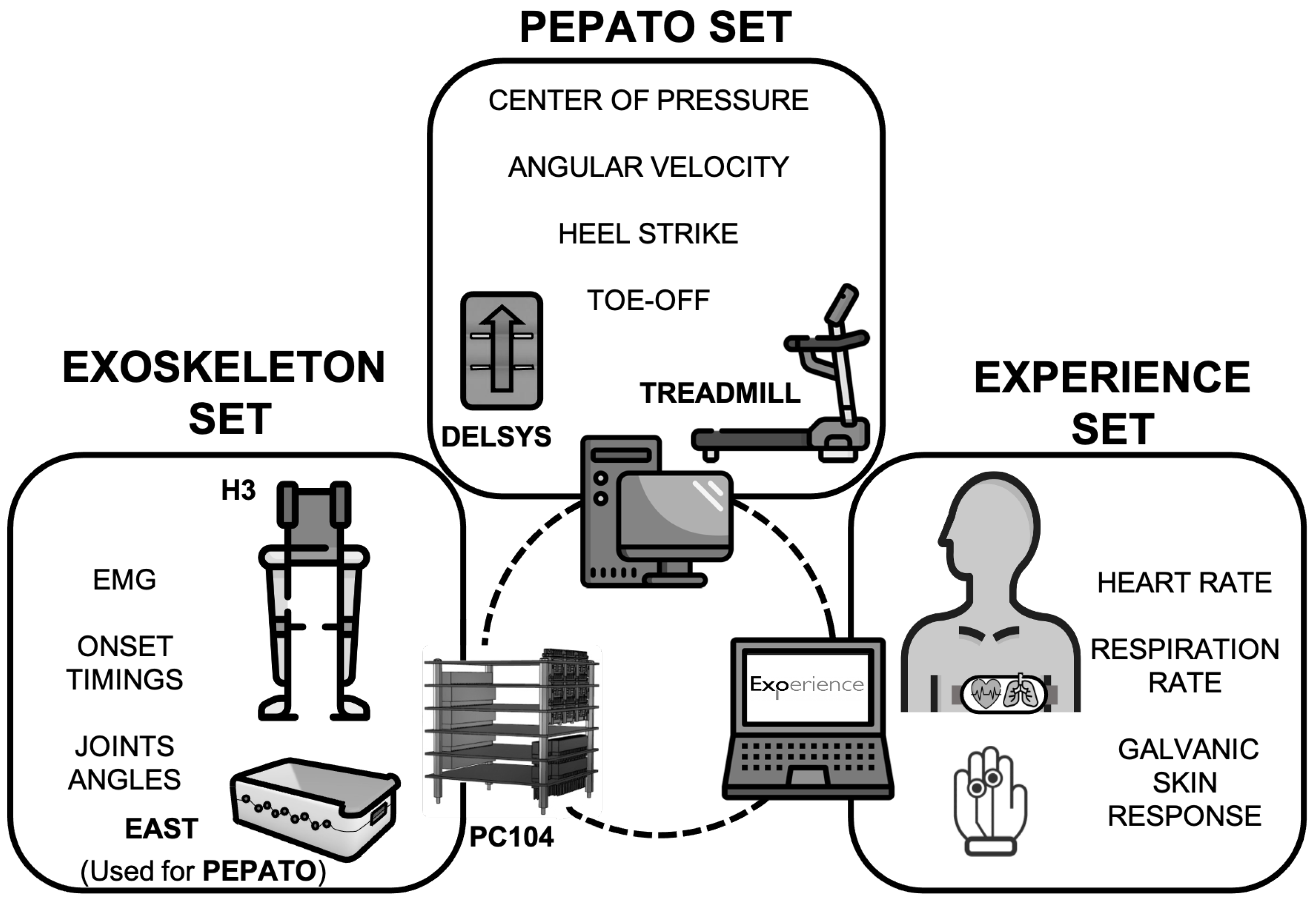
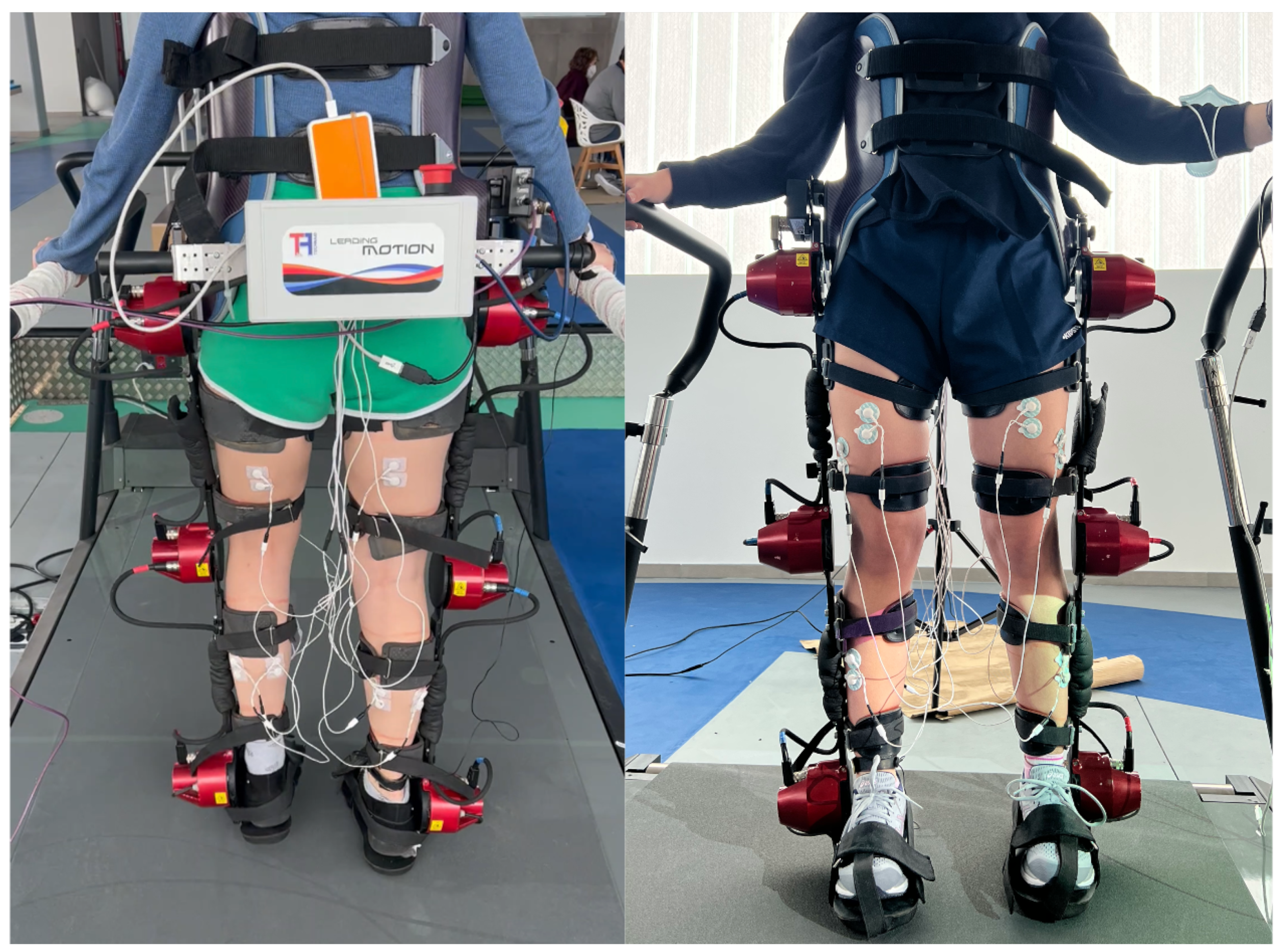
- EMG signal. A customized embedded processing unit that included an EMG amplifier and a voltage-controlled electrical stimulator (EAST, OT Bioelettronica, Turin, Italy) [40]. Surface electrodes Ambu® WhiteSensor™ (Ambu®, Ballerup, Denmark).
- ECG signal and BR. A Zephyr BioHarness™ (Medtronic plc, Minneapolis, MN, USA) sensor was used. It is comprised of a fabric strap that incorporates the textile-type ECG electrodes and the breathing sensor. An electronic module placed at the strap acquires, converts and sends the ECG and BR data through Bluetooth.
- GSR signal. A Shimmer GSR+ Module (Shimmer, Dublin, Ireland) was used. It measures skin conductance between two electrodes attached to two fingers of one hand and converts and sends the GSR through Bluetooth data.
2.2.3. Treadmill
2.3. Control Strategies
2.3.1. Trajectory Controller (TC)
2.3.2. Admittance Controller (AC)
2.3.3. EMG-Onset Controller (OC)
2.4. Experimental Protocol
2.4.1. Experimental Setup
2.4.2. Study Design
2.5. Data Analysis
2.5.1. Exoskeleton
2.5.2. EXPERIENCE Testbed
- Usability: This is defined as the extent to which the exoskeleton can be used by the users to achieve specified goals with effectiveness, efficiency, and satisfaction in this specified context of use. High value of this PI indicates that the robot is highly usable.
- Acceptability: This relates to how the users perceive robots when interacting directly with them and how much you would be willing to introduce one into your everyday life. High value of this PI indicates that the robot is highly acceptable. This PI is comprised of four related constructs: attitude towards technology, self-efficacy, motivation, comfort, safety, and acceptability.
- Perceptibility: This evaluates the effects and influences that walking with the exoskeleton has on the user’s emotions, perceptions and quality of life. High value of this PI indicates that the robot positively influences emotion, perception and quality of life. The constructs associated with this PI are: embodiment and ownership, agency, emotion and attachment, health and quality of life.
- Functionality: This measures the perception of the characteristics of the exoskeleton in terms of ease of learning, the flexibility of interaction, reliability and workload. High value of this PI indicates positive features of the robot in terms of analyzed aspects. The constructs associated with this PI are: learnability, flexibility, robustness and reliability, workload, and functionality.
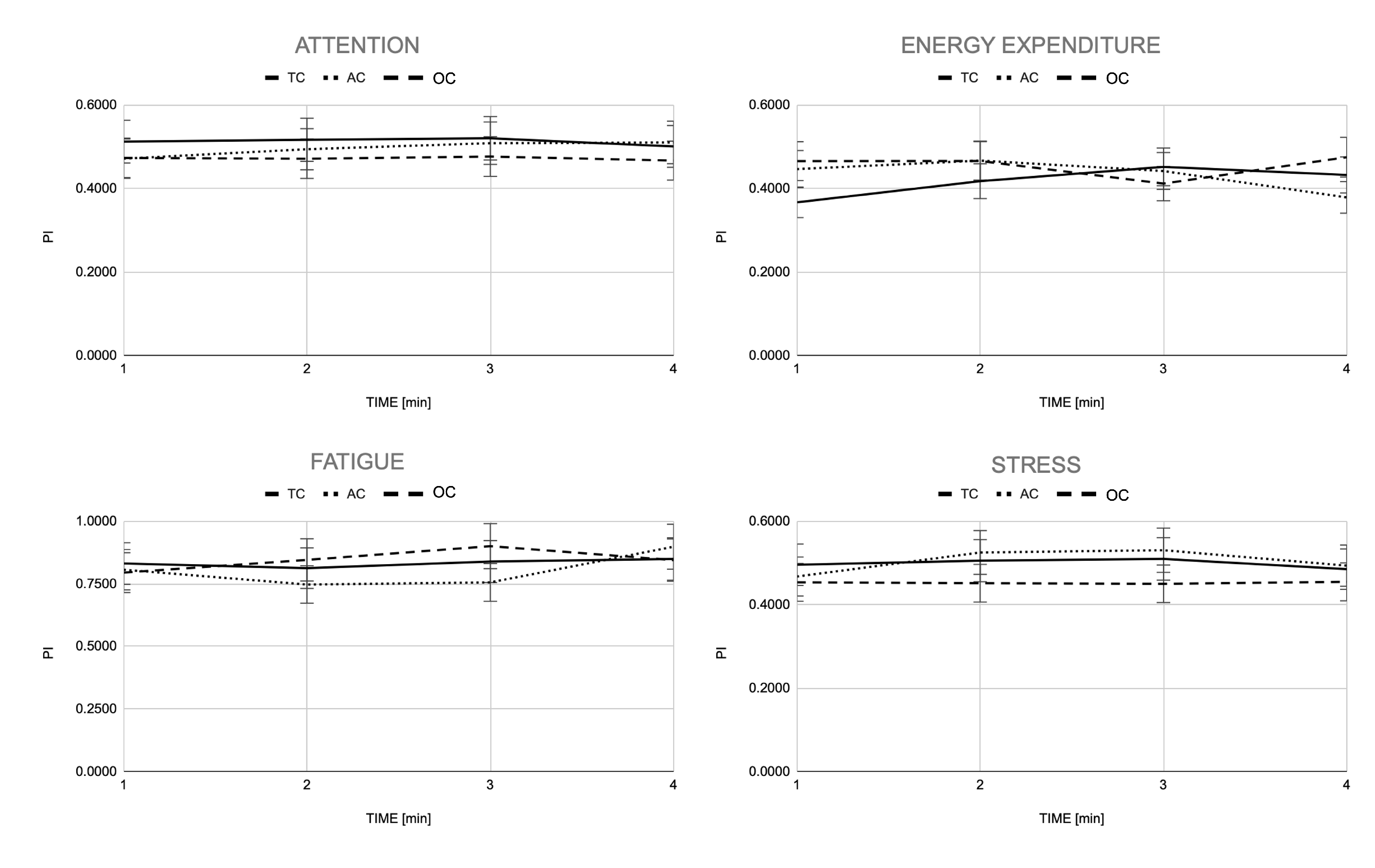
- Stress: This is defined as a state of mental or emotional strain caused by adverse circumstances. High value of this PI indicates that using the robot is stressful.
- Energy expenditure: This is defined as the amount of energy that is needed to carry out physical functions. High value of this PI indicates that using the robot requires high effort.
- Attention: This refers to the degree to which the user is consciously and continuously involved in the task. High value of this PI indicates that the robot use requires high attention.
- Physical Fatigue: This is defined as a type of distress generally conditioned by the exhaustion of one’s muscles due to the execution of a task. High value of this PI indicates that using the robot induces fatigue.
2.5.3. PEPATO Testbed
- EMG reconstruction quality.
- Full width at half maximum (FWMH): Estimated duration of basic patterns.
- Center of activity (CoA) of the basic patterns.
2.5.4. Electromyography
2.5.5. Statistical Analysis
3. Results
3.1. Exoskeleton Kinematics
3.2. EMG-Onset Controller
3.3. Muscle Synergies (PIs Obtained from PEPATO Testbed Software)
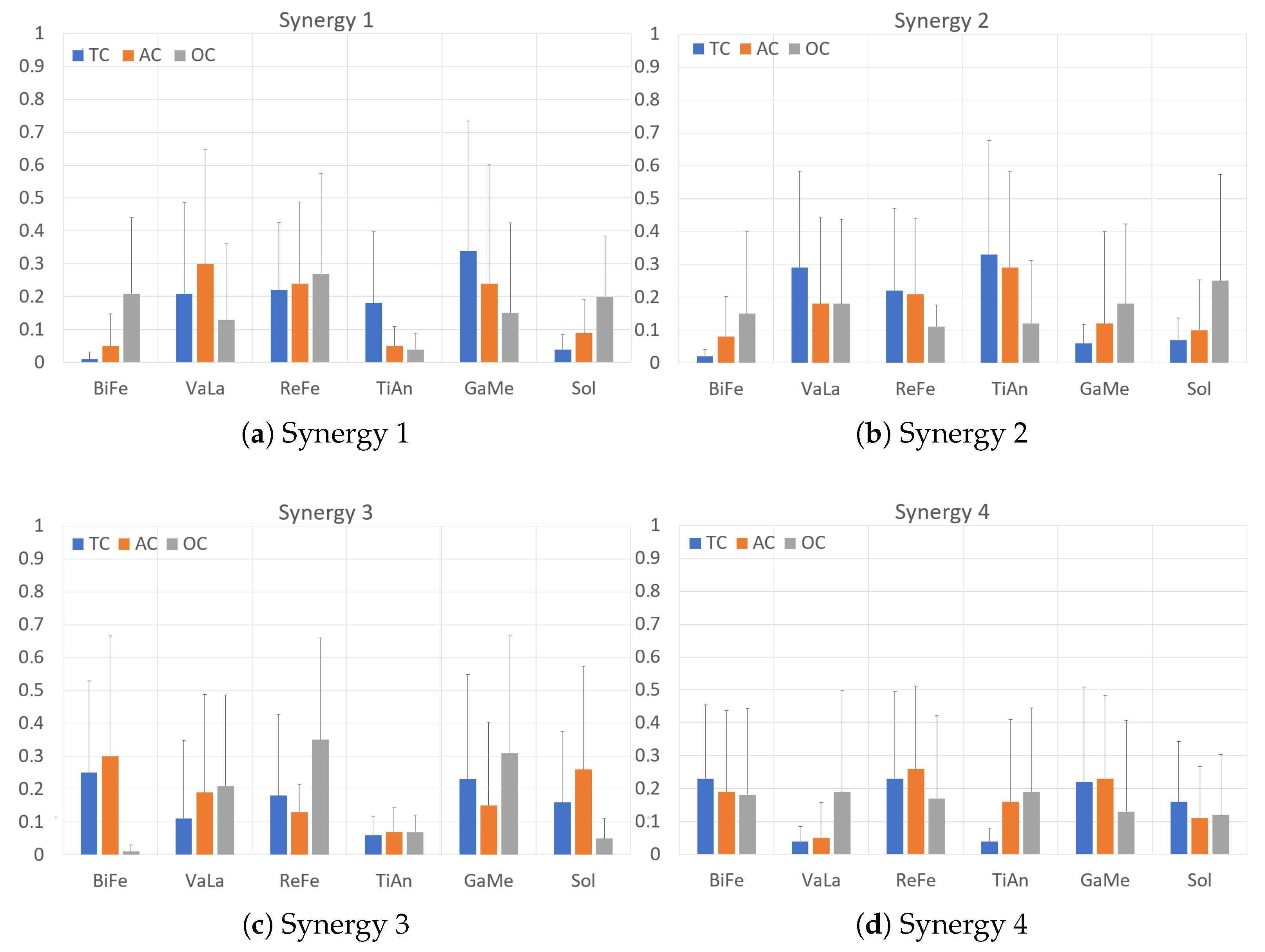
- Synergy 1 is mainly comprised of an ankle plantarflexion (GaMe) and knee extension activity (VaLa and ReFe muscles) with certain antagonist dorsiflexion activity (TiAn) for the TC walking condition. This activity is maintained for the AC walking condition and changes towards a knee antagonist co-contraction (BiFe vs VaLa and ReFe muscles) and increased ankle plantarflexion activity (increased contribution of the Sol muscle). The average mean duration of this synergy remains for the TC and AC walking conditions but shows a non-significant decrease for the OC (Table 4).
- Synergy 2 is mainly comprised of ankle dorsiflexion (TiAn) and knee extension (VaLa and ReFe muscles) for the TC walking condition. Similarly to Synergy 1, this activity mostly remains for the AC walking condition and changes in the OC walking condition towards ankle plantarflexion (increase in GaMe and Sol, reduction in TiAn contributions) and knee flexion (increased BiFe, reduction in VaLa and ReFe contributions). Similarly, the average mean duration synergy 2 remains for the TC and AC walking conditions, showing a significant decrease for the OC walking condition (Table 4).
- Synergy 3 shows a marked ankle plantarflexion (GaMe and Sol muscles) and knee flexion activity (BiFe muscle) for the TC walking condition. Similarly to Synergies 1 and 2, this activity remains with slight variations for the AC walking condition, but changes to a marked knee extension activity (decrease in the BiFe and increase in the VaLa and ReFe contributions), while ankle activity remains unchanged although a lesser contribution of the Sol muscle is observed. The average mean duration of this synergy remains for the TC and AC walking conditions but shows a non-significant decrease for the OC.
- Synergy 4 shows, for the TC walking condition, a noticeable ankle plantarflexion activity (GaMe and Sol muscles and a small TiAn contribution), whereas the knee shows an agonist–antagonist co-contraction (BiFe and ReFe muscles). Again, this activity remains with slight variations for the AC walking condition, whereas the OC walking condition shifts towards ankle dorsiflexion (increase in the TiAn contribution, decreasing in the GaMe and Sol muscles) with an increase in knee extension activity (VaLa muscle), although the contribution of the BiFe to co-contraction remains. Similarly, the average mean duration synergy 4 remains for the TC and AC walking conditions, showing a significant decrease for the OC walking condition (Table 4).
3.4. Subjective Perception (PIs Obtained from EXPERIENCE Testbed Software)
4. Discussion
5. Conclusions
Author Contributions
Funding
Institutional Review Board Statement
Informed Consent Statement
Data Availability Statement
Acknowledgments
Conflicts of Interest
Abbreviations
| AC | Compliant assistance |
| BiFe | Biceps Femoris |
| BR | Breathing rate |
| CAN | Controller Area Network |
| F CNS | Central neural system |
| CoA | Center of activity |
| DC | Direct current |
| DT | Double threshold |
| ECG | Electrocardiographic |
| EMG | Electromyography |
| FWMH | Full width at half maximum |
| GaMe | Gastrocnemius Medialis |
| GSR | Galvanic skin response |
| PI | Performance indicators |
| OC | EMG-Onset control |
| PID | Proportional–integral–derivative |
| ReFe | Rectus Femoris |
| ROM | Range of motion |
| sEMG | Superficial EMG |
| Sol | Soleus |
| ST | Single threshold |
| TC | Trajectory assistance |
| TiAn | Tibialis Anterior |
| VaLa | Vastus Lateralis |
References
- Morone, G.; Paolucci, S.; Cherubini, A.; De Angelis, D.; Venturiero, V.; Coiro, P.; Iosa, M. Robot-assisted gait training for stroke patients: Current state of the art and perspectives of robotics. Neuropsychiatr. Dis. Treat. 2017, 13, 1303–1311. [Google Scholar] [CrossRef]
- Labruyère, R. Robot-assisted gait training: More randomized controlled trials are needed! Or maybe not? J. Neuroeng. Rehabil. 2022, 19, 58. [Google Scholar] [CrossRef]
- Dijkers, M.P.; Akers, K.G.; Dieffenbach, S.; Galen, S.S. Systematic Reviews of Clinical Benefits of Exoskeleton Use for Gait and Mobility in Neurologic Disorders: A Tertiary Study. Arch. Phys. Med. Rehabil. 2021, 102, 300–313. [Google Scholar] [CrossRef] [PubMed]
- Federici, S.; Meloni, F.; Bracalenti, M.; De Filippis, M.L. The effectiveness of powered, active lower limb exoskeletons in neurorehabilitation: A systematic review. NeuroRehabilitation 2015, 37, 321–340. [Google Scholar] [CrossRef]
- Moreno, J.C.; Asin, G.; Pons, J.L.; Cuypers, H.; Vanderborght, B.; Lefeber, D.; Ceseracciu, E.; Reggiani, M.; Thorsteinsson, F.; Del-Ama, A.; et al. Symbiotic Wearable Robotic Exoskeletons: The Concept of the BioMot Project. In Lecture Notes in Computer Science (including Subseries Lecture Notes in Artificial Intelligence and Lecture Notes in Bioinformatics); Springer: Cham, Switzerland, 2014; Volume 8820, pp. 72–83. [Google Scholar] [CrossRef]
- Wu, A.R.; Dzeladini, F.; Brug, T.J.; Tamburella, F.; Tagliamonte, N.L.; Van Asseldonk, E.H.; Van Der Kooij, H.; Ijspeert, A.J. An adaptive neuromuscular controller for assistive lower-limb exoskeletons: A preliminary study on subjects with spinal cord injury. Front. Neurorobot. 2017, 11, 30. [Google Scholar] [CrossRef]
- Haufe, F.L.; Kober, A.M.; Wolf, P.; Riener, R.; Xiloyannis, M. Learning to walk with a wearable robot in 880 simple steps. J. Neuroeng. Rehabil. 2021, 18, 1–14. [Google Scholar] [CrossRef] [PubMed]
- Ingraham, K.A.; Remy, C.D.; Rouse, E.J. The role of user preference in the customized control of robotic exoskeletons. Sci. Robot. 2022, 7, 3487. [Google Scholar] [CrossRef] [PubMed]
- Koelewijn, A.D.; Audu, M.; Del-Ama, A.J.; Colucci, A.; Font-Llagunes, J.M.; Gogeascoechea, A.; Hnat, S.K.; Makowski, N.; Moreno, J.C.; Nandor, M.; et al. Adaptation Strategies for Personalized Gait Neuroprosthetics. Front. Neurorobot. 2021, 15, 1–8. [Google Scholar] [CrossRef]
- Moreno, J.C.; Barroso, F.; Farina, D.; Gizzi, L.; Santos, C.; Molinari, M.; Pons, J.L. Effects of robotic guidance on the coordination of locomotion. J. Neuroeng. Rehabil. 2013, 10, 79. [Google Scholar] [CrossRef] [PubMed]
- Maggioni, S.; Lunenburger, L.; Riener, R.; Melendez-Calderon, A. Robot-aided assessment of walking function based on an adaptive algorithm. In Proceedings of the 2015 IEEE International Conference on Rehabilitation Robotics, Singapore, 11–14 August 2015; pp. 804–809. [Google Scholar] [CrossRef]
- Jamwal, P.K.; Xie, S.Q.; Hussain, S.; Parsons, J.G. An Adaptive Wearable Parallel Robot for the Treatment of Ankle Injuries. IEEE/ASME Trans. Mechatron. 2014, 19, 64–75. [Google Scholar] [CrossRef]
- Hussain, S.; Xie, S.Q.; Jamwal, P.K. Robust Nonlinear Control of an Intrinsically Compliant Robotic Gait Training Orthosis. IEEE Trans. Syst. Man, Cybern. Syst. 2013, 43, 655–665. [Google Scholar] [CrossRef]
- Tang, Z.; Shi, D.; Liu, D.; Peng, Z.; He, L.; Pei, Z. Electro-hydraulic servo system for Human Lower-limb Exoskeleton based on sliding mode variable structure control. In Proceedings of the 2013 IEEE International Conference on Information and Automation for Sustainability, Yinchuan, China, 26–28 August 2013; pp. 559–563. [Google Scholar] [CrossRef]
- Shi, D.; Zhang, W.W.; Zhang, W.W.; Ding, X. A Review on Lower Limb Rehabilitation Exoskeleton Robots. Chin. J. Mech. Eng. 2019, 32, 74. [Google Scholar] [CrossRef]
- Huo, W.; Mohammed, S.; Amirat, Y. Impedance Reduction Control of a Knee Joint Human–Exoskeleton System. IEEE Trans. Control Syst. Technol. 2019, 27, 2541–2556. [Google Scholar] [CrossRef]
- Sposito, M.; Toxiri, S.; Caldwell, D.G.; Ortiz, J.; De Momi, E. Towards Design Guidelines for Physical Interfaces on Industrial Exoskeletons: Overview on Evaluation Metrics; Springer: Cham, Switzerland, 2019; pp. 170–174. [Google Scholar] [CrossRef]
- Huo, W.; Mohammed, S.; Moreno, J.C.; Amirat, Y. Lower Limb Wearable Robots for Assistance and Rehabilitation: A State of the Art. IEEE Syst. J. 2016, 10, 1068–1081. [Google Scholar] [CrossRef]
- Basalp, E.; Wolf, P.; Marchal-Crespo, L. Haptic training: Which types facilitate (re)learning of which motor task and for whom Answers by a review. IEEE Trans Haptics 2021, 14, 722–739. [Google Scholar] [CrossRef] [PubMed]
- Fukuda, O.; Tsuji, T.; Kaneko, M.; Otsuka, A. A human-Assisting manipulator teleoperated by EMG signals and arm motions. IEEE Trans. Robot. Autom. 2003, 19, 210–222. [Google Scholar] [CrossRef]
- Sartori, M.; Llyod, D.G.; Farina, D. Neural data-driven musculoskeletal modeling for personalized neurorehabilitation technologies. IEEE Trans. Biomed. Eng. 2016, 63, 879–893. [Google Scholar] [CrossRef]
- Kirchner, E.A.; Tabie, M.; Seeland, A. Multimodal movement prediction—Towards an individual assistance of patients. PLoS ONE 2014, 9, e85060. [Google Scholar] [CrossRef]
- Cavallaro, E.E.; Rosen, J.; Perry, J.C.; Burns, S. Real-time myoprocessors for a neural controlled powered exoskeleton arm. IEEE Trans. Biomed. Eng. 2006, 53, 2387–2396. [Google Scholar] [CrossRef]
- Benitez, L.M.V.; Will, N.; Tabie, M.; Schmidt, S.; Kirchner, E.; Albiez, J. An EMG-based Assistive Orthosis for Upper Limb Rehabilitation. Proc. Int. Conf. Biomed. Electron. Devices 2013, 1, 323–328. [Google Scholar] [CrossRef]
- Vaca Benitez, L.M.; Tabie, M.; Will, N.; Schmidt, S.; Jordan, M.; Kirchner, E.A. Exoskeleton technology in rehabilitation: Towards an EMG-based orthosis system for upper limb neuromotor rehabilitation. J. Robot. 2013, 2013, 610589. [Google Scholar] [CrossRef]
- Kiguchi, K.; Imada, Y.; Liyanage, M. EMG-based neuro-fuzzy control of a 4DOF upper-limb power-assist exoskeleton. In Proceedings of the 2007 29th Annual International Conference of the IEEE Engineering in Medicine and Biology Society, Lyon, France, 22–26 August 2007; pp. 3040–3043. [Google Scholar] [CrossRef]
- Simon, S.R.M. Gait Analysis, Normal and Pathological Function. J. Bone Jt. Surg. 1993, 75, 476–477. [Google Scholar] [CrossRef]
- Reaz, M.B.I.; Hussain, M.S.; Mohd-Yasin, F. Techniques of EMG signal analysis: Detection, processing, classification and applications. Biol. Proc. Online 2006, 8, 11–35. [Google Scholar] [CrossRef] [PubMed]
- Safavynia, S.; Torres-Oviedo, G.; Ting, L. Muscle Synergies: Implications for Clinical Evaluation and Rehabilitation of Movement. Spinal Cord 2011, 17, 16–24. [Google Scholar] [CrossRef] [PubMed]
- De Luca, A.; Bellitto, A.; Mandraccia, S.; Marchesi, G.; Pellegrino, L.; Coscia, M.; Leoncini, C.; Rossi, L.; Gamba, S.; Massone, A.; et al. Exoskeleton for gait rehabilitation: Effects of assistance, mechanical structure and walking aids on muscle activations. Appl. Sci. 2019, 9, 2868. [Google Scholar] [CrossRef]
- Del-Ama, A.J.; Asin-Prieto, G.; Pinuela-Martin, E.; Perez-Nombela, S.; Lozano-Berrio, V.; Serrano-Munoz, D.; Trincado-Alonso, F.; Gonzalez-Vargas, J.; Gil-Agudo, A.; Pons, J.L.; et al. Muscle activity and coordination during robot-assisted walking with h2 exoskeleton. In Biosystem Biorobotics; Springer: Berlin, Germany, 2017; Volume 15, pp. 349–353. [Google Scholar] [CrossRef]
- Englehart, K.; Hudgins, B. A Robust, Real-Time Control Scheme for Multifunction Myoelectric Control. IEEE Trans. Biomed. Eng. 2003, 50, 848–854. [Google Scholar] [CrossRef]
- Androwis, G.J.; Pilkar, R.; Ramanujam, A.; Nolan, K.J. Electromyography Assessment During Gait in a Robotic Exoskeleton for Acute Stroke. Front. Neurol. 2018, 9, 1–12. [Google Scholar] [CrossRef]
- Eurobench. EUROBENCH Testbeds. 2022. Available online: https://eurobench2020.eu (accessed on 15 August 2022).
- Eurobench FSTP-1. Benchmarking Exoskeleton-Assisted Gait Based on User’s Subjective Perspective and Experience. Available online: https://eurobench2020.eu/developing-the-framework/benchmarking-exoskeleton-assisted-gait-based-on-users-subjective-perspective-and-experience-experience/ (accessed on 15 August 2022).
- Pisotta, I.; Tagliamonte, N.L.; Bigioni, A.; Tamburella, F.; Lorusso, M.; Bentivoglio, F.; Pecoraro, I.; Argentieri, P.; Marri, F.; Zollo, L.; et al. Pilot Testing of a New Questionnaire for the Assessment of User Experience During Exoskeleton-Assisted Walking. In Biosystem Biorobotics; Springer: Berlin, Germany, 2022; Volume 28, pp. 195–199. [Google Scholar] [CrossRef]
- Pecoraro, I.; Tagliamonte, N.L.; Tamantini, C.; Cordella, F.; Bentivoglio, F.; Pisotta, I.; Bigioni, A.; Tamburella, F.; Lorusso, M.; Argentieri, P.; et al. Psychophysiological Assessment of Exoskeleton-Assisted Treadmill Walking. In Biosystem Biorobotics; Springer: Berlin, Germany, 2022; Volume 28, pp. 201–205. [Google Scholar] [CrossRef]
- EUROBENCH PEPATO Testbed. 2021. Available online: https://eurobench2020.eu (accessed on 15 August 2022).
- Technaid, S.L. EXO-H3—Especificaciones Técnicas (EN); Technical Report; TECHNAID: Arganda del Rey, Spain, 2022. [Google Scholar]
- Pascual-Valdunciel, A.; Gonzalez-Sanchez, M.; Muceli, S.; Adan-Barrientos, B.; Escobar-Segura, V.; Perez-Sanchez, J.R.; Jung, M.K.; Schneider, A.; Hoffmann, K.P.; Moreno, J.C.; et al. Intramuscular Stimulation of Muscle Afferents Attains Prolonged Tremor Reduction in Essential Tremor Patients. IEEE Trans. Biomed. Eng. 2021, 68, 1768–1776. [Google Scholar] [CrossRef]
- Bortole, M.; Antonio, J.d.-A.; Rocon, E.; Moreno, J.C.; Brunetti, F.; Pons, J.L. A robotic exoskeleton for overground gait rehabilitation. In Proceedings of the IEEE International Conference on Intelligent Robots and Systems, Tokyo, Japan, 3–7 November 2013; pp. 3356–3361. [Google Scholar] [CrossRef]
- Hodges, P.W.; Bui, B.H. A comparison of computer-based methods for the determination of onset of muscle contraction using electromyography. Electroencephalogr. Clin. Neurophysiol. 1996, 101, 511–519. [Google Scholar]
- Marple-Horvat, D.E.; Gilbey, S.L. A method for automatic identification of periods of muscular activity from EMG recordings. J. Neurosci. Methods 1992, 42, 163–167. [Google Scholar] [CrossRef]
- Morantes, G.; Fernández, G.; Altuve, M. A threshold-based approach for muscle contraction detection from surface EMG signals. IX Int. Semin. Med. Inf. Process. Anal. 2013, 8922, 89220C. [Google Scholar] [CrossRef]
- Dow, D.E.; Petrilli, A.M.; Mantilla, C.B.; Zhan, W.Z.; Sieck, G.C. Electromyogram-triggered inspiratory event detection algorithm. In Proceedings of the 6th International Conference on Soft Computing and Intelligent Systems and The 13th International Symposium on Advanced Intelligence Systems SCIS/ISIS 2012, Kobe, Japan, 20–24 November 2012; pp. 789–794. [Google Scholar] [CrossRef]
- Micera, S.; Vannozzi, G. Improving detection of muscle activation intervals. IEEE Eng. Med. Biol. Mag. 2001, 20, 38–46. [Google Scholar] [CrossRef] [PubMed]
- Staude, G.; Wolf, W. Objective motor response onset detection in surface myoelectric signals. Med. Eng. Phys. 1999, 21, 449–467. [Google Scholar] [CrossRef] [PubMed]
- Rasool, G.; Iqbal, K.; White, G.A. Myoelectric activity detection during a Sit-to-Stand movement using threshold methods. Comput. Math. Appl. 2012, 64, 1473–1483. [Google Scholar] [CrossRef]
- Grasso, R.; Ivanenko, Y.P.; Zago, M.; Molinari, M.; Scivoletto, G.; Castellano, V.; Macellari, V.; Lacquaniti, F. Distributed plasticity of locomotor pattern generators in spinal cord injured patients. Brain 2004, 127, 1019–1034. [Google Scholar] [CrossRef]
- Barroso, F.O.; Torricelli, D.; Bravo-Esteban, E.; Taylor, J.; Gómez-Soriano, J.; Santos, C.; Moreno, J.C.; Pons, J.L. Muscle Synergies in Cycling after Incomplete Spinal Cord Injury: Correlation with Clinical Measures of Motor Function and Spasticity. Front. Hum. Neurosci. 2016, 9, 706. [Google Scholar] [CrossRef] [PubMed]
- Ramanujam, A.; Cirnigliaro, C.M.; Garbarini, E.; Asselin, P.; Pilkar, R.; Forrest, G.F. Neuromechanical adaptations during a robotic powered exoskeleton assisted walking session. J. Spinal Cord Med. 2018, 41, 518–528. [Google Scholar] [CrossRef]
- Knaepen, K.; Beyl, P.; Duerinck, S.; Hagman, F.; Lefeber, D.; Meeusen, R. Human–robot interaction: Kinematics and muscle activity inside a powered compliant knee exoskeleton. IEEE Trans. Neural Syst. Rehabil. Eng. 2014, 22, 1128–1137. [Google Scholar] [CrossRef]
- Zhang, D.; Gao, F.; Zhang, H.; Zhu, J.; Li, Z.; Liu, H.; Yin, Z.; Chen, K. Muscle Synergy Alteration of Human During Walking With Lower Limb Exoskeleton. Front. Neurosci. 2019, 1, 1050. [Google Scholar] [CrossRef]
- Zulkifli, S.S.; Loh, W.P. A state-of-the-art review of foot pressure. Foot Ankle Surg. 2020, 26, 25–32. [Google Scholar] [CrossRef]




| Controller | ||||||
|---|---|---|---|---|---|---|
| Mean | SD | Median | Max | Min | ||
| TC | ||||||
| Ankle | Max 1,2 | 19.40 | 0.32 | 19.43 | 19.56 | 19.31 |
| Min 1,2,3 | −12.97 | 0.12 | −12.97 | −12.92 | −13.01 | |
| ROM 1,2 | 32.40 | 0.40 | 32.40 | 32.57 | 32.23 | |
| Knee | Max 1,2 | 60.45 | 0.89 | 60.55 | 60.64 | 60.46 |
| Min 1,2,3 | −0.03 | 0.14 | −0.03 | 0.01 | −0.07 | |
| ROM 1,2 | 60.48 | 0.87 | 60.58 | 60.63 | 60.53 | |
| Hip | Max 1,2 | 27.20 | 1.81 | 27.11 | 27.12 | 27.10 |
| Min 1,2,3 | −13.53 | 0.80 | −13.54 | −13.48 | −13.60 | |
| ROM 1,2 | 60.48 | 0.87 | 60.58 | 60.63 | 60.53 | |
| AC | ||||||
| Ankle | Max 1,2 | 14.65 | 1.68 | 14.86 | 14.93 | 14.79 |
| Min 1,2,3 | −2.92 | 3.32 | −3.41 | −3.22 | −3.60 | |
| ROM 1,2 | 17.58 | 4.97 | 18.28 | 18.54 | 18.01 | |
| Knee | Max 1,2 | 49.02 | 4.11 | 49.56 | 49.56 | 49.56 |
| Min 1,2,3 | 2.67 | 1.90 | 2.44 | 2.69 | 2.18 | |
| ROM 1,2 | 46.35 | 5.48 | 47.13 | 47.38 | 46.87 | |
| Hip | Max 1,2 | 24.68 | 2.60 | 24.52 | 24.73 | 24.31 |
| Min 1,2,3 | −11.26 | 2.07 | −11.77 | −11.13 | −12.40 | |
| ROM 1,2 | 35.94 | 3.32 | 36.30 | 37.15 | 35.45 | |
| OC | ||||||
| Ankle | Max 1,2 | 14.04 | 1.49 | 14.17 | 15.62 | 13.72 |
| Min 1,2,3 | −1.45 | 2.26 | 3.69 | 3.85 | 3.52 | |
| ROM 1,2 | 44.17 | 4.04 | 44.63 | 44.69 | 44.56 | |
| Knee | Max 1,2 | 47.91 | 2.67 | 48.31 | 48.54 | 48.08 |
| Min 1,2,3 | 3.74 | 2.62 | 3.69 | 3.85 | 3.52 | |
| ROM 1,2 | 44.17 | 4.04 | 44.63 | 44.69 | 44.56 | |
| Hip | Max 1,2 | 25.06 | 2.61 | 24.47 | 24.76 | 24.19 |
| Min 1,2,3 | −11.00 | 1.77 | −11.28 | −10.91 | −11.65 | |
| ROM 1,2 | 36.06 | 2.95 | 35.76 | 35.85 | 35.68 | |
| Right Step | Left Step | ||
|---|---|---|---|
| Right ReFe | Left Sol | Left ReFe | Right Sol |
| 59.24 ± 4.35% | 61.74 ± 5.22% | No Detection | 12.18 ± 5.92% |
| Right Step | Left Step | ||
|---|---|---|---|
| Right ReFe | Left Sol | Left ReFe | Right Sol |
| 17.4% | 32.85% | 0.00% | 49.74% |
| PEPATO PI | ||||||||||
|---|---|---|---|---|---|---|---|---|---|---|
| EMG Reconst. Quality | FWHM 1 | FWHM 2 3 | FWHM 3 | FWHM 4 3 | CoA 1 | CoA 2 | CoA 3 | CoA 4 | ||
| TC | Mean | 0.95 | 16.07 | 18.57 | 22.21 | 14.79 | 16.74 | 31.49 | 57.61 | 57.25 |
| SD | 0.03 | 8.12 | 15.54 | 13.95 | 12.98 | 11.06 | 9.09 | 29.55 | 39.98 | |
| Median | 0.94 | 14.00 | 14.50 | 23.00 | 13.00 | 15.34 | 30.23 | 69.18 | 76.50 | |
| Max | 0.987 | 32.50 | 50.50 | 44.00 | 35.00 | 30.02 | 48.76 | 97.39 | 94.49 | |
| Min | 0.90 | 6.50 | 6.00 | 6.50 | 0.00 | 3.39 | 16.37 | 6.79 | 0.07 | |
| AC | Mean | 0.94 | 15.07 | 18.21 | 19.50 | 26.86 | 31.40 | 46.44 | 59.18 | 51.27 |
| SD | 0.05 | 11.87 | 6.81 | 16.27 | 12.80 | 36.21 | 24.04 | 17.45 | 37.91 | |
| Median | 0.95 | 16.50 | 19.50 | 26.50 | 24.50 | 18.51 | 37.96 | 64.70 | 59.82 | |
| Max | 0.98 | 29.00 | 29.00 | 40.50 | 43.00 | 99.78 | 94.59 | 80.07 | 96.19 | |
| Min | 0.84 | 0.00 | 6.50 | 0.00 | 6.50 | 3.79 | 22.17 | 31.61 | 7.60 | |
| OC | Mean | 0.95 | 6.29 | 6.29 | 7.14 | 5.29 | 25.66 | 36.38 | 55.49 | 67.26 |
| SD | 0.03 | 12.89 | 4.94 | 7.81 | 6.64 | 31.35 | 21.41 | 18.45 | 29.50 | |
| Median | 0.95 | 0.00 | 5.00 | 3.00 | 1.00 | 20.26 | 46.55 | 54.06 | 73.56 | |
| Max | 0.99 | 35.00 | 16.00 | 18.00 | 17.50 | 88.89 | 59.14 | 87.24 | 92.34 | |
| Min | 0.90 | 0.00 | 1.00 | 0.00 | 0.50 | 0.01 | 5.51 | 33.95 | 7.74 | |
| EXPERIENCE PI | |||||
|---|---|---|---|---|---|
| Acceptability | Funcionality | Perceptibility | Usability | ||
| TC | Mean | 4.63 | 3.97 | 2.85 | 4.38 |
| SD | 0.35 | 0.65 | 0.30 | 0.35 | |
| Median | 4.60 | 4.06 | 3.37 | 4.23 | |
| Max | 5.40 | 4.67 | 4.69 | 4.75 | |
| Min | 3.87 | 3.09 | 0.00 | 4.17 | |
| AC | Mean | 4.63 | 3.96 | 2.84 | 4.40 |
| SD | 0.36 | 0.52 | 0.33 | 0.46 | |
| Median | 4.63 | 4.06 | 3.36 | 4.17 | |
| Max | 4.63 | 4.83 | 4.63 | 5.00 | |
| Min | 4.63 | 2.89 | 0.00 | 4.03 | |
| OC | Mean | 4.63 | 3.95 | 2.81 | 4.43 |
| SD | 0.36 | 0.56 | 0.40 | 0.47 | |
| Median | 4.60 | 4.03 | 3.39 | 4.20 | |
| Max | 5.40 | 4.83 | 4.49 | 4.96 | |
| Min | 3.87 | 2.91 | 0.00 | 4.13 | |
Disclaimer/Publisher’s Note: The statements, opinions and data contained in all publications are solely those of the individual author(s) and contributor(s) and not of MDPI and/or the editor(s). MDPI and/or the editor(s) disclaim responsibility for any injury to people or property resulting from any ideas, methods, instructions or products referred to in the content. |
© 2023 by the authors. Licensee MDPI, Basel, Switzerland. This article is an open access article distributed under the terms and conditions of the Creative Commons Attribution (CC BY) license (https://creativecommons.org/licenses/by/4.0/).
Share and Cite
Rodrigues-Carvalho, C.; Fernández-García, M.; Pinto-Fernández, D.; Sanz-Morere, C.; Barroso, F.O.; Borromeo, S.; Rodríguez-Sánchez, C.; Moreno, J.C.; del-Ama, A.J. Benchmarking the Effects on Human–Exoskeleton Interaction of Trajectory, Admittance and EMG-Triggered Exoskeleton Movement Control. Sensors 2023, 23, 791. https://doi.org/10.3390/s23020791
Rodrigues-Carvalho C, Fernández-García M, Pinto-Fernández D, Sanz-Morere C, Barroso FO, Borromeo S, Rodríguez-Sánchez C, Moreno JC, del-Ama AJ. Benchmarking the Effects on Human–Exoskeleton Interaction of Trajectory, Admittance and EMG-Triggered Exoskeleton Movement Control. Sensors. 2023; 23(2):791. https://doi.org/10.3390/s23020791
Chicago/Turabian StyleRodrigues-Carvalho, Camila, Marvin Fernández-García, David Pinto-Fernández, Clara Sanz-Morere, Filipe Oliveira Barroso, Susana Borromeo, Cristina Rodríguez-Sánchez, Juan C. Moreno, and Antonio J. del-Ama. 2023. "Benchmarking the Effects on Human–Exoskeleton Interaction of Trajectory, Admittance and EMG-Triggered Exoskeleton Movement Control" Sensors 23, no. 2: 791. https://doi.org/10.3390/s23020791
APA StyleRodrigues-Carvalho, C., Fernández-García, M., Pinto-Fernández, D., Sanz-Morere, C., Barroso, F. O., Borromeo, S., Rodríguez-Sánchez, C., Moreno, J. C., & del-Ama, A. J. (2023). Benchmarking the Effects on Human–Exoskeleton Interaction of Trajectory, Admittance and EMG-Triggered Exoskeleton Movement Control. Sensors, 23(2), 791. https://doi.org/10.3390/s23020791


